role of thoracomyoplasty procedures in modern surgery …cdn.intechweb.org/pdfs/28647.pdf · role...
TRANSCRIPT

18
Role of Thoracomyoplasty Procedures in Modern Surgery for Intrathoracic Suppurations
Petre Vlah-Horea Botianu and Alexandru Mihail Botianu Surgical Clinic 4, University of Medicine and Pharmacy from Targu-Mures,
Romania
1. Introduction
Both thoracoplasty and muscle transposition are rarely performed procedures in modern thoracic surgery (Deslauriers et al., 2002). Their importance comes from the fact that these procedures are usually indicated in desperate cases, with failed medical treatments and who cannot be cured through standard procedures such as resection or decortication. For these patients, thoracomyoplasty operations are often the last chance for cure and sometimes even life-saving procedures (Botianu et al., 2010a).
2. Modern indications of thoracomyoplasty procedures
In our days, most of the intrathoracic suppurations can be managed without surgery, through antibiotics and minor procedures such as thoracocenthesis or tube-thoracostomy (Davies et al., 2011). Overall, about 10-15% of patients with intrathoracic suppurations still require some form of major surgical treatment. Out of them, most can be managed through less invasive and mutilating procedures (Pardos-Gea et al., 2011, Zahid et al., 2011), which make thoracomyoplasty a quite rarely indicated procedure: - for pleural empyema: in cases not amenable to decortication – usually chronic cases
with no cleavage plane and subjacent lesions in the parenchyma limiting the re-expansion of the lung;
- for pulmonary lesions (abscesses, tuberculosis, aspergilloma etc.): in cases not amenable to lung resection – usually a combination of poor biological and cardio-pulmonary status (including pulmonary hypertension), contralateral disease, fixed and adherent lesions with major technical difficulties.
Tuberculosis (TB) requires a special attention. Although TB infection by itself is not an indication for such procedures, many cases with chronic disease present with features making them candidates for thoracomyoplasty procedures. In our experience, almost half of the patients requiring thoracomyoplasty operations had different forms of TB lesions (Botianu P. et al., 2010a). Bronchial fistula is frequently solved with the use of muscle flaps, alone or in combination with thoracoplasty. Simple closure by suturing through an inflamed tissue has very few chances of success, making reinforcement with a viable flap almost mandatory in most cases. In many cases, the muscle flaps are used as a plug to close the bronchial defect, without direct suture of the edges of the fistula (Hollaus et al., 1999, Zaheer et al., 2009).
www.intechopen.com

Topics in Thoracic Surgery
310
Postoperative empyema is also an indication for thoracomyoplasty procedures in selected cases (Garcia-Yuste et al., 1998, Regnard et al., 2000). A particular technical aspect is that the previous thoracotomy reduces the availability of the neighbourhood flaps. The need to have some flaps available in case of postoperative complications is the main argument for the use of muscle-sparing thoracotomies (Nosotti et al., 2010). Intrathoracic muscle transposition (without thoracoplasty) has also some other indications such as reinforcement of high-risk sutures (Abolhoda et al., 2009, Thingnam et al., 2011), repair of esophageal and tracheal defects (Kotzampassakis et al., 2009, Meyer et al., 2004), pericardial and diaphragmatic reconstruction (Kobayashi et al., 2009), dynamic cardiomyoplasty for end-stage heart failure (Chachques et al., 2008), salvage of infected vascular prosthesis (Mitra et al., 2005) etc.
3. Thoracoplasty
3.1 Historical background
The term “thoracoplasty” was introduced by Estlander (1879) who performed resection of multiple fragments of ribs (“resectio costorum multiplex”) to achieve obliteration of an underlying empyema (Estlander, 1879). The operation became popular mainly as a method of lung collapse to achieve healing of tuberculosis before the introduction of modern TB chemotherapy. In fact, thoracoplasty procedures dominated chest surgery before the 1950’s and played an essential role in the development of thoracic surgery as a distinct specialty. After 1950-60’s, the interest for this procedure decreased due to the introduction of medical treatment (antibiotics and specific TB drugs) and development of other less mutilating and more effective surgical procedures (Deslauriers et al., 2002, Horrigan & Snow, 1990).
3.2 Terminology
Thoracoplasty is a procedure that targets the resolution of a cavity (pleural or pulmonary) by collapsing the chest wall through rib resection and/or plombage; according to the way this collapse is achieved, there are different terms which are used to describe the procedure: - in one or more stages – according to the number of operative steps used. In the past
many authors preferred to perform the thoracoplasty in more steps, with resection of 2-3 ribs in each step, in order to lower the magnitude of the operative aggression and to reduce mortality (Alexander 1936). This was a reasonable attitude in the early years of thoracic surgery, when a lot of things that are today standard were not available (general anesthesia, oro-tracheal intubation, blood transfusion, antibiotics, electrocautery etc). Besides the need for more procedures, a specific disadvantage is the cavity movement phenomenon, in which the cavity just moves in one direction without any resolution; at the end, the patient will have some ribs resected and the same cavity in another position (Archibald 1926). In our days, most of these procedures are done in a single step.
- extra-pleural or intra-pleural – according to the intact preservation or opening of the parietal pleural. In the past, opening of the pleural space during thoracoplasty was considered as a major accident (fear for wound contamination, pneumothorax etc.) (Archibald 1926). In our days, in most cases the pleura is opened deliberately to achieve a direct access to the pleural and/or pulmonary lesion.
www.intechopen.com

Role of Thoracomyoplasty Procedures in Modern Surgery for Intrathoracic Suppurations
311
- sub-periostal or extra-periostal – according to the plane used for rib resection, with or without preservation of the periosteum. In our days, in most cases the ribs are resected using a subperiostal plane. Leaving the periosteum intact allows some form of bone regeneration which helps the long-time stabilization of the chest wall (Alexander 1936).
Many authors use the term "classic" thoracoplasty, with different meanings depending on time and geographical location. For example, in Europe, in the 1910-20’s, "classic" thoracoplasty referred to the procedures described by Estlander and Schede, which were then replaced by the procedures described by Andre Maurer and Sauerbruch; the later became also "classic" for the American surgeons who trained in Europe. In the USA, after 1930, the technique described by Alexander became the standard thoracoplasty performed for lung collapse in order to heal lung TB. In the 1950’s, the osteoplastic techniques and plombage with different materials became popular, being then almost abandoned. In our days, most thoracoplasties are done in a single operative step, intrapleural and using a subperiosteal plane for rib resection (Hopkins et al., 1985).
3.3 Main types of thoracoplasty
Over the time, more than 100 thoracoplasty procedures were described, many of them being in fact minor modifications or combinations of previously described techniques; such a big number of techniques is by itself an indication for the dificulties encountered in this kind of surgery. We present a brief description of the most important techniques, all of them being very popular at a certain moment of the development of thoracic surgery. The Schede thoracoplasty is a very "radical" procedure developed for empyema and involving the resection of ribs, intercostal spaces and parietal pleura overlying the empyema cavity. The wound was left opened and the visceral pleura from the empyema cavity was placed in direct contact with healthy tissues represented by extracostal chest wall muscles and subcutaneous tissue (Schede 1890). For small empyema cavities it may be a reasonable procedure but for big cavities the procedure is a very mutilant one. With different modifications, it is still used today for highly selected cases of empyema (Stobernack et al., 1997, Botianu 2005, 2008). The Sauerbruch thoracoplasty (paravertebral) was indicated for empyema cavities that extended more in the vertical axis than in the horizontal one and involved a 4-6 cm length resection of the posterior part of the ribs. The main disadvantages were the risk of respiratory failure due to the paradoxical movements of the chest wall and the secondary scoliosis. The Andre Maurer thoracoplasty (descendant) involves complete excision of the first 2 ribs and partial removal of the ribs 3-5, leaving intact only the anterior part of them; it was performed extrapleurally, in one or more steps according to the patient’s biological status. The Alexander thoracoplasty was designed for treatment of lung TB and involved a subperiosteal and extrapleural removal of the first 8-9 ribs; for a better collapse the removal of the first rib and the transverse vertebral apophyses was considered to be mandatory. For a better tolerance of the procedure, it was performed in 3 steps at 3 weeks intervals. With this strategy and a good selection of the patients, Alexander achieved an important improvement of the results, with a 10% mortality and 90% rate of healing in survivors (Alexander 1936). The Archibald thoracoplasty involved resection of the first 3 ribs and introduction of the pectoral muscles in the extrapleural space to maintain the collapse of the lung; a particular
www.intechopen.com

Topics in Thoracic Surgery
312
aspect is the ingenious separate mobilization of the two flaps: the pectoralis major was mobilized based on the branches from the internal mammary vessels and the pectoralis minor was mobilized based on the thoraco-acromial pedicle (Entin 1995). Extrapleural plombage (plombage thoracoplasty) achieves lung collapse with the use of different materials that are introduced inside the chest using an extrafascial or extraperiosteal plane (fig. 1). The main advantages of the plombage thoracoplasties are the absence of chest wall mutilation and the fact that the procedure is easy, quick and well-tolerated, even by patients with poor biological status; the procedure was quite popular in the 1950-60’s, with immediate good results (Wilson et al., 1956). The main disadvantage is the high risk of complications related to the introduction inside the chest of a foreign material, including overinfection, migration and erosion of major vessels, which may frequently require the removal of the plombage material (Massard et al., 1997).
Fig. 1. CT aspect of an 81 years-old patient who underwent a plombage thoracoplasty 46 years ago for left upper lobe tuberculosis. The patient presented no TB recurrence and no chest complaints.
Fig. 2. Osteoplastic thoracoplasty developed by Naftali (1964) for apical TB lesions. Note the rib grafts placed in a paramediastinal position which induce also a transversal collapse.
www.intechopen.com

Role of Thoracomyoplasty Procedures in Modern Surgery for Intrathoracic Suppurations
313
The osteoplastic thoracoplasty was popularized by Holst and Björk in the 1950’s and aimed to create a new roof for the chest cavity. The posterior parts of the upper ribs were resected in a growing length, from superior towards inferior and their new posterior ends were sutured to the first rib that was left intact. This resulted in a smaller thoracic cage with a stable wall that prevented the reexpansion of the lung above the new roof (Holst 1952, Bjork 1954, Krasnov et al., 1989). Besides lung TB, these procedures were also frequently performed for the prophylaxis or treatment of pleural space problems after upper lobectomies. In our unit, Naftali Zoltan has developed and used in the 1960’s an original technique of osteoplastic thoracoplasty (fig. 2) which involved also the use of rib grafts placed in a paramediastinal position, achieving a collapse not only in the vertical plane, but also in the transversal one – from medial towards lateral (Naftali, 1964). Apycolisis was introduced by Semb (1937) and involved the division of the adhesions between the pleural dome at the apex and the soft tissues from the base of the neck. This manoeuver, which was used by many surgeons during thoracoplasties for apical TB lesions, allowed a more complete collapse of the lung apex. Resection of the first rib was mandatory in many of the classic thoracoplasties, being considered the key to achieve an adequate collapse in the vertical plane. However, it’s resection is associated with severe adverse effects such as scoliosis, asymmetry and functional impairment of the shoulder and upper extremity. For these reasons, we believe that it’s preservation is nowadays manatory; if there is a space below the first rib it can be easily filled with local flaps (Botianu P. et al., 2010a, c, Deslauriers et al., 2002).
3.4 Modern thoracoplasty – The Andrews procedure and it's modifications
In our time, thoracoplasty is almost always performed for pleural or pleuro-pulmonary cavities, with opening of the pleura and subperiostal rib resection. Most surgeons use the Andrews thoracoplasty, with or without various modifications (Cornet et al., 1965, Icard et al., 1999). The technique was first described as a solution for postpneumonectomy empyema / thoracomediastinal plication (Andrews 1961) and then used for other types of empyema (Andrews 1965). The original technique involved: - thoracotomy; - subperiostal rib resection, limited to the portion of the chest wall located above the
empyema cavity; - wide opening of the suppurated pleural cavity with careful removal of the pus and
detrituses; - closure of the bronchial fistula (if present); - removal of the parietal pleura; - obliteration of the cavity by fixation of the remaining periosto-intercostal plane to the
visceral/mediastinal pleura; this is achieved by mattressing using separate “U” stitches; - drainage of the subscapulary space and closure of the wound. The main original idea of this technique was to open the infected pleural cavity, clean it and obliterate it, with primary wound closure. In our unit, we have tried to improve the results of the Andrews thoracoplasty by several modifications which resulted in a personal procedure (Botianu A.M., 1996 - licence no. 100297/1989/RO) which was used with good results in the last 25 years. The main steps of the procedure are:
www.intechopen.com

Topics in Thoracic Surgery
314
- postero-lateral incision and opening of the empyema cavity (fig. 3a); - wide debridement and toilet of the cavity; if bronchial fistulas are present, they are
temporary closed using small gauzes to avoid bronchial inundation. - subperiosteal removal of ribs overlying the empyema cavity (fig. 3b); - the remaining chest wall (consisting of parietal pleura, periosteum and intercostal
spaces) is sectioned using several cranio-caudal and transversal incisions, which are placed according to how we plan to use the resulting intercostal flaps. The transversal incisions are made through the bed of the resected ribs to avoid damage to the intercostal vessels. The parietal pleura is carefully cleaned but without attempting a complete excision. The resulting pleuro-periosto-intercostal flaps must remain well vascularised and are used for: - closure-reinforcement of the bronchial fistulas, using always atraumatic needles
and late resorbable suture materials; - plombage of the cul-de-sacs and dead angles (such as below the first rib or
paravertebral), diminishing the extent of the rib resection; - plombage of intrapulmonary cavities (fig. 3c);
A B
C D
Fig. 3. Main steps of the thoracopleuroplasty procedure used in Surgical Clinic 4, University of Medicine and Pharmacy from Targu-Mures, Romania (Botianu A.M., 1996 /licence no. 100297/1989/RO).
www.intechopen.com

Role of Thoracomyoplasty Procedures in Modern Surgery for Intrathoracic Suppurations
315
- installation of a closed-circuit irrigation-aspiration system which consists of 1-2 usual drains which are placed in a declive position and are connected to a standard chest drainage system and a separate smaller drain placed in the upper part of the cavity and connected to a standard perfusion set (fig. 3d). This system allows postoperative elimination of pus and secretions, as well as lavages with different antibiotic and antiseptic solutions; in case of postoperative bleeding, local hemostatics may also be introduced in the cavity. Before having CT and US highly available, we also used this system to introduce contrast inside the chest to follow the resolution of the lesion.
- we have completely abandoned the matressing described by Andrews by suturing with "U" stitches the remaining chest wall to the visceral or mediastinal pleura. We consider that this manoeuver is not only time-consuming but has several disadvantages such as the danger to damage the underlying lung or mediastinal structures, ischemia of the remaining pleuro-periosto-intercostal plane and an overall fixation of the chest wall in a non-physiologic position.
- suturing of the remaining planes (skin, subcutaneous fat and chest wall muscles) in a single layer with separate stitches;
Fig. 4. Postoperative aspect at 10 years after a thoracomediastinal plication for a left post-pneumonectomy empyema, performed according to the technique described previously. The cosmetic result is acceptable, with no soliosis and no major chest difformity.
- temporary fixation of the chest wall using compressive bandage and an external contention (fig. 3d); this allows a definitive fixation of the new chest wall in a more natural position, after the cavity is obliterated and the patient starts to breathe normally (fig. 4).
4. Intrathoracic muscle transposition
4.1 Historical background
The idea to bring muscle flaps inside the chest is not a new one, as it was described at the beginning of the 20th century by surgeons like Abrashanoff (1911), Robinson (1915) or Archibald (1921). However, these techniques did not become very popular, mainly due to the fear of infection (large dissections without the possibility to use antibiotics) and absence of precise anatomical knowledge (Arnold & Pairolero, 1989). In the 1960-70’s, plastic and reconstructive surgeons developed the techniques for extensive mobilization of muscle and musculo-cutaneous flaps, based on precise anatomic knowledge.
www.intechopen.com
Topics in Thoracic Surgery
316
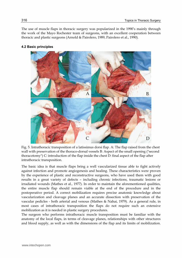
The use of muscle flaps in thoracic surgery was popularized in the 1990’s mainly through the work of the Mayo Rochester team of surgeons, with an excellent cooperation between thoracic and plastic surgeons (Arnold & Pairolero, 1989, Pairolero et al., 1990).
4.2 Basic principles
A B
C D
Fig. 5. Intrathoracic transposition of a latissimus dorsi flap. A: The flap raised from the chest wall with preservation of the thoraco-dorsal vessels B: Aspect of the small opening ("second thoracotomy") C: introduction of the flap inside the chest D: final aspect of the flap after intrathoracic transposition.
The basic idea is that muscle flaps bring a well vascularized tissue able to fight actively against infection and promote angiogenesis and healing. These characteristics were proven by the experience of plastic and reconstructive surgeons, who have used them with good results in a great variety of defects – including chronic infections, traumatic lesions or irradiated wounds (Mathes et al., 1977). In order to maintain the aforementioned qualities, the entire muscle flap should remain viable at the end of the procedure and in the postoperative period. A correct mobilization requires precise anatomic knowledge about vascularization and cleavage planes and an accurate dissection with preservation of the vascular pedicles - both arterial and venous (Mathes & Nahai, 1979). As a general rule, in most cases of intrathoracic transposition the flaps do not require such an extensive mobilization as it is needed in plastic surgery procedures. The surgeon who performs intrathoracic muscle transposition must be familiar with the anatomy of the local flaps, in terms of cleavage planes, relationships with other structures and blood supply, as well as with the dimensions of the flap and its limits of mobilization.
www.intechopen.com

Role of Thoracomyoplasty Procedures in Modern Surgery for Intrathoracic Suppurations
317
Most muscles have several vascular sources, not all of them allowing a complete mobilization of the flap. Anatomical variations may also play an important role. The nutritive pedicle must be carefully prepared using a combination of blunt and sharp dissection, avoiding any damage since this is equivalent to the loss of the flap (McCraw & Arnold, 1987). At the end of the mobilization, the viability of the entire flap must be carefully evaluated. In case of ischemic changes, the affected portion of the flap must be resected; from our experience, severe ischemia of extended portions of the flap is very rare if the mobilization is correctly performed. Any severe ischemia leads to flap necrosis with subsequent recurrence of the intrathoracic suppuration and failure of the surgical procedure. Introduction of the flap inside the thorax requires usually a second window created through a limited rib resection (less than 10 cm length, no more than one rib). When introducing the flap inside the chest, care must be taken to avoid any compression, kinking or twisting of the vascular pedicle in order to avoid thrombosis and loss of the flap. The muscle must reach the defect without any tension and without torsion. Haemostasis must be carefully checked, since postoperative bleeding from the flap will lead to hemothorax. The wound is usually closed, with separate chest and subcutaneous drainage to avoid the postoperative development of seroma in the space resulted from muscle flap dissection (fig. 5). Mobilization of any flap and it's transposition in a different position involves a certain degree of functional morbidity. For most of the flaps used for intrathoracic transposition, these functional sequelae are minor, since there are some other muscles with synergic action that compensate their absence. When put in balance, these functional sequelae are minor compared to the gravity of the situation of the patients that require thoracomyoplasty procedures. However, these aspects must be discussed with the patient before the procedure and a signed informed consent should be obtained (Pairolero & Arnold, 1989, Arnold & Pairolero, 1989).
4.3 Surgical anatomy and techniques of mobilization of the most important flaps
The techniques of muscle flap mobilization were described in detail by the plastic surgeons (Mathes & Nahai, 1979, McCraw & Arnold, 1987), and are based on very precise anatomical knowledges concerning the blood supply (Mathes & Nahai, 1981). The latissimus dorsi flap is probably the most used flap in thoracic surgery. It may be prepared using a skin incision running parallel with the anterior border of the muscle or through a standard postero-lateral skin incision. The standard latissimus dorsi flap involves detachment of the muscle from the iliac crest and sacrum and mobilization from the chest wall keeping intact the main blood supply represented by the thoraco-dorsal vessels, which run parallel with the anterior margin on the deep side of the muscle. If necessary, the insertion tendon and the vascular branch for the serratus anterior may be sectioned in order to increase the length. The flap is quite easy to prepare since there are distinct cleavage planes and the nutrient blood vessels are large enough and with very few anatomical variants; it has a large volume and a long arch of rotation, which allows it to reach almost every part of the thorax (Abolhoda et al., 2008, Seify et al., 2007). The reversed latissimus dorsi flap is much rarely used. It involves sectioning of the flap in the upper part and mobilization based on the secondary blood supply represented by some perforator branches from the last intercostal and first lumbar vessels; these branches are
www.intechopen.com
Topics in Thoracic Surgery
318
small and have an extremely variable anatomy, which make this mobilization more limited and difficult compared to the standard one. However, it may be used with good results in certain circumstances, the main indication being repair or filling of defects located in the supradiaphragmatic area (Botianu P et al., 2010b). Due to the synergic activity of other muscles, mobilization of the latissimus dorsi is not associated with serious functional impairment, having no impact on daily activities. The serratus anterior flap has two main sources of blood supply: the thoracic lateral artery (originating from the axillary artery) and a branch originating from the thoraco-dorsal artery. Since the two vessels have an almost parallel traject, we prefer to preserve both of them if the entire muscle is mobilized; the veins run parallel to the arteries, draining in the axillary vein. The muscle can be easily dissected from the surrounding structures and detached from the chest wall and the scapula. It reaches easily the hilum and is excellent for defects located in the upper half of the thorax. Another advantage is that almost the entire muscle remains intact after the standard postero-lateral thoracotomy, making it suitable for postoperative complications. A specific complication described in the literature is the winged scapula ("scapula allata"), which is very unpleasant for the patient and difficult to treat (Arnold et al., 1984). If several prophylactic measures are taken, this complication is very rare. In our personal experience with over 70 serratus anterior flaps, we had no case of true winged scapula, only some cases of minor asymmetry (Botianu P et al., 2010c). If necessary, the serratus anterior and the latissimus dorsi can be mobilized together using the common thoraco-dorsal vascular pedicle. The pectoralis flap may also be mobilized in more ways. The most used is the mobilization of the pectoralis major, with or without the pectoralis minor muscle, by detachment from the chest wall and subcutis using the thoracoacromial vessels for blood supply. For an increased mobilization, it is necessary to section the insertion tendon on the humerus. Another common way of mobilization is based on the perforator branches that arise from the internal thoracic and anterior intercostal vessels in the parasternal region. The pectoralis flap is usefull for defects located in the apical region (Kalweit et al., 1994, Nomori et al., 2001), although it’s mobilization is difficult through a standard thoracotomy incision; it is also frequently used to prevent and treat infectious complications after median sternotomy (Gao et al., 2010). The rectus abdominis flap can be mobilized based on the superior epigastric vessels, which continue the internal thoracic ones. The second pedicle, represented by the inferior epigastric vessels, can also support the entire flap but does not allow it’s mobilization inside the chest and therefore cannot be used for intrathoracic transposition. Usually, a paramedian vertical incision is made in the abdominal wall and the anterior rectus sheath is opened, allowing exposure of the muscle. The rectus abdominis is raised from the posterior sheath and the inferior epigastric vessels are ligated. A special care must be taken when the flap is turned towards the chest in order to avoid damaging of the vascular pedicle. Specific complications for this flap are represented by the abdominal wall problems, with the risk of developing an incisional hernia. It is preferred by some authors for the treatment of sterno-mediastinal infections following sternotomy (Oh et al., 2004) but it is also usefull for different infections located in the inferior part of the thorax (Ojika et al., 1995).
www.intechopen.com

Role of Thoracomyoplasty Procedures in Modern Surgery for Intrathoracic Suppurations
319
Intercostal flaps are very easy to prepare during thoracomyoplasty procedures (Sarkar et al., 1985). These flaps usually contain the intercostal spaces (including the muscles), the parietal pleura and the periosteum remaining after the rib resection. The arterial blood supply is represented by the posterior intercostal arteries arising from the thoracic aorta and the anterior intercostal arteries arising from the internal thoracic artery; the veins have a parallel traject and drain into the azygos veins, respectively the internal thoracic veins. There is a very rich network connecting the anterior and the posterior intercostal vessels, as well as the vessels from adjacent intercostal spaces and neighbourhood muscles. According to the location of the defect, both the anterior and the posterior intercostal vessels may be used as blood supply for the flaps. When creating the intercostal flaps, the incision should be always made through the bed of the resected ribs in order to minimize the risk of damaging the main intercostal vessels. We found these intercostal flaps particularly usefull for bronchial fistula closure and filling of some dead angles and cul-de-sacs (Botianu P et al., 2010a, c). The omentum flap – although it is not a muscle, it should be included here since it is preferred in many instances with the same purposes and principles as the muscle flaps (Petrov et al., 1999). The main vessels are represented by the left gastro-epiploic artery (originating from the splenic artery) and the right gastro-epiploic artery (originating from the gastro-duodenal artery). These two arteries create the gastro-epiploic arcade which runs along the great curvature of the stomach and gives small branches for the stomach and the so-called epiploic arteries for the omentum. The veins run parallel with the arteries, creating similar arcades. These epiploic vessels have a descendant traject and form a second arcade along the inferior edge of the omentum (Barkow’s arcade). There are many ways to mobilize the omentum based on the vessels and arcades described. However, the anatomy of the omental vascularization is extremely variable and it should be carefully evaluated before planning the flap and making any incision (Kirikuta, 1980). Specific complications are related to the risk of incisional hernia and development of adhesions with intestinal obstruction. The omentum flap is preferred by some authors for sterno-mediastinal infections (Hountis et al., 2009), closure of large bronchial fistulas - especially after pneumonectomy (Chichevatov & Gorshenev, 2005) and reinforcement of high-risk sutures (D'Andrilli et al., 2009). Recent publications have shown good results with the laparoscopic mobilization, which makes the omental flap more attractive (van Wingerden et al., 2010). Other rarely used flaps have been reported in case-reports or small series, with more or less good results: trapezius, subscapularis, infraspinatus, external oblique, teres major etc. Such flaps should be taken into consideration especially when other standard flaps are not available or have failed (Fuchs et al., 2010, Schreiner et al., 2010).
5. Thoracomyoplasty – combining thoracoplasty and muscle flaps
The idea of this kind of procedures is to combine thoracoplasty with intrathoracic muscle transposition in an attempt to achieve safe obliteration of the diseased intrathoracic space. Since for suppurated defects complete filling is mandatory for safe primary wound closure, combining thoracoplasty and muscle transposition acts as a compromise between: - an extended thoracoplasty (rib resection) with major chest wall mutilation; - the use of multiple muscle flaps, with added donor-site morbidity and postoperative
functional deficits.
www.intechopen.com

Topics in Thoracic Surgery
320
Fig. 6. Intraoperative aspects of a complex space-filling procedure / thoracomyoplasty: a limited 5-ribs thoracoplasty, transposition of the latissimus dorsi based on the thoracodorsal vessels and 2 intercostal flaps with posterior vascularization.
There is no standardized technique of thoracomyoplasty. Both rib resection and muscle transposition should be performed trying to reduce the chest wall mutilation and functional morbidity to minimum, but without compromising the definitive obliteration of the cavity (fig. 6). The local anatomy should be carefully evaluated when planning the procedure: - location and dimensions of the cavity, which can be easily assessed on preoperative CT
scans; - presence of bronchial fistula, whose safe closure is mandatory. The presence of a
bronchial fistula may be suggested preoperatively by the clinical course; bronchoscopy and CT may detect some large and centrally located fistula but the exact position of the smaller ones can be evaluated only intraoperative.
- available flaps – in many cases, the neighbourhood muscles may be compromised by previous procedures; typical examples are postero-lateral thoracotomy which divides the latissimus dorsi, upper digestive surgery which compromises the omentum, subcostal laparotomy or myocardial revascularization with the internal mammary artery which compromise the rectus abdominis flap.
The terminology used for this kind of procedures is not very clear. The term "thoracoplasty" should be reserved for cases not associating any muscle flap mobilization. Some authors talk about myoplasty, muscle flaps or intrathoracic muscle transposition, which can quite rarely solve alone a large suppurated cavity without associating any rib resection. As well as other
www.intechopen.com

Role of Thoracomyoplasty Procedures in Modern Surgery for Intrathoracic Suppurations
321
authors, we find the terms "thoracomyoplasty" and "complex space-filling procedures" are the most appropriate to describe this kind of procedures (Botianu P et al., 2010a, García-Yuste et al., 1998, Naumov et al., 1991, Riquet 2010).
6. Personal experience and results from the literature
During the last 8 years we have performed thoracomyoplasty procedures in 102 patients, with almost one half of them having different TB lesions. The procedure was adapted to the local anatomy of the lesion, with an average of 5 resected ribs/patient and 1.9 flaps/patient. Our recently published analysis of the first 76 patients showed an acceptable mortality (5%) and rate of recurrence requiring an open-window procedure (5%); other minor local complications included a few skin necrosis and persistent small thoracic fistulae solved under local anesthesia with no need for a major reoperation. Postoperative hospitalization ranged between 4 and 180 days, with an average of 40 ± 5 days; all the patients with hospitalizations longer than 60 days presented recurrence of the infection requiring on open-window procedure. At the moment of discharge, all the patients had healed wounds with no need for any other surgical care. TB patients were referred to our pneumology colleagues to continue the specific chemotherapy. Five patients presented a mild impairment of the shoulder function, but without interfering with the daily activities; 4 patients presented a minor asymmetry of the two shoulders and scapulas but we had no case of true winged scapula and no major functional disturbance secondary to the extrathoracic muscle flap mobilization. None of our patients presented severe scoliosis or a major chest deformity. At 3 months follow-up, 91% (66 patients) of the survivors returned to an almost normal life compared with their preoperative status. A comparative evaluation showed no statistically significant difference between the pre- and post-operative values of the the functional respiratory tests (VC preoperative – mean 1050 ml/62% of predicted vs postoperative – mean 1100 ml/63% of predicted, FEV1 preoperative – mean 850 ml/61% of predicted vs postoperative - mean 890 ml/62% of predicted, Wilcoxon signed-rank test, p>0.05). The main explanation is that the parenchyma underlying the thoracomyoplasty is more or less diseased, therefore with a lower contribution to the respiration; also, closure of the bronchial fistula improves the respiratory function (Botianu P et al., 2010a). Other authors have also recently published important series with quite similar results, with mortality, morbidity and hospitalization falling within an acceptable range of values. These data recommend the use of thoracomyoplasty procedures to solve difficult cases of intrathoracic suppurations (García-Yuste et al., 1998, Icard et al., 1999, Jadczuk 1998, Krassas et al., 2010, Regnard et al., 2000, Stefani et al., 2011).
7. Future challenges
During the recent years, a few new ideas emerged: - the use of free-transfers, which increases the number and volume of the flaps available
for filling of the infected spaces (Walsh et al., 2011). However, they involve a multidisciplinary approach and complicated procedures; the microsurgical anastomoses of the vessels require experience and special technical skills, are time-consuming and involve a specific morbidity.
www.intechopen.com

Topics in Thoracic Surgery
322
- the use of less-invasive procedures for flap mobilization, using special retractors, light sources, instruments and dissection techniques borrowed from endoscopic surgery. Proponents consider that with the use of these techniques the mobilization of the muscle flaps may be performed using shorter skin incisions and less donor-site morbidity, making them more acceptable (Blidisel et al., 2008).
A major problem is the difficulty to study these procedures according to the modern principles of evidenced-based medicine. These procedures are rarely performed and address to a small number of desperate cases, not comparable to those submitted to the standard treatment consisting of lung resection and/or pleural decortication. The rarity of these procedures and the great heterogenicity of the patients (in terms of local anatomy, etiology and biological status) makes any kind of randomised trial or fair comparison impossible (Botianu P. et al., 2010a, b). The most important challenge remains probably training, since many surgeons are not familiar with the techniques of thoracoplasty or muscle flaps mobilization; most young surgeons have neither performed, nor seen such procedures. Specific training and/or a good cooperation with a plastic and reconstructive surgery colleague is mandatory.
8. Conclusions
Both thoracoplasty and muscle transposition remain in the armamentarium of modern thoracic surgery (Riquet 2010). Due to the recrudescence of TB and other infectious diseases of the chest, it is possible that the number of patients requiring this kind of surgery will increase in the near future. We believe that sooner or later any thoracic surgeon will meet a patient requiring such a procedure. Thoracomyoplasty may solve a complicated case with good immediate and long-term outcome. In such situations, training, careful evaluation and an accurate surgical technique are essential to achieve good results (Botianu P. et al., 2010a, Krassas et al., 2010, Stefani et al., 2011).
9. References
Abolhoda A,; Bui T.D.; Milliken J.C. & Wirth G.A. (2009) Pedicled latissimus dorsi muscle flap: routine use in high-risk thoracic surgery, Tex Heart Inst J; 36(4):298-302
Abolhoda A.; Wirth G.A.; Bui T.D. & Milliken J.C. (2008) Harvest technique for pedicled transposition of latissimus dorsi muscle: an old trade revisited, Eur J Cardiothorac Surg; 33(5):928-30
Abrashanoff G. (1911) Plastische Methode der Schliessung von Fistelgangen, welche von inneren Organen kommen, Zentralbl Chir; 38:186-7
Alexander J. (1936) Some advances in the technic of thoracoplasty, Ann Surg; 104(4):545-51 Andrews NC. (1961) Thoracomediastinal plication: a surgical technique for chronic empyema, J
Thorac Cardiovasc Surg; 41:809-16 Andrews NC. (1965) The surgical treatment of chronic empyema, Dis Chest; 47:533-8 Archibald E. (1926) On extra-pleural thoracoplasty, Can Med Assoc J;16(4)433-5 Arnold P.G. & Pairolero P.C. (1989) Intrathoracic muscle flaps: a 10-year experience in the
management of life-threatening infections, Plast Reconstr Surg; 84(1):92-8 Arnold P.G.; Pairolero P.C. & Waldorf J.C. (1984) The serratus anterior muscle: intrathoracic and
extrathoracic utilization, Plast Reconstr Surg; 73(2):240-8 Bjork V.O. (1954) Thoracoplasty, a new osteo plastic technique, J Thorac Surg; 28(2):194-211
www.intechopen.com

Role of Thoracomyoplasty Procedures in Modern Surgery for Intrathoracic Suppurations
323
Blidisel A.; Maciuceanu B.; Jiga L.; Papurica M. & Ionac M. (2008) Endoscopy-assisted harvesting and free latissimus dorsi muscle flap transfer in reconstructive microsurgery, Chirurgia (Bucur); 103(1):67-72
Botianu A.M. (1996) Procedeu de toracopleuroplastie pentru tratamentul chirurgical al empiemului toracic cu sau fara fistula bronsica/Personal procedure of thoracopleuroplasty for the treatment of the thoracic empyema, with or without bronchial fistula, Jurnalul de Chirurgie toracică/Journal of Thoracic Surgery; 1:3:251-260
Botianu P. V.-H. (2005) Re-evaluation of the Schede thoracoplasty, Jurnalul de Chirurgie Toracica – Journal of Thoracic Surgery; 7(1):71-78
Botianu P.V.-H.; Butiurca A.; Dobrica A.; Damian V.; Ionica S.; Lupu C. & Stoica S. (2008) Schede procedure and muscular plombage for a multirelapsed TB empyema with pleuro-cutaneous fistula, Revista de Medicina şi Farmacie-Orvosi es Gyogyszereszeti Szemle (Acta Medica Marisiensis); 54(S3): 69-72
Botianu P.V.-H.; Botianu A.M.; Bacarea V. & Dobrica A.C. (2010) Thoracodorsal versus reversed mobilisation of the latissimus dorsi muscle for intrathoracic transposition, Eur J Cardiothorac Surg; 38(4):461-5
Botianu P.V.-H.; Botianu A.M.; Dobrica A.C. & Bacarea V. (2010) Intrathoracic transposition of the serratus anterior muscle flap--personal experience with 65 consecutive patients, Eur J Cardiothorac Surg; 38(6):669-73
Botianu P.V.-H.; Dobrica A.C.; Butiurca A. & Botianu A.M. (2010) Complex space-filling procedures for intrathoracic infections - personal experience with 76 consecutive cases, Eur J Cardiothorac Surg; 37(2):478-81
Chachques J.C.; Jegaden O.J.; Bors V.; Mesana T.; Latremouille C.; Grandjean P.A.; Fabiani J.N. & Carpentier A. (2008) Heart transplantation following cardiomyoplasty: a biological bridge, Eur J Cardiothorac Surg; 33(4):685-90
Chichevatov D. & Gorshenev A. (2005) Omentoplasty in treatment of early bronchopleural fistulas after pneumonectomy, Asian Cardiovasc Thorac Ann; 13(3):211-6
Cornet E.; Dupon H.; Coiffard P. & Rembeaux A. (1965) Thoracopleuroplasties for empyema according to Andrews' method (18 cases), Ann Chir Thorac Cardiovasc; 4(4):509-15
D'Andrilli A.; Ibrahim M.; Andreetti C.; Ciccone A.M.; Venuta F. & Rendina E.A. (2009) Transdiaphragmatic harvesting of the omentum through thoracotomy for bronchial stump reinforcement, Ann Thorac Surg; 88(1):212-5
Davies H.E.; Rosenstengel A. & Lee Y.G. (2011) The diminishing role of surgery in pleural disease, Curr Opin Pulm; 17(4):247-54
Deslauriers J.; Jacques L.F. & Grégoire J. (2002) Role of Eloesser flap and thoracoplasty in the third millennium, Chest Surg Clin N Am; 12(3):605-23
Entin MA. (1995) Romance and tragedy of tuberculosis: Edward Archibald's contribution to the surgical treatment of pulmonary tuberculosis, The Canadian Journal of Plastic Surgery; 3(4):213-6
Estlander J.A. (1879) Résection des côtes dans l’empyème chronique, Rev Med Chir (Paris); 3:157-70
Fuchs P.; Schreiner W.; Wolter T.P.; Autschbach R.; Sirbu H. & Pallua N. (2011) A four-muscle-flap for thoracomyoplasty in patients with sacrificed thoracodorsal vessels, J Plast Reconstr Aesthet Surg; 64(3):335-8
Gao J.; Wang Y.L.; Lu S.Q.; Cai A.B., Yang Z.F., Han Z.Y., Li J.J., Wen Y.M., Geng F.Y. & Wang W.Z. (2010) Management of sternal osteomyelitis and mediastinal infection following median sternotomy, Chin Med J (Engl); 123(20):2803-6
www.intechopen.com

Topics in Thoracic Surgery
324
García-Yuste M.; Ramos G.; Duque J.L.; Heras F.; Castanedo M.; Cerezal L.J. & Matilla J.M. (1998) Open-window thoracostomy and thoracomyoplasty to manage chronic pleural empyema, Ann Thorac Surg; 65(3):818-22
Hollaus P.H.; Huber M.; Lax F.; Wurnig P.N.; Böhm G. & Pridun N.S. (1999) Closure of bronchopleural fistula after pneumonectomy with a pedicled intercostal muscle flap, Eur J Cardiothorac Surg; 16(2):181-6
Holst J. (1952) Technique of so-called dome thoracoplasty; preliminary report, Nord Med; 19:48(38):1290-3
Hopkins R.A.; Ungerleider R.M.; Staub E.W. & Young W.G. Jr. (1985) The modern use of thoracoplasty, Ann Thorac Surg; 40(2):181-7
Horrigan T.P. & Snow N.J. (1990) Thoracoplasty: current application to the infected pleural space, Ann Thorac Surg; 50(5):695-9
Hountis P.; Dedeilias P. & Bolos K. (2009) The role of omental transposition for the management of postoperative mediastinitis: a case series, Cases J; 23;2(1):142
Icard P.; Le Rochais J.P.; Rabut B.; Cazaban S.; Martel B. & Evrard C. (1999) Andrews thoracoplasty as a treatment of post-pneumonectomy empyema: experience in 23 cases, Ann Thorac Surg; 68(4):1159-63
Jadczuk E. (1998) Posptneumonectomy empyema, Eur J Cardiothorac Surg; 14(2):123-6 Kalweit G.; Feindt P.; Huwer H.; Volkmer I. & Gams E. (1994) The pectoral muscle flaps in the
treatment of bronchial stump fistula following pneumonectomy, Eur J Cardiothorac Surg; 8(7):358-62
Kirikuta I. (1980) Use of the omentum in plastic surgery, Ed. Medicala/Medical Publishing House, Bucuresti
Kobayashi H.; Nomori H.; Mori T.; Shibata H.; Yoshimoto K. & Ohba Y. (2009) Extrapleural pneumonectomy with reconstruction of diaphragm and pericardium using autologous materials, Ann Thorac Surg 2009; 87(5):1630-2
Kotzampassakis N.; Christodoulou M.; Krueger T.; Demartines N.; Vuillemier H.; Cheng C.; Dorta G. & Ris H.B. (2009) Esophageal leaks repaired by a muscle onlay approach in the presence of mediastinal sepsis, Ann Thorac Surg; 88(3):966-72
Krasnov V.A.; Kozhevnikov N.N.; Gorbunov G.M. & Andrenko A.A. (1989) Long-term results of the use of osteoplastic thoracoplasty in the treatment of destructive tuberculosis of the lungs in patients with antisocial behavior, Probl Tuberk; (1):41-4
Krassas A.; Grima R.; Bagan P.; Badia A.; Arame A.; Barthes F.P. & Riquet M. (2010) Current indications and results for thoracoplasty and intrathoracic muscle transposition, Eur J Cardiothorac Surg; 37(5):1215-20
Massard G.; Thomas P.; Barsotti P.; Riera P.; Giudicelli R.; Reboud E.; Morand G.; Fuentes P.A. & Wihlm J.M. (1997) Long-term complications of extraperiosteal plombage, Ann Thorac Surg; 64(1):220-4
Mathes S.J. & Nahai F. (1981) Classification of the vascular anatomy of muscles: experimental and clinical correlation, Plast Reconstr Surg; 67(2):177-87
Mathes S.J. & Nahai S. (1979) Clinical atlas of muscle and musculocutaneous flaps, CV Mosby Co, St. Louis
Mathes S.J.; Vasconez L.O. & Jurkiewicz M.J. (1977) Extensions and further applications of muscle flap transposition, Plast Reconstr Surg; 60:6-13
McCraw J.B. & Arnold P.G. (1987) McCraw and Arnold's Atlas of Muscle and Musculocutaneous Flaps, Hampton Press, Norfolk, VA
www.intechopen.com

Role of Thoracomyoplasty Procedures in Modern Surgery for Intrathoracic Suppurations
325
Meyer A.J.; Krueger T.; Lepori D.; Dusmet M.; Aubert J.D.; Pasche P. & Ris H.B. (2004) Closure of large intrathoracic airway defects using extrathoracic muscle flaps, Ann Thorac Surg; 77(2):397-404
Mitra A.; Spears J.; Perrotta V.; McClurkin J. & Mitra A. (2005) Salvage of infected prosthetic grafts of the great vessels via muscle flap reconstruction, Chest; 128(2):1040-3
Naftali Z. (1964) Toracoplastia osteoplastica cu transplant paramediastinal de coastă in tratamentul tuberculozei cavernoase (Osteoplastic thoracoplasty with paramediastinal transplantation of rib grafts in the treatment of cavernous tuberculosis), PhD Thesis
Naumov V.N.; Shaikhaev A.I. & Testov V.V. (1991) Thoracomyoplastic operations in the surgery of tuberculosis of the lungs, Grud Serdechnososudistaia Khir; (7):46-8
Nomori H.; Horio H.; Hasegawa T. & Suemasu K. (2001) Intrathoracic transposition of a pectoralis major and pectoralis minor muscle flap for empyema in patients previously subjected to posterolateral thoracotomy, Surg Today; 31(4):295-9
Nosotti M.; Baisi A.; Mendogni P.; Palleschi A.; Tosi D. & Rosso L. (2010) Muscle sparing versus posterolateral thoracotomy for pulmonary lobectomy: randomised controlled trial, Interact Cardiovasc Thorac Surg; 11(4):415-9
Oh A.K.; Lechtman A.N.; Whetzel T.P. & Stevenson T.R. (2004) The infected median sternotomy wound: management with the rectus abdominis musculocutaneous flap, Ann Plast Surg; 52(4):367-70
Ojika T.; Mukoyama N.; Suzuki M.; Senda Y. & Namiki Y. (1995) Two cases of empyema treated with rectus abdominis myocutaneous flap and muscle flap, Kyobu Geka; 48(5):394-6
Pairolero P.C. & Arnold P.G. (1989) Intrathoracic transfer of flaps for fistulas, exposed prosthetic devices, and reinforcement of suture lines, Surg Clin North Am; 69(5):1047-59
Pairolero P.C.; Arnold P.G.; Trastek V.F.; Meland N.B. & Kay P.P. (1990) Postpneumonectomy empyema. The role of intrathoracic muscle transposition. J Thorac Cardiovasc Surg; 99(6):958-66
Pardos-Gea J.; Maza Ú.; Pérez-López J. & San José Laporte A. (2011) Home intravenous antibiotic therapy of empyema and lung abscess: safety and efficacy, Enferm Infecc Microbiol Clin; 29(3):237-9
Petrov D.; Dzhambazov V.; Minchev T.; Plochev M.; Goranov E.; Krupev M. & Petkov R. (1999) Omentoplasty in surgical management of postpulmonectomy pleural empyema, Khirurgiia (Sofiia); 55(6):13-5
Regnard J.F.; Alifano M.; Puyo P.; Fares E.; Magdeleinat P. & Levasseur P. (2000) Open window thoracostomy followed by intrathoracic flap transposition in the treatment of empyema complicating pulmonary resection, J Thorac Cardiovasc Surg; 120(2):270-5
Riquet M. (2010) Thoracomyoplasty (Editorial comment), Eur J Cardiothorac Surg; 37(2):482 Sarkar S.K.; Sharma T.N.; Singh H.; Singh A.; Purohit S.D. & Sharma V.K. (1985)
Thoracoplasty with intercostal myoplasty for closure of an empyema cavity and bronchopleural fistula, Int Surg; 70(3):219-21
Schede M. (1890) Die behandlung der empyeme, Verh Long Innere Med Wiesbaden; 9:41-141 Schreiner W.; Fuchs P.; Autschbach R.; Pallua N. & Sirbu H. (2010) Modified technique for
thoracomyoplasty after posterolateral thoracotomy, Thorac Cardiovasc Surg; 58(2):98-101
Seify H.; Mansour K.; Miller J.; Douglas T.; Burke R.; Losken A.; Culbertson J.; Jones G.; Nahai F. & Hester T.R. (2007) Single-stage muscle flap reconstruction of the postpneumonectomy empyema space: the Emory experience, Plast Reconstr Surg; 120(7):1886-91
www.intechopen.com

Topics in Thoracic Surgery
326
Semb C. (1937) Thoracoplasty with extrafascial apicolysis, Br Med J; 2(4004):650-6 Stefani A.; Jouni R.; Alifano M.; Bobbio A.; Strano S.; Magdeleinat P. & Regnard J.F. (2011)
Thoracoplasty in the current practice of thoracic surgery: a single-institution 10-years experience, Ann Thorac Surg; 91(1):263-8
Stobernack A.; Achatzy R. & Engelmann C. (1997) Delayed complications after extrapleural pneumonolysis for lung tuberculosis, Chirurg; 68(9):921-7
Thingnam S.K.; Mohite P.N.; Raju G.; Ranade S.D. & Saklani R. (2011) Triple reinforcement of bronchial stump, Thorac Cardiovasc Surg; 59(3):169-71
van Wingerden J.J.; Coret M.E.; van Nieuwenhoven C.A.; Totté E.R. (2010) The laparoscopically harvested omental flap for deep sternal wound infection, Eur J Cardiothorac Surg 2010; 37(1):87-92
Walsh M.D.; Bruno A.D.; Onaitis M.W.; Erdmann D.; Wolfe W.G.; Toloza E.M. & Levin L.S. (2011) The role of intrathoracic free flaps for chronic empyema, Ann Thorac Surg; 91(3):865-8
Wilson N.J.; Armada 0.; Vindzberg V. & O'Brien WB (1956) Extraperiosteal plombage thoracoplasty: operative technique and results with 161 cases with unilateral surgical problems, J Thorac Surg; 32:797-813
Zaheer S.; Allen M.S.; Cassivi S.D.; Nichols F.C. 3rd; Johnson C.H.; Deschamps C. & Pairolero P.C. (2006) Postpneumonectomy empyema: results after the Clagett procedure, Ann Thorac Surg; 82(1):279-86
Zahid I.; Nagendran M.; Routledge T. & Scarci M. (2011) Comparison of video-assisted thoracoscopic surgery and open surgery in the management of primary empyema, Curr Opin Pulm Med; 17(4):255-9
www.intechopen.com

Topics in Thoracic SurgeryEdited by Prof. Paulo Cardoso
ISBN 978-953-51-0010-2Hard cover, 486 pagesPublisher InTechPublished online 15, February, 2012Published in print edition February, 2012
InTech EuropeUniversity Campus STeP Ri Slavka Krautzeka 83/A 51000 Rijeka, Croatia Phone: +385 (51) 770 447 Fax: +385 (51) 686 166www.intechopen.com
InTech ChinaUnit 405, Office Block, Hotel Equatorial Shanghai No.65, Yan An Road (West), Shanghai, 200040, China
Phone: +86-21-62489820 Fax: +86-21-62489821
Thoracic Surgery congregates topics and articles from many renowned authors around the world coveringseveral different topics. Unlike the usual textbooks, Thoracic Surgery is a conglomerate of different topics fromPre-operative Assessment, to Pulmonary Resection for Lung Cancer, chest wall procedures, lung cancertopics featuring aspects of VATS major pulmonary resections along with traditional topics such as Pancoasttumors and recurrence patterns of stage I lung disease, hyperhidrosis, bronchiectasis, lung transplantationand much more. This Open Access format is a novel method of sharing thoracic surgical information providedby authors worldwide and it is made accessible to everyone in an expedite way and with an excellentpublishing quality.
How to referenceIn order to correctly reference this scholarly work, feel free to copy and paste the following:
Petre Vlah-Horea Botianu and Alexandru Mihail Botianu (2012). Role of Thoracomyoplasty Procedures inModern Surgery for Intrathoracic Suppurations, Topics in Thoracic Surgery, Prof. Paulo Cardoso (Ed.), ISBN:978-953-51-0010-2, InTech, Available from: http://www.intechopen.com/books/topics-in-thoracic-surgery/role-of-thoracomyoplasty-procedures-in-modern-surgery-for-intrathoracic-suppurations