role of vdr in host immune response to ... of vdr in host immune response to porphyromonas...
TRANSCRIPT
ROLE OF VDR IN HOST IMMUNE RESPONSE TO PORPHYROMONAS
GINGIVALIS INFECTION
by
GULAM YEZDANI
PING ZHANG, CHAIR
AMJAD JAVED
JANNET KATZ
SUZANNE MICHALEK
A THESIS
Submitted to the graduate faculty of The University of Alabama at Birmingham,
in partial fulfillment of the requirements for the degree of
Master of Science
BIRMINGHAM, ALABAMA
2011
ii
ROLE OF VDR IN HOST IMMUNE RESPONSE TO PORPHYROMONAS
GINGIVALIS INFECTION
GULAM YEZDANI
ORAL BIOLOGY
ABSTRACT
Porphyromonas gingivalis is one of the etiologic factors of periodontal disease, a chronic
inflammatory disorder characterized by the destruction of periodontal connective tissue
and the subsequent loss of alveolar bone. Epidemiological and genetic studies have
indicated an association between periodontal disease and the vitamin D system. However,
little is known regarding how vitamin D signaling regulates the inflammatory immune
process of this disease. The purpose of the study was to understand the role of the vitamin
D receptor (VDR) in regulating the host response to P. gingivalis infection by using a
dual chamber mouse model. These chambers served both as infection and sample
collection sites. Chamber exudates of wild type (WT) and VDR knock out (VDR-/-
) mice
were sampled for neutrophil and monocyte levels, bacterial clearance and levels of TNF-
α, IL-6, IL-12p40, and IL-10 cytokines. In addition, serum cytokines, P. gingivalis
specific CD4+ T cell cytokine responses and serum antibody responses were assessed.
Our results showed similar levels of neutrophil and monocyte infiltration in WT and
VDR-/-
mice following P. ginigvalis infection. However, VDR-/-
mice exhibited a
significantly higher level of bacteria than WT mice. Furthermore, significant higher
iii
levels of IL-6, IL-10, IL-12p40 and TNF-α were observed in the chamber exudates of
VDR-/-
mice. Although a similar level of serum IL-6 was seen in WT and VDR-/-
mice 4
hr after P. gingivalis infection, 24 hr post infection, the levels were increased in VDR-/-
mice. Assessment of P. gingivalis-specific CD4+ T cell cytokine responses suggested that
in the presence of the VDR a Th1 response was favored following P. gingivalis infection.
Furthermore, VDR-/-
mice exhibited lower levels of serum IgG anti-P. gingivalis
antibodies than that observed in WT mice. Taken together, these results suggest that
VDR deficient mice have an increased susceptibility to P. gingivalis infection,
implicating the VDR in the control of bacterial growth and in modulating inflammatory
responses that could be detrimental to the host during infection by P. ginigvalis. These
findings provide valuable insight on the role of the VDR in P. gingivalis infection.
iv
DEDICATION
I dedicate this thesis to my parents, brothers and sisters who supported every aspect of
my education and pushed me further. To my wife whose love, support and prayers kept
me strong throughout.
v
ACKNOWLEDGMENTS
I would like to acknowledge several people who helped to make this thesis
possible. Dr. Ping Zhang, who is my mentor and chair of my committee, has helped me
throughout the research progress, thank you. I am thankful for my committee members
Dr. Jannet Katz, Dr. Suzanne Michalek, Dr. Amjad Javed for their kind assistance on the
project. My thanks go to Qingan Xu and Amit Ashtekar for their help and guidance
throughout my graduate work. My lab mates Mike, Dalia also deserve vast amount of
praise for their help and support. Special thanks and I am grateful to Dr. Jannet Katz and
Dr. Ping Zhang who gave me motivation, listened and helped me with the difficulties I
faced at the beginning of my masters programme. Also many thanks to Dr. Suzanne
Michalek, for everything she provided to me in my research. Last person I would
acknowledge is Greg for his excellent technical assistance. These people I will always be
indebted my life. Thank you all.
vi
TABLE OF CONTENTS
Page
ABSTRACT ........................................................................................................................ ii
DEDICATION ................................................................................................................... iv
ACKNOWLEDGMENTS ...................................................................................................v
LIST OF FIGURES .......................................................................................................... vii
LIST OF ABBREVIATIONS .......................................................................................... viii
INTRODUCTION ...............................................................................................................1
Immune system, vitamin D and its effects on disease ..................................................1
Vitamin D receptor .......................................................................................................3
Vitamin D receptor knockout mice ...............................................................................4
Periodontal apparatus ....................................................................................................6
Periodontal disease........................................................................................................7
Hypothesis…………………………………………………………………………….8
Rationale to use mouse chamber model………………………………………………9
MATERIAL AND METHODS…………………………………………………………...9
RESULTS………………………………………………………………………………..15
DISCUSSION……………………………………………………………………………18
GENERAL LIST OF REFERENCES ...............................................................................21
APPENDIX: NOTICE OF APPROVAL FROM INSTITUTIONAL ANIMAL CARE
AND USE COMMITTEE..................................................................................................33
vii
LIST OF FIGURES
Figure Page
1 Periodontal structure ................................................................................................7
2 Infection and sample collection diagram ...............................................................12
3 Cytokine production in bone marrow-derived macrophages .................................26
4 Neutrophil and monocyte infiltration.....................................................................27
5 Bacterial culture .....................................................................................................28
6 Cytokine production in chamber exudates .............................................................29
7 Cytokine production in serum……………………………………………………30
8 CD4+
T cell cytokine production………………… ………………………...........31
9 Serum antibody to P. gingivalis………………………………………………….32
.
viii
LIST OF ABBREVIATION
WT wild type
VDR-/-
vitamin D receptor knock out
ELISA enzyme-linked immunosorbent assay
IFN interferon
TNF-α tumor necrosis factor alpha
IL interleukin
Th t helper
Ig immunoglobulin
BMMs bone marrow derived macrophages
PBS phosphate buffered saline
1
INTRODUCTION
Immune function, vitamin D and its effects on disease.
The main function of the immune system is to protect the host from infection by
pathogenic organisms. It also serves to rid the body of altered self cells, the uncontrolled
division of which can lead to cancer. Therefore, the immune system is comprised of
several organs and cell types, which communicate through cell surface and soluble
molecules such as cytokines and chemokines. Host responses involve the innate and
adaptive immune system (9, 11). The innate system is constitutively present and deals
non-specifically with the invading pathogen, whereas the adaptive system involves the
activation and differentiation of antigen specific CD4+ or CD8
+ T cells and antibody
secreting B cells and is characterized by a delayed response tailored to the pathogen that
induces memory (32). CD4+ T cell are important in responses to bacteria, fungi and
parasites, and ensures that subsequent reinfection by the same pathogen is dealt quickly
before symptoms develop. Loss of immunity results in recurrent infections and severe
immunodeficiencies that can also lead to death. Equally, inappropriate is the excessive
activation of the immune response. Thus the immune system can be viewed as a double
edge sword and maintaining equilibrium is essential for a state of health (7, 11, 48).
Vitamins have been known to have an effect on the host, its deficiencies have been
attributed to diseases, affecting overall host immune mechanism. Of lately vitamin D as
such have been studied in the context of immunity (1, 3, 25, 32). Such information could
2
be useful in the design of treatments for active disease. Although vitamin D has been
traditionally associated with calcium and phosphate homeostasis, in recent years, it has
been shown to be involved in host immunity, influencing both the innate and adaptive
arms of the response (3, 25, 27).
Important co-relations between vitamin D and the immune system were observed in the
1960’s when patients having a chronic granulomatous disorder such as sarcoidiosis,
tuberculosis or histoplasmosis, had high levels of circulating vitamin D in their blood
(32). It is an accepted fact that prior to the development of drugs, the treatment of choice
for tuberculosis was sunlight. This was proven by Lie et al. (49, 39) who showed that
vitamin D had antimicrobial properties that could inhibit the growth of Mycobacterium
tuberculosis in macrophage cultures. Furthermore, low levels of the principal vitamin D
metabolite (25-hydroxy vitamin D) in the circulation, is associated with a high incidence
of autoimmune disorders e.g., multiple sclerosis, inflammatory bowel disease and
diabetes (7). Many investigators have reported that a functioning VDR system helps
control inflammation and modulates the immune system in certain types of cancers and
infectious diseases (27). Furthermore, genetic studies have shown an association between
VDR gene polymorphisms and periodontal infections (12, 14, 26). It has been shown that
a defective VDR codon causes an increase susceptibility to generalized aggressive
periodontitis moreover ongoing periodontal studies are using the VDR gene risk marker
as a susceptibility test to periodontal disease (2). Human studies of vitamin D have shown
that its deficiency is directly responsible for a higher incidence of periodontal diseases
and further, VDR ligands have been used for the treatment of periodontal diseases (12, 2,
14).
3
Vitamin D receptor
The vitamin D receptor (VDR) was discovered in 1969 as a binding protein and was later
cloned and sequenced in 1987 (6) and further investigations led to the discovery of its
binding (38) to the active vitamin D hormone, which confirmed that the receptor and
1,25-(OH)2D3 functions like many other steroid hormones, acts by binding to and
activating a nuclear transcription factor (5). VDR expression has been identified in every
human tissue and nearly all nucleated cells express VDR (32).
VDR structure
The VDR is a molecule of approximately 50-60 kDa depending on the species. The VDR
protein consists of 3 major domains, an N-terminal DNA-binding domain, a ligand
binding domain and a C-terminal transaction domain which can mediate co-factor
interaction (24). The most conserved domain is the DNA binding domain, which is
composed of 2 zinc fingers. The first zinc finger confers specificity for DNA binding to
the vitamin D responsive elements (VDREs) while the second zinc finger is the region for
the heterodimerization to the retinoid X receptor (RXR) (5, 35, 13). The ligand binding
domain is responsible for the binding to 1,25(OH)2D3. Interaction with 1,25(OH)2D3
causes the VDR to heterodimerize with the RXR, inducing or inhibiting transcription of
the target gene. Although the VDR is capable of dimerizing with other nuclear receptors,
it has been observed to prefer interaction with RXR (25, 30). Thus the VDR is a member
of the nuclear receptor family of transcripton factors. These receptors function as ligand
activated, transcriptional regulatory proteins. VDR is localized in both the cytosol and
nucleus, and accumulates in the nucleus in response to 1,25(OH)2D3 binding. It has been
4
seen that mutations of this receptor results in hereditary vitamin D resistant rickets. In
humans, multiple polymorphisms of the VDR gene have been identified and thoroughly
studied that link the VDR to disorders like cancer and autoimmune diseases (10, 27, 25).
Data obtained from VDR-/-
has been instrumental in elucidating the physiological actions
of VDR. VDR-/-
animal models have the full phenotype of severe vitamin D deficiency.
Vitamin D receptor knockout (VDR-/-
) mice
VDR-/-
mice are viable and fertile with a normal survival of approximately 14 months.
The RNA isolated from these mice shows a truncated deletion of the second zinc finger
coding region followed by a premature termination codon, resulting in the absence of
receptor protein (30). At 21 days of age theses mice, show hypocalcemia,
hypophosphatemia, hyperparathyroidism, increased serum parathyroid hormone,
abnormal blood mineral levels, and growth retardation (5). After at 4 weeks of age, VDR-
/- mice exhibit perioral and periorbital alopecia that progresses over the entire body by 4
months of age. Introducing a rescue diet enriched for calcium, phosphorus, and lactose to
young mice prevents hyperparathyroidism, rickets, and osteomalacia, but not alopecia.
Vitamin D metabolism
Vitamin D is obtained through diet, supplements, and exposure to sunlight. Vitamin D2
(ergocalciferol) is found in plants and fungi, and vitamin D3 (cholecalciferol) in meats (5,
32). Vitamin D3 is also produced endogenously when the skin is exposed to ultraviolet
light. Vitamin D3 is hydroxylated at the 25-position by the hepatic vitamin D3
hyrdoxylase enzyme (CYP27A1) to yield 25-hydroxyvitaminD3 (25-
hydroxycholecalciferol), the major form of vitamin D in the circulation (4). This 25-
hydroxyvitaminD3 is further hyroxylated to the final form of 1,25-hydroxyvitamin D with
5
the help of enzyme 1 α-hydroxylase (CYP27B1) occurring exclusively in the kidneys to
form the active vitamin D metabolite 1,25-dihydroxyvitaminD3 also known as calcitriol
(32). This 1,25-dihydroxyvitaminD3 binds to the vitamin D receptor VDR , a nuclear
receptor highly expressed in the target organs of calcium homeostasis, such as the
intestine, bone ,kidney parathyroid glands and cells of the immune system (24, 32). The
ligand bound VDR activates transcription by heterodimerization with RXRs, which is
essential for high-affintiy DNA binding to cognate vitamin D response elements VDREs
located in the regulatory regions of 1,25D target genes. The VDR/RXR heterodimes
recruit numerous coregulatory proteins which control histone modifications, chromatin
remodeling and RNA polymerase II binding and transcriptional initiation (3, 4, 5, 7, 32).
There is strong evidence that shows vitamin D signaling in the innate and adaptive
immune systems. The VDR is present in most cells of the immune system, including
neutrophils, macrophages, dendritic cells and T and B lymphocytes (1, 28, 36). Studies
have shown that inflammatory bowel disease (IBD), arthritis and multiple sclerosis in
VDR-/-
mice have a negative influence on the outcome of the diseases (7, 17). However,
other infection studies showed that vitamin D has no effect on the susceptibility of mice
to infections with herpes simplex virus or Candida albicans (5) and an unsubstantiated
link between vitamin D deficiency and cases of tuberculosis (18, 39, 40) has been noted.
Experimentally, vitamin D deficiency and host resistance to infectious diseases have not
been studied extensively. Surprisingly, little is known about the effect of the vitamin D
status on the ability of the host to fight infections. Moreover little is known about the role
of vitamin D and 1,25(OH)2D3 in regulating immune responses to infectious diseases.
6
Periodontal apparatus
The periodontal ligament is the fibrous connective tissue structure, with neural and
vascular components, that joins the cementum covering the root to the alveolar bone. The
periodontal ligament serves primarily a supportive function by attaching the tooth to the
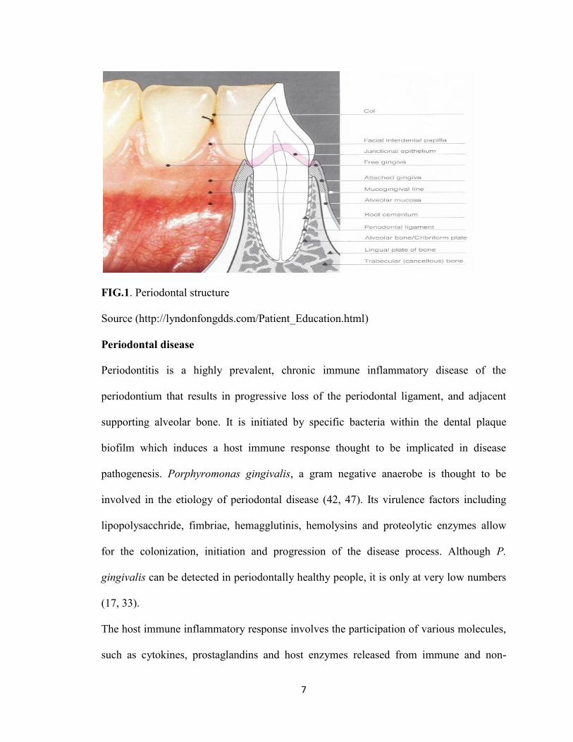
surrounding alveolar bone proper. (Fig. 1) This function is mediated primarily by the
principal fiber groups (horizontal, oblique, apical and interradicular) of the periodontal
ligament that form a strong fibrous union between the root cementum and the bone
(37).The periodontal ligament also serves as a shock-absorber by mechanisms that
provide resistance from light, moderate and heavy forces. Light forces are cushioned by
intravascular fluid that is forced out of the blood vessels. Moderate forces are absorbed
by extravascular tissue fluid that is forced out of the periodontal ligament space into the
adjacent marrow spaces (41). The heavier forces are taken up by the principal fibers. The
periodontal ligament also serves as a reservoir by providing cells that are able to form all
the tissues that make up the attachment apparatus i.e. bone, cementum and the
periodontal ligament. In addition, the periodontal ligament has a sensory function by
nerve endings that are primarily receptors for pain and pressure, and it also provides a
nutritive function that maintains the vitality of its various cells via dental arteries that
enter the ligament through the alveolar bone.
7
FIG.1. Periodontal structure
Source (http://lyndonfongdds.com/Patient_Education.html)
Periodontal disease
Periodontitis is a highly prevalent, chronic immune inflammatory disease of the
periodontium that results in progressive loss of the periodontal ligament, and adjacent
supporting alveolar bone. It is initiated by specific bacteria within the dental plaque
biofilm which induces a host immune response thought to be implicated in disease
pathogenesis. Porphyromonas gingivalis, a gram negative anaerobe is thought to be
involved in the etiology of periodontal disease (42, 47). Its virulence factors including
lipopolysacchride, fimbriae, hemagglutinis, hemolysins and proteolytic enzymes allow
for the colonization, initiation and progression of the disease process. Although P.
gingivalis can be detected in periodontally healthy people, it is only at very low numbers
(17, 33).
The host immune inflammatory response involves the participation of various molecules,
such as cytokines, prostaglandins and host enzymes released from immune and non-
8
immune cells. These factors further increase the extent of inflammation and contribute to
tissue destruction. It is well known that proinflammatory cytokines produced by host
cells play an important role in periodontal tissue destruction (16). Cytokines produced in
response to periodontal pathogens like P. gingivalis are believed to act not only in host
defense, but also in periodontal tissue breakdown (41). Activated lymphocytes,
macrophages and neutrophils infiltrate periodontal tissue and secrete inflammatory
mediators such as interleukin-1 (IL-1) and prostaglandin E2. In addition, T helper1 (Th1)
and Th2 cells, up-regulate the production of the pro-inflammatory IL-6 and TNF-α,
cytokines that can induce alveolar bone resorption (44).
Keeping in view all the epidemiological and experimental studies of VDR influencing
infectious disease stated above, I wanted to investigate if VDR has any role in
periodontal infection, so hence forth I state my hypothesis for the present study
Hypothesis
In the present study, we hypothesized that VDR plays an immunomodulator role to P.
gingivalis infection. We investigated the role of VDR in response to the periodontal
pathogen P. gingivalis using a dual subcutaneous chamber model of infection in WT and
VDR-/-
mice. To investigate this hypothesis we had 2 aims:
1. To assess the role of the VDR in the cellular host immune response to P.
gingivalis infection in vivo
We evaluated the neutrophil and monocyte infiltration, bacterial clearance and CD4+ T
cell cytokine production and antibody production.
9
2. To assess how the VDR signaling in macrophage respond to P. gingivalis in
vitro
To do this we stimulated macrophages in vitro for the production of pro-inflammatory
IL-6, TNF-α and IL-12p40 and anti-inflammatory cytokine production IL-10 in WT and
VDR-/-
mice.
Rationale to use mouse chamber model
The subcutaneous chamber model allows the sampling of the chamber contents
throughout the course of infection for microbiological, immunological and cytological
examination (20, 21, 28, 36,). Thus allowing for examination of specific host factors
produced locally in response to bacterial challenge during P. gingivalis infection.
Although, the lesions are not located in the oral cavity, this model is acceptable for
examining bacterially induced infections that are localized. This is well established model
used in periodontal-related studies (36).
MATERIALS AND METHODS
Bacteria
P. gingivalis ATCC 33277 was cultured and maintained on enriched trypticase soy agar
(ETSA) plates containing trypticase soy agar, 1% yeast extract , 5% defribrinated sheep
blood, 5 µg/ml of hemin, and 1 µg/ml of menadione at 37◦C in an anerobic atmosphere of
10% H2, 5%CO2, and 85% N2. For the preparation of P. gingivalis for cell stimulation,
the bacteria were harvested, centrifuged, and washed in PBS. The number of bacteria
(CFU/ml) was determined by measuring the optical density (OD) at 580 nm and
extrapolating using a standard curve.
10
Mice
C57BL/6 wild type (WT) and VDR-/-
mice were bred and maintained within an
environmentally controlled pathogen free animal facility at the University of Alabama at
Birmingham with the genotyped checked by polymerase chain reaction. Female mice,
age 8-12 weeks old were used in these studies. All studies were performed according to
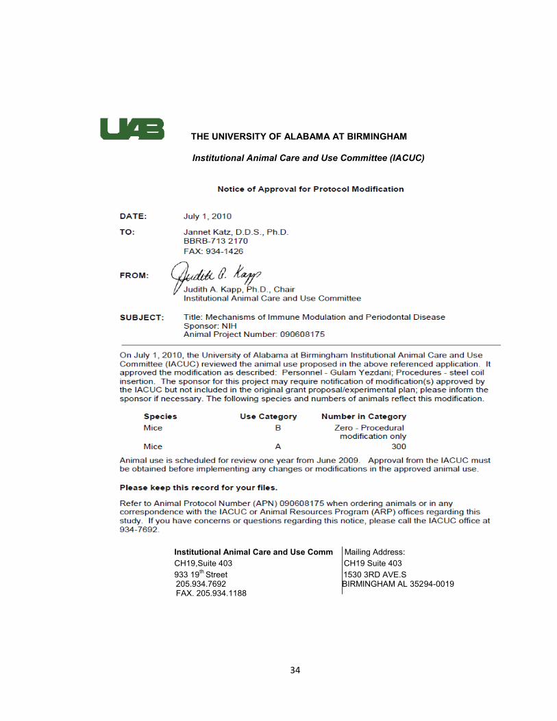
the National Institute of Health guidelines, and protocols were approved by the
University of Alabama at Birmingham Institutional Animal Care and Use Committee.
Generation and stimulation of bone marrow-derived macrophage
Femurs and tibiae from C57BL/6 WT and VDR-/-
mice (8 to 10 weeks old) were removed
and dissected free from adhering soft tissue. After soaking the bones in 70% ethanol for 2
min and rinsing then in PBS, both ends were cut and the marrow was flushed out with
PBS using a 25-gauge needle. Single-cell suspensions were prepared by mechanically
dispersing the bone marrow through a 100-μm cell strainer. Erythrocytes were lysed
using M-lysis buffer (R&D systems, Minneapolis, MN). Bone marrow cells were
cultured in α-10 medium (α-MEM, 10% FCS, 1 x PenStrep) in a humidified 7.5% CO2
incubator at 37°C overnight. The next day, non-adherent cells were harvested and
cultured with α-10 medium supplemented with 10% culture supernatant from CMG14-12
cells as the source of macrophages colony stimulating factor (M-CSF). After 5 days,
floating cells were removed and attached cells were used as BMMs. BMMs (2x10⁵
cells/well) were cultured in 96-well plates in a α-10 medium supplemented with 10% M-
CSF. Cultures were stimulated with live P. gingivalis at an multiplicity of infection
11
(MOI) of 50 or were left untreated. Culture supernatants were harvested after 24h to
assess IL-6, IL-10, IL-12p40 and TNF-α levels by ELISA.
Dual chamber model
Two coils / mouse were surgically implanted in the dorsolumbar region approximately
10mm apart from each other, to create subcutaneous chambers in C57BL/6 WT and
VDR-/-
female mice (8-10 weeks old). These coils were made of stainless steel surgical
wire that was 10 mm long and 5 mm in diameter. After a two week healing period, mice
were infected with P. gingivalis and cellular responses assessed by sampling the exudates
in the chamber (14).
Experimental design
The implanted chambers in WT and VDR-/-
mice were infected with P. gingivalis
suspended in PBS (0.1 ml; 5 x10⁹ CFU/ml) on day 0 and a second infection was given on
day 14th
. (Fig. 2)
Exudates were collected from one chamber at 24 h after the first infection, from the other
chamber 48 h and from both chambers on day 7. An aliquot of chamber fluid was
immediately analyzed for viable P. gingivalis and leukocytes. The remaining fluid was
centrifuged and the supernatant stored at -20◦C for the future analysis of cytokines. In
addition, cytokines were assessed in serum by collecting blood samples from the facial
vein prior to infection and at different time points following infection. Serum was
obtained after centrifugation and stored at -20◦C until cytokine or specific antibody
responses were analyzed.
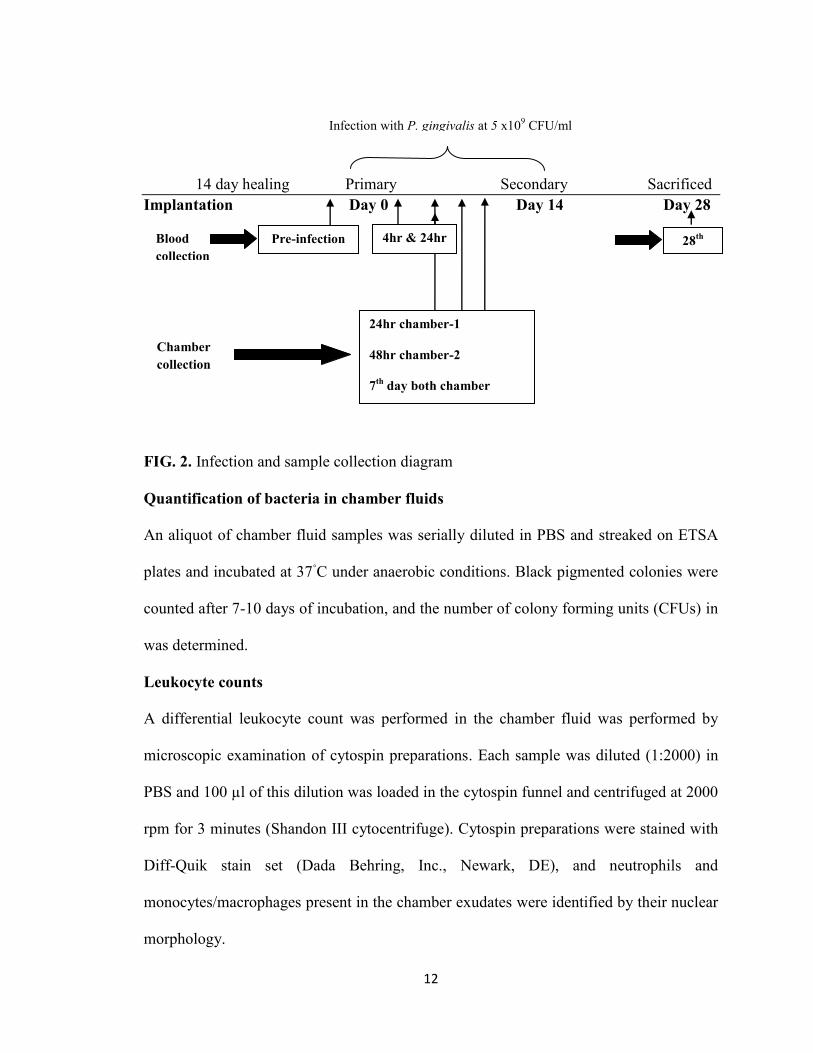
12
14 day healing Primary Secondary Sacrificed
Implantation Day 0 Day 14 Day 28
FIG. 2. Infection and sample collection diagram
Quantification of bacteria in chamber fluids
An aliquot of chamber fluid samples was serially diluted in PBS and streaked on ETSA
plates and incubated at 37◦C under anaerobic conditions. Black pigmented colonies were
counted after 7-10 days of incubation, and the number of colony forming units (CFUs) in
was determined.
Leukocyte counts
A differential leukocyte count was performed in the chamber fluid was performed by
microscopic examination of cytospin preparations. Each sample was diluted (1:2000) in
PBS and 100 µl of this dilution was loaded in the cytospin funnel and centrifuged at 2000
rpm for 3 minutes (Shandon III cytocentrifuge). Cytospin preparations were stained with
Diff-Quik stain set (Dada Behring, Inc., Newark, DE), and neutrophils and
monocytes/macrophages present in the chamber exudates were identified by their nuclear
morphology.
4hr & 24hr Pre-infection Blood
collection
28th
Chamber
collection
24hr chamber-1
48hr chamber-2
7th
day both chamber
Infection with P. gingivalis at 5 x10⁹ CFU/ml
13
Isolation and stimulation of CD4+ T cells
Fourteen days after the second infection, mice were sacrificed and spleens were
aseptically removed for in vitro assessment of the P. gingivalis specific CD4+
T cell
cytokine response Specifically, single-cell suspension from spleens werer prepared by
mechanically dispersing the tissues through 40 µm cell strainers (BD Labware, Franklin
Lakes, NJ) into Hank’s balanced salt solution (HBSS) supplemented with 10% fetal calf
serum. Erythrocytes were removed from the single cell suspensions by using M-lyse
buffer (R&D Systems, Minneapolis, MN). Cells were then washed, suspended in RPMI
1640 supplemented with 10% fetal calf serum, 2mM L-glutamine, 50 µM 2-
mercaptoethanol, 1 mM sodium pyruvate, 1.5 mg/ml of sodium bicarbonate, 50 µg/ml of
gentamicin sulfate, 25mM HEPES, 50 U/ml of penicillin, and 50 µg/ml of streptomycin
(complete medium), and counted in a hemacytometer with trypan blue to estimate
viability. CD4+ T cells were purified by negative selection using a mouse CD4
+ T cell
subset column according to the manufacture’s protocol (R&D System, Minneaspolis,
Minn). For the preparation of feeder cells, splenocytes from naïve WT and VDR-/-
mice
were irradiated with 3000 rad for 42 minutes. Equal numbers of CD4+
T cells and
irradiated feeders were co-cultured in 96-well plates in RMPI 1640 media supplemented
with 10% heat inactivated FCS at 37◦
C in a humidified 7.5% CO2 incubator. Cultures
were immediately stimulated with P. gingivalis at an MOI of 10 and culture supernatant
harvested after 5 days incubation for cytokine analysis.
Quantification of antibody response
The levels of total IgG and subclass IgG subclass specific antibodies in the serum
samples were determined by an ELISA, as previously described (47). Briefly, polystyrene
14
Maxisorp 96-well microtiter plates (Nunc, Roskilde, Denmark) were coated with
formalin-killed P. gingivalis (100 µl of 5 x 10⁸ CFU/ml) in borate-buffer saline (BBS;
100 Mm NaCl, 50 mM boric acide, 1.2 mM Na2B4O7, AND 0.02% azide, pH8.2) at 4◦C
overnight.Total IgG levels in serum samples was determined by coating plates with goat
anti-mouse IgG antibodies (Southern Biotechnology Associates, Inc., Birmingham, AL).
Plates were washed and blocked for 4 h at room temperature with 0.01 M phosphate
buffer (pH 7.2) containing 0.5 M NaCl and 0.15% Tween 20. Serial two fold dilution of
serum samples were added in duplicate and plates were incubated at 4 C overnight.
Afterwards plates were washed with 0.01 M phosphate buffer containing 0.5 M NaCl and
0.015% Tween 20 (pH 7.2) and perioxidase-conjugated goat anti-mouse IgG or IgG
subclass-specific antibodies (Southern Biotechnology Associates, Inc) were added
followed by addition of o-phenylenediamine substrate with H2O2. Optical density was
measured at a wavelength of 490 nm. The concentration of antibodies were calculated by
interpolation on calibration curves generated at the same time using a mouse
immunoglobulin reference serum (ICN Biomedical, Aurora, OH) and constructed by a
computer program based on a four-parameter logistic algorithm ( Softmax/ Molecular
Devices Corp, Menlo Park, CA)
Cytokine analysis
The levels of TNF-α, IL-6 ( eBiosciences, San Diego, CA) IL-10 and IL-12p40 (R&D
Systems) in chamber fluids and BMMs culture supernatants, as wells as the levels of IL-
4, IL-5(R&D Systems ) and IFN-γ ( eBiosciences) in CD4+ T cell culture supernatants
were assessed by an ELISA, according to the manufacturer’s instructions.
15
Statistical analysis
The significance of difference between groups was evaluated by ANOVA and the Tukey
multiple comparison test using the Instat program (GraphPad, San Diego, Calif.)
differences between groups were considered significant at the level of P < 0.05.
RESULTS
Cytokine production by BMMs following P. gingivalis stimulation
To address the role of the VDR in the regulation of P. gingivalis-induced inflammatory
response, we first assessed the induction of cytokines by P. gingivalis stimulated BMMs
from WT and VDR-/-
mice. The levels of IL-6, IL-12p40 and TNF-α in VDR-/-
BMM
cultures following P. gingivalis stimulation were significantly higher (P < 0.05) than the
levels in WT cultures (Fig. 3). However, P. gingivalis induced a significantly lower (P <
0.05) level of the anti-inflammatory cytokine IL-10 in VDR-/-
cell cultures compared to
that of WT cell cultures. These results suggest that VDR plays a regulatory role in the P.
gingivalis –induced cytokine response of macrophages by downregulating the production
of pro-inflammatory cytokines and potentiating the production of anti-inflammatory
cytokine IL-10.
Infiltration of neutrophils and monocytes into the chambers following P. gingivalis
challenge
Neutrophils and monocyte are initial responders in periodontal infections and 1, 25(OH)
2D3 can affect these cells (32). To understand the role of VDR in regulating the cellular
response to P. gingivalis infection, WT and VDR-/-
mice were infected with 5 x 10⁸ CFU
of P. gingivalis in each subcutaneous chamber. Chamber exudates were collected at 24 h,
16
72 h and day 7 for neutrophil and monocyte counts. The total leukocyte count in the
chamber fluids of WT and VDR-/-
control mice ( PBS only injected in the chambers) was
very low (data not shown). However following P. gingivalis infection, a significantly
higher number of leukocytes was observed in both WT and VDR-/-
mice; yet there was no
significant difference in the neutrophil or macrophage count between the two groups
(Fig. 4). These results indicate that VDR does not play a role in regulating the
recruitment of leukocytes following P. gingivalis infection.
Analysis of chamber fluid for bacterial killing
To determine the anti-bacterial effect of the VDR on P. gingivalis chamber fluids were
collected from WT and VDR-/-
mice at 24 h, 48 h and day 7 after P. gingivalis infection.
A decrease in the number of recovered P. gingivalis was observed in the chamber fluid
from both the WT and VDR-/-
mice at 24 h to day 7 (Fig. 5). However, the number of P.
gingivalis in VDR-/-
mice was significantly higher (P < 0.05, P < 0.05) than that of WT
mice at all times tested. No bacterium was recovered from chamber fluids of control non-
infected (data not shown). These results suggest that mice deficient in VDR has an
impaired ability to clear P. gingivalis from the chambers compared to WT mice.
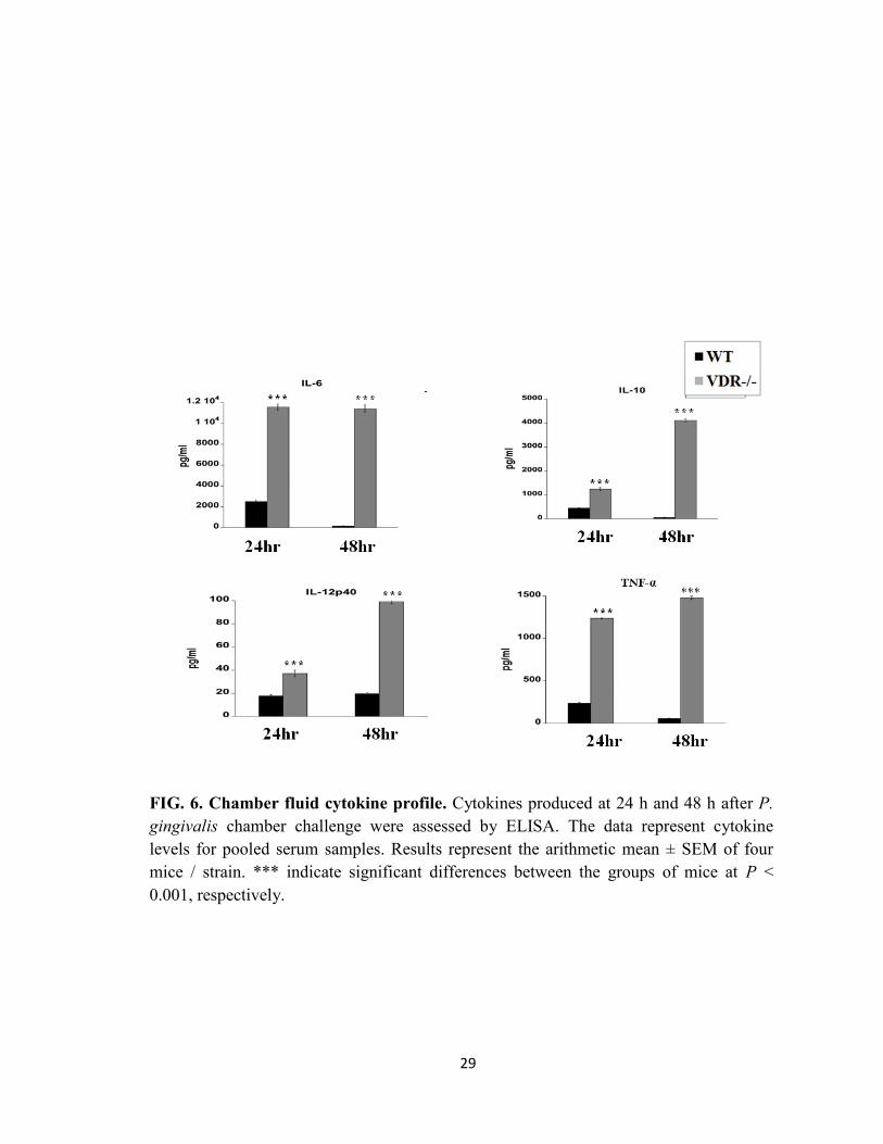
Cytokines levels in the chamber fluid following P. gingivalis infection
Since the VDR has been shown to play a role in the inflammatory host response, we next
investigated the cytokine profile in the chamber exudates after P. gingivalis challenge.
Chamber fluids from non-infected mice had no detectable amounts of tested cytokines
(data not shown). However, P. gingivalis infection induced the production of cytokines in
WT and VDR-/-
mice (Fig. 6). In the WT group, IL-6, IL-10 and TNF-α cytokine levels
peaked at 24 h post infection, but by 48 h the cytokine levels had decreased significantly.
17
However levels of IL-12p40 remained the same at both time points in WT mice. Levels
of TNF-α and IL-6 cytokines were similar 24 h and 48 h in VDR-/-
mice, whereas IL-10
and IL-12p40 gradually increased from 24 h to 48 h. However, VDR-/-
mice showed
cytokines levels that were significantly higher (P < 0.001, P < 0.001) than those of WT
mice. These results demonstrate that VDR down-regulate the inflammatory cytokine
response to P. gingivalis infection.
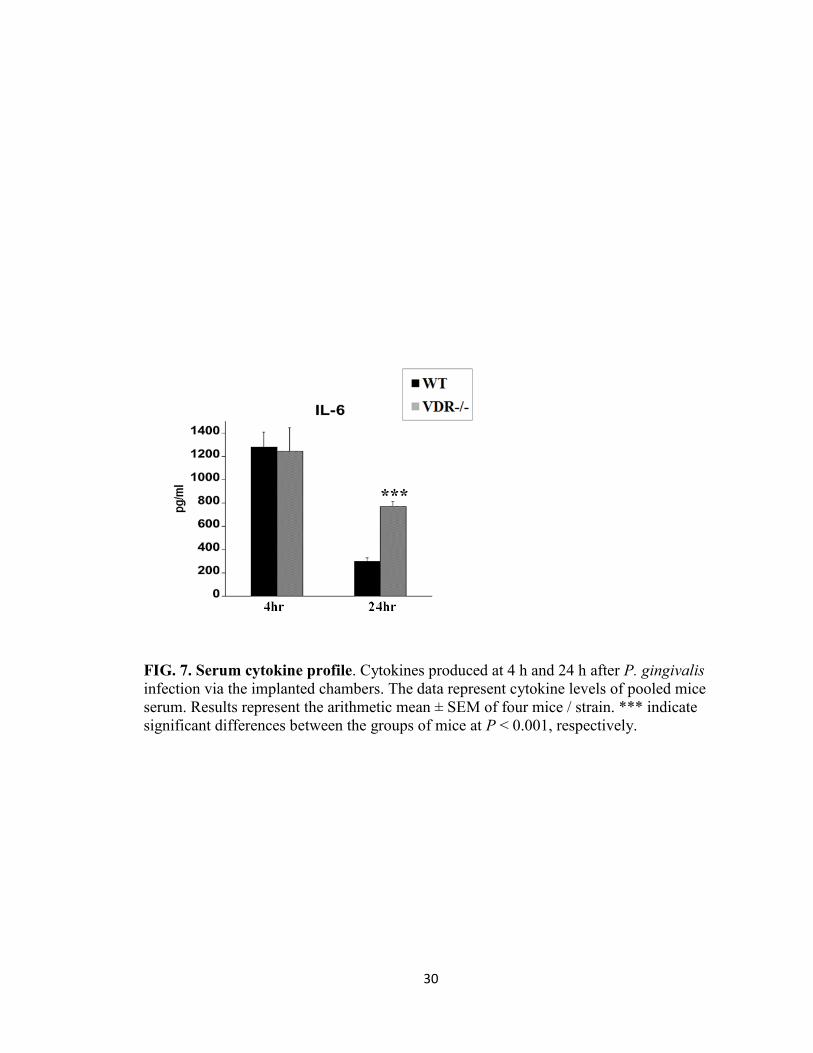
Serum cytokine profile
The serum cytokine response following P. gingivalis infection of the subcutaneous
chambers was also assessed. Our results showed similar levels of serum IL-6 in WT and
VDR-/-
mice at 4 h after P. gingivalis infection (Fig. 7), however the level were
significant higher (P < 0.001) in the VDR-/-
than WT mice at 48 h post-infection.
Minimal levels of serum TNF-α, IL-12p40, IL-10 were detected at 24 h and 48 h in WT
and VDR-/-
mice (data not shown).
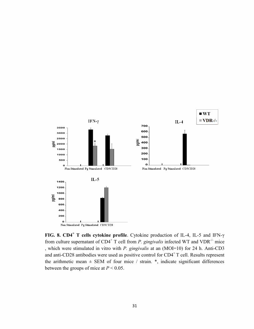
CD4+ T cell cytokine profile
T-cells, especially CD4+
T cells, are implicated in periodontal inflammation and tissue
destruction (40). Furthermore it has been shown that CD4+ T cells can be direct targets of
vitamin D (7). Thus we investigated the production of cytokines by splenic CD4+ T cell
from infected mice following restimulation of cells with P. gingivalis in vitro. We found
a markedly elevated levels IFN-γ in the culture supernatant of both WT and VDR-/-
CD4+
T cell cultures (Fig. 8). However, the levels of IFN-γ in VDR-/-
supernatants was
significantly lower (P < 0.05) than that in WT cultures. Levels of IL-4 and IL-5 were
undetectable in WT and VDR-/-
cultures. These results suggest that VDR may favor a Th1
18
response following a P. gingivalis infection. However, further studies need to be taken to
confirm this result.
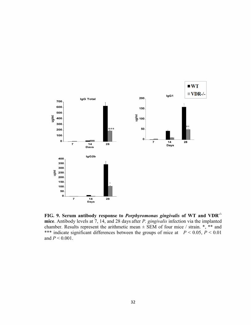
Serum anti -P. gingivalis antibody levels
To understand the role of VDR in regulating the antibody response to P. gingivalis,
serum samples from infected WT and VDR-/-
mice were assessed for specific IgG, IgG1
and IgG2b antibody levels. Very low levels of total IgG1 and IgG2b anti-P. gingivalis
specific antibodies were detected in WT and VDR-/-
mice at 7 and 14 days after the first
infection (Fig. 9). However, two weeks following the second infection (day 28),
significantly higher levels of total IgG and of IgG1 and IgG2b specific antibodies were
observed. Nevertheless WT mice showed significantly higher levels of total IgG (P <
0.001), IgG1 (P <0.01) and IgG2b (P < 0.05) antibodies than VDR-/-
mice. These results
suggest that VDR plays a role in the up-regulation of serum IgG, IgG1and IgG2b specific
antibodies following P. gingivalis infection.
DISCUSSION
The active form of vitamin D 1,25(OH)2D3, not only plays an essential role in bone
metabolism, but also facilitates phagocytosis by neutrophils (9, 25, 31, 45) and
monocytes (3, 4, 18) as well as monocyte differentiation (25,31), thereby exerting a great
influence on the host immune function. However it is not known if the VDR plays a role
in the host response to periodontal pathogens such as P. gingivalis. In the present study
using a mouse subcutaneous chamber infection model, we have shown the involvement
of the VDR on various aspects of the host immune response to P. gingivalis.
19
Polymorphisms in the human VDR gene have been associated with increased resistance
or susceptibility to a number of infectious diseases. Case-control studies suggested links
between certain alleles of the VDR gene and susceptibility to periodontal disease (2, 11,
12, 14, 26), tuberculosis (39), leprosy, dengue fever and hepatitis B virus-induced chronic
hepatitis. Furthermore, strong evidence shows that TLR1/2 signaling in human
macrophages, leads to an up-regulated expression of the VDR that in turn induced the
expression of the antimicrobial peptide cathelicidin, which was shown to mediates
intracellular killing of Mycobacterium tuberculosis (31, 39, 40). In this experiment we
investigated the ability of the host to clear bacteria and disease progression; we observed
that VDR-/-
mice showed significantly enhanced containment of these bacteria in the
chambers as compared to their wild-type. This was evident in accordance to our culture
data that shows VDR deficiency or inefficiency would contribute to increased bacteria
burden. In our further experiment highlights our observation of up regulated levels of pro
inflammatory cytokine production of IL-6, TNF-α, IL12p40 present at the local infection
site, as well as cytokine increase of IL-6 in serum of VDR-/-
mice following P. gingivalis
challenge. This relevance of our findings in vivo was further analyzed in vitro where
macrophages were stimulated with P. gingivalis which showed a similar response. Our
findings are also supported and correlative with another study of salmonella infection
where in VDR-/-
mice exhibited a similar pro-inflammatory bias after Salmonella
infection and had increased bacterial burden and mortality (46).
Studies show that Th1 and Th2 cell responses have a tremendous influence on the
outcome of P. gingivalis infection, wherein Th1 response dramatically results in
increased bone resorption (15, 19, 44). We wanted to observe the cytokine profile
20
induced by P.gingivalis in CD4+ T cell cultures to help us understand the effect of the
VDR on T cell cytokine production (33, 34). We compared the production of the Th2
associated cytokines (IL-4, IL-5) and Th1 associated cytokine production (IFN-γ) by
CD4+T cells from infected VDR
-/- and wild-type mice after in vivo stimulation with P.
gingivalis antigen. It has been shown in recent data that VDR can inhibit the secretion of
cytokines that are involved in the activation and differentiation of a subset of T helper
cells (2). Autoimmune in vivo models of mice has shown that VDR inhibits the induction
of Th1 and directly inhibits IFN-γ production and prevents IgG2 antibody responses (23).
Our results indicate that WT mice had an IFN-γ cytokine up regulation, demonstrating
that VDR-/-
mice neither displayed neither enhanced Th1 nor elevated Th2 responses in
this infection model. This result does support studies with leishmania major infection in
which higher IFN-γ was seen in VDR-/-
(16). Furthermore we wanted to evaluate if the
effects of VDR have on antibody responses and reflects which Th response. With regards
to the serum IgG and subclass, higher IgG1 and IgG2b responses against P. gingivalis
were observed in WT mice, suggesting VDR supports Th1 response (29, 33, 34). This
view is inconsistent with our results from many other infection studies that showed a
preferential IgG2b or Th2 response after infection in presence of VDR signaling.
In summary, we concluded that VDR contributes in regulating host immune system
against P. gingivalis infection. It plays a important and complex anti inflammatory role
at the infected site suppressing inflammation and helps clear bacteria. It supports our
hypothesis that it modulates inflammation, but many questions remain still unanswered
for the adaptive responses and more further research would be needed to evaluate VDR’s
role in P. gingivalis induced antibody and T cell responses.
21
REFERENCES
1. Adams J. S, Liu P. T, Chun R, Modlin R. L, Hewison M. 2007. Vitamin D in
defense of the human immune response. Ann. N. Y. Acad. Sci. 17:94-105
2. Amano. Y, Komiyama. K and Makahasima. M. 2009. Vitamin D and
periodontal disease. J. Oral. Sci. 51:11-29
3. Bikle D. D. 2008. Vitamin D and the immune system: role in protection against
bacterial infection. Curr. Opin. Nephrol. Hypertens. 17:348-352
4. Bikle D. D. 2009. Vitamin D: production, metabolism, and mechanism of action
source. www.endotext.org
5. Bouillon. R, Carmeliet. G, Verlinden. L, Van. Etten. E, Verstuy. A, Luderer
H.F, Lieben. L, Mathieu. C, Demay. M. 2008. Vitamin D and human health:
lessons from vitamin D receptor null mice. Endocri. Rev. 29:726-76
6. Brumbaugh P. F, Haussler M. R. 1975. Specific binding of 1α, 25-
dihydroxycholecalciferol to nuclear components of chick intestine. J. Biol. Chem.
250:1588-94
7. Catorna M. T. 2006. Vitamin D and its role in immunology: multiple sclerosis,
and inflammatory bowel disease. Prog. Biophys. Mol. Biol. 92:60-4
8. Catorna M.T, Hullett D. A, Redaelli C, Brandt C. R, Humpal-Winter J. 1998.
1,25-Dihydroxyvitamin D3 prolongs graft survival without compromising host
resistance to infection or bone mineral density. Transplantation. 66:828-31
9. Cassatella, M.A.1995. The production of cytokines by polymorphonuclear
neutrophils. Immunol. Today. 16:21–26
10. Cheng J. B, Motola D. L, Mangelsdorf D. J, Russell D. W. 2003. De-
orphanisazation of cytochromoe p450 2r1: a microsomal vitamin d 25-
hydroxylase. J. Biol. Chem. 278:38081-38093
11. Delves, P.J, Roitt I.M. 2000. The immune system. First of two parts N. Engl J.
Med. 343:37-49
22
12. De Brito Junior R. B, Scaret-Caminaga R. M, Trevillato P. C, De Souza A. P,
Barros S.P. 2004. Polymorphisms in the vitamin D receptor gene are associated
with periodontal disease. J. Periodontol 75:1090-1095
13. Dilworth, F. J, and P. Chambon. 2001. Nuclear receptors coordinate the
activities of chromatin remodeling complexes and coactivators to facilitate
initiation of transcription. Oncogene. 20:3047–3054
14. Dietrich. T, Joshipura. K. J, Dawson-Hughes B, and Bischoff-Ferrari HA.
2004. Association between serum concentrations of 25 hydroxyvitamin D3 and
periodontal disease in the US population. Am. J. Clin. Nutr. 80:108-113
15. Eastcott J.W, Yamashita K, Taubman M.A, Haraday & Smith D.J. 1994.
Adoptive transfer of cloned T helper cells ameliorates periodontal disease in nude
rats. Oral. Microbiol. Immunol. 9:284-289
16. Ehrchen. J, Helming. L, Varga. G, Pasche. B, Loser. K, Gunzer. M,
Sunderkotter. C, Sorg. C, Roth. J, Lengeling. A. 2007 Vitamin D receptor
signaling contributes to susceptibility to infection with Leishmania major. Faseb
J. 12:3208-18
17. Froicu M, Weaver V, Wynn T. A, McDowell M. A, Welsh J. E, Cantorna M.
T. 2003. A crucial role for the vitamin D receptor in experimental inflammatory
bowel diseases. Mol. Endocrinol. 17:2386-92
18. G.A. Rook, J. Steele, L. Fraher, S. Barker, R. Karmali, J. O'Riordan and J.
Stanford. 1986. Vitamin D3, gamma interferon, and control of proliferation of
mycobacterium tuberculosis by human monocytes. Immunology. 57:159–163
19. Gemmell E, Yamazaki K, Seymour G. J. 2002. Destructive periodontitis lesions
are determined by the nature of the lymphocytic response. Crit. Rev. Oral. Biol.
Med. 13:17-34
20. Genco C. A, Arko R. J. 1994. Animal chamber models for study of host parasite
interactions. Methods. Enzymol. 235:120-140
21. Genco C. A, Van Dyke. T, Amar S. 1998. Animal models for porphyromonas
gingivalis-mediated periodontal disease. Trends. Microbiol. 6:444-449
22. Gorska, R., H. Gregorek, J. Kowalski, A. Laskus-Perendyk, M. Syczewska,
and K. Madalinski. 2003. Relationship between clinical parameters and cytokine
profiles in inflamed gingival tissue and serum samples from patients with chronic
periodontitis. J. Clin. Periodontol. 30:1046–1052
23
23. Glass, C. K, and M. G. Rosenfeld. 2000. The coregulator exchange in
transcriptional functions of nuclear receptors. Genes. Dev. 14:121–141
24. Haussler M. R, Norman A. W. 1969. Chromosomal receptor for a vitamin D
metabolite. Proc. Natl. Acad. Sci. U.S.A. 62:155-162
25. Hayes C. E, Nashold F. E, Spach K. M, Pedersen. 2003. The immunological
functions of vitamin D endocrine system. Cell. Mol. Biol. 49:277-300
26. Hennig B. J, Parkhill J. M, Chapple l L, Heasman P. A, Taylor J. J. 1999.
Association of vitamin D receptor and gene polymorphism with localized early
onset periodontal disease. J. Periodontol. 70:1032-1038
27. Hewison M. 2011. Vitamin D and innate and adaptive immunity. Vitamin Horm.
86:23-62
28. Houri-Haddad. Y, Soskolne. WA, Halabi. A, Barak. V, Shapira. L. 2000.
Repeat bacterial challenge in a subcutaneous chamber model results in augment
tumour necrosis factor-α and interferon-γ response and suppression of interleukin-
1. Immunology. 99:215-220
29. Horiuchi, H, Nagata, Komoriya K. 1991. Protective effect of vitamin D3
analogues on endotoxin shock in mice. Agents. Actions. 33:343–348
30. Issa L. L., Leong G.M, Eisman J.A. 1998. Molecular mechanism of vitamin D
receptor action. Inflamm. Res. 12:451-75
31. Zhang D. E, Hetherington C. J, Gonzalez D. A, Chen H. M and Tenen D. G.
1994. Regulation of CD14 expression during monocytic differentiation induced
with 1 alpha, 25-dihydroxyvitamin D3. J. Immunol. 153:3276–3284
32. John H. White. 2008. Vitamin D signaling, infectious diseases and regulation of
innate immunity. Infect. Immun. 76:3837-3843
33. Lemire J. M, Archer D. C, Lucinda Beck, Spiegelberg H.L. 1995.
Immunosuppressive actions of l, 25-dihydroxy vitamin D3: preferential inhibition
of Th1 functions.J. Nutr. 125:1704-1708
34. Mahon B. D, Wittke A, Weaver V, Catorna M. T. 2003. The targets of vitamin
D depend on the differentiation and activation status of CD4+T cells. J.Cell.
Biochem. 89:922–932
35. McKenna. N. J., and O’Malley B.W. 2002. Combinatorial control of gene
expression by nuclear receptors and coregulators. Cell. 108:465–474
24
36. Oz H. S, Puleo D. A. 2011. Animal models for periodontal disease. J. Biomed
Biotechnol. 2011:754-857
37. Pavasant. P, Yongchaitrakul. T. 2000. Role of mechanical stress on the function
of periodontal ligament cells. Periodontol. 56:154–165
38. Rachez, C., and L. P. Freedman. 2000. Mechanisms of gene regulation by
vitamin D3 receptor: a network of coactivator interactions. Gene. 246:9–21
39. Rockett K. A, Brookes R, Udalova. I, Vidal. V, Hill A.V and Kwiatkowski. D.
1998. 1, 25-Dihydroxyvitamin D3 induces nitric oxide synthase and suppresses
growth of Mycobacterium tuberculosis in a human macrophage-like cell line.
Infect. Immun. 66: 5314–5321
40. Sasidharan PK, Rajeev E, Vijayakumari V. 2002. Tuberculosis and vitamin D
deficiency. J. Assoc. Physicians. India. 50:554-8
41. Seymour G. J. 1991. Importance of the host response in the Periodontium.
J.Clin.Periodontol. 18:421-426
42. Socransky S. S, Haffajee A. D. 1992. The bacterial etiology of destructive
periodontal disease. J. Periodontol. 63:2033-2041
43. Stashenko. P, Gonçalves R.B, Lipkin. B, Ficarelli. A, Sasaki. H, Campos-
Neto. A. 2007. Th1 immune response promotes severe bone resorption caused by
porphyromonas gingivalis. Am. J. Pathol. 170:203–213
44. Taubman M. A, Eastcott J. W, Shimauchi H, Takeichi. O, Smith D. J. 1994.
Modulatory role of T lymphocytes in periodontal inflammation. J. Immunolog .
9:284-289
45. Takahashi. K, Nakayama. Y, Horiuchi. H, Ohta.T, Komoriya. K, Ohmori. H
and Kamimura. T. 2002. Human neutrophils express messenger RNA of vitamin
D receptor and respond to 1,25-Dihydroxyvitamin D3 Immunopharmacology.
Immunotoxicology. 3:335–347
46. Wu. S, Liao A.P, Xia. Y, Li Y. C, Li J. D, Sartor R.B, Sun. J. 2010. Vitamin D
receptor negatively regulates bacterial-stimulated NF-kappa B activity in
intestine. Am. J. Pathol. 177:686-697
47. Zhang. P, Martin. M, Yang Q.B, Michalek. S.M and Katz. J. 2004. Role of B7
costimulatory molecules in immune responses and T-helper cell differentiation in
response to recombinant Hagb from Porphyromonas gingivalis. Infect Immun.
72:637–644
25
48. Zigmond, S.H.1978. Chemotactic response of neutrophils. Am. J. Respir. Cell.
Mol. Biol. 6:451–453
49. Liu P. T, Stenger S, Li H, Wenzel L, Tan B. H, Krutzik S.R, Ochoa M. T,
Schauber J, Wu K,Meniken C, Kamen D. L, Wagner M, Balts R, Steinmeyer
A, Zugel U, Gallo R. L, Eisenberg D, Hewison M, Hollis B. W, Adams J. S,
Bloom B. R, Modulin R. L. 2006. Toll-like receptor triggering of a vitamin D-
mediated human antimicrobial response. Science. 311:1770-1773
26
FIG. 3. Cytokines production from BMMs. Cytokines produced in culture supernatant
of bone marrow-derived macrophages (BMMs), after 24 h stimulation with
Porphyromonas .gingivalis (MOI=50). * indicates significant differences at P < 0.05.
between P. gingivalis infected and non infected groups.
27
FIG. 4. Differential count of neutrophil and monocytes in the chamber fluids. These
were carried out on smears stained using Diff-Quik stain, and cells were identified by cell
morphology. The chamber fluid samples were obtained at 24hr, 48hr and 7th
day. Results
represent the mean count at 10 different fields under 10 x magnifications.
28
FIG. 5. Bacterial count in chamber exudates. Chambers fluid samples obtained at 24h,
48h and 7 days after infection with P. gingivalis were cultured on ETSA plates following
incubation, the number of CFUs were enumerated. Results represent the arithmetic mean
± SEM of samples from four mice / strain. *, indicate significant differences between the
WT and VDR-/-
mice at P < 0.05.
29
FIG. 6. Chamber fluid cytokine profile. Cytokines produced at 24 h and 48 h after P.
gingivalis chamber challenge were assessed by ELISA. The data represent cytokine
levels for pooled serum samples. Results represent the arithmetic mean ± SEM of four
mice / strain. *** indicate significant differences between the groups of mice at P <
0.001, respectively.
30
FIG. 7. Serum cytokine profile. Cytokines produced at 4 h and 24 h after P. gingivalis
infection via the implanted chambers. The data represent cytokine levels of pooled mice
serum. Results represent the arithmetic mean ± SEM of four mice / strain. *** indicate
significant differences between the groups of mice at P < 0.001, respectively.
***
31
FIG. 8. CD4+ T cells cytokine profile. Cytokine production of IL-4, IL-5 and IFN-γ
from culture supernatant of CD4+ T cell from P. gingivalis infected WT and VDR
-/- mice
, which were stimulated in vitro with P. gingivalis at an (MOI=10) for 24 h. Anti-CD3
and anti-CD28 antibodies were used as positive control for CD4+
T cell. Results represent
the arithmetic mean ± SEM of four mice / strain. *, indicate significant differences
between the groups of mice at P < 0.05.
*
32
FIG. 9. Serum antibody response to Porphyromonas gingivalis of WT and VDR-/-
mice. Antibody levels at 7, 14, and 28 days after P. gingivalis infection via the implanted
chamber. Results represent the arithmetic mean ± SEM of four mice / strain. *, ** and
*** indicate significant differences between the groups of mice at P < 0.05, P < 0.01
and P < 0.001.
***
33
APPENDIX
NOTICE OF APPROVAL FROM INSTITUTIONAL ANIMAL CARE AND USE
COMMITTEE
34
THE UNIVERSITY OF ALABAMA AT BIRMINGHAM
Institutional Animal Care and Use Committee (IACUC)
Institutional Animal Care and Use Comm Mailing Address:
CH19,Suite 403 CH19 Suite 403
933 19th
Street 1530 3RD AVE.S 205.934.7692 BIRMINGHAM AL 35294-0019 FAX. 205.934.1188