roleofcellularcholesterolmetabolisminvascularcell ... ·...
TRANSCRIPT

Role of Cellular Cholesterol Metabolism in Vascular CellCalcification*
Received for publication, June 9, 2011, and in revised form, August 4, 2011 Published, JBC Papers in Press, August 11, 2011, DOI 10.1074/jbc.M111.269639
Yifan Geng‡1, Jeffrey J. Hsu‡1, Jinxiu Lu§, Tabitha C. Ting¶, Makoto Miyazaki¶, Linda L. Demer‡§�, and Yin Tintut‡2
From the Departments of ‡Medicine, §Physiology, and �Bioengineering, UCLA, Los Angeles, California 90095-1679 and the¶Department of Medicine, University of Colorado-Denver, Aurora, Colorado 80045
Vascular calcification impairs vessel compliance and in-creases the risk of cardiovascular events. We found previouslythat liver X receptor agonists, which regulate intracellular cho-lesterol homeostasis, augment PKA agonist- or high phosphate-induced osteogenic differentiation of vascular smooth musclecells. Because cholesterol is an integral component of thematrixvesicles that nucleate calcium mineral, we examined the role ofcellular cholesterol metabolism in vascular cell mineralization.The results showed that vascular smooth muscle cells isolatedfrom LDL receptor null (Ldlr�/�) mice, which have impairedcholesterol uptake, had lower levels of intracellular cholesteroland less osteogenic differentiation, as indicated by alkalinephosphatase activity andmatrix mineralization, compared withWT cells. PKA activation with forskolin acutely induced genesthat promote cholesterol uptake (LDL receptor) and biosynthe-sis (HMG-CoA reductase). InWTcells, inhibition of cholesteroluptake by lipoprotein-deficient serum attenuated forskolin-in-duced matrix mineralization, which was partially reversed bythe addition of cell-permeable cholesterol. Prolonged activationof both uptake and biosynthesis pathways by cotreatment with aliver X receptor agonist further augmented forskolin-inducedmatrix mineralization. Inhibition of either cholesterol uptake,using Ldlr�/� cells, or of cholesterol biosynthesis, using mevas-tatin-treated WT cells, failed to inhibit matrix mineralizationdue to up-regulation of the respective compensatory pathway.Inhibition of both pathways simultaneously using mevastatin-treated Ldlr�/� cells did inhibit forskolin-induced matrix min-eralization. Altogether, the results suggest that up-regulation ofcholesterolmetabolism is essential formatrixmineralization byvascular cells.
Vascular calcification is frequently found in advanced ath-erosclerotic lesions and is an independent predictor of cardio-vascular morbidity and mortality in patients with chronic kid-ney disease (1–3). Long considered to be a passive process,there now exists much evidence to suggest that vascular calci-fication is an active cell-mediated phenomenon that is highlyregulated through complex mechanisms under active investi-
gation (4). In vitro and in vivo studies have implicated numer-ous positive and negative regulatory factors in the pathogenesisof calcification (5–8).A common complication of chronic kidney disease is hyper-
parathyroidism. Interestingly, parathyroid hormone, an activa-tor of the PKA pathway, has been shown to have a stimulatoryrole in vascular calcification both in vitro and in vivo. Whenparathyroid hormone levels are continuously elevated, theyinduce vascular calcification in rat models, irrespective of renalfunction (9). Additionally, parathyroid hormone-related pro-tein, also a PKA activator, has been found in calcified athero-sclerotic lesions (10). Accordingly, we and others have demon-strated previously that mineralization of vascular smoothmuscle cells (VSMCs)3 is induced by PKA agonists in vitro (11,12) as well as in vivo (13).Cholesterol is an integral component of cell membranes and
matrix vesicles (14). The latter are secreted by VSMCs, chon-drocytes, and osteoblasts, and they are instrumental in biomin-eralization, including that of skeletal bone, cartilage, and theartery wall (15–17). In a tightly regulated cholesterol homeo-static process, cells obtain essential cholesterol by endogenoussynthesis or uptake from the extracellular milieu. For endoge-nous synthesis, the rate-limiting enzyme is HMG-CoA reduc-tase, which is blocked by the class of drugs known as statins.Alternatively, if circulating cholesterol levels are high or cho-lesterol synthesis is inhibited by statins, cells take up cholesterolfrom their extracellular environment in the form of the choles-terol-rich LDL particle via the LDL receptor (18, 19). Thus,statins are highly effective at lowering circulating levels of LDLand are among the most commonly prescribedmedications forpatients with atherosclerotic cardiovascular disease.We demonstrated previously that both bovine and murine
VSMCs undergo osteogenic differentiation and mineralizationspontaneously as well as in the presence of PKA activators orhigh phosphate concentrations (11, 20–22). Furthermore, wefound that activation of liver X receptor (LXR), which up-reg-ulates the expression of genes involved in cholesterol efflux (23,24), augments PKA- and high phosphate-induced mineraliza-tion of VSMCs (21, 25). Consistent with these findings, inhibi-tion of LXR by the dominant-negative form of LXR� and/orLXR� inhibits mineralization of VSMCs (21). In this study, weinvestigated the role of cholesterol metabolism in vascular cellcalcification and demonstrated that both cellular biosynthesis
* This work was supported, in whole or in part, by National Institutes ofHealth Grants DK081346 and DK081346-S1 (to Y. T.) and HL081202 (toL. L. D.) and NIGMS Initiative for Maximizing Student DevelopmentGrant R25GM083333 (to T. C. T.). This work was also supported byAmerican Heart Association Grant 10BGIA458005 (to M. M.).
1 Both authors contributed equally to this work.2 To whom correspondence should be addressed: Center for Health Sciences,
UCLA, A2–237, 10833 Le Conte Ave., Los Angeles, CA 90095-1679. Tel.:310-206-9964; Fax: 310-825-4963; E-mail: [email protected].
3 The abbreviations used are: VSMC, vascular smooth muscle cell; LXR, liver Xreceptor; CVC, calcifying vascular cell; LPDS, lipoprotein-deficient serum;ALP, alkaline phosphatase; qPCR, quantitative PCR.
THE JOURNAL OF BIOLOGICAL CHEMISTRY VOL. 286, NO. 38, pp. 33701–33706, September 23, 2011© 2011 by The American Society for Biochemistry and Molecular Biology, Inc. Printed in the U.S.A.
SEPTEMBER 23, 2011 • VOLUME 286 • NUMBER 38 JOURNAL OF BIOLOGICAL CHEMISTRY 33701
by guest on May 31, 2018
http://ww
w.jbc.org/
Dow
nloaded from

and uptake of cholesterol are essential to the mineralization ofvascular cells.
EXPERIMENTAL PROCEDURES
Reagents—Forskolin was purchased from Calbiochem,T0901317 from Cayman Chemical (Ann Arbor, MI), and mev-astatin fromBIOMOL (PlymouthMeeting, PA).Water-solublecholesterol was purchased from Sigma-Aldrich.Cell Culture—Bovine calcifying vascular cells (CVCs) were
isolated and maintained as described previously (20). Murineaortic cells (passages 6–10) were isolated from the aortas ofC57BL6 (WT) and Ldlr�/� mice as described previously (11).Cells weremaintained inDMEMcontaining 20%FBS. 3–4 daysafter plating, cells were treated with forskolin (10 �M, unlessindicated otherwise) in �-Minimum Essential Medium con-taining 10% FBS and 5 mM �-glycerophosphate. Lipoprotein-deficient serum (LPDS) was kindly provided by the UCLAAtherosclerosis Research Unit and was used at a final concen-tration of 0.8 mg/ml. LDL receptor deficiency in Ldlr�/� cellswas confirmed by RT-PCR following protocols established byThe Jackson Laboratory. Construction of adenoviral constructsexpressing VP16-LXR�, VP16-LXR�, and SREBP-1c (a consti-tutively active form of SREBP-1 (sterol regulatory element-binding protein 1c); Addgene) and transduction to CVCs werecarried out as described previously (21).Matrix Calcium Quantitation—After the indicated periods,
matrix calcium levels were analyzed by the o-cresolphthaleincomplexone method (Teco Diagnostics, Anaheim, CA). Eachcondition was assayed in quintuplicate and normalized to totalprotein using the Bradford method (11).Alkaline Phosphatase Activity—After the indicated periods,
alkaline phosphatase (ALP) activity was assessed colorimetri-cally using Sigma 104 phosphatase substrate. Each conditionwas assayed in quintuplicate and normalized to total proteinusing the Bradford method (11).Intracellular Cholesterol Measurement—Total cellular cho-
lesterol content was measured using the Amplex Red choles-terol assay kit from Invitrogen (Carlsbad, CA). Cell lysate wascollected with the reaction buffer provided in the kit.Gene Expression—Total RNA was isolated from cells using
TRIzol reagent (Invitrogen). Real-time RT-quantitative PCR(qPCR) was performed using a One-Step qRT-PCR kit (Bio-Chain Institute, Inc. Hayward, CA) in an Mx3005P system(Stratagene La Jolla, CA). �-Actin was used for normalization.Data Analysis—Each experiment was performed in at least
quadruplicate wells and repeated at least three times (n � 3).Data are expressed asmeans� S.E. Student’s t test was used forcomparison between two groups. For more than two groups,mean valueswere compared using one-way analysis of variance,with comparison of different groups by Fisher’s protected leastsignificant difference test. A value of p � 0.05 was consideredsignificant.
RESULTS
Effects of Cholesterol Uptake Deficiency on Osteoblastic Dif-ferentiation and Matrix Mineralization—To investigate theeffects of impaired cholesterol uptake on osteoblastic differen-tiation andmineralization, aortic smoothmuscle cells were iso-
FIGURE 1. Effects of cholesterol uptake deficiency on osteoblastic differ-entiation and matrix mineralization. A, ALP activity of murine WT andLdlr�/� cells at 4 and 6 days in culture. B, matrix calcium levels of murine WTand Ldlr�/� cells at 7 days in culture. C, intracellular cholesterol levels ofmurine WT and Ldlr�/�cells after 4 days in culture. D, matrix calcium levels ofbovine aortic cells (CVCs) at 9 days in culture in medium with normal serum orLPDS. The levels of ALP activity, matrix calcium, and cholesterol were normal-ized to total protein concentration. #, p � 0.05; ##, p � 0.005; **, p � 0.0005;*, p � 0.0001.
Cholesterol Metabolism in Vascular Cell Calcification
33702 JOURNAL OF BIOLOGICAL CHEMISTRY VOLUME 286 • NUMBER 38 • SEPTEMBER 23, 2011
by guest on May 31, 2018
http://ww
w.jbc.org/
Dow
nloaded from

lated from C57BL/6 (WT) and Ldlr�/� mice, and osteoblasticdifferentiation and mineralization were assessed. ALP activity,an early marker of osteoblastic differentiation, was assessedafter 4 days in culture. The results showed that WT cells had2.5–3-fold greaterALP activity thanLdlr�/� cells (Fig. 1A). Thelevel of matrix calcium mineral was also reduced by 4-fold inLdlr�/� cells (Fig. 1B). The impaired cholesterol uptake inLdlr�/� cells was evident by the reduced intracellular choles-terol levels (Fig. 1C).
Because murine aortic cells have low base-line levels ofmatrix calcification, we repeated the effects of reduced choles-terol uptake using a subpopulation of bovine CVCs that havehigher base-line levels. These cells have been characterized pre-viously as capable of undergoing spontaneous osteoblastic dif-ferentiation and mineralization (20). The results showed thatCVCs cultured in LPDS had significantly less matrix calciummineral than those cultured in normal serum (Fig. 1D). Thesteady-state intracellular cholesterol levels of CVCs in LPDSculture were also less than those in normal serum (0.96 � 0.01versus 1.11 � 0.04 �g/ml; p � 0.05).
FIGURE 2. Effects of PKA activation on cholesterol metabolism and osteo-blastic differentiation. A, real-time RT-qPCR analysis of LDL receptor expres-sion in murine WT cells treated with control vehicle or forskolin (Fsk; 10 �M) for6 h or 7 days. Gene expression was normalized to �-actin. B, real-time RT-qPCRanalysis of HMG-CoA reductase in murine WT cells treated with control vehi-cle or forskolin (10 �M) for 6 h or 7 days. Gene expression was normalized to�-actin. C, ALP activity and matrix calcium levels in murine WT cells treatedwith control vehicle or forskolin (10 �M) for 4 or 7 days, respectively. #, p �0.001; *, p � 0.0001.
FIGURE 3. Effects of LPDS on osteoblastic differentiation and mineraliza-tion. A, ALP activity of murine WT cells cultured in normal serum (10% FBS) orLPDS and treated with control vehicle or forskolin (Fsk; 10 �M) for 4 days.B, matrix calcium levels in murine WT cells cultured in medium with normalserum or LPDS and treated with control vehicle or forskolin (10 �M) for 10days. C, matrix calcium levels of murine WT cells cultured in LPDS and treatedwith control vehicle, forskolin (10 �M), and/or cell-permeable cholesterol (10�M) for 18 days. #, p � 0.05; *, p � 0.0001.
Cholesterol Metabolism in Vascular Cell Calcification
SEPTEMBER 23, 2011 • VOLUME 286 • NUMBER 38 JOURNAL OF BIOLOGICAL CHEMISTRY 33703
by guest on May 31, 2018
http://ww
w.jbc.org/
Dow
nloaded from

Effects of Osteogenic Activators on Cholesterol Metabolismand Osteoblastic Differentiation—Real-time RT-qPCR analysisshowed that treatment of murine WT cells with forskolin, aPKA agonist, induced expression of LDL receptors and HMG-CoA reductase acutely at 6 h but not after prolonged treatment(7 days) (Fig. 2, A and B). As shown in our previous study (11),forskolin treatment of WT cells increased ALP activity andmatrix mineralization (Fig. 2C).Effects of LPDS on Osteoblastic Differentiation and
Mineralization—We also tested the effects of cholesteroluptake on forskolin-induced matrix calcification by culturingmurine WT cells in medium containing LPDS versus normalserum. The results showed that forskolin-induced ALP activityand matrix mineralization were attenuated in LPDS (Fig. 3, Aand B). This effect was rescued by the addition of cell-perme-able cholesterol, a cholesterol-cyclodextrin complex (Fig. 3C),suggesting that cholesterol uptake is necessary for forskolin-induced matrix calcification. Treatment with cyclodextrinalone did not have any significant effect (data not shown).Effects of Activation of Both Cholesterol Uptake and Synthesis
on Matrix Mineralization—To investigate the effects of acti-vating cholesterol metabolism, murine WT cells were treatedwith T0901317, an LXR agonist. Treatment with T0901317alone induced expression of the LDL receptor and HMG-CoAreductase at 7 days (Fig. 4A). Cotreatment of cells withT0901317 and forskolin sustainedLDL receptor expression andHMG-CoA reductase at 7 days (Fig. 4A). This cotreatment alsoaugmented forskolin-induced matrix mineralization (Fig. 4B).T0901317 alone did not inducematrix mineralization (Fig. 4B),suggesting that, in the absence of forskolin, increased choles-terol uptake and synthesis are not sufficient for inducingmatrixcalcification. Interestingly, the steady-state intracellular cho-lesterol levels in T0901317-cotreated cells were lower thanthose in cells treated with forskolin alone (Fig. 4C). This is pos-sibly due toT0901317 induction of cholesterol efflux by up-reg-ulating an ATP-binding cassette transporter, ABCA1 (Fig. 4D).This effect was not observed with forskolin alone (Fig. 4D).Recently, we found that LXR activation augments phos-
phate-induced mineralization of CVCs through an SREBP-1-dependent mechanism (21). SREBP-1 is known to be a majorregulator of the LDL receptor, particularly in the liver (26).Overexpression of LXR�, LXR�, or SREBP-1c (a constitutivelyactive form of SREBP-1), which increases phosphate-inducedmineralization (21), also induced expression of the LDL recep-tor in CVCs (Fig. 4E).Effects of Inhibition of Cholesterol Synthesis on Matrix
Mineralization—To investigate the effects of inhibition oncholesterol biosynthesis, cells were cotreated with mevastastinand forskolin. The results showed that cotreatment for 4 days
FIGURE 4. Effects of activation of cholesterol uptake and synthesis onmatrix mineralization. A, real-time RT-qPCR analysis of LDL receptor andHMG-CoA reductase (HMG-CoAR) expression in murine WT cells treated withcontrol vehicle, forskolin (Fsk; 10 �M), and/or T0901317 (5 �M) as indicated.
Gene expression was normalized to �-actin. B, matrix calcium levels of murineWT cells treated with control vehicle, forskolin (10 �M), and/or T0901317 (5�M) as indicated for 7 days. C, intracellular cholesterol levels of murine WTcells after 4 days in culture. D, real-time RT-qPCR analysis of ABCA1 expressionin murine WT cells treated with control vehicle, forskolin (10 �M), and/orT0901317 (5 �M) as indicated. Gene expression was normalized to �-actin.E, real-time RT-qPCR analysis of LDL receptor expression in bovine CVCs trans-duced with a mock vector, control LacZ, VP16-LXR� (LXRa), VP16-LXR� (LXRb),or SREBP-1c. Gene expression was normalized to �-actin. #, p � 0.05; **, p �0.01; ##, p � 0.001; *, p � 0.0001.
Cholesterol Metabolism in Vascular Cell Calcification
33704 JOURNAL OF BIOLOGICAL CHEMISTRY VOLUME 286 • NUMBER 38 • SEPTEMBER 23, 2011
by guest on May 31, 2018
http://ww
w.jbc.org/
Dow
nloaded from
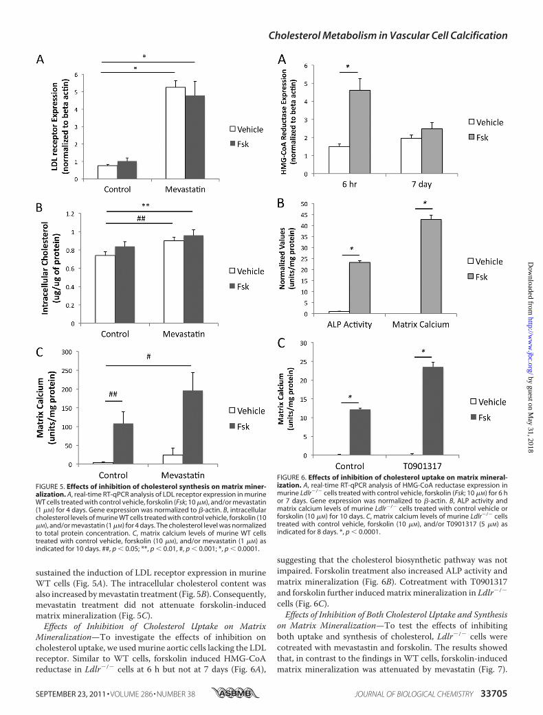
sustained the induction of LDL receptor expression in murineWT cells (Fig. 5A). The intracellular cholesterol content wasalso increased bymevastatin treatment (Fig. 5B). Consequently,mevastatin treatment did not attenuate forskolin-inducedmatrix mineralization (Fig. 5C).Effects of Inhibition of Cholesterol Uptake on Matrix
Mineralization—To investigate the effects of inhibition oncholesterol uptake, we usedmurine aortic cells lacking the LDLreceptor. Similar to WT cells, forskolin induced HMG-CoAreductase in Ldlr�/� cells at 6 h but not at 7 days (Fig. 6A),
suggesting that the cholesterol biosynthetic pathway was notimpaired. Forskolin treatment also increased ALP activity andmatrix mineralization (Fig. 6B). Cotreatment with T0901317and forskolin further inducedmatrixmineralization in Ldlr�/�
cells (Fig. 6C).Effects of Inhibition of Both Cholesterol Uptake and Synthesis
on Matrix Mineralization—To test the effects of inhibitingboth uptake and synthesis of cholesterol, Ldlr�/� cells werecotreated with mevastastin and forskolin. The results showedthat, in contrast to the findings in WT cells, forskolin-inducedmatrix mineralization was attenuated by mevastatin (Fig. 7).
FIGURE 5. Effects of inhibition of cholesterol synthesis on matrix miner-alization. A, real-time RT-qPCR analysis of LDL receptor expression in murineWT cells treated with control vehicle, forskolin (Fsk; 10 �M), and/or mevastatin(1 �M) for 4 days. Gene expression was normalized to �-actin. B, intracellularcholesterol levels of murine WT cells treated with control vehicle, forskolin (10�M), and/or mevastatin (1 �M) for 4 days. The cholesterol level was normalizedto total protein concentration. C, matrix calcium levels of murine WT cellstreated with control vehicle, forskolin (10 �M), and/or mevastatin (1 �M) asindicated for 10 days. ##, p � 0.05; **, p � 0.01, #, p � 0.001; *, p � 0.0001.
FIGURE 6. Effects of inhibition of cholesterol uptake on matrix mineral-ization. A, real-time RT-qPCR analysis of HMG-CoA reductase expression inmurine Ldlr�/� cells treated with control vehicle, forskolin (Fsk; 10 �M) for 6 hor 7 days. Gene expression was normalized to �-actin. B, ALP activity andmatrix calcium levels of murine Ldlr�/� cells treated with control vehicle orforskolin (10 �M) for 10 days. C, matrix calcium levels of murine Ldlr�/� cellstreated with control vehicle, forskolin (10 �M), and/or T0901317 (5 �M) asindicated for 8 days. *, p � 0.0001.
Cholesterol Metabolism in Vascular Cell Calcification
SEPTEMBER 23, 2011 • VOLUME 286 • NUMBER 38 JOURNAL OF BIOLOGICAL CHEMISTRY 33705
by guest on May 31, 2018
http://ww
w.jbc.org/
Dow
nloaded from

Intracellular cholesterol levels were also not further induced bymevastastin (data not shown).
DISCUSSION
Differentiating chondrocytes and osteoblasts produce amin-eralizing matrix by shedding matrix vesicles, which bud offfrom the plasmamembrane and serve as nucleation centers formineral crystals. Because cholesterol content is greater inmatrix vesicles than in the cytoplasm (14), vesicle productionmay drain the cell of its cholesterol stores, unless cholesterolsynthesis is up-regulated.In this study, we found that cholesterol metabolism plays an
important role in vascular cellmineralization. Spontaneous cal-cification was inhibited when cholesterol metabolism wasreduced. The differentiation activator forskolin acutely up-reg-ulated genes involved in both cholesterol uptake andbiosynthe-sis. In the presence of forskolin, it appears that when eitherpathway (cholesterol uptake or biosynthesis) is inhibited, theother pathway overcompensates, yielding even greater matrixmineralization. Inhibition of both pathways appears to berequired to attenuatemineralization. Interestingly, steady-stateintracellular cholesterol levels did not correlate well withthe propensity for forskolin-induced mineralization. This maybe due to a high-throughput state, inwhich cholesterol is incor-porated into matrix vesicles, which are exported as part of theprocess of extracellular matrix mineralization.It is interesting to note that statins have been shown to
inhibit humanVSMC calcification induced by an inflammatory“mixture” containing interferon-�, tumor necrosis factor-�,and oncostatin M (27). Such inhibitory effects may be due totheir known anti-inflammatory effects (28).We found previously that LXR agonists, which regulate cel-
lular cholesterol homeostasis, accelerate PKA-induced matrixmineralization of murine aortic cells (25) and high phosphate-induced bovine CVCs (21). In this study, we have shown thatsustained up-regulation of the LDL receptor and HMG-CoAreductase augments matrix mineralization. This mechanismmay work in concert with a parallel mechanism in which lipo-genesis is up-regulated by LXR agonists (21).These findings provide insight into howmatrix vesicles form
in the process of vascular calcification. They also have broader
implications becausematrix vesicles are a type ofmicroparticle,which is now recognized as a novel mechanism of intercellularcommunication, transcellular delivery, and interaction of cellswith their local extracellular environment.
REFERENCES1. Johnson, R. C., Leopold, J. A., and Loscalzo, J. (2006) Circ. Res. 99,
1044–10592. Jono, S., Shioi, A., Ikari, Y., andNishizawa, Y. (2006) J. BoneMiner. Metab.
24, 176–1813. Mizobuchi, M., Towler, D., and Slatopolsky, E. (2009) J. Am. Soc. Nephrol.
20, 1453–14644. Sage, A. P., Tintut, Y., and Demer, L. L. (2010) Nat. Rev. Cardiol. 7,
528–5365. Abedin,M., Tintut, Y., andDemer, L. L. (2004)Arterioscler. Thromb. Vasc.
Biol. 24, 1161–11706. Giachelli, C. M. (2009) Kidney Int. 75, 890–8977. Hsu, J. J., Tintut, Y., and Demer, L. L. (2008) Curr. Drug Targets 9,
224–2288. Neven, E., and D’Haese, P. C. (2011) Circ. Res. 108, 249–2649. Neves, K. R., Graciolli, F. G., dos Reis, L. M., Graciolli, R. G., Neves, C. L.,
Magalhaes, A. O., Custodio, M. R., Batista, D. G., Jorgetti, V., andMoyses,R. M. (2007) Kidney Int. 71, 1262–1270
10. Rattazzi, M., Bennett, B. J., Bea, F., Kirk, E. A., Ricks, J. L., Speer, M.,Schwartz, S. M., Giachelli, C. M., and Rosenfeld, M. E. (2005) Arterioscler.Thromb. Vasc. Biol. 25, 1420–1425
11. Huang, M. S., Sage, A. P., Lu, J., Demer, L. L., and Tintut, Y. (2008)Biochem. Biophys. Res. Commun. 374, 553–558
12. Tintut, Y., Patel, J., Parhami, F., and Demer, L. L. (2000) Circulation 102,2636–2642
13. Siddappa, R., Martens, A., Doorn, J., Leusink, A., Olivo, C., Licht, R., vanRijn, L., Gaspar, C., Fodde, R., Janssen, F., van Blitterswijk, C., and de Boer,J. (2008) Proc. Natl. Acad. Sci. U.S.A. 105, 7281–7286
14. Peress, N. S., Anderson, H. C., and Sajdera, S.W. (1974)Calcif. Tissue Res.14, 275–281
15. Chen, N. X., O’Neill, K. D., Chen, X., andMoe, S. M. (2008) J. BoneMiner.Res. 23, 1798–1805
16. Reynolds, J. L., Joannides, A. J., Skepper, J. N., McNair, R., Schurgers, L. J.,Proudfoot, D., Jahnen-Dechent,W.,Weissberg, P. L., and Shanahan, C.M.(2004) J. Am. Soc. Nephrol. 15, 2857–2867
17. Sawada, N., Taketani, Y., Amizuka, N., Ichikawa, M., Ogawa, C., Nomoto,K., Nashiki, K., Sato, T., Arai, H., Isshiki, M., Segawa, H., Yamamoto, H.,Miyamoto, K., and Takeda, E. (2007) Bone 41, 52–58
18. Brown, M. S., and Goldstein, J. L. (1984) Sci. Am. 251, 58–6619. Yamamoto, T., Davis, C. G., Brown, M. S., Schneider, W. J., Casey, M. L.,
Goldstein, J. L., and Russell, D. W. (1984) Cell 39, 27–3820. Tintut, Y., Parhami, F., Bostrom, K., Jackson, S.M., andDemer, L. L. (1998)
J. Biol. Chem. 273, 7547–755321. Ting, T. C.,Miyazaki-Anzai, S.,Masuda,M., Levi,M., Demer, L. L., Tintut,
Y., and Miyazaki, M. (2011) J. Biol. Chem. 286, 23938–23944922. Sage, A. P., Lu, J., Tintut, Y., and Demer, L. L. (2011) Kidney Int. 79,
414–42223. Joseph, S. B., McKilligin, E., Pei, L., Watson, M. A., Collins, A. R., Laffitte,
B. A., Chen, M., Noh, G., Goodman, J., Hagger, G. N., Tran, J., Tippin,T. K.,Wang, X., Lusis, A. J., Hsueh,W.A., Law, R. E., Collins, J. L.,Willson,T.M., andTontonoz, P. (2002)Proc. Natl. Acad. Sci. U.S.A. 99, 7604–7609
24. Terasaka, N., Hiroshima, A., Koieyama, T., Ubukata, N., Morikawa, Y.,Nakai, D., and Inaba, T. (2003) FEBS Lett. 536, 6–11
25. Hsu, J. J., Lu, J., Huang, M. S., Geng, Y., Sage, A. P., Bradley, M. N., Ton-tonoz, P., Demer, L. L., and Tintut, Y. (2009) FEBS Lett. 583, 1344–1348
26. Yokoyama, C., Wang, X., Briggs, M. R., Admon, A., Wu, J., Hua, X., Gold-stein, J. L., and Brown, M. S. (1993) Cell 75, 187–197
27. Kizu, A., Shioi, A., Jono, S., Koyama, H., Okuno, Y., and Nishizawa, Y.(2004) J. Cell. Biochem. 93, 1011–1019
28. Jain, M. K., and Ridker, P. M. (2005) Nat. Rev. Drug Discov. 4, 977–987
FIGURE 7. Effects of inhibition of both cholesterol uptake and synthesison matrix mineralization. Matrix calcium levels of murine Ldlr�/� cellstreated with control vehicle, forskolin (Fsk; 10 �M), and/or mevastatin (1 �M)as indicated for 10 days. #, p � 0.0001.
Cholesterol Metabolism in Vascular Cell Calcification
33706 JOURNAL OF BIOLOGICAL CHEMISTRY VOLUME 286 • NUMBER 38 • SEPTEMBER 23, 2011
by guest on May 31, 2018
http://ww
w.jbc.org/
Dow
nloaded from

Demer and Yin TintutYifan Geng, Jeffrey J. Hsu, Jinxiu Lu, Tabitha C. Ting, Makoto Miyazaki, Linda L.
Role of Cellular Cholesterol Metabolism in Vascular Cell Calcification
doi: 10.1074/jbc.M111.269639 originally published online August 11, 20112011, 286:33701-33706.J. Biol. Chem.
10.1074/jbc.M111.269639Access the most updated version of this article at doi:
Alerts:
When a correction for this article is posted•
When this article is cited•
to choose from all of JBC's e-mail alertsClick here
http://www.jbc.org/content/286/38/33701.full.html#ref-list-1
This article cites 28 references, 11 of which can be accessed free at
by guest on May 31, 2018
http://ww
w.jbc.org/
Dow
nloaded from