roles of microrna across prenatal and postnatal periods
TRANSCRIPT
International Journal of
Molecular Sciences
Review
Roles of MicroRNA across Prenatal andPostnatal PeriodsIlaria Floris †,‡, Jamie D. Kraft † and Illimar Altosaar *
Biochemistry, Microbiology & Immunology Department, Faculty of Medicine, University of Ottawa,451 Smyth Road, Ottawa, ON K1H8M5, Canada; [email protected] (I.F.); [email protected] (J.D.K.)* Correspondence: [email protected]; Tel.: +1-613-562-5800 (ext. 6371)† These authors contributed equally to this work.‡ Current address: Labo’Life France, Pescalis, Les Magnys, 79320 Moutiers-sous-Chantemerle, France.
Academic Editor: Martin PichlerReceived: 16 October 2016; Accepted: 17 November 2016; Published: 28 November 2016
Abstract: Communication between mother and offspring in mammals starts at implantation via thematernal–placental–fetal axis, and continues postpartum via milk targeted to the intestinal mucosa.MicroRNAs (miRNAs), short, noncoding single-stranded RNAs, of about 22 nucleotides in length,are actively involved in many developmental and physiological processes. Here we highlight therole of miRNA in the dynamic signaling that guides infant development, starting from implantationof conceptus and persisting through the prenatal and postnatal periods. miRNAs in body fluids,particularly in amniotic fluid, umbilical cord blood, and breast milk may offer new opportunitiesto investigate physiological and/or pathological molecular mechanisms that portend to open novelresearch avenues for the identification of noninvasive biomarkers.
Keywords: maternal-offspring crosstalk; microRNA; noninvasive biomarkers; breast milk
1. Background
In developing mammals, the vulnerability and immature immune status of the fetus and newbornare compensated by maternal factors that are transferred via the placenta or amniotic fluid, duringintrauterine life, and by milk after birth [1].
During development, mother-fetal cross-talk begins at implantation, via the maternal–placental–fetal axis [1]. The biological dialogue initiates between the maternal reproductive tissue and theconceptus [2]. The placenta plays a fundamental and regulatory role by producing a wide variety ofhormones, growth factors, and cytokines [3], as well as other biologically active molecules, includingmiRNA [4,5]. These biomolecules act both locally and systemically, exchanging information andregulating the physiological and metabolic processes required during intrauterine fetal life [3–5].The intrauterine environment strongly influences growth and fetal development, in addition to thehealth status of both the mother and child long after birth [5–8].
During fetal development, the gastrointestinal (GI) tract is characterized by higher permeabilitywhich may allow amniotic fluid to be absorbed by the fetus. Both amniotic fluid and breast milk havenutritive, protective, and regulatory roles [9]. Strikingly, amniotic fluid administered postpartum inthe preterm piglet model is comparable to colostrum in its ability to modulate intestinal inflammation,to increase body weight, to alter bacterial colonization, and to induce differential expression of mRNAcoding for genes involved in gut inflammatory responses [10].
After birth, the mother’s milk continues to provide oral biochemical signaling previously providedto the fetus via amniotic fluid [1]. Milk undoubtedly offers the nutritional requirements for growth andinfant development, but it is much more than food, providing protection and long-term programming
Int. J. Mol. Sci. 2016, 17, 1994; doi:10.3390/ijms17121994 www.mdpi.com/journal/ijms
Int. J. Mol. Sci. 2016, 17, 1994 2 of 12
agents for the infant [11]. The term “lactocrine” has been proposed to indicate the comprehensivematernal signals sent to the offspring via milk [12].
The biochemical factors strongly influencing offspring development in both pre- and postnatalperiods are interpreted as an ancient adaptation of mammals, starting with the origin of lactation [13,14],and enhanced with the evolution of the placenta. Particularly, human infants are born much earlierthan nonhuman primates and are in need of additional support. For example, comparative proteomestudies in humans and rhesus macaques revealed that human milk is richer in proteins involved inthe development of the GI tract, the immune system, and the brain [15]. Indeed, lactation strategiesand milk composition vary among species and individuals, reflecting the biological process of naturalselection [16,17]. In this review, after summarizing the current knowledge of miRNA expressionand potential participation in the regulation of various cell mechanisms, we will present recentfindings suggesting that miRNAs are involved in maternal-infant crosstalk during both pregnancyand suckling periods.
2. Micro-Intro to the MicroRNA World
miRNAs are short, noncoding, single-stranded RNA of about 22 nucleotides in length. They aretranscribed in the nucleus from miRNA genes as longer hairpin precursors known as primary miRNA,which are processed by the endoribonuclease Drosha to form precursor miRNA. These are exportedby Exportin-5 into the cytoplasm, where they are further processed by the endoribonuclease Dicer toform mature miRNA duplexes [18,19]. The duplex consists of a guide strand and a messenger strandof which the latter ultimately is degraded and the former becomes a mature miRNA. The maturemiRNA becomes part of the RNA-induced silencing complex (RISC) after being loaded onto Argonaute.Through partial complementation, miRNA becomes a guide for the RISC to target the 3′ untranslatedregion of messenger RNA (mRNA) [18]. Their main function is to repress target mRNA via translationinhibition and/or transcript degradation, and to silence gene expression [19]. A single miRNA canbind and target multiple mRNAs and in turn, one mRNA can be regulated by multiple miRNAs [20,21].Transcription, translation, and protein degradation are prominent examples of how gene activityis controlled by miRNAs, considered to be “meta-regulators” [22]. Importantly, miRNAs cantarget epigenetic regulators, which play a role in fetal metabolic programming [23,24], mediatinglong-term effects on target cells [24,25], and on developing organs and tissues. Epigenetic factors andmiRNAs have been shown to be reciprocally regulated [24,25]. Thus, miRNAs are recognized as keyregulators of diverse biological and developmental processes in eukaryotes (cell proliferation anddifferentiation, maintenance of tissue identity, apoptosis, immune system development, and responses)and are associated with pathologies, including different types of cancer [26], vascular diseases [27,28],and diabetes [24,29].
Nomenclature and other important information about precursor hairpin sequences,and experimentally identified mature miRNA sequences, are available online in a database calledmiRBase (http://www.mirbase.org/). In October 2016, miRBase had a total of 28,645 entries ofhairpin precursor miRNAs profiled in a vast range of plant and animal species. For Homo sapienswe counted 1881 precursors and 2588 mature miRNAs. In addition, miRBase provides links todatabases for validated miRNA targets (TarBase) and miRNA target prediction (DIANA-microT,microRNA.org, miRDB, RNA22, TargetMiner, PicTar-vertebrates) for in silico studies. Other in silicotools are progressively becoming available for functional miRNA analysis. For example, MAGIA83(miRNA And Genes Integrated Analysis) (http://gencomp.bio.unipd.it/magia/start/) integratesexpression profiles of miRNA and mRNA to investigate more deeply into biological networks andpathways. Online databases, such as miR2disease86 (http://www.mir2disease.org/), can displaymiRNA expression profiles in human disease scenarios. Ingenuity Pathway Analysis (IPA) is acommonly used tool to identify mRNAs as targets for miRNAs in silico.
miRNAs are expressed not only in tissues and cells, but also in all body fluids tested: milk, plasma,urine, saliva, tears, seminal fluid, amniotic fluid [4], and umbilical cord blood [30]. RNA molecules were
Int. J. Mol. Sci. 2016, 17, 1994 3 of 12
once considered to be very unstable due to the presence of ribonucleases (RNases), which are abundantand ubiquitous to neutralize viral and bacterial nucleic acids. However, RNA molecules are now knownto circulate in a “safe and stable” manner in body fluids [4]. RNases are implicated in various functionsof innate and acquired immunity, in stimulating host defense, and in sustaining mucosal barriers [31].In early 1988, Steven Benner proposed a hypothesis regarding “extracellular communicator RNA”.He suggested that RNA molecules might be messengers involved in cell–cell communication, the balancebetween RNase/RNase inhibitors. He also proposed that RNA molecules were related to diseases,including cancer and angiogenesis [32]. During the following 20 years, researchers around the worldworking in different fields, continued to investigate RNA control of gene expression [33] and its role incell-to-cell signaling [34]. Today we are aware that extracellular noncoding RNA, such as miRNA andlong non-coding RNA (lncRNA) [35,36], are involved in cellular communication, may be involved inchildhood development, and are protected from RNases through association with RNA-binding proteinsand/or by their encapsulation inside extracellular vesicles [36–38].
During the last decade, extracellular vesicles (EV) have been studied thoroughly regarding theirbiogenesis, content, and biological function. They can mediate the transfer of proteins, lipids, andnucleic acids, including miRNAs and lncRNAs [36], and have been recognized as potent vehicles ofintercellular communication, both in prokaryotes and eukaryotes [39]. EV have biological functionslinked to sperm maturation and motility, follicular growth, oocyte meiosis, steroidogenesis, and theprevention of polyspermy after fertilization. In the uterus, EV mediate the crosstalk between embryoand endometrium during implantation [40] and are crucial regulators throughout pregnancy [41,42].
3. New Kids on the Block: miRNAs Really Do Matter
During the dynamics of pregnancy, miRNAs appear to play important regulatory roles inmaternal-fetal crosstalk [5]. miRNAs are involved in endometrial receptivity, implantation, placentalfunction, and labor in eutherian organisms [43,44]. Once fertilization has taken place it may be possiblethat seminal fluid or sperm cell miRNAs affect gene expression in the zygote, with downstreameffects on successful implantation. The abundance and identity of miRNA that may be present in asingle spermatocyte remains unknown [2]. miRNAs contribute to establishing immune tolerance atconception [45]. Altered immune-regulatory miRNAs during preconception and the first trimesterare indicative of later pregnancy outcomes in humans (n = 48) [45]. During placental development,miRNAs can bind to target genes responsible for cellular invasion, proliferation, apoptosis, andangiogenesis [44]. Placenta and maternal plasma are characterized by specific miRNA patterns [46].Many of these miRNA genes form clusters, likely controlled by the same promoters to work in asynergistic manner, which are mainly expressed during pregnancy [47]. Their rapid decrease afterparturition suggests a placental-fetal origin and an expression pattern that is specifically inducedduring pregnancy [48]. The regulation of miRNA expression remains to be determined as somemiRNAs are expressed constitutively and others are expressed in a tissue-specific manner in responseto environmental stimuli. Notably, miRNAs contribute to normal term and pre-term labor. For example,in a study with 17 patients (8 term patients, 9 spontaneous term labour patients), expression of miR-223and miR-34 was induced in the cervix during term parturition [48]. In a murine model, expression of themiR-200 family acts to control uterine quiescence and contractility via regulation of the progesteronereceptor as well as the transcription factors ZEB1 and ZEB2, which was further validated usingmyometrial biopsies from pregnant women subject to caesarean section, both during and prior toactive labour [49]. Interestingly, the fetal genome, including loci for miRNA, can generate molecularsignals that are sent through the placenta and amniotic fluid into maternal circulation [50]. In additionto endogenous miRNA, exogenous non-coding RNA consumed by the mother can cross the placentaand gain access to the fetus, regulating fetal development [51]. miRNA profiles in amniotic fluidreveal their involvement in pathways (such as axon guidance, focal adhesion, and mitogen-activatedprotein kinase signaling pathways) related to the normal development of the nervous system andother organs (Figure 1) [52]. Even though the effect of labour on miRNA profiles has been reported
Int. J. Mol. Sci. 2016, 17, 1994 4 of 12
in plasma, miRNA profiles have yet to be identified in milk or amniotic fluid based on vaginal orcaesarean-section modes of delivery [53]. The mode of delivery has been shown to affect the bacterialpresence and abundance in human milk [54]. This suggests potential for differing miRNA expressionin milk depending on the mode of delivery, however, this intriguing aspect remains to be explored [55].
Early enteral feeds appear to have long-term health effects regarding epigenetic mechanisms.Interestingly, in mice, a low protein diet from weaning resulted in an altered miRNA profile (miR-98,miR-199, miR-21, let-7, and miR-210), most of which are involved with cellular proliferation [56].Recent studies have revealed miRNA involvement in chromatin remodeling. Gene expression may beregulated by intertwined mechanisms of miRNAs, DNA methylation, and histone modification [56].Previously, research focused on the changes in DNA methylation based on diet, however, there is now anincrease in research emphasizing the role of nutrition in the variation of miRNAs [57]. In a sheep study,undernourishment at the time of conception led to altered miRNA expression in the skeletal muscle ofoffspring which was suggested to be linked to insulin resistance development [58]. Additionally, a sheepmodel of maternal obesity also lead to differentiation of fetal miRNA expression in muscles thoughtto intensify intramuscular adipogenesis [59]. These variations in miRNA profiles could be due to thesignaling from the mother in utero via amniotic fluid. However, more studies need to be conducted todemonstrate the direct effects of maternal health on the miRNA profiles of offspring.
4. miRNA-Associated Pregnancy Complications
While assessing miRNA roles in normal pregnancy, one cannot overlook the miRNAs associatedwith pregnancy-related pathologies including gestational diabetes (GD) [24], implantation failure,pre-eclampsia, preterm labor, and intrauterine growth restriction [44]. GD, a glucose intolerancefirst recognized during pregnancy, is an independent risk factor of disease in both offspring andmother [60,61]. The hyperglycemia occurring during pregnancy induces long-term phenotypicalterations in fetal endothelial cells, changes in miR-101 expression levels, as well as in those ofEnhancer of Zester Homolog 2 (EZH2), its target [24]. EZH2 is the only protein of the PolycombRepressor Complex 2 (PRC2), known to date, to have catalytic activity [62]. PRC2 works in concertwith chromatin regulators and other proteins to initiate and maintain the methylation of histone H3 onlysine 27 (H3K27), an epigenetic mark which mediates long-term gene silencing [62]. EZH2 is both atarget of miR-101 and a transcriptional repressor of miR-101 via binding with the regulatory sequence(s)in the ‘miR-101 locus’ [24]. miR-101 and EZH2 are reciprocally controlled through a regulatory feedbackloop. The disturbance of this homeostatic loop is associated with cell phenotypic alterations, observedafter five to six passages in culture under normal glucose conditions. These alterations includelower proliferation, impaired migration and angiogenic capacity, as well as increased apoptosis [24].Alterations of noncoding RNA, including miRNA, are implicated in persistent and long lastingepigenetic changes, that can be transferred during cell division [63], and that are associated with celldysfunction and disease [24,63]. In diabetic conditions, the prolonged exposure to hyperglycemiagenerates a long-lasting impression on vascular cells and the progression of vascular complications [64],leading to the ‘glycemic memory’ theory that explains how chronic hyperglycemic conditions lead topersistent outcomes, through epigenetic changes [65] and miRNA alterations [64].
Seven miRNAs in peripheral blood (miR-1, miR-133b, miR-199a-5p, miR-1267, miR-1229, miR-223,and miR-148a-3p) are related to the predisposition for pregnancy complications such as miscarriageand early- and late-onset pre-eclampsia [45]. They have a predictive value for adverse pregnancyoutcomes and therefore have a role as potential biomarkers. In addition, they are associated withlow regulatory T cell numbers and low TNF/IL-10 levels, that implies an altered immunologicalmechanism [39]. These observations are in line with the emerging viewpoint regarding the conundrumof preterm birth, still poorly understood. Labour onset is thought to depend on the balance betweenmaternal cells, myometrial cells, and immune cells circulating in the tissues of the mother and fetus.As there appear to be multifactorial causes for preterm birth, one systematic approach to comprehendthe phenomenon and decipher its intrinsic signaling is to focus on pro-inflammatory signals andimmunological pathways [66], and also explore the role(s) of related miRNAs to better understand themolecular mechanisms involved in uterine exposure to endo-/exo-genous factors [67].
Int. J. Mol. Sci. 2016, 17, 1994 5 of 12
5. Breast Milk miRNA: Possible Key Regulators and Noninvasive Biomarkers
The neonatal and infant period is substantial for growth and development, especially in humans.Human milk has the indisputable role of protecting the infant against infection and confers bothshort and long-term benefits, reducing morbidity and mortality while contributing to cognitiveattributes [11,68–70]. Breast milk contains the highest concentration of total RNA of the 12 body fluidstested, more than 80 times the concentration found in amniotic fluid [4]. More than 1400 maturemiRNAs are expressed in human milk [71]. These miRNAs are thought to originate from mammarygland epithelial cells [72]. Their resistance to degradation stems from their assembly with proteins(Ago2 and other RNA-binding proteins) in skim milk [72,73], packaged inside milk cells [74] andinside the heterogeneous population of milk vesicles, which include milk fat globules [75] andexosomes [76,77]. Human milk somatic cells or exosomes which carry miRNA [74] are thoughtto survive in the acidic environment of the stomach, to be transferred into the circulation and maythen be integrated in various tissues [78]. Using a model GI tract, bovine milk exosomes have beenshown to withstand the harsh conditions of the gut [79]. There are a number of known and unknownmiRNAs conserved in human milk cells and lipids that are differentially expressed during lactation [80].Exogenous food-derived miRNAs appear to be stable in the oral cavity and in the GI tract and aretransferred to the blood of adults, thought to influence gene expression in different tissues [81–83].Recent evidence noted that bovine milk exosomes are taken up by human intestinal cells and vascularendothelial cells via endocytosis [84]. However, more studies are required to demonstrate that milkmiRNAs, once ingested, can cross the GI tract of the infant [83,85]. Recent studies have shown theresistance of miRNA to degradation and that uptake of bovine milk exosomes by intestinal cellsin vitro is mediated by temperature dependent-endocytosis [79,86]. A more recent study has providedevidence that porcine milk exosomal miRNAs modify gene expression and promote proliferation inintestinal cells in vitro and in vivo [87]. Importantly, miRNAs are thought to either have a functionalrole or a nutritional role. The functional hypothesis suggests that milk miRNAs are absorbed by thesuckling baby to imply specific actions: for example, to modulate and shape the immune system [85,88]by regulating T-cells, inducing B-cell differentiation [71,77], and preventing the development ofallergies [89]. The nutritional hypothesis suggests miRNAs simply provide nutrition and refutes thetransfer and the absorption of milk miRNA into the systemic circulation of offspring [85].
The fate and the role of miRNAs and other non-coding RNA components in breast milk stillremain unknown. Interestingly, breast milk is rich with immune-related miRNAs [71,77,88] andmiRNAs are implicated in multiple physiological processes [75], including cellular differentiationand proliferation, tissue identity, metabolism, and developmental programming [72]. Some miRNAspresent in milk are involved in nervous system pathways and may mediate brain development.For example, miR-118.2 [75] targets the Teneurin Transmembrane Protein 2, a protein abundant in thecentral nervous system and important in neural function [90]. Some other miRNAs in human milk aretissue-specific, such as miR-142-5p [73], which is hematopoiesis-specific [91]. These findings suggestthat milk miRNA may target specific organs and tissues of the feeding infant. In addition, there aremilk miRNAs that are involved in metabolic pathways. For instance, miR-21 [73,75,77], appears toplay an important role in promoting postnatal growth via mTORC1 signaling [92], while componentsof the let-7 family, also abundant in milk [73,75], are involved in glucose metabolism and can regulateglucose tolerance and insulin-sensitivity [93]. Furthermore, miR-33, miR-122, miR-370, miR-378-3p,and miR-125a-5p, abundant in the human milk lipid fraction [75] and milk exosomes [77], are potentialregulators of lipid metabolic pathways in the developing infant and/or are possible factors involvedin mammary gland function during lactation [94].
Where miRNAs are found to target epigenetic regulators the effect on the recipient target canbe persistent and durable [24,63]. miR-148-3p, one of the most highly expressed miRNAs in humanmilk [75], targets the DNA methyltransferase 3b, a key component of the epigenetic machinery duringdevelopment [95] and an essential DNA methyltransferase in the intestinal epithelium [96]. Throughmilk, the infant may benefit from these bioactive molecules, which may act as key regulators to setup important processes during the maturation of organs, such as the GI tract. The epithelium of
Int. J. Mol. Sci. 2016, 17, 1994 6 of 12
the GI system is continuously renewed and dynamically regulated by epigenetic modifications andtranscription factors, which influence intestinal cells throughout the crypt-villus axis [97]. Histonemethylation and acetylation are crucial in controlling proliferation and differentiation of intestinalcells of the crypts [97]. DNA methylation sites are key regulators of postnatal epigenetic changes inintestinal stem cells [98]. Promisingly, miRNAs in milk that target epigenetic regulators involved inhistone modification and DNA methylation are possible candidates for the process of gut maturation.
Daily fluctuations of mRNA [99] and miRNA [73] in human milk suggest that other physiologicalroles can be explored. Circadian variations of milk components, including miRNA, may help toestablish and/or fine-tune gastrointestinal circadian rhythmicity of the breast fed infant, mediatingwhat we now refer to as lactocrine circadian signals [73].
During the course of the lactation period the expression levels of miRNAs such as miR-25,miR-155, miR-182, miR-191, miR-221, and miR-223, change in the mammary glands of cows [100],pigs [101], rats [102], and humans [71]. The expression of cellular and extracellular miRNA in bovinemammary epithelial cells changes under the influence of lactogenic hormones such as prolactin [103].These findings suggest that miRNA is implicated in the physiological processes of milk synthesis undernormal conditions. In this context, it is noteworthy that the deregulation of miRNA has been associatedwith disease and pathological status [21,26], supporting their use as biomarkers for evaluating theperformance and health status of the mammary gland during lactation [11,99]. miRNA levels inblood are measured and successfully used to predict breast cancer susceptibility [104] and otherdiseases [105], for example, mastitis [106]. In a similar way, miRNA in breast milk deserves to bestudied in neonatal and perinatal medicine to identify biomarkers for infant diseases [11] and to betterunderstand the unique dyadic exchange between mothers and their babies.
Int. J. Mol. Sci. 2016, 17, 1994 6 of 12
transcription factors, which influence intestinal cells throughout the crypt-villus axis [97]. Histone methylation and acetylation are crucial in controlling proliferation and differentiation of intestinal cells of the crypts [97]. DNA methylation sites are key regulators of postnatal epigenetic changes in intestinal stem cells [98]. Promisingly, miRNAs in milk that target epigenetic regulators involved in histone modification and DNA methylation are possible candidates for the process of gut maturation.
Daily fluctuations of mRNA [99] and miRNA [73] in human milk suggest that other physiological roles can be explored. Circadian variations of milk components, including miRNA, may help to establish and/or fine-tune gastrointestinal circadian rhythmicity of the breast fed infant, mediating what we now refer to as lactocrine circadian signals [73].
During the course of the lactation period the expression levels of miRNAs such as miR-25, miR-155, miR-182, miR-191, miR-221, and miR-223, change in the mammary glands of cows [100], pigs [101], rats [102], and humans [71]. The expression of cellular and extracellular miRNA in bovine mammary epithelial cells changes under the influence of lactogenic hormones such as prolactin [103]. These findings suggest that miRNA is implicated in the physiological processes of milk synthesis under normal conditions. In this context, it is noteworthy that the deregulation of miRNA has been associated with disease and pathological status [21,26], supporting their use as biomarkers for evaluating the performance and health status of the mammary gland during lactation [11,99]. miRNA levels in blood are measured and successfully used to predict breast cancer susceptibility [104] and other diseases [105], for example, mastitis [106]. In a similar way, miRNA in breast milk deserves to be studied in neonatal and perinatal medicine to identify biomarkers for infant diseases [11] and to better understand the unique dyadic exchange between mothers and their babies.
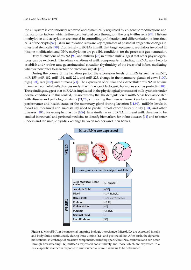
Figure 1. MicroRNA in the maternal-offspring biologic interchange. MicroRNA are expressed in cells and body fluids continuously during intra-uterine (a,b) and post-natal life. After birth, the dynamic, bidirectional interchange of bioactive components, including specific miRNA, continues and can occur through breastfeeding. (c) miRNAs expressed constitutively and those which are expressed in a tissue-specific manner in response to environmental stimuli remains to be determined.
Figure 1. MicroRNA in the maternal-offspring biologic interchange. MicroRNA are expressed in cellsand body fluids continuously during intra-uterine (a,b) and post-natal life. After birth, the dynamic,bidirectional interchange of bioactive components, including specific miRNA, continues and can occurthrough breastfeeding. (c) miRNAs expressed constitutively and those which are expressed in atissue-specific manner in response to environmental stimuli remains to be determined.
Int. J. Mol. Sci. 2016, 17, 1994 7 of 12
6. Conclusions
The biological dialogue between a mother and her offspring starts from the implantation of theembryo in the uterus and continues during fetal life via the maternal–placental–fetal axis [1]. In thisreview, we summarize the current knowledge concerning the role(s) of miRNA in the regulation ofvarious physiological and pathological processes involved in maternal-infant crosstalk during bothpregnancy and suckling periods. The expression of miRNAs is ubiquitous; they are present in cellsand body fluids of both maternal and fetal origin. Here we focus on their potential participation inoffspring development throughout prenatal and post-natal life. In particular, we discuss and citestudies congruent with the functional hypothesis [85], which proposes that milk miRNAs are absorbedby the GI of the suckling baby to ensure and regulate specific physiological processes.
Acknowledgments: We thank Jean-Francois Beaulieu and Elizabeth Herring (University of Sherbrooke, Canada)for providing comments and suggestions. The human milk biology program in the senior author’s laboratory wassupported by Operating Grant 82816 from the Canadian Institutes of Health Research.
Author Contributions: The first two authors contributed equally to the manuscript. Ilaria Floris conceived anddrafted this review and the figure, Jamie D. Kraft edited and prepared the manuscript, and Illimar Altosaarsupervised, wrote and revised the manuscript.
Conflicts of Interest: The authors declare no conflict of interest.
Abbreviations
miRNA microRNAEV Extracellular VesiclesGD Gestational DiabetesEZH2 Enhancer of Zester Homolog 2PRC2 Polycomb Repressor Complex 2H3K27 Histone H3 on Lysine27GI Gastrointestinal
References
1. Power, M.L.; Schulkin, J. Maternal regulation of offspring development in mammals is an ancient adaptationtied to lactation. Appl. Transl. Genom. 2013, 2, 55–63. [CrossRef]
2. Fazeli, A.; Holt, W.V. Cross talk during the periconception period. Theriogenology 2016, 86, 438–442. [CrossRef][PubMed]
3. Petraglia, F.; Pasquale, F.; Wylie, W.V. Placental expression of neurohormones and other neuroactivemolecules in human pregnancy. In Birth, Distress and Disease; Power, M.L., Schulkin, J., Eds.; CambridgeUniversity Press: Cambridge, UK, 2005; pp. 16–73.
4. Weber, J.A.; Baxter, D.H.; Zhang, S.; Huang, D.Y.; Huang, K.H.; Lee, M.J.; Galas, D.J.; Wang, K. The microRNAspectrum in 12 body fluids. Clin. Chem. 2010, 56, 1733–1741. [CrossRef] [PubMed]
5. Forbes, K. IFPA Gabor than Award lecture: Molecular control of placental growth: The emerging role ofmicroRNAs. Placenta 2013, 34, S27–S33. [CrossRef] [PubMed]
6. Thornburg, K.L.; Marshall, N. The placenta is the center of the chronic disease universe. Am. J. Obstet. Gynecol.2015, 213, S14–S20. [CrossRef] [PubMed]
7. Janssen, A.B.; Kertes, D.A.; McNamara, G.I.; Braithwaite, E.C.; Creeth, H.D.J.; Glover, V.I.; John, R.M. A rolefor the placenta in programming maternal mood and childhood behavioural disorders. J. Neuroendocrinol.2016, 28, 1–6. [CrossRef] [PubMed]
8. Tung, J.; Archie, E.A.; Altmann, J.; Alberts, S.C. Cumulative early life adversity predicts longevity in wildbaboons. Nat. Commun. 2016, 7, 11181. [CrossRef] [PubMed]
9. Underwood, M.A.; Gilbert, W.M.; Sherman, M.P. Amniotic fluid: Not just fetal urine anymore. J. Perinatol.2005, 25, 341–348. [CrossRef] [PubMed]
10. Siggers, J.; Ostergaard, M.V.; Siggers, R.H.; Skovgaard, K.; Mølbak, L.; Thymann, T.; Schmidt, M.; Møller, H.K.;Purup, S.; Fink, L.N.; et al. Postnatal amniotic fluid intake reduces gut inflammatory responses andnecrotizing enterocolitis in preterm neonates. Am. J. Physiol. Gastrointest. Liver Physiol. 2013, 304, G864–G875.[CrossRef] [PubMed]
Int. J. Mol. Sci. 2016, 17, 1994 8 of 12
11. Altosaar, I.; Siggers, J. Micromolecules to nanoparticles-human milk: More than nutrition. In Proceedings ofthe 3rd Annual International Conference on Human Milk Science and Innovation; German, B., Rhine, W., Eds.;Prolacta Bioscience, City of Industry, CA: Pasadena, CA, USA, 2015; pp. 6–8.
12. Bartol, F.F.; Wiley, A.A.; Bagnell, C.A. Epigenetic programming of porcine endometrial function and thelactocrine hypothesis. Reprod. Domest. Anim. 2008, 43, 273–279. [CrossRef] [PubMed]
13. Vorbach, C.; Capecchi, M.R.; Penninger, J.M. Evolution of the mammary gland from the innate immunesystem? BioEssays News Rev. Mol. Cell. Dev. Biol. 2006, 28, 606–616. [CrossRef] [PubMed]
14. Sale, S.; Pavelic, K. Mammary lineage tracing: The coming of age. Cell. Mol. Life Sci. 2015, 72, 1577–1583.[CrossRef] [PubMed]
15. Beck, K.L.; Weber, D.; Phinney, B.S.; Smilowitz, J.T.; Hinde, K.; Lönnerdal, B.; Korf, I.; Lemay, D.G.Comparative proteomics of human and macaque milk reveals species-specific nutrition during postnataldevelopment. J. Proteome Res. 2015, 14, 2143–2157. [CrossRef] [PubMed]
16. Hinde, K.; Milligan, L.A. Primate milk: Proximate mechanisms and ultimate perspectives. Evol. Anthropol.2011, 20, 9–23. [CrossRef] [PubMed]
17. Hinde, K.; German, J.B. Food in an evolutionary context: Insights from mother’s milk. J. Sci. Food Agric.2012, 92, 2219–2223. [CrossRef] [PubMed]
18. Finnegan, E.F.; Pasquinelli, A.E. MicroRNA biogenesis: Regulating the regulators. Crit. Rev. Biochem.Mol. Biol. 2013, 48, 51–68. [CrossRef] [PubMed]
19. Guo, H.; Ingolia, N.T.; Weissman, J.S.; Bartel, D.P. Mammalian microRNAs predominantly act to decreasetarget mRNA levels. Nature 2010, 466, 835–840. [CrossRef] [PubMed]
20. Krol, J.; Loedige, I.; Filipowicz, W. The widespread regulation of microRNA biogenesis, function and decay.Nat. Rev. Genet. 2010, 11, 597–610. [CrossRef] [PubMed]
21. Lewis, B.P.; Burge, C.B.; Bartel, D.P. Conserved seed pairing, often flanked by adenosines, indicates thatthousands of human genes are microRNA targets. Cell 2005, 120, 15–20. [CrossRef] [PubMed]
22. Enright, A.J.; John, B.; Gaul, U.; Tuschl, T.; Sander, C.; Marks, D.S. MicroRNA targets in Drosophila.Genome Biol. 2003, 5, R1. [CrossRef] [PubMed]
23. Sookoian, S.; Gianotti, T.F.; Burgueño, A.L.; Pirola, C.J. Fetal metabolic programming and epigeneticmodifications: A systems biology approach. Pediatr. Res. 2013, 73, 531–542. [CrossRef] [PubMed]
24. Floris, I.; Descamps, B.; Vardeu, A.; Mitic, T.; Posadino, A.M.; Shantikumar, S.; Sala-Newby, G.;Capobianco, G.; Mangialardi, G.; Howard, L.; et al. Gestational diabetes mellitus impairs fetal endothelialcell functions through a mechanism involving microRNA-101 and histone methyltransferase enhancer ofzester homolog-2. Arterioscler. Thromb. Vasc. Biol. 2015, 35, 664–674. [CrossRef] [PubMed]
25. Pasquinelli, A.E. MicroRNAs and their targets: Recognition, regulation and an emerging reciprocalrelationship. Nat. Rev. Genet. 2012, 13, 271–282. [CrossRef] [PubMed]
26. Xue, Z.; Yilan, D.; Ping, J.; Fei, M. Bioinformatic analysis of cancer-related microRNAs and their target genes.Yi Chuan 2015, 37, 855–864. [PubMed]
27. Spinetti, G.; Fortunato, O.; Caporali, A.; Shantikumar, S.; Marchetti, M.; Meloni, M.; Descamps, B.; Floris, I.;Sangalli, E.; Vono, R.; et al. MicroRNA-15a and microRNA-16 impair human circulating proangiogenic cellfunctions and are increased in the proangiogenic cells and serum of patients with critical limb ischemia.Circ. Res. 2013, 112, 335–346. [CrossRef] [PubMed]
28. Ovchinnikova, E.S.; Schmitter, D.; Vegter, E.L.; Ter Maaten, J.M.; Valente, M.A.E.; Liu, L.C.Y.; van der Harst, P.;Pinto, Y.M.; de Boer, R.A.; Meyer, S.; et al. Signature of circulating microRNAs in patients with acute heartfailure. Eur. J. Heart Fail. 2016, 18, 414–423. [CrossRef] [PubMed]
29. Shantikumar, S.; Caporali, A.; Emanueli, C. Role of microRNAs in diabetes and its cardiovascularcomplications. Cardiovasc. Res. 2012, 93, 583–593. [CrossRef] [PubMed]
30. Merkerova, M.; Vasikova, A.; Belickova, M.; Bruchova, H. MicroRNA expression profiles in umbilical cordblood cell lineages. Stem Cells Dev. 2010, 19, 17–26. [CrossRef] [PubMed]
31. Rosenberg, H.F. Vertebrate secretory (RNAse A) ribonucleases and host defense. In Ribonucleases;Nicholson, A.W., Ed.; Springer: Berlin/Heidelberg, Germany, 2011; Volume 26, pp. 35–53.
32. Benner, S.A. Extracellular “communicator RNA”. FEBS Lett. 1988, 233, 225–228. [CrossRef]
Int. J. Mol. Sci. 2016, 17, 1994 9 of 12
33. Diederichs, S.; Bartsch, L.; Berkmann, J.C.; Fröse, K.; Heitmann, J.; Hoppe, C.; Iggena, D.; Jazmati, D.;Karschnia, P.; Linsenmeier, M.; et al. The dark matter of the cancer genome: Aberrations in regulatoryelements, untranslated regions, splice sites, non-coding RNA and synonymous mutations. EMBO Mol. Med.2016, 8, 442–457. [CrossRef] [PubMed]
34. Mittelbrunn, M.; Sánchez-Madrid, F. Intercellular communication: Diverse structures for exchange of geneticinformation. Nat. Rev. Mol. Cell Biol. 2012, 13, 328–335. [CrossRef] [PubMed]
35. Karlsson, O.; Baccarelli, A.A. Environmental health and long non-coding RNAs. Curr. Environ. Health Rep.2016, 3, 178–187. [CrossRef] [PubMed]
36. Karlsson, O.; Rodosthenous, R.S.; Jara, C.; Brennan, K.J.; Wright, R.O.; Baccarelli, A.A.; Wright, R.J.Detection of long non-coding RNAs in human breastmilk extracellular vesicles: Implications for earlychild development. Epigenetics 2016, 11, 721–729. [CrossRef] [PubMed]
37. Mitchell, P.S.; Parkin, R.K.; Kroh, E.M.; Fritz, B.R.; Wyman, S.K.; Pogosova-Agadjanyan, E.L.; Peterson, A.;Noteboom, J.; O’Briant, K.C.; Allen, A.; et al. Circulating microRNAs as stable blood-based markers forcancer detection. Proc. Natl. Acad. Sci. USA 2008, 105, 10513–10518. [CrossRef] [PubMed]
38. Valadi, H.; Ekström, K.; Bossios, A.; Sjöstrand, M.; Lee, J.J.; Lötvall, J.O. Exosome-mediated transfer ofmRNAs and microRNAs is a novel mechanism of genetic exchange between cells. Nat. Cell Biol. 2007, 9,654–659. [CrossRef] [PubMed]
39. Yáñez-Mó, M.; Siljander, P.R.-M.; Andreu, Z.; Zavec, A.B.; Borràs, F.E.; Buzas, E.I.; Buzas, K.; Casal, E.;Cappello, F.; Carvalho, J.; et al. Biological properties of extracellular vesicles and their physiologicalfunctions. J. Extracell. Vesicles 2015, 4, 27066. [CrossRef] [PubMed]
40. Machtinger, R.; Laurent, L.C.; Baccarelli, A.A. Extracellular vesicles: Roles in gamete maturation, fertilizationand embryo implantation. Hum. Reprod. Update 2016, 22, 182–193. [CrossRef] [PubMed]
41. Nardi Fda, S.; Michelon, T.F.; Neumann, J.; Manvailer, L.F.S.; Wagner, B.; Horn, P.A.; Bicalho Mda, G.;Rebmann, V. High levels of circulating extracellular vesicles with altered expression and function duringpregnancy. Immunobiology 2016, 221, 753–760. [CrossRef] [PubMed]
42. Burnett, L.A.; Nowak, R.A. Exosomes mediate embryo and maternal interactions at implantation and duringpregnancy. Front. Biosci. 2016, 8, 79–96.
43. Mouillet, J.-F.; Chu, T.; Sadovsky, Y. Expression patterns of placental microRNAs. Birth Defects Res. A Clin.Mol. Teratol. 2011, 91, 737–743. [CrossRef] [PubMed]
44. Bidarimath, M.; Khalaj, K.; Wessels, J.M.; Tayade, C. MicroRNAs, immune cells and pregnancy. Cell. Mol. Immunol.2014, 11, 538–547. [CrossRef] [PubMed]
45. Winger, E.E.; Reed, J.L.; Ji, X. First-trimester maternal cell microRNA is a superior pregnancy marker toimmunological testing for predicting adverse pregnancy outcome. J. Reprod. Immunol. 2015, 110, 22–35.[CrossRef] [PubMed]
46. Chim, S.S.C.; Shing, T.K.F.; Hung, E.C.W.; Leung, T.-Y.; Lau, T.-K.; Chiu, R.W.K.; Lo, Y.M.D. Detection andcharacterization of placental microRNAs in maternal plasma. Clin. Chem. 2008, 54, 482–490. [CrossRef][PubMed]
47. Morales-Prieto, D.M.; Ospina-Prieto, S.; Chaiwangyen, W.; Schoenleben, M.; Markert, U.R. Pregnancy-associated miRNA-clusters. J. Reprod. Immunol. 2013, 97, 51–61. [CrossRef] [PubMed]
48. Hassan, S.S.; Romero, R.; Pineles, B.; Tarca, A.L.; Montenegro, D.; Erez, O.; Mittal, P.; Kusanovic, J.P.;Mazaki-Tovi, S.; Espinoza, J.; et al. MicroRNA expression profiling of the human uterine cervix after termlabor and delivery. Am. J. Obstet. Gynecol. 2010, 202, 80.e1–80.e8. [CrossRef] [PubMed]
49. Williams, K.C.; Renthal, N.E.; Condon, J.C.; Gerard, R.D.; Mendelson, C.R. MicroRNA-200a serves a key rolein the decline of progesterone receptor function leading to term and preterm labor. Proc. Natl. Acad. Sci. USA2012, 109, 7529–7534. [CrossRef] [PubMed]
50. Wong, F.C.K.; Lo, Y.M.D. Prenatal diagnosis innovation: Genome sequencing of maternal plasma.Annu. Rev. Med. 2016, 67, 419–432. [CrossRef] [PubMed]
51. Li, J.; Zhang, Y.; Li, D.; Liu, Y.; Chu, D.; Jiang, X.; Hou, D.; Zen, K.; Zhang, C.-Y. Small non-coding RNAstransfer through mammalian placenta and directly regulate fetal gene expression. Protein Cell 2015, 6,391–396. [CrossRef] [PubMed]
52. Sun, T.; Li, W.; Li, T.; Ling, S. MicroRNA profiling of amniotic fluid: Evidence of synergy of microRNAs infetal development. PLoS ONE 2016, 11, e0153950. [CrossRef] [PubMed]
Int. J. Mol. Sci. 2016, 17, 1994 10 of 12
53. Morisaki, S.; Miura, K.; Higashijima, A.; Abe, S.; Miura, S.; Hasegawa, Y.; Yoshida, A.; Kaneuchi, M.;Yoshiura, K.; Masuzaki, H. Effect of labor on plasma concentrations and postpartum clearance of cell-free,pregnancy-associated, placenta-specific microRNAs. Prenat. Diagn. 2015, 35, 44–50. [CrossRef] [PubMed]
54. Cabrera-Rubio, R.; Collado, M.C.; Laitinen, K.; Salminen, S.; Isolauri, E.; Mira, A. The human milkmicrobiome changes over lactation and is shaped by maternal weight and mode of delivery. Am. J. Clin. Nutr.2012, 96, 544–551. [CrossRef] [PubMed]
55. Cretoiu, D.; Xu, J.; Xiao, J.; Suciu, N.; Cretoiu, S.M. Circulating microRNAs as potential molecular biomarkersin pathophysiological evolution of pregnancy. Dis. Markers 2016, 2016, 1–7. [CrossRef] [PubMed]
56. Lillycrop, K.A.; Burdge, G.C. Epigenetic mechanisms linking early nutrition to long term health. Best Pract.Res. Clin. Endocrinol. Metab. 2012, 26, 667–676. [CrossRef] [PubMed]
57. Vickers, M.H. Early life nutrition, epigenetics and programming of later life disease. Nutrients 2014, 6,2165–2178. [CrossRef] [PubMed]
58. Lie, S.; Morrison, J.L.; Williams-Wyss, O.; Suter, C.M.; Humphreys, D.T.; Ozanne, S.E.; Zhang, S.;Maclaughlin, S.M.; Kleemann, D.O.; Walker, S.K.; et al. Periconceptional undernutrition programs changes ininsulin-signaling molecules and microRNAs in skeletal muscle in singleton and twin fetal sheep. Biol. Reprod.2014, 90, 5. [CrossRef] [PubMed]
59. Yan, X.; Huang, Y.; Zhao, J.-X.; Rogers, C.J.; Zhu, M.-J.; Ford, S.P.; Nathanielsz, P.W.; Du, M. Maternal obesitydownregulates microRNA let-7g expression, a possible mechanism for enhanced adipogenesis during ovinefetal skeletal muscle development. Int. J. Obes. 2013, 37, 568–575. [CrossRef] [PubMed]
60. Nahum Sacks, K.; Friger, M.; Shoham-Vardi, I.; Abokaf, H.; Spiegel, E.; Sergienko, R.; Landau, D.;Sheiner, E. Prenatal exposure to gestational diabetes mellitus as an independent risk factor for long-termneuropsychiatric morbidity of the offspring. Am. J. Obstet. Gynecol. 2016, 215, 380.e1–380.e7. [CrossRef][PubMed]
61. Jensen, L.A.; Chik, C.L.; Ryan, E.A. Review of gestational diabetes mellitus effects on vascular structure andfunction. Diab. Vasc. Dis. Res. 2016, 13, 170–182. [CrossRef] [PubMed]
62. Brooun, A.; Gajiwala, K.S.; Deng, Y.-L.; Liu, W.; Bolaños, B.; Bingham, P.; He, Y.-A.; Diehl, W.; Grable, N.;Kung, P.-P.; et al. Polycomb repressive complex 2 structure with inhibitor reveals a mechanism of activationand drug resistance. Nat. Commun. 2016, 7, 11384. [CrossRef] [PubMed]
63. Weinberg, M.S.; Morris, K.V. Transcriptional gene silencing in humans. Nucleic Acids Res. 2016, 44, 6505–6517.[CrossRef] [PubMed]
64. Berezin, A. Metabolic memory phenomenon in diabetes mellitus: Achieving and perspectives. DiabetesMetab. Syndr. 2016, 10, S176–S183. [CrossRef] [PubMed]
65. Rajasekar, P.; O’Neill, C.L.; Eeles, L.; Stitt, A.W.; Medina, R.J. Epigenetic changes in endothelial progenitorsas a possible cellular basis for glycemic memory in diabetic vascular complications. J. Diabetes Res. 2015,2015, 436879. [CrossRef] [PubMed]
66. Wallenstein, M.B.; Shaw, G.M.; Stevenson, D.K. Preterm birth as a calendar event or immunologic anomaly.JAMA Pediatr. 2016, 170, 525–526. [CrossRef] [PubMed]
67. Kappil, M.; Chen, J. Environmental exposures in utero and microRNA. Curr. Opin. Pediatr. 2014, 26, 243–251.[CrossRef] [PubMed]
68. Isaacs, E.B.; Fischl, B.R.; Quinn, B.T.; Chong, W.K.; Gadian, D.G.; Lucas, A. Impact of breast milk onintelligence quotient, brain size, and white matter development. Pediatr. Res. 2010, 67, 357–362. [CrossRef][PubMed]
69. Luby, J.L.; Belden, A.C.; Whalen, D.; Harms, M.P.; Barch, D.M. Breastfeeding and childhood IQ: The mediatingrole of gray matter volume. J. Am. Acad. Child Adolesc. Psychiatry 2016, 55, 367–375. [CrossRef] [PubMed]
70. Parylak, S.L.; Deng, W.; Gage, F.H. Mother’s milk programs offspring’s cognition. Nat. Neurosci. 2014, 17,8–9. [CrossRef] [PubMed]
71. Alsaweed, M.; Hartmann, P.E.; Geddes, D.T.; Kakulas, F. MicroRNAs in breastmilk and the lactating breast:Potential immunoprotectors and developmental regulators for the infant and the mother. Int. J. Environ. Res.Public. Health 2015, 12, 13981–14020. [CrossRef] [PubMed]
72. Alsaweed, M.; Lai, C.T.; Hartmann, P.E.; Geddes, D.T.; Kakulas, F. Human milk miRNAs primarily originatefrom the mammary gland resulting in unique miRNA profiles of fractionated milk. Sci. Rep. 2016, 6, 20680.[CrossRef] [PubMed]
Int. J. Mol. Sci. 2016, 17, 1994 11 of 12
73. Floris, I.; Billard, H.; Boquien, C.Y.; Joram-Gauvard, E.; Simon, L.; Legrand, A.; Boscher, C.; Roze, J.C.;Bolanos-Jimenez, F.; Kaeffer, B. miRNA analysis by quantitative PCR in preterm human breast milk revealsdaily fluctuations of hsa-miR-16–5p. PLoS ONE 2015, 10, e0140488. [CrossRef] [PubMed]
74. Alsaweed, M.; Lai, C.T.; Hartmann, P.E.; Geddes, D.T.; Kakulas, F. Human milk cells contain numerousmiRNAs that may change with milk removal and regulate multiple physiological processes. Int. J. Mol. Sci.2016, 17, 956. [CrossRef] [PubMed]
75. Munch, E.M.; Harris, R.A.; Mohammad, M.; Benham, A.L.; Pejerrey, S.M.; Showalter, L.; Hu, M.; Shope, C.D.;Maningat, P.D.; Gunaratne, P.H.; et al. Transcriptome profiling of microRNA by next-gen deep sequencingreveals known and novel miRNA species in the lipid fraction of human breast milk. PLoS ONE 2013, 8,e50564. [CrossRef] [PubMed]
76. Kosaka, N.; Iguchi, H.; Ochiya, T. Circulating microRNA in body fluid: A new potential biomarker for cancerdiagnosis and prognosis. Cancer Sci. 2010, 101, 2087–2092. [CrossRef] [PubMed]
77. Zhou, Q.; Li, M.; Wang, X.; Li, Q.; Wang, T.; Zhu, Q.; Zhou, X.; Wang, X.; Gao, X.; Li, X. Immune-relatedmicroRNAs are abundant in breast milk exosomes. Int. J. Biol. Sci. 2012, 8, 118–123. [CrossRef] [PubMed]
78. Hassiotou, F.; Beltran, A.; Chetwynd, E.; Stuebe, A.M.; Twigger, A.-J.; Metzger, P.; Trengove, N.; Lai, C.T.;Filgueira, L.; Blancafort, P.; et al. Breastmilk is a novel source of stem cells with multilineage differentiationpotential. Stem Cells 2012, 30, 2164–2174. [CrossRef] [PubMed]
79. Benmoussa, A.; Lee, C.H.C.; Laffont, B.; Savard, P.; Laugier, J.; Boilard, E.; Gilbert, C.; Fliss, I.; Provost, P.Commercial dairy cow milk microRNAs resist digestion under simulated gastrointestinal tract conditions.J. Nutr. 2016, 146, 2206–2215. [CrossRef] [PubMed]
80. Alsaweed, M.; Lai, C.T.; Hartmann, P.E.; Geddes, D.T.; Kakulas, F. Human milk cells and lipids conservenumerous known and novel miRNAs, some of which are differentially expressed during lactation. PLoS ONE2016, 11, e0152610. [CrossRef] [PubMed]
81. Zhang, L.; Hou, D.; Chen, X.; Li, D.; Zhu, L.; Zhang, Y.; Li, J.; Bian, Z.; Liang, X.; Cai, X.; et al. Exogenous plantMIR168a specifically targets mammalian LDLRAP1: Evidence of cross-kingdom regulation by microRNA.Cell Res. 2012, 22, 107–126. [CrossRef] [PubMed]
82. Baier, S.R.; Nguyen, C.; Xie, F.; Wood, J.R.; Zempleni, J. MicroRNAs are absorbed in biologically meaningfulamounts from nutritionally relevant doses of cow milk and affect gene expression in peripheral bloodmononuclear cells, HEK-293 kidney cell cultures, and mouse livers. J. Nutr. 2014, 144, 1495–1500. [CrossRef][PubMed]
83. Zempleni, J.; Baier, S.R.; Howard, K.M.; Cui, J. Gene regulation by dietary microRNAs. Can. J. Physiol. Pharmacol.2015, 93, 1097–1102. [CrossRef] [PubMed]
84. Kusuma, R.J.; Manca, S.; Friemel, T.; Sukreet, S.; Nguyen, C.; Zempleni, J. Human vascular endothelialcells transport foreign exosomes from cow’s milk by endocytosis. Am. J. Physiol. Cell Physiol. 2016, 310,C800–C807. [CrossRef] [PubMed]
85. Melnik, B.C.; Kakulas, F.; Geddes, D.T.; Hartmann, P.E.; John, S.M.; Carrera-Bastos, P.; Cordain, L.; Schmitz, G.Milk miRNAs: Simple nutrients or systemic functional regulators? Nutr. Metab. 2016, 13, 1–5. [CrossRef][PubMed]
86. Wolf, T.; Baier, S.R.; Zempleni, J. The intestinal transport of bovine milk exosomes is mediated by endocytosisin human colon carcinoma Caco-2 cells and rat small intestinal IEC-6 cells. J. Nutr. 2015, 145, 2201–2206.[CrossRef] [PubMed]
87. Chen, T.; Xie, M.-Y.; Sun, J.-J.; Ye, R.-S.; Cheng, X.; Sun, R.-P.; Wei, L.-M.; Li, M.; Lin, D.-L.; Jiang, Q.-Y.; et al.Porcine milk-derived exosomes promote proliferation of intestinal epithelial cells. Sci. Rep. 2016, 6, 33862.[CrossRef] [PubMed]
88. Na, R.S.; E, G.X.; Sun, W.; Sun, X.W.; Qiu, X.Y.; Chen, L.P.; Huang, Y.F. Expressional analysis ofimmune-related miRNAs in breast milk. Genet. Mol. Res. 2015, 14, 11371–11376. [CrossRef] [PubMed]
89. Melnik, B.C.; John, S.M.; Schmitz, G. Milk: An exosomal microRNA transmitter promoting thymic regulatoryT cell maturation preventing the development of atopy? J. Transl. Med. 2014, 12, 43. [CrossRef] [PubMed]
90. Silva, J.-P.; Lelianova, V.G.; Ermolyuk, Y.S.; Vysokov, N.; Hitchen, P.G.; Berninghausen, O.; Rahman, M.A.;Zangrandi, A.; Fidalgo, S.; Tonevitsky, A.G.; et al. Latrophilin 1 and its endogenous ligand Lasso/teneurin-2form a high-affinity transsynaptic receptor pair with signaling capabilities. Proc. Natl. Acad. Sci. USA 2011,108, 12113–12118. [CrossRef] [PubMed]
Int. J. Mol. Sci. 2016, 17, 1994 12 of 12
91. Ramkissoon, S.H.; Mainwaring, L.A.; Ogasawara, Y.; Keyvanfar, K.; McCoy, J.P.; Sloand, E.M.; Kajigaya, S.;Young, N.S. Hematopoietic-specific microRNA expression in human cells. Leuk. Res. 2006, 30, 643–647.[CrossRef] [PubMed]
92. Melnik, B.C.; John, S.M.; Schmitz, G. Milk is not just food but most likely a genetic transfection systemactivating mTORC1 signaling for postnatal growth. Nutr. J. 2013, 12, 103. [CrossRef] [PubMed]
93. Zhu, H.; Shyh-Chang, N.; Segrè, A.V.; Shinoda, G.; Shah, S.P.; Einhorn, W.S.; Takeuchi, A.; Engreitz, J.M.;Hagan, J.P.; Kharas, M.G.; et al. The Lin28/let-7 axis regulates glucose metabolism. Cell 2011, 147, 81–94.[CrossRef] [PubMed]
94. Fernández-Hernando, C.; Suárez, Y.; Rayner, K.J.; Moore, K.J. MicroRNAs in lipid metabolism.Curr. Opin. Lipidol. 2011, 22, 86–92. [CrossRef] [PubMed]
95. Duursma, A.M.; Kedde, M.; Schrier, M.; le Sage, C.; Agami, R. miR-148 targets human DNMT3b proteincoding region. RNA 2008, 14, 872–877. [CrossRef] [PubMed]
96. Elliott, E.N.; Sheaffer, K.L.; Kaestner, K.H. The ‘de novo’ DNA methyltransferase Dnmt3b compensates theDnmt1-deficient intestinal epithelium. Elife 2016, 5, e12975. [CrossRef] [PubMed]
97. Roostaee, A.; Benoit, Y.D.; Boudjadi, S.; Beaulieu, J.-F. Epigenetics in intestinal epithelial cell renewal.J. Cell. Physiol. 2016, 231, 2361–2367. [CrossRef] [PubMed]
98. Yu, D.-H.; Gadkari, M.; Zhou, Q.; Yu, S.; Gao, N.; Guan, Y.; Schady, D.; Roshan, T.N.; Chen, M.-H.;Laritsky, E.; et al. Postnatal epigenetic regulation of intestinal stem cells requires DNA methylation and isguided by the microbiome. Genome Biol. 2015, 16, 211. [CrossRef] [PubMed]
99. Maningat, P.D.; Sen, P.; Rijnkels, M.; Sunehag, A.L.; Hadsell, D.L.; Bray, M.; Haymond, M.W. Gene expressionin the human mammary epithelium during lactation: The milk fat globule transcriptome. Physiol. Genom.2009, 37, 12–22. [CrossRef] [PubMed]
100. Li, Z.; Liu, H.; Jin, X.; Lo, L.; Liu, J. Expression profiles of microRNAs from lactating and non-lactating bovinemammary glands and identification of miRNA related to lactation. BMC Genom. 2012, 13, 731. [CrossRef][PubMed]
101. Gu, Y.; Li, M.; Wang, T.; Liang, Y.; Zhong, Z.; Wang, X.; Zhou, Q.; Chen, L.; Lang, Q.; He, Z.; et al.Lactation-related microRNA expression profiles of porcine breast milk exosomes. PLoS ONE 2012, 7, e43691.[CrossRef] [PubMed]
102. Izumi, H.; Kosaka, N.; Shimizu, T.; Sekine, K.; Ochiya, T.; Takase, M. Time-dependent expression profiles ofmicroRNAs and mRNAs in rat milk whey. PLoS ONE 2014, 9, e88843. [CrossRef] [PubMed]
103. Muroya, S.; Hagi, T.; Kimura, A.; Aso, H.; Matsuzaki, M.; Nomura, M. Lactogenic hormones alter cellularand extracellular microRNA expression in bovine mammary epithelial cell culture. J. Anim. Sci. Biotechnol.2016, 7, 8. [CrossRef] [PubMed]
104. Singh, R.; Mo, Y.-Y. Role of microRNAs in breast cancer. Cancer Biol. Ther. 2013, 14, 201–212. [CrossRef] [PubMed]105. Chen, X.; Ba, Y.; Ma, L.; Cai, X.; Yin, Y.; Wang, K.; Guo, J.; Zhang, Y.; Chen, J.; Guo, X.; et al. Characterization
of microRNAs in serum: A novel class of biomarkers for diagnosis of cancer and other diseases. Cell Res.2008, 18, 997–1006. [CrossRef] [PubMed]
106. Taga, I.; Lan, C.Q.; Altosaar, I. Plant essential oils and mastitis disease: Their potential inhibitory effects onpro-inflammatory cytokine production in response to bacteria related inflammation. Nat. Prod. Commun.2012, 7, 675–682. [PubMed]
© 2016 by the authors; licensee MDPI, Basel, Switzerland. This article is an open accessarticle distributed under the terms and conditions of the Creative Commons Attribution(CC-BY) license (http://creativecommons.org/licenses/by/4.0/).