rom j morphol embryol r j m e original paper … j morphol embryol 2012, 53(2):369–373 issn ......
TRANSCRIPT
Rom J Morphol Embryol 2012, 53(2):369–373
ISSN (print) 1220–0522 ISSN (on-line) 2066-8279
OORRIIGGIINNAALL PPAAPPEERR
Clinical and paraclinical study regarding the macro- and microscopic diagnosis of various anatomo-clinical forms of
operated uterine fibromyoma DIANA VANIOVA KLIMENTOVA1), ANCA DANIELA BRĂILA1),
CRISTIANA SIMIONESCU2), I. ILIE3), M. B. BRĂILA1)
1)Department of Obstetrics and Gynecology 2)Department of Pathology
University of Medicine and Pharmacy of Craiova
Emergency County Hospital, Craiova 3)Department of Pathology,
Emergency County Hospital, Targoviste
Abstract The research focused on a systematic review of 959 cases operated for uterine fibromyoma (24.8%) from 3859 gynecological surgeries performed over a period of 10 years (2000–2010). Materials and Methods: The important parameters were those related to the general clinical and laboratory data, and especially those focused on the macro- and microscopic histopathological diagnostic methods. Thus, we inserted some details on the methods used for the histopathological examination of surgically removed samples. Results: The age of patients operated for uterine fibromyoma was between 20 and 60 years, with the highest incidence in the 40–50 years group (594 cases – 62.4%). The most frequent anatomo-clinical forms observed were uterine fibromyoma with menometrorrhagia (78.9%), large uterine fibromyoma associated with compression and metrorrhagia (81.1%), uterine fibromyoma with aseptic necrobiosis (33.6%). The other forms showed a reduced frequency. Uterine fibromyoma associated with infertility was found in patients under 40-year-old, their evolution being initially asymptomatic. The incidence of uterine fibromyoma, which degenerated into a leiomyosarcoma, was “0”. Discussion: 1/5 of patients operated after the age of 35 years presented with various anatomo-clinical forms of uterine fibromyoma. Several hypotheses are formulated regarding the etiopathogenesis, morphology and embryogenesis of this benign tumor of the myometrium. The estrogen–progestogen hormonal imbalance after this age in correlation with the genetic predisposition lead to the synthesis of various proteins, enzymes, and growth factors, decrease of apoptosis and stimulation of leiomyomatous cells with the development of a large, even gigantic form of fibromyoma, representing the most common form encountered in our study. The medical treatment with progesterone derivatives did not lead to the expected results, radical surgery usually being the final therapeutical approach. Conclusions: Between 31 and 60-year-old, the incidence of operated uterine fibromyoma was 941 cases out of 959 (98.1%). The correlation between the preoperative clinical and laboratory diagnosis, the intra-operative morphological appearance and especially the post-operative histopathological examination was 100%. In all cases of uterine fibromyoma, histopathological examination was and will remain the sovereign exploration for surgical practice in general and for gynecological surgery in particular. Keywords: uterine fibromyoma, menometrorrhagia, infertility, septic and aseptic necrobiosis, acute abdomen by torsion/
compression, microscopic histopathological examination.
Introduction
The research objectives focused on the reevaluation of the various aspects of uterine fibromyoma in the current surgical practice: the incidence of operated fibromyoma cases over a period of 10 years, its various anatomo-clinical forms, the correlation between pre-operative clinical laboratory diagnosis, macro- and microscopical pathological findings, and the histo-pathological diagnostic methods. We noted a high frequency of uterine fibromyoma, with 959 (24.8%) cases analyzed of 3859 patients operated for genital disorders between 2000 and 2010. From our findings as well as from other studies in both domestic and foreign literature, 1/5 of women after 35-year-old present with uterine fibromyoma for expert advice [1, 2]. In the
absence of appropriate intervention, mostly surgical, local and general suffering of patients is prolonged, social and individual costs increase (absenteeism, multiple
sick leaves, various and costly hormonal, hemostatic, and anti-anemic treatments, mostly inefficient, needlessly
prolonging the initial suffering) [3, 4]. In the study group, we found radical surgical
procedures being performed even on patients under 40-year-old. In a previous study, we have emphasized the opportunity of conservative surgery (single or multiple myomectomy myometrectomy, supra-isthmic hyster-ectomy with uterine cavity reconstruction), especially in patients younger than 40 years who associate uterine fibromyoma with infertility and desire a pregnancy. In such situations, extemporaneous histopathological examinations, as well as the final histopathological
R J M ERomanian Journal of
Morphology & Embryologyhttp://www.rjme.ro/

Diana Vaniova Klimentova et al.
370
examination, are required, both in terms of the endometrium, the myometrium, and fibroid nodules. Uterine fibromyoma is an essentially benign myometrial pathology, which rarely undergoes malignant transfor-mation (1 in 10 000; 0.001%) [5, 6].
In our study, we did not encounter any such case. However, we encountered 611 cases with uterine fibromyomas associated with endometrial hyperplasia (61.7%), and with adenomyosis in 195 cases (20.3%) [7, 8].
The fact is that our study revealed that vaginal bleedings were not caused by the uterine fibromyoma itself but by these two associated entities. Endometrial hyperplasia always required biopsic and hemostatic uterine curettage with preoperative histopathological examination; adenomyosis was diagnosed only by post-operative microscopic histopathological examination both of the myometrium and the uterine fibromyoma. As a practical importance, it should be said that either radical or conservative surgery require significant costs or expenses in terms of the procedures involved, the number of days of hospitalization, sick leaves granted, the recovery time and patient’s reintegration in the family and society. After collecting the data from the accounting service of the Emergency County Hospital of Craiova, we found that: one day of hospitalization costs 245.15 lei (~55 EUR); classical abdominal total hysterectomy costs about 1459.12 lei (~325 EUR); total laparoscopic hysterectomy costs 2076.64 lei (~460 EUR); embolization by embospheres costs 2076.64 lei (~460 EUR).
Based on these data, we estimate both the practical importance of the type of surgery performed, and the number of hospitalization days with all expenses. In the U.S. over 700 000 radical surgeries for uterine fibro-myoma are recorded annually and according to the procedures applied to the costs vary between 10 000 and 15 000 dollars [15, 16].
Materials and Methods
The research was conducted in the second Obstetrics and Gynecology Clinic, from the Emergency County Hospital of Craiova, by analyzing 959 (24.8%) cases with operated uterine fibromyoma out of a total of 3859 gynecological surgical interventions over a period of 10 years (Figure 1).
Figure 1 – The percentage of operated patients for uterine fibromyoma of all hospitalized gynecological patients.
During this time, there was a significant inter-disciplinary collaboration with the pathology department, as well as other hospital services (surgery, radiology,
endocrinology, clinical laboratory). Parameters analyzed were classified as follows:
I. General clinical and laboratory parameters: age (Table 1), area of origin, professional groups, history of obstetrical and gynecological well as other diseases, admission reasons (Figure 1), clinical diagnosis, labora-tory diagnosis.
Table 1 – Distribution of uterine fibromyoma patients according to their age
Age of the patients [years] Year 20–30 31–40 41–50 51–60 >60
Total
2000 1 12 63 17 0 93 2001 0 10 37 12 0 59 2002 0 6 55 12 0 73 2003 2 14 59 17 0 92 2004 0 20 57 13 0 90 2005 1 9 59 26 1 96 2006 3 23 49 11 0 86 2007 1 25 59 23 4 112 2008 0 18 56 18 0 92 2009 2 16 40 15 0 73 2010 2 12 60 17 2 93 Total 12 165 594 181 7 959
II. Methodology: examination methods as well as macro- and microscopic diagnosis. Special attention was paid to this category in the present study in terms of postoperative histopathological examination of the samples removed during surgery.
The samples removed during surgery in the Gyneco-logy Department were transported to the Pathology Laboratory in sealed containers in a mixture of formalin and water in equal proportions, accompanied by the chart of the biopsy material. Surgical samples were processed in three stages: pre-analytical, analytical, and post-analytical. Pre-analytical procedures consisted of reception, recording, handling and transportation of surgical samples, as well as their conservation.
Analytical procedures consisted of orientation of the sample in the laboratory, sectioning it and placing the sections in special boxes, their inclusion in paraffin blocks, microtome sectioning, stretching the sections in the water bath, placing them on albumin coated slides, staining and sealing them using glass coverslips and Canada balm. The classical stain was Hematoxylin and Eosin (HE).
Post-analytical procedures generally consist of the release of the histopathological findings, storage of the remaining samples and waste disposal.
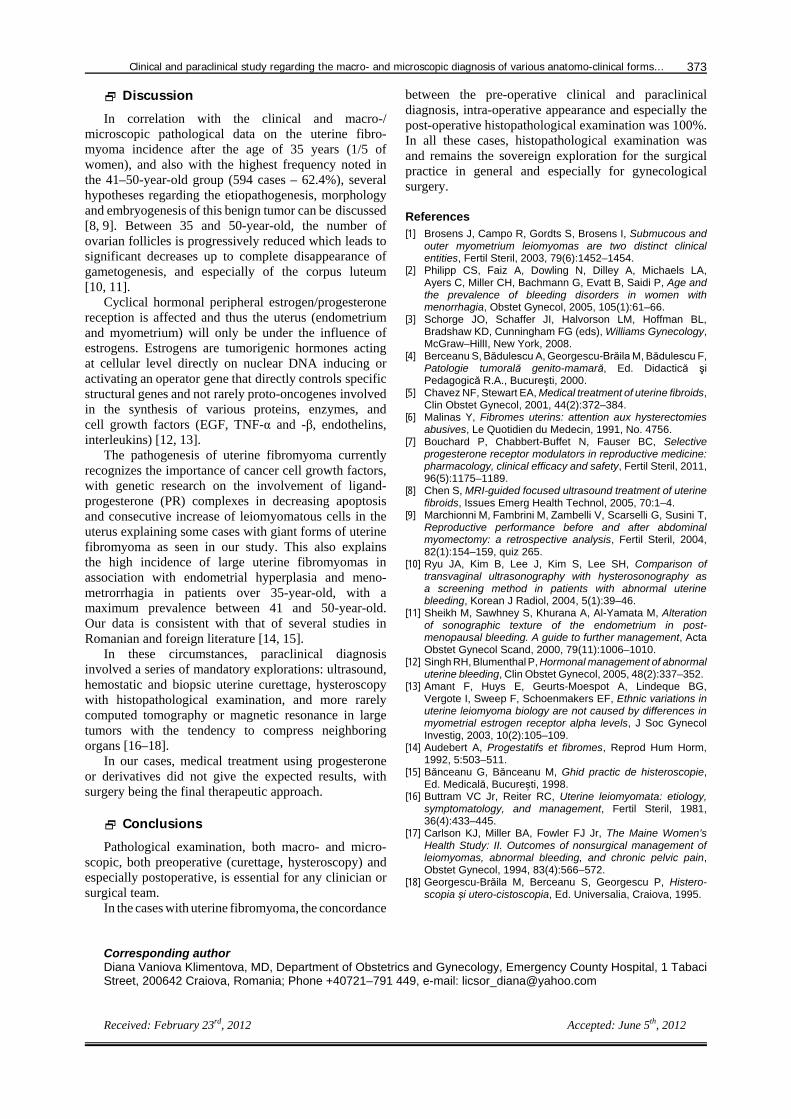
In the orientation room, the macroscopic description of the surgical sample is performed as follows: in the fibromatous uterus, it may be diffusely thickened but also nodular, in which case we describe the shape and size of the uterus and the fibroid nodules with an intra-mural, submucosal or endocavitary location, sometimes associated with the presence of endometrial polyps. Fibroid nodules, especially those measuring over 5–7 cm, besides the “vortex” pattern of smooth muscle fibers, may have a pearly white color due to hyalinization, even diffuse, but also gray areas of necrosis, red hemorrhagic areas, foci of calcification, cystic transfor-mation, etc.
Clinical and paraclinical study regarding the macro- and microscopic diagnosis of various anatomo-clinical forms…
371
The tumor is lined by the pseudocapsule formed by the surrounding tissues, which are compressed by the tumor mass.
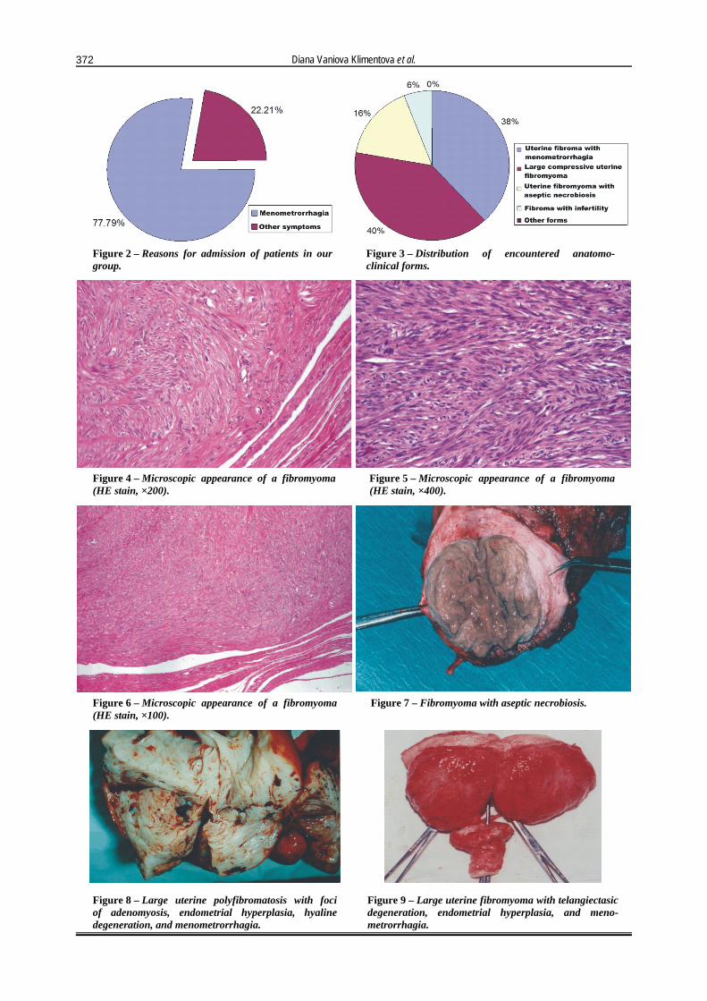
The microscopic characteristics of the uterine fibro-myoma are as follows: besides the “vortex” pattern of smooth muscle fibers, similar to those of the undeveloped embryonic myometrium, there is a great cellular variability ranging from a tumor consisting only of smooth muscle bundles, to a tumor composed mainly of connective stroma. Benign forms with nuclear atypia, giant cells (atypical and bizarre forms of leiomyoma, dense cellular fibroma, etc.), low mitotic index, fibroids with epithelioid pattern, etc. can be seen.
In large tumors secondary to vascularization disorders, degenerative changes occur: diffuse hyalinization, ischemic necrosis, hemorrhagic foci, myxomatous, mucoid or cystic degeneration, foci of calcification, amyloid deposits, etc. Following the orientation of the fragments (2–3 mm in size) in special boxes, the fixation process takes place, usually in 10% formalin solution at room temperature with a duration ranging from four hours to several days. Next, the samples are washed in running tap water from 24 to 48 hours. The embedding of the samples consists of the impregnation with a solidifiable mass (e.g., paraffin), obtaining the paraffin block. This phase includes the dehydration (successive passage through 4–5 tanks with alcohol of increasing concentration from 70% to 100% for variable intervals depending on the size of the sample – from one to five hours), the clarification (three successive xylene or toluene tanks, one to four hours each), and paraffin impregnation. Paraffin impregnation is performed in a hot oven at 560C by passing the samples through three successive baths of molten paraffin. By using a heated forceps, the samples are taken out of the last heated paraffin bath from the oven and placed in a porcelain dish with a flat bottom that already contains melted paraffin. The capsule is carefully sank in cold water for 30–60 minutes and until the paraffin solidifies, thus obtaining a paraffin block which will be subjected to microtome sectioning.
Sectioning is performed using a microtome, thus obtaining thin (5–6 μm) transparent sections, which are placed in a hot water bath and transferred on slides covered with Mayer’s albumin. The slides are placed in an oven at 370C for 24 hours to dry.
HE staining includes the following steps: ▪ deparaffinization (three xylene or toluene baths for
3, 4 and 10 minutes respectively); ▪ hydration in three successive alcohol baths with
decreasing concentrations (100%, 90% and 70%), followed by rinsing in tap water;
▪ staining for 3–4 minutes in Mayer’s hemalaun solution followed by rinsing in tap water;
▪ differentiation in 1% acid alcohol solution for 1–10 seconds, followed by washing in tap water;
▪ fixation in lithium carbonate for 1–2 minutes and another wash in tap water;
▪ Eosin staining for 2–3 seconds and washing in tap water;
▪ dehydration in alcohol with increasing concentra-tions (70%, 90% and 100%);
▪ clarification in three xylene or toluene baths; ▪ mounting the sections in Canada balm (Entellan/
BioMount). The slides are left to dry in an oven and then
examined at ×10, ×20 and ×40 magnification. The nuclei appear blue-violet, the cytoplasm is light red and the connective tissues appear red as can be seen in the microscopic images presented.
Results
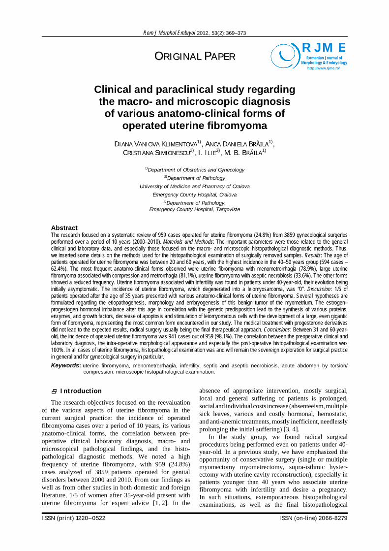
The age of the patients with uterine fibromyoma ranged between 20 and 60-year-old. The highest incidence was recorded in the 41–50 years age group (594 patients – 62.4%), followed by the 51–60 years group (181 cases – 18.8%), and 31–40 years (165 cases – 16.9%). There were 12 (1.2%) cases between 20 and 30-year-old, and 7 (0.7%) cases over the age of 60 years. In 569 (59.6%) cases, there were only one or two births. It is well established that uterine fibromyoma is the prerogative of pauciparity and infertility. Meno-metrorrhagia was the symptom leading to admissions in 746 cases (78.9%) (Figure 2).
Clinical diagnosis was established by traditional gynecological examination. Paraclinical diagnosis was established using abdominal or vaginal ultrasound, uterine hemostatic and biopsic curettage and histopathological examination of sampled fragments, hysteroscopy, colposcopy, and only in some cases, due to the tumor volume, by using computed tomography or MRI.
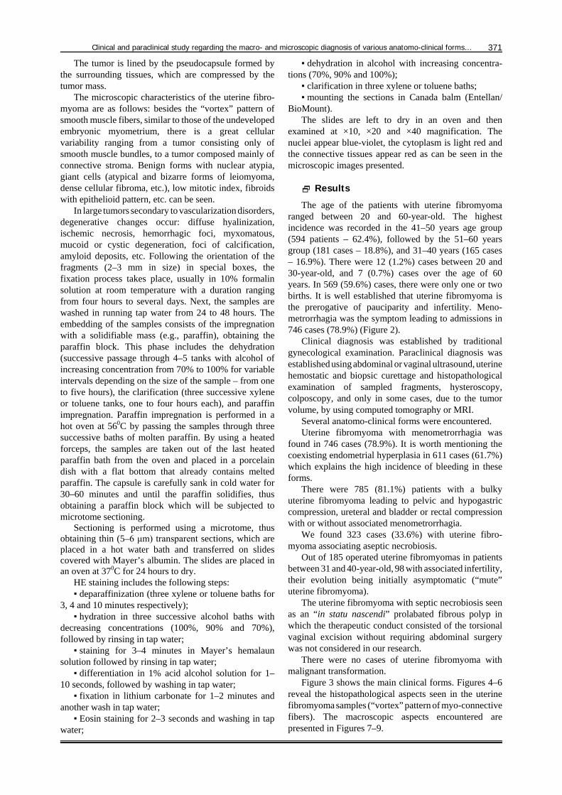
Several anatomo-clinical forms were encountered. Uterine fibromyoma with menometrorrhagia was
found in 746 cases (78.9%). It is worth mentioning the coexisting endometrial hyperplasia in 611 cases (61.7%) which explains the high incidence of bleeding in these forms.
There were 785 (81.1%) patients with a bulky uterine fibromyoma leading to pelvic and hypogastric compression, ureteral and bladder or rectal compression with or without associated menometrorrhagia.
We found 323 cases (33.6%) with uterine fibro-myoma associating aseptic necrobiosis.
Out of 185 operated uterine fibromyomas in patients between 31 and 40-year-old, 98 with associated infertility,
their evolution being initially asymptomatic (“mute” uterine fibromyoma).
The uterine fibromyoma with septic necrobiosis seen as an “in statu nascendi” prolabated fibrous polyp in which the therapeutic conduct consisted of the torsional vaginal excision without requiring abdominal surgery was not considered in our research.
There were no cases of uterine fibromyoma with malignant transformation.
Figure 3 shows the main clinical forms. Figures 4–6 reveal the histopathological aspects seen in the uterine fibromyoma samples (“vortex” pattern of myo-connective fibers). The macroscopic aspects encountered are presented in Figures 7–9.
Diana Vaniova Klimentova et al.
372
Figure 2 – Reasons for admission of patients in our group.
Figure 3 – Distribution of encountered anatomo-clinical forms.
Figure 4 – Microscopic appearance of a fibromyoma (HE stain, ×200).
Figure 5 – Microscopic appearance of a fibromyoma (HE stain, ×400).
Figure 6 – Microscopic appearance of a fibromyoma (HE stain, ×100).
Figure 7 – Fibromyoma with aseptic necrobiosis.
Figure 8 – Large uterine polyfibromatosis with foci of adenomyosis, endometrial hyperplasia, hyaline degeneration, and menometrorrhagia.
Figure 9 – Large uterine fibromyoma with telangiectasic degeneration, endometrial hyperplasia, and meno-metrorrhagia.
Clinical and paraclinical study regarding the macro- and microscopic diagnosis of various anatomo-clinical forms…
373
Discussion
In correlation with the clinical and macro-/ microscopic pathological data on the uterine fibro-myoma incidence after the age of 35 years (1/5 of women), and also with the highest frequency noted in the 41–50-year-old group (594 cases – 62.4%), several hypotheses regarding the etiopathogenesis, morphology and embryogenesis of this benign tumor can be discussed [8, 9]. Between 35 and 50-year-old, the number of ovarian follicles is progressively reduced which leads to significant decreases up to complete disappearance of gametogenesis, and especially of the corpus luteum [10, 11].
Cyclical hormonal peripheral estrogen/progesterone reception is affected and thus the uterus (endometrium and myometrium) will only be under the influence of estrogens. Estrogens are tumorigenic hormones acting at cellular level directly on nuclear DNA inducing or activating an operator gene that directly controls specific structural genes and not rarely proto-oncogenes involved in the synthesis of various proteins, enzymes, and cell growth factors (EGF, TNF-α and -β, endothelins, interleukins) [12, 13].
The pathogenesis of uterine fibromyoma currently recognizes the importance of cancer cell growth factors, with genetic research on the involvement of ligand-progesterone (PR) complexes in decreasing apoptosis and consecutive increase of leiomyomatous cells in the uterus explaining some cases with giant forms of uterine fibromyoma as seen in our study. This also explains the high incidence of large uterine fibromyomas in association with endometrial hyperplasia and meno-metrorrhagia in patients over 35-year-old, with a maximum prevalence between 41 and 50-year-old. Our data is consistent with that of several studies in Romanian and foreign literature [14, 15].
In these circumstances, paraclinical diagnosis involved a series of mandatory explorations: ultrasound, hemostatic and biopsic uterine curettage, hysteroscopy with histopathological examination, and more rarely computed tomography or magnetic resonance in large tumors with the tendency to compress neighboring organs [16–18].
In our cases, medical treatment using progesterone or derivatives did not give the expected results, with surgery being the final therapeutic approach.
Conclusions
Pathological examination, both macro- and micro-scopic, both preoperative (curettage, hysteroscopy) and especially postoperative, is essential for any clinician or surgical team.
In the cases with uterine fibromyoma, the concordance
between the pre-operative clinical and paraclinical diagnosis, intra-operative appearance and especially the post-operative histopathological examination was 100%. In all these cases, histopathological examination was and remains the sovereign exploration for the surgical practice in general and especially for gynecological surgery.
References [1] Brosens J, Campo R, Gordts S, Brosens I, Submucous and
outer myometrium leiomyomas are two distinct clinical entities, Fertil Steril, 2003, 79(6):1452–1454.
[2] Philipp CS, Faiz A, Dowling N, Dilley A, Michaels LA, Ayers C, Miller CH, Bachmann G, Evatt B, Saidi P, Age and the prevalence of bleeding disorders in women with menorrhagia, Obstet Gynecol, 2005, 105(1):61–66.
[3] Schorge JO, Schaffer JI, Halvorson LM, Hoffman BL, Bradshaw KD, Cunningham FG (eds), Williams Gynecology, McGraw–HillI, New York, 2008.
[4] Berceanu S, Bădulescu A, Georgescu-Brăila M, Bădulescu F, Patologie tumorală genito-mamară, Ed. Didactică şi Pedagogică R.A., Bucureşti, 2000.
[5] Chavez NF, Stewart EA, Medical treatment of uterine fibroids, Clin Obstet Gynecol, 2001, 44(2):372–384.
[6] Malinas Y, Fibromes uterins: attention aux hysterectomies abusives, Le Quotidien du Medecin, 1991, No. 4756.
[7] Bouchard P, Chabbert-Buffet N, Fauser BC, Selective progesterone receptor modulators in reproductive medicine: pharmacology, clinical efficacy and safety, Fertil Steril, 2011, 96(5):1175–1189.
[8] Chen S, MRI-guided focused ultrasound treatment of uterine fibroids, Issues Emerg Health Technol, 2005, 70:1–4.
[9] Marchionni M, Fambrini M, Zambelli V, Scarselli G, Susini T, Reproductive performance before and after abdominal myomectomy: a retrospective analysis, Fertil Steril, 2004, 82(1):154–159, quiz 265.
[10] Ryu JA, Kim B, Lee J, Kim S, Lee SH, Comparison of transvaginal ultrasonography with hysterosonography as a screening method in patients with abnormal uterine bleeding, Korean J Radiol, 2004, 5(1):39–46.
[11] Sheikh M, Sawhney S, Khurana A, Al-Yamata M, Alteration of sonographic texture of the endometrium in post-menopausal bleeding. A guide to further management, Acta Obstet Gynecol Scand, 2000, 79(11):1006–1010.
[12] Singh RH, Blumenthal P, Hormonal management of abnormal uterine bleeding, Clin Obstet Gynecol, 2005, 48(2):337–352.
[13] Amant F, Huys E, Geurts-Moespot A, Lindeque BG, Vergote I, Sweep F, Schoenmakers EF, Ethnic variations in uterine leiomyoma biology are not caused by differences in myometrial estrogen receptor alpha levels, J Soc Gynecol Investig, 2003, 10(2):105–109.
[14] Audebert A, Progestatifs et fibromes, Reprod Hum Horm, 1992, 5:503–511.
[15] Bănceanu G, Bănceanu M, Ghid practic de histeroscopie, Ed. Medicală, București, 1998.
[16] Buttram VC Jr, Reiter RC, Uterine leiomyomata: etiology, symptomatology, and management, Fertil Steril, 1981, 36(4):433–445.
[17] Carlson KJ, Miller BA, Fowler FJ Jr, The Maine Women’s Health Study: II. Outcomes of nonsurgical management of leiomyomas, abnormal bleeding, and chronic pelvic pain, Obstet Gynecol, 1994, 83(4):566–572.
[18] Georgescu-Brăila M, Berceanu S, Georgescu P, Histero-scopia și utero-cistoscopia, Ed. Universalia, Craiova, 1995.
Corresponding author Diana Vaniova Klimentova, MD, Department of Obstetrics and Gynecology, Emergency County Hospital, 1 Tabaci Street, 200642 Craiova, Romania; Phone +40721–791 449, e-mail: [email protected] Received: February 23rd, 2012 Accepted: June 5th, 2012