romanian journal of oral rehabilitation -...
TRANSCRIPT

Romanian Journal of Oral Rehabilitation
Vol. 1, No. 4, October 2009
1
Romanian Journal
of Oral Rehabilitation
Vol. 1, No. 4, October 2009
Editor in Chief
Norina Consuela Forna, Iaşi, România
Vice-Editor
Viorel Păun, Bucharest, România
Senior Associate Editors
Pierre Lafforgue, Paris, France
Sammi Sandhaus, Lausanne, Switzerland
Robert Sader, Germania
Zhimon Jacobson, Boston, USA
Editorial Board
Corneliu Amariei, Constanţa, România
Vasile Astărăstoae, Iaşi, România
Mihai Augustin, Bucharest, România
Grigore Băciuţ, Cluj-Napoca, România
Constantin Bălăceanu-Stolnici, Bucharest,
România
Marc Bolla, Nice, France
Dorin Bratu, Timişoara, România
Alexandru Bucur, Bucharest, România
Eugen Carasevici, Iaşi, România
Radu Septimiu Câmpean, Cluj-Napoca,
România
Virgil Cârligeriu, Timişoara, România
Costin Cernescu, Bucharest, România
Yves Comissionat, Paris, France
Marysette Folliguet, Paris, France
Cristina Glavce, Bucharest, România
Emilian Hutu, Bucharest, România
Constantin Ionescu-Tîrgoviste, Bucharest,
România
Michel Jourde, Paris, France
Veronica Mercuţ, Craiova, România
Patrick Missika, Paris, France
Ostin Costin Mungiu, Iaşi, România
Ady Palti, Kraichtal, Germany
Mihaela Păuna, Bucharest, România
Phillipe Pirnay, Paris, France
Constantin Popa, Bucharest, România
Sorin Popşor, Tg. Mureş, România
Dorin Ruse, Vancouver, Canada
Valeriu Rusu, Iaşi, România
Adrian Streinu-Cercel, Bucharest, România
Dragoş Stanciu, Bucharest, România
Mircea Suciu, Tg. Mureş, România
Alin Şerbănescu, Cluj-Napoca, România
General Secretary
Magda Ecaterina Antohe, Iaşi, România
Legislation Committee
Delia Barbu, Bucharest, România
Technical Committee
Andrei Istrate, Bucharest, România
Volum realizat în cadrul Casei Editoriale DEMIURG

Romanian Journal of Oral Rehabilitation
Vol. 1, No. 4, October 2009
2
CUPRINS
FOREWARD (Prof. Univ. Dr. Norina Forna) 3
DENTAL PATIENTS’ INVOLVEMENT IN INFECTION CONTROL: EDUCATIONAL ASPECTS
Lucia Bârlean, Ioan Danilă, Dana Baciu
4
EFFICACY OF LASER IN ROOT CANAL TREATMENT
Sharonit Sahar-Helft, Joshua Moshonov, Adam Stabholz
8
ORAL REHABILITAION ON SMALL SUBSTANCE LOSS CASES
Norina Consuela Forna, Robert Sader
15
FACE TRANSPLANTS: ETHICAL AND SCIENTIFIC STAKES
Philippe Pirnay
21
TELESCOPING IMPLANT PROTHESES WITH GALVANO MESOSTRUCTURES
Bratu Emanuel, Bratu Dorin, Borsanu Ioan
29
PERIODONTAL ASPECTS IN CHILDREN AND ADOLESCENTS WITH DOWN SYNDROME
Vasilica Toma, A. Maxim, Adriana Balan, Diana Gheban, Dana Cristiana Rotaru, Florina Filip,
Liliana Foia
35
THE INTERIM DENTURE – A CASE REPORT
Mihaela Păuna, Gabriela Haghieac, Ruxandra Mărgărit, Zisi Sonila
43
A COMPARATIVE CLINICAL STUDY OF IMPLANTS IN THE POSTERIOR MAXILLA:
METHODS OF SINUS FLOOR AUGMENTATION VERSUS STANDARD IMPLANTATION
F. Atamni, V. Topalo
47
THE ANTERIOR HYPERFUNCTION SYNDROM - FEM SIMULATION
Cătălina Măgureanu Murariu, Elena Preoteasa
54
LATEST FUNCTIONALLY ORTHODONTIC TREATMENT WITHOUT TOOTH EXTRACTION
BEND ON SPECIAL BELONGINGS OF HUMAN BEING!
André Jürgen
59
ASPECTS OF THE BIOACTIVE IMPLANTS INVOLVED IN PERIOINTEGRATION CONCEPT
Sami Sandhaus, Norina Consuela Forna
65
REPORT TO THE NATIONAL ACADEMY OF DENTAL SURGERY
INFECTIOUS COMPLICATIONS OF ARTICULAR PROSTHESIS AND BUCCO-DENTAL
INFECTION
M. Guillain, B. Tomeno, J. P. Courpied, Y. Commissionat, G. Princ, R. Moatty, F. Boukhobza, N. Al-
Zriqat
72
Interview with Prof. Dr. SAMI SANDHAUS,
Founding Member of the European Society of Oral Rehabilitation
82
The 2nd International Congress Of The Romanian Society Of Oral Rehabilitation
Bucharest, November 18-21, 2009
- Dental Medicine Between Standards And Current Practice
84
Present day topics approached during the conference and hand's on of the International Congress -
Bucharest, 18-21 november 2009
87

Romanian Journal of Oral Rehabilitation
Vol. 1, No. 4, October 2009
3
FOREWORD
The fourth issue of the magazine published by the
Romanian Society of Oral Rehabilitation is traditionally
associated with the coming edition of the International
Congress of Dental Medicine in collaboration with the
Autumn DENTA.
This issue focuses on the aspects approached by the
Congress, namely dental medicine between standard and
current practice, the articles dealing with various aspects
of dental medicine providing an accurate image of the standards as well as of
the practical means of oral rehabilitation in particular clinical cases.
In practical activity we are confronted with difficult clinical cases
requiring standards in accordance with the spectacular evolution of the dental
medicine methods and techniques.
Professor Norina FORNA
The President of Romanian Society of Oral Rehabilitation

Romanian Journal of Oral Rehabilitation
Vol. 1, No. 4, October 2009
4
DENTAL PATIENTS’ INVOLVEMENT IN INFECTION CONTROL:
EDUCATIONAL ASPECTS
Lucia Bârlean, Ioan Danilă, Dana Baciu
Department of Oro-Dental Prevention, Faculty of Dental Medicine,
University of Medicine and Pharmacy “Gr.T.Popa” Iasi, ROMANIA
Abstract: The dental patients’ education regarding the involvement in their own health care, based on the
evaluation of their attitudes and knowledge in infection control represents a strategy to strengthen the safety of
the dental treatment. The aim of this survey was to evaluate the knowledge and attitudes of dental patients
towards cross-infection control measures in dental practices; Materials and methods. To assess the patients’
approach a questionnaire-based survey was initiated, a total of 110 patients aged between 16 and 68 years being
interviewed. Data was processed using SPSS 15.0 in terms of age, gender and level of education. Results:
83.6% of the patients trust the medical staff taking into account that it protects them against illness spread.
45.5% of the subjects looking forward to the implementation of infection control procedures. 89,0% of them
require that the dentists wear gloves throughout the dental treatment whilst 63.6% prefer the protection mask .
Conclusions Dental patients’ awareness in terms of infection control must influence the dentists’ choice of
using the equipments and protocols in order to adopt the European safety standards during the dental treatment.
Key words: infection control, patient involvement, safety of dental procedures.
INTRODUCTION
The increase of the educational level
leads to a further preocupation for a health-
oriented way of life, as well as for the
protection against potential infection
sources. The patients’ perception on
infection prevention is particularly
important in motivating the implementation
of specificl procedures and the option of
adopting them by the dentists.
MATERIALS AND METHODS
In order to assess patients' attitudes
regarding the risk of infection and its
prevention during dental treatment, a
questionnaire- based survey was initiated
involving 110 patients aged between 16 and
68 years. In terms of on education level and
occupation 20% of patients had a high
education, 47.3% were students and 9.1%
were retired. 60% of study group were
women, while 40% of them were males.
The confidential questionnaire incorporated
a total of 18 questions about the safety of
the medical procedures in the dental office,
the use of protective equipment, the high
risk procedures, patients' knowledge about
the diseases which may be transmitted
during dental treatment. Data was analyzed
by age, gender and level of education using
SPSS 15.0 (p <0.05).
RESULTS
The majority of the patients (83.6%)
trust the medical staff, considering that it
protects them against the spread of
infectious diseases. Only 10.9% avoid
treatment due to the risk of getting ill and
5.5% consider that they are not at risk
during dental treatment. Men (95.5%)
showed more confidence in the dental staff
than women (75.8%) who manifest an

Romanian Journal of Oral Rehabilitation
Vol. 1, No. 4, October 2009
5
increased anxiety regarding the risk of disease (Fig. 1).
Fig.1. The patients’ confidence in the dental staff
45.5% of the patients are interested in
the enforcement of the measures to prevent
infection transmission during dental
treatment after each patient (changing the
glass for oral rinses, the protective kit, the
disinfection of surfaces). Most of them are
young people situated between 19-35 years
(46.7%) and 36-64 years (39.1%). Older
people (80.2%) and those with medium
education (69.2%) trust that appropriate
measures are applied. 1.8% of the subjects
are not interested in these issues and 10.9%
state they are not familiar with the
appropriate measures. Differences by
gender were significant, female subjects
being twice as interested in the follow up of
infection control procedures as males
(51.1% vs. 27.3%). Also, 36.4% of men
claim that validating the infection control
procedures is not of their competence and
9.1% of them do not want to "offend" the
medical staff. (Fig.2)
Fig. 2. Patients’ involvement in the infection control
The diseases considered by the patients
as being at high risk of transmission during
dental treatment were HIV infection
(67.3%), viral hepatitis B (60.0%), and viral
hepatitis C (47.3%) (Fig.3).
The evaluation of the reactions
regarding the protective equipment, the
main component of Universal Precautions,
revealed that 89.0% of the subjects want the
dentist to wear gloves , 63.6% - masks and
47.2% - glasses during the dental treatment
(Fig.4).
The procedures considered most
important as to prevent diseases during
dental treatment were: hands cleaning
(78.2%), surface disinfection after each
0
10
20
30
40
50
60
70
80
90
100
I trust I do not trust I am not exposed
75,8
15,29,1
95,5
4,50
Woman
Man
0
10
20
30
40
50
60
70
Yes I do not have the competence
I do not want to disturb
I am not at risk
63,6
21,2
6,19,1
50
36,4
9,14,5
Women
Man

Romanian Journal of Oral Rehabilitation
Vol. 1, No. 4, October 2009
6
patient (56.4%) and the handling of the
instruments (45, 5%).
Fig. 3. Patients’ insight on disease transmission in dental office
89.9
63.3
47.2
27.2
0
10
20
30
40
50
60
70
80
90
Gloves Mask Spectacles Hair protection
Fig.4. Patients options about the medical protective equipment
DISCUSSIONS
The study results reveal the patients’
confidence in the medical team and the
implementation of the infection control
measures during the dental treatment.
Patients expressed firm attitudes regarding
infection control measures adopted by
doctors in dental offices. Concerns about
infection control procedures used by
dentists are questioned especially by young
people while older patients claim that they
do not have the required facts or believes
that would allow them to interfere in the
doctor’s procedures.
The proportion of subjects who follow
the completion of these procedures was
significantly lower in men than women.
Also, the high level of education results in a
high patient involvement in their own health
care, with beneficial effects on the safety of
the dental treatment. Most patients perceive
rubber gloves as the indispensable
protective equipment in order to reduce the
risk of infection transmission.
The proportion of patients keen to be
involved in assessing the safety measures
during the dental treatment is low due to
confidence in the medical team, but possibly
also because of the lack of knowledge about
the risks of infection and the measures
required to reduce it.
CONCLUSIONS
1. The implementation of effective
infection control protocols in dental offices
is very important as well as the raise of
public awareness on the benefits of these
practices and the stimulation of their
recognition.
2. Health professionals have the
compulsion to provide adequate information
on the measures taken to reduce the risk of
cross infection in order to increase the trust
in dental treatments.
38,2
60
47,367,3
25,521,8 Hepatitis A
Hepatitis B
Hepatitis C
Hiv infection
Tuberculosis
Influenza

Romanian Journal of Oral Rehabilitation
Vol. 1, No. 4, October 2009
7
3. Effective dentist-patient
communication may provide a constructive
relationship and can shed light on the risk
factors for both medical personnel and
patients.
4. The evaluation of the patient
perception towards infection control must
influence the dentist decision about using
the safety equipments and protocols in order
to meet the European standards in this
domain.
REFERENCES
1. Guidelines for environmental infection control in health-care facilities: recomandations of CDC and the
Healthcare Infection Control Practices Advisory Committee, CDC MMWR 2003 ; 52 (nr.RR-10).
2. Bârlean L., Dănilă I. “Prevenirea transmiterii infecţiei în stomatologie”, Ed. Edict, Iaşi, 2003.
3. *** EU Council Directive 89/391/EEC on Health and Safety of Workers
4. Molinari J.A. Infection Control: its evolution to the current standard precautions. J.Am.Dent. Assoc. 2003
; 134( 5) : 569-574.
5. Danilă Ioan - Dentistica Preventivă – Editura Didactică şi Pedagogică, Bucureşti, 2005
6. Palenik Ch.J. Strategic Planning for Infection Control The Journal of Contemporary Dental -Practice
2000; 1, (4) : 34-37
7. Pistorius A., Willweshausen B., Heffner N. Treatment aspects for patients with infectious diseases in
dental practices-results of a survey . Eur.J.Med.Res. 2002 ; 7 (10) : 457-462.
8. Mousa A.A., Mahmoud N.M., Tag El-Din A.M. Knowledge and attitudes of dental patients towards cross-
infection control measures in dental practice , Eastern Mediterranean Health Journal 1997; (3): 263-273.
9. Pistorius A., Willweshausen B., Heffner N. Treatment aspects for patients with infectious diseases in
dental practices-results of a survey . Eur.J.Med.Res. 2002 ; 7 (10) : 457-462.
10. Lill M., Wilkinson T.J., Judging a book by its cover: descriptive survey of patients preferences for doctors
appearance and mode of address, B.M.J. 2005; (331) : 1524-1527
11. Shulman E.R., Brehm M.S. Dental clinical attire and infection-control procedures. Patients' attitudes.
J.Am.Dent.Assoc. 2001;132 (4):508-516

Romanian Journal of Oral Rehabilitation
Vol. 1, No. 4, October 2009
8
EFFICACY OF LASER IN ROOT CANAL TREATMENT
Sharonit Sahar-Helft, Joshua Moshonov, Adam Stabholz
Endodontics Department, School of Dentistry,
University Hadassah, ISRAEL
Abstract: Although the interest in clinical use of laser systems for endodontic procedures is increasing there are
still some concerns associated with their use, mainly, lack of sufficient well-designed clinical studies, which
clearly demonstrate the advantage of lasers over currently used conventional methods and techniques. Bacterial
contamination of the root canal system is considered the principle etiologic factor for the development of pulpal
and periapical lesions. Obtaining a root canal system free of irritants is a major goal of root canal therapy.
Biomechanical instrumentation of the root canal system has been suggested to achieve this task. However,
because of the complexity of the root canal system, it has been shown that the complete elimination of debris
and achievement of a sterile root canal system is very difficult and a smear layer, which covers the instrumented
walls of the root canal, is formed. The task of cleaning and disinfecting a root canal system which contains
microorganisms gathered in a biofilm became very difficult; certain bacterial species become more virulent
when harbored in biofilm, demonstrating stronger pathogenic potential and increased resistance to antimicrobial
agents since Biofilm has the ability to prevent the entry and action of such agents. Bergmans et al, tried to define
the role of laser as a disinfection tool by using Nd:YAG laser irradiation on some endodontic pathogens ex vivo.
The apparent consensus is that laser irradiation emitted from laser systems utilized in dentistry has the potential
to kill microorganisms. In most cases the effect is directly related to the amount of irradiation and to its energy
level.
Key words: endodontics, root canal, laser, disinfection
INTRODUCTION
The rapid development of laser
technology as well as better understanding
of lasers interaction with biological tissues
widened the spectrum of possible
applications of lasers in endodontics.
The development of new delivery
systems, including thin and flexible fibers as
well as new endodontic tips, made it
possible to apply this technology in various
endodontic procedures such as:
- Pulpal diagnosis
- Pulp capping and pulpotomy,
- Cleaning and disinfecting the root canal
system
- Obturation of the root canal system
- Endodontic retreatment
- Apical surgery.
Although the interest in clinical use of
laser systems for endodontic procedures is
increasing there are still some concerns
associated with their use, mainly, lack of
sufficient well-designed clinical studies,
which clearly demonstrate the advantage of
lasers over currently used conventional
methods and techniques.
Selection of the suitable wavelength
from the various laser systems offered to the
dental practitioners requires advanced
training and good understanding of the
different characteristics of each laser
system. One of the most significant
applications of lasers in endodontics relates
to the cleaning and the disinfection of the
root canal system and this article will focus
on it.

Romanian Journal of Oral Rehabilitation
Vol. 1, No. 4, October 2009
9
MATERIAL AND METHOD
Cleaning and disinfecting the root canal
system
Bacterial contamination of the root
canal system is considered the principle
etiologic factor for the development of
pulpal and periapical lesions (1-3).
Obtaining a root canal system free of
irritants is a major goal of root canal
therapy. Biomechanical instrumentation of
the root canal system has been suggested to
achieve this task. However, because of the
complexity of the root canal system, it has
been shown that the complete elimination of
debris and achievement of a sterile root
canal system is very difficult (4, 5) and a
smear layer, which covers the instrumented
walls of the root canal, is formed (6-8).
The smear layer consists of a
superficial layer on the surface of the root
canal wall approximately 1-2µ thick and a
deeper layer packed into the dentinal
tubules to a depth of up to 40µ (8). It
contains inorganic and organic substances
that include also microorganisms and
necrotic debris (9). In addition to the
possibility that the smear layer itself may be
infected, it can also protect the bacteria
already present in the dentinal tubules by
preventing the application of successful
intra-canal disinfection agents (10). Pashley
(11) considered that a smear layer
containing bacteria or bacterial products
might provide a reservoir of irritants. Thus,
complete removal of the smear layer would
be consistent with the elimination of
irritants from the root canal system (12).
Also, Peters et al. clearly (13) demonstrated
that more than 35% of the canals’ surface
area remained unchanged following
instrumentation of the root canal using four
Ni-Ti preparation techniques. Since most
currently used intra-canal medicaments
have a limited anti-bacterial spectrum and a
limited ability to diffuse into the dentinal
tubules, it was suggested that newer
treatment strategies designed to eliminate
microorganisms from the root canal system
should be considered. These, must include
agents that can penetrate the dentinal
tubules and destroy the microorganisms,
located in an area beyond the host defense
mechanisms, where they cannot be reached
by systematically administered antibacterial
agents (14).
It has also been documented in
numerous studies that CO2 (15), Nd:YAG
(15-17), argon (15,18), Er,Cr:YAG (19) and
Er:YAG (20, 21) laser irradiation has the
ability to remove debris and smear layer
from the root canal walls following
biomechanical instrumentation.
The task of cleaning and disinfecting a
root canal system which contains
microorganisms gathered in a biofilm
became very difficult; certain bacterial
species become more virulent when
harbored in biofilm, demonstrating stronger
pathogenic potential and increased
resistance to antimicrobial agents since
Biofilm has the ability to prevent the entry
and action of such agents (22). Bergmans et
al, tried to define the role of laser as a
disinfection tool by using Nd:YAG laser
irradiation on some endodontic pathogens
ex vivo. They concluded that Nd:YAG laser
irradiation is not an alternative but a
possible supplement to existing protocols
for canal disinfections as the properties of
laser light may allow a bactericidal effect
beyond 1 mm of dentine. Endodontic
pathogens that grow as biofilms, however,

Romanian Journal of Oral Rehabilitation
Vol. 1, No. 4, October 2009
10
are difficult to eradicate even upon direct
laser exposure (23).
RESULTS
However, there are several limitations
that may be associated with the intra- canal
use of lasers that cannot be overlooked (24).
The emission of laser energy from the
tip of the optical fiber or the laser-guide is
directed along the root canal and not
necessary laterally to the root canal walls
(25). Thus, it is almost impossible to obtain
uniform coverage of the canal surface using
a laser (24, 25). Also, since thermal damage
to the periapical tissues is potentially
possible, the safety of such a procedure
always has to be considered (25). Direct
emission of laser irradiation from the tip of
the optical fiber in the vicinity of the apical
foramen of a tooth may result in
transmission of the irradiation beyond the
foramen. This, in turn, may undesirably
affect the supporting tissues of the tooth and
can be hazardous in teeth with close
proximity to the mental foramen or to the
mandibular nerve (25, 26) .In their review,
“Lasers in endodontics”, Matsumoto and his
team (26) also emphasized the possible
limitations of the use of laser in the root
canal system. They suggested that “removal
of smear layer and debris by laser is
possible, however it is difficult to clean all
root canal walls, because the laser is emitted
straight ahead, making it almost impossible
to irradiate the lateral canal walls.” They
strongly recommended improving the
endodontic tip to enable irradiation of all
areas of the root canal walls.
The Er:YAG laser has gained
increasing popularity among clinicians
following its approval by the Food and Drug
Administration (FDA) for use on hard
dental tissues (27).
Stabholz and his colleagues (25, 26)
recently reported the development of a new
endodontic tip which can be used with an
Er:YAG laser system. The beam of the
Er:YAG laser is delivered through a hollow
tube, making it possible to develop an
endodontic tip that allows lateral emission
of the irradiation (side - firing), rather than
direct emission through a single opening at
its far end. This new endodontic side-firing
spiral tip was designed to fit the shape and
the volume of root canals prepared by Ni-Ti
rotary instrumentation. It emits the Er:YAG
laser irradiation laterally to the walls of the
root canal through a spiral slit located all
along the tip. The tip is sealed at its far end,
preventing the transmission of irradiation to
and through the apical foramen of the tooth.
(Fig. 1, 2).
Fig. 1. The prototype of the RCLase
TM Side Firing
Spiral Tip is shown in the root canal of an extracted
maxillary canine in which the side wall of the root was
removed to enable visualization of the tip.
Fig. 2. The RCLaseTM
Side Firing Spiral Tip.

Romanian Journal of Oral Rehabilitation
Vol. 1, No. 4, October 2009
11
The efficacy of the endodontic side-
firing spiral tip in removing debris and
smear layer from distal and palatal root
canals of freshly extracted human molars
was examined. SEM of the lased root canal
walls revealed clean surfaces, free of smear
layer and debris (26) - Figs. 3, 4, 5, 6A, 6B.
Fig. 3. Longitudinally split palatal root of a maxillary molar, sputter coated by gold and ready for a scanning
electron microscope evaluation. The vertical arrow indicated the root canal as shown on the SEM photograph.
Fig. 4, 5. Scanning electron microscope photographs of a lased wall of a root canal demonstrate very clean
surfaces of the root canal walls, free of smear layer and debris and clean open dentinal tubules
(magnification X 300).
Fig.6 A, B. Scanning electron microscope photographs of a non lased wall of a root canal demonstrate unclean
surfaces of the root canal walls with smear layer and debris. The dentinal tubules can not be seen
(magnification X 300).

Romanian Journal of Oral Rehabilitation
Vol. 1, No. 4, October 2009
12
The dentinal tubules in the root run a
relatively straight course between the pulp
and the periphery, in contrast to the typical
S-shaped contours of the tubules in the tooth
crown (11). Studies have shown that
bacteria and their byproducts, present in
infected root canals, may invade the
dentinal tubules. The presence of bacteria in
the dentinal tubules of infected teeth at
approximately half the distance between the
root canal walls and the cementodentinal
junction was also reported (28, 29). These
findings justify the rationale and need for
developing effective means of removing the
smear layer from root canal walls following
biomechanical instrumentation. This would
allow disinfectants and laser irradiation to
reach and destroy microorganisms
harboring in the dentinal tubules.
In various laser systems used in
dentistry, the emitted energy can be
delivered into the root canal system by a
thin optical fiber (Nd:YAG, KTP-Nd:YAG,
Er;YSGG, argon, and diode) or by a hollow
tube (CO2 and Er:YAG). Thus, the potential
bactericidal effect of laser irradiation can be
effectively utilized for additional cleansing
and disinfecting of the root canal system
following biomechanical instrumentation.
This effect was extensively studied
using lasers such as CO2 (30, 31), Nd:YAG
(32-35), KTP-Nd:YAG (36), excimer (37,
38) diode (39) and Er:YAG (40-42).
The apparent consensus is that laser
irradiation emitted from laser systems
utilized in dentistry has the potential to kill
microorganisms. In most cases the effect is
directly related to the amount of irradiation
and to its energy level (Figs. 7A -7H).
Fig. 7A Fig. 7B Fig. 7C
Fig. 7D Fig. 7E Fig. 7F

Romanian Journal of Oral Rehabilitation
Vol. 1, No. 4, October 2009
13
Fig. 7G Fig. 7H
Fig. 7 (A to G). A, Preoperative radiograph of a second left maxillary premolar with chronic apical
periodontitis. A periapical radiolucent area can be clearly seen; a root canal retreatment is indicated.
Following access opening, the old root canal filling material was removed; the occlusal view shows very
unclean root canals B. A length measurement radiograph, C demonstrates the presence of two separate root
canals. Using Er:YAG laser irradiation for cleaning of the root canal system - the RCLaseTM Side-firing Spiral
Tip is introduced to the root canal after biomechanical preparation of the root canal with Ni-Ti (ProTaperTM)
files was completed, D and E (as seen on a radiograph). F and G, Radiographs showing both root canals filled
with gutta-percha. A Six-month postoperative radiograph shows good repair, H.
REFERENCES
1. Kakehashi S, Stanley HR, Fitzgerald RJ: The effect of surgical exposures of dental pulps in germ-free and
conventional laboratory rats. Oral Surg Oral Med Oral Pathol 1965; 20:340-349.
2. Bergenholz G: Microorganisms from necrotic pulps of traumatized teeth. Odontologisk Revy 1974;
25:347-358.
3. Moller AJ, Fabricius L, Dahlen G et al: Influence on periapical tissues of indigenous oral bacteria and
necrotic pulp tissue in monkeys. Scand J of Dent Res 1981; 89:475-484.
4. Bystrom A, Sundquist G: Bacteriologic evaluation of the efficacy of mechanical root canal instrumentation
in endodontic therapy. Scand. J. Dent. Res. 1981; 89:321-328.
5. Sjogren U, Hagglund B, Sundquist G, et al: Factors affecting the long-term results of endodontic treatment.
J. Endod 1990; 16:498- 504.
6. McComb D, Smith DC: A preliminary scanning electron microscope study of root canals after endodontic
procedures. J. Endod 1975; 1:238-242.
7. Moodnik RM, Dorn SO, Feldman MJ et al: Efficacy of biomechanical instrumentation; a scanning electron
microscopy study. J Endod 1976; 2:261-266.
8. Mader CL, Baumgartner JC, Peters DD: Scanning electron microscopic investigation of the smeared layer
on root canal walls. J Endod 1984; 10:477-483.
9. Torabinejad M, Handysides R, Khademi AA et al: Clinical implications of the smear layer in endodontics:
A review. Oral Surg Oral Med Oral Pathol 2002; 94:658-666.
10. Haapasalo M, Orstavik D: In vitro infection and disinfection of dentinal tubules. J Dent Res 1986;
66:1375-1379.
11. Pashley DH: Smear layer: physiological considerations. Oper Dent suppl 1984; 3:13-29.
12. Drake DR, Wiemann AH, Rivera EM et al: Bacterial retention in canal walls in vitro: effect of smear layer.
J Endod 1994; 20:78-82.
13. Peters OA, Schonenberger K, Laib A: Effects of four Ni-Ti preparation techniques on root canal geometry
assessed by micro computed tomography. Int Endod J 2001; 34:221-230.
14. Oguntebi BR: Dentin tubule infection and endodontic therapy implications. Int Endod J 1994; 27:218-222.
15. Anic I, Tachibana H, Matsumoto K, et al: Permeability, morphologic and temperature changes of canal
dentin walls induced by Nd:YAG, CO2 and argon lasers. Int Endod J 1996; 29:13-22.
16. Harashima T, Takeda FH, Kimura, et al: Effect of Nd:YAG laser irradiation for removal of intracanal
debris and smear layer in extracted human teeth. J Clin Laser Med Surg 1997; 15:131-135.

Romanian Journal of Oral Rehabilitation
Vol. 1, No. 4, October 2009
14
17. Saunders WP, Whitters CJ, Strang R, et al: The effect of an Nd:YAG pulsed laser on the cleaning of the
root canal and the formation of a fused apical plug. Int Endod J 1995; 28:213-220.
18. Moshonov J, Sion A, Kasirer J, et al: Efficacy of argon laser irradiation in removing intracanal debris. Oral
Surg Oral Med Oral Pathol 79; 1995:221-225.
19. Yamazaki R, Goya C, Yu DG, et al: Effect of Erbium, Chromium:YSGG laser irradiation on root canal
walls: A scanning electron microscopic and thermographic study. J Endod 2001; 27:9-12.
20. Takeda FH, Harashima T, Kimura Y, et al: Efficacy of Er:YAG laser irradiation in removing debris and
smear layer on root canal walls. J Endod 1998; 24:548-551.
21. Kimura Y, Yonaga K, Yokoyama K, et al: Root surface temperature increase during Er:YAG laser
irradiation of root canals. J Endod 2002; 28:76-78.
22. Svenstater G, Bergenholz G: Biofilms in endodontic infections. Endod Topics 2004; 9:27-36.
23. Bergmans L, Moisiadis P, Teughels W, Van Meerbeek B, Quirinen M, Lambrechts P: Bactericidal effects
of Nd:YAG laser irradiation on some endodontic pthogens ex vivo. Int Endodo J 2006; 39:547
24. Goodis HE, Pashley D, Stabholz A: Pulpal effects of thermal and mechanical irritants. In Seltzer and
Bender’s dental pulp, Eds: Hargreaves KM, Goodis HE, Quintessence Books, 2002; Chapter 16:371- 410.
25. Stabholz A, Zeltzser R, Sela M,et al: The use of lasers in dentistry: principles of operation and clinical
applications. Compend 2003; 24:811-824.
26. Matsumoto K: Lasers in endodontics. Dent Clin of North Am 2000; 44:889-906.
27. Cozean C, Arcoria CJ, Pelagalli J, et al. Dentistry for the 21st century? Erbium:YAG laser for teeth. J Am
Dent Assoc 1997; 128:1080-1087.
28. Ando N, Hoshino E: Predominant obligate anaerobes invading the deep layers of root canal dentine. Int
Endod J 1990; 23:20-27.
29. Armitage GC, Ryder MI, Wilcox SE: Cemental changes in teeth with heavily infected root canals. J Endod
1983; 9:127-130.
30. Zakariasen KL, Dederich DN, Tulip J wt al: Bactericidal action of carbon dioxide laser radiation in
experimental root canals. Can J Microbiol 1986; 32:942-946.
31. Le Goff A, Morazin-Dautel A, Guigand M et al: An evaluation of the CO2 laser for endodontic
disinfection. J Endod 1999;25:105- 108.
32. Moshonov J, Orstavik D, Yamauchi S et al: Nd:YAG laser irradiation in root canal disinfection. Endod
Dent Traumatol 1995; 11:220-224.
33. Fegan SE, Steiman HR: Comparative evaluation of the antibacterial effects of intracanal 22 Nd:YAG laser
irradiation: An in vitro study. J Endod 1995; 21:415-417.
34. Rooney J, Midda M, Leeming J: A laboratory investigation of the actericidal effect of Nd:YAG laser. Br
Dent 1994; J 176:61-64.
35. Gutknecht N, Moritz A, Conrads G: Bactericidal effect of the Nd:YAG laser in in vitro root canals. J Clin
Laser Med Surg 1996;14:77-80.
36. Nammour S, Kowaly k, Powell L, Van Reck J, Rocca JP: external temperature during KTPNd: YAG laser
irradiation in root canals: an in-vitro study. Lasers Med Science 2004; 19:27-32.
37. Stabholz A, Kettering J, Neev J et al: Effects of XeCl excimer laser on Streptococcus mutans. J Endod
1993; 19:232-235.
38. Folwaczny M, Liesenhoff T, Lehn N, et al: Bactericidal action of 308nm excimer-laser radiation: An in
vitro investigation. J Endod 1998; 24:781-785.
39. Gutknecht N, Alt T, Slaus G, Bottenberg P, Rosseel P, Lauwers S Lampert F: A clinical comparison of the
Bactericidal effect of the diode laser and a 5% sodium hypochlorite in necrotic root canals J Oral Laser
Applications2002; 2:151-157.

Romanian Journal of Oral Rehabilitation
Vol. 1, No. 4, October 2009
15
ORAL REHABILITAION ON SMALL SUBSTANCE LOSS CASES
Norina Consuela Forna1, Robert Sader
2
1Faculty of Dental Medicine, University of Medicine and Pharmacy “Gr.T. Popa”,
Iasi, ROMANIA 2Department for Oral, Cranio-Maxillofacial and Facial Plastic Surgery,
Frankfurt, GERMANY
Abstract: The purpose of this study consists of the identification of implantologic and prosthetic methods and
techniques used in substance loss rehabilitation, associated with identifying the specific biomaterials in perfect
accordance with each case particularities, without leaving aside the bone-tissue deficiency etiology. A
representative number of clinical cases were selected, cases which are relevant for the chosen theme. The
possibility of reconstructing the natural parameters of the edentulous alveolar ridge areas is various, starting
with augmentation materials of the autogenous and heterograft type biomaterials(Bio-Oss, Grafton, Cerasorb si
MBCP) including the mixing of these two types of biomaterials, and going to epitheses, which are the best
choise for complex substance loss.
Key words: augmentation materials, biocompatibility, facial prosthesis, implanto-prosthetic therapy.
INTRODUCTION
The implantologic and prosthetic
territory represents a domain of excellence
in operations of complex oral-maxillar-
facial rehabilitation, and it is materialised
during a specific and very important stage
included in this complex algorithm (1).
The causes of substace loss are
represented by oral-maxillar-facial trauma,
by cyst and tumour removal, etiologies
which confer a high degree of difficulty to
these cases (2).
The rehabilitation of the substance
losses has an ascendant way starting from
intra-orally limited defects up to aspects
having a crescendo character with the
perturbation of the functions of the
stomatognathic system without eluding two
well delimited forms, namely the mutilating
resorbtion and atrophy processes triggering
serious facial modifications and the absence
of a significant bony capital caused by the
tumor ablation.
PURPOSE
The purpose of this study consists of
the identification of implantologic and
prosthetic methods and techniques used in
substance loss rehabilitation, associated
with identifying the specific biomaterials in
perfect accordance with each case
particularities, without leaving aside the
bone-tissue deficiency etiology.
The scientific activity unfolded abides
by the objectives provided in the initial plan
aiming at finishing the mathematical
modeling in full compliance with the real
clinical situations of a group of patients
diagnosed with substance losses, of
different sizes anchored in the intra or oral
territory, their solving and the biomaterials
involved being different.
Romanian Journal of Oral Rehabilitation
Vol. 1, No. 4, October 2009
16
MATERIAL AND METHODS
For the three - dimensional
reconstruction of different types of intra and
extra-oral maxillofacial substance losses we
used the universal programme Amira for 3D
reconstructions for any type of Computer
Tomograph.
A representative number of clinical
cases were selected, cases which are
relevant for the chosen theme. The
reconstruction of substance loss is of critical
importance in re-establishing the optimal
parameters which characterise the
edentulous alveolar ridge areas.
The possibility of reconstructing the
natural parameters of the edentulous
alveolar ridge areas is various, starting with
augmentation materials of the autogenous
and heterograft type biomaterials, including
the mixing of these two types of
biomaterials, and going to epitheses, which
are the best choise for complex substance
loss.
RESULTS
The dispersion of forces at the level of
the mucous-bony support is fully linked to
the masticatory force generated by the
natural dentition, by diverse types of fixed
restorations as well as by the mobile
prostheses inducing low tensions at the level
of the anatogonistic arch, the presence of
the silicon material proposed by us as lining
material for these types of prostheses after
the finishing of the adhesion mechanism
between the two biomaterials being in full
compliance with the biomechanical
principle of reducing pressures at the
mucous-bony level. A high frequency of the
analyzed cases is represented by substance
losses at the mandibular level, the analysis
by finite element revealing tension
concentrators at the level of the edges of
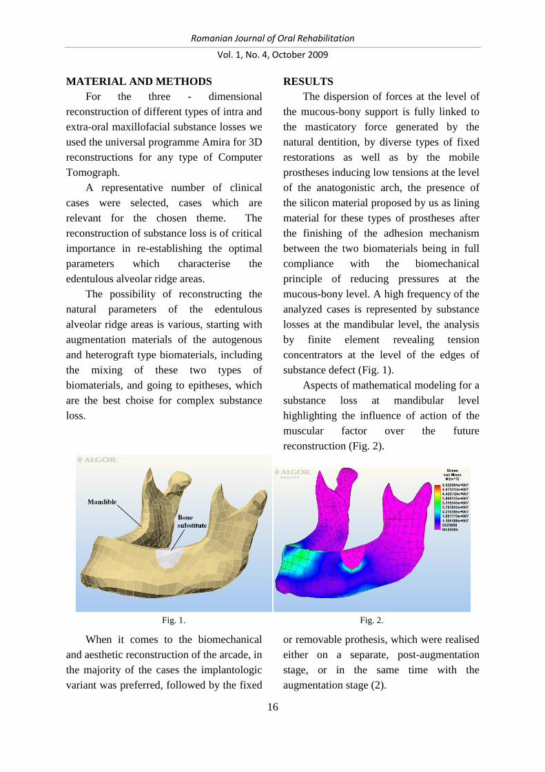
substance defect (Fig. 1).
Aspects of mathematical modeling for a
substance loss at mandibular level
highlighting the influence of action of the
muscular factor over the future
reconstruction (Fig. 2).
Fig. 1. Fig. 2.
When it comes to the biomechanical
and aesthetic reconstruction of the arcade, in
the majority of the cases the implantologic
variant was preferred, followed by the fixed
or removable prothesis, which were realised
either on a separate, post-augmentation
stage, or in the same time with the
augmentation stage (2).
Romanian Journal of Oral Rehabilitation
Vol. 1, No. 4, October 2009
17
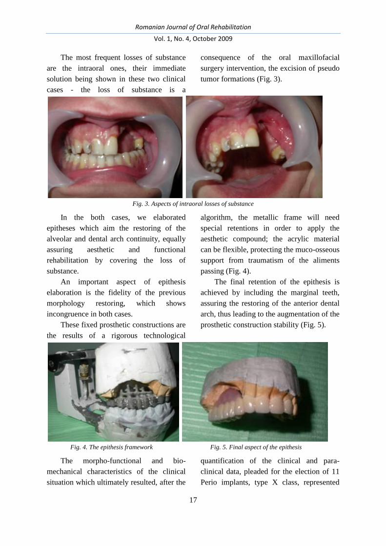
The most frequent losses of substance
are the intraoral ones, their immediate
solution being shown in these two clinical
cases - the loss of substance is a
consequence of the oral maxillofacial
surgery intervention, the excision of pseudo
tumor formations (Fig. 3).
Fig. 3. Aspects of intraoral losses of substance
In the both cases, we elaborated
epitheses which aim the restoring of the
alveolar and dental arch continuity, equally
assuring aesthetic and functional
rehabilitation by covering the loss of
substance.
An important aspect of epithesis
elaboration is the fidelity of the previous
morphology restoring, which shows
incongruence in both cases.
These fixed prosthetic constructions are
the results of a rigorous technological
algorithm, the metallic frame will need
special retentions in order to apply the
aesthetic compound; the acrylic material
can be flexible, protecting the muco-osseous
support from traumatism of the aliments
passing (Fig. 4).
The final retention of the epithesis is
achieved by including the marginal teeth,
assuring the restoring of the anterior dental
arch, thus leading to the augmentation of the
prosthetic construction stability (Fig. 5).
Fig. 4. The epithesis framework Fig. 5. Final aspect of the epithesis
The morpho-functional and bio-
mechanical characteristics of the clinical
situation which ultimately resulted, after the
quantification of the clinical and para-
clinical data, pleaded for the election of 11
Perio implants, type X class, represented

Romanian Journal of Oral Rehabilitation
Vol. 1, No. 4, October 2009
18
another cases, very important for this
subject. The distribution of the
implantations in the two dials was divided
in 5 implants with a diameter of 3,3 and 4
mm, with lengths of 10, 11,5, 13 and 15
mm, with the full consensus of the
dimensions of the bone support and of the
organic structures substituted in the dial
1:14, 15, 16, 17, 18. At the second dial’s
level 6 implantations were applied, having
the diameter of 3,3 and lengths of 10, 13, 15
mm, corresponding to the dental elements
22, 23, 24, 25, 26, 27 (Fig. 6).
Fig. 6. The radiological aspects before and after implantation
The optimal results were obtained
through the rigorous compliance with the
bio-mechanical principals and the
correlation of the increasing method in the
same session with surgical insertion of the
implantations, based on the usage of the
Cerasorb bio-material, having a granulation
of 150-500 µm, with an inorganic, that
reunites the derivatives of the calcium and
phosphate, with a high degree of bio-
compatibility (Fig. 7).
Fig. 7. Aspects of augmentation with Cerasorb
The confirmation from the images of
the clinical and para-clinical jaws offer the
radiography of a deficient jaw prosthetic
field, the remaining odontho-parodontal
elements being characterized by negative
clinical and biological parameters. The
degree of the bone absorption, which stood
at the basis of the pro-prosthetic preparation
stage, will offer the optimal for the
prosthetic bed’s design (3).
An important aspect in the case’s
success was represented by the increase of
the ridge in the frontal zone, with the help
of the Bio-Oss and of the Titan-made
membrane. The manual labour was done at
the same time with the implant, the

Romanian Journal of Oral Rehabilitation
Vol. 1, No. 4, October 2009
19
intervention pleading for the advantage of
the limitation for the surgical timing.
The balance between success and
failure had relatively limited lines, but for
the present case the age and the reaction of
the tissue were in favour of the post-
operation evolution.
The Bio-Oss is substitute of the natural
bone, osteo-inductiv that leads to the
controlled bone increase at the level of the
prosthetic fields characterised by a
parodonthic affection or by substance losses
at the bone level (Fig. 8). This therapeutic
selection was based on the inducement of
the bone crest at the situs’ level, where it
was transplanted (4). This decision was
correlated with the application of the metal
membrane, which is going to adapt at the
future volume of the crest (Fig. 9).
Fig. 8. Aspects of augmentation with Bio-Oss Fig. 9. Aspects of Titan mesch in rehabilitation of
frontal ridge
The immediate prosthetic was an
important stage after the implant and
increase, the final prosthetic offering the
facial harmony and the confidence of the
patient itself (5).
CONCLUSIONS
1. The intraoral losses of substances is a
sever mutilation and its elective immediate
treatment indication is the elaboration of
epitheses.
2. The importance of the substance loss
rehabilitation – a stage which is precedent
or concomitent to that of implantologic
therapy – is reflected in the appreciation of
the resorption and atrophy process on the
edentulous crest level and this appreciation
has a definitory influence upon
accomplishing the final stage of the clinical
case.
3. The mathematical modeling of the
real clinical situations offers optimal data to
choose the treatment solution fully
compliant with the parameters
characterizing the substance loss, the type of
biomaterial involved in the structure of the
prosthetic substitute, and in the case of
maxillofacial prostheses an important role is
played by the fixing means.
REFERENCES:
1. Forna N, Burlui V. Clinical Guidelines and Principles in the Therapy of Partial Extended Edentation. Iaşi:
Ed. Apollonia, 2001, 470-477.
2. Norina Forna, Evaluarea Starii De Sanatate Afectate Prin Edentatie, Editura Demiurg, 2007 ISBN 978-
973-152-017-9, Carte edtitata cu sprijinul Autoritatii Nationale pentru Cercetare Stiintifica, 25-26

Romanian Journal of Oral Rehabilitation
Vol. 1, No. 4, October 2009
20
3. Norina Forna, Magda Antohe, Reabilitarea Pierderilor De Substanta Editura Demiurg, 2007, ISBN 978-
973-152-035-3, Carte edtitata cu sprijinul Autoritatii Nationale pentru Cercetare Stiintifica, 180-186
4. Büchter A, Kleinheinz J, Wiesmann HP, Jayaranan M, Joos U, Meyer U. Interface reaction at dental
implants inserted in condensed bone. Clin Oral Implan Res 2005; 16: 509–517.
5. *** www.clinical-house.com
6. Sethi A, Kaus T, Sochor P. The use of angulated abutments in implant dentistry: five-year clinical results
of an ongoing prospective study. Int J Oral Max Implan 2000; 15: 801–810.
7. Eger DE, Gunsolley JC, Feldman S. Comparison of angled and standard abutments and their effect on
clinical outcomes: a preliminary report. Int J Oral Max Implan 2000; 15: 819–823.

Romanian Journal of Oral Rehabilitation
Vol. 1, No. 4, October 2009
21
FACE TRANSPLANTS: ETHICAL AND SCIENTIFIC STAKES
Philippe Pirnay
Pierre Fochard Academy, National Academy of Dental Surgery, Paris, FRANCE
Abstract: Since the first transplant of the mandible followed by the nose, the mouth and the chin, only some
other rare interventions of face transplants were realized in France and across the world. To the complexity of
the surgical operation, the ethical and deontological aspect that these transplants raise is added. The right to
transplant a jaw, teeth, or mouth removed from a corpse on another human being quickly evolves; it takes into
account the responsibility of the surgeon, the consent of the donor and his family and that of the sick collector.
Keywords: facial allotransplant, facial graft, composite tissues allograft, microsurgery, functional results,
medical ethic
INTRODUCTION
The grafts or organ transplants
represent an ancient dream of humanity.
However, we had to wait until the middle of
the 20th
century for the organ transplants to
be attempted with good chances of success.
The first kidney transplants go back to
1951-1952 while the first heart transplants
were attempted in 1968.
The techniques were largely
experimental and the rate of success was
small. The evolution of the surgical
techniques, a better knowledge of the
incompatibility systems of tissues, the
apparition of medicine which allow a better
control of the rejection phenomenon have
favored the boom of organ transplants.
Today, organ transplants have become
rather dull and they are multiplying. At the
same time, the need for organs has
increased. Grafts are scarce and the risks
can lead to organ trafficking. This crisis
explains the more and more frequent resort
to multiple organs sampling after death.1
1 Essor des greffes, CD permanant de bioéthique et
biotechnologie, Ed. Législatives, 2009
Nevertheless, the face transplants
through allotransplant remains a surgical
treatment which differs dramatically from
the other grafts…It’s about sampling tissue
and vascular and nervous elements from the
donor in cerebral death and grafting them on
a receiver who presents a maximum risk of
rejection, imposing an immunodepressant
treatment for his/her whole life.
Only a few cases of face transplants in the
world…
In January 2003, the daily Figaro
disseminated the information that in Rome a
surgeon had successfully accomplished a
mandible graft on an 80 years old man who
had mouth cancer. The patient would die
few days later. 2
In November 2005, the teams of
Amiens (France) with the professors
Bernard Devauchelle, Testelin and
Dubernard accomplished the first partial
face transplant in the world (graft of the
triangle comprised of the nose and the
mouth) on a 38 years old woman who had
been bit by a dog.
2 Le Figaro, 20.01.2003, p. 12.
Romanian Journal of Oral Rehabilitation
Vol. 1, No. 4, October 2009
22
In April 2006, a bear bit a Chinese
hunter’s face and he subsequently received
a partial face graft comprising a cheek, the
upper lip, the nose and an eyebrow. This
patient died later on.
In January 2007, the team of Pr.
Laurent Lantiéri from Créteil (France) made
the second face transplant, after a 15 hours’
surgery. The patient, aged 27, suffered from
a severe form of the Von Recklinghausen
disease, an incurable pathology that may
deform the face, in its most severe forms.
The patient suffered a graft of the nose-
mouth-chin-cheeks.
In March 2009, the third face transplant
in France was carried out for a 28 years old
patient disfigured by a gunshot. A great part
of the dead donor’s face was grafted as well
as the bone for the repair of the upper jaw.
In April 2009, the team of Pr. Laurent
Lantiéri accomplished simultaneously a
graft of a part of a face (the four eyelids, the
hairy skin to the nape of the neck, the
cheeks and the two ears) and of the two
hands. This intervention required 40
surgeons and 30 hours of surgery. The
eyelids graft was also presented as a world
first. Aged 30, the receiver had suffered
burns following an accident in 2004. 3 The
patient died later on from a cardiac arrest
during the operation aiming at stopping the
infection of the face, which occurred a few
weeks after the graft.
In August 2009, a man whose jaw had
been blown away by a gunshot suffered a
transplant. He had waited for this
intervention for four years.
On November 27th
, 2009, a new
transplant was accomplished by the team of
3 Synthèse de presse bioéthique, Double greffe du visage et
des mains , www.genethique.org, 7 avril 2009
Pr. B. Devauchelle on a seriously burnt man
as a result of an accidental explosion during
a pyrotechnical manifestation in May 2008.
In total, in the whole world, only two
other face transplants were made in the
United States (Fig. 1), another one in China
and one in Spain.
All the current indications of the face
reconstruction concern ballistic
traumatisms, serious burns, face cancers and
certain congenital anomalies. The patient
needs to display a dilapidation
corresponding to a necessity to make a
graft, namely an injury that cannot be
reconstructed by the means of traditional
surgery. 4
MATERIAL AND METHOD5
The allotransplant of composite tissues
(ATC) is a surgical technique described
since 1998. It comprises the sampling of
tissues and of vascular and nervous
elements belonging to a donor in the state of
brain death. The immunitary incompatibility
between the donor and the receiver requires
the use of a life long immunodepressant
treatment in the case of the receiver, whose
potential side effects are the predisposition
to infections, high blood pressure, diabetes
and lymphoproliferative malign disorders
such as skin cancers.
The allotransplant of the entire or only
a part of the face shows a maximum risk of
rejection, which can be estimated at 10%
during the first year, and a risk of chronic
rejection ranging from 30 to 50% in the case
4 Gérard S., Greffe du visage: Il est encore trop tôt pour se
prononcer, www.lexpress.fr 30/03/2009 5 Report of CCNE, Comité consultatif national d'éthique.
The allotansplant of composite tissues (ATC at the level of
the face). Total or partial transplant of a face. Avis no 82
2004.

Romanian Journal of Oral Rehabilitation
Vol. 1, No. 4, October 2009
23
of grafted people between 5 to 10 years
after the transplant.
The first French transplant was given
large media publicity and its realization was
presented by the teams of Pr. Dubernard
and Deveauchel.
Fig. 1. Montage photo de Connie Culp avant et après sa greffe de 80% du visage, subie en décembre
2008./CLEVELAND CLINIC / AFP
" 6 On May 31
st, 2005, at Amien
(France), a patient aged 38 showed,
following a dog bite, a vast substance loss
of all the soft parts of the centro-facial
region. Exposing largely her maxillofacial
skeleton, the gums and the dental arches,
the amputation concerned the distal half of
the nasal pyramid, the upper and lower lips,
the chin and extended laterally to the
cheeks, especially in the right jugal region
rather than the left one. From a functional
point of view, the patient was unable to
drink, eat or talk. Although her facial
6 Lengelé B.-G, Testelin S., Dakpe S., CartonS. , Beziat, L.
Badet J-L., Morelon E., Dubernard J-M., Devauchelle B.,
«Greffe de visage»: views on the first partial composite
allotransplant of the face, Annales de chirurgie plastique
esthétique,Volume 52, numéro 5 pages 475-484 (octobre
2007)
expressivity was restraint only to the
frontal-orbital region, the initial clinical
examination showed that she had kept intact
the proximal stumps of her lifting muscles of
the lip and of the zygomatic.
The request for the authorization of an
allograft had been filed to the competent
health French authorities: l'Agence
française de sécurité sanitaire des produits
de santé (Afssaps), l'Agence de la
biomédecine (ABM) and Le Comité
consultatif de protection des personnes dans
le cadre des recherches biomédicales
(CCPPRB) which gave their consent and
approved the proposed protocol.

Romanian Journal of Oral Rehabilitation
Vol. 1, No. 4, October 2009
24
All the steps were taken to ensure that
the patient is informed clearly and
completely with regard to the surgical
intervention, to the psychological risks and
the constraints related, particularly on the
immunological plan, especially the need to
follow a life long immunodepressant
treatment as well as to the publicity inherent
to such an intervention. A long-term
psychological post-surgery follow-up was to
be expected. The patient expressed her free
and clear consent. 7
During the waiting period for a
compatible graft, the contour of the graft to
be collected was modeled on the computer.
(Fig. 2) and all the phases of the
intervention were studied on anatomical
subjects in order to determine the optimum
plans of dissection and perfect integrity of
the vascular network and of all tissues
involved in the transplant.
Fig. 2. Description schématique du greffon
transplanté lors de la 1ere greffe de visage.
(www.canalacademie.com/Semaine-speciale-
Recherche-sante.html)
7 The French Agency of Sanitary Safety of the products
and the Agency of Biomedicine wish to remind of the
different stages of expertise previously conducted for the
face partial graft, made on November 27 2005.
http://www.agence-biomedecine.fr/article/361
After six months of waiting, a potential
donor was found. Aged 48, in the state of
brain death, her skin texture and color
corresponded to that of the patient. She also
had the same blood type and five HLA
antigens out of six.
The facial graft was collected following
the protocol established in the laboratory
(Fig. 3). The exact contour of the tegument
surface was drawn on the face in conformity
with the substance loss of the receiver. After
that, the facial vessels were exposed
towards the lower rim of the mandible with
the marginal branch of the facial nerve.
Fig. 3. Crédit : REUTERS
Laterally, after having reached the
proximal insertions of the peri-oral lifting
muscles, the dissection showed all the facial
nerve bundles on the right and on the left,
followed to the surface by the fascia
maseterina and sectioned in their
emergence from the parotidian lodge.
Deeper, after the division of the buccinator
muscle, the facial mask was eventually lifted
on a sub perinosteum plan, easily exposing,
coming out from the foramina infra-orbitary
and the mental, the sensitive terminal
bundles of the maxillary and mandibular
nerves, elongated by endo-bony dissection
in order to obtain satisfactory stumps of

Romanian Journal of Oral Rehabilitation
Vol. 1, No. 4, October 2009
25
microsurgical coaptation. The final frontal
cut of the nasal and circumferencial mucosa
and of the oral vestibule were enough to
render the transplant autonomous.
The surgical intervention lasting for 16
hours, the patient wakes up peacefully and
discovers, starting the next day, impatiently
and fearlessly, her new face.
In order to prevent the rejection of the
graft, she received an immunodepressant
treatment and a triple anti-infectious
prophylaxis.
After a brief transitory edema, the facial
graft healed very fast, with no sign of
sufferance or necrosis. No acute rejection
occurs in the critical period of the first 12
days.
On the 18th
post-surgery day, however,
an erythema occurring simultaneously on
the facial transplant and on the sentinel graft
leads to a suspicion of rejection which is
easily controlled by a drug re-orientation.
The patient is able to eat four days after
the intervention. During the sixteenth
month, the patient shows a complete active
and passive labial occlusion (Fig.4). In
parallel, her speech impediment ameliorates
significantly and, during the eighteenth
month, she displays a spontaneous and
symmetrical smile. A second phenomenon
of rejection appears later on. As in the case
of other face transplants, the two
manifestations of rejection were dealt with
and controlled.
Fig. 4. Isabelle Dinoire, première greffée, en 2005 et aujourd’hui. AFP
DISCUSSIONS
The face transplant was considered as a
serious therapeutic option during the
Congress of Plastic Surgical Research
Council from Boston (USA) in April 2002.
However, the report of the Royal College,
triggered by the publicity made around the
declarations of Doctor Butler in December
2002 in front of the British Association of
Plastic Surgeons, expresses the deepest
concerns and underlines the fact that it is
not simply a surgical reparatory technique
like any other.
In France, Le Comité Consultatif
National d'Ethique concluded on February
6th
, 2004:

Romanian Journal of Oral Rehabilitation
Vol. 1, No. 4, October 2009
26
«Facial transplants are not organ
transplants and they are far from limb
transplants. That is why they shouldn’t be
attempted as long as more complete
researches are made on the procedures
themselves and as long as we fail to have
the elements that allow us to appreciate
correctly the risks accompanying this type
of graft and to validate the results. (…)
A complete facial transplant (ATC)
doesn’t make too much sense at the moment.
The question is not raised from a medical or
technical point of view. The possibility of a
partial ATC reconstructing the mouth –
nose triangle which grants the face a
certain new morphological identity still
belongs to the domain of research and high-
risk experimentation. It shouldn’t be
presented as a future ideal and accessible
solution for the painful problems related to
the face alteration. In the event such
possibility is envisaged, it should be done
within a strict multi-disciplinary and multi-
centric protocol, approved by the
Etablissement Français des Greffes or other
instances with the same attributions. »8
We shall also note that the National
Council for the Order of Doctors had made
public, on December 15, 2005, “a solemn
reminder to the ethics, deontology and the
law” with regard to the partial face
transplant. The organism admitted that it is
about “an undeniable surgical
performance” which “sparks off a legitimate
hope for other people”. However, the
council “deplores that a premature,
uncontrolled communication has led to an
augmentation of the technical realization
over the respect due to the patient, to the
8 CCNE avis n°82 du 6 février 2004, www. ccne-
ethique.org
person of the donor, to his/her generosity
and that of the family, contrary to the
professional rules stipulated by the
deontological code”.9
These interventions have therefore
aroused ethical disputes:
Ethical questions:
For the receiver:
- The face is unique, it fundamentally
characterizes an individual. The patient who
first benefited from a face transplant stated
in 2009: “before the transplant I represented
nothing. Six months without a face. A mask
covered my face. You cease to exist when
you don’t have a face”.10
Can a face be
transmitted to somebody else? How can one
live in somebody else’s skin? Will the
receiver be able to accept this new
appearance which will turn him/her into
somebody else? The patient would never
discover the traits of his/her former face,
therefore, what can be done if the patient
doesn’t accept this new face? How will the
patient deal with the way the others perceive
him/her? How to deal with the
psychological difficulties? 11
- Does the state of the patient really allow
him/her to consent to all risks, hazards,
constraints related to this difficult surgical
intervention?
- The patient will have to deal with the
constant risk of rejection. These grafts
weigh against the real prejudice of the
patient and the prejudice generated by the
treatment. Can we accept the fact that the
9 Conseil National de l’Ordre des médecins, avis du 15
décembre 2005,www.conseil-national.medecin.fr 10 Dinoire I, Les défis de la science Paris Match 30
décembre 2009 p:84-85 11 CCNE avis n°82 du 6 février 2004, www. ccne-
ethique.org

Romanian Journal of Oral Rehabilitation
Vol. 1, No. 4, October 2009
27
patient “who must take five pills in the
morning, one at noon and five in the
evening”"10
should make out of this a daily
re-education and that the patient should be
treated all life long with immunodepressants
with serious side effects?
- The secret of the intervention cannot be
kept for a long time. As soon as the press
conference12
organized by the medical teams
was over, photographs were made public.
The image of the body or the corporal
appearance is classically protected by the
human right13
which aims at providing the
respect to the dignity of the human person.14
Are we heading for a “spectacle surgery”
where the grafted patient will allow
him/herself to be photographed for money?
- As far as the small number of donors is
concerned; who should suffer the
transplant? The young or the old? The
employees or the retired? The people who
suffered deep burns or those with cancer?
The choice will be morally acceptable if the
principles of this choice are universal
(identical and invariant for all), irrevocable
and public.15
- At last, a question of social legitimacy of
the organ donation is raised: isn’t it more
advantageous to invest more in the
prevention of certain serious maladies rather
than to compensate them belatedly by the
transplant?
Other questions are also raised for the
donor and his/her family. They refer to the
12 Pirnay P., De l'éthique au droit et du droit à l'éthique
Ed hospitalières, soumis 2010 13 Hassler T., note sous TGI Paris, 15 avr. 1987, D. 1987,
Jur. p. 551. 14 Corpart I., La dignité de la victime photographiée face à
la liberté de la presse, note sous Civ. 2ème 4 novembre
2004, D. 2005, 696 ; JAC n° 49, décembre 2004 15 Course of the Faculty of Medicine from Bichat –
Lariboisière, Greffes et transplantations d’organes, 2009
image of the corpse, the definition of death,
to consent and the principle of non-
merchantability of the body16
, to the issue of
the anonymity of the organ donors.
CONCLUSION
Beyond the moral question, this
technique represents the hope of all
mutilated people. Ugliness is not admitted
in our society. Without a face, a person is
nothing but a curious animal.
In the case of the first transplant on a
woman, can we say that it wouldn’t have
been vital to the extent that particular
woman could no longer cope with her
destructed face? It is not about talking of
this surgery, experimenting, but giving a
therapeutic answer to a patient in pain, who
nowadays is thankful to the medical team
and about opening other ways for progress.
Depending on the grafted organs, the
questions are raised differently. Numerous
people who had face transplants are dead or
have attempted to commit suicide. Who
remembers that the first project of cardiac
cathererism was rejected as contrary to
ethics towards 1935 by an eminent scientific
society?17
Otherwise, it stands as an
inspiration today for all heart surgeries.
History therefore has the duty to
validate a technique, to warrant its ethics
and morality and to grant it the right of legal
existence.
16 Read on the subject the article of Nancy Scheper-
Hughes, anthropologist at the University of california,
Berkeley (United States), Le commerce du corps en
«pièces détachées», www.unesco.org 17 Bernard J., Les conséquences morales de la révolution
biologique, CD - Encyclopedia universalis, 2000

Romanian Journal of Oral Rehabilitation
Vol. 1, No. 4, October 2009
28
REFERENCES
1. CD permanant de bioéthique et biotechnologie. Essor des greffes, Ed. Législatives, 2009
2. Le Figaro, 20.01.2003: 12.
3. Synthèse de presse bioéthique, « Double greffe du visage et des mains », www.genethique.org, 7 avril
2009.
4. Gérard S, Greffe du visage: Il est encore trop tôt pour se prononcer, www.lexpress.fr 2009 .
5. Rapport du Comité consultatif national d'éthique, L'allotansplantation de tissus composites (ATC au niveau
de la face). Greffe totale ou partielle d'un visage. Avis no 82 2004.
6. Lengelé B-G, Testelin S, Dakpe S, Carton S, Beziat J-L, Badet L, Morelon E, Dubernard J-M,
Devauchelle B. «Greffe de visage»: regards portés sur la première allotransplantation composite partielle
de la face. Annales de chirurgie plastique esthétique 2007; 52,5: 475-484.
7. L’Agence française de sécurité sanitaire des produits de santé et l’Agence de la biomédecine tiennent à
rappeler les différentes étapes et expertises qui ont été menées préalablement à la greffe partielle de la face,
réalisée le 27 novembre 2005. http://www.agence-biomedecine.fr/article/361.
8. Conseil National de l’Ordre des médecins, avis du 15 décembre 2005,www.conseil-national.medecin.fr.
9. Pirnay P., De l'éthique au droit , du droit à l'éthique, Ed. Hospitalières, soumis 2010.
10. Hassler T, note sous TGI Paris, 1987, D. 1987, Jur.: 551.
11. Corpart I, La dignité de la victime photographiée face à la liberté de la presse, note sous Civ. 2ème 2004,
D. 2005, 696 ; JAC n° 49, décembre 2004.
12. Dinoire I, Les défis de la science, Paris Match 2009; 30 décembre:84-85.
13. Commission Nationale de Santé Publique et de Bioéthique du Grand Orient de France. Humanisme 2007;
276: 71.
14. Scheper-Hughes N, Le commerce du corps en «pièces détachées», www.unesco.org.
15. Bernard J, Les conséquences morales de la révolution biologique, CD - Encyclopedia universalis, 2000.

Romanian Journal of Oral Rehabilitation
Vol. 1, No. 4, October 2009
29
TELESCOPING IMPLANT PROTHESES WITH GALVANO
MESOSTRUCTURES
Bratu Emanuel1, Bratu Dorin
2, Borsanu Ioan
3
1Department at the Clinic Of Oral Implantology, UMF ”Victor Babes” Timisoara
2Department at the Clinic Of Prosthodontics, UMF ”Victor Babes” Timisoara
3Private Practice at Prof. Dr. Bratu Dental Clinic, Timisoara, ROMANIA
Abstract: This article will describe the technique for the fabrication of the telescoping implant prostheses with a
very precise fit, achieved by luting intraorally the galvano mesostructures. The main result is the strain
reduction, similar to the cemented fixed prostheses (12, 21). By the use of galvano-telescopic copings, the
advantages of retrievability can be combined with the improved fit of a cemented superstructure. The telescopic
system permits simple retrievability for peri-implant hygiene, and repair procedures, if necessary. The precision
of implant superstructures is determined by the entire clinical and laboratory fabrication process. Errors may
occur during the impression making, fabrication of the definitive cast, casting of the framework and ceramic
veneering (14). Also, after extended edentulous periods, the replacement of hard and soft tissues is often
necessary for esthetic or phonetic reasons. To compensate for the resorptive processes of the maxilla, a buccal
flange may be required for adequate support of the lips and the facial profile. In these situations, a removable
prosthesis may be given preference over a cemented fixed prosthesis or a screw retained prothesis.
Key words: telescopic implant prostheses, galvano-telescopic copings.
INTRODUCTION
Dental implants are made to be placed
in the jaw bones to give prosthetic
restorations stability and retention. The
passive-fit of the implant superstructures is
a prerequisite for the dental implant
prostheses (2, 9). The lack of fitting of the
frameworks to the implant abutments
generates stress in two directions: in the
superstructures (ceramic fissure) and
connections (loosening and fractures of the
screws) (1) and on the other hand it
compromises the implant/bone interface
with the result of bone loss and eventually
implant failure. Because of the
discrepancies of the screw-retained
restorations, cementation of implant
frameworks has been brought forward.
However, the cement-retained prostheses
have also disadvantages, including the lack
of retrievability in case of the implant
failure.
MATERIAL AND METHOD
Altrough telescoping restaurations was
first proposed americans (18) the
application of galvano-electroforming for
telescoping units is primarily described in
the German language literature. The
galvano process is an electro-deposition of
metal ions of an electrolyte solution to a
negatively charged cathode, resulting in a
pure metal structure on the cathode surface.
It was introduced in the early 1960s for the
fabrication of inlays and onlays. Only after
the development of automatic systems with
cyanid-free gold-sulfide baths could the
electro-forming procedure be used in
clinical practice (6, 21).

Romanian Journal of Oral Rehabilitation
Vol. 1, No. 4, October 2009
30
The main advantage of the
galvanoformed coping is their retention,
which is a non-friction one. The retention is
an adhesion between the galvano coping
and the prosthetic abutment, through the
saliva pellicle. If one tries to take the
prothesis off the prosthetic field, a vacuum
effect is created because the saliva is
inextensible and the restoration remains in
place. A similar effect is obtained by the full
denture’s „suction”. This type of retention is
also known as hydraulic retention (3) or
adhesive retention (fig.1). This kind of
hydraulic retention can be obtained only
when the fitting of the two structures (the
galvano coping and the prosthetic abutment
of the implant) is very precise and the
marginal fit is between 20-30 µm.
Fig. 1. Hidraulic retention
TECHNIQUE
A number of 3 edentulos patients were
reabilitated using this technology (2 males
and 1 female with ages betwen 49 and 60
years) – Table 1.
Table 1. The patients characteristics
Name Age Sex Diagnostic Tipe of restauration
M.I. 54 M Maxilary subtotal edentation Partial fixed-removble
prothesis
P.I. 60 M Maxilary complete edentation Fixed-removable restauration
C.P. 49 F Mandibulary complete edentation Fixed-removable restauration
Make the indirect impression after
second-stage surgery (Fig.2 a,b).Fabricate a
stone cast of the artificial tooth arrangement
as a guide for the dimensions of the
framework and to preserve the desired
position of the artificial teeth. Carve the
prosthetic abutments with a 2-degree
milling titanium cutter in the planned path
of insertion, and place the abutment
shoulder at the soft tissue level. (Fig.2 c,d).
Fabricate an acrylic resin (Pattern Resin,
GC) positioning jig over the completed
abutments.Close the base of the abutments
and the screw access openings with
autopolymerizing acrylic resin (Pattern
Resin, GC) and connect the abutments to an
insulated wire (AGC Contact rod, Wieland).
Carefully apply a thin layer of silver
conductor (AGC Elektroforming System,
Wieland) to the abutments and the acrylic
resin of the screw openings. Perform the
electroforming process in the galvanobath.
Create copings with a thickness of 0.3 mm
(Fig. 2 e).
Detach the galvano copings from the
abutments and remove the silver connector
with 25% nitric acid. Position the abutments
and the galvano copings on the working cast
and number the copings (Fig.2 f).
Fabricate the frame for the prosthesis
from a rigid nonprecious cobalt-chromium
alloy. Allow enough space (approximately

Romanian Journal of Oral Rehabilitation
Vol. 1, No. 4, October 2009
31
0.1 mm) between framework and galvano
copings for passive fit and the luting agent.
Verify the clearance between the framework
and the galvano copings.(Fig.2 g).Silanize
(Rocatec, 3M ESPE) the framework and
copings to prepare for intraoral
luting.Attach the abutments to the implants
by using the acrylic resin positioning jigs to
verify the abutment position. Torque the
abutments afterwards as recommended by
the manufacturer. Place the galvano
secondary copings intraorally and verify the
passive fit of the tertiary framework. Bond
the secondary copings intraorally to the
framework with composite resin (AGC
Cem; Wieland). Verify the final wax
arrangement on the cast (Fig.2 h) and
intraorally.Complete the acrylic resin
portions of the denture with auto-
polymerizing polymer. Finish and polish the
prostheses in the conventional way. (Fig.2
i)Attach and torque the abutments to the
implants as previously described. Cover the
abutment screws with a thin layer of white
gutta-percha material for retrievability.
Place the completed telescoping denture
intraorally (Fig. 2 j). Verify esthetics,
function, and appropriate retention. Instruct
the patient on the use and maintenance of
the protheses.
A B
C D
E F

Romanian Journal of Oral Rehabilitation
Vol. 1, No. 4, October 2009
32
G H
I J
Fig. 2. Healing after surgery (a); indirect impression (b); master cast with gingival mask(c);
prosthetic abutments prepared at 2 degrees and the silicone markup(d);galvano forming bath with the
prosthetic abutments placed(e);numbering of the galvanoformed copings (f);
the galvanoformed copings placed in the metal cast (g);
the wax probe (h); final restoration-mucozal aspect (i); and extraoral aspect(j)
RESULTS
The described technique permits the
fabrication of a retrievable implant-
supported denture with a passive fit
comparable to cemented restorations.
Retrievability allows simple repairs and
modifications of the acrylic resin dentures
and easy access for periimplant hygiene.
The telescopic design with intraoral luted
galvano copings provides excellent
prosthesis retention and stability.
All the patients were pleased with the
quality of the restaurations witch are
excelent looking after about one year of use.
DISCUSSIONS
The correlation between misfit of
implant-supported prostheses and an
increased rate of mechanical failures is
established, but the degree of fit accuracy
necessary to prevent mechanical
complications remains unclear (1).
Although cementation of implant prostheses
can compensate for fit discrepancies and
cemented metal-ceramic prostheses can
have esthetics superior to metal-resin
dentures, fixed dental prostheses may not be
indicated in all situations. The high costs of
full arch metal-ceramic restorations are a
limitation for many patients. In addition,
porcelain failures remain a common
problem. Esthetics and durability of
adhesive systems for intraoral porcelain
repair may not be satisfactory, and the
removal and laboratory repair of a cemented
metal-ceramic prosthesis is a potentially
hazardous and costly procedure.
For patients with extensive residual
ridge resorption, replacement of hard and
soft tissues with a removable prosthesis may

Romanian Journal of Oral Rehabilitation
Vol. 1, No. 4, October 2009
33
be considered a more suitable option than a
cemented restoration. No screw access
openings interfere with occlusal surfaces,
improving esthetics and occlusion compared
to conventional screw-retained prostheses.
The thin galvano copings allow adequate
space to be completely covered with the
framework, which in association with the
silanization procedure allows a durable
connection.
Altrough telescoping using
galvanoformated copings is relativley new
in implantology it is starting to accaparate
implantology because of the major
advantages witch it offers: passove fit, high
mechanic resistance, permise for good
hygene and the posibility of numerous
adjustments
CONCLUSIONS
1. With this kind of restaurations it is
possible to obtain the Passive Fit
Adaptation witch is a decisive factor
for implant suported protheses;
2. The restaurations are removable
witch gives the patients the
posibility for very good oral hygene,
the medic posibility to work around
the implants and technicians to
correct whatever failure may ocour
3. We recomand to extend the use of
these restaurations in our country
because of the numerous advantages
even trough the cost is a little higher.

Romanian Journal of Oral Rehabilitation
Vol. 1, No. 4, October 2009
34
REFERENCES
1. Bragger U, Aeschlimann S, Burgin W, Lang NP. Biological and technical complications and failures with
fixed partial dentures (FPD) on implants and teeth after four to five years of function. Clin Oral Implants
Res 2001; 12: 26-34.
2. Branemark PI. Osseointegration and its experimental background. J Prosthet Dent 1983; 50: 399-410.
3. Bratu D, Bratu E, Antonie S. Restaurarea edentațiilor parțiale prin proteze mobilizabile. Ed Medicală, Buc
2008.
4. Bratu D. Scurtu S- Gallavoformarea Partea a II-a. Proteze parțiale fixe. Revista Națională de Stomatologie,
vol I, nr. 3, 2003, 142-147
5. Chee W, Felton DA, Johnson PF, Sullivan DY. Cemented versus screw-retained implant prostheses: which
is better? IntJ Oral Maxillofac Implants 1999; 14: 137-41.
6. Dolger J, Gadau C, Rathmer R. Treatment behavior and complete-mouth rehabilitation using AGC crowns:
a case report. IntJ Periodontics Restorative Dent 2001; 21: 373-9.
7. Erpenstein H, Borchard R, Kerschbaum T. Long-term clinical results of galvano-ceramic and glass-ceramic
individual crowns. Prosthet Dent 2000;83:530-4.
8. Geering AH, Kundert M, Kelsey CC. Complete denture and overdenture prosthetics. New York: Thieme
Medical Publishers Inc; 1993. p. 110-6, 204-5.
9. Greven B, Luzpke M, von Dorche S: Telescoping implant protheses with intraoral luted galvano
mezostructures to improve passive fit. The Journal of Prosthetic Dentistry vol 98, 3, 2007, 240-244.
10. Guichet DL, Caputo AA, Choi H, Sorensen JA. Passivity of fit and marginal opening in screw- or cement-
retained implant fixed partial denture designs. IntJ Oral Maxillofac Implants 2000; 15: 239-46.
11. Hebel KS, Gajjar RC. Cement-retained versus screw-retained implant restorations: achieving optimal
occlusion and esthetics in implant dentistry. J Prosthet Dent 1997; 77: 28-35.
12. Heckmann SM, Karl M, Wichmann MC, Winter W, Graef F, Taylor TD. Cement fixation and screw
retention: parameters of passive fit. An in vitro study of three-unit implant-supported fixed partial dentures.
Clin Oral Implants Res 2004; 15:466-73.
13. Kan JY, Rungcharassaeng K, Bohsali K, Goodacre CJ, Lang BR. Clinical methods for evaluating implant
framework fit. J Prosthet Dent 1999; 81: 7-13.
14. Karl M, Rosch S, Graef F, Taylor TD, Heckmann SM. Static implant loading caused by cast metal and
ceramic-veneered superstructures. J Prosthet Dent 2005; 93: 324-30.
15. Jaert I, Quirynen M, van Steenberghe, Darius P. A study of 589 consecutive implants supporting complete
fixed prostheses. Part II: Prosthetic aspects. J Prosthet Dent 1992; 68: 949-56.
16. Pietrabissa R, Gionso L, Quaglini V, Di Martino E, Simion M. An in vitro study on compensation of
mismatch of screw versus cement-retained implant supported fixed prostheses. Clin Oral Implants Res
2000; 11: 448-57.
17. Setz J, Diehl J, Weber H. The marginal fit of cemented galvanoceramic crowns. IntJ Prosthodont 1989; 2:
61-4.
18. Setz J, Kramer A, Benzing U, Weber H. Complete dentures fixed on dental implants: chewing patterns and
implant stress. Int J Oral Maxillofac Implants 1989; 4: 107-11.
19. Taylor TD. Prosthodontic problems and limitations associated with osseointegration. J Prosthet Dent
1998;79:74-8.
20. Wee AG, Aquilino SA, Schneider RL. Strategies to achieve fit in implant prosthodontics: a review of the
literature. IntJ Prosthodont 1999; 12: 167-78.
21. Wirz J, Hoffmann A. Electroforming in restorative dentistry: New dimensions in biologically based
prostheses. Chicago: Quintessence; 2000. p. 132-61, 238-67.
22. Zitzmann NU, Marinello CP. Treatment plan for restoring the edentulous maxilla with implant-supported
restorations: removable overdenture versus fixed partial denture design. J Prosthet Dent 1999;82:188-96.

Romanian Journal of Oral Rehabilitation
Vol. 1, No. 4, October 2009
35
PERIODONTAL ASPECTS IN CHILDREN AND ADOLESCENTS
WITH DOWN SYNDROME Vasilica Toma
1, A. Maxim
1, Adriana Balan
1, Diana Gheban
1,
Dana Cristiana Rotaru1
, Florina Filip2,Liliana Foia
3
Faculty of Dental Medicine 1Pedodontics Dept.,
2Family Medicine Dept,
3Chemistry and
Biochemistry of Oral Cavity Dept.
University of Medicine and Pharmacy „Gr. T. Popa” Iaşi
Abstract:
The Down syndrome is the most frequent genetic anomaly, presenting an incidence of 1/650 births (Winston,
2004). The individuals affected by DS frequently develop a form of aggressive periodontitis which affect both
temporary and permanent teeth (Saxen, 1977, Svantum and Gjermo, 1978 a.o.) and can lead to the precocious
expulsion of the teeth. Starting with these data, we tried to clinically evaluate the periodontal status in a group of
12 patients with DS, compared to the control group (which comprised 24 children without general diseases) and
establish on the basis of clinical indicators (QHI, PBI, CAL) the forms of periodontal disease. Our results
showed the preponderance of superficial chronic periodontits (66.67%) in the children with DS, followed by
gingivitis (33.3%), the aggressive forms (aggressive periodontitis) not being probably encountered due to the
fact that the group was too young for the juvenile stage and however the children were hospitalized in the dental
office of Sfânta Maria Policlinics, Iasi.
Key words: Down Syndrome, children, teenagers, clinical evaluation, periodontal disorders
INTRODUCTION The Down Syndrome – 21 Trisomy is
the first genetic disease described in 1866
by John Langdon Down. DS is the most
frequent genetic anomaly, presenting an
incidence of 1/650 births, being
characterized by an autosomal
transmission of the 21 chromosome
trisomy. The individuals affected by DS
frequently develop an aggressive form of
disease which affects both temporary and
permanent teeth (Cohen, 1961, Johnson şi
Young, 1973, Saxen, 1977, Svantum and
Gjermo, 1978 a.o.) and can lead to the
precocious expulsion of the teeth.
Starting with these data, we tried to
clinically evaluate the periodontal status
and to establish the forms of periodontal
diseases in a group of children with DS.
MATERIAL AND METHOD
For realizing that purpose we
constituted two groups of study:
I. Group I (control) – comprised 24
children with ages between 6-18 years old,
without general diseases, being under
dental treatment in the assistance requested
by the Infantile Dentistry Clinics Iasi,
Dept. of Pedodontics.
II. Group II (active) – comprised 12
patients with age comprised between 5-16
years old being in the evidence of
Neuropsychiatry – Genetic Diseases
Office and the Dental Office of Sfânta
Maria Policlinics.
The evaluation of the periodontal
status was performed in the groups of
children and adolescents through the
calculation of the following indexes of
diagnostic for periodontal disease:
The Quiqley and Hein coloured
bacterial plaque index
Papillary bleeding Index (Saxen and
Muhlemann)
The level of attachment loss (CAL)
evaluated through periodontal probing and
radiological exam.
All the evaluations of clinical indexes
were performed at the level of Ramfjörd
teeth, mesial sites. The evaluation of
plaque deposits were made with the help

Romanian Journal of Oral Rehabilitation
Vol. 1, No. 4, October 2009
36
of coloured plaque indexes Quiqley – Hein, as it follows:
QH = The sum of values for every tooth of B and L surfaces
The total number of examined surfaces
This index is evaluated on the basis of
scores:
0 – absence of plaque
1 – separated isles of plaque at the
level of cervical area
2 – fine band of plaque of up to 1 mm
height at cervical area
3 – band of plaque comprised
between 1 mm and 1/3 from the crown of
the tooth
4 – band of plaque comprised
between 1/3 and 2/3 from the tooth crown
5 – bacterial plaque which covers
more than 2/3 of the tooth crown
The intensity of inflammation was
appreciated through the intensity of
papillary bleeding (PBI index). Indicator
of gingival bleeding severity, it notes from
0 – 4 different progressive phases of
bleeding
0 = without bleeding
1 = punctiform blleding
2 = line/points, fine line of bleeding or
several points at gingival margin
4 = drop that fllows – massive
bleeding
The clinical attachment loss was
evaluated through periodontal probing
appreciating the depth of the pockets and
also the degree of recession, at the level of
every test tooth in at least 6 sites (B, L,
MB, ML, DL) the reference element for
evaluating the attachment loss was amelo-
cementar jonction (AML) expressing the
distance between the bottom of the
sulcus/pocket in millimetres.
The Rx exam, useful in appreciating
the importance of attachment loss was
performed on retro-dento-alveolar cliches
in isometric and orthoradial incidence or
through ortopantomography (OPT).
Comparisons were made between the two
groups for every calculated index, the
values for every patient were calculated
together for creating the average of the
group, using â common ground estimated
as standard of correction for the error of
unequal variances. Under this
circumstance the patients were in the
centre of the study and not the number of
sites.
The statistical differences between the
averages of the values of interest
according to the groups were tested using
the One-Way ANOVA test completed of
KRUSKAL-Wallis test (or Mann-
Whitney/Wilcoxon test) for the average
age and clinical values. The statistically
signifficant differences were considered
for the values of the level of significance
(p) lower than 0.05, according to a level of
confidence of 95%.
RESULTS
The bacterial plaque index
For evaluating the patients with Down
syndrome from the point of view of the
clinical indexes, their values were
compared to the values recorded in the
control group. In the analysis of every
clinical index the statistical indicators
which describe correctly the characteristics
of the group were calculated, comparing
them at the same time with the values of
the respective indicators of the control
group.

Romanian Journal of Oral Rehabilitation
Vol. 1, No. 4, October 2009
37
Table I. The statistical indicators of the Quiqley-Hein index of bacterial plaque (QHI) for the group
of patients with Down Syndrome
QHI
Group Average -95% +95% Std. Dev Variance
Std.
Error Min Max Q25
Median
Value Q75
Control 2.995 2.859 3.130 0.576 0.332 0.068 2.166 4.133 2.500 3.000 3.500
Down Syndrome 3.622 3.408 3.836 0.632 0.400 0.105 3.000 5.000 3.083 3.495 3.830
Total 3.204 3.077 3.330 0.663 0.439 0.064 2.166 5.000 2.660 3.000 3.660
Categ. Box & Whisker Plot:
Indicele de placa bacteriana IQH
Mean
±SE
±SD
2.995
3.622
Martor Sindrom Down
LOT
2.2
2.4
2.6
2.8
3.0
3.2
3.4
3.6
3.8
4.0
4.2
4.4
I QH
Fig.1. Average values of the bacterial plaque index in the control group (QHI) and the group of
patients with Down Syndrome
Clinical Attachment loss (CAL)
Table II. Statistical indicators of the attachment loss for the group of patients with Down Syndrome
CAL
Group Average -95% +95% Std Dev. Variance Sst Er. Min Max Q25
Median
Value Q75
Control 0.388 -0.067 0.842 1.077 1.160 0.220 0.000 4.000 0.000 0.000 0.000
Down Syndrome 1.417 0.562 2.272 1.346 1.811 0.388 0.000 3.500 0.000 1.750 2.500
Total 0.731 0.306 1.155 1.254 1.574 0.209 0.000 4.000 0.000 0.000 1.750

Romanian Journal of Oral Rehabilitation
Vol. 1, No. 4, October 2009
38
Categ. Box & Whisker Plot:
Pierderea de atasament
Mean
±SE
±SD
0.39
1.42
Martor Sindrom Down
LOT
-1.0
-0.5
0.0
0.5
1.0
1.5
2.0
2.5
3.0
PA
C
Fig.2. The average values of attachment loss (CAL) in the control group and the group of patients
with Down Syndrome
From the above image an increase of
3.6 times higher of the average value for
the CAL index in the Down group
compared to the control group can be
noticed.
Papillary bleeding index
Table. III. The statistical indexes pf the papillary bleding index for the group of patients with Down
Syndrome
ISP
Group Average -95% +95% Std. Dev. Variance Std.Er. Min Max Q25
Median
Value Q75
Control 1.560 1.271 1.850 0.686 0.470 0.140 0.500 2.660 0.833 1.500 2.165
Down Syndrome 2.678 2.291 3.064 0.609 0.370 0.176 2.000 4.000 2.248 2.580 2.830
Total 1.933 1.647 2.218 0.843 0.711 0.141 0.500 4.000

Romanian Journal of Oral Rehabilitation
Vol. 1, No. 4, October 2009
39
Categ. Box & Whisker Plot:
Indicele de sangerare
Mean
±SE
±SD
1.56
2.68
Martor Sindrom Down
LOT
0.6
0.8
1.0
1.2
1.4
1.6
1.8
2.0
2.2
2.4
2.6
2.8
3.0
3.2
3.4IS
P
Fig.3. The average values of PBI in the control group and the group of patients with Down syndrome
Corroborating the clinical exam with
the values of the calculated clinical
indexes (QHI, PBI, CAL) and with the
radiological exam, we established the
periodontal diagnosis for every patient.
The analysis of the parameters
connected with the repartition depending
on diagnostic and systemic condition
showed the following:
Control group
The highest weight belongs to the
microbial inflammatory diseases induced
by bacterial plaque, gingivitis respectively
(87.50%), followed by the superficial
alteration of the sustaining periodontium
(chronic superficial periodontitis) (8.33%),
and aggressive periodontitis (4,17%)
(Table 4).

Romanian Journal of Oral Rehabilitation
Vol. 1, No. 4, October 2009
40
Table IV. The incidence of the cases depending on the diagbosis in the control group
Diagnostic (control group) No. of cases %
Generalized bacterial gingivitis 21 87.50%
Chronic superficial periodontitis 2 8.33%
Aggressive (juvenile) periodontitis 1 4.17%
Total 24
Fig.4. The incidence of the diseases in the control group
The group of patients with Down Syndrome
Although the data from literature show
that in the patients with Down Syndrome
aggressive forms of periodontitis are
encountered (Cohen et al. 1961, Johnson and
Young 1963, Saxen et al. 1967, Snajder et al.,
1968, Svatum and Gjermo,1978) in a
proportion of 90% (Dow 1951) and between
90-100% (Kisling and Krebs, 1963, Cohen and
Goldmann, 1960, Johnson and Young 1963),
in our study for the patients with Down
Syndrome, the highest weight is represented
by chronic superficial periodontitis(66.67%),
followed by gingivitis (33.33%), the
aggressive forms of disease being absent.
Table V. The repartition of cased depending on diagnosis
Diagnosis No of. cases %
Generalized bacterial gingivitis 4 33.33%
Chronic marginal periodontitis 8 66.67%
Aggressive periodontitis 0 0.00%
Total 12
0 20 40 60 80 100
87,5
8,33
4,17
Witness sample
generalized bacterial gingivitis
chronic superficial periodontitis
aggressive (juvenile) periodontitis

Romanian Journal of Oral Rehabilitation
Vol. 1, No. 4, October 2009
41
Fig. 5. The repartition of the cases depending on diagnosis
DISCUSSIONS
The Down Syndrome – 21 Trisomy -
the first genetic disease described in 1866
by John Langdon Down. DS is the most
frequent genetic anomaly,(1/650 new
bourns). The affected individuals
frequently develop a form of aggressive
periodontal disease which affects both
temporary and permanent teeth (Cohen
1961, Johnson and Young 1973, Saxen
1977, Snajder 1968, Svatum and Gjermo
1978) and can lead to the precocious
expulsion of the teeth, loss of alveolar
bone measured on orthopatomography,
being found in 69% from the patients with
21 trisomy (Saxen et al. 1977).
The periodontal destructions are
characterized by the formation of deep
periodontal pockets, associated to
increased quantities of bacterial plaque and
intense gingival inflammation (maximum
values QHI=5, PBI=4, CAL=4 mm),
values which cannot be explained only on
local factors (bacterial plaque, calculus)
(Ulseth, 1991).
Dow in 1951 reported that more than
90% from the children with Down
syndrome, having ages between 8-12 years
old, develop some forms of periodontal
disease. Subsequent studies also reported
the prevalence of periodontal disease in
percentages of 90-100% in the patients
with 21 trisomy (Kisling and Krebs, 1963;
Johnson and Young, 1963; Cohen and
Goldman 1960) and 76.5% of the cases
(Rusu et al. 1972); Orner (1976) reported
that the periodontal index in the children
with Down Syndrome was 4.5 times
higher than in the healthy patients.
Our results enrol as patient data in
those from literature, the prevalence of
periodontal disease being 33.33%
generalized bacterial gingivitis and
66.67% chronic marginal periodontitis.
Numerpus other studies have shown
an increased prevalence of periodontal
disease in patients with Down syndrome
compared to other deficiencies (Johnson
and Young 1963; Snajder 1968, Cutress
1971, Brown 1973; Reuland – Bosma and
Van Diyk 1986, Cichon 1998 and others)
and Shallow (1964) found an increased
prevalence of periodontal disease in
hospitalized children with Down
Syndrome compared to those not
hospitalized.
CONCLUSIONS
Although the data from literature
shows that in the patients with Down
Syndrome aggressive forms of
periodontitis are encountered Cohen 1961,
Johnson and Young 1973, Saxen 1977,
Snajder 1968, Svatum and Gjermo 1978)
in a proportion of 90% (Dow 1951) and
between 90-100% (Kisling and Krebs,
1963, Cohen and Goldmann, 1960,
Johnson and Young 1963), in our study of
patients with Down Syndrome, the weight
is represented by chronic marginal
0
20
40
60
80
100
33,33
66,67
chronic marginal periodontitis
generalized bacterian gingivitis

Romanian Journal of Oral Rehabilitation
Vol. 1, No. 4, October 2009
42
periodontitis (66.67%), followed by
gingivitis (33.33%), the aggressive forms
of periodontal disease being absent;
probably due to the fact that our group was
too small for the juvenile stage and
however our patients were allready
hospitalized in the dental office of Sfânta
Maria Policlinics, Iasi.
REFERENCES 1. Dow R.S., A preliminary study of periodontoclasia in Mongolian children at Polk State Scholl, Am J Ment
Defic., 1951, Apr; 55(4):535-8.
2. Kisling E., Krebs G., Periodontal Conditions In Adult Patients with Mongolism (Down’s Syndrome), Acta
Odontol Scand., 1963 Nov; 21:391-405.
3. Rusu M., Cura E., Fratu A., Roşu M., Luchian A., Orientări şi perspective în profilaxia afecţiunilor
stomatologice în România. Culegere de probleme de stomatologie infantilă, 1990.
4. Toma V., Modificări ale compoziţiei lichidului crevicular în boala parodontală la copil şi adolescent, Teză
doctorat, 2004.
5. Ulseth JO, Hestnes A., Stovner LJ, Storhaug K., Dental caries and pedodontitis in persons with Down
Syndrome, Spec. Care Dentist, 1991 Mar-Apr;11(2):71-3.
6. Winston RM, Conn CM, Cozzi J., Harper JC, Delhanty JD, Preimplantation genetic diagnosis for couples
at high risk of Down Syndrome pregnancy owing to parental translocation or mosaicism, J Med Genet.,
1999 Jan;36(1):45-50.

Romanian Journal of Oral Rehabilitation
Vol. 1, No. 4, October 2009
43
THE INTERIM DENTURE – A CASE REPORT
Mihaela Păuna, Gabriela Haghieac, Ruxandra Mărgărit, Zisi Sonila
Department of Removable Prosthodontics, Faculty of Dentistry,
“Carol Davila” University of Medicine and Pharmacy Bucharest, ROMANIA
Dedicated to the memory of Victor Săvulescu
Abstract: There are a lot of clinical situations when a denture made entirely in acrylic resin is the most
appropriate answer due to the low cost and the easy way to modify it. Using an interim denture the dentist can
appreciate if a higher VDO will be supported by the edentulous patient, can decide what teeth are useful for the
final RPD and can have a clearer idea about retention, support and esthetic solutions.
Key words: interim denture, vertical dimension of occlusion, esthetics
INTRODUCTION
The need and demand for removable
partial denture (RPD) is a common problem
in the dental offices1. The replacement of
missing teeth is often very complex or/and
costly. Sometimes the difficulty of the case
supposes a complicated decision and
postpones a definitive treatment plan.
In this situation the solution can be an
interim (diagnostic) denture2, 3
. This
provisional, temporary denture used for a
short interval of time will provide esthetics,
mastication, occlusal support and
convenience3. Sometimes this denture can
be helpful in conditioning the patient to
accept the final prosthesis. The patient may
wear the interim RPD for a very short
period or for a more extended period of
years, depending on the situation.
MATERIAL AND METHOD
In our case the patient was a 32 years
old woman, with a modest social position,
whose dental care was neglected in her
childhood and young age, due to the
material possibilities of her mother. The
only dental treatments provided were as
emergency solutions, so a lot of extractions
were done over the years. The long-term
absence of antagonists has resulted in an
over-eruption of remaining maxillary and
mandibular teeth. The situation at the first
visit of our patient is illustrated on the
documentary casts (Fig. 1a, b, c).
a b c
Fig. 1. Documentary casts at the first visit of the patient – some of the remaining teeth are contacting the
opposing edentulous ridges. Note the 23/33 reverse contact.

Romanian Journal of Oral Rehabilitation
Vol. 1, No. 4, October 2009
44
The reason that determined the patient
to begin a treatment was the imminent loss
of the right upper incisor, with subsequent
esthetic consequences. The radiologic
examination confirmed the fact that the
extraction of the tooth was inevitable (Fig.
2).
Fig. 2. Radiological examination for the right upper
central incisor - an accentuated recession
complicated with an apical lesion.
RESULTS AND DISCUSSIONS
After the removal of the upper central
right incisor emerged a need for immediate
replacement of the extracted tooth. Due to
the labially drifting of the anterior teeth and
the 90º rotation of the right upper lateral
incisor (1. 2) the remaining edentulous span
was very large and with a major bone
defect. At the VDO the prosthetic space was
very limited in the anterior area and
practically inexistent in the lateral area both
in the dentate area as in the right distal area
where the maxillary tuberosity was
oversized in the vertical direction (Fig 3a, b,
c; Fig 1a). Our intention was to preserve as
much as it was possible the remaining teeth
and supporting structures, to restore
esthetics and phonetics and to improve
mastication. We explained all our options to
the patient and we had his informed
consent. We expected a good prognosis due
to the fact that our patient was young, co-
operant and motivated.
We decided for the moment to keep all
the remaining teeth so endodontic treatment
was provided to all teeth excepting the four
mandibular incisors (Fig. 4).
The main problem was to obtain a
minimal vertical prosthetic space, so we
reshaped the occlusal surfaces of the
involved teeth. Shortening the crown we
also provided a better crown/root ratio
considering that all these teeth had also a
reduced periodontal support. Minimal
preparation also provided an economic
advantage for the patient (crowns were not
required). The difference of needed vertical
prosthetic space was obtained by the
alteration (increasing) of vertical dimension
of occlusion.
a b c
Fig. 3. Clinical aspect after the removal of the tooth 1.1

Romanian Journal of Oral Rehabilitation
Vol. 1, No. 4, October 2009
45
Fig. 4. Radiological examination after the
endodontic treatment of the over-erupted teeth.
We selected a design that fitted the
present situation of teeth and soft tissues,
but having in mind the subsequent required
tissue alterations. Because teeth of
questionable prognosis were present, the
design was chosen so that it would enable
the partial denture to be adapted if such a
tooth was lost.
In the design of denture base we used
broad tissue base support. Maximizing the
denture base coverage provided better stress
distribution and resistance to displacement
by lateral forces. Distally the denture base
was extended over the maxillary right
tuberosity and bilaterally included the
hamular notches.
We decided that the teeth 1. 5. and 2. 3.
to be used as abutments and that option
provided sufficient retention for the partial
denture. We placed retentive arms as upper
as possible for esthetics.
Denture acrylic teeth were selected to
harmonize with the shade, shape, length and
width of the remaining dentition. Acrylic
teeth are easier to arrange, modify and
adjust.
Because appearance was compromised
and we were confronted with an unusual
long edentulous span and excessive alveolar
bone loss complicated with the 1. 2. teeth
rotation, in order to improve esthetics, we
made an unusual teeth arrangement using
two artificial teeth (a central and a lateral
incisor) that fitted perfectly in the available
space (Fig. 5.). So we obtained not only an
improvement of esthetics but also better
conditions for bone healing after the central
incisor extraction due to better protection
provided by the largest saddle.
Fig. 5. Note the improved appearance of the dental
arch due to the atypical teeth arrangement
After the denture delivery we obtained
teeth contacts simultaneously and
bilaterally, having contact on both natural
and partial replacement teeth in centric
position.
Very important was to determine how
the patient reacted to the changes of the
VDO. As expected, we have not acceptable
phonetics due to the disappearance of the
closest speaking distance. So we decided to
adjust as soon as possible the VDO. If it
was easy to modify the remaining teeth and
the denture teeth, the main problem was to
obtain posterior space. So we needed to
modify the right tuberosity by surgical
reduction. In our case it was relatively
simple because the big volume of the
tuberosity was provided by an excessive

Romanian Journal of Oral Rehabilitation
Vol. 1, No. 4, October 2009
46
amount of fibrous tissues, as is obvious on
the Orthopantomography (Fig. 4) and the
bony tuberosity was not involved.
After surgery, the base of the acrylic
denture was modified (Fig. 6a, b), one of the
important advantages of acrylic resin bases
being the fact that they allow relining to
follow the supporting tissues changes and to
maintain a close mucosal support.
a b
Fig. 6. The interim denture after relining the right tuberosity area a. general view; b.detail
CONCLUSIONS
After minor adjustments, the patient felt
comfortable and had no evidence of tissue
irritation. During the post insertion period
we also reshaped the two left canines in
reverse contact in order to allow an
unrestricted closing movement (Fig. 7).
The final denture, an acrylic denture
too, was made two years later on a
stabilized situation. No tooth was lost
during this period and no TMJ disturbance
was observed.
The final denture restored our patient
health and comfort and subsequently
improved her quality of life.
Fig. 7. The reshaped reverse contact of the two left
canines
REFERENCES
1. DAVENPORT J.C., BASKER R.M., HEATH J.R., RALPH J.P., GLANTZ P-O - Need and demand for
treatement. Br. Dent. J, 2000; 189: 364-368
2. DAVENPORT J.C., BASKER R.M., HEATH J.R., RALPH J.P., GLANTZ P-O - Connectors. Br. Dent.
J, 2001; 190: 184-191
3. MCGIVNEY G. P., CARR A. B. – McCracken’s Removable Partial Prosthodontics, Tenth Ed, Mosby
Inc 2000, 3

Romanian Journal of Oral Rehabilitation
Vol. 1, No. 4, October 2009
47
A COMPARATIVE CLINICAL STUDY OF IMPLANTS IN THE
POSTERIOR MAXILLA: METHODS OF SINUS FLOOR
AUGMENTATION VERSUS STANDARD IMPLANTATION
F. Atamni, V. Topalo
Department of Oro-Maxillo Facial Surgery, Faculty of Stomatology,
USMF “Nicolae Testemiţanu”, Chişinău, Rep. MOLDOVA
Abstract: The purpose of this study was to evaluate the secondary stability of implants placed in the posterior
Maxilla according to different surgical techniques of sinus floor augmentation versus standard implantation. 128
patients had been treated between the years 2005 to 2009 with SLA screw implants in the posterior maxilla. 3
surgical techniques have been performed depending on the residual bone height: lateral approach sinus floor
elevation with graft material (Bio-Oss) in 30 patients with 88 simultaneous placed implants, flapless
transalveolar sinus floor elevation without graft material in 50 patients with 66 placed implants, 79 Implants in
48 patients with standard implantation(control group). Periotest values (PV) of each implant in all groups were
analyzed. No statistically differences were found between the groups. Clinical evaluations of the results showed
stable implants according to PV.
Key words: Dental Implants, Sinus Floor Augmentation, Osteotome Technique.
INTRODUCTION
Implant insertion in the posterior
maxilla is a challenging procedure. The
reduced bone quantity and low bone quality
are limitary factors (1, 2). Sinus
pneumatisation and alveolar ridge
resorption associated with removable
prostheses can contribute to decrease bone
height. Due to these restrictions, different
methods such as tilted, short implants or
additionary vertical bone augmentation have
been described. Many techniques have been
introduced addressing vertical bone
augmentation: sinus floor augmentation
with lateral access (3) or transalveolar
approach using osteotome technique, onlay
graft (4), guided bone regeneration,
oppositional bone graft or combination of
these techniques.
The sinus floor augmentation and graft
technique with a lateral access is now
considered a state-of-art surgery and is the
most commonly used bone augmentation
technique, which was first presented in 1977
by Tatum (5) and first published in 1980 by
Boyne & James (3). Misch (6), Smiler &
Holmes (7) Wood & Moore (8), Kent &
Block (9) and Misch & Dietsh (10) later
modified this technique. The sinus lift
procedure can result in a dramatic increase
in the height of bone available for implant
placement. Among the variety of techniques
that have been described, the most three that
are widely used are the 2-step sinus lift and
the 1 step sinus lift with lateral access (7)
and the osteotomy technique (transalveolar
approach) (11). The 2 step sinus lift is the
treatment of choice when the residual bone
height is less than 4 mm (12). In this
procedure the implants are usually placed at
least 6 months after augmentation. The 1-
step sinus lift is applied when the residual
bone height ranges between 4-6 mm. In this
procedure implants can be placed

Romanian Journal of Oral Rehabilitation
Vol. 1, No. 4, October 2009
48
simultaneously with sinus floor elevation
(12). In cases, where the residual bone
height is more than 6 mm the osteotome
technique can be performed with
simultaneous implantation (11). The most
important difference between the 1 to the 2
steps technique is that less time needed
before prostheses. The long-term success of
this often modified augmentation procedure
has been documented (13). Different bone
graft materials types of implants, timing of
implant placement, failure analysis,
radiographic analysis, indications or
contraindications and prosthetic aspects
have all been analyzed (13). A less invasive
alternative osteotome technique can obtain a
localized elevation of the sinus floor
through a 3 to 6 mm diameter transalveolar
osteotomy (14). This prevents the need of a
larger lateral access. This technique
proposed initially by Summers in 1994 (11)
offers the advantages of more localized
augmentation, a conservative surgical entry
(15), a lesser degree of postoperative
mediated morbidity and the ability for
earlier implant loading. Transalveolar sinus
floor elevation is performed using with
elevation of the mucoperiost flap,
preventing the contact between the
osteotomes and Schneidarian membrane.
Osteotomes expand gradually the osteotomy
compressing and displacing apically
cancellous maxillary bone. This act of
compaction of bone increases the implant’s
primary stability. When residual bone
height ranges between 5 to 7 mm, 2 to 7 mm
of localized sinus floor elevation with
simultaneous implant placement can be
successfully achieved (16). As with lateral
access sinus floor elevation, several grafting
materials have successfully been used:
autogenous bone, allografts, xenografts, and
combinations of these materials (15). High
implant survival rates have been reported by
several authors (17). There are some
differences among 3 techniques described
regarding surgical procedures and healing
times. The one of the various techniques
which provides the desired results in less
time, least invasive, easiest to perform
should be routinely applied. The
measurement of SS of placed implants
provides indications for the prosthesis phase
and also for the survival rate prognosis. To
date, there is no data have been published
comparing the SS of implants placed in
augmented sinus by lateral approach to
implants placed with flapless transalveolar
sinus floor augmentation or implants placed
in pristine bone. At present the lack of
comparative research makes it difficult to
select the most appropriate procedure.
The purpose of our study was to
evaluate the secondary stability (SS) of
implants placed in the posterior maxilla in
conjunction with different methods of sinus
floor augmentation and to compare the
results with standard implant placement in
non augmentation bone.
MATERIAL AND METHODS
128 patients with atrophied posterior
maxilla, 65 women and 63 men with
medium age of 61.5 (range 25 – 86 years)
had been treated between September 2005
and August 2008 with 233 SLA screw
implants (Alpha-Bio) in the posterior
maxilla. Both partially and completely
edentulous patients were included. All
patients had been treated at least 6 months
prior to the examination with one or more
implants in the premolar or molar regions in

Romanian Journal of Oral Rehabilitation
Vol. 1, No. 4, October 2009
49
the posterior maxilla. Clinical and
radiographic examinations of all 233
implants were performed 6 months after
implants placement. Patient with systemic
diseases exhibiting risk factors for surgical
procedures, as well as patients with
untreated sinusitis was excluded. SS of the
implants in both groups was measured with
the Periotest (Medizintechnik Gulden,
Germany) after screwing the healing caps.
According to the residual bone height
between the alveolar crest and sinus floor
determined by radiographical examination
of the vertical dimension one of three
surgical procedures was chosen for implant
placement for each group:
1. Lateral approach sinus floor
elevation with simultaneous implantation
applied in cases with residual bone height
less than 5 mm. The elevated sinus was
filled with deproteinized bovine bone
mineral as a grafting material (Bio-Oss,
Geistlich, Wolhusen, Switzerland) and the
lateral window was covered with a bio
resorbable collagen membrane (Bio – Gide,
Geistlich, Wolhusen, Switzerland). SLA
screw implants with 3.75 to 6 mm in
diameter and 11.5 to 16 mm in length were
simultaneously placed at the time of sinus
floor augmentation with force range 40 – 45
N cm. PV are demonstrated in Table 1. This
group includes 30 patients with 88 implants.
TABLE 1. PV of Inserted Implants in the first group.
D L Periotest Value
13
11.5 -6 -6 -6 -6
10 -4 -3 -5 -4 -5
16 -5 -5
-4 -6
-4 -4
-6 -5
-4-7-5
-7
-4 -6 -7 -6
-5 -6
-5 -6
-5
-5
4.2
13 -5
11.5 -5 -3
-4
-6 -4 -4-6-5
-7 -4
-5 -3
-6 -5
-6 -5 -7 -5 -4
-6
-5-6-4
-3 -6
-5-5-5
-2-4-5
10 -4 -3-5-4 -4 -4-3-4 -4
3.75
13 -4 -4 -5-5-3 -4
11.5 -2 -6 -4 -4
-3
-6 -5 -5 +2 -2
10
Teeth number 17 16 15 14 24 25 26 27
Total implants 14 16 13 8 4 11 13 9 88
implants
30 patients
PV of inserted implants with lateral sinus elevation and simultaneous implantation.
2. Flapless transalveolar sinus floor
elevation without graft material performed
in cases with residual bone height between 5
to 10 mm. SLA screw implants 3.75 to 6
mm in diameter and 8-13 mm in length
were used. Bone quality was evaluated
during surgery and implant sites were
underprepared to achieve maxim primary
stability. Elevation was performed with only
1 osteotome. The diameter of the osteotome
was similar with the implant apex port. The
integrity of the sinus membrane was
appreciated through Valsalva maneuver.
The 2nd
stage was performed no less than 6

Romanian Journal of Oral Rehabilitation
Vol. 1, No. 4, October 2009
50
months of implant insertion. PV are demonstrated in Table 2.
TABLE 2. PV of Inserted Implants in the second group.
D L Periotest Value
5
13
11.5 -4 -4 -5 -4-4-5
10 -4 -2 -5-5-5 -4 -4-5-4
-7-4-4
-6 -5
- 6 -7
8 -3 -4
4.2
13
11.5 -5 -5 -5-6 -4 -6 -4 -6 -5-6-3 -7-4-5
-7-6-4
-6 -2
10 -5-7-7 -5-4-5
-5
-4-5-5 -5-4-4
-2
0-4-4 -
6
-5 -4
3.75
13
11.5 -2 0-6 -6 -6
10 -2 -5
Teeth number 17 16 15 14 24 25 26 27
Total implants 5 11 13 5 4 16 14 2 70
50 patients
PV of inserted implants by flapless transalveolar sinus floor elevation without graft materials
3. Standard implantation procedure
carried out in cases where the residual bone
height was more than 8 mm in non
augmented posterior maxilla. 79 SLA screw
implants with 3.75 – 6 mm in diameter and
8 to 16 mm in length were inserted in 48
patients included in this study. PV are
demonstrated in Table 3.
TABLE 3. PV of Inserted Implants in the third group.
D L Periotest Value
5.0
13 -5 -6
11.5 -5 -5 -5 -5 -4-7
10 -6 -5-4
4.2
13 -6 -5 -4-4
-4-4
-6-5-7
-4-4-6
-4-3-6
-5 -4 -4
-3 -6
11.5 -4 -3 -4-5-4
-4
-5 -6 -4 -6 -6- 4 -4
-6 -6 -5
-4-3
10 -3 -4 -7 -6 -5 -5 -5
3.75
13 -2 -4 -5-4-4 -5-3-4
-4-5-4
-6 -5
11.5 -5-4-4
-3
-5 -5-4 -5 -6
10 -5
Teeth number 17 16 15 14 24 25 26 27
Total implants 1 5 17 14 18 16 7 1 79
48 patients
PV of inserted implants by standard implantation.

Romanian Journal of Oral Rehabilitation
Vol. 1, No. 4, October 2009
51
Mean values between the groups were
tested using analysis of the t-Student test.
RESULTS
According to analysis of implant
distribution due to implant location it was
observed that in the first group the most
implants were placed in the first molar
region, in the second group the most
implants were placed in the second
premolar and first molar, in the third group
most implants were placed in the premolar
region (Table 4).
TABLE 4. Implant Distribution.
Group First Premolar % Second Premolar % First Molar % Second Molar %
1. 13,6 27,3 32,9 26,2
2. 12,8 41,5 35,7 10,0
3. 40,5 41,8 15,2 2,5
Implant distribution due to location and groups.
The average PV due to implant location
showed different results, every group has
specific characteristic: in the first group the
highest PV of inserted implants were noted
in the premolar region and logically the
lowest PV were noted in the molar region, a
higher PV in the 2nd
molar than in the
premolar and 1st molar was noted in the 2
nd
group. In the 3rd
group the lowest PV was
noted in the second molar region (Table 5).
TABLE 5. Average PV
Group First Premolar Second Premolar First Molar Second Molar
1. -5,0 -5,2 -4,3 -4,4
2. -4,7 -4,6 -4,3 -5,2
3. -4,6 -4,6 -4,5 -4,0
Average PV due to implant location and groups.
In the sinus lift group, 2 implants failed.
In the osteotome group 1 implant failed 2
months after insertion because of
postoperative infection. 4 implants failed in
the standard implantation group. A mild
degree of postoperative nasal bleeding
occurred in few patients with membrane
perforation. Mean PV for SS of the implants
from the osteotome group was -4.6. In sinus
lift group the mean value for SS of the
implants was -4.7 and- 4.6 for the standard
implant group. No statistically significant
difference was noted between the groups.
Comparative study of SS between the 1st
and 2nd
group was t-Student=0.3584, P>0.05
and between the 1st and 3
rd group was t-
Student =0.0745, P>0.05 and between the
second and the third group was t-
Student=0.4585, P>0.05. The
osseointegration rate of implants for the
lateral approach sinus lift group was 97.7%,
98.5% for transalveolar group and 95% for
standard implantation group.
DISCUSSION
According to the average PV due to
implants location relating to the different
groups a higher PV (-5.2) was noted in the
premolar region in the first group, in the 2nd
molar in the second group and almost
similar values were noted in the 3rd
group.
Logically explanations of that results in the
2nd
molar in 2nd
group is the fact, that in
those regions, where a higher PV were

Romanian Journal of Oral Rehabilitation
Vol. 1, No. 4, October 2009
52
noted more non augmented residual
autogenous bone is existing, which permit
the bone formation after elevating the sinus
floor around the implant apices in a shorter
period of time than in sinus lift procedures
with bone graft. Further possible
explanations of the lower PV in the 2nd
molar (-4.4) in the first group may be
suggested is the low density of augmented
bone 6 months postoperative. We suppose
that augmented sinuses with different graft
materials and simultaneous implantations
need more healing time than six months to
provide better PV of inserted implants (18,
19, 20). Long-term studies reporting on
survival of implants in augmented sinuses
and non augmented areas are needed to
evaluate the outcome of survival rates.
CONCLUSION
Comparison of the SS of the inserted
implants measured with Periotest showed
no differences between implants in grafted
sinuses, implants placed using flapless
transalveolar osteotome technique and
implants placed in non augmented bone.
According to the results of this study, SS
provided by augmented bone through a
sinus lift procedure or flapless transalveolar
sinus elevation without bone graft is
biomechanically identical to that found for
standard implantation in non augmented
bone in the posterior maxilla. The results
from this study indicate that the implant SS
measured with Periotest in grafted sinuses
with different technique and in non
augmented bone were similar after 6 months
of insertion. The implant stability provided
by the bone, regardless whether it came
through a bone graft procedure or non
augmented bone was suitable for prosthetic
loading.
REFERENCES
1. Esposito M, Hirsch J, Lekholm U, Thomsen P. Biological factors contributing to failures of
osseointegrated oral implans. (II). Etiopathogenesis. Eur J Oral Sci 1998; 106: 721 – 764.
2. Sennerby L, Roos J. Surgical determinants of clinical success of osseointegrated oral implants: A review of
the literature. Int J Prosthodont 1998; 11; 408 – 420.
3. Boyne PJ, James RA. Grafting of the maxillary sinus floor with autogenous marrow and bone. J Oral Surg
1980; 30: 613 – 616
4. Jensen L, Sindet-Pedersen S. Autogenous mandibular bone grafts and osseointegrated implants for
reconstruction of the severely atrophied maxilla: A prelominary report. J Oral Maxillofac Surg 1991; 49:
1277 – 1287.
5. Tatum H. Maxillary and sinus implant reconstruction. Dent Clin North Am 1986; 30: 207 – 229
6. Misch C. Maxillary sinus augmentation for endosteal implants: Organized alternative treatment plans, Int J
Oral Implaptol. 1987; 4:49 – 58.
7. Smiler D, Holmes R. Sinus lift procedure using porous hydroxyapatite: A preliminary report. J Oral
Implantol. 1987; 13: 239 – 253.
8. Wood R, Moore D. Grafting of the maxillary sinus with intraorally harvested autogenous bone prior to
implant placement. Int J Oral Maxillofac Implants. 1988; 3: 209 – 214.
9. Kent J, Block M. Simultaneous maxillary sinus floor bone grafting and placement of hydroxyapatite-
coated implants. J Oral Maxillofac Surg. 1989; 47: 2238 – 2242.
10. Misch C, Dietsh F. Bone-grafting in implant dentistry. Impl Dent. 1993; 2:158-162.

Romanian Journal of Oral Rehabilitation
Vol. 1, No. 4, October 2009
53
11. Summers R. A new concept in maxillary implant surgery: The osteotome technique. Compend Cotin Educ
Dent 1994; 15: 152 –160.
12. Zitzmann N, Scharer P. Sinus elevation procedures in the resorbed maxilla: Comparison of the crestal and
lateral approaches. Oral Surg Oral Med Oral Pathol Oral Radiol Endod 1998; 85:8-17.
13. Jensen OT, Shulman L, Block M, Iacono V. Report of the Sinus Consensus Conference of 1996. Int J Oral
Maxillofac Implants 1998;13; 11-45.
14. Albrektsson T, Zarb G, Worthington P, Eriksson A. The long-term efficacy of currently used dental
implants: A review and proposed criteria of success. Int J Oral Maxillofac Implants 1986; 1:11-25.
15. Ioannidou E, Dean J. Osteotome sinus floor elevation and simultaneous, non-submerged implant
placement: Case report and literature review. J Periodontol 2000; 71: 1613-1619.
16. Komarnyckyi O, London R. Osteotome single-stage dental implant placement with and without sinus
elevation: A clinical report. Int J Oral Maxillofac Implants 1998; 13: 799 – 804.
17. Schmidlin P. R, Muller J, Bindl A, Imfeld Th. Sinus floor elevation using an osteotome technique without
grafting materials or membranes. Int J Periodontics Restorative Dent 2008; 28: 401-409.
18. Fugazzotto P. Augmentation of the posterior maxilla: A proposed hierarchy of treatment selection. J
Periodontol 2003; 74: 1682-1691.
19. Winter A, Pollack A, Odrich R. Placement of implants in the severely atrophic posterior maxilla using
localized management of the sinus floor: A preliminary study. Int J Oral Maxillofac Implants 2002; 17:
687-695.
20. Leblebicioglu B, Ersanli S, Karabuda C, Tosun T, Gokdeniz H. Radiographic evaluation of dental implants
placed using an osteotome technique. J Periodontol 2005; 76: 385 – 390.

Romanian Journal of Oral Rehabilitation
Vol. 1, No. 4, October 2009
54
THE ANTERIOR HYPERFUNCTION SYNDROM - FEM
SIMULATION
Cătălina Măgureanu Murariu, Elena Preoteasa
Removable Prosthodontic Department, Dental Medical School,
Discipline of Complete Denture, “Carol Davila” University of Medicine and Pharmacy
Bucharest, ROMANIA
Abstract: Objectiv: Establishing a corelation of biomechanic behaviour with clinical observations, of the
support area of masticatory presions, through out specific changes which characterise the anterior hyperfunction
syndrom. Material and method: Starting with some oclusal considerations there are presented clinical
observations concerning the anterior hyperfunction syndrom with predisposant factors, manifestation and
consequenses. There are evaluated, using finite element method FEM, the oclusal presions, analizing the support
area and their efects on oral structures, for oclusal forces of 100N. Results and conclusions: The biomechanic
results with FEM are confirming the role of residual ridge in lateral area as zone of primary support and the
important resorbtion in frontal ridge leading to flaby ridge, that caracterise the anterior hyperfunction syndrom.
The biomechanic results by FEM confirmes the lateral ridge as primary support, and also the accelerated
resorbtion in frontal crest, that appears in anterior hyperfunction syndrom.
Key words: anterior hyperfunction syndrome, FEM simulation
GENERAL CONSIDERATIONS
The anterior hyperfunction syndrom is
most of the time asociated with the
combination syndrom (CS, described by
Kelly). In 1972, Kelly presents the clinical
case, most frequent, of the combination of
complete maxilar edentulous with cls I
Kenedy, termino -terminal mandibular
edentulous, which he calls the combination
syndrome (known as Kelly`s syndrom). This
combination of edentulous areas and
removable dentures implies some specific
changes, morphological and functional,
beeing characterised by Kelly through five
clinical simptoms:
- the important resorbtion of the frontal
ridge, frequently apearing the flabby
ridge with mobility in horizontal plane;
- papilary hyperplasia (on the palatal
mucosa)
- inferior hypertrophy of maxillary
tuberosities;
- extrusion and bucal migration of anterior
mandible teeth (with periodontal
affectation of them) and frequently
disorder of occlusal plane.
- considerable resorbtion under denture`s
base, especialy mandibular. He describes
for the first time the loss of bone and
changes in soft tissues of the maxilar and
mandible, in this combination of
edentulouism treated with removabal
dentures.
Later, some authors (Saunders 1979,
Budts 1981, Keating 1997, M. Martin,
Cunha 2003) completed with their
observations the clinical picture of
combination syndrom, such as:
- anterior position of mandible, with
mandibular disfunction;

Romanian Journal of Oral Rehabilitation
Vol. 1, No. 4, October 2009
55
- diminution of vertical dimension of
inferior floor of face;
- changes of occlusal plane because of
diving the mandibular denture and over-
eruption of mandibular anterior teeth.
- epulis fissuratum, in the labial mucosa of
the maxilar;
- periodontal afectation of the anterior
mandibular teeth;
- difficulties in accomodation with
dentures.
Cuhna in 2003 showes that 85% of
pacients with this kind of edentulouism
treated with removabal dentures are
presenting the clinical signs described by
Kelly, and some 5% are presenting only
one sign. He showes that in over 7% of
cases there are signs of mandibular
disfunction, from wich 32% where
moderate, 50% where reduced and 11%
where none.
Shen and Gogloff (1989), by examing
150 patients with complete maxilary
denture, they found:
- a frequency of 24% patients with
complete maxilary denture oposing cls I
Kennedy – bilateral edentulous areas
located posterior to the remaining natural
teeth, in mandible, with partial denture.
- in presence of a complete maxilary
denture oposing a partial denture , with
presence of molars, it was not described
the simptomatology of the combination
syndrom.
We can apreciate in this case, the
importance of lateral teeth in preventing the
combination syndrom.
The effects of oclusal pressions are
depending of the size of muscular forces, of
length of action, site and direction in wich it
is working.
Lewin, Hartwel and Rahn divided the
bearig area of support for the denture, in
principal supporting areas, wich receives
majority of oclusal pressions (the lateral
ridge) and secundar supporting areas, wich
receives more reduces pressions (the hard
palat, tuberosities, frontal ridge).
The size of pressions applied to muco-
bony structures is different, bbeing implied
different factors(age, sex, muscular tonicity,
the tipe of mastication, thopografic zones,
frontal or lateral), by Gateau, Chokroun,
1998.
A study of Bakke (1986) developed on
63 women and 59 men, of different ages,
showes that mastication force depends on
age. It is average of 356,9N at 5-10 years
old, then it groes at 610,8N ,at 40-50 years
old then it reduces at 373,8 N at 60-70 years
old.
Depending on sex, Waltimo and Konen
(1993) by mesuring the average loading in
molar region, they found values of 847N for
men and 597N for women. By decreasing
the muscular tonicity of mandibular
elevation muscles, wich come with time and
by reducing the contribution of proteins, the
mastication forces are limited.
The pain in maxilar bones and mucosa,
in muscles or temporo-mandibular joint,
limits the masticatory effort.
Erhardson (1993), showes in EMG
study, that maximum oclusal forces are
obtaind only in MI (maximum
intercuspidation). In case of unilateral
mastication this forces are reduced with
25%. He affirmed that second molar
supports maximum contractions and this
one diminishes by incisal zone (with 20%).
On premolars the loading is about 400N
(Kovarik).

Romanian Journal of Oral Rehabilitation
Vol. 1, No. 4, October 2009
56
The time of loading the support
structures can be prolonged in case of heavy
abrasion of teeth, in bruxism, dentures lack
of stability or increasing of oclusal vertical
dimension. By prolonging the action time
on support structures, after Jores rules, it
produces ridge resorbtion and replacing the
bony tissue with fibrous tissue, leading to
fibrous ridge.
The vertical and intermitent pressions
are generaly well supported by the muco-
bony structures. By alteration of occlusal
relations or by heavy wearing of teeth
asociated with tendency of mandibular
propulsion, can appear horizontaly
pressions, usually with anterior direction.
This kind of pressions are producing oclusal
instability and denture`s lack of stability
with difficulties in wearing accomodation
and distructive effects on the support
structures.
The determinating factors of structural
and functional changes in this syndrom are
constitued by the presence of anterior
mandibular teeth, or anterior prosthetic
reconstructions (bridges) with antagonist, a
complete maxilary denture. The artificial
teeth from the denture are producing
oversolicitation of the frontal ridge in
maxila, by developing high pressions and
denture instability.
The asociated modifications in
combination syndrom are explained by the
moving of the denture on the support areas
and diving of the mandibular denture, with
changes in the occlusal field (missing of
contacts in lateral zone and mooving the
occlusal field in frontal region) and
instability of the maxilar denture. By
loosing the occlusal contacts in lateral zone
there are using in mastication the frontal
teeth. Because of that the combination
syndrom is calling the anterior
hyperfunction syndrom.
BYOMECHANICAL STUDY
The aim of this syudy is to corelate the
clinical issues with biomechanic isues. The
byomechanic study consisted in numerical
analisys of the effort in bone surface at
complete edentulous, which appear under
the occlusal pression, throught digital
investigation.
MATERIAL AND METHOD
For evaluation of deformation and
tensions in bone structure of maxillar we
used the FEM.
With a FORTRAN program we
acomplished a geometrical model formed by
volumes, throught which was represented
the complete denture (base and artificial
teeth), and the bony support of the denture
(fig. 1).
Fig. 1. Global geometrical model: artificial teeth,
denture’s base and area of support.
The volumes where discretisated in
finite elements; the bony support, which is
the study object, was shaped in volum and
discretisated in tetraedric tridimensional
elements. (fig. 2 a and b).

Romanian Journal of Oral Rehabilitation
Vol. 1, No. 4, October 2009
57
Fig. 2 a, b. The bony support of the denture discretisated in volume elements.
For loading of the model we considered
a force F of 100N, applied as vertical
occlusal pression on the occlusal surface in
the lateral area (a phisiological aspect
because lateral area is the one used in
mastication) and frontal area (a modified
functional situation as it happened in
anterior hyperfunction syndrom).
The results are presented like
equivalent von Misses tensions, in numbers
and grafic in MPa.
THE BYOMECHANIC STUDY concerns:
1. Observations aiming aplication of
pressions in lateral zone.
Was verified the solicitation throught
occlusal surface by unilateral application.
The loading of the model was on the lateral
right side on the geometrical model, with a
pression coresponding at F of 100 N, in
vertical direction, in premolar-molar region,
without incisal participation (fig.3).
The results are showing the
deformation and tenssions field limitated on
a half of the rest area, between the passiv
mobile mucosa and the middle line of hard
palate, 1/3 from incisal zone and 1/3 to
distal area, the Ah zone.
The tenssions values are decreasing
from middle to perifery. The maximum is
0,85 MPa on the ridge area, in intern crest
between molar and premolar. The values are
decreasing in vestibular retrozygomatic
area- 0,47 MPa untill 0,19 MPa.
Fig. 3. The tention field in lateral crest on bearing
area
On palatinal zone it is observed a
decreasing of tensions arriving on middle
line at 0,009 MPa.
In the other regions of the rest field of
the denture, the values of tenssions are
insignificant on a surface of 52%, this
phenomen could affecting the denture
stability.
2. Observations aiming application of
pressions in frontal zone.
The frontal application of pressions can
appear in anterior guiding or in food

Romanian Journal of Oral Rehabilitation
Vol. 1, No. 4, October 2009
58
incision, but as we know, most of the
authors are forbiden remaking the anterior
guiding at dentures, as same as incisal
function.
We applied a force of 100N, in incisiv
zone. It showes o increasing of bone
tenssions in frontal ridge, with values of
1,735MPa (fig.4), which are reducing in
lateral way, in canine zone is 0,77MPa. In
other areas the values of tenssions are
insignificant.
Fig. 4. The frontal tenssions
CONCLUSIONS
We can say that:
- the oclusal presions are beginning on
oclusal level throught tooth to tooth
contacts or tooth- food-tooth contact;
- these oclusal presions are transmited
throught denture’s base on the support
structures;
- in normal, phisiological raport (with
mastication in lateral region), the
pressions are supported first by lateral
ridge, capabal to sustain vertical
pressions, in some valoric limits;
- the pressions transmited in frontal region
are not functional, especialy when they
became horizontal, they are distructive
on the support structures, explaining the
great resorbtion on frontal ridge with
aspect of fibrous ridge, from anterior
hyperfunction syndrom;
- the byomecanical simulations confirmes
the clinical observations concerning, the
capacity of support structures and
modifications at this level in
unphysiological situations, of excesiv
pressions, in the anterior hyperfunction
syndrom.
REFERENCES:
1. Budts-Jorgensen E. - Prosthodontics for the Elderly, Quintessence Publiching, Co., Inc. 1999.
2. Gateau Ph, H Choukroun - Le point sur les reconstitutions corono-radiculaires. AOS , 1998, nr 204.
3. Kelly E. - Changes caused by a mandibular removable partial denture apposing a maxilary complete
denture. J. Prosthet. Dent., 1972, 27, 140.
4. Langer Y, Laufer BZ, Cardash HS: Modalities of treatment for the combination syndrome. J Pros Dent.
1995,4:76-81.
5. Levin B – Impressions for complete dentures, Quintessence, 1984
6. Magureanu-Murariu C, M Păuna, M, Glăvan – Edentaţia totală. Aspecte de analiză biomecanică,
Bucureşti, Ed Cerma, 2002
7. Ottenhoff, A. van der Bilt, H.W. van der Glas, F. Bosman and J.H. Abbink - The relationship between jaw
elevator muscle surface electromyogram and simulated food resistance during dynamic condition in
humans. Journal of Oral Rehabilitation, 1996, 23: 270-279.
8. Pauna M, Preoteasa E – Aspecte practice în protezarea edentaţiei totale, Ed Cerma, Bucureşti, 2002
9. Preoteasa E - Sindromul combinaţiei, Medicina stomatologică, 2004, 5, 15
10. Saunders TR, Gillis RE, Desjardins RP: The maxillary complete denture opposing the mandibular bilateral
distal extension partial denture: Treatment considerations. J. Prosthet Dent 1979, 41:124-128.
11. Shen K, Goggloff RK: Prevalence of the combination syndrome among denture patients. J Prosthet Dent
1989;62:642-644.

Romanian Journal of Oral Rehabilitation
Vol. 1, No. 4, October 2009
59
LATEST FUNCTIONALLY ORTHODONTIC TREATMENT WITHOUT
TOOTH EXTRACTION BEND ON SPECIAL BELONGINGS OF
HUMAN BEING!
André Jürgen,
Private Surgery, Salzburg, AUSTRIA
Abstract: This article is about the modeling of jaws and positions of teeth as the orthodontic treatment with
holistic global aspects. In order to achieve a neuro-functionally reorganization (NFR) of human being, it is
necessary to consider in which way we could sustain the development of individual growth. Only human beings
who have learned to take care for their own life, will be able to continue and to support society. Orthopedic and
orthodontic is the art to prevent and to adjust while childhood the deformations of the body. In this respect
parents and the social environment involved in elevation are playing an important role. Besides a special dental
treatment can be used in these circumstances and the dentistry may help to create self-conscious and self-
responsible people. My personal aspect in this field is, to point out before all, that not dentists are the ones who
claim for long-term results but patients themselves. Orthopedic and orthodontic is not the alignment of teeth by
technical features but by individual ways of progresses in different human areas. These are BODY, SOUL and
SPIRIT, the only entrance to each individual universe. These three partials are the main sources for self-healing
processes. To sustain these aspects is what dentistry and orthodontic should do, instead of alignments with only
technical views and regards.
Key words: orthodontics, anomalies, treatment, prevention
AIM
Reflection on human being!
Determination is various!
Personality!
Equilibrity!
Prevention!
As these theses are common to all
treatments, I want to explain the specialty of
my way of treatment. It needs an
understanding in a different way. The
difference is, that in the past we had the
functional orthodontic treatment that has
been overwhelmed by the technical
treatment with braces. But there is no need
to treat every dislocation of teeth with
technical support. Sometimes it is much
easier not to treat with technique but with
mental healing programs. Even if each
culture has developed own ways, by
comparing the three elements body, soul,
spirit we will find similar.
For my way of treatment I use some
well known elements. Each of them has
been founded separately. In the special
combination of these different systems I
found out a way to treat orthodontic
disorders without tooth extraction in a
smooth way only with removables.
The three levels for the neuro-
functionally reorganization are:
- Curing Eurythmy from Dr. Rudolf
Steiner and Dr. Ita Wegmann;
- Primary Motoric Structure from Monika
Prischl;
- Activator from Prof. Soulet and Prof.
Besombes from France.

Romanian Journal of Oral Rehabilitation
Vol. 1, No. 4, October 2009
60
Essentials of Soulet & Besombes
In order to give a structure to the
alignment of teeth, both of them introduced
a chewing piece based on caoutchouc in the
1950th, nowadays called “Activator Soulet-
Besombes” (S-B/see picture above). On this
apparatus people have to chew and by the
form given through this S-B they receive the
formation wished to get. As it is always the
harmonic half bow, you may use 25 various
forms to come to the final aim. For the
starting is different, the S-B is partitioned
into four main sections, comparable to the
Angle classes. The four sections are:
Transversal Expansion, Retromorphose,
Antemorphose and Conformation. As the
description is quite well the real meaning of
what is done with each form, the difference
within each section is made by the size.
There are different sizes in order to support
different ages too. The younger patients are,
the smaller the S-B has to be, the older the
bigger normally it is. With the grow and the
appearance of the permanent teeth S-B´s
sizes up, until the final use of the form
“Conformation”.
This functionally orthodontic system
does mean a low cost system for everybody,
a flexible formation system for the
elastically human system, the theory of
„occlusale balancing“, rehabilitation of the
neuro-occlusale systemand the development
of rules for the stomatognathe system.
Before all it does mean modeling versus
standardized regulation, the interior and
exterior muscle balancing of tongue and
cheek and a universal simple technique to
receive a consideration of a harmonic
occlusion and arch of teeth.
Furthermore it means the neuro-
occlusale rehabilitation designed by Prof.
Planas from Spain.Two fundamental
precepts are introduced by Planas, the
“Principle of Minimal Elevation” and the
“Functional Masticating Angle Planas
(FMAP)“. To show how the function is,
Planas built two plates on lane to get the
most effective shift to the lower jaw.
Therefore and in order to move he made one
lane to each side a lower and an upper
plate.
The theory behind the approach of
Soulet-Besombes described by Planas.
The “Principle of Minimal Elevation”:
- Every lower jaw side shift causes a
vertical change in elevation which is
steady and habitual.
- Everyone tries to reproduce this
positioning.
- In best case the maximum
intercuspidation will be reached by the
centered occlusion.
- In case of non-centering: damages of the
jaw junction occur, because of forcing
the jaw to search for the best position.
The “Functional Masticating Angle
Planas (FMAP) “
- Angle between the horizontal depending
to each side shift of the mandible seen by
the incisal interdental contact 31-41.

Romanian Journal of Oral Rehabilitation
Vol. 1, No. 4, October 2009
61
- 15 – 20 degree by the elevation of
deciduous (milk) teeth.
- Angle decrease until the age of 6.
- Enhancement during the mixed dentition.
- Decrease to „0“ degree until the 65th –
70th age.
- Sameness in angles means a bilateral
mastication.
- In case of dissimilarity mastication
happens to the side with the smaller
angle.
AIM
Release of the mandible side shift.
Equalization of the Functional
Masticating Angle Planas (FMAP).
Aquirement for a peak of contacts
while side shifts from mandible.
Therapy concept
- Selective looping-in.
- Prevention of early side shift contacts.
- Receipt of the natural abrasion which is
not achievable by civilized food.
- Activating with stimulation of the
transversal increase: especially M.
temporalis, M. masseter, M.
pterygoideus.
“Selective looping in” does mean the
equalization of the occlusion in order to
receive an almost identical harmonic side
shift. Only if the side shift can be used in a
bilateral same way, people can chew
without oncoming pain and stress. To get
those results either you have to use a plate
or to elongate the occlusale part of side
teeth. To increase the occlusion one side of
the natural teeth becomes a surface like a
veneer, for example left side lower teeth and
right side upper teeth. With this kind of
elevation you will retain a minimum of
intercuspation, but you are be able to size up
the sagittal distance. It is the best
compromise for either to treat but also to
keep almost natural functions.
If it is necessary to decrease the sagittal
distance it is much easier. You only have to
grind teeth very carefully. This work mostly
has not to be done, as almost all patients are
in an abrasive situation. Furthermore rarely
often you will find prosthetics or fillings
which are milled off. These grindings
always are accompanied by a decrease of
the sagittal distance (chin closer to the
nose). The facial view is either with
skinfolds or skindent caused by stronger
muscle force to bring teeth to the occlusion.
You may also see a stronger/bigger M.
masseter. The head is becoming more
squared.
For clearer understanding I take a
special view to the embryology part of
human growth. Embryological development
first starts with conjunctive tissue and the
muscular system. Subordinately cartilage
and bone is growing. Muscles are
conducting the direction of bone growth!
(fig. 1).
Therefore muscles are positioners for
teeth and jaws and their relations.
Ossification and/or bone growth is a product
of muscles functions and immune defense.
In opposite ossification encourages muscles
to hold on them. It is a conjunctive system
in order to structurate the body. Invalide
muscles injurers bone (lordosis, skyphosis,
multiple sklerosis, spasticity,etc.) and may
show us, what happens if diseases are
involved. You even may see the result of
tetracycline within the enamel structure
when given while early childhood.
Therefore it is no longer allowed to treat

Romanian Journal of Oral Rehabilitation
Vol. 1, No. 4, October 2009
62
children under the age of 3 with those
tetracyclines. The same you may see with
other irregulations. Can you imagine which
small quantities of detoxifying materials
within food or air or natural water can
provoke disorientation to muscles and so to
bone?
Fig. 1.
The neuro-vegetative function takes
part in a lot of different regions. Below I
will mention the most important one.
Ectoderm: i.e. enamel, sebaceous and
sweat gland
Mesoderm: head musculature, tongue,
dental papilla
Innervation: Nerv. III / IV / V / VI /
VII / IX / X / XII - Somitomere 1-7
Striated muscles: torso = Somites /
head = Somitomere
Extremities: Myoblasts went off the
somites
Because of the overwhelming effects to
the body it is important to understand, that
herewith you only take attention to one third
of all if only you take care to the
physiological disturbances and physical
factors. In addition to the body you should
also sustain mind and soul as important
parts of human being.
Analysis of Soulet-Besombes for the body
level (S-B)
Both Soulet and Besombes defined four
main classes for the orthodontic
examination and therapy. They took simply
the reason why to treat a certain situation
for the description of this class. The classes
are named (in French): “Retromorphose”,
“Antemorphose”, “Expansion Transversale”
and “Conformateur”. There is no translation
necessary. Each activator is built to both
upper and lower jaw concomitantly.
1. Retromorphose
To be comparable and because of the
international specification this class is
comparable with the Angle II class. It is the
treatment of the retrognathe lower jaw in
order to elongate 16 26 36 46, to get the
intrusion of 13-23 and 33-43, to receive the
protrusion of the lower jaw and to
harmonize the alveolar bow.
2. Antemorphose
Romanian Journal of Oral Rehabilitation
Vol. 1, No. 4, October 2009
63
This is the class Angle III andmade to
treat the pseudo-prognathism. The form of
the activator is done to sustain the extrusion
of 13-23 and 33-43. At once this type does
intrude all sections from 14-17, 24-27, 34-
37, 44-47. Simultaneously you may achieve
a harmonic arch.
3. Expansion transversale
The meaning of the specification is
wide spreaded in Angle I + II + III and is
made especially for the widening of the
palatinum. This enlargement does include
the mandibule and accelerate the
strengthening of the tongue by the special
butterfly form that is built to the oral part of
upper and lower jaw.
4. Conformation
Angle I is the class that is the
international standard specification for this
situation. It is the easiest and mostly final
treatment to get to the desired position of
teeth. With this kind of activator you claim
for the harmonic bow and for the adaption
from jaws and muscles to guarantee a long-
term confidential patient.
Additional therapies for “ Mind and
Soul”
Curing Eurythmy for the mind level
Comparable with Tai Chi / Chi Gong
this therapy is the one who claims energy
towards itself by itself. Similar to the Asian
style curing eurythmy is a healing process to
sustain the transformation from the past to
the now. Developed by Dr. I.Wegmann and
Dr. R.Steiner you may live to see what
happens to children having done this. With
only curing eurythmy you see childrens’
incontinence is going off or even dyslexia. I
have seen a boy where jammed teeth 12 and
22 with vestibular protrusion went back to
their normal positions.
“Eurythmy differs from other arts of
movement in that it is an interpretation of
speech as well as of music.”(R.Steiner)
It is a therapy forecast of 10 weeks
(once a week) where patients are involved
into and to work with.
Primary mobilization for the soul level -
(PRIMO®)
Training for the muscle balancing with
regard to the neuro-functional system, this
therapy does mean to sustain people to
receive a muscular equilibrity all around.
The main problem for the equilibrity is to
understand, by looking onto the long way
growth, from fertilization to birth to upright
standing. While staying within the uterus
each human being normally lays in a head-
down-position. From birth on a person is
laying flat. With the age of 4-6 month
human starts to set up for a walking
position, this means a head-up-position. So
human being has to do a turnaround of 180
degrees. It is imaginable very easily that a
lot of “defects” or “wrong programmings”
are achievable. These programmings do
have the effect by the muscular system,
because every function done by the body
must be “informed” by the “harddisk”,
called brain. To set up a new programming
is the aim of this therapy. Therefore people
have to learn once again all these things
they should have done in early childhood
(birth to walk). To eliminate programming
defaults the particular examination is done
by audit checkings and physical exercises.
The therapy is to be done by itself at
home for at least three month every day.
Each day you should spend 15 minutes. The

Romanian Journal of Oral Rehabilitation
Vol. 1, No. 4, October 2009
64
less time you spend each day the longer you
need. People have to re-learn such things as
crawling on all four, creeping, rolling and to
use baby-soother.
Following we may see different times
of treatment and the results obtained by this
period.
Fig. 2. Therapy – 3 months
Fig. 3. Therapy – 3 years
CONCLUSION
This therapy should help to implement
patients’ expectations with esthetics, low
costs and functions. It is not necessary to
achieve the same esthetic with all patients.
As we are individuals we may have
different types of tooth positions. To be a
little different is what is full of curiosity. If
we looked all the same, it would be a very
boring world. To treat for a long-term health
is what is required in order to receive this
aim. I propose to more reflect on side-
effects to the muscular system and to all the
neuro-vegetative functions in order to
pretend pain and especially headaches
introduced by fixed ligatures over long time
braces.

Romanian Journal of Oral Rehabilitation
Vol. 1, No. 4, October 2009
65
ASPECTS OF THE BIOACTIVE IMPLANTS INVOLVED IN
PERIOINTEGRATION CONCEPT
Sami Sandhaus1, Norina Consuela Forna
2
1Forum Odondologicum Intenational, Lausanne, SWITZERLAND
2Faculty of Dental Medicine, University of Medicine and Pharmacy Iasi, ROMANIA
Abstract: Introduction. The change of the implants’ surface becomes a sine-qua-non condition for the
realisation of the perio-integration concept, essential aspect for the viability of the implant-prosthetic therapeutic
solutions. Methods: The experiments were conducted on dental implants manufactured from titanium Ti6Al4V
and Periotype implants from the Kavo Company, with a micro RBM (resorbable Blasting Media) surface. The
process of total bio-mimic covering made use of two solutions which can deposit calcium phosphate from
watery solution, at the body’s temperature the synthetic biological fluid – FBS and the oversaturated solution in
ions of SCS-calcium. Results: From the study of the microscopic structures obtained and researched it resulted
that the micro bio-mimic ceramic layer is continuous, porous and adherent to the metal surface.Through AFM
electronic microscope studies it was highlighted, within 3D coordinates, the relief of the ceramic layer
deposited. This phenomenon confirms itself also through in vivo experiments conducted on animals.The
determining of the chemical nature for the ceramic layer was conducted through a difractometric study with X-
XRD ray. The evolution in time of the reconstruction process of the bone tissue and of bone integration was
measured through micro-X-raying of the implanted areas at a 30-days interval; [3]In the case of the experiments
done on animals, these determinations. Discussion & Conclusions: Following the specific operating times
specific to such type of therapeutic manual labour, after the finalization of the implant’s application it was radio-
physiologically ascertain at four months after the very good intervention of the implant, that the bone structure
presented an evident condensation, a fact which pleads in favour of the correct integration in full harmony with
the sustaining structures. The bio-active apatite’s layer led to considerable results regarding the surface’s state. It
is remarkable the highly degree of bio-compatibility, facilitating a very good osseous-integration of implants
with the surface’s state thus modified.
Key words: bioactive implants, periointegration, prosthetic
INTRODUCTION
Edentation, along with its distinct
clinical forms: unilateral, partial, subtotal or
total is a complex clinical phenomenon
with deep impact on the appearance of the
dento-maxillary apparatus, also influencing
the patient”s social insertion in an age
governed by aesthetic demands. In the past
few years the progress achieved in the
precision techniques, biology, biomaterial
sciences, masticatory function analysis as
well as in research focused on both basic
and applied sciences have allowed a
significant increase in the number of
patients who recover the functions affected
by edentation due to implanto-prothetic
rehabilitation.
In 1981 Albrektsson established the
classical requirements that should be met in
order to attain long term osteointegration:
1. implant biocompatibility
2. implant design
3. implant surface treatment
4. osseous bed treatment of removable
prosthesis
5. insertion surgical technique

Romanian Journal of Oral Rehabilitation
Vol. 1, No. 4, October 2009
66
6. occlusive loadingconditions attained
by means of rehabilitation
The complex aspects of tissue
conditioning induced by the implant shape
and surface in order to have a firm
attachment, a real bareer against bacterial
colonization, pleads in favour of
periointegration as opposed to
osteointegration associated with
unidirectional bond implant-osseous capital.
A complex of factors contribute to this
aspect starting with the implantory surface,
the implant type, the augmentation
biomaterials associated with the prothetic
field preparation in the view of implant
insertion.
AIM
This study has in view the practical use
of the periointegration concept by forming
an active bioceramics layer on the implant
surface as well as the analysis of its
biocompatibility and higher quality
compared with the untreated surface
implants.
METHODS
The experiments were conducted on
dental implants manufactured from titanium
Ti6A14 V and Periotype implants from the
Cavo Company with a micro RBM
(resorbable blasting media) surface.
The process of biomimetic coverage
confirmed and completed the working
parameters for the three covering stages:
1. The controlled oxidation of the implant
surface in diluted solution of NAOH with
Sodium Titanate formation according to the
given reaction:
2. The thermal treatment of the implant
surface at 600C so that the Sodium Titanate
layer may bond to the metal
3. The formation and fixation on the implant
oxidated surface of the micronic ceramic
layer of Calcium Phosphates in calcic
biologic solutions at the normal human
body temperature of 36-37C according to
the basic reactions.
A highly important aspect was the
clinical application of the periotype implant
at a patient diagnosed with partial reduced
edentation where the implanto-prosthetics
solution was best fitted. The clinical
experiments were conducted in two steps:
on animals, namely rabbits, using titanium
metal thighbone implants under the form of
screw samples either covered or not by an
apatite-like bone layer and on human
patients under the form of actual dental
implants covered by bioactive ceramic film.
RESULTS
The micronic layer of apatite formed on
the implant surface was studied as follows:
- The physical properties of the layer
thickness, consistence, continuity and
roughness were obtained by means of
optical microscopy, electronic microscopy
SEM (in collaboration with the Technical
Physics Institute, Iasi), and by means of
electronic microscopy AFM (in
collaboration with the “A. I. Cuza”
University, Iasi), confirming the proper
form of a continuous and consistent layer of
ceramic material on the implant surface.
Fig 1 shows the physical aspect of the
ceramic layer formed on the studied
implants starting from the geometric shape
of the dental implant.

Romanian Journal of Oral Rehabilitation
Vol. 1, No. 4, October 2009
67
a) b) c)
Fig. 1. Physical aspect of the studied implants: a) the geometric form of the covered implant
b) the optical microscopy aspect (X50) of the applied ceramic layer
c) the electronic microscopy SEM of the formed ceramic layer
From the study of the obtained and
analyzed microscopic structures resulted
that the applied biomimetic microceramic
layer was continuous, rough, porous and
adherent to the metallic surface.
The electronic microscopy studies
AFM presented in 3D the relief of the
applied ceramic layer with an uneven rough
aspect necessary for the growth and
development of the biologic tissue at the
living tissue-implant interface, a
phenomenon confirmed by the “in vivo”
experiments on animals (fig. 2).
Fig. 2. The aspect of electronic microscopy AFM for the ceramic layer applied on dental implants
Qualitative Determinations By Means of
XRay Difractiometry
The chemical nature of the ceramic
layer was determined by means of X-XRD
ray difractometric study (in collaboration
with the Institute of Technical Study, Iasi),
as shown in fig 3. The modern method of
analysis used in research highlighted the
formation and presence of a superficial
ceramic layer of apatite on the implant
surface.
These determinations brought into
evidence the difference in calcium
phosphate components formed in synthetic
fluid solution in the two different
procedures. The ceramic layer immersed in
SBF solutions is richer in hidroxyapatite
than the layer introduced in SCS that
presents a higher concentration of calcium

Romanian Journal of Oral Rehabilitation
Vol. 1, No. 4, October 2009
68
phosphates of the Ca3PO4type. The
influence of the organic phosphatic
compounds on the ceramics bioactivity was
shown during the “in vivo” experiments on
animals, in collaboration with the Faculty of
Veterinary Medicine of the Agronomic
Institute Iasi.
Fig. 3. Difractogrames obtained by means of XRD analysis showing the presence of hidroxyapatite and calcium
phosphates in the ceramic layer.
The Study of Initial Biocompatibility of
the Bioceramic Implant “in Vitro”
The laboratory experiments conducted
to verify if, in accordance with the
techniques presented above, the covered
dental implants are completely
biocompatible in salivary fluids having a
composition similar with the human oral
cavity. The tested implants were immersed
in a prepared salivary solution of the
Fusayama Neyer type and kept at 37C in a
thermostatic bath for 7 days. No chemical
interaction between implants and solution
were reported.
Phenomena indicating poor
biocompatibility with the salivary medium
are:
- a change of the salivary solution
colour
- a change of colour in the surface of
the implant covered by a white layer of
hidroxyapatite.
- electrochemical corrosion at the
sample surface.
There was concluded that the dental
implant samples covered with a bioactive
apatite layer immersed in artificial salivary
solution showed no reaction between fluid
and samples. The secondary stage of the
biocompatibility test consisted in implants
on rabbits.
The Study of Biocompatibility and
Bioactivity “in Vivo” for the Bioactive
Metallo- Ceramic Implants
This test is the decisive stage of
research in the field of bioactive implants
and focuses simultaneously on the bioactive
and the biocompatible behaviour of the
implants in contact with living tissue.
Preliminary biologic tests were conducted in
collaboration with the Faculty of Veterinary
Medicine of the Agronomic Institute, Iasi.
The implant samples were made of
titanium in the form of rectangular plates of
2 by 10 mm, biomimetically covered with a
micro film of bioactive apatite by means of

Romanian Journal of Oral Rehabilitation
Vol. 1, No. 4, October 2009
69
SBF and SCS procedures in accordance
with the technique presented at ch1.
After visual and microscopic evaluation
the samples were handed to the specialists
from the Faculty of Dental Medicine and the
Faculty of Veterinary Medicine who
conducted the “in vivo” experiments.
During this secondary stage of implant
biocompatibility and bioactivity
experiments (the 1st stage being done in
salivary synthetic fluid medium), the
samples were implanted subcutaneously in
the thoracic area on a 2 month female
rabbit. Two symmetrically positioned
incisions were made.
There were taken samples by means of
circular cutaneous and adjacent conjunctive
tissue incision after 14 days. The tissue
fragments along with the extracted samples
were immersed in formol 10% for fixation,
then taken out from the seal. The histologic
fragments were subsequently introduced in
parafine, sectioned by microtome up to 5
micrometres each. The fragments were
displayed on lamellas and HEA coloured.
After the fixation and consolidation of
Canada balm the resulting histologic
fragments (6 for each sample) were
interpreted at the optical microscope.
The samples SCS-3 and SBF presented
reduced lymphohistiocitary and fibroblastic
proliferation with a tendency of foreign
body incapsulation into thin fibrous
conjunctive tissue. The implanted samples
did not affect the animal”s general health
condition, were well tolerated and,
histopathologically, they did not induce
major inflammatory tissue reactions
suggesting a good tissue compatibility as
shown in figs 4, 5.
Fig. 4. The histologic aspect, sample SCS-3: reduced lymphohistiocitary proliferation with a tendency of foreign
body incapsulation into thin fibrous conjunctive tissue
Fig. 5. Histological aspect
From a group of 5 patients diagnosed
with partial edentation presenting a
favourable general condition who gave their
accord to the experiment with periotype

Romanian Journal of Oral Rehabilitation
Vol. 1, No. 4, October 2009
70
implants covered in active hidroxiapatite
there was selected a clinical case of 3rd
class
Kennedy partial edentation where the
therapeutic solution was implanto-prothetic.
The periotype implant surface was treated
as described above. The general algorhythm
followed the classical steps of implanto-
prothetic rehabilitation. The post-implantory
and subsequent 4-month radiovisiographic
images showed uniform trabeculation, a
marker of osteo- and periointegration with
no negative symptomatology, preserving an
osseous capital favourable to successful
implantoprothetic rehabilitation. There was
evidenced both better osseous capital
recovery and higher biological integration
as compared to the cases where this new
type of optimization was not implemented.
There was also noted a good cellular
integration and 70-90%adaptation to the
implant surface. The tissue integration in the
context of local growth factors allowed for
the development and maturation of blood
vessels between bone and implant.
In the first stage an extracellular matrix
layer with precollagen fibres was inserted in
the implant rough surface covered with
active hidroxiapatite leading to local
conjunctive transformation.
The tissue integration took place at the
osseous, conjunctive and epithelial tissue
levels. Fibroblastes, differentiated into
osteoblastes with osseous matrix
mineralization, favoured the implant
stabilization.
In the first osteointegration stage of the
spongeous tissue incorporation there is
formed a 150-400micrometre gap between
the preexistent osseous tissue and the
implant surface, subsequently filled in by
the newly formed spongeous tissue. The
next stage consists of remodellation of the
spongious tissue formed from the lamellary
tissue, which is rendered radiographically.
CONCLUSIONS
1. The technology of the biomimetic
filming of titanium dental implants was
conducted by means of two procedures
namely immersion in SBF and SCS
solutions.
2. The physico-chemical properties of
the apatite ceramic layer were studied by
means of modern techniques in authorized
laboratories and confirmed the quality,
adherence and chemical composition of the
micro film.
3. The biocompatibility properties of
the implant samples were tested in salivary
synthetic fluid and “in vivo”, i.e. in living
biologic tissue, by means of both
technologies of biomimetic filming.
4 The essential aspects of active
bioceramics layer application are tied to the
periointegration concept materialization – a
sine-qua-non condition for the therapeutic
solution viability.
REFERENCES
1. Forna N, Burlui V. Clinical Guidelines and Principles in the Therapy of Partial Extended Edentation. Iaşi:
Ed. Apollonia, 2001, 470-477.
2. Norina Forna, Evaluarea Starii De Sanatate Afectate Prin Edentatie, Editura Demiurg, 2007 ISBN 978-
973-152-017-9, Carte edtitata cu sprijinul Autoritatii Nationale pentru Cercetare Stiintifica, 25-26
3. Norina Forna, Magda Antohe, Reabilitarea Pierderilor De Substanta Editura Demiurg, 2007, ISBN 978-
973-152-035-3, Carte edtitata cu sprijinul Autoritatii Nationale pentru Cercetare Stiintifica, 180-186

Romanian Journal of Oral Rehabilitation
Vol. 1, No. 4, October 2009
71
4. Büchter A, Kleinheinz J, Wiesmann HP, Jayaranan M, Joos U, Meyer U. Interface reaction at dental
implants inserted in condensed bone. Clin Oral Implan Res 2005; 16: 509–517.
5. *** www.clinical-house.com
6. Sethi A, Kaus T, Sochor P. The use of angulated abutments in implant dentistry: five-year clinical results
of an ongoing prospective study. Int J Oral Max Implan 2000; 15: 801–810.
7. Eger DE, Gunsolley JC, Feldman S. Comparison of angled and standard abutments and their effect on
clinical outcomes: a preliminary report. Int J Oral Max Implan 2000; 15: 819–823.

Romanian Journal of Oral Rehabilitation
Vol. 1, No. 4, October 2009
72
REPORT TO THE NATIONAL ACADEMY OF DENTAL SURGERY
INFECTIOUS COMPLICATIONS OF ARTICULAR PROSTHESIS AND
BUCCO-DENTAL INFECTION
Professor M. GUILLAIN, President of the Academy.
Professors B. TOMENO and J. P. COURPIED, orthopedic surgeons
Professors Y. COMMISSIONAT and G. PRINC, dentists
Doctors R. MOATTY, F. BOUKHOBZA and N. AL-ZRIQAT, odontologists
One of the major developments in
postwar surgery was the replacement of
seriously deteriorated articulations with
articular prostheses.
The practice of mounting articular
prostheses is currently met.
Nevertheless, the infectious
complications at the level of the articular
prostheses still raise problems. In effect, the
consequences may be disastrous. We can
arrive to the point of changing the
prosthesis, if that’s possible. In the contrary
case, after the retraction of the prosthesis
without replacement, a pseudoarthrosis
appears and triggers a skeletal deficiency,
shrinkage of the limbs and an important
physical deterioration (Salvati 1984).
In the worst case, the infection of an
articular prosthesis may lead to a chronic
osteomyelitis which can cause amputation
or death.
We can distinguish between primary
and secondary infections.
- the primary infections appear within less
than two months after the replacement of
the articular prostheses. They are due to
post-operatory contamination. They are
the most frequent. Yet, due to the use of
the pre-operatory antibioprophylaxis, on
one hand, and of the evolution of
techniques on the other, their incidence
decreased during the last two decades.
- The secondary infections, due to bacteria
coming from distant infectious sites:
bucco-dental, urinary, oto-rhino-
laryngological, respiratory, cutaneous or
others.
It is crucial to identify this possible
bucco-dental etiology in order to come up
with strict therapeutic rules.
Orthopedic surgeons, odontologists and
dentists are perfectly aware of the risk, but
this risk is difficult to assess, even more
difficult in the case of infectious
endocarditis. This latter affection has made
the object of two conferences in the field,
one in March 1992, the other revised in
2002.
This difference in evaluation comes
from several factors.
The endocarditis appears at patients
who show a previous cardiopathy; the
mounting of valvular prostheses does
nothing but increase the risk. On the
contrary, the mounting of articular
prostheses is often conducted on patients
who show no general pathology.

Romanian Journal of Oral Rehabilitation
Vol. 1, No. 4, October 2009
73
Moreover, if the bacteriological
evidence is difficult to evaluate in the
presence of an endocarditis, it is infinitely
more difficult to assess in the presence of an
infection around an articular prosthesis.
It results that the medical literature, so
abundant in the case of endocarditis, is
scarce in articular matters.
These facts impact on the relationships
between the orthopedic surgeons and
odonto-dentists. The former require the
latter to search and eliminate all infectious
dental sources, patent or latent. The latter
are caught between two fires: they either
enlarge the indications and suppress, except
obvious sources, all suspect sources, thus
edentating a patient maybe too much and for
no good reason, or they are more
economical and see, in case of an articular
infection, their responsibility challenged.
There has to be drawn a separation
between three categories of patients:
- research of infectious sources of dental
origin prior to the mounting of an
articular prosthesis
- dental treatments appeared after a bucco-
dental infection at a patient carrier of an
articular prosthesis
- around an articular prosthesis. Research
of a possible bucco-dental etiology.
RESEARCH OF INFECTIOUS
SOURCES OF DENTAL ORIGIN
PRIOR TO THE MOUNTING OF AN
ARTICULAR PROSTHESIS
The 1997 conference of the American
Dental Association and the American
Academy of Orthopedic Surgeons simply
suggests:
“The patients who need the mounting
of an articular prosthesis must have a good
dental health before the intervention and
should be encouraged to solve their dental
issues if necessary”.
These directives are vague and force us
to turn to those issued for the infectious
endocarditis with a corrective capital. We
shall further see the rarity of articular
infections not certainly but probably of
dental origin and the difficulty to prove it.
That’s why it seems to us that we should
keep the broad lines of the directives issued
for the endocarditis but apply them with less
severity.
There are two categories of patients:
patients without risks and patients with a
risk of infection on articular prosthesis.
Patients without risks:
They are patients who, beside their
articular pathology, do not show any general
pathology.
There is no counter-indication for non
hemorrhagic dental acts (Table 1).
Table 1.
Acts of prevention
application of fluoride
sealing of fissures
Conservative care (coronary restauration)
Non hemorrhagic prosthetic care (plaster cast)
Mounting of orthodontic removable prosthesis
Mounting or adjustment of orthodontic devices
For the hemorrhagic acts the attitude
should be as follows:
Two general rules:
- the antibiotic treatments have the same
indications as in the case of a fault free
patient
- it would be advisable to apply the
general rule of a mouth wash with a
chlorexidine based solution for 30
seconds before the dental intervention.

Romanian Journal of Oral Rehabilitation
Vol. 1, No. 4, October 2009
74
Detailed indications:
- contrary to the rules indicated when
confronted with a risk of endocarditis,
the edodontic care can be thought of.
Ideally, the care should be done at least 3
months prior to the intervention on the
articulation to follow the treatment
evolution and extract the tooth in case of
an infectious complication. We remind
the fact that the interventions of
orthopedic surgery are not necessarily
urgent and collaboration between the
odontologist and the surgeon avoids ill-
timed acts.
- Retake of radicular treatments: the same
rules may be admitted
- Pulped teeth with perfectly treated
channels. DESCROZAILLES doesn’t
consider as an infectious source a tooth
having suffered a correct channel
obstruction more than a year before and
which shows no periapical lesion
- Pulped teeth with channels not treated
totally. It seems to us possible to be
relatively indulgent on condition that the
treatment lasts for more than one year
and a minute examination shows no peri-
radicular lesion
- Peri-apical surgery: it would be great to
perform it 3 moths prior to the mounting
of an articular prosthesis. The tooth can
be kept if after such delay there is a hint
of calcification, if not, it needs to be
extracted.
- Parodontal care. THYNE and
FERGUSSON insist on the role of the
parodontal malady as factor of articular
infection. The tartar removal doesn’t
pose any problems, on the contrary, as
the prognostic of parodontal surgery acts
is not always assured, the extractions
have broad indications.
- Irrecoverable teeth: to be extracted
- Teeth included in an open pericoronary
sack: to be extracted
- Dental implants: the articular prosthesis
is a foreign body similar to an implant,
but this foreign body is completely
buried in the organism, while the dental
implant communicates with the oral
cavity. The apparition of a peri-
implantitis is unpredictable. Abstention
should be the rule.
- Trauma: extra-cameral coronary
fractions do not raise any problems. The
endodontic treatments will be conducted
conforming to the above mentioned
directives. Radicular fractions will need
extraction. Reimplants should be
discouraged.
Patients with risks:
Their list is to be found in the table
below:
Table 2.
Patient carrying articular prostheses for less than
two years.
Patients with antecedents of infection on articular
prostheses.
Patients with general pathologies such as:
- Insulin dependant diabetes
- Malnutrition.
- Hemophilia.
- Cancer
- AIDS
- Kidney failure
- Heart failure
- Liver failure.
Immuno-depressants patients: immuno-depression
gained, constitutional or therapeutic
Rheumatic arthritis (rheumatoid polyarthritis,
erythematous lupus …)
- Long term intake of medicine (cortical
therapy, chemo therapy…)

Romanian Journal of Oral Rehabilitation
Vol. 1, No. 4, October 2009
75
Selon Ching and coll., Murray and coll., Jacobson
and coll., Johnson and Bannister, Berbari and coll.
It is obvious that the directives are too strict.
- the antibiotic treatments have too broad
indications
- the endodontic care is inspired from the
rules issued for endocarditis: treatments
carried out under surgery, in one sitting,
on perfectly accessible channels
(monoradicular)
- intake of radicular treatments, periapical
surgery: counter-indicated
- pulped teeth with perfectly treated
channels: to keep acoording to the norms
of DESCROZAILLES.
- Pulped teeth with channels not totally
treated: extraction
- Parodontal care: no parodontal surgery
DENTAL TREATMENTS APPEARED
AFTER A BUCCO-DENTAL
INFECTION AT A PATIENT
CARRIER OF AN ARTICULAR
PROSTHESIS
After the conference held by the
American Dental Association and the
American Academy of Orthopedic
Surgeons, the antibiotic prophylaxis is not
indicated in the case of patients carriers of
plaques and screws or in the case of patients
carriers of articular prosthesis.
The risks of antibiotherapy are well
known: digestive problems, toxicity,
allergy, development, selection and
transmission of a microbial resistance.
The reports risks/benefits and
cost/efficiency do not justify such a
prescription. The comparison between the
articular infection and the infectious
endocarditis is not valid, because the
anatomy, blood circulation, germs and the
mechanism of infection are totally different.
Any patient showing an acute oral-facial
infection must be energetically treated
through the elimination of the source of
infection (incisions and drainage,
endodontic treatments, extractions) and
antibiotherapy. Usual signs at the level of
the articulation must raise an alarm:
tumefaction, pain, fever and temperature.
If the authors blame the systematic
antibiotic prophylaxis at the moment of
dental treatments, they make an exception
for the patients with a risk of infection on
the articular prosthesis (see table above).
A new reunion took place in 2003. The
conclusions remain the same except for a
modification in the classification of patients
with a potential risk.
ROTHSTEIN insists on these
conclusions and on the distinction between
patients with a high risk and the patients
with a low risk.
FIELD and MARTIN insist as well on
the fact that the antibiotic prophylaxis is not
systematically justified.
SEYMOUR and coll. Go a little bit
further. The relations between the articular
infections and the dental treatments are
doubtful and there is no evidence that an
antibiotic prophylaxis protects the patients.
The relationship between dental treatments
which provoke a bacterial disease and the
articular infections stands and if an oral
germ is involved it may come either from a
spontaneous bacterial disease or from a
dental infection. The antibiotic prescriptions
recommended by orthopedists haven’t been
evaluated in a study with control-placebo.
There is therefore little reason to institute an
antibiotic prophylaxis. Besides, the risk of

Romanian Journal of Oral Rehabilitation
Vol. 1, No. 4, October 2009
76
prescribing such a prophylaxis is bigger
than the risk of an articular infection.
In effect, for SEYMOUR, from 100
000 patients carrying an articular prosthesis,
only 30 developed an infection which
imposed its replacement.
From another point of view, the
prescription of antibiotics would be the
cause of 40 cases of anaphylaxis and 4 cases
of death.
This statement is confirmed by
JACOBSON and coll. According to these
authors, the infectious risk is minimum:
29.3 cases for 10(6) dental acts. The risk of
death from an oral therapy with penicillin is
bigger. The prophylactic antibiotherapy is
therefore a costly strategy. The author
makes nevertheless certain concessions in
particular cases.
LITTLE marks the same reserves with
regards to the antibiotherapy because its
benefit is not proven and the antibiotics may
have side effects. The final decision is taken
by the odontologist.
THYNE and FERGUSON have shown
in the literature 21 cases of articular
infection of dental origin. In only 3 cases a
viridians streptococcus was found in the
articulation.
The patients showed a marked
parodontal malady. These authors quote a
study by AINSCOW and DENHAM who
followed 1000 patients, carriers of 1112
articular prostheses. No antibiotic was
prescribed for covering a dental intervention
or surgery. Only 3 patients showed an
articular infection associated with an
established cutaneous infection.
WAHL goes further in an article called
“the myths of the infection of articular
prostheses of dental origin”.
The first myth is the similarity with the
infectious endocarditis.
The bacteriology contradicts this myth:
the viridians streptococcus was found only
in 1.6 – 6% of the cases of articular
infection. On the other hand, the
staphylococcus found in 60% of the cases of
articular infection doesn’t form but 0.005%
of the oral flora and was not isolated in the
acute dental infections.
For the same author, less than 25 cases
were recorded to prove these relationships.
The physiological causes (mastication, tooth
brushing, use of dental floss) provoke more
endocarditis than the interventions. Why
than accuse the interventions in the articular
field?
No animal experiment showed that a
bacterial disease of dental origin triggers an
infection on the articular prosthesis.
For WAHL, only 0.5% of the patients
who carry an articular prosthesis are
attacked by an infection after a dental
treatment.
The systematic antibioprohylaxis is
therefore not justified, for two reasons: the
inherent risks and, more importantly for the
Americans than the Europeans, its cost.
WAHL, following TSEVAT, insists on the
countless failures of the antibiotic
prophylaxis in the case of endocarditis;
therefore it isn’t certain whether the results
are better in the articular pathology.
WAHL’s conclusions are final:
“It is now time to stop the practice of
antibiotic prophylaxis in order to prevent an
articular infection around prosthesis after a
dental intervention”.
Other authors seem less hostile against
antibioprophylaxis. Their opinion is based

Romanian Journal of Oral Rehabilitation
Vol. 1, No. 4, October 2009
77
on the results of investigations conducted by
various practitioners.
SCHAAF and YODER estimate that
there is a menace of infection through
hematogens after dental interventions. There
is no shared protocol to guide odontologists
to determine the methods of treatment.
That’s why they carried out an investigation
to which 121 orthopedists answered. Most
of them recommend an antibiotic
prophylaxis in the case of oral surgery,
extractions and acute dental infection. The
British Orthopedic Association advises on
such a prophylaxis when the dental
treatment is complex and long termed (more
than 45 minutes).
A similar study was made in Great
Britain by SANDHU and coll. on 250
dentists and orthopedic surgeons. 77.7% of
the dentists recommended an antibiotic
prophylaxis and only 29% of the orthopedic
surgeons. The authors concluded that the
cooperation between the specialties turns
out to be necessary in order to set up strict
rules.
SCHROUT and coll. conducted an
investigation on 110 orthopedic surgeons
and 63 odontologists. 9.3% of the former
considered that the bacterial diseases after
dental infections could affect the articular
prostheses as opposed to 75% of the latter.
The two groups thought the same about the
consultation of the orthopedist before any
dental treatment.
98% of the orthopedists are partisans of
an antibiotherapy prophylaxis before a
dental treatment, as well as for the whole
duration of the patient’s life. Certain authors
make a difference between the great
articulations (knees and hips) and the other
articulations for which this rule would be
less strict.
NORDEN distinguishes between
“ordinary” dental interventions which do
not call for antibiotherapy as opposed to
periodontal affections or potential dental
infections.
Clinical consequences
What should therefore be the attitude of
the odontologist or the dentist when they
have to act on or treat an infectious accident
at a subject who carries an articular
prosthesis?
Antibioprophylaxis
It seems to be rejected by several
authors on grounds that its possible
inefficiency and side effects may be serious.
It must be noted that Anglo-Saxon authors
seem to be more troubled by the seriousness
of these side effects than the European
practitioners.
In reality it seems that it is advisable to
prescribe it before any infectious
phenomenon of oral dental origin.
Regarding the interventions, it seems
pointless, except for complex interventions
or long term interventions (more than 45
minutes).
This attitude is valid for the patients
without risks. The indications of antibiotics
for patients with risks are much broader.
Nature of the intervention
The endodontic care may be practical.
An antibiotherapy will be instituted at the
slightest menace of an infection. The
periapical, parodontal surgery, the implants
seem to us counter-indicated.

Romanian Journal of Oral Rehabilitation
Vol. 1, No. 4, October 2009
78
INFECTION AROUND AN
ARTICULAR PROSTHESIS.
RESEARCH OF A POSSIBLE BUCCO-
DENTAL ETIOLOGY.
For SHROUT and coll., the bacteria
provoking most of the late infections around
the articular prostheses have but few
representatives in the oral cavity.
For the Conference of the American
Dental Association and American Academy
of Orthopedic Surgeons a bacterial disease
of dental origin may trigger an articular
infection either precociously after the
intervention or several years after. The most
critical period would appear 2 years after.
SKIEST and COYKENDALL state an
observation of the infection of a hip
prosthesis following a dental intervention.
They remind the fact that the literature
could reveal beforehand 21 cases of such an
infection.
It was about a man of 39 years of age
affected by lupus, treated by corticoids,
carrier of a hip prosthesis and showing fever
for more than 2 weeks, pain and limitation
of movement of the hip. Seven weeks
before, algia at the level of the 2nd
upper
right molar had been extracted. Because of a
penicillin allergy, he had taken 2 hours
before the extraction 3g of erythromycin
and 500mg 6 hours after. Several dental
interventions were conducted the following
weeks, with an intake of erythromycin
before each. The hip prosthesis had been
placed ten years before and replaced 5 years
later because of an infection with the golden
staphylococcus. He was treated with
irrigation, drainage and replacement of the
prosthesis. The examination would show an
oralis streptococcus, part of the viridians
streptococcus which was resistant to
erythromycin.
According to SKIEST, 1 million people
in the US are carriers of articular prostheses.
The infection incidence is estimated around
0.5%.
Most of the infections were due to
Staphylococci followed by
Corynebacterium, hemolytic β and
anaerobic streptococci. The viridians
streptococci are recorded only in 2% of the
cases and only 0.04% of these infections
admit a dental etiology. For these reasons,
the systematic antibioprophylaxis is not
justified for an oral dental intervention
except the case of obvious infection. This
opinion is confirmed by the Academy of
Oral Medicine, The Council of Dental
Therapeutics. Despite these
recommendations, the author quotes a
survey indicating that 93% of the American
orthopedists recommend such a prophylaxis.
SEYMOUR and WHITWORTH
confirm the distinction between precocious
articular infection (within the two months
following surgery) and late infection. The
precocious infection is the consequence of
the surgery; the late one comes from a
hematogenic dissemination starting from a
distant infectious source.
On the bacterial plan, more than 66% of
the articular infections are caused by the
staphylococcus and only 4.9% by the
viridians streptococcus of possible oral
origin.
The authors quote a record of 21 cases
of articular infection attributable to a dental
act. With one of these patients, the same
microorganism was found in the cultures
coming from saliva, blood and the articular

Romanian Journal of Oral Rehabilitation
Vol. 1, No. 4, October 2009
79
prosthesis. It was the hemolytic β
streptococcus.
Another study incriminates primarily an
infection of the skin and of the soft tissues.
In 110 cases, 4 could be attributed to
viridians streptococcus. These 4 patients
had been victims of a recent acute dental
infection.
ROUGERIE was able to record 28
cases of infected hip prostheses.
In 3 cases, the germs identified were
certainly not of oral origin: the golden
staphylococcus after spondylodiscitis,
Escherichia coli, epidermis staphylococcus
after the leg ulcer.
The author couldn’t take samples from
more than 6 other patients. There was no
germ identification except one case: a 21
years old patient, affected by stiff
spondylarthritis. A non-classifiable
streptococcus was found at the level of a
residual lacteal root and at the level of his
hip prosthesis. Nevertheless, the
antibiograms revealed a different sensitivity
to certain antibiotics. From where the
following conclusion:
“If certain authors have described
clinical cases of late infections of total hip
prosthesis, with strong suspicions of oral
dental origin, none has recorded absolute
evidence in reality”.
Odonto-stomatological consequences
It is clear that the infection around an
articular prosthesis requires the conduction
of a rigorous oral dental examination. The
patient will be considered as a subject with
risks and treated as such. The indications of
extractions are therefore enlarged.
The extractions should be taken
advantage of in order to make a
bacteriological study of the extracted teeth.
The sampling technique through immersion
of the apex should be applied (LEPOIVRE,
COMMISSIONAT, CHIKHANI and coll.).
One should try to confirm a similarity
between the germs at the level of the
articular prosthesis and the germs collected
at the level of the dental apex.
The antibiotic coverage will be
prescribed with the consent of the
orthopedic surgeon.
GENERAL THERAPEUTICAL
CONSIDERATIONS
After the conference mentioned above,
the rules of dental hygiene before and after
the intervention must be strict: elimination
of tartar, manual or electrical brushing,
cleaning of contact points, and use of
hydropulsers.
We stated the reserves of a good many
American authors concerning the
antibioprophylaxis.
Still, when the antibiotics are
prescribed, mainly in the case of high risk
patients, the conference proposes the
following scheme in order to classify the
dental interventions (Table 3):
Table 3.
SITUATION ANTIBIOTIC POSOLOGY AND WAY OF
ADMINISTRATION
Standard prophylaxis Cephalexin, cephradin or
amoxicillin
2g per os one hour before
surgery
Unusable oral way Cephalozin or amoxicillin Cephalozin 1g or amoxicillin
2g IM ouIV one hour before

Romanian Journal of Oral Rehabilitation
Vol. 1, No. 4, October 2009
80
surgery.
Allergy at lactamines B Clindamycine 600mg per os one hour before
surgery
Allergy at lactamines B and
unusable oral way
Clindamycine 600mg IV one hour before
surgery
A second dose is not recommended
except in cases of infection.
It seems to us that in France it is wiser
to rely on the advice issued by the
conferences in 1992 and 2002 on the
prophylaxis of the infectious endocarditis:
- amoxicillin 3 g one hour before surgery
- in case of allergy at betalactamines:
clindamycin 600mg per os one hour
before surgery or pristinamycine: 1g per
os.
The germs of dental origin are indeed
sensitive to these antibiotics.
We should add to the
antibioprophylaxis a local pre-operatory
antiseptic under the form of a mouthwash
chlorexidine based.
BENDER quoted by DECROZAILLES
and coll.:
“It was demonstrated that certain local
antiseptics based on chlorexidine applied on
the gum 3 to 5 minutes before the dental
extraction reduce the possibility of bacterial
infection after extraction. It is recommended
to use them in conjunction with the
antibioprophylaxis and not as a replacement
of it”.
Post-operatory the patient should be
asked to check the temperature the next
morning and the following days. In case of
abnormally high temperature, the patient
has to revisit the orthopedist. Anyways, the
patient should be consulted the day
following the intervention, In case of
normal cicatrisation, no treatment is
recommended. In case of late cicatrisation,
with blood clots associated to: fetidity, vivid
pain, peri-maxillary edema, a
complementary antibiotherapy is suggested
according to the conference.
“Except certain invasive gestures in an
infected spot, it will be necessary to extend
the antibiotherapy”.
Moreover, 3g of amoxicillin one hour
before surgery will be completed by 1g
every 8 hours or 3g for several days until
the healing of the operatory wound:
- elimination of all suspect necrotic areas
- disappearance of all fetidity
- disappearance of a peri-maxillary edema
The control of the temperature should
indicate a coming back to normal; otherwise
the orthopedist must be consulted.
In case of allergy to betalactamines,
clindamycine or pristinamycine shall be
used according to the same principles.
CONCLUSIONS
For the American authors the infections
on articular prostheses are rare: 30 from
100000 carriers of prostheses according to
SEYMOUR. For SKIEST, 1 million people
in the US are carriers of articular prostheses.
The infection incidence is estimated at
0.5%. The dental etiology is even rarer. 29.3
cases for 10(6) dental acts. Since the
infection on the articular prosthesis is rare,
for these authors it is difficult to prove a
link between this infection and a dental
etiology. The germs found at the level of the
articular prosthesis are rarely considered as
factors of dental infection. The streptococci
viridians are not found in more than 2% of

Romanian Journal of Oral Rehabilitation
Vol. 1, No. 4, October 2009
81
the cases and only 0.04$ of them admit a
dental etiology. (SKIEST)
For SEYMOUR the incidence of the
viridians streptococci is of 4.9% and of 1.6
to 6% for WAHL.
In front of such statistic results the strict
rules suggested for the infectious
endocarditis should be kept?
It seems to us that for the patients
showing no pathology except their articular
pathology, these rules should be softened
according the scheme mentioned above. On
the contrary, at the high risk subjects, the
rules must be kept as strict as possible.
Another problem is raised by the
American authors. The systematic antibiotic
coverage of dental acts at subjects who are
carriers of an articular prosthesis provokes
in them an ever greater reticence. It is not
only useless, but it seems that more
Europeans fear the side effects associated
with it. They still admit that a large number
of practitioners recommend it in the
presence of invasive dental acts or in the
case of a dental infection.
We recommend in this report the
observance by the carriers of articular
prostheses of the rules elaborated for the
infectious endocarditis, keeping at the same
time the same prescription protocol, this
prescription being valid in effect for the
germs of dental origin.
Since the articular infection is declared,
it is obvious that the same strict rules should
apply. Extractions are generally necessary.
It would therefore be advisable to use the
technique of sampling through immersion of
the apex to show the germs and to compare
them avec those recorded at the level of the
articular prosthesis.

Romanian Journal of Oral Rehabilitation
Vol. 1, No. 4, October 2009
82
Interview with Prof. Dr. SAMI SANDHAUS,
Founding Member of the European Society of Oral
Rehabilitation
R. Prof. S, you are an outstanding figure of
modern dental medicine, your name being
associated with the concept of complex oral
rehabilitation. How would you define this
concept
SS. My concept derives from the basic
notions of dental medicine, especially the
aspects of anatomy and immunology
involved in the organism acceptance of
implanto-prohtetic substitutes.There should
be also considered aspects of functionality
aspects such occlusion and temporo
mandibulary joint problems.
R. Which is the main aspect that governs
the complex oral rehabilitation concept
SS. In my opinion material biocompatibility
is essential to obtain the cascade effect in
oral rehabilitation. Thus biocompatibility
and imunocompatibility are closely related
to prothetic bioarchitecture which refers to
the therapeutic solution of selection
conceivedin a physiological context which
relates matter and biology to dental material
selection.Zirconiumis highly biocompatible
being used in prothetic suprastructure as
well as in dental implants, influencing
decisively their biomechanical and biologic
behaviour.
R. What is your opinion concerning
titanium implants and non metallic
protheses such as ceramic on Zirconium
SS. Oral rehabilitation is a whole therefore
we cannot speak of implantology associated
to conventional protheses .the integrative
concepts related to biocompatibility and the
principle of stomatology medicalization is
valid for the entire treatment period.
Stomatology medicalization has
implications on the patient ,s general
condition.We cannot use an aesthetic
prothesis if it affects metabolism.
R. What does the European Society for Oral
Rehabilitation mean
SS. This society founded 35 years ago is a
team of professionals each with a definite
role .We have frequent meetings ,organize
post universitary courses where the
practitioners are taught practical and
theoretical aspects of oral rehabilitation
found at the basis of an individualized
approach to the clinical case.Most of our

Romanian Journal of Oral Rehabilitation
Vol. 1, No. 4, October 2009
83
members teach at prestigious European
Universities and they are carefully selected.
R. Do you think a Romanian Society of
Oral Rehabilitation is needed
SS. Definitely.This is a target for each
country as each practitioner must know and
use oral rehabilitation as this concept does
not include any stomatologic treatment.
Besides our society a new perspective of the
dental technician viewed as a biorchitect
(bioarchitect) CS, my collaborator is a
biorchitect, he uses a biological technique,
understands and uses properly the oral
rehabilitation concept and is not a
conventional dental technician. The dentist
and dental technician must learn that oral
rehabilitation is not separated from
cytological, biochemical, histological and
tribological implications. Special care
should be paid to materials which must be
observed for a longer period and selected
according to general condition and saliva
structure. To conclude I would be pleased if
the Romanian Society of Oral Rehabilitation
joined The European Society as they share
common goals and I sincerely hope that
Romania will gain its deserved place in the
context of European dental medicine.

Romanian Journal of Oral Rehabilitation
Vol. 1, No. 4, October 2009
84
THE 2ND INTERNATIONAL CONGRESS OF THE ROMANIAN
SOCIETY OF ORAL REHABILITATION
BUCHAREST, NOVEMBER 18-21, 2009
DENTAL MEDICINE BETWEEN STANDARDS AND CURRENT
PRACTICE
THE 2ND INTERNATIONAL
CONGRESS OF THE ROMANIAN
SOCIETY OF ORAL
REHABILITATION constitutes an
influential scientific event which will take
place in Bucharest, at ROMEXPO, between
November 18 and November 21, 2009, in
collaboration with the Dental Medicine
International Exhibition, Denta 2009 – the
fall edition, a not at all random association
which offers the participants a unitary view
on the actualities in the field of dental
medicine and a clear image of the
contemporary therapeutic limits and
possibilities, with a profound practical
character, reflected in the increased number
of hands-on, on one hand, and the latest
materials and equipment on the other hand,
challenging and connecting the practitioner
to all that is new and modern.
This congress is becoming a traditional
scientific event organized by the Romanian
Society of Oral Rehabilitation under the
effigy of the International Society of Oral
Rehabilitation in collaboration with the
Romanian Dental Association for Education
and the College of Dentists from Romania,
together with the Romanian Society of Oral
Implantology and Materials, the Society of
Esthetic Stomatology of Romania, the
Romanian National Association of
Orthodontics, the Romanian Society of
Endodontics and the Romanian Society of
Stomatology, the mixed efforts of all these
famous professional societies being an
argument in favor of the complex vision of
rehabilitation.
The hands on which are part of the
program of the congress will be
materialized in an optimum environment for
the practitioners in order to assist and
effectively participate to maneuvers and
avant-garde techniques of the modern
stomatology and the round tables will
generate the proper context to analyze the
legislative aspects as well as the new
specialties approved for the first time in
Romania.
This Congress is individualized through
the new manner of approach which reunites
conferences held by most famous names of
European and Romanian Stomatology,
doubled by practical demonstrations for
each specialty of dental medicine,
corroborated with the biggest exhibition of
materials and equipment in Dental Medicine
on the territory of Romania, putting together
a representative number of companies from

Romanian Journal of Oral Rehabilitation
Vol. 1, No. 4, October 2009
85
Romania, Germany, Sweden, China, France,
United States of America, Ireland and
Pakistan.
The solemn opening of the scientific
event will take place on Wednesday,
November 18, 2009, 10:00 in “Nicolae
Balcescu” Hall at ROMEXPO, followed
by conferences mixed with hands-on.
The themes of the conferences are
anchored in the current aspects of implanto-
prothetic rehabilitation, an important
emphasis being placed on the
success/failure ratio in this avant-garde field
of dental medicine, the restorative therapy,
surgery, dental prosthetics, orthodontics
being part of the themes approached at the
congress; the participants will also take a
deep interest in the details regarding the
aspects related to malpractice and defensive
stomatology.
The hands`on develop and deepen the
practical aspects of the implanto-prothetic
rehabilitation using Mis and Ankilos
implants, the use of the diga being
especially useful in the competitive dental
medicine. The new endodontic techniques
and the CAD-CAM methods will also be of
interest in dental prosthesis.
In parallel with the themes of the
congress there are post-university lectures in
the field of oral rehabilitation delivered by
personalities of national and international
dental medicine.
Location of Congress: ROMEXPO, Bd. Marasti 65-67 sector 1 Bucharest. Web site :
asrro.ro si www.quartzmatrix.ro/congres09
Account: RO03BRDE240SV05533752400
Contact: Secretary SRRO: Jana Condurache
Fax : 0232218876
Phone: 0232218876
Mobile :0735523366
Prof. Univ. Dr. Norina Forna
President of Congress

Romanian Journal of Oral Rehabilitation
Vol. 1, No. 4, October 2009
86

Romanian Journal of Oral Rehabilitation
Vol. 1, No. 4, October 2009
87
PRESENT DAY TOPICS APPROACHED DURING
THE CONFERENCE AND HAND'S ON
OF THE INTERNATIONAL CONGRESS
BUCHAREST, 18-21 NOVEMBER 2009
3d Navigation-Impla Sistem
The benefits of the use of 3D
navigation in the current practice: virtual
guiding and planning: Shortening surgical
time, Planning the exact cost, Easier
communication with the patient, Economic
stock of implants.
Perfect documentation, 3D planning
provides the necessary data for the
acknowledgement of the following
dimensions, Bone thickness, Sinus position,
Bone quality, Position of veins, nerves,
roots.
IMPLA™ 3D
The benefits of Impla 3D system
• Impla 3D is a perfect system (all the
components come from an experienced
developper, Impla, Schütz Dental)
• Production of prostheses (partner dental
laboratory)
• Production of 2D and 3D prostheses in
your lab (3 or 4 axes)
• Good graphic (easy to understand for
patients, marketing tool)
• Open system (2D planning for any
possible implant)
Just a second... Immediate charging with the
Impla system!
• (teeth in just one session – fixation of
prosthesis in one session)
• Benefits of the practitioner: Overall view,
Safety, Shortening the treatment time for the
patient, Marketing, Additional value,
Documenting. Esthetics.
WHY 3D?
• The only system which is compatible with
any implant system
• You make your own preoperatory guide in
a very easy way
• Information export to other colleagues (ex.
for information update)
• The soft has no expiry date
A single CT/DVT is necessary
Impression in implantoprothetic
rehabilitation
During the algorithm of implanto-
prothetic rehabilitation the impression stage
is extremely important having a high degree
of difficulty. It is essential for the dentist to
know each step of this stage that combines
various techniques and involves the correct
application of prothetic accessories as well
as the use of certain impression materials.

Romanian Journal of Oral Rehabilitation
Vol. 1, No. 4, October 2009
88
Stages of impression using MISS
implants
The biomechanical characteristics of
the impression material in use have great
influence on the clinical finality, a special
role having the present day therapeutic
solutions rooted in implanto-prosthetics that
use high precision impression material.
The impression material IMPREGNUM
is associated with the automatic
MIXING/MALAXATING equipment
pentamix TM that mixes and doses
homogeneously the impression material.
This impression material was used mainly
in the case of implant prothesing. The use of
Pentamix is associated with a simple
procedure. The material reserves are
introduced in the cartridge, the cartridge is
introduced in Pentamix,the mixing end and
the cartridge are fixed in Pentamix and the
material is dosed in tray.
The impregnum Penta Soft material
composition includes Aluminium Sulphate,
as retraction agent (for high precision
mixtures).
The tixotropic properties make it easy
to use.
The impression stability is up to 14
days allowing for the collaboration with
dental technique laboratories placed at a
distance from the clinic.
The material is hydrophilic, the slightly
humid prosthetic field being acceptable
Aspects of using Pentamix and polyeteric
material Impregnum

Romanian Journal of Oral Rehabilitation
Vol. 1, No. 4, October 2009
89
Guided osseous regeneration using BIO-
OSS and BIO-GIDE.
The augmentation biomaterials have an
essential role in prothetic surgery of
optimization the postextractionally resorbed
alveolar ridges as well as in implanto
prothetic rehabilitation allowing for an
optimum implant integration
(periointegration).
The Bio Oss is a material of comblation
being elected in osseous defects associated
with immediate or belated implanting
-it has osteoconductive properties and a
high degree of biocompatibility
-the Bio Oss is a biologic guide of
osseous remodelling
-it is a bovine anorganic substitute ABB
-due to slow resorbtion it maintains the
optimum parameters of the osseous
structure
-the resemblance with the human bone
allows for the good regeneration with
Bio Oss.
The TRABECULAR architecture and the
crystaline structure of the Bio Oss allow for
the formation of matrixes with high degree
of osteoconductivity.
1. Bio Oss induces the process of
revascularization, osteoblast migration and
creation of a fine osseous network.
2. the micro SIMACROP interconnection
stabilizes the blood clot, the premise of
effective remodeling
3. the perfect integration of the new osseous
structure associated with Bio Oss is
completed in 6 months.

Romanian Journal of Oral Rehabilitation
Vol. 1, No. 4, October 2009
90
Indications of collagen membrane use
SINUSAL lifting
Graft protection in immediate or
belated implants
Protecting grafted osseous defects
Protecting grafted SPONGY block

Romanian Journal of Oral Rehabilitation
Vol. 1, No. 4, October 2009
91

Romanian Journal of Oral Rehabilitation
Vol. 1, No. 4, October 2009
92

Romanian Journal of Oral Rehabilitation
Vol. 1, No. 4, October 2009
93

Romanian Journal of Oral Rehabilitation
Vol. 1, No. 4, October 2009
94