roy (2001) selective processing of vestibular reafference
TRANSCRIPT

Selective Processing of Vestibular Reafference duringSelf-Generated Head Motion
Jefferson E. Roy and Kathleen E. Cullen
Aerospace Medical Research Unit, McGill University, Montreal, Quebec, Canada H3G 1Y6
The vestibular sensory apparatus and associated vestibularnuclei are generally thought to encode head-in-space motion.Angular head-in-space velocity is detected by vestibular haircells that are located within the semicircular canals of the innerear. In turn, the afferent fibers of the vestibular nerve project toneurons in the vestibular nuclei, which, in head-restrained an-imals, similarly encode head-in-space velocity during passivewhole-body rotation. However, during the active head-on-bodymovements made to generate orienting gaze shifts, neurons inthe vestibular nuclei do not reliably encode head-in-space mo-tion. The mechanism that underlies this differential processingof vestibular information is not known. To address this issue, westudied vestibular nuclei neural responses during passive headrotations and during a variety of tasks in which alert rhesusmonkeys voluntarily moved their heads relative to space. Neu-rons similarly encoded head-in-space velocity during passive
rotations of the head relative to the body and during passiverotations of the head and body together in space. During allmovements that were generated by activation of the neckmusculature (voluntary head-on-body movements), neuronswere poorly modulated. In contrast, during a task in which eachmonkey actively “drove” its head and body together in space byrotating a steering wheel with its arm, neurons reliably encodedhead-in-space motion. Our results suggest that, during activehead-on-body motion, an efferent copy of the neck motorcommand, rather than the monkey’s knowledge of its self-generated head-in-space motion or neck proprioceptive infor-mation, gates the differential processing of vestibular informa-tion at the level of the vestibular nuclei.
Key words: vestibular nucleus; self-motion; reafference; effer-ence copy; gaze shift; gaze pursuit; vestibular reflexes;head-unrestrained
On the basis of behavioral experiments, von Holst and Mittel-staedt (1950) proposed that the sensory signals that arise from ananimal’s own movement, which they termed reafference, could bedistinguished from sensory signals generated by external sources.They postulated that a copy of the motor command (efferencecopy) is combined with the afferent sensory signal to selectivelyremove the reafferent component caused by the motor behavior.Indeed, it is now well established that, in certain model systems,for example the electrosensory system of the electric fish (Bell,1981) and mechanosensory system of the crayfish (Krasne andBryan, 1973; Edwards et al., 1999), sensory signals are suppressedat the level of afferent fibers and/or the central neurons to whichthey project, via an efference copy signal. Accordingly, sensoryinformation is filtered such that signals arising from externalsources are emphasized.
Whether reafferent signals from the vestibular semicircularcanals are selectively processed during self-generated motion isan issue of continuing controversy. Because it is technically dif-ficult to maintain isolation of a single vestibular receptor cell orprimary afferent during active motion, this question has beenaddressed only at the level of second-order neurons in the vestib-ular nucleus. Recent experiments in squirrel monkey (Boyle et
al., 1996; McCrea et al., 1996, 1999) have shown that a specificsubclass of second-order neurons [vestibular-only (VO) neurons]are significantly less sensitive to the head motion generated dur-ing active eye–head gaze shifts than during passive whole-bodyrotations. In contrast, in rhesus monkey, single-unit recordingsshowed that VO neurons similarly encode head velocity duringpassive whole-body rotation and combined eye–head pursuit(Khalsa et al., 1987). Unfortunately, the interpretation of theseprevious studies is limited, because neuronal responses weretested during different voluntary behaviors, i.e., gaze shifts (Boyleet al., 1996; McCrea et al., 1999) or gaze pursuit (Khalsa et al.,1987).
Although the above findings in squirrel monkey are consistentwith the hypothesis that an efference copy of the neck motorcommand selectively suppresses vestibular signals that arise fromactive head motion, it is also possible that other mechanismsmediate the observed attenuation of VO neuron responses. First,VO neurons could be selectively inhibited by the premotor cir-cuitry that generates gaze shifts. This would account for theapparent discrepancy between previous studies, because thebrainstem burst generator is active throughout gaze shifts but issilent during smooth pursuit (Keller, 1974; Cullen and Guitton,1997a). Therefore, we recorded from the same VO neuronsduring gaze shifts and gaze pursuit to determine whether vestib-ular reafferent signals were similarly suppressed. In addition,neuronal responses were characterized immediately after gazeshifts in which the burst generator is silent and the axis of gaze isstable, yet the head is still moving in space (Cullen et al., 1993b;Cullen and Guitton, 1997b). Alternatively, it is possible that themonkey’s knowledge of its self-generated head motion attenuatesthe sensitivity of VO neurons to head velocity during gaze shifts.
Received Oct. 16, 2000; revised Dec. 22, 2000; accepted Dec. 22, 2000.This study was supported by the Medical Research Council of Canada. We thank
Drs. D. Guitton, D. Watt, and G. Mandl for many helpful discussions, P. A.Sylvestre, M. Huterer, and A. Dubrovsky for critically reading this manuscript, andM. Drossos, W. Kucharski, J. Knowles, and A. Smith for excellent technicalassistance.
Correspondence should be addressed to Kathleen E. Cullen, Aerospace MedicalResearch Unit, 3655 Drummond Street, Montreal, Quebec, Canada H3G 1Y6.E-mail: [email protected] © 2001 Society for Neuroscience 0270-6474/01/212131-12$15.00/0
The Journal of Neuroscience, March 15, 2001, 21(6):2131–2142

To test this hypothesis, we designed a novel behavioral task inwhich a head-restrained monkey voluntarily “drove” its head andbody together in space by manually rotating a steering wheel.Finally, we investigated whether inputs from the neck muscleproprioceptors might contribute, at least in part, to the attenua-tion of VO neuron responses during gaze shifts. To date, therehas been no clear agreement on how strongly this input influencesthe activity of VO neurons in alert animals (cat: Fuller 1988;squirrel monkey: McCrea et al., 1999; Gdowski and McCrea,1999; rhesus: Khalsa et al., 1987, 1988). To address this question,neurons were characterized during passive rotation of the headrelative to the body while the monkey was generating minimalneck motor commands.
MATERIALS AND METHODSThree rhesus monkeys (Macaca mulatta) were prepared for chronicextracellular recording using aseptic surgical techniques. All experimen-tal protocols were approved by the McGill University Animal CareCommittee and were in compliance with the guidelines of the CanadianCouncil on Animal Care.
Surg ical procedures. The surgical techniques were similar to thosedescribed previously by Sylvestre and Cullen (1999). Briefly, surgicallevels of anesthesia were achieved using isoflurane gas (2–3% initially)and maintained for the duration of the surgery (0.8–1.5%). A dentalacrylic implant was fastened to each animal’s skull using stainless steelscrews. A stainless steel post, which was used to restrain the animal’shead, and a stainless steel recording chamber, which was positioned toprovide access to the medial vestibular nucleus (posterior and lateralangles of 30°), were attached to the implant. In the same procedure, an18–19 mm in diameter eye coil (three loops of Teflon-coated stainlesssteel wire) was implanted in the right eye behind the conjunctiva. Afterthe surgery, buprenorphine (0.01 mg/kg, i.m.) was used for postoperativeanalgesia. Animals were given 2 weeks to recover from the surgery beforeany experiments were performed.
Data acquisition. During the experiments, the monkey was comfortablyseated in a primate chair, which was fixed to the suprastructure of avestibular turntable. The monkey’s head was initially restrained during eachexperiment, and the room was dimly lit. Extracellular single-unit activitywas recorded using enamel-insulated tungsten microelectrodes (7–10 MVimpedance; Frederick Haer Co., Bowdoinham, ME) as has been describedpreviously (Sylvestre and Cullen, 1999). The locations of the medial andlateral vestibular nuclei were determined relative to the abducens nucleus,which was identified based on its stereotypical discharge patterns duringeye movements (Cullen et al., 1993a; Sylvestre and Cullen, 1999). Gaze andhead position were measured using the magnetic search coil technique(Fuchs and Robinson, 1966). Turntable velocity was measured using anangular velocity sensor (Watson Inc.). Unit activity, horizontal and verticalgaze and head positions, target position, and table velocity were recordedon digital audio tape for later playback. The isolation of each unit wascarefully reevaluated off-line. During playback, action potentials were dis-criminated using a windowing circuit (BAK Electronics Inc., Germantown,MD) that was manually set to generate a pulse coincident with the risingphase of each action potential. Gaze position, head position, target position,and head velocity signals were low-pass filtered at 250 Hz (eight-pole Besselfilter) and sampled at 1000 Hz.
Behavioral paradigms. All monkeys were trained to follow a target lightfor a juice reward. The activity of vestibular neurons was initially re-corded with the monkey in the head-restrained condition during volun-tary eye movements and passive whole-body rotations. A target light(HeNe laser) was projected, via a system of two galvanometer-controlledmirrors, onto a cylindrical screen located 60 cm away from the monkey’shead. Neuronal responses were recorded during eye movements made totrack a target that was (1) stepped between horizontal positions over arange of 630° and (2) moved sinusoidally (0.5 Hz, 80°/sec peak velocity)in the horizontal plane. Neuronal sensitivities to head velocity weretested by passively rotating monkeys about an earth vertical axis (0.5 Hz,80°/sec peak velocity) in the dark [ passive whole-body rotation (pWBR)]and while they cancelled their vestibulo-ocular reflex by fixating a targetthat moved with the vestibular turntable (pWBRc). Target and turntablemotion and on-line data displays were controlled by a UNIX-basedreal-time data acquisition system (REX; Hays et al., 1982).
After a neuron was fully characterized in the head-restrained condi-
tion, the monkey’s head was slowly and carefully released. Once released,the monkey was free to rotate its head through the natural range ofmotion in the yaw (horizontal), pitch (vertical), and roll (torsional) axes.The response of the same neuron was then recorded during the voluntaryhorizontal head movements made during combined eye–head gaze shifts(15–65° in amplitude) and combined eye–head gaze pursuit of a sinu-soidal target (0.5 Hz, 80°/sec peak velocity). Neuronal responses tocombined passive and active head motion were tested by passivelyrotating (0.5 Hz, 80°/sec peak velocity) monkeys in the head-unrestrainedcondition, such that they could simultaneously generate voluntary head-on-body movements. We analyzed those intervals in which head motionvelocity differed from turntable velocity by .10°/sec. The active compo-nent of head motion was calculated by subtracting the turntable velocityfrom the head-in-space velocity. The passive component of head motionwas the velocity of the passive turntable rotation. This analysis assumesthat the vestibulo-collic reflex (VCR) response is minimal in rhesusmonkeys, which is consistent with our preliminary data (our unpublishedobservations). Finally, to investigate the influence of neck proprioceptiveinputs on neural discharges, two different paradigms were used: (1) themonkey’s head was held stationary relative to the earth while its body waspassively rotated below (0.5 Hz, 80°/sec peak velocity), and (2) theexperimenter manually rotated the monkey’s head to induce rapid mo-tion of the head relative to a stationary body. In the first of these twoparadigms, the torque produced against the head restraint was measuredusing a reaction torque transducer (Sensotec, Columbus, OH).
Neuronal responses to voluntary head-in-space motion generated bybehaviors that did not involve activation of the neck musculature werecharacterized using one of two head-restrained paradigms. In the first,monkeys fixated a light-emitting diode (LED) target that was attached tothe turntable. When the LED began to flash, the monkey depressed aswitch initiating a constant velocity–direction rotation of the turntable(40°/sec). In the second, monkeys were trained to operate a steeringwheel that controlled the rotation of the turntable on which they wereseated to track a laser or a food target. During this latter “drivingparadigm,” the position of the steering wheel was fed into the turntableservo, which in turn controlled the position of the turntable. Thus,monkeys controlled the initiation of the movement and also the rota-tional velocity of the turntable, via the speed at which they rotated thesteering wheel.
Analysis of neuron discharges. Before analysis, recorded gaze and headposition signals were digitally filtered at 125 Hz. Eye position wascalculated from the difference between gaze and head position signals.Gaze, eye, and head position signals were digitally differentiated toproduce velocity signals. The neural discharge was represented using aspike density function in which a Gaussian function was convolved withthe spike train (SD of 5 msec for saccades and gaze shifts and 10 msec forthe remainder of the paradigms) (Cullen et al., 1996). Saccade and gazeshift onsets and offsets were defined using a 620°/sec gaze velocitycriterion. Subsequent analysis was performed using custom algorithms(Matlab; MathWorks Inc., Natick, MA). Statistical significance was de-termined using a paired Student’s t test.
In this study, we only present data from neurons that were not sensitiveto eye position during ocular fixation and eye position or velocity duringsmooth pursuit. To verify that a neuron was unresponsive to eye positionand/or velocity, we analyzed periods of steady fixation and saccade-freesmooth pursuit using a multiple regression analysis (Roy and Cullen,1998). A least-squared regression analysis was then used to determine thephase shift of each unit relative to head velocity, resting discharge (bias,spikes per second), and head velocity sensitivity [gpWBR (spikes persecond)/(degrees per second)]. This analysis was done during pWBR andpWBRc to obtain two estimates of head velocity sensitivity of a neuron.Only unit data from periods of slow-phase vestibular nystagmus (pWBR)or steady fixation (pWBRc) that occurred between quick phases ofvestibular nystagmus and/or saccades were included in the analysis.
Confirmation of neuron isolation. To confirm that isolation of the sameneuron was maintained before and after the head-restrained–head-unrestrained transition, resting discharge rates were compared. Valueswere not significantly different before and after head release on a neuron-by-neuron basis (population mean, 61 6 33 vs 64 6 33 spikes/sec,respectively; R 2 5 0.9; p . 0.8). In addition, the pWBRc paradigm wasrepeated for the majority (77%) of neurons after head release, and theneuronal modulation was found to be comparable with that observedduring the initial head-restrained characterization [mean head velocitysensitivity, 0.53 6 0.24 vs 0.50 6 0.25 (spikes/sec)/(°/sec), respectively;R 2 5 0.86; p . 0.6].
2132 J. Neurosci., March 15, 2001, 21(6):2131–2142 Roy and Cullen • Differential Processing in the Vestibular Nuclei

RESULTSVestibular-only neurons: head-restrained conditionA distinct population of vestibular nuclei neurons, VO neurons,are known to receive direct monosynaptic projections from ves-tibular nerve afferents (Scudder and Fuchs, 1992; Cullen andMcCrea, 1993; McCrea et al., 1999). To date, VO neurons havebeen well characterized in head-restrained monkeys (Fuchs andKimm, 1975; Keller and Kamath, 1975; Tomlinson and Robinson,1984; Scudder and Fuchs, 1992; Cullen and McCrea, 1993); theirfiring frequency is modulated by head-in-space motion duringwhole-body rotation but not by eye-in-head motion. An exampleof VO neuron discharge is illustrated in Figure 1. During pWBRabout an earth vertical axis, the neuron was strongly modulated inresponse to ipsilaterally directed head velocity [head velocitysensitivity, 0.62 (spikes/sec)/(°/sec)] (Fig. 1A). Because thepWBR paradigm elicited a compensatory eye motion response[i.e., the vestibulo-ocular reflex (VOR)], each neuron was alsocharacterized during passive whole-body rotation while the mon-key suppressed its VOR by fixating a visual target that moved withits head (pWBRc) (Fig. 1B). This neuron was representative ofthe cells in our sample (n 5 40) in that its head velocity sensitivityduring pWBRc was the same as during pWBR [sample meanhead velocity sensitivity, 0.52 6 0.24 (6SD) and 0.53 6 0.24(spikes/sec)/(°/sec), respectively]. Moreover, all neurons were un-responsive to eye position during steady fixation (Fig. 1C), eyemotion during saccades (Fig. 1C, arrows), and smooth pursuit(Fig. 1D). Depending on whether their activity increased duringipsilaterally (n 5 23) or contralaterally (n 5 17) directed passivewhole-body rotation, neurons were further classified as type I ortype II, respectively. For the purpose of this paper, type I and IIneurons were considered collectively, because they encoded sim-ilar signals during each behavioral task.
Vestibular-only neurons: voluntaryhead-on-body motionThe sensitivity of each VO neuron to voluntary head motion wasalso characterized during combined eye–head gaze shifts. Themonkey’s head was released from its restraint, allowing rotationthrough the natural range of motion in all three axes. During thecritical transition between the head-restrained and head-unrestrained conditions, the waveform of the action potential ofeach neuron was carefully monitored to ensure that the cell re-mained undamaged and well isolated (see Materials and Methods).Our example neuron was typical of all neurons tested in that it waspoorly modulated by voluntary head motion during small (25–35°)and large (55–65°) horizontal gaze shifts (Fig. 2A, filled arrows, lef tand right panels, respectively). For each cell, we determinedwhether a model (Fig. 1A, thick trace) based on the head velocitysensitivity of the neuron during passive whole-body rotation couldpredict the firing rate of the neuron during combined eye–headgaze shifts. The model is given by the following equation:
fr 5 bias 1 ~gpW BR p head velocity) (pWBR prediction)
where f r is the firing rate, and gpWBR is the head velocity sensi-tivity of the neuron during passive whole-body rotation. Thismodel consistently overestimated the discharge of the neuronduring active head-in-space motion (Fig. 2A, pWBR prediction,thick traces). To quantify this observation, we determined the bestestimate of head velocity sensitivity of each neuron ( gest) duringgaze shifts (range, 15–65°) using the following equation:
fr 5 bias 1 ~gest p head velocity) (estimate)
VO neurons were less modulated for the voluntary head-on-bodymovements made during gaze shifts than during passive whole-body rotation (Fig. 2A, compare pWBR prediction with estimate).The mean head velocity sensitivity of our sample of neurons wassignificantly reduced compared with that observed during pWBR[0.17 6 0.16 vs 0.53 6 0.24 (spikes/sec)/(°/sec); p , 0.005], inagreement with previous studies in rhesus monkey (Roy andCullen, 1999) and squirrel monkey (Boyle et al., 1996; McCrea etal., 1996, 1999).
Here and for all subsequent tasks (see below) during which VOneurons were tested, head velocity sensitivity of each cell wasnormalized relative to passive whole-body rotation (pWBR) tofacilitate comparison (normalized sensitivity, gest for a giventask/gpWBR). For example, during gaze shifts, the normalizedhead velocity sensitivity of VO neurons was 0.17 6 0.16/0.53 60.24 5 0.32 6 0.35 (spikes/sec)/(°/sec), corresponding to an atten-uation of 68%.
Figure 2B illustrates that the attenuation of neural sensitivitiesto head velocity that we observed during gaze shifts was indepen-dent of gaze shift amplitude. For each neuron, gaze shifts weresorted into separate data sets, each spanning 10° and containing atleast 10 and generally 15 or more examples. The head velocitysensitivities were estimated separately for each amplitude range.For our entire sample of VO neurons, the attenuation of headvelocity sensitivity was not significantly different for large gazeshifts than for small ones [e.g., normalized gest, 0.36 6 0.33(spikes/sec)/(°/sec) for 55–65° vs 0.28 6 0.36 (spikes/sec)/(°/sec)for 15–25°); p . 0.2] and was always significant relative to pWBR( p , 0.005).
To determine whether the same neurons demonstrated similarattenuation during all voluntary motion of the head on the neck,neural responses were characterized during (1) voluntary headmovements made when gaze was immobile and (2) voluntaryhead movements made during combined eye–head gaze pursuit.To address whether VO neuron responses to head motion wereattenuated during the period that immediately followed a gazeshift in which the head was still moving but gaze was immobile(Fig. 2A, open arrows), a quantitative analysis was performed.The pWBR prediction provided a poor estimate of VO neuronactivity during this interval (thick traces). We obtained an esti-mate of head velocity sensitivity ( gest) over the interval of 10–80msec that immediately followed each gaze shift and found thatthe mean calculated attenuation in neural modulation was com-parable with that observed during gaze shifts [normalized gest,0.35 6 0.3 (spikes/sec)/(°/sec); p . 0.6]. Figure 2C illustrates theattenuation in head velocity sensitivities that we observed imme-diately after gaze shifts for our population of VO neurons. As wasthe case during gaze shifts, the level of attenuation did not varysystematically with gaze shift amplitude ( p . 0.5). Furthermore,for our entire sample of VO neurons, attenuation was comparableduring the postgaze shift interval and the gaze shift itself for allamplitudes ( p . 0.9) (Fig. 2, compare B, C).
We characterized VO neuron responses to head motion duringcombined eye–head gaze pursuit and obtained an analogousresult (Fig. 3A). All neurons tested (n 5 31) were less sensitive tohead-in-space (or head-on-body) motion during gaze pursuit[normalized gest, 0.32 6 0.44 (spikes/sec)/(°/sec)] than duringpassive whole-body rotation ( p , 0.005). This is illustrated forour example neuron in which the pWBR prediction (Fig. 3A,thick trace) consistently overestimated the modulation of theneuron during gaze pursuit. The mean peak head movementvelocities generated in this task were significantly less than those
Roy and Cullen • Differential Processing in the Vestibular Nuclei J. Neurosci., March 15, 2001, 21(6):2131–2142 2133

generated during gaze shifts larger than 35° (Fig. 3B) and weremuch less stereotyped. Nevertheless, the estimated head velocitysensitivities during gaze pursuit were comparable with thoseobserved during gaze shifts on a neuron-by-neuron basis (H0,slope of 1; p . 0.9) (Fig. 4).
Figure 5 summarizes the head velocity information carried byVO neurons during passive whole-body rotation paradigms versusself-generated head motion resulting from activation of the neckmusculature. We found that the head velocity signal carried byour population of neurons was similarly attenuated for all behav-iors in which head-in-space motion resulted from active motion ofthe head-on-body ( gray columns). Furthermore, the observedattenuation was not dependent on whether the behavioral goalwas to redirect gaze (gaze shifts and gaze pursuit) or stabilizegaze in space (i.e., in the interval after gaze shifts). VO neuronresponses were similarly decreased during and immediately aftergaze shifts [normalized gest averaged across all amplitudes, 0.32 60.33 vs 0.35 6 0.3 (spikes/sec)/(°/sec)] and gaze pursuit [normal-ized gest, 0.32 6 0.44 (spikes/sec)/(°/sec)]. In contrast, when gazewas redirected during passive whole-body rotation to cancel theVOR, VO neurons showed little or no attenuation in their headvelocity sensitivity [black column; normalized gpWBRc, 0.97 6 0.12(spikes/sec)/(°/sec)]. Thus, the discharge of VO neurons de-pended on whether head-in-space motion resulted from an activehead-on-body movement and not the monkey’s gaze strategy (i.e.,to stabilize or redirect gaze). The implications of this differentialprocessing of head velocity information will be considered inDiscussion.
Vestibular-only neurons: simultaneous voluntary andpassive head motionWe next addressed whether VO neurons selectively encode ves-tibular inputs that arise from external sources (i.e., passivelyapplied motions) when the vestibular system is simultaneouslystimulated by passive and self-generated head motion. Neuronswere recorded while the monkey generated voluntary head move-ments on its body (Fig. 6A, dashed arrow in schema) whileundergoing passive whole-body rotation (Fig. 6A, filled arrow inschema). During this paradigm, the head-in-space movement(HS) is the sum of the passive whole-body rotation (BS) and thevoluntarily generated head-on-body motion (HB). Remarkably,our example neuron responded robustly to only the component ofhead-in-space motion produced by passive rotation of the body(Fig. 6A, BS prediction, thin dark trace and M in bottom lef t panel).This finding is in agreement with studies in squirrel monkey(Boyle et al., 1996; McCrea et al., 1999). In contrast, the neuronalresponse to the component of head-in-space motion generated bythe monkey’s own voluntary head-on-body movements was rela-tively weak or negligible (Fig. 6A, HS prediction, thick dark traceand F in bottom lef t panel). For the sample of neurons (n 5 24),responses to the voluntary component of head-on-body motionwere significantly reduced when gaze was redirected [normalizedgvol, 0.33 6 0.33 (spikes/sec)/(°/sec)] (Fig. 6B, black column) orstabilized [normalized gvol, 0.33 6 0.36 (spikes/sec)/(°/sec)] (Fig.6B, gray column), whereas responses to the passive component of
Figure 1. Activity of an example VO neuron (unit 79 5) during thehead-restrained condition. A, B, Passive whole-body rotation was used tocharacterize the response of the neuron to head movements during VORin the dark (A) and head movements while the monkey cancelled its VORby fixating a target that moved with the table (B). A model based onhead-restrained head movement sensitivities during VOR in the dark( pWBR prediction, thick trace) is superimposed on the firing rate traces.
4
C, D, The neuron was unresponsive to eye movements during saccades(arrows in C) and smooth pursuit (D). Note that neurons were alsounresponsive to vestibular quick phases (arrows in A). Traces directedupward are in the ipsilateral direction. E, Eye position; H, head position;E, eye-in-head velocity; H, head velocity; G, gaze velocity (E 1 H ); FR,firing rate.
2134 J. Neurosci., March 15, 2001, 21(6):2131–2142 Roy and Cullen • Differential Processing in the Vestibular Nuclei

the motion were not attenuated, regardless of monkey’s gazestrategy [normalized gpass, 0.98 6 0.12 and 0.95 6 0.23 (spikes/sec)/(°/sec) gaze redirected and stabilized, respectively] (Fig. 6B,white columns). Thus, the vestibular afferent input to VO neuronswas not cancelled in its entirety (i.e., gated out) during self-generated head motion; vestibular afferent signals related to thevoluntary head-on-body motion were effectively suppressed, but
neurons continued to respond to unexpected perturbations of thehead.
Vestibular-pause neuronsTo emphasize the implications of the above findings, we recordedfrom an additional 10 neurons termed vestibular-pause (V-pause)neurons, which have been well characterized in head-restrained
Figure 2. Activity of an example VO neuron (unit 79 5) during and after voluntary combined eye–head gaze shifts. A, Superimposed on the firing rateare fits for the pWBR prediction and the estimate of the head velocity sensitivity. The pWBR prediction overestimated the discharge of this neuronduring small (middle panel ) and large (right panel ) amplitude gaze shifts. Dotted vertical lines indicate the onset and offset of gaze shifts using a 620°/seccriterion. Two time intervals are denoted: the duration of the gaze shift (large filled arrows) and 10–80 msec immediately after the gaze shift (large openarrows). B, During gaze shifts, the head velocity sensitivity of our sample of VO neurons was similarly attenuated for all gaze shift amplitudes from 15to 65° ( gray columns), and responses were significantly ( p , 0.005) smaller than those resulting from pWBR (black column). C, Comparable attenuation wasobserved in the postgaze shift interval ( gray columns). Error bars show SEM. Abbreviations as in Figure 1.
Figure 3. Responses of a typical VO neuron (unit 79 5) to voluntary head-on-body motion during combined eye–head gaze pursuit. A, The responseof the unit to self-generated head motion was reduced compared with that predicted by the sensitivity of the neuron to passive whole-body rotation(compare pWBR prediction and estimate). B, Comparison of head velocities generated during gaze shifts and gaze pursuit. The mean peak head velocitiesgenerated during gaze shifts (.35° in amplitude; gray columns) were significantly larger than those generated during gaze pursuit (black column). T,Target velocity. All other abbreviations as in Figure 1.
Roy and Cullen • Differential Processing in the Vestibular Nuclei J. Neurosci., March 15, 2001, 21(6):2131–2142 2135

animals (Fuchs and Kimm, 1975; Keller and Kamath, 1975;Scudder and Fuchs, 1992; McCrea et al., 1999). V-pause neuronsdiffer from VO neurons only in that they stop firing (pause) forsaccades and vestibular quick phases (Fig. 7 arrows, A and B,
respectively). Like VO neurons, V-pause neurons are not sensi-tive to eye position (Fig. 7A) or eye velocity during smoothpursuit (data not shown) and are strongly modulated by head-in-space motion during pWBR (Fig. 7B). Moreover, their responsesduring pWBR and cancellation of the VOR (pWBRc) are com-parable [head velocity sensitivities, 0.90 6 0.60 and 0.87 6 0.47(spikes/sec)/(°/sec)]. V-pause neurons further resembled VO neu-rons in that their vestibular responses to active head-on-bodymovements immediately after gaze shifts (Fig. 7C, open arrows)and during gaze pursuit were similarly attenuated [normalizedgest, 0.47 6 0.23 vs 0.56 6 0.19 (spikes/sec)/(°/sec), respectively;p . 0.1]. Figure 7C shows that the normalized head velocitysensitivities estimated for these two tasks were well correlated forthe population of neurons (right panel) (H0, slope of 1; p . 0.2).However, during gaze shifts, V-pause neurons paused (Fig. 7C,filled arrows) as they did during head-restrained saccades. Ac-cordingly, the normalized head velocity sensitivity of all V-pausecells was nearly zero [normalized gest, 0.12 6 0.14 (spikes/sec)/(°/sec)] and was poorly correlated with vestibular sensitivities mea-sured during other self-generated movements of the head-on-body. Head velocity sensitivities of V-pause neurons wereestimated using a model that included an eye velocity term, aswell as a bias term and a head velocity term. In addition, theneural responses of V-pause neurons during simultaneous stim-ulation with passive and voluntary head motion were similar tothose of VO neurons when the monkey’s goal was to eitherstabilize its gaze or redirect its gaze using slow eye movements.Otherwise, during rapid eye movements, the neurons paused inactivity. Thus, V-pause neurons differ from vestibular-only neu-rons in that they appear to receive an additional source ofinhibition from the saccadic premotor circuitry during gaze shifts,as well as during saccades and vestibular quick phases. Theresponses of V-pause neurons were comparable with VO neu-rons, except during rapid eye movements, for the remainder ofthe tasks in this study.
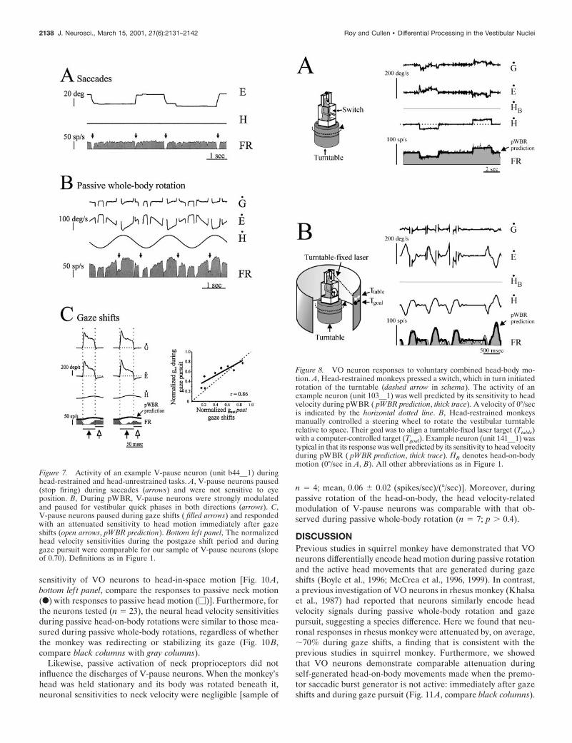
Self-generated motion of the head and body in spaceTo determine whether the monkey’s knowledge of its self-generated head motion attenuates the sensitivity of VO neuronsto head velocity, we characterized neural activity during twovoluntary tasks in which the head and body moved togetherrelative to space. In the first task (Fig. 8A, schema), monkeys weretrained to depress a switch at the appearance of a light cue. Bypushing the switch, the monkey initiated a vestibular turntablerotation of a constant velocity, direction, and duration, afterwhich it received a juice reward. The example neuron was typicalof the three neurons tested in that it was strongly activated by theresultant head motion; its activity could be reliably predicted byits head velocity sensitivity during passive whole-body rotation(Fig. 8A, pWBR prediction, thick trace). During this task, themonkey anticipated the head movement, because it initiatedthe motion of the turntable. However, it is arguable whether theresultant head movement was truly voluntary, because the mon-key had no control over the actual trajectory of table motion.
To address this issue, we designed a second task in which themonkey directly controlled the velocity and direction of thevestibular turntable. Monkeys drove their own body motion byrotating a steering wheel connected to the motor controller of thevestibular turntable (Fig. 8B, schema). Each animal was welltrained in this task and would generate accurate head-in-spacemotion to align its head–body position with either a laser target[laser task (Fig. 8B)] or a food reward (banana task; data not
Figure 4. VO neuron responses during gaze shifts and gaze pursuit.Despite the differences in peak head velocities and type of eye motiongenerated during the two tasks, the normalized head velocity sensitivitieswere not significantly different. Comparable trends were found for type I(l) and type II (u) VO neurons. Dashed line represents unity.
Figure 5. During self-generated head-on-body motion, the suppressionof the head velocity sensitivity was not dependent on whether the monkeywas stabilizing or redirecting its gaze. During the pWBRc paradigm(black column) in which the monkey was redirecting its gaze, the neuronalresponses were not significantly different from those observed duringpWBR, yet when gaze was redirected during gaze shifts and gaze pursuit( gray columns), neuronal responses were significantly attenuated. VOneurons response were selectively attenuated during active motion of thehead-on-body: gaze shifts, gaze pursuit, and the period immediately afterthe gaze shifts (rightmost column) in which gaze was immobile but thehead continued to move. Error bars show SEM.
2136 J. Neurosci., March 15, 2001, 21(6):2131–2142 Roy and Cullen • Differential Processing in the Vestibular Nuclei

shown). Thus, the trajectory of the head motion was the directresult of a goal-directed action taken by the monkey. All neuronstested (n 5 15) were strongly modulated by the resultant volun-tary head-in-space motion [normalized gest, 1.04 6 0.18 and1.05 6 0.16 (spikes/sec)/(°/sec), laser and banana task, respective-ly]. Moreover, the activity of each neuron was predicted well by amodel based on the modulation of the neuron during passivewhole-body rotation (Fig. 8B, pWBR prediction, thick trace). To-gether, these results indicate that vestibular-related modulationwas not attenuated by the knowledge of self-generated motion ofthe head relative to space and that the specific motor command(i.e., activation of the neck musculature) used to produce thebehavior must be considered.
Effect of neck afferent activationIt is known that in decerebrate animals neck muscle spindleafferents influence the activity of vestibular nuclei neurons (Boyleand Pompeiano, 1981; Anastasopoulos and Mergner, 1982; Wil-son, 1991) via a disynaptic pathway (Sato et al., 1997). It isconceivable that in alert monkeys neck afferent inputs contributeto the suppression of VO neuron responses during and after gazeshifts (Fig. 2A) and during gaze pursuit (Fig. 3A). To address thispossibility, we tested neurons during two experiments in whichthe head was rotated relative to the body. It is likely that thepassive neck rotation resulted in even greater activation of neckproprioceptors than did active head-on-body rotations, becausegamma motoneurons are more active during passive than activemovements (Prochazka et al., 1987), and, in turn, the sensitivityof muscle spindles to passive neck rotation is likely to be greater(Hulliger et al., 1977).
In the first experiment, the monkey’s head was held stationaryrelative to space and its body was rotated beneath it (Fig. 9A, filledarrow in schema). The example VO neuron was representative of
our sample in that it vigorously responded to passive whole-bodyrotation (Fig, 1A) but was unresponsive to passive rotation of thebody under the neck (Fig. 9A, bottom lef t panel, compare F withM). The activity of the neuron was well described by its sponta-neous discharge rate (Fig. 9A, HB prediction, thick trace) andpoorly predicted by a model that would be consistent with neckafferent inputs suppressing vestibular responses during voluntaryhead-on-body motion (Fig. 9A, BS prediction, thin trace). Neckrotation sensitivities were negligible [normalized sample mean,0.12 6 0.18 (spikes/sec)/(°/sec)] (Fig. 9B, white column) for allneurons tested (n 5 15) and thus did not significantly contributeto the attenuation that was observed during self-generated headmotion (Fig. 9B, gray columns). The torque produced by themonkey against the head restraint was concurrently measured andfound to be small (less than 60.5 Nm) compared with thatproduced when monkeys oriented to food targets (torque of morethan 63.5 Nm). Thus, the neck motor commands, and by exten-sion neck motor efference copy signals, generated by the monkeyswere minimal during these passive rotations.
The influence of neck afferent inputs was further investigatedusing a paradigm during which the experimenter rapidly rotatedthe animal’s head on its neck (Fig. 10A, hand in schema). Thepassively elicited head-on-body movements had head-in-spacetrajectories and velocities comparable with those generated dur-ing large voluntary gaze shifts (mean peak head velocity, 167 6 63vs 153 6 55 °/sec for 55–65° gaze shifts). Torque was not mea-sured during this paradigm, but the analysis was restricted tointervals in which monkeys exhibited little resistance to rotation.The neuronal firing rate of the example neuron was well predictedby the head velocity sensitivities during passive head-on-bodyrotation (Fig. 10A, pWBR prediction, thick trace), indicating thatthe passive activation of the neck muscle spindles did not alter the
Figure 6. VO neuron responses to combined voluntary and passive head-in-space motion. A, Head-unrestrained monkeys generated voluntaryhead-on-body movements (dashed arrow in schema) while being passively rotated by the vestibular turntable (thick arrow in schema). Head-in-spacemovement (HS ) is the sum of the body-in-space motion (BS ) generated by passive rotation and voluntary head-on-body movements (BB ). Themodulation of the example neuron (unit 79 5) was well correlated with the passive body-in-space motion (BS prediction, thin dark trace) but was poorlyrelated to the voluntary head-on-body component of head-in-space motion (HS prediction, thick dark trace). Bottom lef t panel, Modulation in responseto passive body-in-space motion (M) and to voluntary head-on-body motion during combined stimulation (F). B, The response to the voluntarycomponent of head-in-space motion was significantly attenuated when the monkey was either redirecting (black column) or stabilizing ( gray column) itsgaze. In contrast, the neurons showed no attenuation in response to the passive rotation component (white columns). Error bars show SEM. All otherdefinitions as in Figure 1.
Roy and Cullen • Differential Processing in the Vestibular Nuclei J. Neurosci., March 15, 2001, 21(6):2131–2142 2137
sensitivity of VO neurons to head-in-space motion [Fig. 10A,bottom lef t panel, compare the responses to passive neck motion(F) with responses to passive head motion (M)]. Furthermore, forthe neurons tested (n 5 23), the neural head velocity sensitivitiesduring passive head-on-body rotations were similar to those mea-sured during passive whole-body rotations, regardless of whetherthe monkey was redirecting or stabilizing its gaze (Fig. 10B,compare black columns with gray columns).
Likewise, passive activation of neck proprioceptors did notinfluence the discharges of V-pause neurons. When the monkey’shead was held stationary and its body was rotated beneath it,neuronal sensitivities to neck velocity were negligible [sample of
n 5 4; mean, 0.06 6 0.02 (spikes/sec)/(°/sec)]. Moreover, duringpassive rotation of the head-on-body, the head velocity-relatedmodulation of V-pause neurons was comparable with that ob-served during passive whole-body rotation (n 5 7; p . 0.4).
DISCUSSIONPrevious studies in squirrel monkey have demonstrated that VOneurons differentially encode head motion during passive rotationand the active head movements that are generated during gazeshifts (Boyle et al., 1996; McCrea et al., 1996, 1999). In contrast,a previous investigation of VO neurons in rhesus monkey (Khalsaet al., 1987) had reported that neurons similarly encode headvelocity signals during passive whole-body rotation and gazepursuit, suggesting a species difference. Here we found that neu-ronal responses in rhesus monkey were attenuated by, on average,;70% during gaze shifts, a finding that is consistent with theprevious studies in squirrel monkey. Furthermore, we showedthat VO neurons demonstrate comparable attenuation duringself-generated head-on-body movements made when the premo-tor saccadic burst generator is not active: immediately after gazeshifts and during gaze pursuit (Fig. 11A, compare black columns).
Figure 7. Activity of an example V-pause neuron (unit b44 1) duringhead-restrained and head-unrestrained tasks. A, V-pause neurons paused(stop firing) during saccades (arrows) and were not sensitive to eyeposition. B, During pWBR, V-pause neurons were strongly modulatedand paused for vestibular quick phases in both directions (arrows). C,V-pause neurons paused during gaze shifts ( filled arrows) and respondedwith an attenuated sensitivity to head motion immediately after gazeshifts (open arrows, pWBR prediction). Bottom lef t panel, The normalizedhead velocity sensitivities during the postgaze shift period and duringgaze pursuit were comparable for our sample of V-pause neurons (slopeof 0.70). Definitions as in Figure 1.
Figure 8. VO neuron responses to voluntary combined head-body mo-tion. A, Head-restrained monkeys pressed a switch, which in turn initiatedrotation of the turntable (dashed arrow in schema). The activity of anexample neuron (unit 103 1) was well predicted by its sensitivity to headvelocity during pWBR ( pWBR prediction, thick trace). A velocity of 0°/secis indicated by the horizontal dotted line. B, Head-restrained monkeysmanually controlled a steering wheel to rotate the vestibular turntablerelative to space. Their goal was to align a turntable-fixed laser target (Ttable)with a computer-controlled target (Tgoal). Example neuron (unit 141 1) wastypical in that its response was well predicted by its sensitivity to head velocityduring pWBR ( pWBR prediction, thick trace). HB denotes head-on-bodymotion (0°/sec in A, B). All other abbreviations as in Figure 1.
2138 J. Neurosci., March 15, 2001, 21(6):2131–2142 Roy and Cullen • Differential Processing in the Vestibular Nuclei

The discrepancy between our study and that of Khalsa et al.(1987) may be the result of two factors: a sampling bias in theirdata set and/or a difference in the analysis approach. First, it ispossible that these investigators inadvertently excluded the VOneurons that showed the greatest attenuation by assuming thatthey had lost neuronal isolation during the transition from thehead-restrained to head-unrestrained condition. Indeed, we wereinitially concerned that this might have been the case in thepresent study and were therefore careful to confirm neuronal
isolation (see Materials and Methods). Second, these authorsused a regression analysis to relate mean firing frequencies tomean head velocities, whereas we used a more sensitive dynamicanalysis method to quantify VO neuron discharges. Thus, if theirdata set included only those VO neurons that showed the leastattenuation, it is possible that their analysis was not sufficient toprovide evidence that neuronal head velocity sensitivities werealtered.
In the present study, we also describe the behavior of a second
Figure 9. Response of VO neurons to passive neck rotation. A, The response of example neuron (unit br31 2) was typical in that it was not modulatedwhen the monkey’s body was passively rotated beneath its stationary head; the mean and variance of the discharge of the neuron was comparable duringthis paradigm and head-restrained eye movement paradigms (mean interspike interval, 18 6 13 vs 17 6 13 msec, respectively). The neural discharge waswell described by the HB prediction, which was based on head motion (zero in the paradigm), and was overestimated by the BS prediction, which wasgenerated using the hypothetical neck sensitivity required to account for the attenuated vestibular response of the neuron during active head-on-bodymotion. Bottom lef t panel, Comparison of neural modulation in response to pWBR (M) and to passive neck rotation (F). B, The normalized neck velocitysensitivity (white column) is insufficient to account for the attenuation observed during gaze shifts, after gaze shifts, and during gaze pursuit ( graycolumns). Error bars show SEM. Abbreviations as in Figure 1.
Figure 10. Response of VO neurons to passive rotation of the head-on-body. A, The experimenter (hand in schema) passively rotated the monkey’s headrelative to its earth stationary body. The discharge of example neuron (unit br31 2) was reliably predicted by the sensitivity of the neuron to pWBR(thick trace). Bottom lef t panel, Relationships between neural modulation (M) and residual modulation (F; total discharge 2 HB-related modulation) andhead-in-space motion (where HS 5 HB ). B, For our sample of VO neurons, responses during passive head-on-body rotation were comparable with thoseresulting passive whole-body rotation [pWBR (used for normalization) and pWBRc (lef tmost black column)], regardless of whether the monkey wasredirecting (black columns) or stabilizing ( gray column) its gaze. Error bars show SEM. Abbreviations as in Figure 1.
Roy and Cullen • Differential Processing in the Vestibular Nuclei J. Neurosci., March 15, 2001, 21(6):2131–2142 2139

class of vestibular nuclei neurons, V-pause neurons, that differedfrom VO neurons in that their sensitivity to head velocity infor-mation is reduced to a much greater extent during gaze shiftsthan during other active motion of the head-on-body. We proposethat the preferential suppression of V-pause neuron responsesduring gaze shifts is mediated via two mechanisms: one similar tothat of VO neurons and the other a suppression of activity by thesaccadic burst generator (Cullen and Guitton, 1997b). This lattermechanism could be mediated, in part, by projections of premotorburst neurons to type II vestibular neurons (Sasaki and Shimazu,1981), which in turn send inhibitory projections to neurons withinthe vestibular nucleus (Shimazu and Precht, 1966). However, thispathway is unlikely to contribute to the attenuation of VO neuronhead velocity responses given that they are similarly reduced forall active head-on-body movements.
Negligible role of neck afferent inputsOur finding that the activation of neck proprioceptors did notsignificantly influence the firing patterns of VO neurons wasunexpected in light of previous studies (McCrea et al., 1996, 1999;Gdowski and McCrea, 1999). These studies reported that insquirrel monkey most, if not all, secondary vestibular neurons(including VO neurons) are sensitive to passive rotation of theneck. In contrast, we used a comparable experimental approachand concluded that passive activation of neck proprioceptors hadlittle effect on VO responses in rhesus monkey. We found that (1)passive rotation of the monkey’s head on its body elicited re-sponses comparable with those elicited by passive whole-bodyrotation (Fig. 11A, diagonally striped column), and (2) VO neu-rons were not sensitive to passive rotation of the monkeys bodyunder its stationary head. We propose that the discrepancy be-tween our results and those of McCrea and colleagues mightresult from either of two factors. First, squirrel monkeys have arelatively small oculomotor range (approximately 620°) (Cullenet al., 1991) compared with humans and rhesus monkeys (approx-imately 650°) and thus rely more heavily on head motion toredirect gaze. Therefore, it is conceivable that neck propriocep-tion information is processed differently in the two species. Sec-ond, because these investigators did not measure neck torqueduring the paradigms they used to passively activate neck pro-prioceptors, the possibility that the monkeys generated someresistance (i.e., a non-negligible efference copy signal) cannot beruled out.
Neck efference copy and the principle of reafferenceOur results are consistent with von Holst and Mittelstaedt’s(1950) original idea of reafference, in which an efference copy
Figure 11. Vestibular reafferent information is differentially processed atthe level of the vestibular nuclei. A, The responses of VO neurons weresignificantly suppressed during voluntary head-on-body movements (blackcolumns) compared with pWBR ( gray columns). Similarly, when monkeysmade voluntary head-on-body movements during passive rotation, neu-ronal responses to the voluntary component (open column) remainedattenuated, whereas the responses to the passive component (horizontally
4
striped column) remained encoded. In contrast, neurons continued toencode head-in-space motion when the head was passively rotated on thebody (diagonally striped column) and when the monkey generated volun-tary head-in-space motion by driving the motion of the vestibular turn-table (vertically striped column). Error bars show SEM. B, Proposedmechanism for the selective processing of vestibular information by VOneurons during voluntary head-on-body motion. An efferent copy of theneck motor command (voluntary HB) is subtracted from the vestibularafferent-related signal (HS ) at the level of either the semicircular canals(A), presynaptic to the VO neurons (B), and/or at the VO neuron itself(C). Neither neck proprioceptive information nor knowledge of self-generated head-in-space motion contributes to the differential processingof self-generated head-on-body motion. (Note that the hypothetical path-ways that have been eliminated are indicated by X.) C, Alternatively, anefference copy of the neck motor command could function to selectivelygate in inhibitory neck proprioceptive inputs.
2140 J. Neurosci., March 15, 2001, 21(6):2131–2142 Roy and Cullen • Differential Processing in the Vestibular Nuclei

signal is combined with the vestibular afferent signal to selectivelyremove the reafferent component caused by the monkey’s behav-ior. We propose that attenuation is mediated by a direct efferencecopy of the neck motor command input to the VO neurons (Fig.11B) or alternatively by efference copy signal that functions toselectively “gate in” inhibitory neck proprioceptive inputs (Fig.11C). However, the site of this behaviorally dependent gating ofvestibular information is, as yet, unknown. Modulation of sensoryinformation could occur at the level of the semicircular canalsthemselves (Fig. 11B, site A) via the vestibular efferent system. Itis possible that this efferent feedback is used to tune the sensi-tivity of the vestibular nerve (or a subset of afferents) to voluntaryhead-on-body movements (Goldberg and Fernandez, 1980). Al-though this idea is supported by the finding that the toadfishefferent system selectively modulates the activity of afferent fibersduring self-motion (i.e., swimming behavior; Highstein, 1992), ithas not been tested in primates. Conversely, modulation mayoccur in the vestibular nucleus itself, either presynaptically (Fig.11B, site B) or at the level of second-order vestibular neurons(Fig. 11B, site C). Additional experiments are needed to deter-mine the location of the behaviorally dependent modulation.Additional experiments will also be required to determinewhether vestibular reafference is suppressed during other natu-rally occurring behaviors. Although neurons responded robustlyduring the cognitively demanding driving task (Fig. 11A, verti-cally striped column), it is possible that, during locomotion and/orcombined eye–head–body gaze shifts, the motor efference signalsgenerated by the activation of the head, torso, and limb muscu-lature might collectively influence the response of vestibular nu-clei neurons to self-generated head motion in space. Pending theresults from these experiments, we suggest that a more suitablename for VO neurons would be vestibular reafference gated(VRG) neurons.
Behaviorally dependent modulation in thevestibular nucleusThe attenuation of head velocity signals encoded by VO neuronswas similar during all active head-on-body movements, regardlessof whether the animal was stabilizing its gaze or redirecting itsgaze to a new point in space. For example, the head movementsensitivity of VO neurons is attenuated not only during combinedeye–head gaze shifts (Fig. 2A, filled arrows) and pursuit (Fig. 3A)but also immediately after gaze shifts, when gaze is stable inspace but the head is still moving (Fig. 2A, open arrows). Incontrast, the head velocity-related activity of another class of ves-tibular nuclei neurons, VOR interneurons (i.e., position-vestibular-pause neurons that also receive direct inputs from the vestibularnerve but project to the extraocular motor nuclei) is attenuatedonly while gaze is being redirected in space (Roy and Cullen,1998), regardless of whether the head motion is actively or pas-sively generated. The difference in behaviorally dependent mod-ulation of these two cell types is consistent with the their role inmediating the vestibulo-collic reflex (see below) and VOR,respectively.
Functional implicationsEmerging evidence suggests that the VCR, a reflex that functionsto stabilize the head in space via activation of the neck muscula-ture during head motion, is mediated, at least in part, by VOneurons that project directly to the ventromedial funiculus ofsegments C1–C2 of the spinal cord (Wilson et al., 1990; Boyle etal., 1996; Gdowski and McCrea, 1999). Because the stabilization
response produced by the VCR would be counterproductiveduring voluntary behaviors in which an animal’s goal is to moveits head on its body, it would be logical to selectively attenuate VOneuron responses to voluntary head-on-body movements. Accord-ingly, the VCR would be effectively suppressed for self-generatedhead motion but would remain responsive to unexpected pertur-bations of the head. Indeed, we found that, when a monkeyactively rotates its head on its body while undergoing passivewhole-body rotation, VO neurons continued to encode the pas-sive component (BS) of the head-in-space movement (Fig. 11A,horizontally striped column). This result is in agreement withprevious studies (Boyle et al., 1996; McCrea et al., 1999). Fur-thermore, we discovered for combined active and passive headmotion that neuronal responses to the component of head-in-space velocity produced by active head-on-body motion wereweak and comparable with those observed during active head-on-body motion in the absence of simultaneous passive rotation (Fig.11A, compare open columns with black columns).
The information encoded by VO neurons could also be com-bined with other vestibular pathways to produce an estimate ofour current orientation in space during self-generated motion.VO neurons are well situated within extensive cerebellar andcortical recursive networks. The nodulus–uvula of the cerebellumreceives inputs from vestibular afferents (Korte and Mugnaini,1979) and is thought to be reciprocally connected to VO neurons(Wylie et al., 1994; Voogd et al., 1996; Wearne et al., 1998). Thetransformation of head-centered motion information into an in-ertial (gravity-centered) coordinate frame requires the nodulus–uvula (Angelaki and Hess, 1995; Wearne et al., 1998). In addition,many cortical areas involved in spatial representation, navigation,and gaze control receive vestibular information and in turnproject back to the vestibular nuclei (for review, see Fukushima,1997). For example, posterior parietal neurons have been shownto encode body-referenced and world-referenced information intwo separate streams (Snyder et al., 1998) and project via directand polysynaptic pathways to the vestibular nuclei (Faugier-Grimaud and Ventre, 1989). Indeed, it has been suggested re-cently that VO neurons transform vestibular head-in-space infor-mation into body-in-space coordinates (Gdowski and McCrea,1999). However, although this proposal is consistent with theobservation that VO neurons reliably encode passive head-in-space motion during simultaneous passive whole-body rotationand active head-on-body motion, it cannot account for our findingthat neurons continue to encode head-in-space velocity duringpassive rotation of the head on a stationary body. Thus, VOneurons do not simply transform head-in-space signals into body-in-space coordinates but rather encode vestibular informationfrom which self-generated head-on-body motion has been selec-tively eliminated. We suggest that this behaviorally dependentprocessing of vestibular information contributes to both the con-trol and maintenance of posture and the computation of aninternal estimate of spatial orientation.
REFERENCESAnastasopoulos D, Mergner T (1982) Canal–neck interaction in vestib-
ular nuclear neurons of the cats. Exp Brain Res 46:269–280.Angelaki DE, Hess BJM (1995) Inertial representation of angular mo-
tion in the vestibular system of rhesus monkeys. II. Otolith-controlledtransformation that depends on an intact cerebellar nodulus. J Neuro-physiol 73:1729–1751.
Bell C (1981) An efference copy which is modified by reafferent input.Science 214:450–453.
Boyle R, Pompeiano O (1981) Responses of vestibulospinal neurons tosinusoidal rotation of the neck. J Neurophysiol 44:633–649.
Boyle R, Belton T, McCrea RA (1996) Responses of identified vestibu-
Roy and Cullen • Differential Processing in the Vestibular Nuclei J. Neurosci., March 15, 2001, 21(6):2131–2142 2141

lospinal neurons to voluntary eye and head movements in the squirrelmonkey. Ann NY Acad Sci 781:244–263.
Cullen KE, Guitton D (1997a) Analysis of primate IBN spike trainsusing system identification techniques. I. Relationship to eye movementdynamics during head-fixed saccades. J Neurophysiol 78:3259–3282.
Cullen KE, Guitton D (1997b) Analysis of primate IBN spike trainsusing system identification techniques. II. Relationship to gaze, eye, andhead movement dynamics during head-free gaze shifts. J Neurophysiol78:3283–3306.
Cullen KE, McCrea RA (1993) Firing behavior of brain stem neuronsduring voluntary cancellation of the horizontal vestibulo-ocular reflex.I. Secondary vestibular neurons. J Neurophysiol 70:828–843.
Cullen KE, Belton T, McCrea RA (1991) A non-visual mechanism forvoluntary cancellation of the vestibulo-ocular reflex. Exp Brain Res83:237–252.
Cullen KE, Chen-Huang C, McCrea RA (1993a) Firing behavior ofbrain stem neurons during voluntary cancellation of the horizontalvestibulo-ocular reflex. II. Eye-movement related neurons. J Neuro-physiol 70:844–856.
Cullen KE, Guitton D, Rey CG, Jiang W (1993b) Gaze-related activityof putative inhibitory burst neurons in the head-free cat. J Neurophysiol70:2678–2683.
Cullen KE, Rey CG, Guitton D, Galiana HL (1996) The use of systemidentification techniques in the analysis of oculomotor burst neuronspike train dynamics. J Comput Neurosci 3:347–368.
Edwards DH, Heitler WJ, Krasne FB (1999) Fifty years of a commandneuron: the neurobiology of escape behavior in the crayfish. TrendsNeurosci 22:153–161.
Faugier-Grimaud S, Ventre J (1989) Anatomic connections of inferiorparietal cortex (area 7) with subcortical structures related to vestibulo-ocular function in a monkey (Macaca fascicularis). J Comp Neurol280:1–14.
Fuchs AF, Kimm J (1975) Unit activity in vestibular nucleus of the alertmonkey during horizontal angular acceleration and eye movement.J Neurophysiol 38:1140–1161.
Fuchs AF, Robinson DA (1966) A method for measuring horizontal andvertical eye movements in the monkey. J Physiol (Lond) 191:609–631.
Fukushima K (1997) Corticovestibular interactions: anatomy, electro-physiology, and functional considerations. Exp Brain Res 117:1–16.
Fuller JH (1988) Chronic recording of neck sensory input to vestibularneurons. In: Control of head movement (Peterson BW, Richmond FJ,eds), pp120–128. Oxford: Oxford UP.
Gdowski GT, McCrea RA (1999) Integration of vestibular and headmovement signals in the vestibular nuclei during whole-body rotation.J Neurophysiol 81:436–449.
Goldberg JM, Fernandez C (1980) Efferent vestibular system in thesquirrel monkey: anatomical location and influence on afferent activity.J Neurophysiol 43:986–1025.
Hays AV, Richmond BJ, Optican LM (1982) A UNIX-based multipleprocess system for real-time data acquisition and control. W ESCOMConf Proc 2:1–10.
Highstein SM (1992) The efferent control of the organs of balance andequilibrium in the toadfish, Opsanus tau. Ann NY Acad Sci656:108–123.
Hulliger M, Matthews PBC, Noth J (1977) Static and dynamic fusimotoraction on the response of IA fibers to low frequency sinusoidal stretch-ing of widely ranging amplitude. J Physiol (Lond) 267:811–838.
Keller EL (1974) Participation of medial pontine reticular formation ineye movement generation in monkey. J Neurophysiol 37:316–332.
Keller EL, Kamath BY (1975) Characteristics of head rotation and eyemovement-related neurons in alert monkey vestibular nucleus. BrainRes 100:182–187.
Khalsa SB, Tomlinson RD, Schwarz DW, Landolt JP (1987) Vestibularnuclear neuron activity during active and passive head movement in thealert rhesus monkey. J Neurophysiol 57:1484–1497.
Khalsa SB, Tomlinson RD, Scharwz DW (1988) Secondary vestibularand neck position signals in the vestibular nuclei of alert rhesus mon-keys performing active head movements. Acta Otolaryngol106:269–275.
Korte G, Mugnaini E (1979) The cerebellar projection of the vestibularnerve in the cat. J Comp Neurol 184:265–278.
Krasne FB, Bryan JS (1973) Habituation: regulation through presynapticinhibition. Science 182:590–592.
McCrea RA, Chen-Huang C, Belton T, Gdowski GT (1996) Behaviorcontingent processing of vestibular sensory signals in the vestibularnuclei. Ann NY Acad Sci 781:292–303.
McCrea RA, Gdowski GT, Boyle R, Belton T (1999) Firing behavior ofvestibular neurons during active and passive head movements:vestibulo-spinal and other non-eye-movement related neurons. J Neu-rophysiol 82:416–428.
Prochazka A, Hulliger M, Trend P, Durmuller N (1987) Dynamic andstatic fusimotor set in various behavioural contexts. In: Mechanorecep-tors. Development, structure, and function (Hnik P, Soukup T, VejsadaR, Zelena J, eds), pp 417–430. New York: Plenum.
Roy JE, Cullen KE (1998) A neural correlate for vestibulo-ocular reflexsuppression during voluntary eye–head gaze shifts. Nat Neurosci1:404–410.
Roy JE, Cullen KE (1999) Differential processing of head velocity sig-nals in the vestibular nuclei: voluntary head/neck versus head/spacemotion. Soc Neurosci Abstr 25:265.
Sasaki S, Shimazu H (1981) Reticulovestibular organization participat-ing in generation of horizontal fast eye movement. Ann NY Acad Sci374:130–145.
Sato H, Ohkawa T, Uchino Y, Wilson VJ (1997) Excitatory connectionsbetween neurons of the central cervical nucleus and vestibular neuronsin the cat. Exp Brain Res 115:381–386.
Scudder CA, Fuchs AF (1992) Physiological and behavioural identifica-tion of vestibular nucleus neurons mediating the horizontal vestibu-loocular reflex in trained rhesus monkeys. J Neurophysiol 68:244–264.
Shimazu H, Precht W (1966) Inhibition of central vestibular neuronsfrom the contralateral labyrinth and its mediating pathway. J Neuro-physiol 29:467–492.
Snyder LH, Grieve KL, Brotchie P, Andersen RA (1998) Separatebody- and world-referenced representations of visual space in parietalcortex. Nature 394:887–891.
Sylvestre PA, Cullen KE (1999) Quantitative analysis of abducens neu-ron discharge dynamics during saccadic and slow eye movements.J Neurophysiol 82:2616–2632.
Tomlinson RD, Robinson DA (1984) Signals in vestibular nucleus me-diating vertical eye movements in the monkey. J Neurophysiol51:1121–1136.
Voogd J, Gerrits M, Ruigrok JH (1996) Organization of the vestibulo-cerebellum. Ann NY Acad Sci 781:553–579.
von Holst E, Mittelstaedt H (1950) Das reafferenzprinzip. Naturwissen-schaften 37:464–476.
Wearne S, Raphan T, Cohen B (1998) Control of spatial orientation ofthe angular vestibuloocular reflex by the nodulus and uvula. J Neuro-physiol 79:2690–2715.
Wilson VJ (1991) Vestibulospinal and neck reflexes: interaction in thevestibular nuclei. Ach Ital Biol 129:43–52.
Wilson VJ, Yamagata Y, Yates BJ, Schor RH, Nonaka S (1990) Re-sponse of vestibular neurons to head rotations in vertical planes. III.Response of vestibulocollic neurons to vestibular and neck stimulation.J Neurophysiol 64:1695–1703.
Wylie DR, De Zeeuw CI, Digiorgi PL, Simpson JI (1994) Projections ofindividual purkinje cells of identified zones in the ventral nodulus to thevestibular and cerebellar nuclei in the rabbit. J Comp Neurol 349:448–463.
2142 J. Neurosci., March 15, 2001, 21(6):2131–2142 Roy and Cullen • Differential Processing in the Vestibular Nuclei