royal society of chemistry nmr discussion group
TRANSCRIPT
1
Royal Society of Chemistry
NMR Discussion Group
Postgraduate Meeting 2012
School of Chemistry University of Bristol
14th June 2012

2
3
Dear Delegate,
Welcome to the fifth Postgraduate Symposium of the RSC NMR Discussion Group!
This meeting has a broadly similar format to the previous meetings and brings
together early career researchers, broadly defined as postgraduates, early career post
doctorial workers, and young industrialists, who all have a strong research interest in
magnetic resonance. The idea is that it provides a forum to showcase their work and
networking opportunities within the NMR community.
This year we are continuing to have two overview lectures, given by leaders in their
field, to highlight the power of magnetic resonance methods across a broad range of
topics. Dr John Hollerton (GlaxoSmithKline) and Dr Melanie Britton (University of
Birmingham) will present overviews of the use of NMR in drug discovery and
applications of NMR and MRI to soft matter, emulsions and suspensions.
The programme is designed to be varied, with a wide range of talks. Posters allow
delegates to discuss their work with other early career researchers and more
established colleagues in a friendly and informal setting. There is also ample
opportunity for further discussions over tea/coffee and lunch.
We hope that you will make the most of this opportunity and that you enjoy the
meeting.
Iain Day University of Sussex Meeting Organiser
Craig Butts University of Bristol Local Organiser
James Keeler University of Cambridge NMRDG Chairman

4

5
Local organisation and acknowledgements Meeting coordinated by: Iain Day, University of Sussex Local organisation coordinated by: Craig Butts, University of Bristol Online registration coordinated by: John Parkinson, University of Strathclyde The organisers would like to thank Stephen Byard (Covance) for significant help “behind the scenes”. Thanks go to James Keeler (University of Cambridge), Iain Day (University of Sussex) and John Parkinson (University of Strathclyde) for acting as Judges for the prize giving. The NMR Discussion Group gratefully acknowledges the following sponsorship for their generous support of this meeting:
Agilent Technologies UK Ltd
Dalton Transations
Posters Posters should be mounted on the poster boards during the arrival period prior to the formal welcome and start of the program and should be attached to the board for which the poster number has been designated. Posters should be removed after the close of the meeting.

6
7
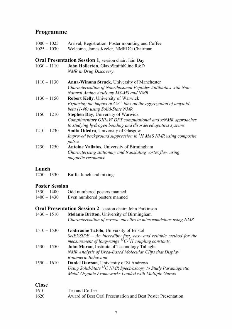
Programme 1000 – 1025 Arrival, Registration, Poster mounting and Coffee 1025 – 1030 Welcome, James Keeler, NMRDG Chairman Oral Presentation Session 1, session chair: Iain Day 1030 – 1110
John Hollerton, GlaxoSmithKline R&D NMR in Drug Discovery
1110 – 1130
Anna-Winona Struck, University of Manchester Characterization of Nonribosomal Peptides Antibiotics with Non-Natural Amino Acids my MS-MS and NMR
1130 – 1150
Robert Kelly, University of Warwick Exploring the impact of Cu2+ ions on the aggregation of amyloid-beta (1-40) using Solid-State NMR
1150 – 1210
Stephen Day, University of Warwick Complimentary GIPAW DFT computational and ssNMR approaches to studying hydrogen bonding and disordered apatites systems
1210 – 1230
Smita Odedra, University of Glasgow Improved background suppression in 1H MAS NMR using composite pulses
1230 – 1250
Antoine Vallatos, University of Birmingham Characterising stationary and translating vortex flow using magnetic resonance
Lunch 1250 – 1330 Buffet lunch and mixing Poster Session 1330 – 1400 Odd numbered posters manned 1400 – 1430 Even numbered posters manned Oral Presentation Session 2, session chair: John Parkinson 1430 – 1510
Melanie Britton, University of Birmingham Characterisation of reverse micelles in microemulsions using NMR
1510 – 1530
Godiraone Tatolo, University of Bristol SelEXSIDE – An incredibly fast, easy and reliable method for the measurement of long-range 13C-1H coupling constants.
1530 – 1550
John Moran, Institute of Technology Tallaght NMR Analysis of Urea-Based Molecular Clips that Display Rotameric Behaviour
1550 – 1610
Daniel Dawson, University of St Andrews Using Solid-State 13C NMR Spectroscopy to Study Paramagnetic Metal-Organic Frameworks Loaded with Multiple Guests
Close 1610 Tea and Coffee 1620 Award of Best Oral Presentation and Best Poster Presentation

8
Index to Abstracts Berwick, M. 022 Sneddon, S. 016
Struck, A-W. 002
Britton, M. M. 007
Tatola, G. 008
Dawson, D. M. 010
Day, S. P. 004 Vallatos, A. 006
Hawarden, L. 023 Yeasmin, Z. 015
Hollerton, J. C. 001
Jones, C. R. 011
Joyce, R. E. 012
Katz, J. R. 018
Kelly, R. T. 003
King, S. P. 013
Lamley, J. M. 017
Law, S. J. 019
Mills, A. 021
Moran, J. 009
Odedra, S. 005
Romer, F. H. 014
Rose, H. 020

9
Abstracts of Talks (Talk 001)
John Hollerton, GlaxoSmithKline R&D, [email protected] NMR in Drug Discovery John Hollerton GlaxoSmithKline R&D, Gunnels Wood Road, Stevenage, Herts, SG1 2NY, UK NMR is so versatile that it has huge application across Drug Discovery. This was not always the case and this presentation highlights some of the advances in NMR that have made this possible as well as touching on some of the diverse areas in which NMR has found a role.

10

11
(Talk and Poster 002) Anna-Winona Struck, University of Manchester, [email protected] Characterization of Nonribosomal Peptides Antibiotics with Non-Natural Amino Acids my MS-MS and NMR Anna-Winona Struck,1 Matthew Styles,1 Jenny Thirlway,1 Richard Lewis,2 Laura Nunns,1 Majid Al Nakeeb,1 Colin P. Smith, 2 Jason Micklefield1 1School of Chemistry & Manchester Interdisciplinary Biocentre, The University of Manchester, 131 Princess Street, Manchester M1 7DN 2FHMS, University of Surrey, Guildford, Surrey, GU27XH Nonribosomal peptides are a group of structurally diverse secondary metabolites, which are synthesised by nonriobosomal peptide synthetases (NRPS). We engineered the biosynthesis of calcium-dependent antibiotics (CDA) to incorporate a synthetic non-natural amino acid into the decapeptide lactone core of CDA. Our strategy focused on altering the specificity of the Module 10 A-domain, of CdaPS3, so that it preferentially incorporates (2S,3R)-3-methyl glutamine (mGln) and Gln over the natural substrates (2S,3R)-3-methyl glutamic acid (mGlu) and Glu. The new product was subsequently purified by HPLC and subjected to high-resolution MS, confirming the proposed molecular formula. The sequence of the CDAs was obtained by tandem MS and confirmed by 2D 1H NMR spectroscopy. Reference: J. Thirlway, R. Lewis, L. Nunns, M. Al Nakeeb, M. Styles, A.-W. Struck, C. P. Smith and J. Micklefield, Angew. Chem. Int. Ed. 2012, in press.

12

13
(Talk and Poster 003) Robert Kelly, University of Warwick, [email protected] Exploring the impact of Cu2+ ions on the aggregation of amyloid-beta (1-40) using Solid-State NMR Robert T. Kelly,a Ray Dupree,a Józef R. Lewandowski,a Steven P. Brown,a Oleg N. Antzutkin a,b aDepartment of Physics, University of Warwick, Coventry CV4 7AL, United Kingdom bDivision of Chemistry, Luleå University of Technology, Luleå SE 971 87, Sweden Aggregation of amyloid-beta1-40 (Aβ1-40) is linked to the pathology of Alzheimer’s disease with current ssNMR research focusing on the interaction of oligomers of Aβ with membranes.1 In this work, 2D and 3D solid-state NMR (ssNMR) spectra, chemical shift values and electron microscopy images of Aβ1-40 aggregates formed in the presence of Cu2+ are presented. Short (20 ms) and long (200 ms) mixing time 13C-13C MAS DARR experiments were performed on Bruker Avance II+ 600 MHz and Bruker Avance III 850 MHz spectrometers so as to separate inter- and intra-residue cross peaks. 13C chemical shift values are compared to those obtained for other Aβ fibrils 2-4 and Aβ1-40 fibrils which, after formation, had Cu2+ ions introduced,5 noting that carbon chemical shifts are sensitive to the secondary structure. 1. Sandberg, A. et al “Stabilization of neurotoxic Alzheimer amyloid-beta oligomers by protein engineering” Proc. Natl. Acad. Sci. USA. 107, 15595-15600 (2010) 2. Petkova, A., Yau, W. M. and Tycko, R. “Experimental constraints on quaternary structure in Alzheimer’s beta-amyloid fibrils” Biochemistry 45, 498-512 (2006) 3. Paravastu, A. K. et al “Molecular structural basis for polymorphism in Alzheimer’s beta- amyloid fibrils” Proc. Natl. Acad. Sci. USA. 105, 18349-18354 (2008) 4. Bertini, I. et al “A New Structural Model of Aβ40 Fibrils” J. Am. Chem. Soc. 133, 16013-16022 (2011) 5. Parthasarathay, S. et al “Molecular-Level Examination of Cu(2+) Binding Structure for Amyloid Fibrils of 40-Residue Alzheimer's beta by Solid-State NMR Spectroscopy” J. Am. Chem. Soc. 133, 3390-3400 (2011)

14

15
(Talk and Poster 004) Stephen Day, University of Warwick, [email protected] Complimentary GIPAW DFT computational and solid state NMR approaches to studying hydrogen bonding and disordered apatites systems Stephen P. Day, Gregory J. Rees, John D. Wallis, David Quigley, John V. Hanna Department of Physics, University of Warwick, Coventry CV4 7AL, United Kingdom The combination of GIPAW DFT computation and solid state NMR (SSNMR) has been shown to be a powerful combination when dealing with many different types of chemical and materials systems. Solid organic structures which are strongly influenced by hydrogen bonding, and materials systems characterised by structural and positional disorder are very amenable to this type of analytical approach. This investigation into hydrogen bonded structure uses the benzoic acid motif as a starting model to investigate more complex Group I hemobenzoate systems, where the effect of the alkali metal ion on the hydrogen bonded network will be reported. This project has used SSNMR techniques such as fast MAS, MQMAS and DOR in combination with GIPAW DFT calculations facilitated with the CASTEP code. In contrast, rare earth apatites (A10-x(Si/Ge)6O26+y (A = rare earth)) are potential oxide-ion electrolytes for solid oxide fuel cells which exhibit positional and structural disorder, and these pose very different computational issues. Recent 17O MAS NMR, DFT and MD studies4 on La8Y2Ge6O27 have shown that the oxide-ion excess(y) is preferentially incorporated as an interstitial species between two GeO4 tetrahedra. These complex systems require a systematic and efficient strategy to handle large DFT computational load needed to accurately describe these structures and thus predict the NMR parameters characterising the relevant species. We have statistically addressed the computed results by constraining them within Boltzmann distributions to identify structurally probable realisations based on their final energy, and weighted the average NMR parameters over these configurations according to their respective probabilities. 1 J. C. Speakman, The hydrogen bond and other intermolecular forces, Chemical Society, London, 1975. 2 R. K. Harris, P. Jackson, L. H. Merwin, B. J. Say, G. Hagele, J Chem Soc Farad T 1 84, 1988, 3649- 3672. 3 W. Fu, P Sun, Frontiers of Chemistry in China, 6 (2011) 173-189 4 Panchmatia, P. M., Orera, A., Rees, G. J., Smith, M. E., Hanna, J. V., Slater P. R. and Islam M. S. ,Angew. Chem., 50 (2011) 9328–9333

16

17
(Talk and Poster 005) Smita Odedra, University of Glasgow, [email protected] Improved background suppression in 1H MAS NMR using composite pulses Smita Odedra and Stephen Wimperis School of Chemistry and WestCHEM, University of Glasgow, Glasgow G12 8QQ, United Kingdom A well known feature of 1H MAS NMR spectroscopy, particularly of solids where the concentration of 1H nuclei is low, is the presence in the spectrum of a significant broad "background" signal arising from 1H nuclei that are outside the MAS rotor and radiofrequency coil, probably located on the surfaces of the static components of the probehead. A popular method of suppressing this unwanted signal is the "depth pulse" experiment,1,2 consisting of a 90° pulse followed by two 180° pulses that are phase cycled according to the "Exorcycle" scheme,3 which removes signal associated with imperfect 180° pulses. Consequently, only spins in the centre of the radiofrequency coil contribute to the 1H MAS spectrum, while those experiencing a low B1 field outside the coil are suppressed. Although very effective at removing background signal from the spectrum, one drawback with this approach is that significant loss of the desired signal from the sample also occurs owing to the spatial inhomogeneity of the B1 field produced by the coil. By using novel antisymmetric passband composite pulses4,5 to replace the simple pulses in the "depth pulse" experiment, we achieve improved intensity of the 1H signals of interest while still maintaining effective background suppression. We expect that our results will be relevant to 1H MAS NMR studies of, for example, nominally perdeuterated biological samples or nominally anhydrous inorganic materials. 1. M. R. Bendall and R. E. Gordon, J. Magn. Reson. 53, 365 (1983). 2. D. G. Cory and W. M. Ritchey, J. Magn. Reson. 80, 128 (1988). 3. G. Bodenhausen, R. Freeman, and D. L. Turner, J. Magn. Reson. 27, 511 (1977). 4. S. Wimperis, J. Magn. Reson. 83, 509 (1989). 5. S. Wimperis, J. Magn. Reson. A 109, 221 (1994).

18

19
(Talk and Poster 006) Antoine Vallatos, University of Birmingham, [email protected] Characterising stationary and translating vortex flow using magnetic resonance Antoine Vallatos1, Marc C.T. Wilson2, Annette F. Taylor3, Melanie M. Britton1 1 School of Chemistry, University of Birmingham, Birmingham, B15 2TT, UK 2 School of Mechanical Engineering, University of Leeds, Leeds, LS2 9JT, UK 3 School of Chemistry, University of Leeds, Leeds, LS2 9JT, UK Understanding molecular displacements within complex flows is of great importance in fluid mechanics, engineering, biology and chemistry, and there is an increasing interest in oscillating and time periodic vortical flows. High-resolution, three-dimensional velocity mapping, which can characterise the transport properties in these systems, remains a challenge. Also, where mechanical instabilities generate dispersion and anomalous diffusion it has been found that enhanced molecular displacements are produced, which are not predicted from velocity maps alone. This is especially the case for vortical and periodic flows. In this work1, we report magnetic resonance (MR) velocity and diffusion maps in three directions for stationary vortices (Taylor Vortex Flow) and velocity maps (fig. b) for translating vortices (Vortex Flow Reactor) produced in a Couette cell (fig. a). Imaging translating vortices is difficult due to the artefacts produced by unsteady flow. However, these motion artefacts were removed by synchronising data acquisition with the translation period of the vortices. MR propagator experiments, which measure the conditional probability density for displacement, were performed to characterise molecular displacements in these systems. Simulations were performed using the experimental velocity and diffusion maps to aid interpretation of experimentally measured propagators and enable characterisation of the macro-mixing properties. These simulations enabled molecular transport to be assessed over longer-time scales than are accessible experimentally (fig. c), allowing plug-flow, by-pass flow and inter-vortex mixing to be quantified.
References: 1. A. Vallatos, M. C. T. Wilson, A. F. Taylor, and M. M. Britton, EuroPhys Lett (submitted)

20

21
(Talk 007) Melanie Britton, University of Birmingham, [email protected] Characterisation of reverse micelles in microemulsions using NMR Melanie M. Britton School of Chemistry, University of Birmingham, Edgbaston, Birmingham, B30 2ES Water-in-oil reverse micelles (RMs), in a microemulsion, are composed of nanosized water droplets surrounded by surfactant molecules in a continuous organic phase. These self-assembled structures form thermodynamically stable droplets of water ranging in intramicellar diameter from approximately 1 to 20 nm, depending on the molar water-to-surfactant ratio (ω). The aqueous core of reverse micelles provides a highly adaptable environment for a variety of chemical and biochemical reactions, protein extraction, synthesis of nanoparticles, as well as providing a model for biological systems. For all of these applications, an understanding of the chemistry, dynamics and structure within the interior of the droplets is important, and typically a variety of molecular probes and spectroscopic techniques have been used to probe intramicellar parameters such as pH, ionic strength, microviscosity and micropolarity. However, the use of probe molecules has a number of limitations. The first is associated with the location of the probe molecule and difficulties in the getting the probe molecule to reside in the region of interest. The other problem is that the probe molecule may adversely affect the size, stability or environment of the reverse micelle. In this talk, I will discuss these issues and present methods1, 2 for probing the chemistry of reverse micelles using NMR spectroscopy, without the need for a probe molecule. In addition to characterising the chemistry and physical environment within reverse micelles, knowledge of their structure, size and polydispersity is also extremely important. Typically, methods such as dynamic light scattering (DLS) or small-angle x-ray scattering (SAXS) have been used to determine these properties. However, these techniques also have a number of limitations. Recent developments3 for measuring size distributions using the inverse Laplace transformation of NMR measurements of diffusion will be presented. 1. Halliday, N. A.; Peet, A. C.; Britton, M. M., "Detection of pH in Microemulsions, without a Probe Molecule, Using Magnetic Resonance." J. Phys. Chem. B 2010, 114, 13745–13751. 2. Binks, D. A.; Spencer, N.; Wilkie, J.; Britton, M. M., "Magnetic Resonance Studies of a Redox Probe in a Reverse Sodium Bis(2-ethylhexyl)sulfosuccinate/Octane/Water Microemulsion." J. Phys. Chem. B 2010, 114, (39), 12558-12564. 3. Law, S. J.; Britton, M. M., "Determination of Droplet Size Distributions in Microemulsions using NMR Measurements of Diffusion." Langmuir 2012, submitted.

22

23
(Talk and Poster 008) Godiraone Tatola, University of Bristol, [email protected] SelEXSIDE – An incredibly fast, easy and reliable method for the measurement of long-range 13C-1H coupling constants. Godiraone Tatolo,1 Craig P. Butts,1 Berte Heise 2 1 Department of Chemistry, University of Bristol, Cantocks Close, BS8 1TS 2 Agilent Technologies UK Ltd.,6 Mead Road, Yarnton, Oxford OX5 1QU, UK The application of 3-bond 13C-1H scalar coupling constants has seen a tremendous growth in elucidation of 3-dimensional structures of organic molecules. Unfortunately, in practical terms 3JCH values are difficult to extract – they are relatively small and of same magnitude as 3JHH coupling constants but also made more complicated by the low sensitivity of the 13C nucleus. Many new experiments for simplifying the measurement of 3JCH have been reported in literature(1) and their main setbacks are that the interpretation of the resulting spectra is not straightforward, long selective pulse sequences lead to the loss of signal due to t2 relaxation and high-resolution 2-dimensional methods typically require extended experiment times. Of these methods, the most straightforward in our experience is the EXSIDE,(2) which gives rise to simple in-phase doublets in the F1 (13C) dimension from which the nJCH value can be read directly from the spectrum, however the extremely high F1 resolution required, means typical experiments take around 7 hours. A new, fast, easy to interpret approach is reported for determination of long-range 13C-1H coupling constant where EXSIDE is converted to be doubly-selective in both the 13C and 1H domains. This sequence termed SelEXSIDE, is easy to interpret and reduces a multi-hour (> 6 hrs) experiment to a matter of minutes (as short as 4 mins) for each coupling constant measured.
Figure 1: (a) EXSIDE spectrum (~7.5 hours) for H11b-C10 of strychnine with 200 ppm (spectral width, 1675 t1 increments, 4 scans/inc. (b) SelEXSIDE spectrum (9
7.5 hours 9 minutes 4 minutes
a c b
nJCH* J-scale nJCH* J-scale

24
mins) for H11b-C10 of strychnine with 4 ppm (500 Hz) 13C spectral width, 32 t1 increments, 4 scans/inc. (c) SelEXSIDE spectrum (4 mins) for H11b-C10 of strychnine with 4 ppm (500 Hz) 13C spectral width, 32 t1 increments, 2 scans/inc. References [1] For example, see: (a) Marquez B.L., Gerwick W.H., Williamson R.T., Magn. Reson. Chem. 2001; 39, 499–530. (b) Edden R.A.E., Keeler J., J. Magn. Reson. 2004; 166, 53–68. (c) Vidal P., Esturau N., Parella T., Espinosa J.F., J. Org. Chem. 2007, 72, 3166-3170. [2] Krishnamurthy V.V., J. Magn. Reson. 1996, Series A 121, 33-41.
25
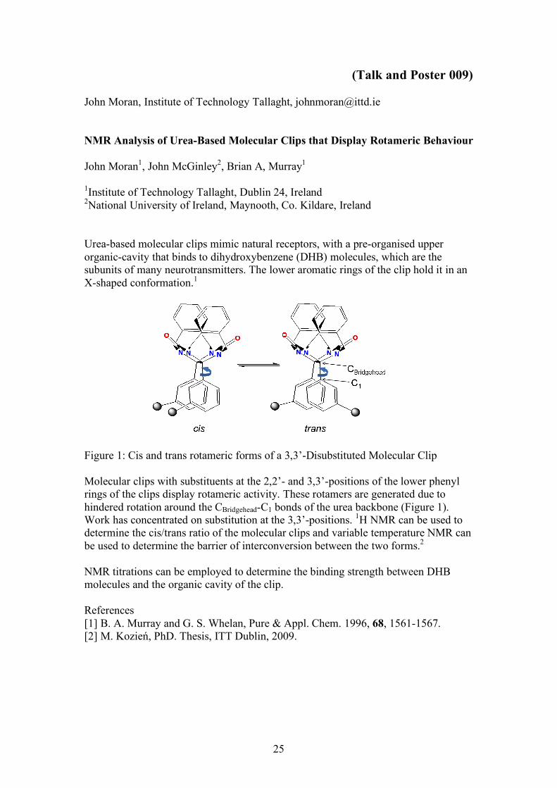
(Talk and Poster 009) John Moran, Institute of Technology Tallaght, [email protected] NMR Analysis of Urea-Based Molecular Clips that Display Rotameric Behaviour John Moran1, John McGinley2, Brian A, Murray1 1Institute of Technology Tallaght, Dublin 24, Ireland 2National University of Ireland, Maynooth, Co. Kildare, Ireland Urea-based molecular clips mimic natural receptors, with a pre-organised upper organic-cavity that binds to dihydroxybenzene (DHB) molecules, which are the subunits of many neurotransmitters. The lower aromatic rings of the clip hold it in an X-shaped conformation.1
Figure 1: Cis and trans rotameric forms of a 3,3’-Disubstituted Molecular Clip Molecular clips with substituents at the 2,2’- and 3,3’-positions of the lower phenyl rings of the clips display rotameric activity. These rotamers are generated due to hindered rotation around the CBridgehead-C1 bonds of the urea backbone (Figure 1). Work has concentrated on substitution at the 3,3’-positions. 1H NMR can be used to determine the cis/trans ratio of the molecular clips and variable temperature NMR can be used to determine the barrier of interconversion between the two forms.2 NMR titrations can be employed to determine the binding strength between DHB molecules and the organic cavity of the clip. References [1] B. A. Murray and G. S. Whelan, Pure & Appl. Chem. 1996, 68, 1561-1567. [2] M. Kozień, PhD. Thesis, ITT Dublin, 2009.

26
27
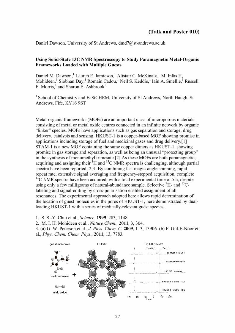
(Talk and Poster 010) Daniel Dawson, University of St Andrews, [email protected] Using Solid-State 13C NMR Spectroscopy to Study Paramagnetic Metal-Organic Frameworks Loaded with Multiple Guests Daniel M. Dawson,1 Lauren E. Jamieson,1 Alistair C. McKinaly,1 M. Infas H, Mohideen,1 Siobhan Day,1 Romain Cadou,1 Neil S. Keddie,1 Iain A. Smellie,1 Russell E. Morris,1 and Sharon E. Ashbrook1 1 School of Chemistry and EaStCHEM, University of St Andrews, North Haugh, St Andrews, Fife, KY16 9ST Metal-organic frameworks (MOFs) are an important class of microporous materials consisting of metal or metal oxide centres connected in an infinite network by organic “linker” species. MOFs have applications such as gas separation and storage, drug delivery, catalysis and sensing. HKUST-1 is a copper-based MOF showing promise in applications including storage of fuel and medicinal gases and drug delivery.[1] STAM-1 is a new MOF containing the same copper dimers as HKUST-1, showing promise in gas storage and separation, as well as being an unusual “protecting group” in the synthesis of monomethyl trimesate.[2] As these MOFs are both paramagnetic, acquiring and assigning their 1H and 13C NMR spectra is challenging, although partial spectra have been reported.[2,3] By combining fast magic-angle spinning, rapid repeat rate, extensive signal averaging and frequency-stepped acquisition, complete 13C NMR spectra have been acquired, with a total experimental time of 5 h, despite using only a few milligrams of natural-abundance sample. Selective 2H- and 13C-labeling and signal-editing by cross-polarisation enabled assignment of all resonances. The experimental approach adopted here allows rapid determination of the location of guest molecules in the pores of HKUST-1, here demonstrated by dual-loading HKUST-1 with a series of medically-relevant guest species. 1. S. S.-Y. Chui et al., Science, 1999, 283, 1148. 2. M. I. H. Mohideen et al., Nature Chem., 2011, 3, 304. 3. (a) G. W. Peterson et al., J. Phys. Chem. C, 2009, 113, 13906. (b) F. Gul-E-Noor et al., Phys. Chem. Chem. Phys., 2011, 13, 7783.

28

29
Abstracts of Posters Poster 011
Catharine Jones, University of Bristol, [email protected] Accurate NOE-Derived Interproton Distances – Fact or Fiction? Catharine R. Jones,1 Craig P. Butts1 1Department of Chemistry, University of Bristol, Cantock’s Close, BS8 1TS Establishing an Initial Level of Accuracy We present a simple method to derive accurate interproton distances from within rigid and flexible systems using NOE data. Strychnine is used as a rigid model system to test the validity of this method. A comparison of the 1D NOE-derived distances and the computed structure of strychnine[1] (right) gives an average error of only 2.3% (0.07Å).[2] Low Level Conformer Identification A second low-level conformer of strychnine is identified using NOE data and confirmed computationally, showing the potential of accurate NOE measurements to determine minute contributions to structure ensembles in solution.[3] Modelling Populations in Flexible Compounds NOE data is further applied to the small flexible molecule, 4-propylaniline, to confirm and predict the relative populations of the multiple possible conformers.[4] It is suggested that with the highly accurate interproton distances determined using this method, there is less need for reliance on the large numbers of loose restraints, such as scalar couplings, typically used in the dynamical analysis of flexible molecules. Monitoring Changes in Conformer Populations with Temperature NOE-distance relationships are also shown to be sufficiently accurate to monitor very small changes in conformer populations in solution (<0.5%/10 °C) – in response to temperature – in good agreement with Boltzmann-predictions, illustrating the effectiveness of accurate NOE-distance measurements in obtaining high quality dynamical, as well as structural, information for small molecules. [1] Bagno et al., Chem. Eur. J., 2006. [2] Butts et al.,Org. Biomol. Chem., 2011. [3] Butts et al., Chem. Comm., 2011. [4] Jones et al., Belstein J. Org. Chem., 2011.

30
Poster 012 Rebecca Joyce, University of Sussex, [email protected] Monitoring Membrane Permeation using a Phosphorous-31 NMR and a Paramagnetic Shift Reagent Rebecca Joyce, Louise C. Serpell and Iain J. Day. School of Life Sciences, University of Sussex, Falmer, Brighton, BN1 9QJ Aggregation of amyloidogenic peptides has been found to be a cause of several diseases; for instance, Aβ plaques have been found in the brains of Alzheimer's patients1 and αSynuclein in those patients with Parkinson's disease2. The mechanism of toxicity of these peptides is not confirmed, however, many studies suggest that it is the small soluble oligomeric species in the aggregation pathway which are cytotoxic.3 In this work NMR has been applied to assist in the elucidation of the mechanism of membrane permeation. Small unilamellar phospholipid vesicles have been used as biomimetic membranes. The 31P peaks of the interior and exterior leaflets are separated by addition of PrCl3, a paramagnetic shift reagent, to the exterior solution of the vesicles only. Changes to the lipid distribution in the vesicles, caused by membrane permeation or instability, leads to changes in the ratio of peak areas. Monitoring the peak distributions over time with and without the addition of aβ illustrates the membrane permeation ability of the amyloidogenic protein.
1. M. Sakono and T. Zako, FEBS Journal, 2010, 277, 1348–1358 2. .B. Lucking and A. Brice, Cell. Mol. Life Sci. 2000, 57, 1894–1908 3. S.M. Butterfield and H.S. Lashuel, Angew. Chem. Int. Ed. 2010, 49, 5628 – 5654

31
Poster 013 Scott King, University of Warwick, [email protected] A Multinuclear Solid State NMR Investigation into the Speciation and Structure of Aluminium Doped Phosphate Bioactive Glasses Scott P. King,1 Jodie M. Smith,2 David M. Pickup,2 Robert J. Newport,2 Steven P. Brown,1 John V. Hanna1
1. University of Warwick, Department of Physics, Coventry, CV4 7AL, England 2. School of Physical Sciences, University of Kent, Canterbury, CT2 7NH, England. Calcium phosphate glasses exhibit interesting properties and compatibility for use as bioactive materials; in particular, they provide bone support and also demonstrate a tendency to stimulate bone regeneration. Aluminium doping of these glass systems induces more favourable properties, due to the strengthening of the structure and through subsequent greater control of dissolution rates from the glass network. A multinuclear solid state MAS NMR study of a glass series with the nominal stoichiometry x(Al2O3) (11-x)(Na2O) 44.5(CaO) 44.5(P2O5) (with x = 0, 3, 5, 8) has been undertaken in order to construct a picture of the glass network, and to determine how the structure may influence the bioactive properties. 23Na and 27Al MAS NMR provide insight on the coordination and disorder within the glass network. Information on the phosphorus Q speciation present within the glasses is evidenced by 31P MAS NMR, which shows both chain like Q2 species and less polymerised Q1 units. Further information is obtained, however, by the implementation of the newly developed 31P refocused INADEQUATE Spin Echo (REINE) technique that reveal 2D correlations of the 2JPP couplings with the 31P chemical shifts of the coupled nuclei. These measurements have been performed for the first time on a coherent suite of samples thus enabling the evolving speciation and polymerisation of the phosphate network to be mapped out throughout the entire series. The structural information obtained from this study provides a much greater insight to support their development as bioactive materials.

32
Poster 014 Frederik Romer, University of Warwick, [email protected] Dynamics and Structure of Novel Polymer Electrolyte Membranes Investigated using Multinuclear Solid-State NMR Frederik H. Romer,1 Jonathan Turley,2 David M. Antonelli,2 John V. Hanna1 1University of Warwick, Coventry, CV4 7AL 2University of Glamorgan, Pontypridd, CF37 1DL Fuel cells have the potential to revolutionise power generation due to their high energy efficiency and low Greenhouse emissions footprint. A key component of any fuel cell is the electrolyte membrane separating the electrodes which must exhibit excellent proton or oxide ion conduction characteristics depending on the material and nature of the fuel cell. The most commonly used proton conducting electrolytes are water-based which limits their use to low temperatures yet temperatures above 130°C are desirable to limit CO poisoning of the Pt catalyst [1]. The dynamics and structure of novel polymer electrolyte membranes based on naphthalene sulfonate formaldehyde (NSF) resin inserted into mesopourous metal oxides creating a metal-organic framework (MOF) have been investigated using 1H pulsed field gradient (PFG) NMR and 1H, 17O and 13C MAS NMR techniques. The 1H PFG data from pure NSF shows that this material is characterised by high proton diffusion behaviour which are only two orders of magnitude smaller than that of H2O (2.0x10⁻¹¹ m2 s-1 vs. 5.6x10⁻⁹ m2 s-1 at 70°C [2]). In a mesoporous TiO2/H2O assisted inclusion of NSF system clear two component diffusional behaviour is observed with the faster 1H diffusional process being characterised by D values which are higher than that of H2O (8.7x10⁻⁹ m2 s-1, 70°C), and a second slower diffusional process characterised by a much smaller D value (1.4x10⁻¹⁰ m2 s-1). This second slower process is ascribed to areas of the MOF with reduced NSF concentrations or less efficient NSF contact near the pore structure to assist in the proton conduction process. These results will be correlated with structural findings obtained from 1H, 17O and 13C MAS NMR measurements. [1] L. Carrette, K. A. Friedrich, and U. Stimming, “Fuel Cells - Fundamentals and Applications,” Fuel Cells, vol. 1, no. 1, pp. 5-39, May 2001. [2] M. Holz, S. R. Heil, and A. Sacco, “Temperature-dependent self-diffusion coefficients of water and six selected molecular liquids for calibration in accurate 1H NMR PFG measurements,” Phys. Chem. Chem. Phys., vol. 2, no. 20, pp. 4740-4742, 2000.

33
Poster 015 Zenefar Yeasmin, Institute of Technology Tallaght, [email protected] Conformational Analysis of New Rotameric Molecular Clips Using Variable Temperature NMR Study Zenefar Yeasmin, Brian Murray, Bernie Creaven Department of Science, Institute of Technology Tallaght, Dublin 24, Ireland. Key words: molecular clips, rotamers, energy barrier, interconversion, VT-NMR. Molecular clips are well-known artificial receptors for dihydroxyaromatics, e.g. resorcinol, catechol and other biomolecules bearing a dihydroxyaromatic unit. Two xylylene walls and two carbonyl groups, making a pre-organized cavity, are the key features which bind the substrate via two aryl-stacking & two hydrogen-bonding interactions[1] (Figure 1).
NNN N
O OOHHO
Figure 1: a
molecular clip binds resorcinol
Figure 2: interconversion from cis (middle) to trans (left and right)
Figure 3: the free energy was found to be same
Figure 1: a molecular clip binds resorcinol Figure 2: interconversion from cis (middle) to trans (left and right) Figure 3: the free energy was found to be same Recently we have prepared[2] new clips which exist as "rotamers" (isomers interconverted by rotation about a single bond; Figure 2). A VT-NMR study shows that the barrier for the interconversion of these rotamers, together with free energy calculations, is almost the same for any substituents at 3,3-positions of the locking phenyl (Figure 3). The energy barrier was found to be too high to access by VT for 2,2-disubstituted clip. References: [1] (a) R. P. Sijbesma, S. S. Wijmenga, R. J. M. Nolte, J. Am. Chem. Soc., 1992, 114, 9807; (b) R. P. Sijbesma, W. P. Bosman, R. J. M. Nolte, J. Org. Chem., 1991, 56, 3199; (c) J. W. H. Smeets, L. van Dalen, V. E. M. Kaats-Richter, R. J. M. Nolte, J. Org. Chem., 1990, 55, 454. [2] Kozien, M. PhD Thesis, ITT Dublin, 2009.
exo endo
34
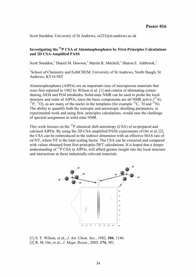
Poster 016 Scott Sneddon, University of St Andrews, [email protected] Investigating the 31P CSA of Aluminophosphates by First-Principles Calculations and 2D CSA-Amplified PASS Scott Sneddon,1 Daniel M. Dawson,1 Martin R. Mitchell,1 Sharon E. Ashbrook,1 1School of Chemistry and EaStCHEM, University of St Andrews, North Haugh, St Andrews, KY16 9ST Aluminophosphates (AlPOs) are an important class of microporous materials that were first reported in 1982 by Wilson et al. [1] and consist of alternating corner-sharing AlO4 and PO4 tetrahedra. Solid-state NMR can be used to probe the local structure and order of AlPOs, since the basic components are all NMR active (27Al, 31P, 17O), as are many of the nuclei in the templates (for example 13C, 1H and 15N). The ability to quantify both the isotropic and anisotropic shielding parameters, in experimental work and using first- principles calculations, would ease the challenge of spectral assignment in solid-state NMR. This work focuses on the 31P chemical shift anisotropy (CSA) of as-prepared and calcined AlPOs. By using the 2D CSA-amplified PASS experiments of Orr et al. [2], the CSA can be reintroduced in the indirect dimension with an effective MAS rate of ωr/NT, where NT is the total scaling factor. The CSA can be extracted and compared with values obtained from first-principles DFT calculations. It is hoped that a deeper understanding of 31P CSA in AlPOs, will afford greater insight into the local structure and interactions in these industrially-relevant materials.
[1] S. T. Wilson, et al., J. Am. Chem. Soc., 1982, 104, 1146. [2] R. M. Orr, et al., J. Magn. Reson., 2005, 174, 301.

35
Poster 017 Jonathan Lamley, University of Warwick, [email protected] Simultaneous Acquisition of Homonuclear and Heteronuclear Long-Distance Contacts with Time-Shared Third Spin Assisted Recoupling Jonathan M. Lamley,1 Józef R. Lewandowski1 1Department of Chemistry, University of Warwick It is well known that low inherent sensitivity is a major limiting factor of NMR and its application to the study of large biomolecules, often leading to acquisition timescales on the order of days or weeks for 2D and higher dimensionality spectra. In solution-state NMR, so-called time-shared experiments have been shown to cut experimental times by effectively multiplying the amount of information gained per experiment[1-3], but their use is uncommon in the solid state. We present a time-shared Third Spin Assisted Recoupling (TSTSAR) experiment that allows for simultaneous acquisition of long-distance homonuclear (13C-13C) and heteronuclear (15N-13C) contacts in biomolecular solids under magic angle spinning, with a view to gaining complementary distance constraints for biological systems. TSTSAR is demonstrated to lead to substantial time savings and to increase the information content of 2D correlation spectra, without or with very little loss of peak intensity compared to each experiment performed separately. References: 1. Frueh D.P., Arthanari H. and Wagner G., Journal of Biomolecular NMR, 33, 187 – 196 (2005) 2. Parella T. And Nolis P., Concepts in Magnetic Resonance Part A, 36A, 1 – 23 (2010) 3. Kodama Y., Reese M.L., Shimba N., Ono K., Kanamori E., Dotsch V., Noguchi Y., Fukunishi Y., Suzuki E., Shimada I. And Takahashi H., Journal of Structural Biology, 174, 434 – 442 (2011) Acknowledgements: We thank Prof. Steven Brown for providing the samples. This research was supported by the EPSRC (UK).

36
Poster 018 Jonathan Katz, University of Sussex, [email protected] Probing Small Molecule Aggregation Phenomena using NMR Spectroscopy and Small Molecule Probes Jonathan R. Katz, Iain J. Day School of Life Science, University of Sussex,Falmer, Brighton, BN1 9QJ Aggregation, one form of which is the grouping of large planar aromatic molecules through a π − π non-covalent interaction, is known to occur in systems such as dyes and pigments1,2, and various drug molecules3,4. Increasingly larger aggregates present with slower rates of diffusion and amplified magnetic shielding due to an enhancement of the ring shielding affect in neighbouring molecules. Nuclear Magnetic Resonance can be employed as an effective tool in the investigation of such changes. Using a pulsed field gradient echo experiment to monitor diffusion coefficients5 or tracking concentration dependent changes in chemical shifts6 it becomes possible to quantify the size and degree of aggregation. These approaches however, can fall victim to complications such as spectral crowding and extensive line broadening, making detailed analysis difficult or impossible. Here we aim to apply the already accepted NMR toolbox in a new way. By introducing a reporter probe molecule with a unique magnetically active nucleus such as 19F or 31P into a self-aggregating system it becomes possible to ameliorate those spectral complications and hence provide a new means of monitoring these changes. With the introduction of Fluorophenol (1 mol%) into a range of samples with varying Sunset Yellow concentrations, we will show that it becomes possible to uncover information about the aggregates via the monitoring of the new unique fluorine marker. References [1] M. P Renshaw and I. J. Day, The Journal of Physical Chemistry B, 2010, 114, 10032–10038. [2] D. Hazafy, M.-v. Salvia, A. Mills, M. G. Hutchings, M. P Evstigneev and J. A. Parkinson, Dyes and Pigments, 2011, 88, 315–325. [3] D. B. Davies, L. N. Djimant and A. N. Veselkovb, Journal of Chememical Society, Faraday Transactions, 1996, 92, 383–390. [4] J. Chairs and N. Dattagupta, Biochemistry, 1982, 21, 3927–3932. [5] C. J. Johnson, Progress in Nuclear Magnetic Resonance Spectroscopy, 1999, 34, 203–256. [6] A. Veselkov, A. Lantushenko, D. Veselkov and D. Davies, Journal of Structural Chemistry, 2002, 43, 234–241.

37
Poster 019 Susan Law, University of Birmingham, [email protected] Size Distributions of Reverse Micelles using NMR Diffusion Measurements. Susan J. Law and Melanie M. Britton School of Chemistry, University of Birmingham, Edgbaston, Birmingham, B15 2TT, U.K. This work reports a novel method for evaluating the size distributions of water-in-oil (w/o) reverse micelles (RMs) using NMR diffusion measurements.1 Reverse micelles (w/o) are nano-sized water droplets surrounded by surfactant molecules in an organic continuous phase. They are of great interest, as they provide adaptable environments for chemical reactions, as well as providing a model for biological systems.2 Knowing the RM size distribution is crucial in understanding the chemistry, properties and reaction kinetics of RMs. Traditionally, Dynamic Light Scattering (DLS) has been used to characterise the size distributions of RMs, however, this technique has a number of limitations, which include a need for samples to be dust-free and transparent. Frequently errors can also be introduced into size distributions by the analysis used. In this poster, we show an alternative method for droplet sizing using NMR. This technique is not affected by many of the problems which affect DLS, but also has several advantages. Diffusion coefficients (D) of RMs were determined by measuring D for surfactant molecules contained in the RMs, acquired using pulsed gradient stimulated echo (PGSTE) experiments. Diffusion coefficient distributions, G(D), were produced using the inverse Laplace transform (a constrained regularization method)3. Size distributions were produced from the G(D) data using the Stokes-Einstein relation. AOT/n-octane/H2O and CTAB/hexanol/H2O microemulsions were studied and the effect of ω (ω = [H2O]/[surfactant]) and the presence of additives were investigated.4,5 Our method produced distributions in good agreement with the literature values produced by DLS, and was able to detect monomodal and bimodal distributions.
Figure 1. Droplet size distributions for RMs in AOT/n-octane/H2O microemulsions at (a) ω = 15, droplet fraction = 0.15 and (b) at ω = 12 and droplet fraction = 0.5, loaded with [H2SO4] = 0.25M ,[NaBrO3] = 0.16 M and [malonic acid] = 0.25 M . (1) Law, S.J.;Britton, M.M. Langmuir 2012 (submitted) (2) Levinger, N. E. Science 2002, 298, 1722. (3) Fordham, E. J.; Sezginer, A.; Hall, L. D. J. Magn. Reson., Ser. A 1995, 113, 39. (4) Alvarez, E. V.; Carballido-Landeira, J. et. al. J. Chem. Phys. 2011, 134, 94512 (5) Vanag, V. K.; Epstein, I. R. Phys. Rev. Lett. 2003, 90, 098301.
38
Poster 020 Heather Rose, University of Birmingham, [email protected] Magnetic Resonance Imaging of Hydrodynamic Instabilities in Porous Media H Rose* and MM Britton* *School of Chemistry, University of Birmingham, Edgbaston, Birmingham, B15 2TT Viscous fingering is a hydrodynamic instability formed when there is an interface between two solutions of different viscosity. At such an interface flow becomes non-linear and the interface deforms producing finger-like tendrils. Such instabilities are of interest in a variety of applications, such as extraction of oil from pipes[1], instabilities in combustion[2] and growth of microorganism colonies[3]. In recent years, there is an increasing interest in systems that involve chemical reactions at the interface. The products of these reactions effect the formation of the fingers, either through changing the viscosity or altering the composition of the porous media. Both experimental[4, 5] and theoretical[6] studies have been conducted on these systems. However, experimental work has typically relied on investigating systems optically using Hele-Shaw cells[4, 5]. This pseudo-2D experiment provides limited information on the formation of instabilities in a 3D system. We have used magnetic resonance imaging (MRI) to visualise the formation of viscous fingers at various reactive interfaces. The production of both worm-like micelles and precipitate at the interface has been investigated. MRI’s ability to probe opaque systems makes it uniquely amenable to directly investigate flow instabilities in these systems in ‘real’ porous media, such as a packed bed. 3D imaging was used to monitor the development of these instabilities and characterise their growth. [1] G. Paşa, and O. Titaud, Transport in porous media 58 (2005). [2] A. Goriely, and M. Tabor, Physical Review Letters 90, 4 (2003). [3] O. Zik, Z. Olami, and E. Moses, Physical Review Letters 81, 3868 (1998). [4] Y. Nagatsu et al., Physical Review E 77 (2008). [5] T. Podgorski et al., in Physical Review E2007). [6] T. Gerard, and A. De Wit, Physical Review E 79, 10 (2009).

39
Poster 021 Amanda Mills, University of Birmingham, [email protected] Aggregation behaviour of lipid and MALDI matrices in methanol using NMR Amanda Mills*, Melanie Britton* *School of Chemistry, University of Birmingham, Edgbaston, Birmingham, B15 2TT NMR diffusion experiments are reported which have been used to probe possible aggregation of MALDI matrix compounds with lipids at various concentrations. Matrix Assisted Laser Desorption Mass Spectrometry (MALDI-MS) is a soft ionisation technique employed for the analysis of biomolecules and large organic molecules like lipids1. MALDI uses a matrix compound to promote ionisation of the analyte1 e.g. lipid, and the matrix and analyte are solvated using methanol. Investigations of lipids show that some matrix compounds produce more abundant lipid peaks than others. This was thought to be due to interactions between lipid and matrix compounds through self-aggregation. As lipids possess both hydrophobic and hydrophilic regions, they are expected to form self-aggregated assemblies in solution. Possible assemblies include lipid bilayers, micelles and liposomes (vesicles) in methanol. The formation of these aggregated structures was tested using pulsed field gradient stimulated echo (PGSTE)experiments which measure the translational diffusion of the lipid, matrix and solvent2. The diffusion coefficient of the solvent methanol was found to decrease on increasing concentration, suggesting the formation of aggregated structures. No change in the diffusion coefficient of the lipid or matrix molecules was observed. The behaviour of the methanol can be explained by the formation of a liposome (vesicle) aggregation assembly, where some of the methanol molecules can reside both inside and outside the liposome.
(1) Stroobant, E. d. H. a. V. Mass Spectrometry: Principles and Applications; 3rd ed.; John Wiley & Sons Ltd, 2007. (2) Price, W. S. Concepts in Magnetic Resonance 1998, 10, 197.
40
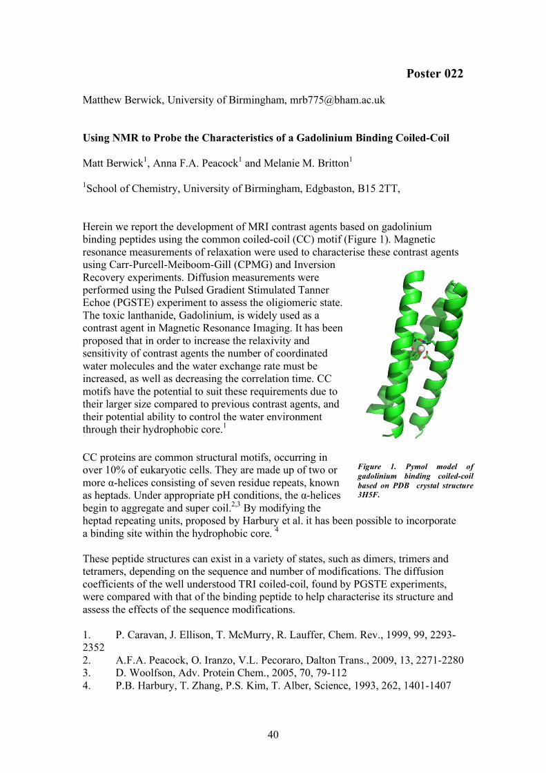
Poster 022 Matthew Berwick, University of Birmingham, [email protected] Using NMR to Probe the Characteristics of a Gadolinium Binding Coiled-Coil Matt Berwick1, Anna F.A. Peacock1 and Melanie M. Britton1 1School of Chemistry, University of Birmingham, Edgbaston, B15 2TT, Herein we report the development of MRI contrast agents based on gadolinium binding peptides using the common coiled-coil (CC) motif (Figure 1). Magnetic resonance measurements of relaxation were used to characterise these contrast agents using Carr-Purcell-Meiboom-Gill (CPMG) and Inversion Recovery experiments. Diffusion measurements were performed using the Pulsed Gradient Stimulated Tanner Echoe (PGSTE) experiment to assess the oligiomeric state. The toxic lanthanide, Gadolinium, is widely used as a contrast agent in Magnetic Resonance Imaging. It has been proposed that in order to increase the relaxivity and sensitivity of contrast agents the number of coordinated water molecules and the water exchange rate must be increased, as well as decreasing the correlation time. CC motifs have the potential to suit these requirements due to their larger size compared to previous contrast agents, and their potential ability to control the water environment through their hydrophobic core.1
CC proteins are common structural motifs, occurring in over 10% of eukaryotic cells. They are made up of two or more α-helices consisting of seven residue repeats, known as heptads. Under appropriate pH conditions, the α-helices begin to aggregate and super coil.2,3 By modifying the heptad repeating units, proposed by Harbury et al. it has been possible to incorporate a binding site within the hydrophobic core. 4 These peptide structures can exist in a variety of states, such as dimers, trimers and tetramers, depending on the sequence and number of modifications. The diffusion coefficients of the well understood TRI coiled-coil, found by PGSTE experiments, were compared with that of the binding peptide to help characterise its structure and assess the effects of the sequence modifications. 1. P. Caravan, J. Ellison, T. McMurry, R. Lauffer, Chem. Rev., 1999, 99, 2293-2352 2. A.F.A. Peacock, O. Iranzo, V.L. Pecoraro, Dalton Trans., 2009, 13, 2271-2280 3. D. Woolfson, Adv. Protein Chem., 2005, 70, 79-112 4. P.B. Harbury, T. Zhang, P.S. Kim, T. Alber, Science, 1993, 262, 1401-1407
Figure 1. Pymol model of gadolinium binding coiled-coil based on PDB crystal structure 3H5F.

41
Poster 023 Lucy Hawarden, University of East Anglia, [email protected] Predicting the Physical Stability of Polymer-Stabilised Amorphous Dispersions for Oral Drug Delivery: Part I - Indomethacin studies Lucy Hawarden1, 2 Sarah Nicholson 3 Yaroslav Khimyak,1,2 1 University of East Anglia 2 University of Liverpool 3 Bristol-Myers Squibb Pharmaceuticals, Moreton Poor physical stability is a huge limiting factor in the development and use of amorphous drugs within the pharmaceutical industry. Amorphous drugs are attractive due to increased solubility and bioavailability; however they unpredictably revert to the thermodynamically stable insoluble crystalline form [1]. Accurate prediction of physical stability on a molecular level would be a great advantage – polymers can be used to stabilise amorphous systems but we need a firm understanding of the mechanism of stabilisation on a molecular level. Indomethacin was systematically chosen as the initial investigative compound as it has been widely formulated and studied both as an amorphous drug and dispersion [1- 3]. There is a wealth of information available, however only a handful of studies have utilised solid-state NMR [1, 3, 4]. This technique has the ability to provide molecular level information on structure and dynamics and could therefore be a powerful tool to predict the physical stability of polymer-stabilised amorphous systems. Indomethacin was successfully formulated as a pure amorphous drug [2] and as multiple solid dispersions (varying drug:polymer ratios) with PVP-VA [2]. Initial characterisation with PXRD, DSC and solid-state NMR confirmed amorphous behaviour and determined Tg values and spectral peak assignments. Variable temperature solid-state NMR studies highlighted a possible recrystallisation of amorphous indomethacin to the α-polymorph. VT 1H-13C CP/MAS NMR, 13C {1H} and relaxation studies highlighted a change in mobility of the drug when formulated with different levels of polymer, indicating the possible formation of drug-rich domains within the higher loaded dispersions. CP kinetics studies also showed a difference in relaxation behaviour between pure amorphous indomethacin, solid dispersions and pure PVP-VA. 1. Guilbaud, J.-B.C., Linda; Khimyak, Yaroslav, Encapsulation of Indomethacin in PVP: Solid-State NMR Studies. Macromol. Symp., 2007. 251: p. 41 - 46. 2. Matsumoto, T. and G. Zografi, Physical properties of solid molecular dispersions of indomethacin with poly(vinylpyrrolidone) and poly(vinylpyrrolidone-co-vinylacetate) in relation to indomethacin crystallization. Pharmaceutical Research, 1999. 16(11): p. 1722-1728. 3. Apperley, D.C., et al., Characterisation of indomethacin and nifedipine using variable-temperature solid-state NMR. Magnetic Resonance in Chemistry, 2005. 43(11): p. 881-892. 4. Pham, T.M.e.a., Analysis of Amorphous Solid Dispersions Using 2D Solid-State NMR and 1H T1 Relaxation Measurements. Molecular Pharmaceutics, 2010. 7(5): p. 1667 - 1691.

42

43
Delegates Richard Arnott Agilent Technologies Michael Beaumont University of Liverpool Matthew Berwick University of Birmingham Craig Butts University of Bristol Melanie Britton University of Birmingham Tim Claridge University of Oxford Anna Codina Bruker UK Eirian Curzon Bruker UK Daniel Dawson University of St Andrews Iain Day University of Sussex Stephen Day University of Warwick Rob Evans University of Manchester Caroline Fost University of Bristol Andrew Gibbs Bruker UK Peter Gierth Bruker UK Lucy Hawarden University of East Anglia
John Hollerton GlaxoSmithKline R&D Peter Howe Syngenta Ltd Catharine Jones University of Bristol Rebecca Joyce University of Sussex Jonathan Katz University of Sussex James Keeler University of Cambridge Robert Kelly University of Warwick Alan Kenwright University of Durham Yaroslav Khimyak University of East Anglia Scott King University of Warwick Jonathan Lamley University of Warwick Susan Law University of Birmingham John Lowe University of Bath Sandra van Meurs Bruker UK Amanda Mills University of Birmingham John Moran Institute of Technology Tallaght Brian Murray Institute of Technology Tallaght
44
Smita Odedra University of Glasgow Victoria Paget ACD/Labs John Parksinson University of Strathclyde Gregory Rees Bruker UK Nick Rees University of Oxford Frederik Romer University of Warwick Heather Rose University of Birmingham Scott Sneddon University of St Andrews Anna-Winona Struck University of Manchester Paul Tan Lilly UK Godiraone Tatolo University of Bristol Antoine Vallatos University of Birmingham Neil Wells University of Southampton Corinne Wills Newcastle University Zenefar Yeasmin Institute of Technology Tallaght