rtca card an 12 prod 2 - ebiotrade.com · xcelligence system rtca cardio instrument as an assay...
TRANSCRIPT

xCELLigence System RTCA Cardio Instrument Preclinical Cardiac Safety Assessment using Mouse ES-Derived Cor.At® Cardiomyocytes
Application Note No. 12 /September 2010
For life science research only.Not for use in diagnostic procedures.
System
2
Over the last two decades, a number of high-profile drugs have either been withdrawn or have been issued safety warnings by regulatory agencies due to adverse cardiac effects.1 In addition, lead compounds or drug candidates are frequently terminated at late stages of drug development due to cardiac safety concerns. Both of these factors can have significant financial impact on the overall cost of drug discovery. Consequently, both pharmaceutical companies and regulatory agencies have taken steps and imple-mented procedures to address these issues. The underlying causes of adverse drug effects on the heart vary, but can be broadly categorized into either direct or indirect effects. Most of the drugs that have been withdrawn from the market due to cardiotoxicity appear to impact heart function by directly interfering with well-coordinated electrical activity in the heart.2 The electrical activity of the heart itself results from directional and selective movement of calcium, sodium, and potassium ions across channels in the membrane of cardio-myocytes. Interfering with this highly orchestrated movement of ions across the cardiomyocyte membrane will ultimately affect the excitation/ contraction coupling cycle of the heart. This in turn can lead to a life-threatening form of ventricular arrhythmia called Torsades de Pointes (TdP).1
While the exact molecular mechanisms resulting in TdP are subject to debate and still under investigation, it is well accepted that delayed ventricular repolarization (otherwise, known as QT prolongation) is a surrogate biomarker linked with an enhanced proarrhythmic risk.2 The main component of ventricular repolarization in humans is the rapidly activated delayed rectifier potassium current IKr , and the human hERG gene product is the principal ion channel responsible.2 A block of the hERG channel in cardiomyocytes is thought to prolong ventricular action potential duration (APD). The subsequent defective repolar-ization gives rise to early after depolarization (EAD), triggering TdP.2 Recognizing the central role of the hERG channel in the repolarization phase of the action potential and its propensity to interact with drugs, current regulatory guidelines consider hERG assays a major component of an integrated cardiac risk assessment.
It must be noted, however, that hERG channel interaction alone is not exclusively predictive of ventricular arrhythmia. Certain compounds may be able to inhibit multiple ion channels and therefore counteract the adverse effect of the hERG channel block, as has been shown for verapamil.2 Also, induction of arrhythmia by mechanisms other than hERG block cannot be discounted. It would be highly desirable to utilize an in vitro model system that can be used to assess the integrated response of all ion channels and non-ion channel targets involved in the process of excitation/contraction coupling earlier in the drug discovery process. The recent introduction of stem cell technology and importantly, stem cell-derived cardiomyocytes for safety testing could provide a viable solution.3 In this application note, we have used mouse embryonic stem cell-derived cardiomyocytes (Cor.At®) from Axiogenesis together with the xCELLigence System RTCA Cardio Instrument as an assay system for preclinical in vitro cardiac safety assessment. Cor.At® cells are a 100% pure population of spontaneously beating cardio- myocytes that contain all the relevant ion channels and cytoskeletal components found in a typical cardiomyocyte. Our data show that combining a physiologically relevant cardiomyocyte model system such as Cor.At® cells, together with the capabilities of the xCELLigence System RTCA Cardio Instrument, can provide predictive and mechanistic toxicity data. This in turn would allow researchers to make more accurate assessments of cardiac toxicity profiles of lead candidate compounds.
Introduction
3
Materials and Methods
Cor.At® Cells: Mouse ES cell-derived cardiomyocytes (Cor.At®) were obtained from Axiogenesis AG (Cat. No. XCAC-1010E, Lonza Cologne, Germany). Cells were kept in liquid nitrogen until thawed, and cultured according to the protocol provided by Axiogenesis with slight modifications. Briefly, each well of the E-Plate Cardio 96 was coated with 50 µl of a 1:100 diluted fibronectin (FN) solution (F1114, Sigma-Aldrich, USA), and incubated at +4˚C overnight. Subsequent to FN removal, culture wells were washed with PBS and followed by cell seeding. Cells were thawed at +37˚C in a water bath, transferred to a 15 ml conical tube containing 9 ml fresh Cor.At® complete culture medium (XCAM-250E, Lonza Cologne, Germany), and centrifuged at 100 x g for 5 minutes. Culture medium was replaced with a small volume of fresh Cor.At® complete culture medium containing puromyocin at a final concentration of 10 µg/ml. Cells were counted and the percent viable cells determined using the trypan blue exclusion method.
Dynamic Monitoring of Cor.At® Cardiomyocyte Beating using the xCELLigence System RTCA Cardio Instrument. About 40,000–60,000 viable cells were seeded per well of an E-Plate Cardio 96 (Roche and ACEA Biosciences). Cells were continually monitored using the RTCA Cardio Instrument (Roche Applied Science and ACEA Biosciences). Cell culture medium was replaced once a day. Typically, drug treatment was initiated 60–80 hours after cell seeding depending on seeding density.
Data collection is controlled by the RTCA Cardio Software, which operates the hardware and allows the user to define the sampling frequency and sampling window. Sampling frequency is defined as the number of times during an experimental run that cell beating is sampled. The sampling window is defined as the duration of time that cell beating is measured. Ten minutes prior to treatment, cells were sampled every minute for 20 seconds to establish a baseline recording. After treatment, sampling frequency was adjusted to every minute for the first hour, every 5 minutes for the second hour, and every 15 minutes for 3–24 hours. The sampling window for each recording was fixed at 20 seconds. After data acquisition, the RTCA Cardio Software was used to calculate parameters, such as beating frequency, amplitude, normalized beating frequency, normalized amplitude, beat duration, and the beating rate irregularity index. In addition, the RTCA Cardio Software performs basic statistics (average, standard deviation), and calculates half-maximal response concentrations.

4
Results
Real-Time and Dynamic Monitoring of Cor.At® Cardiomyocyte Attachment, Growth, and Beating using the xCELLigence System RTCA Cardio Instrument
The RTCA Cardio Instrument extends the current capabilities of the xCELLigence System portfolio (including RTCA SP, MP, and DP Instruments) to the functional monitoring of cardiomyocyte beating for quantitatively assessing cardiotoxicity. At the core of the system are the microelectronic cell sensor arrays that are integrated into the bottom of 96-well microplates (E-Plate Cardio 96). With its improved data acquisition rate (12.5 millisecond update rate/plate), the RTCA Cardio Instrument can temporally resolve the contraction/relaxation cycle of cardiomyocyte beating in the context of short-term and long-term assays.
The RTCA Cardio Instrument consists of the following main components (see Figure 1):
RTCA Cardio Control Unit
RTCA Cardio Analyzer
RTCA Cardio Station
E-Plate Cardio 96
To characterize cell beating activity, Cor.At® cells were seeded in wells of the E-Plate Cardio 96 at a density of 60,000 cells/well. Cells were monitored up to 96 hours in culture (see Figure 2A), and beating activity was recorded once every hour for 20 seconds. Snapshots of beating activity at 12, 24, 48, 72, and 96 hours are shown in Figure 2B. Within 24 hours after seeding the cells, no concerted cell beating activity could be detected even though clusters of asynchronously beating cardiomyocytes (cells which have not yet formed a syncytium) could be detected by microscopy (data not shown). Within 48 hours, individual clusters formed clear connections and the entire monolayer of cardiac cells in the bottom of the well was beating in synchrony. Correspondingly, using impedance recording, reproducible cell beating activity was detected by 48 hours (see Figure 2B). The beating rate for these cardiomyocytes is approximately 80 beats/minute, progressively increasing to 250 beats/minute after a month in culture (data not shown).
Figure 1: The xCELLigence System RTCA Cardio Instrument.The RTCA Cardio Instrument is a label-free, real-time system for dynamic monitoring of cardiomyocyte beating and assessment of cardiotoxicity. It is composed of the RTCA Cardio Control Unit, the RTCA Cardio Analyzer, the RTCA Cardio Station (which is placed in a CO2 incubator), and the E-Plate Cardio 96 which contains interdigitated gold microelectrodes in the bottom of each well.

5
Figure 2: Characterization of Cor.At® attachment, growth, and beating using the RTCA Cardio Instrument.(A) Cor.At® cells were seeded in the wells of the E-Plate Cardio 96 and allowed to adhere and form a syncytium. Cells were cultured for up to 96 hours and monitored using the RTCA Cardio Instrument at regular intervals. Growth medium in the wells was changed daily. (B) Beating activity and the CI profile of Cor.At® cells recorded by the RTCA Cardio Instrument at the indicated time points after cell seeding. The beating rate (1/minute) and amplitude (delta CI) were quantified using the RTCA Cardio Software. The data is the average of 8 wells -/+ standard deviation. A duration of 5 seconds of recording time is shown.
Results continuedC
ell I
nd
ex
Time (hours)
A
- - - - - -
16-14-12-10-8-6-4-2-0-
0 20 40 60 80 100
Post Cultivation
(hours)Beating Pattern
Beating Frequency
(1/minute ± SD)N=8
Amplitude(Delta CI)
N=8
Beat Duration(ms)
Time to Max (Tr)(ms)
Decay Time (Td)(ms)
12 NA NA NA NA NA
24NA
NA NA NA NA
48 80 ± 5.7 0.14 ± 0.003 142 ± 4.6 38 ± 1.4 124 ± 12.0
72 145 ± 2.7 0.24 ± 0.005 98 ± 3.0 29 ± 5.1 88 ± 7.2
96 127 ± 2.8 0.33 ± 0.007 105 ± 2.4 31 ± 3.3 97 ± 6.4
B

6
Pharmacological Assessment of Cor.At® Beating Activity using the xCELLigence System RTCA Cardio Instrument
Using a pharmacological approach, ion channel and non-ion channel targets involved in the regulation of excitation/contraction coupling in Cor.At® cells were evaluated. For these experiments, Cor.At® cells were thawed, seeded in the wells of the E-Plate Cardio 96. Cells were cultured for 3 days, treated with increasing concentrations of the compound to be tested, and monitored for 12 hours using the RTCA Cardio Instrument.
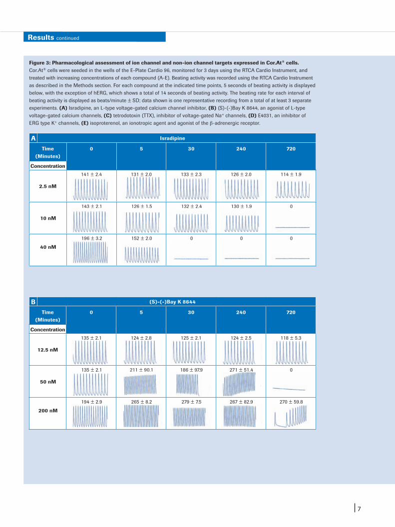
Assessment of voltage-gated calcium channelsIsradipine, a well known voltage activated L-type calcium channel blocker of the dihydropyridine class, induces progressive time- and dose-dependent decrease and inhibition of beating activity. Isradipine was applied to show that calcium entry through L-type calcium channels is required for cell beating (see Figure 3A). The IC50 for isradipine-induced inhibition of beating activity relative to the normalized beating rate and amplitude is 19.7 nM and 42.3 nM, respectively (at 5-minute time points after compound addition; see Table 1). These values are consistent with the efficacy of isradipine mea-
sured in isolated rabbit heart4 and in recombinant HEK-293 cells stably expressing the human Cav1.2.5 The compound (S)-(-)Bay K 8644 is also of the dihydropyridine class, but acts in an agonistic mode to activate voltage-gated calcium channels. Treatment of Cor.At® cells with (S)-(-)Bay K 8644 resulted in a dose- and time-dependent increase in the beating rate which persisted for up to 12 hours at higher concentrations (see Figure 3B). The increase in beating rate is consistent with the reported iono-tropic action of this compound and the half-maxi-mal concentration obtained (77 nM) for beating rate (see Table 1) is similar to previously published reports using rat ventricular myocytes.6
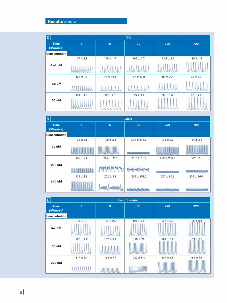
Assessment of sodium channel modulatorsVoltage-gated Na+ channels are primarily responsible for the Na+ current and the depolarization phase of cardiac action potential. Treatment of Cor.At® cells with tetrodotoxin (TTX), a potent and selec-tive inhibitor of voltage-gated Na+ channels, led to a dose-dependent decrease in the beating rate of Cor.At® cells, which is sustained at the higher concentrations for the entire duration of 12 hours (see Figure 3C). The apparent isradipine concen-tration value obtained for inhibition of Cor.At® beating is approximately 0.3 uM (see Table 1).
Table 1: Dose-response profiling of modulators of ion channel and non-ion channel targets in Cor.At® cells. RTCA Cardio Software was used to analyze dose-dependent effects of the indicated compounds on the beating activity of Cor.At® cells and to derive IC50 and EC50 values. The parameters, normalized beating rate, normalized amplitude, beat duration, and beating rhythm irregularity, are part of the analysis package of the RTCA Cardio Software for quantifying cell beating activity.
Results continued
Parameter Normalized Beating Rate
(IC50; M)
Normalized Amplitude (IC50; M)
Beat Duration(IC50; M)
Beat Rate Irregularity (IC50; M)
Compound
Isradipine 19.7 x 10⁻⁹ M 42.3 x 10⁻⁹ M N.A. N.A.
(S)-(-)-Bay K8644
77.3 x 10⁻⁹ M N.A. N.A. 24.2 x 10⁻⁹ M
E4031 26.9 x 10⁻⁹ M N.A. 2.4 x 10⁻⁷ M 56.6 x 10⁻⁹ M
TTX 2.8 x 10⁻⁷ M 5.3 x 10⁻⁸ M N.A. N.A.
Isoproterenol 1.4 x 10⁻⁸ M N.A. 7.1 x 10⁻⁹ M N.A.
7
Results continued
Figure 3: Pharmacological assessment of ion channel and non-ion channel targets expressed in Cor.At® cells.Cor.At® cells were seeded in the wells of the E-Plate Cardio 96, monitored for 3 days using the RTCA Cardio Instrument, and treated with increasing concentrations of each compound (A-E). Beating activity was recorded using the RTCA Cardio Instrument as described in the Methods section. For each compound at the indicated time points, 5 seconds of beating activity is displayed below, with the exception of hERG, which shows a total of 14 seconds of beating activity. The beating rate for each interval of beating activity is displayed as beats/minute ± SD; data shown is one representative recording from a total of at least 3 separate experiments. (A) Isradipine, an L-type voltage-gated calcium channel inhibitor, (B) (S)-(-)Bay K 8644, an agonist of L-type voltage-gated calcium channels, (C) tetrodotoxin (TTX), inhibitor of voltage-gated Na+ channels, (D) E4031, an inhibitor of ERG type K+ channels, (E) isoproterenol, an ionotropic agent and agonist of the β-adrenergic receptor.
Isradipine
Time (Minutes)
0 5 30 240 720
Concentration
2.5 nM
141 ± 2.4 131 ± 2.0 133 ± 2.3 126 ± 2.0 114 ± 1.9
10 nM
143 ± 2.1 126 ± 1.5 132 ± 2.4 130 ± 1.9 0
40 nM
196 ± 3.2 152 ± 2.0 0 0 0
A
(S)-(-)Bay K 8644
Time (Minutes)
0 5 30 240 720
Concentration
12.5 nM
135 ± 2.1 124 ± 2.8 125 ± 2.1 124 ± 2.5 118 ± 5.3
50 nM
135 ± 2.1 211 ± 90.1 186 ± 97.9 271 ± 51.4 0
200 nM
194 ± 2.9 265 ± 8.2 279 ± 7.5 267 ± 82.9 270 ± 59.8
B
8
Results continued
TTX
Time (Minutes)
0 5 30 240 720
Concentration
0.41 uM
127 ± 2.3 100 ± 1.7 106 ± 1.7 113.2 ± 1.9 110 ± 1.5
4.5 uM
126 ± 3.5 77 ± 12.1 87 ± 13.0 91 ± 2.2 89 ± 4.8
50 uM
124 ± 2.8 87 ± 2.6 95 ± 3.1 90 ± 1.6 88 ± 2.6
C
E4031
Time (Minutes)
0 5 30 240 720
Concentration
50 nM
125 ± 2.5 100 ± 2.3 334 ± 219.2 135 ± 2.3 124 ± 2.0
200 nM
132 ± 2.4 244 ± 82.7 267 ± 78.3 459 ± 293.6 122 ± 2.0
800 nM
129 ± 1.9 202 ± 3.1 266 ± 252.6 224 ± 66.8 226 ± 86.6
D
Isoproterenol
Time (Minutes)
0 5 30 240 720
Concentration
3.7 nM
104 ± 2.8 104 ± 2.5 121 ± 2.5 127 ± 3.7 125 ± 3.8
33 nM
106 ± 2.6 157 ± 5.2 179 ± 7.6 164 ± 5.8 163 ± 5.8
300 nM
117 ± 3.1 183 ± 7.7 207 ± 5.4 187 ± 5.6 182 ± 7.9
E
9
Results continued
Assessment of potassium channel modulatorsThe rapid activating component of the delayed rectifier current (IKr) is involved in the repolarization phase of the cardiac action potential. This compo-nent is mainly mediated through the ERG channel. The effect of E4031, a potent hERG channel inhibitor, was also tested using Cor.At® cells in a time- and dose-dependent manner (see Figure 3D). As shown, E4031 treatment interrupted the normal rhythmicity of beating, especially at high concen-trations (200 nM–800 nM), resulting in prolonged beat durations accompanied by plateau oscillations. This phenomenon is typical of other hERG blockers (see next section). At the doses tested, the cells appear to recover from the effect of E4031 by 12 hours after treatment. Based on the normalized beating rate and beat rate irregularity parameters, the half-maximal concentrations obtained are 27 nM and 57 nM, respectively (see Table I), consistent with the reported IC50 for E4031 using stem cell-derived human cardiomyocytes using the patch clamp technique.7
Assessment of chronotropic agentsActivation of the sympathetic nervous system and neuro-hormonal regulation through the β-adrenergic receptor is a major mechanism controlling contractility and beating rate of cardiac tissue.8 The protein machinery responding to β-adrenergic receptor stimulation is present and functional within Cor.At® cells, and the corresponding receptor agonists are well-characterized chronotropic and ionotropic agents. We therefore sought to test whether the effect of β-adrenergic receptor stimula-tion on cardiomyocyte beating could be detected by the RTCA Cardio Instrument.
Treatment of Cor.At® cells with isoproterenol, a β-adrenergic receptor agonist, increased the beating frequency of Cor.At® cells in a dose- and time-dependent manner simultaneously decreasing the overall duration of each beat (see Figure 3E). This effect is similar to the L-type calcium channel agonist (S)-(-)Bay K 8644 (see Figure 3B), consistent with the observation that stimulation of β-adrenergic receptors leads to activation of L-type calcium channels.
Using the xCELLigence System RTCA Cardio Instrument for Preclinical Safety Studies
To test the utility of the RTCA Cardio Instrument for preclinical cardiac safety screening, two complementary approaches were undertaken. First, four drugs withdrawn from the market due to increased incidence of TdP, were screened in a dose-response manner using Cor.At® cells (see Figure 4A). These compounds have subsequently been shown to directly inhibit hERG channel activity. All four compounds significantly were found to affect beating rate in a dose-dependent manner (see Figure 4A). Each produced beating irregularities consistent with those observed for E4031 in terms of morphology, reflecting a common underlying mechanism.
Next, we tested pentamidine which has been shown to affect the transport of the hERG channel to the membrane in heterologous expression systems and in cardiomyocytes, due to delayed repolarization. Since this compound affects hERG channel activity indirectly, its effect is likely to be manifested in a time-dependent manner making it difficult to capture using patch clamp techniques, which are limited to only a few minutes of recording time.
Administration of pentamidine at a final concentration of 20 uM had no noticeable effect on beating rate and amplitude through the 240-minute time point (see Figure 4B). By 900 minutes after compound addition, the beating rate slowed down and the beat duration was significantly delayed, most likely due to an extended repolarization phase. These observations highlight the importance of monitoring compound effects in a time-dependent manner to resolve the effect on both early and longer term function of cardiomyocytes and gain greater mechanistic insight.

10
Results continued
Droperidol Astemizole Cisapride Sertindole
A PentamidineBeating Profile
Norm Beating
Rate
Beat Duration
(Sec)
0 min 1 ± 0.03 0.37 ± 0.02
5 min 0.91 ± 0.02 0.41 ± 0.03
30 min 0.92 ± 0.02 0.41 ± 0.01
240 min 0.97 ± 0.02 0.36 ± 0.03
900 min 0.064 ± 0.01 5.8 ± 0.04
1200 min 0.04 5.3
B
Figure 4: Mechanism-based cardiotoxicity profiling using the RTCA Cardio Instrument. (A) The indicated drugs, which have been withdrawn from the market due to increased incidence of TdP arrhythmia, were screened in a dose-response manner in Cor.At® cells. For each compound, a total of 5 seconds of beating activity is displayed. For astemizole, cisapride, droperide, and sertindole, the dose-response profiles are shown at 30 minutes, 15 minutes, 180 minutes, and 165 minutes after compound addition, respectively. Each compound was serially diluted twofold from a 20 μM starting concentration, except cisapride, which started at 10 μM. (B) Cor.At® cells were seeded in the wells of E-Plate Cardio 96 and treated on day 3 with 20 μM pentamidine. Beating activity was monitored at the indicated time windows after compound treatment and quantified with respect to beat duration as described in the Methods section.
Low
High
400 ms
Co
ntr
ols
11
Discussion
In this application note, we introduce a micro- electronic-based cardiomyocyte monitoring system, the RTCA Cardio Instrument, for use with stem cell-derived cardiomyocytes (Cor.At®) to assess the cardiac safety of lead compounds and drug candidates during preclinical drug development. This instrument system is designed to capture real-time information regarding cardiomyocyte beating, viability, including morphological changes that may occur as a result of compound treatment. The real-time aspect of this system allows capturing both acute and long term effects of compound exposure.
Most if not all in vitro assay systems for cardiac safety screen for surrogates of arrhythmia, such as hERG channel interaction, rather than arrhythmia itself. Assays designed to screen for compounds that may affect repolarization and induce arrhythmia in the context of the whole heart or heart tissue are not implemented until much later in drug development. These include sophisticated, technically demanding, low-throughput and costly procedures such as the Purkinje fiber assay, ventricular wedge assay, and the Langendorff whole heart assay, as well as telemetry experiments in live and anesthetized animals.1 The field of preclinical cardiac safety can greatly benefit from an assay system that assesses in integrated fashion compound action on ion-channel and non-ion channel targets involved in cardiac excitation- contraction coupling.
Data presented here using the RTCA Cardio Instru-ment in conjunction with Cor.At® cells permits an integrated assessment of compound action on multiple targets involved in heart function. This assay system can sensitively and quantitatively detect the effect of compounds on the major ion channels involved in heart function, specifically the calcium, sodium, and potassium channels. Furthermore, ionotropic and chronotropic
compounds that impact the force and rate of heart beating can also be analyzed by this system. From a preclinical cardiac safety perspective, drugs removed from the market due to the induction of ventricular arrhythmias or TdP could be identified. These compounds produced a characteristic beat-ing profile that may be indicative of arrhythmia.
Another major advantage of the assay system described here is its extraordinary time resolution. The RTCA Cardio Instrument has a data update rate of 12.5 ms per 96-well plate. At the same time, the instrument can be used to monitor longer term drug effects up to days and weeks. The utility of time-dependent monitoring of compound action on cardiomyocytes was demonstrated by testing the compound pentamidine, which affects the trans-port of hERG to the plasma membrane, rather than affecting hERG activity directly. The RTCA Cardio Instrument was able to detect the effect of this compound on beating rate and beat duration of Cor.At® cells, which occurred around 15 hours after treatment. Traditional patch clamp experiments would probably not have been able to detect the adverse effect of this drug on cardiomyocytes.
In summary, the xCELLigence System RTCA Cardio Instrument, in conjunction with Cor.At® cells, is a physiologically relevant and predictive assay system for preclinical cardiac safety assessment of lead compounds. The features of this assay system, including time resolution, dynamic monitoring of mechanical beating activity of cardiomyocytes, as well as the 96-well throughput, will provide valuable mechanistic and cytotoxicity information with respect to compound action on the heart.
12
Published byRoche Diagnostics GmbHRoche Applied ScienceWerk Penzberg82372 PenzbergGermany
© 2010 Roche Diagnostics. All rights reserved.
www.xcelligence.roche.com
06480942001 1010
For life science research only.Not for use in diagnostic procedures.
References
Product Cat. No. Pack Size
RTCA Cardio Station 06 417 019 001 1 instrument
RTCA Cardio Analyzer 06 416 993 001 1 instrument
RTCA Cardio Control Unit 06 200 184 001 1 instrument
E-Plate Cardio 96 06 417 051 00106 417 035 001
6 plates 6 x 6 plates
Ordering Information
1. Fermini B, Fossa AA. (2003). “The impact of drug-induced QT interval prolongation on drug discovery and development.” Nat Rev Drug Discov. 2, 439–447.
2. Brown AM. (2005). “HERG block, QT liability and sudden cardiac death.” Novartis Found Symp. 266, 118–131; discussion 131–115, 155–118.
3. Kettenhofen R, Bohlen H. (2008). “Preclinical assessment of cardiac toxicity.” Drug Discovery Today. 13, 702–707.
4. Mellemkjaer S, Bang L, Nielsen-Kudsk F. (1992). “Isradipine dynamics and pharmacokinetics in the isolated rabbit heart.” Pharmacol Toxicol. 70, 366–372.
5. Balasubramanian B, Imredy JP, Kim D, Penniman J, Lagrutta A, Salata JJ. (2009). “Optimization of Ca(v)1.2 screening with an automated planar patch clamp platform.” J Pharmacol Toxicol Methods. 59, 62–72.
6. Zahradnikova A, Minarovic I, Zahradnik I. (2007). “Competitive and cooperative effects of Bay K8644 on the L-type calcium channel current inhibition by calcium channel antagonists.” J Pharmacol Exp Ther. 322, 638–645.
7. Peng S, Lacerda AE, Kirsch GE, Brown AM, Bruening-Wright A. (2010). “The action potential and comparative pharmacology of stem cell-derived human cardiomyocytes.” J Pharmacol Toxicol Methods. 61, 277–286.
8. Bers DM. (2002). “Cardiac excitation-contraction coupling.” Nature. 415, 198–205.
Trademarks
Cor.At® is a registered trademark of AxiogenesisXCELLIGENCE is a trademark of Roche.E-PLATE and ACEA BIOSCIENCES are registered trademarks of ACEA Biosciences, Inc. in the US.Other brands or product names are trademarks of their respective holders.
Disclaimer of License
The RTCA Cardio Analyzer in combination with the RTCA Cardio Station, an RTCA Cardio Software Package (1.x) and an E-Plate Cardio 96 is a real-time cell based assay system covered by US patent No. 7,192,752 (exp. 11/10/2023), No. 7,470,533 (exp. 1/12/2025), No. 7,560,269 (exp. 10/24/2025).