running head: effects of inflammation in cerebellum and brainstem · 2010-06-11 · effects of...
TRANSCRIPT

Effects of Inflammation in Cerebellum and Brainstem 1
Investigation of consequences of chronic neuroinflammation in the cerebellum and brainstem
regions of young rats
A Senior Honors Thesis
Presented in Partial Fulfillment of the Requirements for graduation with research distinction in
psychology in the undergraduate colleges of The Ohio State University
By
Roxanne Kaercher
The Ohio State University
June 2010
Project Advisor: Professor Gary Wenk, PhD, Department of Psychology and Neuroscience

Effects of Inflammation in Cerebellum and Brainstem 2
Abstract
In neurodegenerative diseases like Alzheimer’s (AD) and Parkinson’s (PD), specific
areas in the brain degenerate, producing disabling physical and mental symptoms. A major factor
of the degeneration is brain inflammation. A recent theory suggests that certain regions in the
brainstem could be vulnerable and become negatively affected first in neurodegenerative
diseases; however this theory has yet to be extensively explored. I predicted that there would be
an elevated amount of inflammation, measured by the number of activated microglia, in the
cerebellum because of vulnerability and consequences of surgery. I also predicted regions in the
brainstem such as the raphe nucleus (RN) and the locus coeruleus (LC) would be damaged based
on previous observations in humans with neurodegenerative diseases. An animal model of
neuroinflammation was used to reproduce the pathology of a neurodegenerative disease in order
to examine the cerebellum and brainstem. These regions have not yet been studied in this model.
An animal model of neuroinflammation is surgically created by diffusing consistent, low-levels
of lipopolysaccharide (LPS) into the 4th
ventricle of a rat. Histological processes and behavioral
tests were conducted. The results show that rats exposed to LPS have more activated microglia in
the cerebellum and brainstem than the controls. Rats exposed to LPS have motor impairment and
perform more poorly on a hanging task than control rats. The condition of the RN and the LC
was qualitatively deduced and the nuclei have fewer neurotransmitters and more activated
microglia in the rats exposed to LPS. Inflammation could have negatively affected the condition
of those nuclei. Abnormalities of the RN and LC have also been found in humans with AD or
PD, thus indicating that the animal model of neuroinflammation I chose for study reproduces
important pathological aspects of neurodegenerative diseases and would be useful for future
investigations of potential drug treatments.

Effects of Inflammation in Cerebellum and Brainstem 3
Introduction
Brain inflammation is a major factor of age-related diseases like AD and PD.
Components of these diseases can be replicated by surgically diffusing an inflammation-inducing
material (LPS) into a young rat’s brain to study and test treatments. My project included
examining the cerebella and brainstems of the rats chronically infused with LPS in order to
investigate 1) the degree of neuroinflammation in the cerebellum and 2) the condition of specific
nuclei in the brainstem, the locus coeruleus (LC) and the raphe nucleus (RN) (See figure 1). This
project was carried out to make further discoveries about an animal model of inflammation and
to make contributions to the knowledge base of treatment for those with debilitating
neurodegenerative diseases.
The pathology of neurodegenerative diseases like AD and PD is extensive. Patients with
AD are confused and have impaired memory. Its main pathology is neurofibillary tangles and
senile plaques in the brain, which can only be diagnosed post-mortem (Freberg, 2006). PD is
defined by neuronal loss, specifically an 80% or more death of dopamine neurons in the
substantia nigra. Once this 80% threshold is reached, patients begin showing motor deficits like
facial freezing and involuntary tremors (Freberg, 2006). Both of these diseases have other major
symptoms, such as depression and poor sleep quality. While dopamine loss is the main pathology
of PD and plaques and tangles define AD, brain inflammation is also a major factor in both. It is
known that neuroinflammatory processes contribute to neurodegenerative diseases (Hirsch &
Hunot, 2009, Gasparini, 2004). Neuroinflammation could just be a consequence of neuronal
changes or degeneration, or neuroinflammatory processes might be a main cause of disease
(Hirsch & Hunot 2009).
Effects of Inflammation in Cerebellum and Brainstem 4
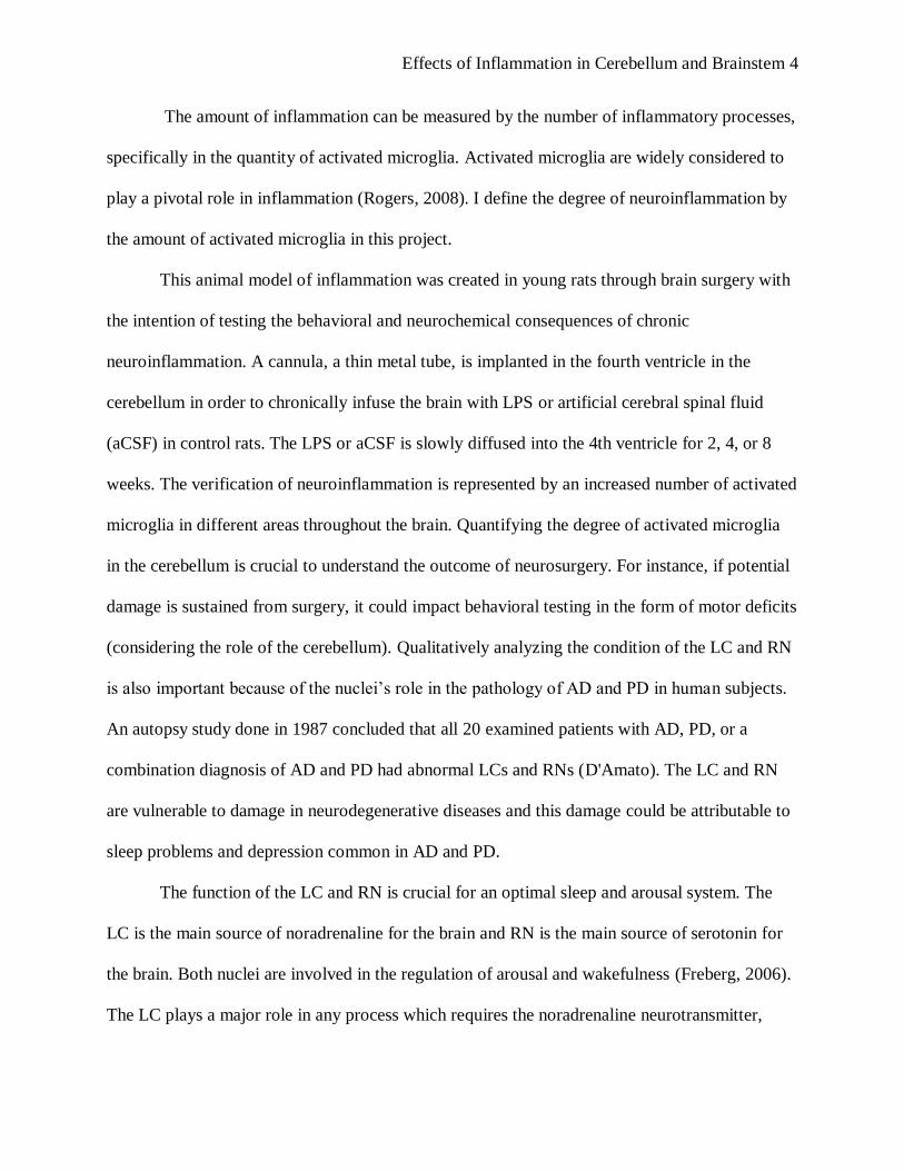
The amount of inflammation can be measured by the number of inflammatory processes,
specifically in the quantity of activated microglia. Activated microglia are widely considered to
play a pivotal role in inflammation (Rogers, 2008). I define the degree of neuroinflammation by
the amount of activated microglia in this project.
This animal model of inflammation was created in young rats through brain surgery with
the intention of testing the behavioral and neurochemical consequences of chronic
neuroinflammation. A cannula, a thin metal tube, is implanted in the fourth ventricle in the
cerebellum in order to chronically infuse the brain with LPS or artificial cerebral spinal fluid
(aCSF) in control rats. The LPS or aCSF is slowly diffused into the 4th ventricle for 2, 4, or 8
weeks. The verification of neuroinflammation is represented by an increased number of activated
microglia in different areas throughout the brain. Quantifying the degree of activated microglia
in the cerebellum is crucial to understand the outcome of neurosurgery. For instance, if potential
damage is sustained from surgery, it could impact behavioral testing in the form of motor deficits
(considering the role of the cerebellum). Qualitatively analyzing the condition of the LC and RN
is also important because of the nuclei’s role in the pathology of AD and PD in human subjects.
An autopsy study done in 1987 concluded that all 20 examined patients with AD, PD, or a
combination diagnosis of AD and PD had abnormal LCs and RNs (D'Amato). The LC and RN
are vulnerable to damage in neurodegenerative diseases and this damage could be attributable to
sleep problems and depression common in AD and PD.
The function of the LC and RN is crucial for an optimal sleep and arousal system. The
LC is the main source of noradrenaline for the brain and RN is the main source of serotonin for
the brain. Both nuclei are involved in the regulation of arousal and wakefulness (Freberg, 2006).
The LC plays a major role in any process which requires the noradrenaline neurotransmitter,

Effects of Inflammation in Cerebellum and Brainstem 5
including attention and stress. The RN is important to sleep regulation and also the pathology of
depression. A type of prescription anti-depressant called Selective Serotonin Reuptake Inhibitors
(SSRIs) acts on serotonin in the RN by blocking the reuptake of serotonin with the purpose of
reducing the symptoms of depression. Depression is a major symptom of AD and PD (National
Institute on Aging, 2008, Mayo Clinic, 2010). If the condition of the LC and RN is
compromised, neurotransmitters noradrenaline and serotonin could be fewer and perform less
optimally. The loss of noradrenergic LC neurons in AD has been known, but LC loss is now
suspected to be occurring limitedly in PD (Galea et al, 2002).This cell loss and subsequent poor
condition could be attributable to the presence of activated microglia in those areas.
The evidence shows that abnormalities in the LC and RN occur in patients who have
clinical AD or PD. However, the development of inflammation in the LC and RN could possibly
occur before the onset of the neurodegenerative disease. It’s theorized that inflammation occurs
first in the pons region of the brainstem, which includes the RN and LC (Szot et al, 2009). The
foundation for this theory is the likely relationship between the LC and RN and the progression
of PD. This relationship can be exemplified in the link between REM Behavior Disorder (RBD)
and PD. RBD consists of sporadic physical movements during REM sleep and this condition of
abnormal sleep, most likely involving the LC and RN, could be a warning sign for an increased
risk of developing PD (Thorpy & Adler, 2005).
Patients with RBD normally produce violent movements in their sleep due to acting out a
vivid dream, but they often report a precursory phase which precedes the onset of RBD by
months or years and includes talking or limb movements in sleep (Thorpy & Adler, 2005). RBD
involves destruction of brainstem structures responsible for the state of paralysis during REM
Effects of Inflammation in Cerebellum and Brainstem 6
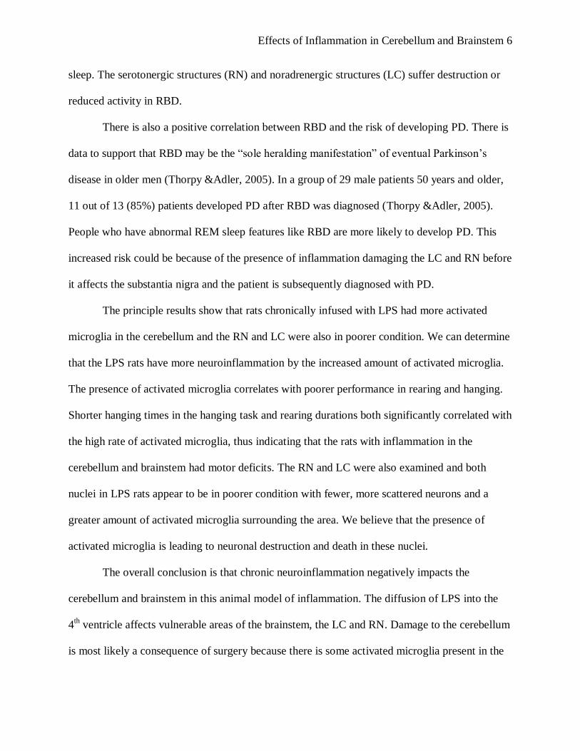
sleep. The serotonergic structures (RN) and noradrenergic structures (LC) suffer destruction or
reduced activity in RBD.
There is also a positive correlation between RBD and the risk of developing PD. There is
data to support that RBD may be the “sole heralding manifestation” of eventual Parkinson’s
disease in older men (Thorpy &Adler, 2005). In a group of 29 male patients 50 years and older,
11 out of 13 (85%) patients developed PD after RBD was diagnosed (Thorpy &Adler, 2005).
People who have abnormal REM sleep features like RBD are more likely to develop PD. This
increased risk could be because of the presence of inflammation damaging the LC and RN before
it affects the substantia nigra and the patient is subsequently diagnosed with PD.
The principle results show that rats chronically infused with LPS had more activated
microglia in the cerebellum and the RN and LC were also in poorer condition. We can determine
that the LPS rats have more neuroinflammation by the increased amount of activated microglia.
The presence of activated microglia correlates with poorer performance in rearing and hanging.
Shorter hanging times in the hanging task and rearing durations both significantly correlated with
the high rate of activated microglia, thus indicating that the rats with inflammation in the
cerebellum and brainstem had motor deficits. The RN and LC were also examined and both
nuclei in LPS rats appear to be in poorer condition with fewer, more scattered neurons and a
greater amount of activated microglia surrounding the area. We believe that the presence of
activated microglia is leading to neuronal destruction and death in these nuclei.
The overall conclusion is that chronic neuroinflammation negatively impacts the
cerebellum and brainstem in this animal model of inflammation. The diffusion of LPS into the
4th ventricle affects vulnerable areas of the brainstem, the LC and RN. Damage to the cerebellum
is most likely a consequence of surgery because there is some activated microglia present in the

Effects of Inflammation in Cerebellum and Brainstem 7
cerebellum of the control rats which were infused with aCSF. There are implications for this
study in terms of future research involving the LC and RN. Implications of neurodegenerative
diseases in humans and prevention against neuroinflammation are also discussed.
Methods
Subjects and surgical procedures
Thirty-seven 3-month old male F-344 rats (Harlan Sprague-Dawley, Indianapolis, IN)
were individually housed in Plexiglas cages with freely available food and water. The rats were
maintained on a 12/12 hour light-dark cycle in a temperature-controlled room (22 C). All of the
rats were given health checks and handled for 3 days before behavioral testing. They were also
given 1 week to adapt to their new environment without any interference. After that week, the
rats underwent surgery to implant a cannula into the 4th ventricle of the brain in order to
Figure 1. Areas of interest in the cerebellum and brainstem of a rat.
The diagram (left) shows the area of the cannula tract and also the areas of the examined nuclei:
locus coeruleus and raphe nucleus. The photomicrograph (right) shows the actual tissue and the
corresponding areas.
Cannula track
Raphe nucleus
Locus coeruleus
Effects of Inflammation in Cerebellum and Brainstem 8
chronically infuse the brain with a low dose of either artificial cerebrospinal fluid (aCSF, n=11)
or lipopolysaccharide (Sigma, St. Louis, MO, USA E.coil, serotype 055:B5, TCA extraction, 1.0
ug/ul dissolved in aCSF, n=26). LPS is gram-negative bacteria which will produce inflammation
over a long-term exposure. The cannula, which is implanted through the cerebellum and
dispenses into the 4th ventricle, is connected (via Tygon tubing, 0.06 O.D.) to an osmotic
minipump (Alzet model #2004, to deliver 0.25 ul/h; Direct Corp., Cupertino, CA, USA). The
aCSF vehicle contained (in mM) 140 NaCl; 3.0 KCl; 2.5 CaCl2; 1.0 MgCl2; 1.2 Na2HPO4,
adjusted to pH 7.4. The research was conducted under the supervision, and with the approval, of
the University of Ohio State University Institutional Animal Care and Use Committee. All
efforts were made to minimize animal suffering and to reduce the number of animal used to a
reasonable minimum regarding statistical constraints.
Behavioral testing
The rats were infused with LPS or aCSF for 2, 4, or 8 weeks. At the end of the exposure,
the rats underwent behavioral testing: hanging task and open field. The hanging task measures
how long the rats can hang on a bar and tests for motor coordination. A wire bar was covered by
rubber tubing and secured over a plastic box, with more than 3 inches from each side. The rat
was gently placed on the bar so that the rat was hanging on with all four feet and then timed until
the rat fell into the box onto several towels. The first 2 timed attempts were for practice only.
The final 3 trials were counted. This task aims to test the rat’s ability to coordinate between the
front and back legs enough to hold onto the bar for a maximum of 60 seconds.
The task of open field requires the researcher to observe the rats’ behavior in a novel
environment. A researcher monitored behavior and recorded the frequency of boli, urine, sleep,
Effects of Inflammation in Cerebellum and Brainstem 9
and the duration of freezing and rearing on their back legs. The purpose of open field is to test
for gross motor impairment, anxiety, and rearing frequency and duration. Open field is
conducted in a dry 170cm diameter tub. Bedding from a few of the rats’ cages was dispersed on
the bottom of the tub to discourage the rats from being attracted to one spot because of the scent
of another animal. One rat at a time was gently positioned at a specific place in the dry tub and
observed for 15 minutes. The actions of each rat were observed and recorded.
Histological procedures
Immediately after behavior testing was completed, each rat was deeply anesthetized with
isoflurane and sacrificed by transcardiac perfusion of the brain. Cold saline containing 1 U/ml
heparin was infused for 8 minutes, followed by 4% paraformaldehyde in 0.1M phosphate buffer,
pH 7.4 for 12 minutes. The harvested brain was post-fixed in 4% paraformaldehyde overnight at
4 ◦C then changed to phosphate buffer saline (PBS 1x), pH 7.4 for long-term storage at 4 ◦C.
The cerebellum and brainstem were separated from the rest of the brain and stored in
PBS 1x solution at 4 ◦C. The cerebellum was cut at its posterior with a brain matrix into a 4 ml
section. The vibratome was set (speed 4 -6, frequency 10, feed 40 um) and slices stored in a 24
well plate (each well filled with .5 ml of antifreeze). The tissue was organized by 5 pieces of
tissue in succession per each well. Tissue was stored at -70 ◦C until used.
Immunohistochemical staining
Six slices of brain tissue per rat were immunohistochemically stained. The first set
consisted of 3 pieces of tissue containing the best sample of the LC. The second set consisted of
3 pieces of tissue per rat with the RN present. The LC is located posterially from the RN so 2
Effects of Inflammation in Cerebellum and Brainstem 10
sets of tissue were used to provide the best example of each. Tissue with the 4th ventricle at its
most prominent point was selected because the RN is located below it. Tissue from 2 wells
previous from the 4th ventricle tissue was selected for the LC sample.
The tissue with the 4th ventricle at its most prominent was immunohistochemically with
tryptophan hydroxylase (TrypH) for serotonin, tyrosine hydroxylase (TH) for norepinephrine and
OX-6 for activated microglia. After the tissue was rinsed with PBS 1x, quenched for endogenous
peroxydase activity in 0.3% H2O2 in 50% methanol and blocked for nonspecific binding in 5%
normal goat serum, primary antibodies to tyrosine hydroxylase (final dilution 1:750, Chemicon)
and tryptophan hydroxylase (final dilution 1:100, BD PharMingen) were mixed and applied to
the free-floating tissue for 48 hours at 4◦C. The tissue was then rinsed with PBS tween,
incubated for 2.5 hours with secondary antibodies biotinylated anti-rabbit (final dilution 1:200,
Vector) and biotinylated anti-sheep (final dilution 1:200, Vector). Sections were incubated for 2
hours (22 ◦C) with avidin-biotinylated horseradish peroxydase (Vectastain, ABC kit, Vector,
Burlingame, CA). The reaction was stopped by washing the tissue with PBS. The tissue was
incubated with SG Blue working solution as chromogen for 2-3 minutes and the reaction was
stopped by rinsing with PBS.
Next, the tissue was stained for activated microglia with primary antibody Ox-6 to label
positive MHC-II receptors. The tissue was incubated in Ox-6 (final dilution 1:200, BD
PharMingen) overnight at 4 ◦C on the shaker. After it was rinsed with PBS the next day, the
tissue was incubated in the secondary antibody anti-mouse (final dilution 1:200, Vector) for 1.5
hours at 22 ◦C. The tissue was rinsed with PBS and the sections were incubated for 1 hour (22
◦C) with avidin-biotinylated horseradish peroxydase (Vectastain, ABC kit, Vector, Burlingame,
CA). After rinsing again in PBS tween, the sections were incubated with 0.05% 3,3 -
Effects of Inflammation in Cerebellum and Brainstem 11
diaminobenzidine tetrahydrochloride (DAB, Vector, Burlingame, CA) as chromogen for 20
minutes. Finally, the tissue was mounted on gel-coated slides, air-dried, dehydrated and
coverslipped with cytoseal (Allan Scientific, Kalamazoo, MI) mounting medium.
The tissue with the best sample of LC was stained for TH to label norepinephrine neurons
and for OX-6 to label activated microglia. The procedure to stain this set of tissue is the same as
the above except TrypH and its secondary antibody (biotinylated anti-sheep) are excluded
because there are no serotonin neurotransmitters in this region.
Image acquisition (confocal microscopy) and Data Collection
The mounted and coverslipped tissue was image acquisitioned using a Nikon microscope.
The photomicrographs were inspected at a high magnification for activated microglia
surrounding damaged neurons. The LC and RN were qualitatively examined to determine the
condition of the neurons and compare the appearances of these nuclei between the control and
LPS rats.
A quantitative analysis determined the amount of activated microglia in the cerebellum
and brainstem tissue. Multiple selections of tissue for each rat were selected for analysis. The
first set of sample tissue (with the 4th ventricle and RN) showed evidence of the completed
neurosurgery because of the cannula tract. Each individual slice was given a rating between 0 (no
activated microglia) and 6 (excessive activated microglia). The individual ratings were averaged
to give each rat one overall activated microglia rating.
The density of activated microglia for a set area of ventral cerebellum (below the 4th
ventricle and above the raphe nucleus) was calculated by ImageJ. The data were compared

Effects of Inflammation in Cerebellum and Brainstem 12
between the control rats to determine if there was more or less inflammation damage in the
cerebellum and brainstem.
Results
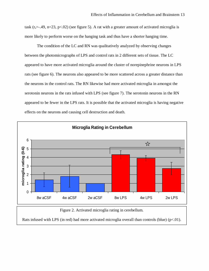
Rats chronically infused with LPS had more activated microglia in the cerebellum and
brainstem compared to the control rats (f(1,5)=16.55, p<.01) (See figure 2 and 3). A quantitative
analysis was calculated by me and Holly Brothers, M.A., to give each rat an appropriate 0-6
rating based on the amount of activated microglia. Brothers separately and blindly rated the
amount of activated microglia for each individual sample of tissue using the same scale. The
averaged overall activated microglia rating Brothers assigned to each rat correlated with my
ratings (rs=0.801, n=35, p=0.0).
The same photomicrographs of cerebellum and brainstem were also analyzed
densitometrically with ImageJ and averaged overall for each rat once again. This densitometry
rating also correlated with my averaged activated microglia rating of 0 – 6 (rs=0.67, n=30,
p=0.0).
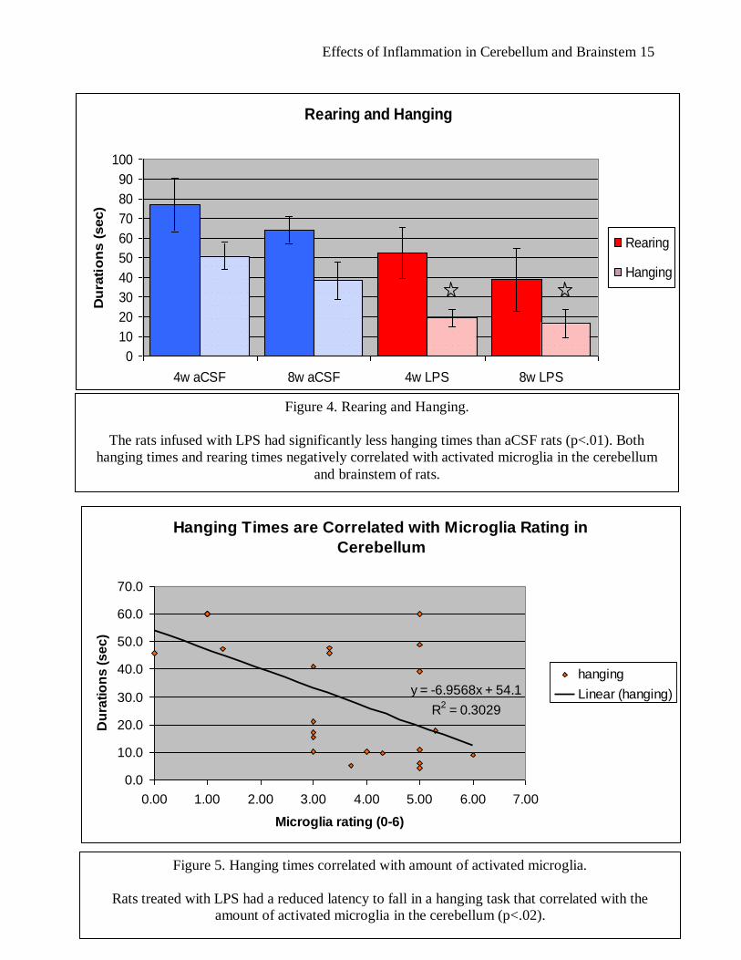
The rats underwent behavioral testing after 2, 4, or 8 weeks of infusion of LPS or aCSF.
The behavioral tests included hanging and open field. There was a significant difference between
the LPS and aCSF groups (4 and 8 weeks) in hanging times during the hanging task
(f(1,19)=27.87, p<.01) (see figure 4). There were correlations between a greater activated
microglia rating and poor performance in rearing during open field and also in hanging. The
more activated microglia in the cerebellum and brainstem of a rat, the fewer times the animal
reared during open field (rp=-0.55, n=22, p<0.01). The rats with more activated microglia also
had less total rearing durations (rs=-0.63, n=22, p<0.01). The higher activated microglia rating in
the cerebellum is negatively correlated with the rat’s ability to hang on a bar during the hanging
Effects of Inflammation in Cerebellum and Brainstem 13
task (rs=-.49, n=23, p<.02) (see figure 5). A rat with a greater amount of activated microglia is
more likely to perform worse on the hanging task and thus have a shorter hanging time.
The condition of the LC and RN was qualitatively analyzed by observing changes
between the photomicrographs of LPS and control rats in 2 different sets of tissue. The LC
appeared to have more activated microglia around the cluster of norepinephrine neurons in LPS
rats (see figure 6). The neurons also appeared to be more scattered across a greater distance than
the neurons in the control rats. The RN likewise had more activated microglia in amongst the
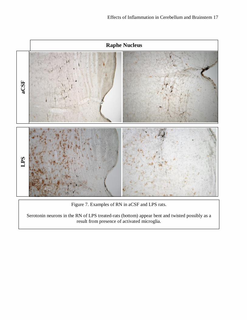
serotonin neurons in the rats infused with LPS (see figure 7). The serotonin neurons in the RN
appeared to be fewer in the LPS rats. It is possible that the activated microglia is having negative
effects on the neurons and causing cell destruction and death.
Microglia Rating in Cerebellum
0
1
2
3
4
5
6
8w aCSF 4w aCSF 2w aCSF 8w LPS 4w LPS 2w LPS
mic
rog
lia r
ati
ng
(0-6
)
Figure 2. Activated microglia rating in cerebellum.
Rats infused with LPS (in red) had more activated microglia overall than controls (blue) (p<.01).

Effects of Inflammation in Cerebellum and Brainstem 14
Activated microglia in the cerebellum and brainstem
Figure 3. Activated microglia in cerebellum and brainstem tissue
Typical amounts of activated microglia in an aCSF rat (left) and rat infused with LPS
(right). Animals treated with LPS had more activated microglia overall. Activated
microglia are the brown cells in this selected tissue.
aCSF LPS
Effects of Inflammation in Cerebellum and Brainstem 15
Figure 4. Rearing and Hanging.
The rats infused with LPS had significantly less hanging times than aCSF rats (p<.01). Both
hanging times and rearing times negatively correlated with activated microglia in the cerebellum
and brainstem of rats.
Rearing and Hanging
0
10
20
30
40
50
60
70
80
90
100
4w aCSF 8w aCSF 4w LPS 8w LPS
Du
rati
on
s (
sec)
Rearing
Hanging
Hanging Times are Correlated with Microglia Rating in
Cerebellum
y = -6.9568x + 54.1
R2 = 0.3029
0.0
10.0
20.0
30.0
40.0
50.0
60.0
70.0
0.00 1.00 2.00 3.00 4.00 5.00 6.00 7.00
Microglia rating (0-6)
Du
rati
on
s (
se
c)
hanging
Linear (hanging)
Figure 5. Hanging times correlated with amount of activated microglia.
Rats treated with LPS had a reduced latency to fall in a hanging task that correlated with the
amount of activated microglia in the cerebellum (p<.02).

Effects of Inflammation in Cerebellum and Brainstem 16
Locus coeruleus
aC
SF
L
PS
Figure 6. Examples of LC in aCSF and LPS rats.
Norepinephrine neurons in the LC appear more scattered in LPS-treated rats (bottom), possibly due
to interaction with the surrounding activated microglia.
Effects of Inflammation in Cerebellum and Brainstem 17
Raphe Nucleus
aC
SF
L
PS
Figure 7. Examples of RN in aCSF and LPS rats.
Serotonin neurons in the RN of LPS treated-rats (bottom) appear bent and twisted possibly as a
result from presence of activated microglia.
Effects of Inflammation in Cerebellum and Brainstem 18
Discussion
The overall conclusion from this experiment is that chronic neuroinflammation has
negative affects on the LC, RN and cerebellum in an animal model of inflammation.
Neuroinflammation in the brainstem affects the LC and RN biochemically and
neuroinflammation in the cerebellum correlates with less optimal performance in rats. Such
evidence in this analogue study creates implications for humans and the risk of
neurodegenerative diseases.
Studies including those involving a genetic focus and epidemiological analysis of humans
indicate that neuroinflammatory processes can be risk factors for PD (Hirsch & Hunot 2009).
Humans could reduce their risk of developing a neurodegenerative disease by leading an anti-
inflammatory lifestyle. Practicing a healthy lifestyle and making choices such as drinking coffee
could help protect a person from chronic neuroinflammation (Ross et al, 2000). Another way to
protect against AD is to take ibuprofen over a lifetime. It is an anti-inflammatory medication
which crosses the blood brain barrier and epidemiological studies have indicated that prolonged
use of non-steroidal anti-inflammatory drugs (NSAIDs) like ibuprofen decreases the risk of AD,
delays dementia onset and reduces the severity of cognitive symptoms (Gasparini et al).
The cerebellum, LC and RN have not yet been extensively examined in this animal
model of inflammation because the main areas of study are the substantia nigra for PD and the
hippocampus for AD. However, I predict that in future research of neurodegenerative diseases
the LC and RN will be recognized as a vital part of the pathology and that will warrant further
study and also pharmacological manipulation of the area.
Examples of valuable future studies would be monitoring the sleep patterns of the
animals and also the condition of the LC and RN. These nuclei are highly involved in the

Effects of Inflammation in Cerebellum and Brainstem 19
regulation of the sleep and wake arousal system; therefore it would be a good idea to determine
if inflammation in the LC and RN would produce sleep disturbances in the rats like what would
most likely happen to humans if these areas sustained damage. It would also be beneficial to
investigate pharmacological effects on the LC and RN to see if the nuclei could be protected
from the neuroinflammation. The LC and RN may be a reliable and valid area to examine for
damage and potential treatments.
Effects of Inflammation in Cerebellum and Brainstem 20
References
D'Amato RJ, Zweig RM, Whitehouse PJ, Wenk GL, Singer HS, Mayeux R, Price DL, Snyder
SH (1987). Aminergic systems in Alzheimer's disease and Parkinson's disease. Ann
Neurol, 22, 229-236.
Freberg, L.A. (2006). Sleep and Waking. In J. Potter (Ed), Discovering Biological Psychology
(pp. 312-341). Boston, Mass: Houghton Mifflin Company.
Gasparini L, Ennio O, Wenk GL (2004). Non-steroidal anti-inflammatory drugs (NSAIDs) in
Alzheimer’s Disease: old and new mechanisms of action. J Neurochem., 91, 521-536.
Hirsch, E.C., Hunot S. (2009). Neuroinflammation in Parkinson’s disease: a target for
neuroprotection? Lancet Neurol, 8, 382-97.
Mayo Clinic (2010). Parkinson’s Disease. Retrieved from
http://www.mayoclinic.com/health/parkinsons-disease/DS00295/DSECTION=coping-
and-support#
National Institute on Aging (2008). Alzheimer's disease fact sheet. Retrieved from
http://www.nia.nih.gov/Alzheimers/Publications/adfact.htm
Rogers, J. (2008). The Inflammatory Response in Alzheimer’s Disease. J Periodontol, 79(8),
153-160.
Effects of Inflammation in Cerebellum and Brainstem 21
Ross, GW, Abbott, RD, Petrovitch, H, Morens, DM (2000). Association of Coffee and Caffeine
Intake With the Risk of Parkinson Disease. JAMA., 283(20), 2674-2679.
Szot ,P, White, SS, Franklin, A, Raskind, MA. (2009, October). Tyrosine hydroxylase and
dopamine b-hydroxlyase mRNA expression in the LC noradrenergic neurons in
Parkinson’s disease. Poster session presented at the annual meeting of the Society for
Neuroscience, Chicago, IL.
Thorpy, M.J., Adler, C.H.. (2005). Parkinson’s Disease and Sleep. Neurol Clon, 23, 1187-1208