running head: mechanical diversity of stomata · given that stomatal movement is ultimately a...
TRANSCRIPT
1
Running head: Mechanical diversity of stomata Author for correspondence: Peter J. Franks School of Tropical Biology James Cook University GPO Box 6811 Cairns, QLD 4870 Australia Phone: +61 7 4042 1237 Fax: + 61 7 4042 1319 e-mail: [email protected] Journal Research area: Whole Plant and Ecophysiology Special Issue: Biology of Transpiration
Plant Physiology Preview. Published on November 17, 2006, as DOI:10.1104/pp.106.089367
Copyright 2006 by the American Society of Plant Biologists
www.plantphysiol.orgon August 25, 2019 - Published by Downloaded from Copyright © 2006 American Society of Plant Biologists. All rights reserved.

2
The mechanical diversity of stomata and its significance in gas exchange control.
Peter J. Franks1 and Graham D. Farquhar2
1 School of Tropical Biology, James Cook University, PO Box 6811, Cairns, Queensland 4870, Australia.
2 Environmental Biology Group, Research School of Biological Sciences, Australian National University,
GPO Box 475, Canberra ACT 2601, Australia.
www.plantphysiol.orgon August 25, 2019 - Published by Downloaded from Copyright © 2006 American Society of Plant Biologists. All rights reserved.

3
Author for correspondence: Peter J. Franks School of Tropical Biology James Cook University GPO Box 6811 Cairns, QLD 4870 Australia Phone: +61 7 4042 1237 Fax: + 61 7 4042 1319 e-mail: [email protected]
www.plantphysiol.orgon August 25, 2019 - Published by Downloaded from Copyright © 2006 American Society of Plant Biologists. All rights reserved.

4
ABSTRACT
Given that stomatal movement is ultimately a mechanical process, and that stomata are
morphologically and mechanically diverse, we explored the influence of stomatal
mechanical diversity on leaf gas exchange, and considered some of the constraints.
Mechanical measurements were conducted on the guard cells of four different species
exhibiting different stomatal morphologies, including three variants on the classical
"kidney" form, and one "dumb-bell" type, and this information, together with gas
exchange measurements, was used to model and compare their respective operational
characteristics. Based on evidence from SEM images of cryo-sectioned leaves that were
sampled under full sun and high humidity, and from pressure probe measurements of the
stomatal aperture versus guard cell turgor relationship at maximum and zero epidermal
turgor, it was concluded that maximum stomatal apertures (and maximum leaf diffusive
conductance) could not be obtained in at least one of the species (the grass Triticum
aestivum) without a substantial reduction in subsidiary cell osmotic (and hence turgor)
pressure during stomatal opening to overcome the large mechanical advantage of
subsidiary cells. A mechanism for this is proposed, with a corollary being greatly
accelerated stomatal opening and closure. Gas exchange measurements on T. aestivum
revealed the capability of very rapid stomatal movements, which may be explained by the
unique morphology and mechanics of its dumb-bell shaped stomata coupled with "sea-
sawing" of osmotic and turgor pressure between guard and subsidiary cells during
stomatal opening or closure. Such properties might underlie the success of grasses.
www.plantphysiol.orgon August 25, 2019 - Published by Downloaded from Copyright © 2006 American Society of Plant Biologists. All rights reserved.
5
INTRODUCTION
Although the morphological diversity of stomata is widely documented (Haberlandt,
1884; Meidner and Mansfield, 1968; Allaway and Milthorpe, 1976; Ziegler, 1987;
Willmer and Fricker, 1996), little is known of how this translates into functional
diversity, and what the environmental context of this might be. Throughout the 400 Ma
history of vascular plants on land, long-term decline in atmospheric CO2 concentration
and shifts in prevailing moisture patterns have placed selective pressures on stomata to
increase epidermal conductance to CO2 diffusion and also to increase transpiration
efficiency (CO2 fixed per unit water transpired). This posed two separate problems, upon
which the combination of mutation and time might have worked to give rise to the
current diversity of stomatal form and function. The first centred on the simple
geometric practicalities of fitting enough functional stomatal units per unit leaf surface
area to meet the desired CO2 flux as atmospheric CO2 concentration changed, or to
service an increase in photosynthetic capacity. The second centred on the performance
characteristics of any new stomatal structure or configuration, in relation to transpiration
efficiency. Here, by examining the mechanical and performance characteristics of
stomata in four different species, we explore the nature of these two problems and how
they might have been resolved.
The mechanical characteristics of stomata are central to their performance in gas
exchange regulation, but relatively little is known about these properties, particularly how
they vary across different stomatal forms. The simple quantitative relationship between
guard cell turgor (Pg), epidermal (or subsidiary) cell turgor (Pe) and stomatal aperture (a),
which defines the operational potential of all stomata, is known for only a few species,
and most of these have structurally similar stomata (Meidner and Bannister, 1979; Franks
et al., 1998; Franks et al., 2001). No such data are available for the distinctive "dumb-
bell" shaped stomata of grasses, for example, or for the more mechanically isolated
stomata that are common in many pteridophytes. Without this information on the
mechanical diversity of stomata, the power to predict the behaviour of stomata in diverse
types of vegetation, or to understand key stages in the evolution of plant gas exchange
www.plantphysiol.orgon August 25, 2019 - Published by Downloaded from Copyright © 2006 American Society of Plant Biologists. All rights reserved.

6
characteristics, remains limited. Our first objective was to address this shortfall by
measuring and analysing the Pg-Pe-a relationship in four species with distinctively
different stomatal morphologies: (i) the lycopod Huperzia prolifera; (ii) the fern
Nephrolepis exaltata; (iii) the herbaceous angiosperm Tradescantia virginiana; (iv) the
grass Triticum aestivum.
Stomata of the four species chosen for the study cover a broad morphological and
evolutionary spectrum (Figure 1). H. prolifera is a living representative of one of the
most ancient vascular plant taxa (Lycopodiaceae), with fossilised remains of its close
relatives (having the same stomatal morphology) dating back to the lower Devonian
(Stubblefield and Banks, 1978; Sun et al., 2005). Its stomata are anomocytic (lacking
subsidiary cells), with large and comparatively fat guard cells, which undergo minimal
swelling or lateral movement during stomatal opening. Based on these characteristics, it
is regarded as archetypal (Ziegler, 1987), perhaps closely resembling the first stomata.
N. exaltata, like H. prolifera, is anomocytic, but fern stomata are structurally and
functionally more advanced than those of Lycopods. Although the pattern of guard cell
deformation during stomatal opening is similar to that of Lycopods, fern stomata of
comparable guard cell dimensions achieve wider apertures, which assists with higher
rates of gas exchange (N. exaltata exhibits photosynthetic rates more than three times that
of H. prolifera under similar conditions; Franks, 2006). T. virginiana and T. aestivum are
distinctive in that they both have a subsidiary cell running parallel to each guard cell.
Their guard cells exhibit substantial lateral movement and physical interaction with the
subsidiary cells during stomatal opening, resulting in wide stomatal pores that facilitate
high rates of photosynthetic gas exchange. While stomata of the other species are
variants on the common "kidney" form, those of T. aestivum exhibit the characteristic
dumb-bell shaped guard cells of grasses (Graminae). The functional significance of this
uniquely different guard cell form remains unknown, although the capacity for rapid
stomatal opening and closure in grasses is thought to be somehow linked to their unusual
guard cell geometry (Johnsson et al., 1976; Hetherington and Woodward, 2003). It has
further been proposed that superior dynamic performance of grass stomata could have
facilitated the relatively recent spread and diversification of grasses during a period of
global aridification 35-40 Ma ago (Hetherington and Woodward, 2003).
www.plantphysiol.orgon August 25, 2019 - Published by Downloaded from Copyright © 2006 American Society of Plant Biologists. All rights reserved.

7
The second objective of the study was to investigate the role of guard cell
morphology and mechanics in stomatal function, particularly in relation to its potential
influence on the speed of stomatal opening and closure. Stomatal response time is
important because it determines the extent to which leaf gas exchange can be optimised
under fluctuating environmental conditions. Fast and appropriately damped response to
changes in the environmental drivers of photosynthesis or transpiration rate can lead to
greater transpiration efficiency. Grasses have long been known for their capacity for
rapid stomatal response (Raschke and Fellows, 1971; Brogardh and Johnsson, 1975;
Johnsson et al., 1976; Karlsson and Assmann, 1990; Grantz and Assmann, 1991;
Assmann et al., 1992) but the underlying mechanism for this has remained a mystery, due
partly to the absence of data on the mechanical interactions between guard and subsidiary
cells in grasses. Our approach to unravelling this centrally important aspect of stomatal
function was to revisit the findings of several classical studies on guard cell solute
transport and, with new information on guard cell mechanical and geometric properties,
formulate an osmo-mechanical model of stomatal movement that could explain the
superior performance of grass stomata under dynamic environmental conditions. To
illustrate these characteristics, the stomatal properties of the grass T. aestivum were
compared against those of the three non-grass species.
RESULTS
The mechanical diversity of the four stomatal types is illustrated dramatically in
the scanning electron micrographs of cryo-sectioned leaf material in Figures 2-5. Here
the signature guard cell swelling and displacement characteristics of the different
stomatal types are evident. In particular, H. prolifera and N. exaltata show little
mechanical interaction between guard cells and epidermal cells as the guard cells swell to
create the stomatal pore. H. prolifera displays the "Psilotum type" guard cell
deformation (Ziegler, 1987), whereby the guard cell lower (leaf inner) wall expands into
the sub-stomatal cavity during stomatal opening. N. exaltata shows the archetypal
"Adiantum-type" guard cell deformation, whereby the guard cell upper and lower (leaf
www.plantphysiol.orgon August 25, 2019 - Published by Downloaded from Copyright © 2006 American Society of Plant Biologists. All rights reserved.

8
inner and outer) walls buckle outwards as the guard cells swell into a more rounded
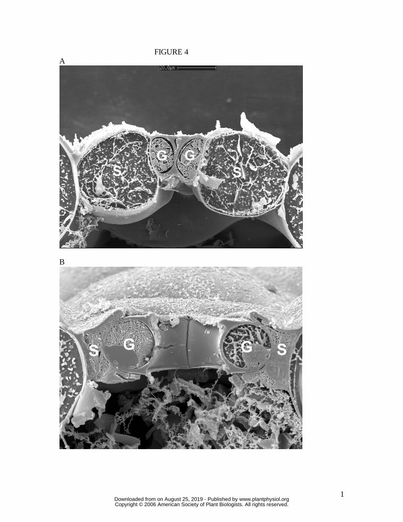
cross-section to create the stomatal pore. By contrast, both T. virginiana and T. aestivum
guard cells undergo substantial lateral displacement into their adjacent subsidiary cells
during stomatal opening, to the extent that the subsidiary cells are almost squashed in
order to accommodate the open stoma. Of great significance is the fact that, since these
leaves were sampled at very high humidity, we know that rates of transpirational water
loss were minimal, and therefore the water potential of all cells in the image was close to
zero (the plants being also well-watered). This means that turgor in all cells was maximal
for the prevailing osmotic conditions. It is shown below that for the stomata in T.
virginiana and T. aestivum to have reached the apertures indicated, turgor in the
subsidiary cells had to be significantly reduced, as full or even partial subsidiary cell
turgor simply would not have allowed such wide apertures. Later, in the Discussion, we
propose how this might be achieved.
The relationship between guard cell pressure (Pg) and stomatal aperture (as pore
width, a; µm) at maximum and zero epidermal turgor is summarised for each species in
Figure 6. A strong linear relationship was found between stomatal pore width and
stomatal pore area in each species (data not shown), as was reported for T. virginiana by
Franks and Farquhar (2001). Consistent with the evidence from the cryo-sections of open
stomata (Figs 2-5), the aperture corresponding to any given Pg was virtually unaffected
by epidermal turgor (Pe) in H. prolifera and N. exaltata, but substantially influenced by Pe
in T. virginiana and T. aestivum. The small offset observed in H. prolifera (Fig 6A)
could be due to a small, generalised expansion of the epidermal cells at high turgor. At
full epidermal turgor, T. virginiana stomata could not attain an aperture above about 7
µm, compared to its maximum of about 20 µm when Pe was zero. Similarly, T. aestivum
stomatal apertures were on average not greater than 2.5 µm at full epidermal turgor,
compared to about 8 µm at zero epidermal turgor. This massive mechanical
counteraction of stomatal opening by epidermal turgor in T. virginiana and T. aestivum is
a negative side effect arising from the need for greater lateral displacement of guard cells
to create a larger stomatal pore. Known technically as the "mechanical advantage" of
epidermal cells over guard cells (DeMichelle and Sharpe, 1973; Cooke, 1976; Wu et al.,
1985; Franks et al., 1998), this epidermal impediment to stomatal opening would
www.plantphysiol.orgon August 25, 2019 - Published by Downloaded from Copyright © 2006 American Society of Plant Biologists. All rights reserved.
9
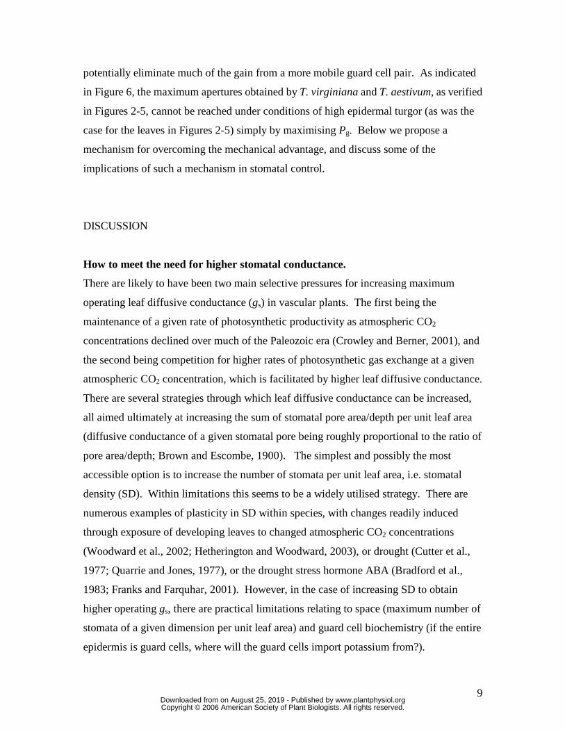
potentially eliminate much of the gain from a more mobile guard cell pair. As indicated
in Figure 6, the maximum apertures obtained by T. virginiana and T. aestivum, as verified
in Figures 2-5, cannot be reached under conditions of high epidermal turgor (as was the
case for the leaves in Figures 2-5) simply by maximising Pg. Below we propose a
mechanism for overcoming the mechanical advantage, and discuss some of the
implications of such a mechanism in stomatal control.
DISCUSSION
How to meet the need for higher stomatal conductance.
There are likely to have been two main selective pressures for increasing maximum
operating leaf diffusive conductance (gs) in vascular plants. The first being the
maintenance of a given rate of photosynthetic productivity as atmospheric CO2
concentrations declined over much of the Paleozoic era (Crowley and Berner, 2001), and
the second being competition for higher rates of photosynthetic gas exchange at a given
atmospheric CO2 concentration, which is facilitated by higher leaf diffusive conductance.
There are several strategies through which leaf diffusive conductance can be increased,
all aimed ultimately at increasing the sum of stomatal pore area/depth per unit leaf area
(diffusive conductance of a given stomatal pore being roughly proportional to the ratio of
pore area/depth; Brown and Escombe, 1900). The simplest and possibly the most
accessible option is to increase the number of stomata per unit leaf area, i.e. stomatal
density (SD). Within limitations this seems to be a widely utilised strategy. There are
numerous examples of plasticity in SD within species, with changes readily induced
through exposure of developing leaves to changed atmospheric CO2 concentrations
(Woodward et al., 2002; Hetherington and Woodward, 2003), or drought (Cutter et al.,
1977; Quarrie and Jones, 1977), or the drought stress hormone ABA (Bradford et al.,
1983; Franks and Farquhar, 2001). However, in the case of increasing SD to obtain
higher operating gs, there are practical limitations relating to space (maximum number of
stomata of a given dimension per unit leaf area) and guard cell biochemistry (if the entire
epidermis is guard cells, where will the guard cells import potassium from?).
www.plantphysiol.orgon August 25, 2019 - Published by Downloaded from Copyright © 2006 American Society of Plant Biologists. All rights reserved.
10
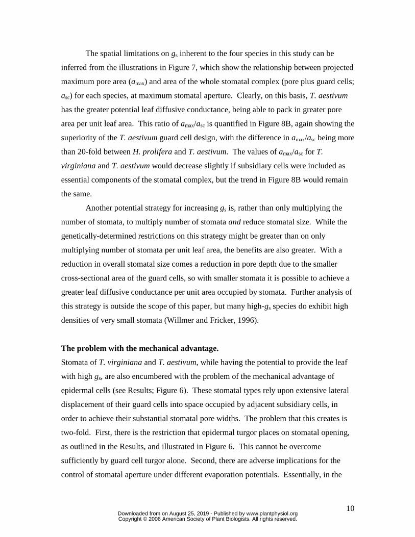
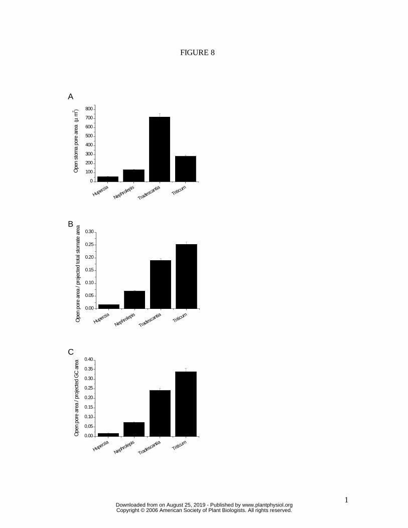
The spatial limitations on gs inherent to the four species in this study can be
inferred from the illustrations in Figure 7, which show the relationship between projected
maximum pore area (amax) and area of the whole stomatal complex (pore plus guard cells;
asc) for each species, at maximum stomatal aperture. Clearly, on this basis, T. aestivum
has the greater potential leaf diffusive conductance, being able to pack in greater pore
area per unit leaf area. This ratio of amax/asc is quantified in Figure 8B, again showing the
superiority of the T. aestivum guard cell design, with the difference in amax/asc being more
than 20-fold between H. prolifera and T. aestivum. The values of amax/asc for T.
virginiana and T. aestivum would decrease slightly if subsidiary cells were included as
essential components of the stomatal complex, but the trend in Figure 8B would remain
the same.
Another potential strategy for increasing gs is, rather than only multiplying the
number of stomata, to multiply number of stomata and reduce stomatal size. While the
genetically-determined restrictions on this strategy might be greater than on only
multiplying number of stomata per unit leaf area, the benefits are also greater. With a
reduction in overall stomatal size comes a reduction in pore depth due to the smaller
cross-sectional area of the guard cells, so with smaller stomata it is possible to achieve a
greater leaf diffusive conductance per unit area occupied by stomata. Further analysis of
this strategy is outside the scope of this paper, but many high-gs species do exhibit high
densities of very small stomata (Willmer and Fricker, 1996).
The problem with the mechanical advantage.
Stomata of T. virginiana and T. aestivum, while having the potential to provide the leaf
with high gs, are also encumbered with the problem of the mechanical advantage of
epidermal cells (see Results; Figure 6). These stomatal types rely upon extensive lateral
displacement of their guard cells into space occupied by adjacent subsidiary cells, in
order to achieve their substantial stomatal pore widths. The problem that this creates is
two-fold. First, there is the restriction that epidermal turgor places on stomatal opening,
as outlined in the Results, and illustrated in Figure 6. This cannot be overcome
sufficiently by guard cell turgor alone. Second, there are adverse implications for the
control of stomatal aperture under different evaporation potentials. Essentially, in the
www.plantphysiol.orgon August 25, 2019 - Published by Downloaded from Copyright © 2006 American Society of Plant Biologists. All rights reserved.
11
absence of an active compensating mechanism in the stomatal control system, the
mechanical advantage dictates that stomatal aperture will open wider as evaporation
potential increases, due to the increase in transpiration rate lowering epidermal turgor and
facilitating the passive widening of the stomatal pores. Using the hydromechanical
stomatal model described in Franks (2004), this effect is simulated for a stomatal opening
sequence in Figure 9A, for a plant with stomatal characteristics similar to T. virginiana,
and assuming no active compensation to counteract the increase in gs with increasing
evaporative demand. Such a mode of operation would be highly destructive if it were to
actually occur. However, it is almost universally observed that plants operate with lower
(not higher) gs under higher evaporative demand (Lange et al., 1971; Schulze et al., 1972;
Hall et al., 1976; Grantz, 1990; Franks and Farquhar, 1999). This is illustrated in Figure
9B and 9C for measured opening sequences on leaves of T. virginiana and T. aestivum,
respectively, under two different values of leaf-to-air vapour pressure difference (D). In
both cases the final steady state conductance to water vapour (gsw) was substantially
lower at higher D. Note also that there appears to be little enhancement of the initial rate
of increase in gsw at higher D, compared to low D, suggesting a highly active
compensating mechanism.
Overcoming the mechanical advantage
Based on the pressure probe results in Figure 6, it seems virtually impossible for T.
virginiana and T. aestivum stomata to reach the apertures observed in Figures 4 and 5
under humid conditions, yet clearly they did. The simplest explanation, suggested by the
degree of deformation of T. aestivum subsidiary cells (Figure 5) is that subsidiary cell
turgor is much lower during maximum stomatal aperture, such as occurs under high
humidity. The only way that this can be achieved at high cellular water potentials is if
the osmotic pressure of subsidiary cells declines substantially, thus reducing the effect of
the mechanical advantage, and allowing maximum lateral displacement of guard cells and
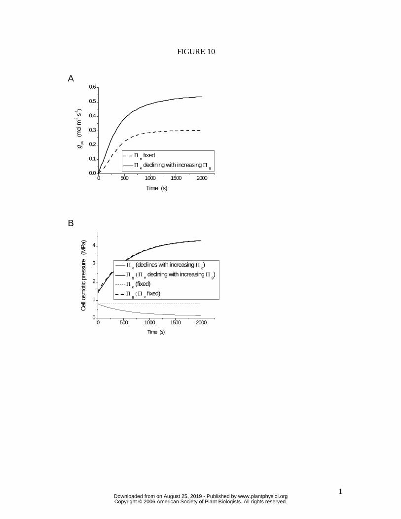
maximum stomatal apertures. This effect is simulated in Figure 10, using the same
hydromechanical feedback model as for the simulations in Figure 9A, but allowing Πe
(effectively the osmotic pressure of subsidiary cells) to decline in association with the
increase in guard cell osmotic pressure (Πg; see Methods). This sea-sawing of relative
www.plantphysiol.orgon August 25, 2019 - Published by Downloaded from Copyright © 2006 American Society of Plant Biologists. All rights reserved.

12
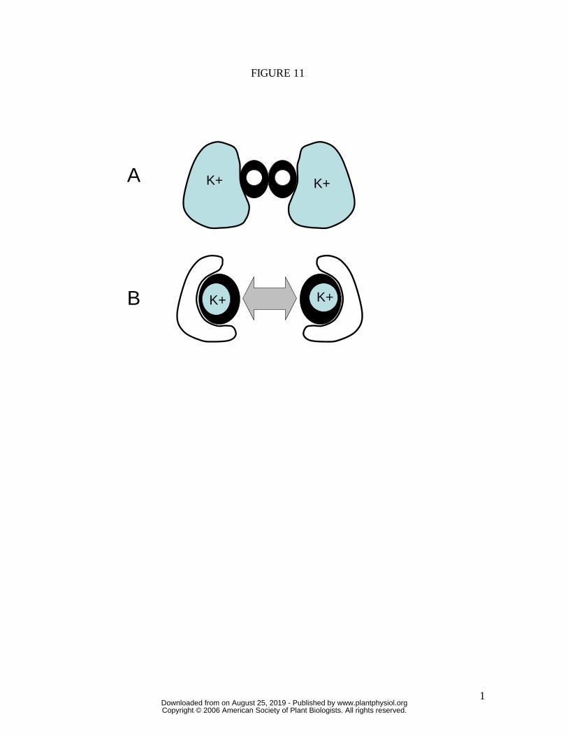
osmotic and turgor pressures between subsidiary cells and guard cells, illustrated
schematically in Figure 11, overcomes the problem of the mechanical advantage, and
would allow T. aestivum to attain full apertures under high humidity, where it otherwise
could not. Transfer of solutes (namely K+) to guard cells via subsidiary and epidermal
cells has long been recognised (Fujino, 1967; Fischer, 1968; Humble and Hsiao, 1969;
Sawhney and Zelitch, 1969; Humble and Raschke, 1971; Pallaghy, 1971; Penny and
Bowling, 1974; Dayanandan and Kaufman, 1975; Macrobbie and Lettau, 1980; Outlaw,
1983) but here we make the case for mandatory and opposite changes in osmotic and
turgor pressure of guard and subsidiary cells where epidermal or subsidiary cells have a
large mechanical advantage over guard cells. This requirement would diminish in
species with diminished mechanical advantage (e.g. H. prolifera and N. exaltata in this
study). A desirable side effect arising from this mechanism is that the rate of stomatal
opening is increased (compare Figure 10A solid line with dashed line), as is the rate of
closure in a reversal of the same process. Thus, the mechanical advantage, which arose
as an unavoidable consequence of increased guard cell lateral movement to create wider
stomatal pores, may have been harnessed in the manner described above to ultimately
facilitate, rather than impede the operation of stomata. This facilitation enabled higher
steady-state stomatal conductances to be achieved, and increased the rate at which
stomatal aperture can change, thereby supporting increased rates of photosynthetic gas
exchange and increased transpiration efficiency. Of course we cannot be sure which of
increased lateral movement and shuttling of osmotica came first, and evolution may well
have occurred in parallel.
Potassium shuttling between guard and subsidiary cells in relation to rapid
stomatal opening was first discussed by Raschke and Fellows (1971) in their study on
Zea mays, and later by Brogardh and Johnsson (1975). However, these authors
considered only the speed of solute transfer and the potentially rapid rate of change of
guard cell osmotic pressure due to the large reservoir of potassium in subsidiary cells
immediately adjacent to guard cells of grasses. The requirement for, and advantages of, a
substantial reduction in both the osmotic and turgor pressure of subsidiary cells during
opening of graminoid stomata was not considered in detail at that time. Later work,
which focussed on rapid responses in grass stomata exposed to a blue light stimulus
www.plantphysiol.orgon August 25, 2019 - Published by Downloaded from Copyright © 2006 American Society of Plant Biologists. All rights reserved.

13
(Johnsson et al., 1976; Karlsson and Assmann, 1990; Grantz and Assmann, 1991;
Assmann et al., 1992) also did not consider, from a quantitative perspective, the role of
the mechanical advantage of subsidiary cells. Our measurements of the Pg-Pe-a
relationship in the graminoid stomatal complex of T. aestivum and the integration of this
information into a mechanistic model of stomatal movement has revealed how the
combination of osmotic shuttling, high epidermal mechanical advantage and unique
guard cell geometry in graminoid stomata allows them to open or close at a substantially
faster rate than perhaps any other stomatal type.
Significance of graminoid stomatal mechanics in the rise of grasses
Several authors have postulated that the spread and diversification of grasses, which
likely began in tropical forest understoreys between 55-70 Ma ago (Kellogg, 2001), but
peaked during a period of global aridification approximately 30-45 Ma ago, could have
been assisted by them having faster and therefore more transpiration-efficient stomata
(Hetherington and Woodward, 2003, and references therein). However, the significance
of the unique mechanical properties of graminoid-type stomata in relation to this superior
performance has not until now been quantified. To illustrate just how superior their
performance can be, we quantified the maximum rate of stomatal opening in response to
light, under standardised environmental conditions, in each of the four species in this
study (Figure 12). Regardless of how the rate of opening was defined, the graminoid
stomata of T. aestivum were about an order of magnitude faster than any of the other
species.
Conclusions and further work
The results demonstrate that the morphological diversity of stomata translates into
considerable mechanical and, ultimately, functional diversity. This study examined only
the stomata of four species and, although it spanned a considerably broad morphological
and evolutionary spectrum, there are many more stomatal forms yet to be examined in
this way, and the picture is far from complete. By the same measure, we do not wish to
generalise too extensively about the function of grass somata from our examination of
just one species, but the results do provide a compelling case for the role of stomatal
www.plantphysiol.orgon August 25, 2019 - Published by Downloaded from Copyright © 2006 American Society of Plant Biologists. All rights reserved.
14
mechanics in the distinctive capabilities of this special group of plants. The findings
highlight the importance of integrating mechanical and quantitative physical information
about guard and adjacent cells in models of stomatal function in order to better describe
and predict gas exchange regulation in diverse vegetation types.
ACKNOWLEDGEMENTS We thank S. C. Wong and C. Huang for excellent technical assistance, A. Hardham for
help with microscope facilities, M. J. Canny for useful advice on cryo-SEM, and B. E. S.
Gunning for generous help and advice in many aspects of the pressure probe work.
METHODS Plant material: Huperzia prolifera (Blume) Trevis, Tradescantia virginiana L. and
Nephrolepis exaltata L. were propagated vegetatively (H. prolifera by layering; T.
virginiana and N. exaltata by division of parent plants). Triticum aestivum L. was grown
from seed, with all measurements performed on the third, fully expanded leaf. Plants
were grown in 1 litre pots in a glasshouse (30/25 °C day/night temperature, full sun, high
humidity, well watered) and fertilised with a slow-release fertiliser (Osmocote; Grace-
Sierra Pty Ltd, Castle Hill, Australia)
Cryo-SEM: Leaves from plants kept either in darkness overnight (closed stomata) or
exposed to full sun for several hours in an enclosed, water-saturated greenhouse
environment (open stomata) were snap frozen in liquid nitrogen. Frozen leaf fragments
were then mounted on a stub in low temperature Tissue Tek, and planed in transverse
section in a cryo-microtome according to (Huang et al., 1994). The planed specimens
were then transferred to the cold stage of a SEM (JEOL 6400), etched for several minutes
at –90 °C, cooled to –170 °C, and coated with gold for observation at 15 kV.
Pressure probe: Measurements of stomatal aperture (a) at controlled guard cell
pressures (Pg) at maximum and zero epidermal turgor were carried out on epidermal
preparations using the equipment and procedures described in Franks et al. (1998) and
Franks and Farquhar (2001). Peels were maintained in 25mM MES pH 6.5 (adjusted
www.plantphysiol.orgon August 25, 2019 - Published by Downloaded from Copyright © 2006 American Society of Plant Biologists. All rights reserved.
15
with NaOH), 1mM KCl, 0.1 mM CaCl2, either without (full epidermal turgor conditions)
or with (zero epidermal turgor conditions) 450 mM Mannitol. Measurements were
obtained for n = 3 to 9 stomata per treatment in each species.
Gas exchange: Well-watered plants were kept in darkness overnight in the lab and then
leaves were clamped into the chamber of an open-flow leaf gas exchange analyser (LI-
6400, Li-Cor, Lincoln, NB, USA). Chamber conditions were controlled at the following
levels, initially keeping the leaf in darkness: ambient CO2 concentration 350 µmol mol-1,
leaf temperature 30 °C; leaf-to-air vapour pressure difference (D) either 1 kPa or 2 kPa.
When conditions stabilised, photosynthetically active radiation (PAR) was increased
instantly to1000 µmol m-2 s-1, and leaf gas exchange parameters logged at 60 s intervals
for 90 minutes. Maximum rates of stomatal opening for D = 1 kPa were measured on n =
3 leaves from three different plants per species by measuring the slope of a line fitted
through the steepest six-minute interval on the gws versus time plot. Rates in Figure 12
were calculated on the basis of three different reference points: (A) moles per unit leaf
area per second per second, (B) moles per stoma per second per second, to remove any
bias due to stomatal density, and (C) moles per guard cell pair projected area per second
per second, to correct for different guard cell sizes.
Stomatal simulation model: All simulations used the simple steady-state hydro-
mechanical feedback model presented in detail in (Franks, 2004). Briefly, steady-state
maximum guard cell osmotic pressure (Πg(max)) is pre-set to a typical value, together with
epidermal osmotic pressure (Πe), hydraulic conductances from soil to epidermal cells (ks-
l) and from soil to guard cells (ks-g), and guard cell pressure versus aperture characteristics
calibrated according to results in (Franks et al., 1998). Actual guard cell osmotic
pressure (Πg) is a function of Πg(max) and subsidiary cell turgor. For a given leaf-to-air
vapour pressure difference, the solution for the simultaneous equations in the feedback
loop comprising transpiration rate (E), guard cell water potential (Ψg), Πg, Pg, epidermal
water potential (Ψe), epidermal turgor (Pe), stomatal aperture and finally stomatal
conductance to water vapour (gsw) is obtained by iteration. Stomatal aperture is scaled to
gsw using a multiplier to acccount for stomatal density and pore depth. The only
www.plantphysiol.orgon August 25, 2019 - Published by Downloaded from Copyright © 2006 American Society of Plant Biologists. All rights reserved.
16
structural modifications made here were 1) for the simulation in Figure 9A, Πg was held
constant to demonstrate the effect of the mechanical advantage in the absence of osmotic
compensation, 2) for the simulation in Figure 11, Πe was either fixed or made a function
of Πg, such that Πe = a(b-Πg)/b, i.e. as Πg approaches b, Πe approaches zero, and 3) to
generate time series for stomatal opening, the model was solved in discrete time steps for
Πg increasing from zero according to
( )Ttggg e /
(var)0 1 −−Π+Π=Π , (1)
where Πg0 is guard cell osmotic pressure at zero aperture (here 1.4 MPa), Πg(var) is the
difference between Πg0 and Πg(max) (here 3.0 MPa), t is time from zero, and T is the time
constant (here 600 s).
www.plantphysiol.orgon August 25, 2019 - Published by Downloaded from Copyright © 2006 American Society of Plant Biologists. All rights reserved.
17
REFERENCES Allaway WG, Milthorpe FL (1976) Structure and functioning of stomata. In TT
Kozlowski, ed, Water Deficits and Plant Growth. Volume IV, Soil Water Measurement, Plant Responses, and Breeding for Drought Resistance. Academic Press, New York, pp 57-102
Assmann SM, Lee DM, Malkus P (1992) Rapid Stomatal Response to Red-Light in Zea-Mays. Photochemistry and Photobiology 56: 685-689
Bradford KJ, Sharkey TD, Farquhar GD (1983) Gas exchange, stomatal behaviour, and d13C values of the flacca tomato mutantin relation to abscisic acid. Plant Physiology 72: 245-250
Brogardh T, Johnsson A (1975) Regulation of transpiration in Avena: Responses to white light steps. Physiologia Plantarum 35: 115-125
Brown HT, Escombe F (1900) Static diffusion of gases and liquids in relation to the assimilation of carbon and translocation in plants. Philosophical Transactions of Royal Society of London, B 193: 223-291
Cooke JR, DeBaerdemaeker, J. G., Rand, R. H., and Mang, H. A. (1976) A finite element shell analysis of guard cell deformation. Transactions of the American Society of Agricultural Engineers 19: 1107-1121
Crowley TJ, Berner RA (2001) Paleoclimate - CO2 and climate change. Science 292: 870-872
Cutter JM, Rains DW, Loomis RS (1977) The importance of cell size in the water relations of plants. Physiologia Plantarum 40: 255-260
Dayanandan P, Kaufman PB (1975) Stomatal movements associated with potassium fluxes. American Journal of Botany 62: 221-231
DeMichelle DW, Sharpe PJH (1973) An analysis of the mechanics of guard cell motion. Journal of Theroetical Biology 41: 77-96
Fischer RA (1968) Stomatal opening in isolated epidermal strips of Vicia faba: I. Response to light and to CO2-free air. Plant Physiology 43: 1947-1952
Franks PJ (2004) Stomatal control and hydraulic conductance, with special reference to tall trees. Tree Physiology 24: 865-878
Franks PJ (2006) Higher rates of leaf gas exchange are associated with higher leaf hydrodynamic pressure gradients. Plant, Cell and Environment 29: 584-592
Franks PJ, Buckley TN, Shope JC, Mott KA (2001) Guard cell volume and pressure measured concurrently by confocal microscopy and the cell pressure probe. Plant Physiology 125: 1577-1584
Franks PJ, Cowan IR, Farquhar GD (1998) A study of stomatal mechanics using the cell pressure probe. Plant, Cell & Environment 21: 94-100
Franks PJ, Farquhar GD (1999) A relationship between humidity response, growth form and photosynthetic operating point in C-3 plants. Plant, Cell & Environment 22: 1337-1349
Franks PJ, Farquhar GD (2001) The effect of exogenous abscisic acid on stomatal development, stomatal mechanics, and leaf gas exchange in Tradescantia virginiana. Plant Physiology 125: 935-942
www.plantphysiol.orgon August 25, 2019 - Published by Downloaded from Copyright © 2006 American Society of Plant Biologists. All rights reserved.

18
Fujino, M (1967) Role of adenosinetriphosphate and adenosinetriphosphatase in stomatal movement. Science Bulletin of the Faculty of Education, Nagasaki University 18: 1-47
Grantz DA (1990) Plant response to atmospheric humidity. Plant, Cell and Environment 13: 667-679
Grantz DA, Assmann SM (1991) Stomatal Response to Blue-Light - Water-Use Efficiency in Sugarcane and Soybean. Plant Cell and Environment 14: 683-690
Haberlandt G (1884) Physiological Plant Anatomy. Jayyed Press, Delhi Hall AE, Schulze E-D, Lange OL (1976) Current perspectives of steady state stomatal
response to environment. In OL Lange, L Kappen, E-D Schulze, eds, Water and Plant Life. Ecological Studies 19. Springer, Berlin, pp 169-185
Hetherington AM, Woodward FI (2003) The role of stomata in sensing and driving environmental change. Nature 424: 901-908
Huang CX, Canny MJ, Oates K, McCully ME (1994) Planing frozen hydrated plant specimens for SEM observation and EDX microanalysis. Microscope Research Techniques 28: 67-74
Humble GD, Hsiao TC (1969) Specific requirement of potassium for light-activated opening of stomata in epidermal strips. Plant Physiology 44: 230-234
Humble GD, Raschke K (1971) Stomatal opening quantitatively related to potassium transport. Evidence from electron probe analysis. Plant Physiology 48: 447-453
Johnsson M, Issaias S, Brogardh T, Johnsson A (1976) Rapid, blue-light-induced transpiration response restricted to plants with grass-like stomata. Physiologia Plantarum 36: 229-232
Karlsson PE, Assmann SM (1990) Rapid and Specific Modulation of Stomatal Conductance by Blue-Light in Ivy (Hedera-Helix) - an Approach to Assess the Stomatal Limitation of Carbon Assimilation. Plant Physiology 94: 440-447
Kellogg EA (2001) Evolutionary history of the grasses. Plant Physiology 125: 1198-1205 Lange OL, Lösch R, Schulze E-D, Kappen L (1971) Responses of stomata to changes
in humidity. Planta 100: 76-86 Macrobbie EAC, Lettau J (1980) Potassium Content and Aperture in Intact Stomatal
and Epidermal-Cells of Commelina-Communis L. Journal of Membrane Biology 56: 249-256
Meidner H, Bannister P (1979) Pressure and solute potentials in stomatal cells of Tradescantia virginiana. Journal of Experimental Botany 30: 255-265
Meidner H, Mansfield TA (1968) Physiology of Stomata. McGraw Hill, London Outlaw WH (1983) Current concepts on the role of potassium in stomatal movement.
Physiologia Plantarum 59: 302-311 Pallaghy CK (1971) Stomatal movement and potassium transport in epidermal strips of
Zea mays: The effect of CO2. Planta 101: 297-295 Penny MG, Bowling DJF (1974) A study of potassium gradients in the epidermis of
intact leaves of Commelina communis L. in relation to stomatal opening. Planta 119: 17-25
Quarrie SA, Jones HG (1977) Effect of abscisic acid and water stress on development and water stress of wheat. Journal of Experimental Botany 28: 192-203
Raschke K, Fellows M (1971) Stomatal movement in Zea mays: Shuttle of potassium and chloride between guard cells and subsidiary cells. Planta 101: 296-316
www.plantphysiol.orgon August 25, 2019 - Published by Downloaded from Copyright © 2006 American Society of Plant Biologists. All rights reserved.
19
Sawhney BL, Zelitch I (1969) Direct determination of potassium ion accumulation in guard cells in relation to stomatal opening in light. Plant Physiology 44: 1350-1354
Schulze E-D, Lange OL, Buschbom U, Kappen L, Evanari M (1972) Stomatal responses to changes in humidity in plants growing in the desert. Planta 108: 259-270
Stubblefield S, Banks HP (1978) The cuticle of Drepanophycus spinaeformis, a long-ranging Devonian Lycopod from New York and Eastern Canada. American Journal of Botany 65: 110-118
Sun T-X, Edwards D, Li C-S (2005) The stomatal apparatus of Lycopodium japonicum and its bearing on the stomata of the Devonian lycophyte Drepanophycus spinaeformis. Botanical Journal of the Linnean Society 149: 209-216
Willmer CM, Fricker M (1996) Stomata, Ed Second. Chapman and Hall, London Woodward FI, Lake JA, Quick WP (2002) Stomatal development and CO2: ecological
consequences. New Phytologist 153: 477-484 Wu H, Sharpe PJH, Spence RD (1985) Stomatal mechanics, III. Geometric
interpretation of the mechanical advantage. Plant, Cell and Environment 8: 269-274
Ziegler H (1987) The evolution of stomata. In E Zeiger, GD Farquhar, IR Cowan, eds, Stomatal Function. Stanford University Press, Stanford, pp 29-57
www.plantphysiol.orgon August 25, 2019 - Published by Downloaded from Copyright © 2006 American Society of Plant Biologists. All rights reserved.

20
FIGURE LEGENDS
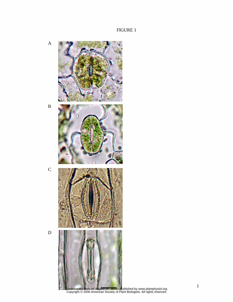
Figure 1. Images of stomata (closed) from each of the four species examined. (A)
Huperzia prolifers, (B) Nephrolepis exaltata, (C) Tradescantia virginiana, (D) Triticum
aestivum. All are in epidermal peels, bathed in 25 mM MES, pH 6.5, 1.0 mM KCl, 0.1
mM CaCl2. Scale bar is 20 µm, all images to same scale.
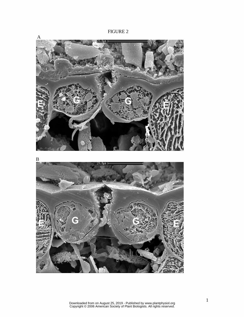
Figure 2. Cross-section of Huperzia prolifera stoma sampled by snap freezing an intact
leaf. (A) closed, (B) open under full sunlight, very high humidity. Note absence of guard
cell mechanical interaction with adjacent epidermal cells. Cryo-SEM. Scale bar is 20
µm. G, guard cell; E, epidermal cell.
Figure 3. Cross-section of Nephrolepis exaltata stoma sampled by snap freezing an
intact leaf. (A) closed, (B) open under full sunlight, very high humidity. Note absence of
guard cell mechanical interaction with adjacent epidermal cells. Cryo-SEM. Scale bar in
(B) is 20 µm. Both images are to the same scale. G, guard cell; E, epidermal cell.
Figure 4. Cross-section of Tradescantia virginiana stoma sampled by snap freezing an
intact leaf. (A) closed, (B) open under full sunlight, very high humidity. Note in (B) the
strong guard cell mechanical interaction with adjacent subsidiary cells. Cryo-SEM.
Scale bar in (A) is 20 µm. Both images are to the same scale. G, guard cell; S,
subsidiary cell.
Figure 5. Cross-section of Triticum aestivum stoma sampled by snap freezing an intact
leaf. (A) closed, (B) open under full sunlight, very high humidity. Note in (B) the strong
guard cell mechanical interaction with adjacent subsidiary cells. Cryo-SEM. Scale bar in
(A) is 10 µm. Both images are to the same scale. G, guard cell; S, subsidiary cell.
Figure 6. Relationship between stomatal pore width (a) and guard cell pressure (Pg), for
the four species, at full epidermal turgor and zero epidermal turgor (mean ± S.E.). Note
www.plantphysiol.orgon August 25, 2019 - Published by Downloaded from Copyright © 2006 American Society of Plant Biologists. All rights reserved.

21
the massive impediment to stomatal opening (mechanical advantage) by epidermal turgor
in T. virginiana and T. aestivum, compared to little or no effect in H. prolifera and N.
exaltata.
Figure 7. A visual impression of the difference, across the four stomatal types, in
maximum pore area (amax) per unit total area of the pore-guard cell complex (asc). This
ratio, amax/asc, sets the upper limit for stomatal conductance per unit leaf area. (A) H.
prolifera, (B) N. exaltata, (C) T. virginiana, (D) T. aestivum. Images were traced from
stomata in which Pg was held at approximately 4 MPa, with zero epidermal turgor. Scale
bar is 20 µm.
Figure 8: (A) maximum stomatal pore area; (B) ratio of maximum stomatal pore area :
projected total stomate area (pore plus guard cells); (C) ratio of maximum stomatal pore
area : projected guard cell area. All measurements were conducted on stomata in which
Pg was held at approximately 4 MPa, with zero epidermal turgor. (Mean ± S.E.)
Figure 9. (A) Simulation of the effect of leaf-to-air vapour pressure difference (D) on
the rate and magnitude of stomatal opening in a plant having stomatal mechanical
characteristics similar to T. virginiana (high epidermal mechanical advantage) but no
mechanism to actively compensate for the effect. gsw, stomatal conductance to water
vapour. (B) Comparison of the opening sequence of T. virginiana stomata at D = 1 kPa
and D = 2 kPa, following a change in PAR from 0 to 1000 µmol m-2 s-1. Plant well
watered; leaf temperature 25 °C. (C) As for (B), with T. aestivum. Arrows indicate the
direction of the difference in gsw at D = 2 kPa relative to D = 1 kPa.
Figure 10. (A), simulated stomatal opening sequence and (B), associated changes in
guard cell (Πg) and epidermal (Πe) osmotic pressures, for a plant with (solid lines) a
mechanism by which epidermal osmotic pressure declines in association with increasing
guard cell osmotic pressure during stomatal opening (see Methods for model details). All
other stomatal functional characteristics, as well as environmental conditions, are
www.plantphysiol.orgon August 25, 2019 - Published by Downloaded from Copyright © 2006 American Society of Plant Biologists. All rights reserved.

22
constant. Dotted lines correspond to the condition in which Πe remains constant during
stomatal opening. A decline in Πe during stomatal opening could allow the epidermal
mechanical advantage to be overcome under conditions of low D, allowing the observed
maximum stomatal apertures to be achieved at low D. Note also that not only does the
mechanism allow gws to reach its full potential, but the rate of opening is also greatly
increased.
Figure 11. Schematic of the proposed "osmotic sea-saw" (combined opposite changes in
both osmotic and turgor pressure between guard and subsidiary cells) in T. aestivum and
other grass-type stomata. The mechanism reduces the large mechanical advantage that
would otherwise prevent stomatal opening under high humidity, even if maximum turgor
were generated in guard cells. A corollary is that the mechanism also greatly accelerates
the rate of stomatal opening, helping to explain why T. aestivum has rates of stomatal
opening more than an order of magnitude faster than any of the other species (Figure 12).
(A), cross-section of guard cells (thick dark walls) and subsidiary cells (thin walls) of
closed stoma; (B) open stoma, showing highly displaced subsidiary cells which have
transferred most of their potassium to the guard cells, undergoing a significant reduction
in turgor as a consequence. Compare SEM images in Figure 5.
Figure 12. Maximum rate of increase in stomatal conductance to water vapor (dgsw / dt),
quantified in relation to (A) leaf area, (B) individual stomata, or (C) projected area of the
inflated guard cell pair, as a proxy for guard cell size. The grass T. aestivum was
substantially faster in all categories. (Mean ± S.E. See Methods for an explanation of the
units.)
www.plantphysiol.orgon August 25, 2019 - Published by Downloaded from Copyright © 2006 American Society of Plant Biologists. All rights reserved.
1
FIGURE 1
A B C
D
www.plantphysiol.orgon August 25, 2019 - Published by Downloaded from Copyright © 2006 American Society of Plant Biologists. All rights reserved.
1
FIGURE 2 A
B
www.plantphysiol.orgon August 25, 2019 - Published by Downloaded from Copyright © 2006 American Society of Plant Biologists. All rights reserved.

1
FIGURE 3 A
B
www.plantphysiol.orgon August 25, 2019 - Published by Downloaded from Copyright © 2006 American Society of Plant Biologists. All rights reserved.
1
FIGURE 4 A
B
www.plantphysiol.orgon August 25, 2019 - Published by Downloaded from Copyright © 2006 American Society of Plant Biologists. All rights reserved.

1
FIGURE 5 A
B
www.plantphysiol.orgon August 25, 2019 - Published by Downloaded from Copyright © 2006 American Society of Plant Biologists. All rights reserved.

1
0 1 2 3 4 50
1
2
3
4
5
6
Stom
atal
ape
rture
, a
(µm
)
Guard cell pressure (MPa)
Pe = max
Pe = 0
AHuperzia prolifera
FIGURE 6
0 1 2 3 4 50
1
2
3
4
5
6
Sto
mat
al a
pertu
re, a
(µ
m)
Guard cell pressure (MPa)
Pe = max
Pe = 0
Nephrolepis exaltata
B
0 1 2 3 4 50
5
10
15
20
25
Stom
atal
ape
rture
, a
(µm
)
Guard cell pressure (MPa)
Pe = max
Pe = 0
Tradescantia virginianaC
0 1 2 3 40
2
4
6
8 P
e = max
Pe = 0
Triticum aestivum
D
Sto
mat
al a
pertu
re, a
(µ
m)
Guard cell pressure (MPa)
www.plantphysiol.orgon August 25, 2019 - Published by Downloaded from Copyright © 2006 American Society of Plant Biologists. All rights reserved.

1
FIGURE 7
A B
C D
www.plantphysiol.orgon August 25, 2019 - Published by Downloaded from Copyright © 2006 American Society of Plant Biologists. All rights reserved.
1
FIGURE 8
Huperzia
Nephrolepis
Tradescantia
Triticum
0
100
200
300
400
500
600
700
800
Ope
n st
oma
pore
are
a (µ
m2 )
A
Huperzia
Nephrolepis
Tradesca
ntiaTritic
um
0.00
0.05
0.10
0.15
0.20
0.25
0.30
Ope
n po
re a
rea
/ pro
ject
ed to
tal s
tom
ate
area
B
Huperzia
Nephrolepis
Tradescantia
Triticum
0.00
0.05
0.10
0.15
0.20
0.25
0.30
0.35
0.40
Ope
n po
re a
rea
/ pro
ject
ed G
C a
rea
C
www.plantphysiol.orgon August 25, 2019 - Published by Downloaded from Copyright © 2006 American Society of Plant Biologists. All rights reserved.

1
FIGURE 9
0 500 1000 1500 20000.0
0.1
0.2
0.3
0.4
0.5S
imul
ated
gsw
(m
ol m
-2 s
-1)
Time (s)
D = 1kPa D = 2kPa
A
..
0 1000 2000 3000 4000 5000 60000.00
0.05
0.10
0.15
0.20
0.25
0.30
g sw (
mol
m-2 s
-1)
Time (s)
D = 1 kPa D = 2 kPa
Tradescantia virginiana
B
0 1000 2000 3000 4000 5000 60000.0
0.2
0.4
0.6
0.8
g sw (
mol
m-2
s-1)
Time (s)
D = 1 kPa D = 2 kPa
Triticum aestivum
C
www.plantphysiol.orgon August 25, 2019 - Published by Downloaded from Copyright © 2006 American Society of Plant Biologists. All rights reserved.
1
FIGURE 10
0 500 1000 1500 20000.0
0.1
0.2
0.3
0.4
0.5
0.6
g sw
(mol
m-2 s
-1)
Time (s)
Πe fixed
Πe declining with increasing Π
g
A
0 500 1000 1500 20000
1
2
3
4
Cel
l osm
otic
pre
ssur
e (
MPa
)
Time (s)
Πe (declines with increasing Π
g)
Πg ( Π
e declning with increasing Π
g)
Πe (fixed)
Πg ( Π
e fixed)
B
www.plantphysiol.orgon August 25, 2019 - Published by Downloaded from Copyright © 2006 American Society of Plant Biologists. All rights reserved.
1
FIGURE 11
K+ K+
K+K+
A
B
www.plantphysiol.orgon August 25, 2019 - Published by Downloaded from Copyright © 2006 American Society of Plant Biologists. All rights reserved.

1
FIGURE 12
Huperzia
Nephrole
pis
Tradesc
antiaTritic
um
0.0
0.5
1.0
1.5
2.0 M
axim
um le
af-a
rea-
spec
ific
d g sw
/ dt
(mm
ol m
-2 s-2
)
A
Huperzia
Nephrolepis
Tradesc
antia
Triticum
0
10
20
30
40
50
60
70
Max
imum
sto
ma-
spec
ific
d g sw
/dt
(pm
ol s
tom
a-1 s
-2)
B
Huperzia
Nephrolep
is
Tradescan
tiaTritic
um
0
10
20
30
40
50
60
70
80
90
Max
imum
gua
rd c
ell-a
rae-
spec
ific
dgsw
/ dt
(mm
ol m
-2
(GC
pai
r) s-2)
C
www.plantphysiol.orgon August 25, 2019 - Published by Downloaded from Copyright © 2006 American Society of Plant Biologists. All rights reserved.