running head: methyl iaa esterases in arabidopsis thaliana · perhaps be involved in the hydrolysis...
TRANSCRIPT

1
Running head: Methyl IAA esterases in Arabidopsis thaliana
Corresponding author:
Eran Pichersky
Department of Molecular, Cellular and Developmental Biology, University of Michigan, Ann
Arbor, MI 48109-1048 USA
Tel. 1-734-936 3522
Fax. 1-734-647 0884
E-mail: [email protected]
Plant Physiology Preview. Published on May 8, 2008, as DOI:10.1104/pp.108.118224
Copyright 2008 by the American Society of Plant Biologists
www.plantphysiol.orgon February 18, 2020 - Published by Downloaded from Copyright © 2008 American Society of Plant Biologists. All rights reserved.

2
Inactive Methyl Indole-3-Acetic Acid Ester Can Be Hydrolyzed and Activated by Several
Esterases Belonging to the AtMES Esterase Family of Arabidopsis thaliana1
Yue Yang, Richard Xu, Choong-je Ma, A. Corina Vlot, Daniel F. Klessig, and Eran Pichersky*
Department of Molecular, Cellular and Developmental Biology, University of Michigan, 830
North University Street, Ann Arbor, MI 48109-1048 (YY, RX, CM, EP)
Boyce Thompson Institute for Plant Research, Ithaca, NY 14853 (ACV, DFK)
*Corresponding author; e-mail [email protected].
1This work was supported by National Science Foundation Arabidopsis 2010 project grant MCB-
0312466 to EP, and by National Science Foundation grant IOB-0525360 to DFK.
The author responsible for distribution of materials integral to the findings presented in this
article in accordance with the policy described in the Instructions for Authors
(www.plantphysiol.org) is: Eran Pichersky ([email protected]).
Key Words:
Esterase, gene family, IAA, methyl IAA, plant hormones, plant biochemistry
www.plantphysiol.orgon February 18, 2020 - Published by Downloaded from Copyright © 2008 American Society of Plant Biologists. All rights reserved.
3
Abstract
The plant hormone auxin (indole-3-acetic acid, IAA) is found both free and conjugated to a
variety of carbohydrates, amino acids and peptides. We have recently shown that IAA could be
converted to its methyl ester (MeIAA) by the Arabidopsis thaliana enzyme IAMT1 (IAA
carboxyl methyltransferase 1). However, the presence and function of MeIAA in vivo remains
unclear. Recently it has been shown that the tobacco protein SABP2 (Salicylic acid binding
protein-2) hydrolyzes methyl salicylate to salicylic acid. There are 20 homologs of SABP2 in the
genome of Arabidopsis thaliana, which we have named AtMES (for methyl esterases). We tested
15 of the proteins encoded by these genes in biochemical assays with various substrates, and
identified several candidate MeIAA esterases that could hydrolyze MeIAA. MeIAA, like IAA,
exerts inhibitory activity on the growth of wild type roots when applied exogenously. However,
the roots of Arabidopsis plants carrying T-DNA insertions in the putative MeIAA esterase gene
AtMES17 (At3g10870) displayed significantly decreased sensitivity to MeIAA compared with
wild type roots, while remaining as sensitive to free IAA as wild type roots. Incubating seedlings
in the presence of [14C]-MeIAA for 30 min revealed that mes17 mutants hydrolyzed only 40% of
the [14C]-MeIAA taken up by plants, whereas wildtype plants hydrolyzed 100% of absorbed
[14C]-MeIAA. Roots of Arabidopsis plants overexpressing AtMES17 showed increased
sensitivity to MeIAA, but not to IAA. Additionally, mes17 plants have longer hypocotyls, and
display increased expression of the auxin-responsive DR5:GUS reporter gene, suggesting a
perturbation in IAA homeostasis and/or transport. mes17-1/axr1-3 double mutant plants have the
same phenotype as axr1-3, suggesting MES17 acts upstream of AXR1. The protein encoded by
AtMES17 had a Km value of 13 µM, and a Kcat value of 0.18 sec-1 for MeIAA. AtMES17 was
expressed at the highest levels in shoot apex, stem and root of Arabidopsis. Our results
demonstrate that MeIAA is an inactive form of IAA, and the manifestations of MeIAA in vivo
activity are due to the action of free IAA that is generated from MeIAA upon hydrolysis by one
or more plant esterases.
www.plantphysiol.orgon February 18, 2020 - Published by Downloaded from Copyright © 2008 American Society of Plant Biologists. All rights reserved.

4
INTRODUCTION
Indole-3-acetic acid (IAA), also known as auxin, is a plant hormone involved in many aspects of
plant growth and development, such as embryogenesis, vascular differentiation, fruit set and
development, and senescence (Woodward and Bartel, 2005; Teale et al., 2006; Delker et al.,
2008). Plants utilize a variety of mechanisms to spatially and temporally regulate IAA
concentrations and gradients, including de novo synthesis, degradation, transport, and synthesis
and hydrolysis of various IAA conjugates (Normanly, 1997; Ljung et al., 2002; Woodward and
Bartel, 2005).
IAA is known to be conjugated to sugars, amino acids and peptides, and some enzymes
that catalyze these conjugating reactions have been characterized (Jackson et al., 2001; Staswick
et al., 2005). Some conjugates such as IAA-Asp and IAA-Glu are not able to induce auxin
responses when applied exogenously, and therefore are considered inactive auxin and
intermediates in IAA degradation (Ljung et al., 2002; Woodward and Bartel, 2005). Recently, a
rice GH3-8 gene encoding IAA-amino acid synthetase has been shown to promote basal
immunity in rice by converting active IAA to inactive IAA-Asp, and thus reducing the auxin-
induced cell wall loosening (Ding et al., 2008). Other IAA conjugates such as IAA-Leu and
IAA-Ala induce auxin responses when applied exogenously to plants. However, the hydrolytic
cleavage of these compounds parallels the activity (Bartel and Fink, 1995; Ljung et al., 2002).
These findings have led to suggestions that these conjugates per se are biologically inactive, and
any response obtained in the assay reflected the degree of hydrolysis and the activity of the
released free hormone.
A family of hydrolases that act on IAA-amino acid conjugates and release IAA from
some IAA-amino acid conjugates have been identified in Arabidopsis (LeClere et al., 2002;
Rampey et al., 2004). The hydrolyzable IAA conjugates have been proposed to function as
storage form to allow the plants to quickly release active IAA when necessary. While the
transport of any IAA conjugates has rarely been reported, IAA-inositol has been shown to be
transported from the endosperm to shoot in Zea mays at a rate much faster than that of free IAA,
and there hydrolyzed to yield free IAA (Nowacki and Bandurski, 1980).
We have recently discovered an IAA-carboxylmethyltransferase (IAMT1) in Arabidopsis
thaliana and several other species that can methylate IAA to form the ester methyl indole-3-
acetate (MeIAA) (Zubieta et al., 2003; Qin et al., 2005; Zhao et al., 2008). Furthermore,
www.plantphysiol.orgon February 18, 2020 - Published by Downloaded from Copyright © 2008 American Society of Plant Biologists. All rights reserved.
5
disruption of the expression levels of IAMT1 led to phenotypes indicative of disruption of IAA
homeostasis (Qin et al., 2005). MeIAA has rarely been reported as an endogenous IAA
metabolite in plants (Narasimhan et al., 2003), probably due to its low abundance or fast turnover,
and therefore its in vivo function remains unknown. MeIAA had been used as a substitute for
IAA in physiological studies (Zimmerman and Hitchcock, 1937), and it has been observed that
various auxin signaling mutants show decreased sensitivity to exogenously applied MeIAA as
well as to IAA (Qin et al., 2005), suggesting that MeIAA and IAA share similar signaling
components. Therefore, either MeIAA itself could initiate the auxin signaling pathway, or it must
be hydrolyzed to IAA to exert hormonal function. If MeIAA hydrolysis occurs in planta, the
reaction is likely to be catalyzed by one or more carboxylesterases.
Carboxylesterases catalyze the hydrolysis of a C-O ester linkage in a wide range of
compounds, and structural analyses have shown that such enzymes are all members of the α/β
hydrolase “superfamily” (Nardini and Dijkstra, 1999). Carboxylesterases have been extensively
studied in animals and microbes. However, the physiological role and substrate specificity of few
plant carboxylesterases have been identified. Several putative plant proteins, including those
encoded by tobacco hsr203J, the tomato and pea homologs of hsr203J, and PrMC3 from Pinus
radiate and pepEST from pepper (Pontier et al., 1994; Pontier et al., 1998; Walden et al., 1999;
Ichinose et al., 2001; Ko et al., 2005) have been annotated as carboxylesterases based on
homology with fungal esterases but with little direct biochemical evidence (Baudouin et al.,
1997). Marshall et. al. (2003), in turn, searched the Arabidopsis genome, which has several
hundred members of the α/β hydrolase superfamily, for genes encoding proteins with the highest
similarities to these previously annotated plant carboxylesterases. This bioinformatic search
identified a branch of the α/β hydrolase superfamily containing 20 genes, which were
collectively named the AtCXE family (Marshall et al., 2003). However, the in vivo substrates of
none of the enzymes in the AtCXE family have been experimentally determined. Recently, two
proteins belonging to the α/β hydrolase superfamily have been identified in Gentiana triflora and
implicated in cold response, but their in vivo substrates remain unknown (Hikage et al., 2007).
Recently, we have demonstrated that a tobacco protein required for development of
systemic acquired resistance, SABP2 (originally identified as salicylic acid binding protein-2), is
a methyl salicylate esterase (Kumar and Klessig, 2003; Forouhar et al., 2005). The amino acid
sequence of SABP2 shares 46-56% similarities to two other confirmed methyl esterases from
www.plantphysiol.orgon February 18, 2020 - Published by Downloaded from Copyright © 2008 American Society of Plant Biologists. All rights reserved.
6
plants, methyl jasmonate esterase (MJE) from tomato and polyneuridine aldehyde esterase
(PNAE) from the medicinal plant Rauvolfia serpentina (Dogru et al., 2000; Stuhlfelder et al.,
2004; Forouhar et al., 2005). Bioinformatic analysis of the Arabidopsis genome revealed 20
genes encoding proteins with sequence similarities to SABP2 (Forouhar et al., 2005; Yang et al.,
2006a). These proteins are distinct from the group of 20 AtCXE proteins, and their sequence
similarity to known methylesterases suggests that they too may be methylesterases and may
perhaps be involved in the hydrolysis of methyl salicylate, methyl jasmonate, or MeIAA in
Arabidopsis thaliana (Yang et al., 2006a).
Here we show that some of the proteins in this group of putative Arabidopsis
methylesterases, which we have named MES (for methyl esterases), are able to hydrolyze
MeIAA. Analysis of mutants with T-DNA insertions in the AtMES genes indicates that at least
one AtMES (AtMES17) is capable of hydrolyzing MeIAA in vivo. We used this mutant to
demonstrate that MeIAA itself is an inactive form of IAA.
RESULTS
The Arabidopsis genome has 20 MES genes
A search of the Arabidopsis genome for genes encoding proteins with the highest identity to
tobacco SABP2 (methyl salicylate esterase), tomato MJE (methyl jasmonate esterase), and
Rauvolfia serpentina PNAE (polyneuridine aldehyde esterase) identified 20 genes forming a
close clade within the α/β hydrolase superfamily, which we named AtMES1 through AtMES20
(MES for methyl esterase) (Fig. 1 and Table I). The amino acid sequences of the AtMES proteins,
which range in length from 256 to 444 aa (with the exception of the proteins encoded by
AtMES19 and 20, which are likely to be pseudogenes, see below), share 30-57% similarity with
tobacco SABP2, 31-42% similarity with tomato MJE, and 29-49% similarity with R. serpentina
PNAE. The tree topology of the AtMES family shows the presence of three clusters of genes,
which we have named Subfamily 1, 2, and 3 (Fig. 1). Members of the previously annotated plant
carboxylesterases (CXE) family, including tobacco jsr203J, PrMC3, and three AtCXE genes
(AtCXE1, 2, and 19), are more divergent and they cluster into a clade that is distant from the
AtMES family (Fig. 1).
The sequence alignment of AtMES1-20 revealed that the catalytic triad Ser-His-Asp, a
characteristic feature of the α/β hydrolase fold family, is conserved in 15 of these proteins (Fig.
www.plantphysiol.orgon February 18, 2020 - Published by Downloaded from Copyright © 2008 American Society of Plant Biologists. All rights reserved.

7
2). In the protein sequences of AtMES11, AtMES13 and AtMES15, the conserved Ser in the
catalytic triad is replaced by Asp, a substitution previously found in active α/β hydrolases in
animals (Holmquist, 2000). AtMES19 and AtMES20 lack part of the N-terminal or C-terminal
region, respectively, and are therefore likely to be inactive enzymes.
Substrate specificities of 15 MES esterases
To examine whether the AtMES genes encode functional esterases, we obtained full-length
cDNAs of 15 AtMES genes, expressed the cDNAs in E. coli, and tested the recombinant proteins
for esterase activity. Since AtMES19 and 20 were likely to be pseudogenes, they were not tested.
Three other AtMES proteins, AtMES6, AtMES13 and AtMES15, were also not tested because we
were not able to obtain full-length cDNAs.
When the 15 AtMES esterases were tested with the chymotryptic synthetic substrate p-
nitrophenyl acetate (PNPA), AtMES1, AtMES2, AtMES3, AtMES4, AtMES7, AtMES8, AtMES9,
AtMES16, and AtMES17 showed activity (Table I). Because the AtMES proteins are homologs
of SABP2 and MJE, esterases that hydrolyze the methylated plant hormones MeSA and MeJA
respectively, we further examined whether the AtMES proteins are active with known
methylated plant hormones, including MeIAA, MeSA, MeJA, MeGA4 and MeGA9 (Shulaev et
al., 1997; Chen et al., 2003; Qin et al., 2005; Yang et al., 2006a; Varbanova et al., 2007). To
assess whether the AtMES proteins are active with any of these substrates, we carried out
preliminary assays for each protein with a number of substrates present at a concentration of 1
mM. Reactions that resulted in product formation that was at least 3 times the value found in
control assays (using boiled enzyme) were scored “+”as indicating enzymatic activity (Table I).
Among the 15 esterases tested, AtMES1, AtMES2, AtMES3, AtMES7, AtMES9,
AtMES16, AtMES17, and AtMES18 displayed hydrolase activity with MeIAA, while AtMES4,
AtMES5, AtMES8, AtMES10, AtMES11, AtMES12 and AtMES14 could not hydrolyze MeIAA
(Table I). In addition, AtMES1, AtMES2, AtMES4, AtMES7, and AtMES9 displayed MeSA
hydrolase activity, while AtMES1, AtMES2, AtMES3, AtMES9, AtMES10, and AtMES16 were
active with MeJA. None of the 15 AtMES esterases was active with MeGA4 or MeGA9. AtMES5,
AtMES8, AtMES11, AtMES12, and AtMES14 were not active with any of these methylated
hormones.
www.plantphysiol.orgon February 18, 2020 - Published by Downloaded from Copyright © 2008 American Society of Plant Biologists. All rights reserved.
8
Atmes17 null mutant plants are more resistant than wildtype to the root inhibition activity
of exogenously supplied MeIAA and they are defective in hydrolysis of such MeIAA in vivo
Since AtMES1, AtMES2, AtMES3, AtMES7, AtMES9, AtMES16, AtMES17, and AtMES18
could all hydrolyze MeIAA in vitro, we examined whether they possess MeIAA hydrolase
activity in vivo. It has been previously shown that both IAA and MeIAA inhibit root growth in
wildtype Arabidopsis seedlings when applied exogenously (Zimmerman and Hitchcock, 1937;
Qin et al., 2005), but it has not been determined if MeIAA itself is active or whether the apparent
activity of MeIAA is due to its hydrolysis in planta, giving rise to active IAA.
T-DNA insertional mutants of AtMES1, AtMES9, AtMES16, and AtMES17 were obtained
as described in materials and methods, including two independent mutant lines each for both
AtMES16 and AtMES17. There was no T-DNA insertional mutant of AtMES3 reported, and the
several T-DNA insertions reported for AtMES2, AtMES7, and AtMES18 turned out upon further
examination (described in materials and methods) not to abolish gene transcriptions (data not
shown).
All mutant lines as well as wildtype Arabidopsis plants were next grown on 1/2 MS
medium containing various concentrations of MeIAA or no MeIAA, and their root lengths were
measured after 7 days. While in unsupplemented medium root length of an AtMES17 T-DNA
mutant mes17-1 (SALK_092550) seedlings were similar to that of wildtype, in the presence of
MeIAA concentration ranging from 0.01-1 µM root length of mutant seedlings was consistently
longer than the root length of wildtype seedlings (Fig. 3B). For example, at 0.5 µM MeIAA, a
concentration that inhibits the root growth of wildtype Arabidopsis by 85% on average, wildtype
seedlings had an average root length of 4.1 mm and mes17-1 plants had an average root length of
12 mm, 3 times as long as that of the wildtype (Fig. 3A, B). Similar results were obtained with a
second independent AtMES17 T-DNA mutant, mes17-2 (SAIL-503-c03) (data not shown). The
root lengths of AtMES1, AtMES2, AtMES7, AtMES9 and AtMES16 mutant lines grown on
MeIAA were the same as wildtype. All mutant plants, including the mes17-1 and mes17-2, when
grown on 1/2 MS medium containing different concentrations of IAA showed no statistically
significant difference in root length from that of wild-type plants, although the mes17 mutants
appeared to have a slightly diminished response to IAA (Fig. 3C, D).
To examine directly the fate of exogenously added MeIAA in wildtype and Atmes17
www.plantphysiol.orgon February 18, 2020 - Published by Downloaded from Copyright © 2008 American Society of Plant Biologists. All rights reserved.

9
mutant plants, we soaked plants in a 0.5 µM solution of [14C]-MeIAA and examined the total
amount of [14C] label taken up by the plant and the relative amounts of [14C]-MeIAA remaining
in the plant tissues. After 30 min of incubation, wildtype plants had no [14C]-MeIAA left but
Atmes17-1 plants still contained 58.5%+/-16.5% of the [14C] label taken up in the form of
MeIAA (Figure 4).
We also obtained several lines that overexpress AtMES17 under the control of the 35S
promoter and tested them for sensitivity to MeIAA and IAA treatments. When AtMES17
overexpressing plants of three independent lines were grown in the presence of 0.5 µM MeIAA
for 7 days, their root growth was more severely inhibited than that of wildtype seedlings (see Fig.
5A for one of the lines). However, both types of seedlings had a similar root length in the
presence of 0.5 µM IAA (Fig. 5B), suggesting that the increased root inhibition of MeIAA on
MES17 overexpressing plants was caused by increased auxin concentration derived from
increased rate of MeIAA hydrolysis.
Atmes17 null mutants have a longer hypocotyl
We observed that mes17-1 mutant plants grown in soil had in general longer hypocotyls than
wildtype plants. The hypocotyls of 4-week old mes17-1 plants were on average 32% longer than
that of wildtype (Fig. 6). Statistical analysis performed by Student’s t test returned p values <
1x10⁻⁹, indicating that the differences are significant. When grown on 1/2 MS medium under
continuous light for 8 days, mes17-1 and mes17-2 mutants had hypocotyls longer on average by
29% than that of wildtype seedlings (Fig. 6), with statistical analysis indicating that the
differences are significant (p values < 1x10⁻⁵). When grown on 1/2 MS medium in the dark,
however, the hypocotyl lengths of mes17-1 and mes17-2 mutants were the same as that of
wildtype (Fig. 6). With the exception of hypocotyl length, mes17 mutants grown under normal
conditions did not display any obvious phenotypic differences compared to wildtype.
The DR5:GUS reporter gene is more highly expressed in Atmes17 null mutants compared
with wildtype plants
DR5 is a synthetic auxin response element, and the DR5:GUS reporter has been widely used as a
marker to study the endogenous distribution of auxin (Ulmasov et al., 1997; Ottenschlager et al.,
2003). We constructed mes17 null mutant plants carrying the DR5:GUS reporter gene and tested
www.plantphysiol.orgon February 18, 2020 - Published by Downloaded from Copyright © 2008 American Society of Plant Biologists. All rights reserved.
10
them for GUS activity. mes17 null plants had much stronger GUS staining overall than wildtype
plants, including in the shoot apex and in the root primordial and leaf tip (Fig. 7).
mes17-1/axr1-3 double mutant plants have the same phenotype as axr1-3
Plants homozygous for the allele axr1-3, which carries a missense mutation in the AXR1 gene,
display resistance to exogenous auxin, as well as a variety of morphological defects due to
compromised auxin signaling (Lincoln et al., 1990; Leyser et al., 1993). We have previously
shown that axr1-3 mutants also have reduced sensitivity to MeIAA (Qin et al., 2005), suggesting
that MeIAA shares similar signaling components as IAA. We therefore constructed plants that
were homozygous for both mes17-1 and axr1-3, and tested them for their phenotype and their
response to IAA and MeIAA. While mes17-1 mutant had a longer hypocotyl than wildtype when
grown in 1/2 MS medium in light, axr1-3 has a shorter hypocotyl than wildtype (Fig. 8, see also
Jensen et al, 1998). The hypocotyl length of mes17-1/axr1-3 was shorter than that of wildtype,
similar with the hypocotyl length of axr1-3 mutant (Fig. 8). In addition, mes17-1/axr1-3 mutant
plants had the same morphological phenotype as the axr1-3 mutant line, which includes irregular
rosette leaves, reduced height, and reduced fertility (data not shown). When plants were tested
for their responses to the root growth inhibition activity of IAA and MeIAA, mes17-1/axr1-3
double mutant displayed reduced sensitivity to both IAA and MeIAA, same as axr1-3 (data not
shown).
Biochemical characterization of AtMES17
Because AtMES17 displays MeIAA hydrolase activity in vitro and likely does so in vivo, we
performed a more detailed in vitro kinetic analysis of the E. coli-expressed and purified
AtMES17. AtMES17 displayed hydrolase activity towards MeIAA, but not MeJA, MeSA, or
MeGAs (Table I). AtMES17 displayed the highest MeIAA hydrolase activity at pH 8.5, and
about 60% of the highest activity at pH 6.5 or 9.5. However, at pH 8.0 or higher, non-enzymatic
hydrolysis of MeIAA was also observed. We therefore used buffers with pH 7.5, which gave
93% of the maximal enzymatic activity, and no observable non-enzymatic hydrolysis. Under
these conditions, AtMES17 had a Km value of 13 µM and a Kcat value of 0.18 sec-1 for MeIAA.
The hydrolase activity was strongly inhibited (44-75%) by 5 mM Fe2+, Fe3+, Zn2+, and Cu2+, and
mildly inhibited by 5 mM Ca2+ and Mn2+ (20% and 34%), respectively. At 5 mM concentration,
www.plantphysiol.orgon February 18, 2020 - Published by Downloaded from Copyright © 2008 American Society of Plant Biologists. All rights reserved.

11
Na+, Mg2+, K+, and NH4+ had no effects on the hydrolase activity.
Expression pattern of AtMES17
Real-time RT-PCR analysis showed that the expression of AtMES17 in 10-d old in seedlings is
app. 5-fold higher in the region of the shoot apex than in the rest of the hypocotyl (Fig. 9). When
AtMES17 transcripts levels in the 8-week mature plants were examined, the highest expression
levels were observed in stem, followed by roots, flowers, rosette leaves, and siliques, and no
AtMES17 transcripts were detectable in cauline leaves (Fig. 9).
DISCUSSION
The Arabidopsis MES methylesterase family
We have identified a family of 20 Arabidopsis proteins which we have designated the AtMES
(for methyl esterase) family, based on the sequence similarity of these proteins to experimentally
identified methyl esterases, including the tobacco methyl salicylate esterase SABP2 (NtSABP2)
and the tomato methyl jasmonate esterase MJE (LeMJE). This AtMES family is distinct from the
previously annotated AtCXE (carboxylesterase) family (Fig. 1), yet both families belong to the
α/β hydrolase superfamily, which is characterized by the structurally conserved “canonical” α/β
hydrolase fold and catalytic residues (Nardini and Dijkstra, 1999).
Most members of the AtMES family encode proteins of approximately 250 aa, similarly
to NtSABP2 and LeMJE. AtMES11, 12, 13, 14 and 15 contain an extra region of 90-190 aa at
their N-termini (Fig. 2). This N-terminal extension does not appear to constitute a targeting
signal peptide, suggesting that these AtMES proteins are localized in the cytosol like the rest of
the members in the family. The AtMES11, 12, 13, 14 and 15 proteins also cluster into a close
clade within the AtMES family (subfamily 3, Fig. 1), and so far we have not been able to ascribe
any enzymatic activity to any of the proteins in this subfamily.
Because the AtMES proteins are closely related to LeMJE and NtSABP2, we
hypothesized that members of the AtMES family could encode MeIAA esterase(s). Of the
members of the AtMES family that we tested, eight AtMES proteins were found to be active with
MeIAA, and they all belong to subfamilies 1 and 2. Some of these proteins also hydrolyze other
methyl esters under the experimental conditions used in this study, and it is likely that many, and
www.plantphysiol.orgon February 18, 2020 - Published by Downloaded from Copyright © 2008 American Society of Plant Biologists. All rights reserved.
12
perhaps all of the AtMES proteins would be found to use multiple substrates upon a more
extensive survey of substrates.
AtMES17 encodes an esterase capable of hydrolyzing MeIAA, which is not itself active
We further showed that AtMES17 encodes an esterase that efficiently and specifically hydrolyzes
MeIAA to IAA in vitro, and is likely to do so in vivo as well. The kinetic parameters of
AtMES17 are comparable to previously characterized esterases (Forouhar et al., 2005), indicating
that MeIAA is likely to be a relevant substrate for AtMES17 in planta. As pointed out above,
although MES17 showed activity only with MeIAA in our limited survey, we cannot yet
conclude that MeIAA is its only substrate.
The growth of roots of two independent mes17 mutants was much less inhibited by
MeIAA than was widltype root growth (Fig. 3A). However, both mes17 null mutants responded
similarly as wildtype to the root inhibition activity of IAA (Fig. 3B), indicating normal auxin
signaling in these mutants. Incubating seedlings in the presence of [14C]-MeIAA also revealed
that mes17 mutants were much less efficient in hydrolyzing [14C]-MeIAA than wildtype plants
(Fig. 4). We thus conclude that the response of Arabidopsis seedlings to the root inhibition
activity of MeIAA is at least partly due to the hydrolytic activity of AtMES17. The observations
that some hydrolysis of [14C]-MeIAA occurred in the mes17 mutant line and that the root growth
of mes17 mutants retained some response to the inhibitory activity of MeIAA also indicate that
there are other esterases in addition to AtMES17 that participate in MeIAA hydrolysis in
Arabidopsis, consistent with our finding that other AtMES proteins could hydrolyze MeIAA in
vitro. These proteins include AtMES16 and AtMES18, which have the highest sequence
similarities to AtMES17. However, we obtained two independent T-DNA insertional lines of
AtMES16 and observed that both of these null mutants responded to MeIAA similarly to
wildtype, including in the root growth assay. Microarray data indicates that transcript levels of
AtMES17 in roots are more than 10 times higher than those of AtMES16 (AtGenExpress
Visualization Tool) (Schmid et al., 2005). In addition, the double mutant mes16/mes17-1 was
phenotypically indistinguishable from mes17-1 mutant in all organs or developmental stages
(data not shown). We were unable to obtain a mutation in AtMES18. It remains to be determined
whether AtMES16 or other AtMES can hydrolyze MeIAA in vivo.
The much reduced (although not completely abolished) response of roots of mes17
www.plantphysiol.orgon February 18, 2020 - Published by Downloaded from Copyright © 2008 American Society of Plant Biologists. All rights reserved.

13
mutants in the root inhibition assay with exogenously supplied MeIAA coupled with the
observation that these mutants are much less efficient in the hydrolysis of MeIAA also suggest
that the inhibition is due to IAA and not MeIAA. Consistent with this interpretation, when
AtMES17 was overexpressed, their roots were even more sensitive to MeIAA than wildtype
plants (Fig. 5), likely because MeIAA was hydrolyzed in these plants even faster than in
wildtype. The observation that the mes17-1/axr1-3 double mutant plants have the same
phenotype as axr1-3 is also consistent with the putative role of MES17 in producing IAA, which
acts upstream of AXR1. A similar albeit more extensive analysis of the effects of MeIAA
treatment on axr1 and other auxin response mutants has also concluded that MeIAA is likely to
be inactive by itself (Li et al., 2008). In addition, the recently solved structure of the auxin
receptor TIR1 supports the notion that MeIAA is not an active auxin, since it was shown that the
carboxyl group of the IAA molecule interacts with two residues in the binding pocket of TIR1,
docking IAA to the bottom of the pocket (Tan et al., 2007). MeIAA has a methyl ester group
instead of a carboxyl group, and therefore is not likely to be accommodated in the binding site of
TIR1 to initiate TIR1-mediated auxin signaling. Analogous to our finding, it was recently shown
that silencing of a tobacco methyl jasmonate esterase (NaMJE) reduced MeJA- but not JA-
induced herbivore resistance, indicating that the resistance elicited by MeJA treatment is directly
elicited not by MeJA but by its de-methylated product, JA (Wu et al., 2008).
Possible role of MeIAA in Arabidopsis
mes17-1 mutants have longer hypocotyls than wildtype plants. The regulation of hypocotyl
length is a complex process that is under the influence of many factors, including light, nutrient,
and hormones such as IAA, ethylene and brassinosteroids (Jensen et al., 1998; Collett et al., 2000;
Vandenbussche et al., 2005). Earlier physiological studies have shown that auxin promotes the
growth of excised hypocotyl segments from various plant species (Evans, 1985). Several
Arabidopsis mutants that accumulate increased overall auxin levels also have longer hypocotyls
than wildtype, although the auxin gradient may be more important than its actual concentration
(Boerjan et al., 1995; Zhao et al., 2001; Zhao et al., 2002). In addition, Jensen et al, (1998) have
demonstrated that auxin transport from the shoot apex to the root is required for hypocotyl
elongation in Arabidopsis during development in the light.
www.plantphysiol.orgon February 18, 2020 - Published by Downloaded from Copyright © 2008 American Society of Plant Biologists. All rights reserved.

14
In seedlings, AtMES17 is expressed at highest levels in the shoot apex, but it is also
expressed at lower levels elsewhere (Fig. 9). mes17-1 plants display a stronger auxin response in
the shoot apex as well as in other parts of the plant (as assessed by the DR5:GUS reporter system,
Fig. 7). Although it seems paradoxical that mes17 mutants appear to have higher levels of IAA, it
may be that the higher GUS staining in this line indicates a higher rate of transport of IAA, rather
than a higher level of IAA concentration, brought about by a higher but transient and localized
concentrations of MeIAA due to the decrease in (but not complete absence of) overall MeIAA
esterase activity. Methylation of IAA to enhance its transport (and subsequent hydrolysis by
MES enzymes) would be analogous to the transport of SA as biologically inactive MeSA from
the site of infection to distal tissue for development of systemic acquired resistance (Park et al.,
2007). For this explanation to be valid, however, it would appear necessary to postulate that there
are distinct types of cells, probably in close proximity to each other, that contain either IAA
methylation activity or MeIAA esterase activity.
A high-resolution spatial map depicting auxin concentration as well as activities of
MES17 and IAMT is needed to validate this hypothesis. Recently, a cell type-specific microarray
analysis has shown that in a given area of roots, genes involved in auxin biosynthesis are
expressed in different cells than genes regulating auxin homeostasis or auxin transport (Brady et
al., 2007). Transient methylation of IAA and small local differences in expression of MES17
(and IAMT) among populations of cells may also explain our failure to detect differences in
MeIAA concentration between wildtype, mes17, and 35S::IAMT lines, which all contain MeIAA
levels that are barely detectable (Yang et al., unpublished). The expression of YUCAA genes,
involved in biosynthesis of auxin, are localized to small populations of cells, and the actual
differences in IAA concentrations in yucca mutants are also difficult to demonstrate, despite
strong defects in organ formation in these mutants (Cheng et al., 2007). Alternatively, it is
possible that other auxin biosynthetic pathways are induced in response to the loss of MES17
activity in the shoot apex as well as in other parts of the plants.
In conclusion, our results suggest that MES17 functions in auxin homeostasis in vivo and
that MeIAA itself is not an active auxin. Because MeIAA is more non-polar than IAA, MeIAA
could more easily diffuse across membranes, and it is therefore possible that transport of IAA (in
the form of MeIAA) to neighboring cells or even to more distant targets could be enhanced,
where it could be hydrolyzed back to the active auxin IAA by esterases belonging to the MES
www.plantphysiol.orgon February 18, 2020 - Published by Downloaded from Copyright © 2008 American Society of Plant Biologists. All rights reserved.
15
family.
MATERIALS AND METHODS
Plant material and growth conditions
Wildtype Arabidopsis thaliana (ecotype Col-0) was used in all experiments. The AtMES17 full
length cDNA was ligated into pCHF3 vector (Varbanova et al., 2007) using the Gateway system
(Hartley et al., 2000). The resulting 35S::AtMES17 construct was then introduced into wildtype
Arabidopsis thaliana using Agrobacterium-mediated transformation by the floral dip method
(Clough and Bent, 1998). Three independent homozygous transgenic lines were selected by
examining the pattern of kanamycin resistance in T2 and T3 generation, and overexpression of
AtMES17 in these homozygous lines was confirmed by northern blot (D'Auria et al., 2002).
Arabidopsis thaliana plants grown in soil were under 16 h light /8 h dark cycles at 22 °C.
Arabidopsis plants grown on 1/2 Murashige and Skoog medium (Murashige and Skoog, 1962)
were subjected to constant light at 22 °C.
Chemicals
All chemicals were purchased from Sigma. MeIAA and IAA were dissolved in 95% ethanol to
make stock solutions of different concentrations. Stock solutions were then diluted 1:1000 into
1/2 MS medium, and the medium poured into square plates. Plates containing chemicals were
wrapped in aluminum foil and stored at 4 °C before use.
Protein Expression and protein purification
Isolation of AtMES cDNAs and construction of E. coli expression vectors of all AtMES genes
except for AtMES11, 12, and 18 are described elsewhere (Vlot et al, in preparation). Full length
cDNA of AtMES11 (U22904), AtMES12 (U15905), and AtMES18 (U50042) were obtained from
ABRC, subcloned into pENTR/D-TOPO (Invitrogen), and subsequently pHis9 vector (a
Gateway adapted derivative of pET28a). The plasmid containing the respective AtMES cDNA
was transformed into E. coli and expressed as previously described (Nam et al., 1999), with the
following minor modifications: All expression constructs in this study were transformed into the
E. coli cell line BL21 Codon plus. E. coli cells were grown to an OD600 of 0.4, then induced with
0.4 mM IPTG and grown at 18 °C for overnight. The cell lysate used in esterase enzyme assays
www.plantphysiol.orgon February 18, 2020 - Published by Downloaded from Copyright © 2008 American Society of Plant Biologists. All rights reserved.
16
or protein purification was first examined by SDS-PAGE to ensure that the protein encoded by
the cDNA was expressed.
For protein purification, Ni-NTA agarose (Qiagen) was loaded into a column, and
washed with 10 bed volumes of water followed by 10 bed volumes of lysis buffer (50 mM Tris-
HCl, pH 8.0, 500 mM NaCl, 20 mM imidazole, pH 8.0, 20 mM β-mercaptoethanol, 10% [v/v]
glycerol, and 1% [v/v] Tween 20). Ten bed volumes of cell lysate was passed over the column,
and subsequently washed with 10 bed volumes of lysis buffer, and 20 bed volumes of wash
buffer (50 mM Tris-HCl, pH 8.0, 500 mM NaCl, 20 mM imidazole, pH 8.0, 20 mM -
mercaptoethanol, and 10% [v/v] glycerol). The protein was eluted with elution buffer (50 mM
Tris-HCl, pH 8.0, 500 mM NaCl, 250 mM imidazole, pH 8.0, 20 mM β-mercaptoethanol, and
10% [v/v] glycerol), and collected in 0.5 ml fractions. After being examined by SDS-PAGE,
elution fractions containing the most abundant purified proteins were pooled and concentrated by
centrifugation in the Amicon Ultra-4 centrifugal filter (Millipore). Concentrated proteins were
finally resuspended in a buffer containing 50 mM Tris-HCl, pH 8.0, 10mM NaCl, 20 mM β-
mercaptoethanol, and 10% [v/v] glycerol. All purification procedures were performed at 4 °C.
Esterase enzyme assay
The chymotryptic substrate p-nitrophenyl acetate was dissolved in acetonitrile to make a stock
solution of 100 mM. An assay was prepared containing 50 mM Tris-HCl, pH 7.5, 0.05% Triton
X-100, 1 mM p-nitrophenyl acetate, and 200 µl expression lysate. Control assays were set up in
parallel with denatured protein. Esterase activity was estimated by the rate of hydrolysis
determined spectrophometrically at 410 nM. The assay was carried out at room temperature, and
OD410 values were measured at two-minute intervals up to 30 min. All assays were performed in
duplicates. An AtMES protein was considered active when the reaction product determined by
OD410 was at least three times that of the control assay.
Esterase assays with MeIAA, MeSA, MeJA, MeGA4 and MeGA9 as substrates were
performed using the coupled methyltransferase assay, as previously described (Forouhar et al.,
2005). All assays were performed in triplicates.
For kinetic analysis of AtMES17, the amount of IAA generated from the esterase assay
was quantified by HPLC analysis on a Waters 2690 Separations Module. HPLC separation of
MeIAA and IAA was achieved over a Waters Nova-Pak C18 column, using an 8-min linear
www.plantphysiol.orgon February 18, 2020 - Published by Downloaded from Copyright © 2008 American Society of Plant Biologists. All rights reserved.

17
gradient from 65% acetonitrile in 1.5% phosphoric acid to 90% acetonitrile, with the flow rate
set at 1 ml/min and the column temperature set to 30 °C. In-line UV light spectra (200 to 450 nm)
were obtained using an attached Waters 996 photodiode array detector. Eluting compounds were
identified by comparison of both UV light spectra and elution volume with authentic MeIAA and
IAA. IAA peak area detected at 278.4 nM (the maximum absorption wavelength for IAA) was
plotted onto a standard curve created at identical parameters to calculate the product of each
reaction.
[14C]-MeIAA uptake and in vivo hydrolysis assays
[14C]-MeIAA was produced by incubating IAA with [14C]-SAM and IAMT under assay
conditions described previously (Zubieta et al., 2003). Seedlings (8-d old) were incubated in a
100 µl solution containing 0.5 µM [14C]-MeIAA and 50 mM Tris-HCl (pH 7.5). After 30 min of
incubation, the solution was removed, the seedlings were washed with 1 ml of ddH2O three times,
and then grounded with a pestle in 100 µl of Tris-HCl buffer. [14C]-MeIAA in the plants were
then extracted with ethyl acetate and analyzed by radio-TLC and in a scintillation counter as
previously described (Fridman et al., 2005). [14C]-MeIAA was loaded on the same TLC plate to
show the position of MeIAA. Each experiment was repeated three times and results calculated on
per fresh weight basis.
Characterization of AtMES17 kinetic parameters
Appropriate enzyme concentrations and incubation time were chosen so that the reaction velocity
was linear over time with no more than 10% of the substrate consumed during the time period.
The determination of kinetic parameters was as described (Yang et al., 2006b), except that 50
mM Bis-tris propane, pH 7.5 was used to examine all steady-state kinetics, because control
assays prepared with denatured enzyme indicated that non-enzymatic hydrolysis occurs at pH 8.0,
and increases as pH increases.
Screening of T-DNA insertional mutants
The following T-DNA insertional mutants Salk_006044 (AtMES1), Salk_030442 (AtMES9),
Salk_151578 (AtMES16), Salk_139756 (AtMES16), Salk_092550 (AtMES17) and SAIL-503-
c03 (AtMES17) were obtained from ABRC. The T-DNA insertion sites in these AtMES genes
www.plantphysiol.orgon February 18, 2020 - Published by Downloaded from Copyright © 2008 American Society of Plant Biologists. All rights reserved.

18
were verified first by PCR using T-DNA-specific primer SALKLBb1 (5'-
GCGTGGACCGCTTGCTGCAACT-3', for SALK lines) or SAILLB3 (5’-
TAGCATCTGAATTTCATAACCAATCT -3’, for SAIL lines), and the genomic primers
designed for each T-DNA insertional lines as follows: Salk_006044: forward (5’-
CACCGAACACTCACCATCCTTCG -3’) and reverse (5’-
TTAAACGAATTTGTCCGCGATTTTCAG -3’). Salk_030442: forward (5’-
ATGAAGCATTATGTGCTAGTTCACGGAGGC -3’) and reverse (5’-
TTAGGGATATTTATCAGCAATCTTTAGAAG -3’). Salk_151578: forward (5’-
TTACTAACTCACCTCTCTTCTTCTTCG -3’) and reverse (5’-
ATACGCTAAGGCATCGAAGGG -3’). Salk_139756: forward (5’-
CTCTCTTGTCCGATCTCCCTCC -3’) and reverse (5’- CCCTGGATTGCTTCGCATG -3’).
Salk_092550: forward (5’- GCGTTTGACAAATGTGACAAGGC -3’) and reverse (5’-
GGTTTGATAATAGCACTGGTGGG -3’). SAIL-503-c03: forward (5’-
ATGGCGGAGGAGAATC -3’) and reverse (5’- TTAGATAGAACCGACGGAAACGGC -3’).
PCR results were verified by sequencing. All homozygous T-DNA insertional lines were
confirmed by PCR with specific primers, and subsequent southern blot. RT-PCR was done with
RNA extracted from homozygous lines to ensure absence of the respective gene transcript (see
Supplemental Fig. 1 for mes17 mutants). Homozygous T-DNA insertional lines were also
obtained for AtMES2 (Salk_050266), AtMES7 (Salk_054303, Salk_036791), and AtMES18
(CS826062). However, full length gene transcripts were detectable in these mutants.
Measurement of root length and hypocotyl length
The root length of seedlings was measured with a ruler, and at least 20 measurements were taken
to calculate the mean and standard deviation values. Hypocotyl lengths of 4-week old plants
grown in soil were measured with a ruler. To measure hypocotyl length of seedlings grown on
plates, seedlings were gently lifted with forceps from plates onto acetate sheets and digitized with
a flat-bed scanner at a resolution of 1,200 dpi. Seedling scans were analyzed by ImageJ 1.37v
software (National Institutes of Health, Bethesda, MD), through which the hypocotyl lengths of
seedling were measured. Twenty seedlings were analyzed for each measurement to calculate
mean and standard deviation values.
www.plantphysiol.orgon February 18, 2020 - Published by Downloaded from Copyright © 2008 American Society of Plant Biologists. All rights reserved.

19
Real-time RT-PCR analysis
RNA extraction, purification, and real-time RT-PCR were performed as described (Varbanova et
al., 2007). Shoot apex and hypocotyl were collected from 10-day old plants grown in soil.
Flowers, siliques, stems, rosette leaves, cauline leaves, and roots were collected from 8-week-old
flowering plants grown in soil. AtMES17 gene-specific primers were designed as follows:
forward 5’- GTTTTGGTCTAGGACCGGAGAATC -3’, and reverse 5’-
CCAAGGAACATTCCTGTTGAGG -3’.
DR5:GUS reporter analysis
DR5:GUS/mes17 plants were obtained by crossing DR5:GUS line into the mes17-1 mutant line.
Plants homozygous for both DR5:GUS and mes17-1 were analyzed for GUS activity and
compared to that of wildtype DR5:GUS. Seedlings were grown on 1/2 Murashige and Skoog
medium for 8 days, and incubated in GUS staining solution (100 mM sodium phosphate buffer,
pH 6.8, 10 mM EDTA, 0.2% Triton X-100, and 0.2 mg/mL 5-bromo-4-chloro-3-indolyl-β-D-
glucuronide) for 16 hours after which chlorophyll were extracted with 75% Ethanol for 24 hr.
Quantitative GUS assay was carried out as described in (Nakamura et al., 2003) except that 1.3
mM 4-methylumbelliferyl-β-d-glucuronide was used and the assay was carried out for 32 min.
ACKNOWLEDGMENTS
We thank Dr. Mark Estelle at the University of Indiana for providing the axr1-3 line. We thank
Dr. Yunde Zhao at the University of California at San Diego for providing the DR5:GUS line.
www.plantphysiol.orgon February 18, 2020 - Published by Downloaded from Copyright © 2008 American Society of Plant Biologists. All rights reserved.

20
Table І. Substrate specificities of AtMES proteins.
Name a Gene ID PNPA MeIAA MeSA MeJA MeGA4 MeGA9
AtMES1 At2g23620 + + + + ─ ─
AtMES2 At2g23600 + + + + ─ ─
AtMES3 At2g23610 + + ─ + ─ ─
AtMES4 At2g23580 + ─ + ─ ─ ─
AtMES5 At5g10300 ─ ─ ─ ─ ─ ─
AtMES6 At2g23550 n.d. n.d. n.d. n.d. n.d. n.d.
AtMES7 At2g23560 + + + ─ ─ ─
AtMES8 At2g23590 + ─ ─ ─ ─ ─
AtMES9 At4g37150 + + + + ─ ─
AtMES10 At3g50440 ─ ─ ─ + ─ ─
AtMES11 At3g29770 ─ ─ ─ ─ ─ ─
AtMES12 At4g09900 ─ ─ ─ ─ ─ ─
AtMES13 At1g26360 n.d. n.d. n.d. n.d. n.d. n.d.
AtMES14 At1g33990 ─ ─ ─ ─ ─ ─
AtMES15 At1g69240 n.d. n.d. n.d. n.d. n.d. n.d.
AtMES16 At4g16690 + + ─ + ─ ─
AtMES17 At3g10870 + + ─ ─ ─ ─
AtMES18 At5g58310 ─ + ─ ─ ─ ─
AtMES19 At2g23570 n.d. n.d. n.d. n.d. n.d. n.d.
AtMES20 At4g37140 n.d. n.d. n.d. n.d. n.d. n.d.
aAtMES family members (AtMES1-20) are listed with the respective gene identification numbers. Fifteen heterologously expressed AtMES proteins were assayed for esterase activities with p-nitrophenyl acetate (PNPA), IAA methyl ester (MeIAA), methyl salicylate (MeSA), methyl jasmonate (MeJA), and methyl gibberellins (MeGA4 and MeGA9), as described in materials and methods (+, active; ─ , not active; n.d., not determined)
www.plantphysiol.orgon February 18, 2020 - Published by Downloaded from Copyright © 2008 American Society of Plant Biologists. All rights reserved.

21
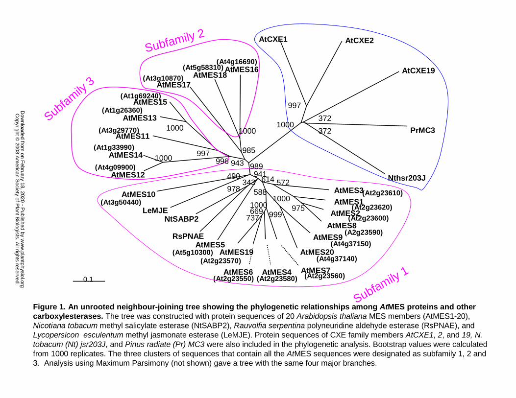
Figure 1. An unrooted neighbour-joining tree showing the phylogenetic relationships
among AtMES proteins and other carboxylesterases. The tree was constructed with protein
sequences of 20 Arabidopsis thaliana MES members (AtMES1-20), Nicotiana tobacum methyl
salicylate esterase (NtSABP2), Rauvolfia serpentina polyneuridine aldehyde esterase (RsPNAE),
and Lycopersicon esculentum methyl jasmonate esterase (LeMJE). Protein sequences of CXE
family members AtCXE1, 2, and 19, N. tobacum (Nt) jsr203J, and Pinus radiate (Pr) MC3 were
also included in the phylogenetic analysis. Bootstrap values were calculated from 1000 replicates.
The three clusters of sequences that contain all the AtMES sequences were designated as
subfamily 1, 2 and 3. Analysis using Maximum Parsimony (not shown) gave a tree with the
same four major branches.
Figure 2. Multiple sequence alignment of tobacco SABP2, tomato MJE and the 20
Arabidopsis AtMES. The sequence alignment was constructed using CLUSTALX program
(Thompson, 1997). Identical amino acids at a given position in 17 or more proteins are shown in
white letters on black. The catalytic triad residues are indicated by asterisks.
Figure 3. Plants with a null mutation in AtMES17 are more resistant to MeIAA but not to
IAA. A. Seedlings were grown on 1/2 MS medium containing 0.5 µM MeIAA for 7 days. B.
Root length (mean ± SD, N≥20) of wildtype (black) and in mes17-1 plants (purple) grown for 7
days on 1/2 MS medium containing various concentrations of MeIAA. C. Seedlings were grown
on 1/2 MS medium containing 0.5 µM IAA for 7 days. D. Root length (mean ± SD, N≥20) of
wildtype (black) and mes17-1 plants (purple) grown for 7 days on 1/2 MS medium containing
various concentrations of IAA.
Figure 4. Plants with a null mutation in AtMES17 hydrolyze exogenously supplied MeIAA
at a much lower efficiency than wildtype plants. Wildtype and mes17-1 seedlings were
incubated in a 0.5 µM solution of [14C]-MeIAA for 30 min. [14C]-MeIAA absorbed by the plants
was extracted and analyzed by radio-TLC. The position of an authentic MeIAA standard is also
shown.
www.plantphysiol.orgon February 18, 2020 - Published by Downloaded from Copyright © 2008 American Society of Plant Biologists. All rights reserved.
22
Figure 5. Plants overexpressing AtMES17 are more sensitive to MeIAA treatment, but not
to IAA. Wildtype plants and plants overexpressing AtMES17 were grown on 1/2 MS medium
containing 0.5 µM MeIAA (A) or IAA (B) for 7 days.
Figure 6. Plants carrying a null mutation in AtMES17 have longer hypocotyls. Left: analysis
of hypocotyl lengths of 4-week old wildtype and mes17-1 Arabidopsis plants grown in soil.
Right, analysis of the hypocotyl length of wildtype, mes17-1, and mes17-2 seedlings grown on
1/2 MS medium under continuous light for 8 days or in the dark for 4 days. Hypocotyl lengths of
the seedlings grown on 1/2 MS plates were measured with Image J as described in materials and
methods. The mean value and standard deviation were calculated from 20 samples.
Figure 7. Histochemical staining of GUS activity in DR5:GUS and DR5:GUS/mes17-1
seedlings. A: DR5:GUS seedlings and DR5:GUS/mes17-1 seedlings were stained for GUS
activity for 16 hours. B: Quantitative GUS assay of DR5:GUS and DR5:GUS/mes17-1 seedlings.
The mean value and standard deviation of GUS activity were calculated from three replicates,
and represented as nM 4-methyl umbelliferone per mg protein per min, as described in materials
and methods.
Figure 8. mes17/axr1-3 double mutant has the same hypocotyl length as axr1-3. Plants were
grown on 1/2 MS medium under continuous light for 9 days. The hypocotyl lengths of the
seedlings were measured with Image J as described in materials and methods. The mean value
and standard deviation were calculated from 20 samples.
Figure 9. Real-time RT-PCR analysis of AtMES17 transcript levels in different plant
organs at different developmental stages. AtMES17 transcript levels were normalized to the
levels of ubiquitin gene expression in respective samples. The levels of AtMES17 transcript in
flowers were arbitrarily set to 1.0. Data are plotted as means ± SD. *, below the detection limit.
Supplemental Figure 1: mes17-1 and mes17-2 are null mutants of AtMES17. A. Location of the
T-DNA insertions in mes17-1 and mes17-2. B. RT-PCR of mes17-1 and mes17-2 using RNA
www.plantphysiol.orgon February 18, 2020 - Published by Downloaded from Copyright © 2008 American Society of Plant Biologists. All rights reserved.

23
from leaves of 4-week old plants, demonstrating lack of MES17 transcript. Tubulin was used as a
control.
www.plantphysiol.orgon February 18, 2020 - Published by Downloaded from Copyright © 2008 American Society of Plant Biologists. All rights reserved.

24
References
Bartel B, Fink G (1995) ILR1, an amidohydrolase that releases active indole-3-acetic acid from conjugates. Science 268: 1745-1748
Baudouin E, Charpenteau M, Roby D, Marco Y, Ranjeva R, Ranty B (1997) Functional expression of a tobacco gene related to the serine hydrolase family-esterase activity towards short-chain dinitrophenyl acylesters. Eur. J. Biochem. 248: 700-706
Boerjan W, Cervera MT, Delarue M, Beeckman T, Dewitte W, Bellini C, Caboche M, Onckelen HV, Montagu MV, Inze D (1995) superroot, A recessive mutation in Arabidopsis, confers auxin overproduction. Plant Cell 7: 1405-1419
Brady SM, Orlando DA, Lee J-Y, Wang JY, Koch J, Dinneny JR, Mace D, Ohler U, Benfey PN (2007) A high-resolution root spatiotemporal map reveals dominant expression patterns. Science 318: 801-806
Chen F, D'Auria JC, Tholl D, Ross JR, Gershenzon J, Noel JP, Pichersky E (2003) An Arabidopsis thaliana gene for methylsalicylate biosynthesis, identified by a biochemical genomics approach, has a role in defense. Plant J. 36: 577-588
Cheng Y, Dai X, Zhao Y (2007) Auxin synthesized by the YUCCA flavin monooxygenases is essential for embryogenesis and leaf formation in Arabidopsis. Plant Cell 19: 2430-2439
Clough SJ, Bent AF (1998) Floral dip: a simplified method for Agrobacterium-mediated transformation of Arabidopsis thaliana. Plant J. 16: 735-743
Collett CE, Harberd NP, Leyser O (2000) Hormonal interactions in the control of Arabidopsis hypocotyl elongation. Plant Physiol. 124: 553-562
D'Auria JC, Chen F, Pichersky E (2002) Characterization of an acyltransferase capable of synthesizing benzylbenzoate and other volatile esters in flowers and damaged leaves of Clarkia breweri. Plant Physiol. 130: 466-476
Delker C, Raschke A, Quint M (2008) Auxin dynamics: the dazzling complexity of a small molecule's message. Planta 227: 929-941
Ding X, Cao Y, Huang L, Zhao J, Xu C, Li X, Wang S (2008) Activation of the indole-3-acetic acid amido synthetase GH3-8 suppresses expansin expression and promotes salicylate- and jasmonate-independent basal immunity in rice. Plant Cell 20: 228-240
Dogru E, Warzecha H, Seibel F, Haebel S, Lottspeich F, Stöckigt J (2000) The gene encoding polyneuridine aldehyde esterase of monoterpenoid indole alkaloid biosynthesis in plants is an ortholog of the alpha/beta hydrolase super family. Eur. J. Biochem. 267: 1397-1406
Evans ML (1985) The action of auxin on plant-cell elongation. Crc Crit. Rev. Plant Sci. 2: 317-365
Forouhar F, Yang Y, Kumar D, Chen Y, Fridman E, Park SW, Chiang Y, Acton TB, Montelione GT, Pichersky E, Klessig DF, Tong L (2005) Structural and biochemical studies identify tobacco SABP2 as a methyl salicylate esterase and implicate it in plant innate immunity. Proc. Natl. Acad. Sci. U.S.A. 102: 1773-1778
Fridman E, Wang J, Iijima Y, Froehlich JE, Gang DR, Ohlrogge J, Pichersky E (2005) Metabolic, genomic, and biochemical analyses of glandular trichomes from the wild tomato species Lycopersicon hirsutum identify a key enzyme in the biosynthesis of methylketones. Plant Cell 17: 1252-1267
Hartley JL, Temple GF, Brasch MA (2000) DNA cloning using in vitro site-specific recombination. Genome Res. 10: 1788-1795
www.plantphysiol.orgon February 18, 2020 - Published by Downloaded from Copyright © 2008 American Society of Plant Biologists. All rights reserved.

25
Hikage T, Saitoh Y, Tanaka-Saito C, Hagami H, Satou F, Shimotai Y, Nakano Y, Takahashi M, Takahata Y, Tsutsumi K (2007) Structure and allele-specific expression variation of novel alpha/beta hydrolase fold proteins in gentian plants. Mol. Genet. Genomics 278: 95-104
Holmquist M (2000) Alpha beta-hydrolase fold enzymes structures, functions and mechanisms. Curr. Protein Pept. Sci. 1: 209-235
Ichinose Y, Hisayasu Y, Sanematsu S, Ishiga Y, Seki H, Toyoda K, Shiraishi T, Yamada T (2001) Molecular cloning and functional analysis of pea cDNA E86 encoding homologous protein to hypersensitivity-related hsr203J. Plant Sci. 160: 997-1006
Jackson RG, Lim E-K, Li Y, Kowalczyk M, Sandberg G, Hoggett J, Ashford DA, Bowles DJ (2001) Identification and biochemical characterization of an Arabidopsis indole-3-acetic acid glucosyltransferase. J. Biol. Chem. 276: 4350-4356
Jensen PJ, Hangarter RP, Estelle M (1998) Auxin Transport Is Required for Hypocotyl Elongation in Light-Grown but Not Dark-Grown Arabidopsis. Plant Physiol. 116: 455-462
Ko MK, Jeon WB, Kim KS, Lee HH, Seo HH, Kim YS, Oh B-J (2005) A Colletotrichum gloeosporioides-induced esterase gene of nonclimacteric pepper (Capsicum annuum) fruit during ripening plays a role in resistance against fungal infection. Plant Mol. Biol. V58: 529-541
Kumar D, Klessig DF (2003) High-affinity salicylic acid-binding protein 2 is required for plant innate immunity and has salicylic acid-stimulated lipase activity. Proc. Natl. Acad. Sci. U.S.A. 100: 16101-16106
LeClere S, Tellez R, Rampey RA, Matsuda SPT, Bartel B (2002) Characterization of a family of IAA-amino acid conjugate hydrolases from Arabidopsis. J. Biol. Chem. 277: 20446-20452
Leyser HM, Lincoln CA, Timpte C, Lammer D, Turner J, Estelle M (1993) Arabidopsis auxin-resistance gene AXR1 encodes a protein related to ubiquitin-activating enzyme E1. Nature 364: 161-164
Li L, Hou X, Tsuge T, Ding M, Aoyama T, Oka A, Gu H, Zhao Y, Qu L-J (2008) The possible action mechanisms of indole-3-acetic acid methyl ester in Arabidopsis. Plant Cell Rep. 27: 575-584
Lincoln C, Britton JH, Estelle M (1990) Growth and development of the Axr1 mutants of Arabidopsis. Plant Cell 2: 1071-1080
Ljung K, Hull AK, Kowalczyk M, Marchant A, Celenza J, Cohen JD, Sandberg G (2002) Biosynthesis, conjugation, catabolism and homeostasis of indole-3-acetic acid in Arabidopsis thaliana. Plant Mol. Biol. 50: 309-332
Marshall SG, Putterill J, Plummer K, Newcomb R (2003) The carboxylesterase gene family from Arabidopsis thaliana. J. Mol. Evol. V57: 487-500
Murashige T, Skoog F (1962) A revised medium for rapid growth and bioassays with tobacco tissue cultures. Physiol. Plant 15: 473-497
Nakamura A, Higuchi K, Goda H, Fujiwara MT, Sawa S, Koshiba T, Shimada Y, Yoshida S (2003) Brassinolide induces IAA5, IAA19, and DR5, a synthetic auxin response element in Arabidopsis, implying a cross talk point of brassinosteroid and auxin signaling. Plant Physiol. 133: 1843-1853
www.plantphysiol.orgon February 18, 2020 - Published by Downloaded from Copyright © 2008 American Society of Plant Biologists. All rights reserved.

26
Nam KH, Dudareva N, Pichersky E (1999) Characterization of benzylalcohol acetyltransferases in scented and non-scented Clarkia Species. Plant Cell Physiol. 40: 916-923
Narasimhan K, Basheer C, Bajic VB, Swarup S (2003) Enhancement of plant-microbe interactions using a rhizosphere metabolomics-driven approach and its application in the removal of polychlorinated biphenyls. Plant Physiol. 132: 146-153
Nardini M, Dijkstra BW (1999) Alpha/beta hydrolase fold enzymes: the family keeps growing. Curr. Opin. Struct. Biol. 9: 732-737
Normanly J (1997) Auxin metabolism. Physiologia Plantarum 100: 431-442 Nowacki J, Bandurski RS (1980) Myoinositol esters of indole-3-acetic-acid as seed auxin
precursors of Zea Mays L. Plant Physiol. 65: 422-427 Ottenschlager I, Wolff P, Wolverton C, Bhalerao RP, Sandberg G, Ishikawa H, Evans M,
Palme K (2003) Gravity-regulated differential auxin transport from columella to lateral root cap cells. Proc. Natl. Acad. Sci. U S A 100: 2987-2991
Park S-W, Kaimoyo E, Kumar D, Mosher S, Klessig DF (2007) Methyl salicylate is a critical mobile signal for plant systemic acquired resistance. Science 318: 113-116
Pontier D, Godiard L, Marco Y, Roby D (1994) Hsr203j, A tobacco gene whose activation is rapid, highly localized and specific for incompatible plant/pathogen interactions. Plant J. 5: 507-521
Pontier D, Tronchet M, Rogowsky P, Lam E, Roby D (1998) Activation of hsr203, a plant gene expressed during incompatible plant-pathogen interactions, is correlated with programmed cell death. Mol. Plant Microbe Interact. 11: 544-554
Qin G, Gu H, Zhao Y, Ma Z, Shi G, Yang Y, Pichersky E, Chen H, Liu M, Chen Z, Qu L (2005) Regulation of Arabidopsis leaf development by an indole-3-acetic acid carboxyl methyltransferase in Arabidopsis. Plant Cell 17: 2693-2704
Rampey RA, LeClere S, Kowalczyk M, Ljung K, Sandberg G, Bartel B (2004) A family of auxin-conjugate hydrolases that contributes to free indole-3-acetic acid levels during Arabidopsis germination. Plant Physiol. 135: 978-988
Schmid M, Davison TS, Henz SR, Pape UJ, Demar M, Vingron M, Scholkopf B, Weigel D, Lohmann JU (2005) A gene expression map of Arabidopsis thaliana development. Nat. Genet. 37: 501-506
Shulaev V, Silverman P, Raskin I (1997) Airborne signalling by methyl salicylate in plant pathogen resistance. Nature 385: 718-721
Staswick PE, Serban B, Rowe M, Tiryaki I, Maldonado MT, Maldonado MC, Suza W (2005) Characterization of an Arabidopsis enzyme family that conjugates amino acids to indole-3-acetic acid. Plant Cell 17: 616-627
Stuhlfelder C, Mueller MJ, Warzecha H (2004) Cloning and expression of a tomato cDNA encoding a methyl jasmonate cleaving esterase. Eur. J. Biochem. 271: 2976-2983
Tan X, Calderon-Villalobos LIA, Sharon M, Zheng C, Robinson CV, Estelle M, Zheng N (2007) Mechanism of auxin perception by the TIR1 ubiquitin ligase. Nature 446: 640-645
Teale WD, Paponov IA, Palme K (2006) Auxin in action: signalling, transport and the control of plant growth and development. Nat. Rev. Mol. Cell Biol. 7: 847-859
Thompson JD, Gibson, T.J., Plewniak, F., Jeanmougin, F. and Higgins, D.G. (1997) The clustalx windows interface: flexible strategies for multiple sequence alignment aided by quality analysis tools. Nucl. Acids Res. 24: 4876-4882
www.plantphysiol.orgon February 18, 2020 - Published by Downloaded from Copyright © 2008 American Society of Plant Biologists. All rights reserved.
27
Ulmasov T, Murfett J, Hagen G, Guilfoyle TJ (1997) Aux/IAA proteins repress expression of reporter genes containing natural and highly active synthetic auxin response elements. Plant Cell 9: 1963-1971
Vandenbussche F, Verbelen P, Van Der Straeten D (2005) Of light and length: regulation of hypocotyl growth in Arabidopsis. BioEssays 27: 275-284
Varbanova M, Yamaguchi S, Yang Y, McKelvey K, Hanada A, Borochov R, Yu F, Jikumaru Y, Ross J, Cortes D, Ma C, Noel JP, Mander L, Shulaev V, Kamiya Y, Rodermel S, Weiss D, Pichersky E (2007) Methylation of gibberellins by Arabidopsis GAMT1 and GAMT2. Plant Cell 19: 32-45
Walden AR, Walter C, Gardner RC (1999) Genes expressed in Pinus radiata male cones include homologs to anther-specific and pathogenesis response genes. Plant Physiol 121: 1103-1116
Woodward AW, Bartel B (2005) Auxin: regulation, action, and interaction. Ann. Bot. 95: 707-735
Wu J, Wang L, Baldwin I (2008) Methyl jasmonate-elicited herbivore resistance: does MeJA function as a signal without being hydrolyzed to JA? Planta 227: 1161-1168
Yang Y, Varbanova M, Ross J, Wang G, Cortes D, Fridman E, Shulaev V, Noel JP, Pichersky E (2006a) Methylation and demethylation of plant signaling molecules. In JT Romeo, ed, Rec. Adv. Phytochem., Ed 1 Vol 40. Elsevier Science Ltd., Oxford, p 253
Yang Y, Yuan JS, Ross J, Noel JP, Pichersky E, Chen F (2006b) An Arabidopsis thaliana methyltransferase capable of methylating farnesoic acid. Arch. Biochem. Biophys. 448: 123-132
Zhao N, Ferrer J, Ross J, Guan J, Yang Y, Pichersky E, Noel JP, Chen F (2008) Structural, biochemical and phylogenetic analyses suggest that indole-3-acetic acid methyltransferase is an evolutionarily ancient member of the SABATH family. Plant Physiol. 146: 455-467
Zhao Y, Hull AK, Gupta NR, Goss KA, Alonso J, Ecker JR, Normanly J, Chory J, Celenza JL (2002) Trp-dependent auxin biosynthesis in Arabidopsis: involvement of cytochrome P450s CYP79B2 and CYP79B3. Genes Dev. 16: 3100-3112
Zhao YD, Christensen SK, Fankhauser C, Cashman JR, Cohen JD, Weigel D, Chory J (2001) A role for flavin monooxygenase-like enzymes in auxin biosynthesis. Science 291: 306-309
Zimmerman P, Hitchcock AE (1937) Comparative effectiveness of acids, esters, and salts as growth substances and methods of evaluating them. Contrib. Boyce Thompson Inst. 8: 337-350
Zubieta C, Ross JR, Koscheski P, Yang Y, Pichersky E, Noel JP (2003) Structural basis for substrate recognition in the salicylic acid carboxyl methyltransferase family. Plant Cell 15: 1704-1716
www.plantphysiol.orgon February 18, 2020 - Published by Downloaded from Copyright © 2008 American Society of Plant Biologists. All rights reserved.
0.1
AtMES3AtMES1
AtMES2
AtMES8
AtMES9
AtMES20
AtMES7AtMES4AtMES6
AtMES19AtMES5
RsPNAE
NtSABP2LeMJE
AtMES10
AtMES12
AtMES14
AtMES11
AtMES13
AtMES15
AtMES17AtMES18
AtMES16
AtCXE1 AtCXE2
AtCXE19
PrMC3
Nthsr203J
(At4g16690)(At5g58310)
(At3g10870)
(At1g69240)
(At1g26360)
(At3g29770)
(At1g33990)
(At4g09900)
(At3g50440)
(At5g10300)(At2g23570)
(At2g23550) (At2g23580) (At2g23560)
(At4g37140)
(At4g37150)
(A2g23590)
(At2g23600)
(At2g23620)
(At2g23610)
Subfamily 1
Subfamily 2
Subfamily 3
997
3721000
372
1000
985
1000
9971000 996 943 989
490 941
978343
7376691000
999975
1000
572588
614
Figure 1. An unrooted neighbour-joining tree showing the phylogenetic relationships among AtMES proteins and other carboxylesterases. The tree was constructed with protein sequences of 20 Arabidopsis thaliana MES members (AtMES1-20), Nicotiana tobacum methyl salicylate esterase (NtSABP2), Rauvolfia serpentina polyneuridine aldehyde esterase (RsPNAE), and Lycopersicon esculentum methyl jasmonate esterase (LeMJE). Protein sequences of CXE family members AtCXE1, 2, and 19, N. tobacum (Nt) jsr203J, and Pinus radiate (Pr) MC3 were also included in the phylogenetic analysis. Bootstrap values were calculated from 1000 replicates. The three clusters of sequences that contain all the AtMES sequences were designated as subfamily 1, 2 and 3. Analysis using Maximum Parsimony (not shown) gave a tree with the same four major branches.
w
ww
.plantphysiol.orgon F
ebruary 18, 2020 - Published by
Dow
nloaded from
Copyright ©
2008 Am
erican Society of P
lant Biologists. A
ll rights reserved.
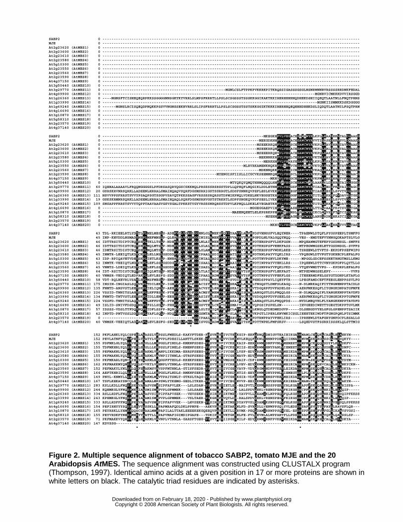
SABP2 0 ------------------------------------------------------------------------------------------------------------------- MJE 0 ------------------------------------------------------------------------------------------------------------------- At2g23620 (AtMES1) 0 ------------------------------------------------------------------------------------------------------------------- At2g23600 (AtMES2) 0 ------------------------------------------------------------------------------------------------------------------- At2g23610 (AtMES3) 0 ------------------------------------------------------------------------------------------------------------------- At2g23580 (AtMES4) 0 ------------------------------------------------------------------------------------------------------------------- At5g10300 (AtMES5) 0 ------------------------------------------------------------------------------------------------------------------- At2g23550 (AtMES6) 0 ------------------------------------------------------------------------------------------------------------------- At2g23560 (AtMES7) 0 ------------------------------------------------------------------------------------------------------------------- At2g23590 (AtMES8) 0 ------------------------------------------------------------------------------------------------------------------- At4g37150 (AtMES9) 0 ------------------------------------------------------------------------------------------------------------------- At3g50440 (AtMES10) 0 ------------------------------------------------------------------------------------------------------------------- At3g29770 (AtMES11) 0 -------------------------------------------------------MGNLCSLFTPPKPVKKRKPITKRQSSIGASSSGSGLNSNRWNNRVRSSSSRRDNKFEDAL At4g09900 (AtMES12) 0 -----------------------------------------------------------------------------------------------MGNRVICMKKKDVVIRSGGD At1g26360 (AtMES13) 0 ----MGNSFTCISHEQEQRPKKSSGGGGNNSGKYKYVRRLSLMPSFRRRTLLPSLSCSGSSSTSSSKKGGIKAKTKKIRERHHHHHQDHEKDSHIIQEQTLAATNLLFNQTPRNS At1g33990 (AtMES14) 0 ------------------------------------------------------------------------------------------------MGNKIISMMKKDSKDGGGG At1g69240 (AtMES15) 0 ------MGNSLRCISQEQDPNQKKPSSVVNGNSSEKHVRRLSLIPSFRRRTLLPSLSCSGSSTSSTSKKGGIKTKKKIRERHHQEQHHHDHEKDSLIQDQTLAATNILFSQTPRN At4g16690 (AtMES16) 0 ------------------------------------------------------------------------------------------------------------------- At3g10870 (AtMES17) 0 ------------------------------------------------------------------------------------------------------------------- At5g58310 (AtMES18) 0 ------------------------------------------------------------------------------------------------------------------- At2g23570 (AtMES19) 0 ------------------------------------------------------------------------------------------------------------------- At4g37140 (AtMES20) 0 ------------------------------------------------------------------------------------------------------------------- SABP2 0 ------------------------------------------------------------------------MKEGKHFVLVHGACHGGWSWYKLKPLLEAAGHKVTALDLAASG MJE 0 ----------------------------------------------------------------------MEKGDKNHFVLVHGACHGAWCWYKVVTILRSEGHKVSVLDMAASG At2g23620 (AtMES1) 0 ---------------------------------------------------------------------MSEEKRKQHFVLVHGSCHGAWCWYKVKPLLEAVGHRVTAVDLAASG At2g23600 (AtMES2) 0 ---------------------------------------------------------------------MSEEKRKQHFVLVHGACHGAWCWYKVKPLLEALGHRVTALDLAASG At2g23610 (AtMES3) 0 ---------------------------------------------------------------------MSEEERKQHVVLVHGACHGAWCWYKVKPQLEASGHRVTAVDLAASG At2g23580 (AtMES4) 0 ----------------------------------------------------------------------MEKNNKKRFVLVHGLCHGAWCWYKVKTHLEAVGHCVTAVDLAASG At5g10300 (AtMES5) 0 ------------------------------------------------------------------------MERKHHFVLVHNAYHGAWIWYKLKPLLESAGHRVTAVELAASG At2g23550 (AtMES6) 0 ---------------------------------------------------------------MLRVRKAMENKNQKRFVLIHGVCHGAWTWDKVKTQLEVAGHCVTAVDLAASG At2g23560 (AtMES7) 0 ----------------------------------------------------------------------MDKNNQKKFVLVHGICHGAWCWYKVKAQLEAAGHSVTAVDLAASG At2g23590 (AtMES8) 0 ---------------------------------------------------MYENGISFIISLLICGCVKSEEMMKQHFVLVHGSCLGAWCWYKVKPLLEASGHRVTALDLAACG At4g37150 (AtMES9) 0 ---------------------------------------------------------------------------MKHYVLVHGGCHGAWCWYKVKPMLEHSGHRVTVFDLTAHG At3g50440 (AtMES10) 0 ---------------------------------------------------------MTYQKQYQMQTHHMQQQQLHHFVFVHGSCHGAWCWFKLAAKLKLDGHRVTAIDLGGSG At3g29770 (AtMES11) 60 IQEHALAAAAVLFRQQNGGGGSLPFDRSASQRYQGSCSKKNQLPRSSSSRSRSSTDPLLQPHQFLNQGIKLDDLETNHFVLVHGGSFGAWCWYKTIALLEEDGFKVTAIDLAGCG At4g09900 (AtMES12) 20 GSRSKRVNRSQRKLLADEENLHRRALSMAIHQAQVSQRFDGSMSRRIGSTSSRRGTLSDSFSNNKQVPEFLESLKVKKFVLVHGEGFGAWCWYKTIASLEESGLSPVTVDLAGSG At1g26360 (AtMES13) 111 NSVVPPSFRRSTSVVYPSAQPSGTSSGPVSAVQTPKKSSAGFVRSSSSRQRSSTDPMIKPNQLVDKELNKVEGSETKRFVLVHGGGFGAWCWYKTITLLEKHGFQVDAVELTGSG At1g33990 (AtMES14) 19 GSKSKRMNRSQRKLLADEEMLHRRALSMAIHQAQLSQRFDGSMSRRVGSTSTRKRTLSDPFSNGKQVPDFSESLIVKKFVLVHGEGFGAWCWYKMVASLEESGLSPVTVDLTGCG At1g69240 (AtMES15) 109 SNSAPPFRRSTSVVYTQPPTAAVAASVGSVSGALTPKKSTYGYVRSSSNRQRSSTDPVLKPNQLLDKELKVEGAETKRFVLVHGGGFGAWCWYKTITLLEKHGFQVDAVDLTGSG At4g16690 (AtMES16) 0 ------------------------------------------------------------------MGGEGGAEPVIHFVFVHGASHGAWCWYKLTTLLDAAGFKSTSVDLTGAG At3g10870 (AtMES17) 0 ----------------------------------------------------------MAEENQEETLELKPSRKPPHFVLIHGMSLGSWCWYKIKCLMEVSGFTVTCIDLKSSG At5g58310 (AtMES18) 0 -------------------------------------------------------------------------MSEHHFVFVHGAGHGGWCWYKLANSLRDNGHKATCIDLKGAG At2g23570 (AtMES19) 0 ------------------------------------------------------------------------------------------------------------------- At4g37140 (AtMES20) 0 ---------------------------------------------------------------------------MKHYVLVHGGCHGAWCWYKVKPVLEASGHRVTVVDLTASG SABP2 43 TDL-RKIEELRTLYDYTLPLMELMESLS-ADEKVILVGHSLGGMNLGLAMEKYPQKIYAAVFLAAFMPDSVHNSSFVLEQYNER----TPAENWLDTQFLPYGSPEEPLTSMFFG MJE 45 INP-KHVDDLNSMADYNEPLMEFMNSLP-QLERVVLVGHSMGGINISLAMEKFPQKIVVAVFVTAFMPGPDLNLVALGQQYNQQ----VES--HMDTEFVYNNGQDKAPTSLVLG At2g23620 (AtMES1) 46 IDTTRSITDIPTCEQYSEPLTKLLTSLP-NDEKVVLVGHSFGGLNLAIAMEKFPEKISVAVFLTAFMPDTEHSPSFVLDKFGSN----MPQEAWMGTEFEPYGSDNSGL-SMFFS At2g23600 (AtMES2) 46 IDTTRSITDISTCEQYSEPLMQLMTSLP-NDEKVVLVGHSFGGLSLALAMDKFPDKISVSVFVTAFMPDTKHSPSFVEEKFASS----MTPEGWMGSELETYGSDNSGL-SVFFS At2g23610 (AtMES3) 46 IDMTRSITDISTCEQYSEPLMQLMTSLP-DDEKVVLVGHSLGGLSLAMAMDMFPTKISVSVFVTAMMPDTKHSPSFVWDKLRKE----TSREEWLDTVFTS-EKPDFPSEFWIFG At2g23580 (AtMES4) 45 INMTR-LEEIQTLKDYCKPLLELLNSLGSDDDKVILVAHSMGGIPAALASDIFPSKIATIVFLTAFMPDTRNLPAYVYQKLIRS----VPQEGWLDTVFGTYGKHECPLEFALFG At5g10300 (AtMES5) 43 IDP-RPIQAVETVDEYSKPLIETLKSLP-ENEEVILVGFSFGGINIALAADIFPAKIKVLVFLNAFLPDTTHVPSHVLDKYME------MPGGLGDCEFSSHETRNGTMSLLKMG At2g23550 (AtMES6) 52 INMTK-VEEIQTLNDYCKPLLEFLSSLGSDDGKVIVVAHSMGGISAALAADSFACKIAAIVFLTAFMPDTINPPAYVYEKLLRS----IPQEEWLDTTCVNYGKPDFPLQYTLLG At2g23560 (AtMES7) 45 VNMTS-LDEIQTLKDYCKPLLEFLSSLGSDDDKVILVAHSMGGISASLAADIFPSKVAAIVFVAAFMPDISNPPAYVFQKLVKD----VTQEVWMDTVFG---KPDRPLEFALFG At2g23590 (AtMES8) 64 IDT-RSITDISTCEQYSEPLIQLMTSLP-NDEKVVLVGHSYGGLTLAIAMDKFPDKISVSVFVTSFMPDTKNSPSFVLEKFAST----MTPEDWMGSELEPY---------VVFS At4g37150 (AtMES9) 40 VNMSR-VEDIQTLEDFAKPLLEVLESFG-SDDKVVLVAHSLGGIPAALAADMFPSKISVAVFVTSFMPDTTNPPSYVFEKFLGS----ITEEERMDFELGSYGTDDHPLKTAFLG At3g50440 (AtMES10) 58 VDT-RQLHEVRLVSAYLEPLMSFMESLP-ENEKVVLVGHSYGGIGTSLAMERFPTKVSVGIFLSAYMPHHDSPPAVLIQEYFTR----LPEGFAMDCEFTFEEGLEHPPSSVLFG At3g29770 (AtMES11) 175 INSIN-INGIASLSQYVKPLTDILEKLP-IGEKVILVGHDFGGACISYAMELFPSKISKAVFLAAAMLTNGQSTLDMFSLKAGQ----N-DLMRKAQIFIYTNGNENPPTAIDLD At4g09900 (AtMES12) 135 FNMTD-ANSVSTLEEYSKPLIELIQNLP-AEEKVILVGHSTGGACVSYALERFPEKISKAIFICATMVTDGQRPFDVFADELGS----AERFMKESQFLIYGNGKDNPATGFMFE At1g26360 (AtMES13) 226 VSSID-TNNITSLAHYSKPLLHFFESLK-PTEKVILVGHDFGGACMSYAMEMFPTKIAKAVFISAAMLANGQSTLDLFNQQLGS----N-DLMQQAQIFLYANGKKNPPTAVDFD At1g33990 (AtMES14) 134 FNMTD-TNTVSTLEEYSKPLIDLLENLP-EEEKVILVGHSTGGASISYALERFPEKISKAIFVCATMVSDGQRPFDVFSEELGS----AERFMKESQFLIYGNGKDKPPTGFMFE At1g69240 (AtMES15) 224 VSSFD-TNNITSLAQYVKPLLHFFDTLK-PTEKVILVGHDFGGACMSYAMEMYPSKIAKAIFISAAMLANAQSTLDLFNQQPDS----NYDLMEQVHLFLYANGKKNPPTAVDFD At4g16690 (AtMES16) 49 ISLID-SNIVFDSDQYNRPLFSLLSDLP-PHHKVILVGHSIGGGSVTEALCKFTDKISMAIYLAASMVQPGSIPSPHLSN--------IHVGEEDIWEYTYGEGTDKPPTGVLMK At3g10870 (AtMES17) 57 IDSSS-VDSLTTFDQYNQPLIDFLSSFP-EQEQVILVGHSAGGLSLTSAIQRFPKKICLAVFIGASMLKNGLQTDEDMKDGVP-----DLSEHGDVYELGFGLGPENPPTSAIIK At5g58310 (AtMES18) 42 INPTD-PNTVSSLDDYDEPLYAFLSQLP-NDQKVILVSHSVGGGSMTAAMCLFPSKVSLAVYVAAAMVKPGTLIPERLKNVMKICSGLIEEETEKIWDFTFGNGPQNLPTSIMMK At2g23570 (AtMES19) 0 ----------------------------------------MGGIPAALAADIFSCKISAVVFLAAFMPDTRNPPAYVFEKLIRS----IPREEWLDTAFGRYGNPDCPLESALLG At4g37140 (AtMES20) 40 VNMSK-VEEIQTLADYAKPLLEVLESFG-SEDKVILVAHSLGGISVGLAADMFPSKISVAVFITSFMPDTTNPHLFMFSKSV------LSQEDVSTFSSRRISSSKLQLSTTMIG * SABP2 152 PKFLAHKLYQLCSPEDLALASSLVRPSSLFMEDLS-KAKYFTDER-FGSVKRVYIVCTEDKGIP-EEFQRWQIDNIGVTEAIEIKGADHMAMLCEPQKLCASLLEIAHKYN---- MJE 152 PEVLATNFYQLSPPEDLTLATYLVRPVPLFDESILLANTTLSKEK-YGSVHRVYVVCDKDNVLKEQQFQKWLINNNPPDEVQIIHNADHMVMFSKPRDLSSCLVMISQKYY---- At2g23620 (AtMES1) 155 PDFMKLGLYQLSPVEDLELGLLLMRPGSLFINDLS-KMKNFSDEG-YGSVPRVFIVCKEDKAIP-EERQRWMIDNFPVNLVMEMEETDHMPMFCKPQQLSDYFLKIADKFV---- At2g23600 (AtMES2) 155 TDFMKHRLYQLSPVEDLELGLLLKRPSSLFINELS-KMENFSEKG-YGSVPRAYIVCKEDNIIS-EDHQRWMIHNYPANLVIEMEETDHMPMFCKPQLLSDHLLAIADNFC---- At2g23610 (AtMES3) 155 PEFMAKNLYQLSPVQDLELAKMLVRANPLIKKDMA-ERRSFSEEG-YGSVTRIFIVCGKDLVSP-EDYQRSMISNFPPKEVMEIKDADHMPMFSKPQQLCALLLEIANKYA---- At2g23580 (AtMES4) 155 PKFMAKNLYQLSPVQDLELAKMLVRVNPIITNNLA-GTRSFSEEG-YGTVTRIYIVCGEDMAVP-EDYQWWMIKNFPPKEVMEIKCADHMAMFSKPHKLCALLVEIACKYA---- At5g10300 (AtMES5) 150 PKFMKARLYQNCPIEDYELAKMLHRQGSFFTEDLS-KKEKFSEEG-YGSVQRVYVMSSEDKAIP-CDFIRWMIDNFNVSKVYEIDGGDHMVMLSKPQKLFDSLSAIATDYM---- At2g23550 (AtMES6) 162 PKFMAKKMYQNSPVQDLEVVKTLVRENPLVTNNLA-GTRSFSEEG-YGSVTRIYIVCREDLVEV-EDYQRWMISNFPPKEVMEIKCADHMPMFSKPQEVCALLLEIANKYCKN-- At2g23560 (AtMES7) 152 PEFMAKYLYNLSPLQDFELAKMSVRVSPFMTNNLA-GTISFSEDR-YGSVTRIYIVCGEDVAVP-VDYQRGMINDFPVKEVLEIKDADHMPMFSKPQELCALLLEIADKYA---- At2g23590 (AtMES8) 164 AEFTKHRILQLSPIEDLELRLLLKRPGSLFLNDLS-RMKNFSEKG-YGSVPRAYIVSKDDHTIS-EEYQRWMIDNYPPNLVIEMEGTDHLPLFCKPQLLSDHLLAIADKFS---- At4g37150 (AtMES9) 149 PNYL-KNMYLLSPIEDYELAKMLMRVTPAITSNLT-GTKSLTAQG-YGSISRVYIVCGEDKGIR-VDFQRWMIENSPVKEVMEIKDADHMPMFSKPHELCDRLLKIADKYP---- At3g50440 (AtMES10) 167 TSFLKEKAYSNCQLEDLELAMALMKPSWLYTKEMG-GEDLITKER-YGSGKRVFIVCEGDNVVP-EEIQKWMISNYEPHEVKRIEEAGHMAMLTKPHELSQLLQEIAAKYN---- At3g29770 (AtMES11) 283 KSLLKDLLFNQSPSKDVALASVSMRSIPFAPVLEK---LSLSDAN-YGSVRRYYIETLED-NAIPVTLQENMINSSPPEKVYRLKGADHAPFFSKPQALHKLLLEIARISPA--- At4g09900 (AtMES12) 244 KQHMKGLYFNQSPNKDIALSMISMRPVPLGPMMEK---LSLSAER-YGKGRRFYVQTLDD-LALSPDVQEKLVRENSPEAVFKIKGSDHCPFFSKPQSLHKILLEIAQIP----- At1g26360 (AtMES13) 334 RSLLRDFLFNQSPPKDLALASVSIRPIPFAPVSEK---VHVSEKN-YGSIRRFYIKTMED-YAVPVLLQEAMIKLNPPEQVFQLKGSDHAPFFSRPQSLNKILVEISQIPFKKSS At1g33990 (AtMES14) 243 KPHMKGLYFNQSPNKDIALAMISMRPVPLGPMMEK---VSLTAER-YGKGRRFYVQTLDD-RALSPDVQEKLVRENSPEGVFKIKGSDHCPFFSKPQSLHKILLEIAQIP----- At1g69240 (AtMES15) 333 RSLLRDFFFNQSPPKDVALASVSMRPIPFAPVVEK---LHVSEKN-YGSIRRFYIKTMEDDYAVPVSLQDAMIKSNPPEQVFHLKGSDHAPFFSRPQSLNRILVEISQLPPKKSS At4g16690 (AtMES16) 154 PEFIRHYYYSQSPLEDVTLSSKLLRPAPMRAFQDLDKLPPNPE---AEKVPRVYIKTAKDNLFD-SVRQDLLVENWPPSQLYVLEDSDHSAFFSVPTTLFAYLLRAVSFLQR--- At3g10870 (AtMES17) 165 PEYRRKLLYHMSPQQECSLAALMMRPAPILALTTAKLEEEEKEKGQEEQVPRVYIKTLLDRVMK-PEQQDAMIRRWPPSQVYEL-ESDHSPFFSNPFVLFGLLIKAAVSVGSI-- At5g58310 (AtMES18) 155 PEYVRDKFYNESPMEDYTLATTLLRPAPVMAFIGIMDIPGAPE---TDKIPRVYVKTGKDHLFE-PVLQEVMLALWPPAHTFLLPDSDHSAFFSQPQELYQFLLQAASSLSP--- At2g23570 (AtMES19) 71 PKFMAKKVYQRSPIEDLELAKMLVRVNPLVTNNLA-GARSFTGEG-YGSVTRIYIISGEDNILP-EDYQRWMIRNFPVKEVMEIKDADHMAMFSKPKELCALLLEIADKYA---- At4g37140 (AtMES20) 147 KDVSSG------------------------------------------------------------------------------------------------------------- * *
Figure 2. Multiple sequence alignment of tobacco SABP2, tomato MJE and the 20 Arabidopsis AtMES. The sequence alignment was constructed using CLUSTALX program (Thompson, 1997). Identical amino acids at a given position in 17 or more proteins are shown in white letters on black. The catalytic triad residues are indicated by asterisks.
www.plantphysiol.orgon February 18, 2020 - Published by Downloaded from Copyright © 2008 American Society of Plant Biologists. All rights reserved.
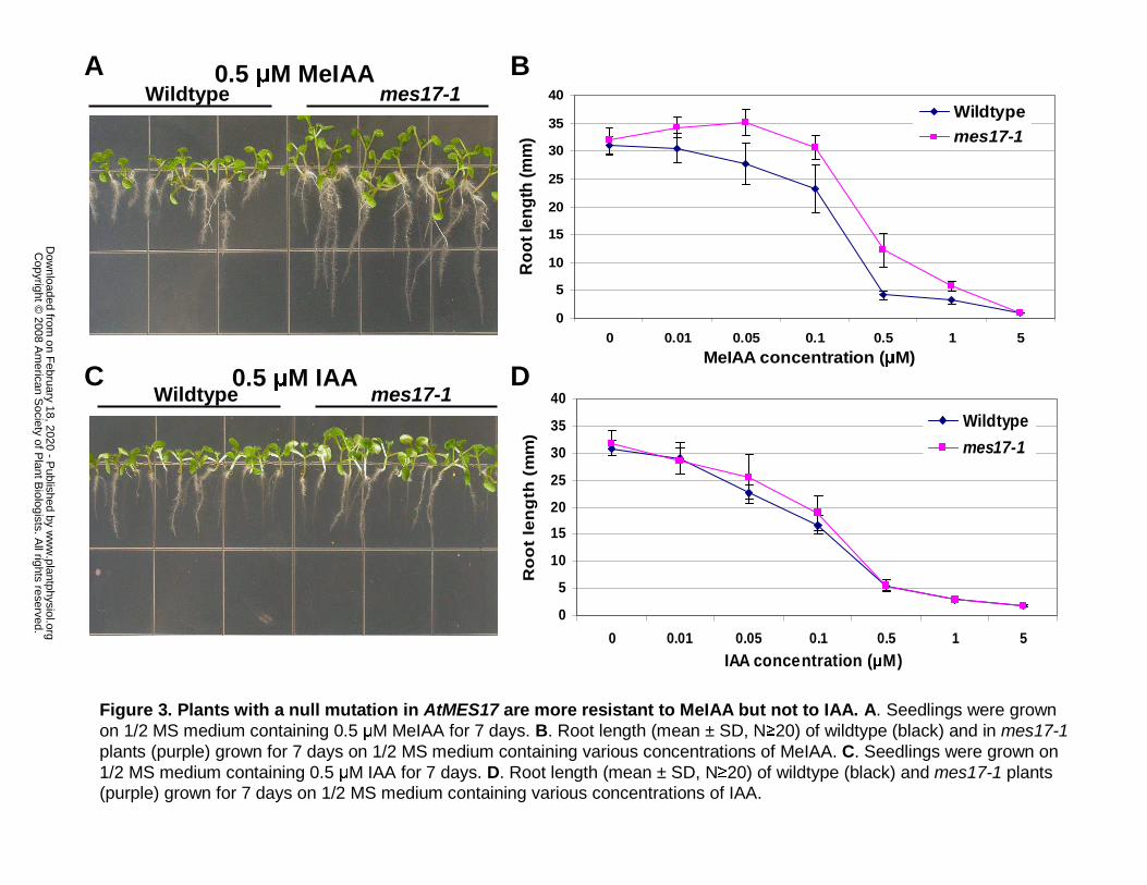
Wildtype mes17-10.5 µM MeIAA
Wildtype0.5 µM IAA
mes17-1
A
C
0
5
10
15
20
25
30
35
40
0 0.01 0.05 0.1 0.5 1 5
MeIAA concentration (µM)
Ro
ot l
eng
th (m
m)
Wildtypemes17-1
B
0
5
10
15
20
25
30
35
40
0 0.01 0.05 0.1 0.5 1 5
IAA concentration (µM)
Ro
ot
len
gth
(m
m)
Wildtype
mes17-1
D
Figure 3. Plants with a null mutation in AtMES17 are more resistant to MeIAA but not to IAA. A. Seedlings were grown on 1/2 MS medium containing 0.5 µM MeIAA for 7 days. B. Root length (mean ± SD, N≥20) of wildtype (black) and in mes17-1plants (purple) grown for 7 days on 1/2 MS medium containing various concentrations of MeIAA. C. Seedlings were grown on 1/2 MS medium containing 0.5 µM IAA for 7 days. D. Root length (mean ± SD, N≥20) of wildtype (black) and mes17-1 plants (purple) grown for 7 days on 1/2 MS medium containing various concentrations of IAA.
w
ww
.plantphysiol.orgon F
ebruary 18, 2020 - Published by
Dow
nloaded from
Copyright ©
2008 Am
erican Society of P
lant Biologists. A
ll rights reserved.

Wt mes17-1
MeIAA
Figure 4. Plants with a null mutation in AtMES17 hydrolyze exogenously supplied MeIAA at a
much lower efficiency than wildtype plants. Wildtype and mes17-1 seedlings were incubated in a
0.5 µM solution of [14C]-MeIAA for 30 min. [14C]-MeIAA absorbed by the plants was extracted and
analyzed by radio-TLC. The position of an authentic MeIAA standard is also shown.
w
ww
.plantphysiol.orgon F
ebruary 18, 2020 - Published by
Dow
nloaded from
Copyright ©
2008 Am
erican Society of P
lant Biologists. A
ll rights reserved.
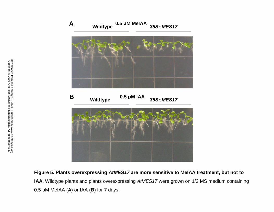
Wildtype 35S::MES17
35S::MES17Wildtype
0.5 µM MeIAA
0.5 µM IAA
A
B
Figure 5. Plants overexpressing AtMES17 are more sensitive to MeIAA treatment, but not to
IAA. Wildtype plants and plants overexpressing AtMES17 were grown on 1/2 MS medium containing
0.5 µM MeIAA (A) or IAA (B) for 7 days.
w
ww
.plantphysiol.orgon F
ebruary 18, 2020 - Published by
Dow
nloaded from
Copyright ©
2008 Am
erican Society of P
lant Biologists. A
ll rights reserved.
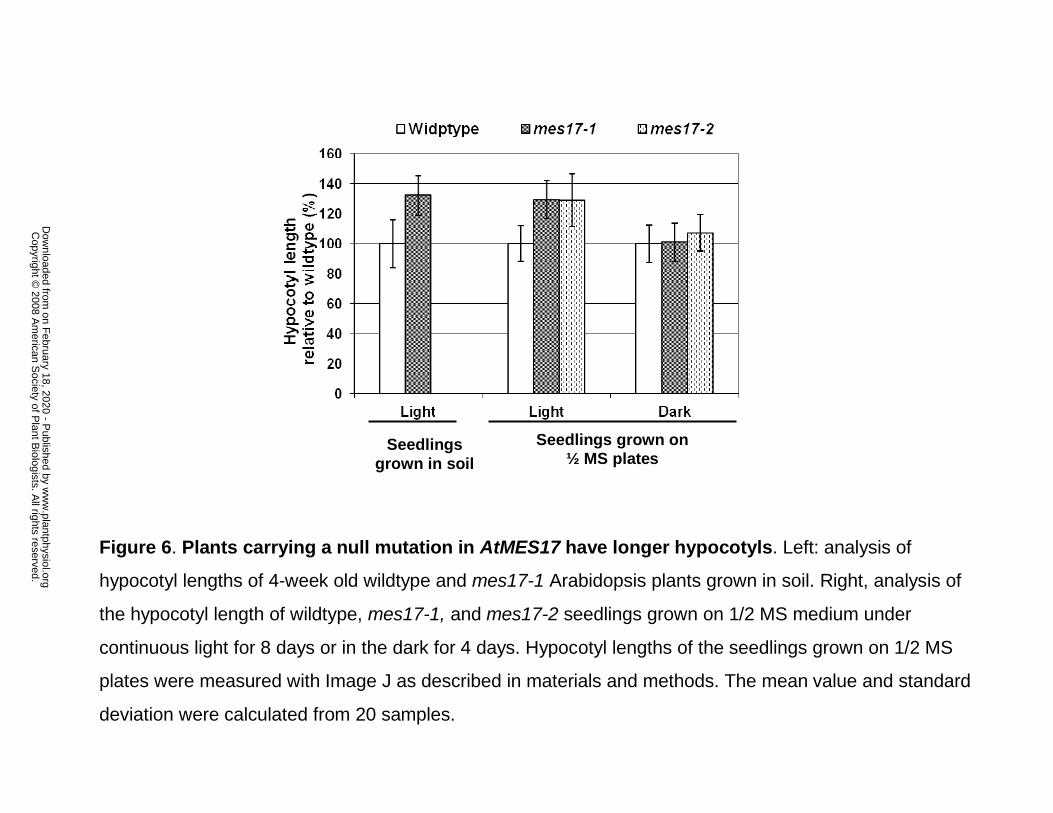
Seedlingsgrown in soil
Seedlings grown on ½ MS plates
Figure 6. Plants carrying a null mutation in AtMES17 have longer hypocotyls. Left: analysis of
hypocotyl lengths of 4-week old wildtype and mes17-1 Arabidopsis plants grown in soil. Right, analysis of
the hypocotyl length of wildtype, mes17-1, and mes17-2 seedlings grown on 1/2 MS medium under
continuous light for 8 days or in the dark for 4 days. Hypocotyl lengths of the seedlings grown on 1/2 MS
plates were measured with Image J as described in materials and methods. The mean value and standard
deviation were calculated from 20 samples.
w
ww
.plantphysiol.orgon F
ebruary 18, 2020 - Published by
Dow
nloaded from
Copyright ©
2008 Am
erican Society of P
lant Biologists. A
ll rights reserved.
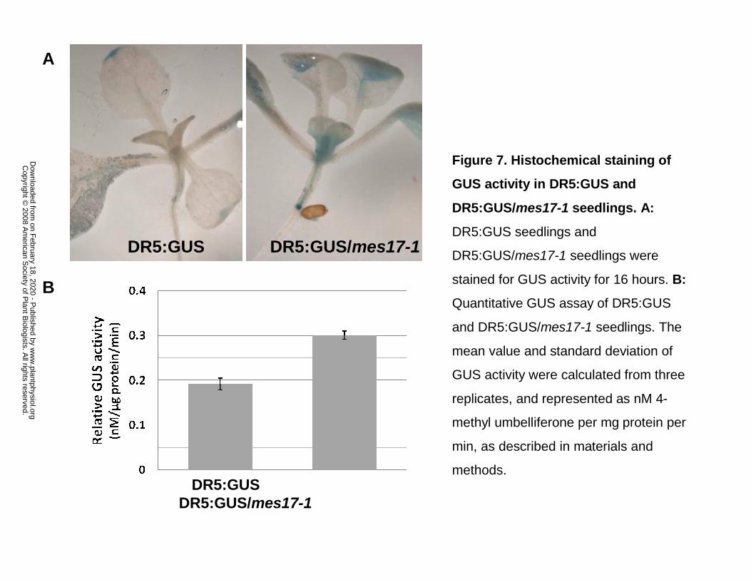
DR5:GUS DR5:GUS/mes17-1
A
B
DR5:GUSDR5:GUS/mes17-1
Figure 7. Histochemical staining of
GUS activity in DR5:GUS and
DR5:GUS/mes17-1 seedlings. A:
DR5:GUS seedlings and
DR5:GUS/mes17-1 seedlings were
stained for GUS activity for 16 hours. B:
Quantitative GUS assay of DR5:GUS
and DR5:GUS/mes17-1 seedlings. The
mean value and standard deviation of
GUS activity were calculated from three
replicates, and represented as nM 4-
methyl umbelliferone per mg protein per
min, as described in materials and
methods.
w
ww
.plantphysiol.orgon F
ebruary 18, 2020 - Published by
Dow
nloaded from
Copyright ©
2008 Am
erican Society of P
lant Biologists. A
ll rights reserved.

0
1
2
3
4
5
6
7
8
Hyp
oco
tyl L
eng
th (m
m)
Wildtype mes17-1 mes17-1/ axr1-3
axr1-3
Figure 8. mes17/axr1-3 double mutant has the same hypocotyl length as axr1-3.
Plants were grown on 1/2 MS medium under continuous light for 9 days. The hypocotyl
lengths of the seedlings were measured with Image J as described in materials and
methods. The mean value and standard deviation were calculated from 20 samples.
w
ww
.plantphysiol.orgon F
ebruary 18, 2020 - Published by
Dow
nloaded from
Copyright ©
2008 Am
erican Society of P
lant Biologists. A
ll rights reserved.
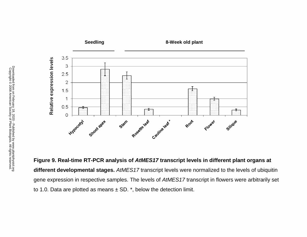
Seedling 8-Week old plant
Figure 9. Real-time RT-PCR analysis of AtMES17 transcript levels in different plant organs at
different developmental stages. AtMES17 transcript levels were normalized to the levels of ubiquitin
gene expression in respective samples. The levels of AtMES17 transcript in flowers were arbitrarily set
to 1.0. Data are plotted as means ± SD. *, below the detection limit.
w
ww
.plantphysiol.orgon F
ebruary 18, 2020 - Published by
Dow
nloaded from
Copyright ©
2008 Am
erican Society of P
lant Biologists. A
ll rights reserved.