running head: regulation of cuc1 stm · knat6, which can have partially overlapping functions in...
TRANSCRIPT

Running head: Regulation of CUC1 by STM
Dr. Javier Palatnik
Instituto de Biología Molecular y Celular de Rosario
Suipacha 531
2000 Rosario
Argentina.
Tel: +54-341-4350661ext#147
Research category: Development and Hormone Action – Associate Editor Richard
Amasino.
Plant Physiology Preview. Published on June 17, 2011, as DOI:10.1104/pp.111.177709
Copyright 2011 by the American Society of Plant Biologists
www.plantphysiol.orgon January 2, 2020 - Published by Downloaded from Copyright © 2011 American Society of Plant Biologists. All rights reserved.

A mechanistic link between STM and CUC1 during Arabidopsis
development
Silvana V. Spinelli1, Ana Paula Martin1, Ivana L. Viola2, Daniel H. Gonzalez2 and Javier F.
Palatnik1*
1Instituto de Biología Molecular y Celular de Rosario, Suipacha 531, 2000 Rosario,
Argentina.
2Instituto de Agrobiotecnología del Litoral, Facultad de Bioquímica y Ciencias Biológicas,
Universidad Nacional del Litoral, CC 242 Paraje El Pozo, 3000 Santa Fe, Argentina.
Financial source: Supported by grants to Javier Palatnik (ANPCyT, HFSP and HHMI).
*To whom correspondence should be directed: [email protected]
www.plantphysiol.orgon January 2, 2020 - Published by Downloaded from Copyright © 2011 American Society of Plant Biologists. All rights reserved.

ABSTRACT
The KNOXI transcription factor SHOOT MERISTEMLESS (STM) is required to establish and
maintain the Arabidopsis thaliana apical meristem, yet little is known about its direct targets.
Using different approaches we demonstrate that the induction of STM causes a significant up-
regulation of the organ boundary gene CUC1, which is specific and independent of other
meristem regulators. We further show that the regulation of CUC1 by STM is direct and
identify putative binding sites in its promoter. Continuous expression of STM in Arabidopsis
leaf primordia also causes the activation of CUC2-3, as well as microRNA MIR164a, which
provides a negative feedback loop by post-transcriptionally regulating CUC1 and CUC2. The
results bring new insights into the mechanistic links between KNOXI and CUC transcription
factors and contribute to the understanding of the regulatory network controlled by STM.
www.plantphysiol.orgon January 2, 2020 - Published by Downloaded from Copyright © 2011 American Society of Plant Biologists. All rights reserved.

INTRODUCTION
In contrast to animals, plants continue to produce new organs throughout their life cycle. All
above ground parts of plants are derived from a small number of stem cells located at the shoot
apical meristem. Two homeodomain transcription factors play key roles in meristem formation
and maintenance. WUSCHEL (WUS) is expressed at the core of the meristem and defines the
stem cell niche in the overlaying cells (Tucker and Laux, 2007), while SHOOT
MERISTEMLESS (STM), is expressed throughout the meristem and prevents the differentiation
of the meristematic cells [reviewed in (Hake et al., 2004; Hamant and Pautot, 2010; Hay and
Tsiantis, 2010)].
STM belongs to class I of KNOX homeodomain transcription factors. In Arabidopsis,
the KNOXI subclass comprises STM, KNAT1 (also called BREVIPEDICELLUS), KNAT2 and
KNAT6, which can have partially overlapping functions in the shoot meristem (Scofield and
Murray, 2006). KNOXI proteins interact with BELL1-like homedomain transcription factors,
and these interactions determine their target affinity and subcellular localization [reviewed in
(Hake et al., 2004; Hay and Tsiantis, 2010)].
At least part of the functions of WUS and STM are performed through the control of
hormone homeostasis and signalling. WUS directly represses a group of type-A ARABIDOPSIS
RESPONSE REGULATORs involved in a negative feedback loop during cytokinin response
(Leibfried et al., 2005; Busch et al., 2010). STM expression induces ISOPENTENYL
TRANSFERASE7 (IPT7), which encodes a key enzyme involved in cytokinin biosynthesis
(Jasinski et al., 2005; Yanai et al., 2005). Conversely, ectopic IPT expression or exogenous
cytokinin can partially rescue weak stm mutants (Jasinski et al., 2005; Yanai et al., 2005). STM
also represses gibberellin activity by reducing the levels of the biosynthetic enzyme GA 20-
oxidase1 and increasing the levels of the catabolic enzyme GA 2-oxidase1, thus providing an
environment of high cytokinin and low gibberellin (Sakamoto et al., 2001; Hay et al., 2002;
Chen et al., 2004; Jasinski et al., 2005; Yanai et al., 2005).
The boundaries of the meristem are defined by members of the NAC family of
transcription factors (Aida and Tasaka, 2006). In Arabidopsis, this function is redundantly
performed by CUP SHAPED COTYLEDON1 (CUC1), CUC2 and CUC3 (Aida et al., 1997;
Aida et al., 1999; Vroemen et al., 2003; Hibara et al., 2006). CUC1 and CUC2 are also post-
transcriptionally regulated by microRNA (miRNA) miR164, which is encoded by a small gene
family comprising three members, MIR164a-c (Laufs et al., 2004; Mallory et al., 2004; Baker et
al., 2005; Nikovics et al., 2006; Sieber et al., 2007; Raman et al., 2008).
In Arabidopsis, CUC genes are required for the activation of STM during embryogenesis
(Aida et al., 1999; Long and Barton, 1998), and it has been proposed that STM can in turn
www.plantphysiol.orgon January 2, 2020 - Published by Downloaded from Copyright © 2011 American Society of Plant Biologists. All rights reserved.

activate CUC expression (Aida et al., 1999; Kwon et al., 2006; Takada et al., 2001). Mutations
in STM or two CUC genes compromise the population of self-renewing stem cells and cause
fusions of the cotyledons. KNOX and CUC genes are recruited again at later stages of
Arabidopsis development and are required for carpel and ovule development (Ishida et al.,
2000; Pautot et al., 2001; Scofield et al., 2007). In many species, KNOXI and CUC genes are
expressed in the leaf primordia and act in concert to sculpt the organ shape and generate
compound leaves (Bharathan et al., 2002; Blein et al., 2008; Berger et al., 2009).
CUC1 and CUC2 share a common ancestor, but have diverged significantly within the
Brassicaceae (Hasson et al., 2011). While both of them are required for organ separation,
specialization has been acquired for certain functions, such as the control of the serrations of an
Arabidopsis simple leaf, which is regulated by the balance between CUC2 and MIR164a genes
(Nikovics et al., 2006; Hasson et al., 2010). STM expression also diverges within the
Brassicaceae. While it is confined to the meristem in Arabidopsis thaliana, closely related
species express STM in the leaf primordia and have more complex organs (Piazza et al., 2010).
Although the biological roles of the versatile developmental regulator STM are well
characterized, little is known about its direct targets. In an attempt to bring insights into the
network regulated by STM, we performed microarray experiments shortly after the induction of
the transcription factor. We found that STM activates CUC1, and demonstrated that it directly
binds to its promoter. Additionally, the long-lasting expression of STM also promotes the
expression of CUC2-3, and MIR164a, which provides a negative feedback loop to adjust the
final CUC level. These results provide new mechanistic insights into the regulatory network
comprised by KNOXI and CUC transcription factors and miRNA miR164.
RESULTS
Genome-wide response to STM levels
To start to explore the network controlled by STM, we analyzed the transcriptome of plants
harboring an ethanol-inducible version of the transcription factor, an approach already used to
identify targets of WUS (Leibfried et al., 2005). The selected transgenic plants did not show any
obvious phenotypes when grown under normal conditions. However, one single treatment with
ethanol was sufficient to cause leaf lobing, as expected for the ectopic expression of STM (Fig.
1A). These morphological changes were obvious one week after the induction (Fig. 1A).
It is known that KNOXI transcription factors interact with other proteins that regulate
their activity [reviewed in (Hake et al., 2004; Hay and Tsiantis, 2010)]. Therefore, we generated
an activated version of STM, by preparing transgenic plants where the transcription factor is
fused to the transactivation domain from the herpes simplex virus VP16. This strategy has been
www.plantphysiol.orgon January 2, 2020 - Published by Downloaded from Copyright © 2011 American Society of Plant Biologists. All rights reserved.

previously used in plants to detect transcription factor activity independently of the presence of
co-activators [e.g., (Parcy et al., 1998)]. Treatment with ethanol of plants harboring an inducible
STM-VP16 transgene caused a higher degree of leaf lobing than that observed for STM alone
(Fig. 1A).
We performed a transcriptome analysis on ATH1 microarrays, 12 hours after the
induction of STM and STM-VP16. For the profiling experiments we used the shoot apex that
includes the meristem and developing leaves, as has been described previously (Leibfried et al.,
2005). Genes that showed per-gene variance p>0,05 (logit-T) (Lemon et al., 2003) and more
than 2-fold change (GCRMA) (Irizarry et al., 2003) compared to control plants were considered
as differentially expressed and were selected for further studies (Table S1, S2).
Analysis of the STM-modified genes using GO term enrichment revealed that there were
no strong over-represented functional categories among them (Table S3). We observed,
however, that At1g50960, which encodes a GA 2-oxidase7 involved in the catabolism of
gibberellin, was significantly induced by both STM and STM-VP16 (Table S1 and S2). IPT7,
which participates in cytokinin biosynthesis, was induced nearly 3 times by STM, although it
did not pass the logit-T filter used for the analysis of the arrays (not shown). These observations
are in agreement with previous results showing that STM increases cytokinin levels while
reducing the gibberellins (Sakamoto et al., 2001; Hay et al., 2002; Jasinski et al., 2005; Yanai et
al., 2005).
With the stringent selection criteria applied, 183 genes were induced by STM and 200 by
STM-VP16 (Fig. 1B). Most of them (129 genes, Table S4) were induced in both conditions. The
higher activation capacity of STM-VP16, which is also correlated with the stronger leaf
phenotypes observed, is likely responsible for the genes that are differentially expressed
between STM and STM-VP16 transgenic plants.
To validate our transcriptome analysis we turned to another inducible system where
STM is fused to the glucocorticoid receptor (GR) (Gallois et al., 2002). The STM-GR fusion
protein is retained in the cytoplasm of transgenic plants, but moves into the nucleus once the
cells are treated with dexamethasone (DEX). We selected thirteen genes induced by both STM
and STM-VP16 (~10 % of the genes induced by both constructs) and tested their response to
STM-GR (Prom35S:STM-GR construct) by RT-qPCR. We found that 10 out of 13 genes were
also induced by this system after 12 hours of DEX application (Fig. 1C, Fig. S1 and Table S4).
These results highlight at least a reasonable reproducibility of the microarray data.
Then, we decided to study in more detail the genes induced by both STM and STM-VP16
(Fig. 1D). We classified these genes in three groups depending on their relative expression in
STM and STM-VP16 samples, using the criteria depicted above, variance p>0,05 (logit-T) and
www.plantphysiol.orgon January 2, 2020 - Published by Downloaded from Copyright © 2011 American Society of Plant Biologists. All rights reserved.

more than 2-fold change (GCRMA). Using this criteria, most genes (101 genes) were similarly
induced by STM and STM-VP16 (cluster #1), 25 genes were more expressed in STM-VP16 than
in STM (cluster #2) and only 3 genes were higher in STM than in STM-VP16 (cluster #3).
As expected, STM was detected as significantly up-regulated in both samples. STM levels
were, however, similar in STM and STM-VP16 arrays (Fig. 1D, cluster #1), indicating that both
plants express their transgenes at similar levels and differences between their transcriptomes are
likely caused by the presence of the VP16 domain. That the VP16 activated version caused
stronger phenotypic defects than STM alone, suggested that the group of genes moderately
induced by STM but strongly by STM-VP16 (Fig. 1D, cluster #2, Table S4) might be
particularly related to the KNOXI pathway. CUC1 (At3g15170) was included in this group and
stood out as a particularly attractive candidate to study in more detail due to its known roles in
the establishment of Arabidopsis meristem.
Specific response of CUC1 to STM levels
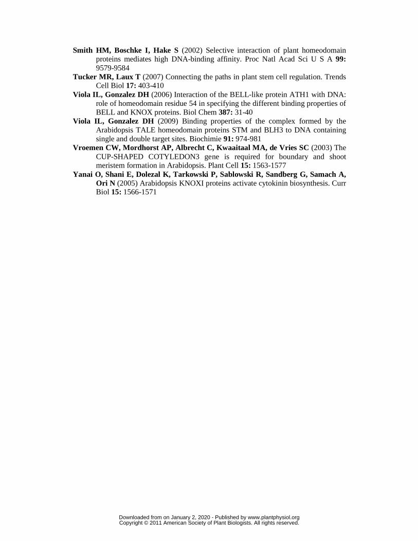
The induction of CUC1 by STM prompted us to study the effects on CUC expression of other
transcription factors known to regulate the meristem function. First, we compared the induction
of CUC1 by STM with the ones caused by other meristem regulators such as WUS and LFY that
were prepared as similar inducible versions (Leibfried et al., 2005). STM was able to induce
CUC1 levels 4-fold, while STM-VP16 further enhanced the response to more than 100-fold in
the microarray experiments (Figure 2A). In contrast to STM, LFY and WUS were not able to
induce CUC1 (Figure 2A). These transcription factors also failed to modify the levels of CUC2
and CUC3, while STM-VP16 caused a moderate up-regulation of CUC3 (Fig. 2A).
We then tested the specificity of CUC1 induction inside the KNOXI family of
transcription factors, which comprises STM, KNAT1, KNAT2 and KNAT6, being KNAT1 the
more closely related to STM, as judged by phylogenetic analyses (Scofield and Murray, 2006).
To test whether other KNOXI genes could activate CUC1 expression we prepared ethanol-
inducible transgenic lines harboring KNAT1 and KNAT2. In contrast to STM, these other KNOXI
genes failed to up-regulate CUC1 (Fig. 2B). These results indicate that there is at least certain
degree of specificity for its induction in planta.
We then prepared an activated version of KNAT1, by fusing to it the VP16 domain. In
this case, we observed that the induction of KNAT1-VP16 caused the activation CUC1 (Fig.
2B). It is known that KNOXI proteins can interact with different partners [reviewed in (Hake et
al., 2004; Hay and Tsiantis, 2010)]. The enhanced activity of the VP16 fusions might indicate
that other factors could be required in vivo for a maximum activation of CUC1 by KNOXI
proteins.
www.plantphysiol.orgon January 2, 2020 - Published by Downloaded from Copyright © 2011 American Society of Plant Biologists. All rights reserved.
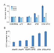
Direct regulation of CUC1 by STM
To test whether STM was directly regulating the expression of CUC1, we turned again to the
STM-GR system. Direct targets of STM-GR should be induced after DEX treatment, even in the
presence of the translational inhibitor, cycloheximide (CYC).
First, we crossed STM-GR transgenics to a CUC1 reporter line (PromCUC1:GUS). We used
1.4 kb CUC1 upstream sequences that have already been described to be sufficient to
complement a cuc1 cuc2 mutant when fused to its coding sequence (Baker et al., 2005). The
CUC1 promoter is normally expressed in the SAM, but DEX treatment during 24 hs caused a
strong induction in whole seedlings (Fig. 3A). As a control of the experimental approach, we
observed that the supplemental addition of CYC largely prevented the burst of GUS protein
activity, which is expected from the inhibition of the translational machinery (Fig. 3A). Note
that these experiments were carried out under long induction and staining periods to ensure the
saturation of the system.
We then analyzed the expression of CUC genes at the RNA level. As CUC1 is post-
transcriptionally regulated by miR164 in a quantitative way (Baker et al., 2005; Nikovics et al.,
2006; Sieber et al., 2007), the potential induction caused by STM should overcome this
repression in order to be detectable. We observed that 4 hs of induction of STM-GR caused the
up-regulation of CUC1 (Fig. 3B). CUC3 was also activated, but after 24 hs. Supplemental
addition of CYC prevented the induction of CUC3, while CUC1 remained unaffected,
confirming CUC1 as a direct target of STM (Fig. 3B). We observed an effect on CUC2 only 96
hours after DEX treatment (Fig. 3B). The longer activation time, which is prevented by
incubation with CYC suggests that both CUC2 and CUC3 are indirectly regulated by STM.
That CUC2 and CUC3 lack obvious STM binding sites in their promoters is in agreement with
this possibility.
In vitro binding of STM to the CUC1 promoter
Next, we searched for potential STM regulatory motifs by analyzing the promoters of genes up-
regulated in the microarray data, as described previously (Schommer et al., 2008). We only
found a potential candidate box when we analyzed genes induced at least 5-fold by STM-VP16,
GTCACT (p= 0,06) (Table S5). Even though the enrichment of this site was not particularly
high, it suggestively overlapped with the preferred binding site of STM, which has already been
investigated in vitro and was found to be CTGTCA (Krusell et al., 1997; Smith et al., 2002;
Viola and Gonzalez, 2006). These sequences share the minimal sequence recognized by KNOX
homeodomains, a GTCA core [reviewed in (Hake et al., 2004)].
www.plantphysiol.orgon January 2, 2020 - Published by Downloaded from Copyright © 2011 American Society of Plant Biologists. All rights reserved.

Interestingly, both six-mer sequences are present in the CUC1 promoter in a narrow
region at -135 (box1, CTGTCA and GTCACT) and -124 (box2, CTGTCA) (Fig. 4A), which
prompted us to perform a more detailed study. We tested whether a recombinant STM protein
could recognize the CUC1 promoter in vitro. EMSA assays showed a strong and specific
interaction between a promoter fragment and the STM homeodomain alone or the complete
recombinant transcription factor (Fig. 4B-C). Binding was competed by a 50-fold molar excess
of the same unlabeled fragment but not by a similar amount of a different fragment, thus
showing the specificity of the interaction (Fig. S2). Mutating box1 caused a significant decrease
in the binding efficiency and a further mutation in box 2 almost completely abolished the
interaction between the CUC1 promoter and STM in vitro (Fig. 4B-C).
We also analyzed the interaction between STM and the CUC1 promoter in a yeast one-
hybrid assay. STM directed CUC1 expression also in this system (Fig. 4D), expression which
was lost when the putative binding box1 was mutated. In sum, these results confirmed that STM
directly regulates CUC1, likely through these two specific binding boxes, and provide a
mechanistic scheme for the regulation of these transcription factors.
CUC1 expression in plants
We then analyzed the expression of CUC1 in the strong stm-1 mutant. As expected, we found
that the levels of CUC1 were reduced in this mutant (Fig. 5A). We also crossed the
PromCUC1:GUS plants to the weak stm allele bum1-3 and found a reduction in the reporter
expression in flowers (Fig. 5B). We tried to rescue the stm-1 mutant by overexpressing a
miR164-resistant version of CUC1. However, the expression of CUC1 alone was not sufficient
to complement the STM deficiency (not shown), as has seen before when overexpressing a
wild-type version of CUC1 in stm-1mutants (Hibara et al., 2003).
To study the role of the STM-binding sites on CUC1 transcription, we turned to
reporters. Previously described transcriptional reporters for CUC1 and CUC2 are expressed in a
broader domain inside the meristem while in situ hybridization assays have shown that CUC
RNA accumulates in the boundaries (Nikovics et al., 2006; Sieber et al., 2007; Raman et al.,
2008). This is at least partially achieved by the post-transcriptional repression carried out by
miR164 (Sieber et al., 2007).
We analyzed the transcription of a wild-type reporter and two mutated versions where
one or both STM-binding sites were removed. Mutations in the putative STM-binding sites
quantitatively decreased its expression levels during vegetative development more than 2-fold
by assaying seven independent transgenic lines for each construct (Fig. 5C-E).
www.plantphysiol.orgon January 2, 2020 - Published by Downloaded from Copyright © 2011 American Society of Plant Biologists. All rights reserved.

The expression of the mutated reporter was reduced 4-fold in inflorescences (Fig. 5C).
Whole-mount stainings showed that the wild-type reporter was expressed at the base of the
flowers and in the carpels (Figure 5F), while the mutated version showed a significantly reduced
staining in the carpels (Fig. 5G).
We then down-regulated CUC1 and other miR164 targets by expressing MIR164b from
different promoters. Expression of MIR164b from the CUC1 promoter (PromCUC1:MIR164b)
caused cotyledon fusions (20 out of 48 T1 plants) (Fig. 6A) and most of them had severe stem-
cauline leaf (Fig. 6B) and sepal fusions (Fig. 6C). In contrast, expression of MIR164b from the
mutated CUC1 promoter did not cause any cotyledon fusions and the defects during
reproductive development were weaker (Fig. 6A-C). Additionally, expression of MIR164b from
a STM promoter (PromSTM:MIR164b) also caused organ fusions (Fig. 6D-F; see Fig. S3 for
PromSTM:GUS stainings).
These phenotypes are similar to some of the phenotypes observed in Prom35S:MIR164
plants (Laufs et al., 2004; Mallory et al., 2004), but still highlight the importance of the STM-
binding sites on the quantitative regulation of CUC1 and the importance of CUC activity inside
the STM domain. We also tried to complement the cuc1/cuc2 double mutant with
PromCUC1:CUC1 and PromCUC1Δbox:CUC1constructs. Unfortunately, the transgenes were silenced
in the mutant background.
A feedback regulatory loop mediated by MIR164a
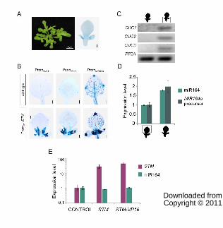
In many plant species with compound leaves, KNOXI transcription factors are expressed in the
leaf primordia (Hareven et al., 1996; Bharathan et al., 2002; Hay and Tsiantis, 2006) where they
interact with CUC genes (Blein et al., 2008). Therefore, we prepared Arabidopsis plants
expressing STM from the LEAFY promoter (PromLFY:STM), which is active in the primordia of
leaves and flowers (Blazquez and Weigel, 2000), and studied the effects on CUC activity.
PromLFY:STM transgenics had lobed leaves as expected from ectopic expression of a
KNOXI gene in leaf primordia (Fig. 7A). Analysis of CUC1 and CUC2 reporters in
PromLFY:STM plants revealed that they were both ectopically expressed in young leaves,
specially at the base of leaf lobes (Fig. 7B, S4) resembling the ectopic pattern of expression of
STM itself (Fig. 7A, right). These plants have constitutively altered levels of STM, so the
induction of CUC2 is likely an indirect effect of STM as we have observed after 96 hs of DEX
treatment of STM-GR plants (Fig 3B).
MIR164a has previously been implicated in the regulation of CUC activity during
Arabidopsis leaf development (Nikovics et al., 2006; Hasson et al., 2010), so we crossed
www.plantphysiol.orgon January 2, 2020 - Published by Downloaded from Copyright © 2011 American Society of Plant Biologists. All rights reserved.

PromLFY:STM plants to a MIR164a reporter. We found that MIR164a was also activated by
STM, in a similar way to the CUC reporters (Fig. 7B, S4).
To validate these results we performed sections of wild-type and PromLFY:STM
developing leaves. We then determined the levels of CUC genes and miR164 by RT-PCR in the
proximal and distal region of the organ. The levels of CUC1-3 as well as miR164 were
increased in the proximal part of the organ, as expected from the whole-mount staining (Fig. 7C
and D). We also determined the levels of the precursor of MIR164a and found that it was also
activated, demonstrating its increased transcription is at least partially responsible for the
elevated miR164 levels (Fig. 7D).
We then tested the short-term response of miR164 to STM levels. We measured miR164
12 h after the induction of STM (Fig. 7E). However, in this case we did not observe an obvious
change in the levels of the miRNA.
These results suggest that MIR164a operates in a negative feedback-loop to adjust the
final CUC levels. The lack of change in miR164 levels when STM is transiently induced
suggests that the activation of MIR164a is an indirect modification caused by STM and the
consequence of the long lasting expression of the KNOXI transcription factor in the leaf
primordia.
We have also performed crosses between PromLFY:STM and cuc1-1 mutants and observed
that the plants still have lobes (Fig. S5), which is in good agreement with the ability of
PromLFY:STM to activate CUC1, CUC2 and CUC3 in leaf primordia (Figure 7C). These results
also indicate that the activation of CUC2 and CUC3 by STM is independent of CUC1.
DISCUSSION
Targets of the KNOXI transcription factors.
The class I of KNOX family of transcription factors comprises a small family of TALE
homeobox genes that are widely distributed in plants. They regulate diverse developmental
processes throughout the Arabidopsis life cycle. KNOXI transcription factors maintain the
activity of the meristem, the boundaries between the stem and the meristem and diverse aspects
of flower development and leaf morphology [reviewed in (Hake et al., 2004; Hamant and
Pautot, 2010; Hay and Tsiantis, 2010)].
Despite their central role as developmental regulators, few downstream effectors of
KNOXI activities are currently known. They regulate the levels of cytokinins and gibberelins
(Sakamoto et al., 2001; Hay et al., 2002; Chen et al., 2004; Jasinski et al., 2005; Yanai et al.,
www.plantphysiol.orgon January 2, 2020 - Published by Downloaded from Copyright © 2011 American Society of Plant Biologists. All rights reserved.

2005) and the deposition of lignin (Mele et al., 2003). Still, precise mechanistic insights into
KNOXI action are lacking in most cases. Here, we identified a direct target of STM in
Arabidopsis. We show that STM directly activates the organ-boundary gene CUC1 and
characterize the process at the biochemical level.
Genetic analyses revealed that KNOX genes conforms a complex network exhibiting both
overlapping and antagonistic activities (Hamant and Pautot, 2010). To add further complexity to
the network, a recently discovered KNOX protein lacking the DNA-binding domain competes
for interacting factors (Magnani and Hake, 2008). Our results indicate that the induction of
CUC1 is relatively specific to STM levels, although the closely related KNAT1 could also
rapidly induce CUC1 when fused to the VP16 domain.
KNOXI transcription factors form complexes with BELL1-like proteins [reviewed in
(Hake et al., 2004; Hay and Tsiantis, 2010)]. Our results cannot rule out that the activation of
CUC1 by STM requires additional factors in vivo, or that the STM-binding site are also
recognized by other TALE transcription factors in certain tissues. Actually, the higher activation
capacity of STM and KNAT1 on CUC1 levels conferred by the addition of VP16 might suggest
that additional factors could be involved in plants.
It has been suggested that KNOXI transcription factors might exert their functions
through the regulation of a few targets at least in certain situations (Hay and Tsiantis, 2010), as
it has been shown for animal homeodomain transcription factors (Lovegrove et al., 2006).
Genome-wide experiments will likely be required to understand the complex KNOX networks.
The transcriptome analysis that we have performed here might also aid in the identification of
other STM regulated genes.
The KNOXI – CUC regulatory network.
It has been shown that both KNOXI and CUC systems reinforce each other, and that the ectopic
expression of CUC1 induces STM (Aida et al., 2002; Hibara et al., 2003; Furutani et al., 2004;
Kwon et al., 2006; Blein et al., 2008). The results obtained showing that STM directly induces
CUC1, while indirectly activates CUC2-3 and MIR164a provide further insights into this
regulatory network.
That STM can directly activate CUC1, while indirectly affecting CUC2-3 indicates that
the CUC genes respond to different signals. Previous evidence has already shown that CUC
genes can be regulated independently of each other by different factors. For instance, CUC1-2
are differentially affected by PIN1 (Aida et al., 2002), CUC1-2 are targets of miR164 but not
CUC3 (Laufs et al., 2004; Mallory et al., 2004), and only CUC2 is regulated by SPLAYED
www.plantphysiol.orgon January 2, 2020 - Published by Downloaded from Copyright © 2011 American Society of Plant Biologists. All rights reserved.

(Kwon et al., 2006). The regulation of redundant factors by different pathways might confer
robustness to a biological process, such as the formation of the meristem.
The activation of MIR164a by STM that we observed, which is likely indirect, might also
contribute to fine-tune the levels of CUC expression as a part of a negative feedback-loop. A
general homeostatic function has been already proposed for miR159 and that as its targets, the
GA-MYB transcription factors, might activate the expression of the miRNA (Achard et al.,
2004).
KNOXI and CUC genes are also versatile developmental regulators whose functions go
beyond the establishment and maintenance of the meristem. They are recruited during carpel
and ovule development in Arabidopsis (Ishida et al., 2000; Pautot et al., 2001; Scofield et al.,
2007) and the formation of complex leaves in many species (Bharathan et al., 2002; Blein et al.,
2008; Berger et al., 2009). Specific relationships between KNOXI, CUC and MIR164 family
members can be established during particular biological processes. The specific function of
MIR164c in the regulation of petal number (Baker et al., 2005) and the role of CUC2 and
MIR164a in the formation of leaf serrations (Nikovics et al., 2006) are in good agreement with
this possibility.
CUC1 and CUC2 have diverged significantly within the Brassicaceae (Hasson et al.,
2011). That CUC1, but not CUC2, responds directly to STM and has STM-binding sites in its
promoter is also consistent with this data. The expression of STM also varies considerably in
different species closely related to Arabidopsis thaliana. Many relatives express STM in the leaf
primordia and have organs with more complex morphology (Piazza et al., 2010). Interestingly,
the STM-binding sites are conserved in the CUC1 promoter in several Brassicaceae species
(Spinelli & Palatnik, unpublished). It might be interesting to determine whether the direct
regulation of CUC1 by STM has a role in the formation of complex leaf morphologies within
the Brassicaceae.
MATERIALS AND METHODS
Plant Material
Plants were grown in long days (16 hours light/8 hours dark) at 23 °C. See Table S6 for a list of
transgenic lines and mutants. For Dex treatments, 2 week-old seedlings were transplanted to MS
plates containing 60μM Dex or 60μM Dex and 10μM CYC. Control plates were treated in the
same way without the addition of Dex or the translational inhibitor. Control plates treated only
with CYC showed no significant differences respect to untreated controls. CYC was also added
www.plantphysiol.orgon January 2, 2020 - Published by Downloaded from Copyright © 2011 American Society of Plant Biologists. All rights reserved.

before the DEX treatment as an additional control without any modification of the results.
Seedlings were collected at different times after treatment for analysis.
Analytical Procedures
GUS stainings, microscopic observations, RNA extraction and analysis by RT-qPCR was
performed as described previously (Rodriguez et al., 2010). PROTEIN PHOSPHATASE 2 was
used as a control to normalize the data (Czechowski et al., 2005). In cases where no expression
was detected in the reference sample after 35 cycles, results were shown as semi-quantitative
data (CUC1 was usually detected around cycles 26-29 with primers flanking the miRNA target
site). GUS activity was assayed in protein extracts by a fluorescence method with 4-
methylumbelliferyl glucuronide as a substrate (Jefferson et al., 1987). Mature miR164 levels
were determined by stem-loop RT-qPCR (Chen et al., 2005). Primers are shown in Table S7
and Table S8 has a description of binary plasmids prepared for this study.
Microarray Analyses
Two week-old seedlings were treated with 0,6 % ethanol during 12 hours. The shoot apex and
the surrounding tissue was analyzed with Affymetrix ATH1 arrays (n=2) (E-MEXP-2550).
Differentially expressed genes (Table S1, S2) and over-represented motifs (table S5) were
identified as described before (Schommer et al., 2008).
DNA binding assays
Proteins were obtained as fusions with glutahione S-transferase as described previously (Viola
and Gonzalez, 2006, 2009). For EMSAs, purified recombinant proteins were incubated with 0,5
ng of labeled CUC1 promoter fragments (-177/-84 respective to the transcription start site).
Binding reactions (20 µl) contained 20 mM HEPES (pH 7.5), 50 mM KCl, 2 mM MgCl2, 0.5
mM EDTA, 1.0 mM DTT, 0.5% Triton X-100, 22 ng/µl BSA, 0.5 μg poly(dI-dC), and 10%
glycerol. EMSAs were performed as described (Viola and Gonzalez, 2006). For the analysis of
STM binding to the CUC1 promoter in yeast, the STM coding sequence was cloned in pGADT7
(Clontech) and introduced into yeast strains constructed using the pHIS3NX/pINT1 vector and
carrying the CUC1 promoter inserted in the PDC6 locus in front of the HIS3 reporter gene
preceded by its own minimal promoter.
Supplemental material
Supplementary Table 1: Genes modified by STM after 12 hours of induction.
Supplementary Table 2: Genes modified by STM-VP16 after 12 hours of induction.
Supplementary Table 3: Gene Ontology (GO) term enrichment for STM and STM-VP16
regulated genes.
Supplementary Table 4: Genes induced by both STM and STM-VP16.
www.plantphysiol.orgon January 2, 2020 - Published by Downloaded from Copyright © 2011 American Society of Plant Biologists. All rights reserved.

Supplementary Table 5: Presence of putative STM-binding motifs in genes induced at least 5
times by STM-VP16.
Supplementary Table 6: List of previously described transgenic lines used in this article.
Supplementary Table 7: Relevant Locus IDs and oligonucleotide primers used in RT-qPCR
assays.
Supplementary Table 8: List of binary vectors prepared in this article.
Supplementary Figure 1: Expression levels of genes analyzed using the STM-GR system by
RT-qPCR.
Supplementary Figure 2: Binding of STM to the CUC1 promoter fragment in vitro.
Supplementary Figure 3: Expression pattern of the STM reporter.
Supplementary Figure 4: Expression pattern of CUC1, CUC2 and MIR164a reporters in
different backgrounds.
Supplementary Figure 5: Phenotypes of PromLFY:STM crossed to cuc1-1 plants.
ACKNOWLEDGEMENTS
We thank Detlef Weigel, in whose lab at the MPI for Developmental Biology (Tübingen) some
of these experiments were started, Jan Lohmann for access to comparative microarray data,
Carla Schommer, Edgardo Bresso, Juan Debernardi, Jean-Luc Gallois and Detlef Weigel for
discussions, Patrick Laufs for CUC2 and MIR164a reporters, Robert Sablowski for the
Prom35S:STM-GR construct. SS and AM are CONICET fellows, and JP, ILV and DHG are
members of CONICET.
www.plantphysiol.orgon January 2, 2020 - Published by Downloaded from Copyright © 2011 American Society of Plant Biologists. All rights reserved.
Figure legends:
Fig. 1: Genome-wide response to STM levels
(A) Phenotype of STM and STM-VP16 inducible lines 9 days after treatment with 0,6% ethanol.
Samples for microarray experiments were collected 12 hours after ethanol induction. The
transgenic line used as control expresses GUS under the ethanol inducible promoter. Bars: 1 cm
(B) Venn diagram showing the overlap of STM and STM-VP16 up-regulated genes.
(C) Heat map representing relative expression levels in gray-scale of 13 genes in three STM
inducible systems: ethanol inducible STM and STM-VP16, and STM-GR. The genes were
selected from those induced in STM and STM-VP16 (Fig. 1B). CUC1 (At3g15170) is depicted
in blue. The data shown are mean of 2 biological replicates ± s.e.m for microarray data (STM
and STM-VP16) and 3 biological replicates ± s.e.m for RT-qPCR experiments in the case of
STM-GR (see Fig. S1 for details). As a control, we used the constitutively expressed gene
PROTEIN PHOSPHATASE 2A.
(D) The 129 genes induced by both STM and STM-VP16 were classified in 3 clusters according to
their relative expression levels in the Affimetrix microarrays. Cluster #1 contains genes with
similar levels in STM and STM-VP16 transgenic plants (101 genes); cluster #2 has genes with
higher expression in STM-VP16 than in STM (25 genes); cluster #3 contains genes with higher
expression in STM than in STM-VP16 (3 genes) using the criteria described in the text to select
differentially expressed genes (logit-T 0.05; 2-fold change with GCRMA). STM is highlighted in
green and CUC1 in blue.
Fig. 2: Specific response of CUC1 to STM levels
(A) Expression levels of CUC1-3 after the induction of LFY, WUS, STM, and STM-VP16 from
Affimetrix microarrays (GCRMA normalization). Expression levels were normalized to plants
carrying inducible GUS grown in the same conditions, which were used as a control. The data
shown are mean of 2 biological replicates ± s.e.m.
(B) Regulation of CUC1 expression by Arabidopsis KNOXI genes. The level of CUC1 was
determined in transgenic lines harboring an inducible version of KNAT1, KNAT1-VP16, KNAT2,
STM and STM-VP16, 12 hours after ethanol treatment. CUC1 levels were determined by RT-
qPCR and expressed relative to plants expressing inducible GUS (Control). The data shown are
mean of 3 biological replicates ± s.e.m.
Fig. 3: Direct regulation of CUC1 by STM.
(A) GUS expression in seedlings of transgenic plants expressing PromCUC1:GUS crossed to STM-GR
after 24 hs treatment with or without DEX and CYC.
www.plantphysiol.orgon January 2, 2020 - Published by Downloaded from Copyright © 2011 American Society of Plant Biologists. All rights reserved.
(B) CUC1-3 expression levels determined by RT-PCR in STM-GR transgenics treated with or
without DEX and CYC. The data shown is representative of at least 3 biological replicates.
Fig. 4: In vitro binding of STM to the CUC1 promoter
(A) Scheme representing the CUC1 promoter. Putative STM-binding sites identified by SELEX are
highlighted with blue squares and the sequence identified to be over-represented in STM-VP16
induced genes (Table S5) is indicated with a dashed line.
(B, C) EMSA with CUC1 promoter using recombinant STM homeodomain (B) or whole protein (C).
(D) One-hybrid experiment in yeast using wild-type and mutated CUC1 promoter. Growth in the
absence of histidine due to activation of the HIS3 gene under the control of the CUC1 promoter
was monitored using serial dilutions of the corresponding yeast strains. 3AT: 3-amino-1,2,4-
triazole.
Fig. 5: CUC1 expression in plants
(A) CUC1 expression levels determined by RT-PCR in apices of wild-type (Ler) and stm-1 plants
grown for 6 days in MS media. The data shown is representative of at least 3 biological
replicates.
(B) CUC1 reporter in a developing wild-type (left) or bum1-3 (rigth) flower.
(C) GUS activity of transgenic plants expressing wild-type and mutant CUC1 reporters in 12 day-old
seedlings and inflorescences. The values correspond to the average of 7 independent lines for
each promoter version ± s.e.m.
(D-G) GUS expression in seedlings (D, E) and inflorescences (F, G) of transgenic plants harboring the
wild-type (D, F) and mutant (Δbox1/2) (E, G) CUC1 reporter.
Bars: 10 mm.
Fig. 6: Missexpression of MIR164b using different promoters
(A-C) Phenotypes of plants expressing MIR164b from a CUC1 and CUC1Δbox1/2 promoter.
(D-F) Phenotypes of PromSTM:MIR164b transgenic plants. (F) wild-type (left) and PromSTM:MIR164b
flowers.
Bars: 10 mm (A, C, F) and 1 cm (B, D, E). Organ fusions are indicated with arrowheads.
www.plantphysiol.orgon January 2, 2020 - Published by Downloaded from Copyright © 2011 American Society of Plant Biologists. All rights reserved.

Fig. 7: CUC and MIR164 genes expression pattern in PromLFY:STM plants.
(A) Altered leaf shape of plants expressing STM under the control of the LFY promoter,
PromLFY:STM. Right panel: PromLFY:STM crossed to PromLFY:GUS, which indicates the domain
of expression of STM in these transgenics.
(B) Expression of CUC1, CUC2 and MIR164a reporters in wild-type and PromLFY:STM plants.
(C) CUC1-3 expression levels determined by RT-PCR in the proximal and distal parts of leaves #3
and #4 of PromLFY:STM plants. The data shown is representative of at least 3 biological
replicates.
(D) miR164 and MIR164a precursor expression levels determined by RT-qPCR in PromLFY:STM
leaves dissected as in C. The data shown are mean of 3 biological replicates ± s.e.m.
(E) Expression levels of miR164 determined by RT-qPCR after 12 hs of induction of STM and STM-
VP16 with ethanol. Expression levels were normalized as described in Fig. 1.The data shown are
mean of 3 biological replicates ± s.e.m.
Bars: 2 mm unless otherwise noted.
www.plantphysiol.orgon January 2, 2020 - Published by Downloaded from Copyright © 2011 American Society of Plant Biologists. All rights reserved.

REFERENCES
Achard P, Herr A, Baulcombe DC, Harberd NP (2004) Modulation of floral
development by a gibberellin-regulated microRNA. Development 131: 3357-3365
Aida M, Ishida T, Fukaki H, Fujisawa H, Tasaka M (1997) Genes involved in organ separation in Arabidopsis: an analysis of the cup-shaped cotyledon mutant. Plant Cell 9: 841-857
Aida M, Ishida T, Tasaka M (1999) Shoot apical meristem and cotyledon formation during Arabidopsis embryogenesis: interaction among the CUP-SHAPED COTYLEDON and SHOOT MERISTEMLESS genes. Development 126: 1563-1570
Aida M, Tasaka M (2006) Genetic control of shoot organ boundaries. Curr Opin Plant Biol 9: 72-77
Aida M, Vernoux T, Furutani M, Traas J, Tasaka M (2002) Roles of PIN-FORMED1 and MONOPTEROS in pattern formation of the apical region of the Arabidopsis embryo. Development 129: 3965-3974
Baker CC, Sieber P, Wellmer F, Meyerowitz EM (2005) The early extra petals1 mutant uncovers a role for microRNA miR164c in regulating petal number in Arabidopsis. Curr Biol 15: 303-315
Berger Y, Harpaz-Saad S, Brand A, Melnik H, Sirding N, Alvarez JP, Zinder M, Samach A, Eshed Y, Ori N (2009) The NAC-domain transcription factor GOBLET specifies leaflet boundaries in compound tomato leaves. Development 136: 823-832
Bharathan G, Goliber TE, Moore C, Kessler S, Pham T, Sinha NR (2002) Homologies in leaf form inferred from KNOXI gene expression during development. Science 296: 1858-1860
Blazquez MA, Weigel D (2000) Integration of floral inductive signals in Arabidopsis. Nature 404: 889-892
Blein T, Pulido A, Vialette-Guiraud A, Nikovics K, Morin H, Hay A, Johansen IE, Tsiantis M, Laufs P (2008) A conserved molecular framework for compound leaf development. Science 322: 1835-1839
Busch W, Miotk A, Ariel FD, Zhao Z, Forner J, Daum G, Suzaki T, Schuster C, Schultheiss SJ, Leibfried A, Haubeiss S, Ha N, Chan RL, Lohmann JU (2010) Transcriptional control of a plant stem cell niche. Dev Cell 18: 849-861
Czechowski T, Stitt M, Altmann T, Udvardi MK, Scheible WR (2005) Genome-wide identification and testing of superior reference genes for transcript normalization in Arabidopsis. Plant Physiol 139: 5-17
Chen C, Ridzon DA, Broomer AJ, Zhou Z, Lee DH, Nguyen JT, Barbisin M, Xu NL, Mahuvakar VR, Andersen MR, Lao KQ, Livak KJ, Guegler KJ (2005) Real-time quantification of microRNAs by stem-loop RT-PCR. Nucleic Acids Res 33: e179
Chen H, Banerjee AK, Hannapel DJ (2004) The tandem complex of BEL and KNOX partners is required for transcriptional repression of ga20ox1. Plant J 38: 276-284
Furutani M, Vernoux T, Traas J, Kato T, Tasaka M, Aida M (2004) PIN-FORMED1 and PINOID regulate boundary formation and cotyledon development in Arabidopsis embryogenesis. Development 131: 5021-5030
www.plantphysiol.orgon January 2, 2020 - Published by Downloaded from Copyright © 2011 American Society of Plant Biologists. All rights reserved.
Gallois JL, Woodward C, Reddy GV, Sablowski R (2002) Combined SHOOT MERISTEMLESS and WUSCHEL trigger ectopic organogenesis in Arabidopsis. Development 129: 3207-3217
Hake S, Smith HM, Holtan H, Magnani E, Mele G, Ramirez J (2004) The role of knox genes in plant development. Annu Rev Cell Dev Biol 20: 125-151
Hamant O, Pautot V (2010) Plant development: a TALE story. C R Biol 333: 371-381 Hareven D, Gutfinger T, Parnis A, Eshed Y, Lifschitz E (1996) The making of a
compound leaf: genetic manipulation of leaf architecture in tomato. Cell 84: 735-744
Hasson A, Blein T, Laufs P (2010) Leaving the meristem behind: the genetic and molecular control of leaf patterning and morphogenesis. C R Biol 333: 350-360
Hasson A, Plessis A, Blein T, Adroher B, Grigg S, Tsiantis M, Boudaoud A, Damerval C, Laufs P (2011) Evolution and Diverse Roles of the CUP-SHAPED COTYLEDON Genes in Arabidopsis Leaf Development. Plant Cell 23: 54-68
Hay A, Kaur H, Phillips A, Hedden P, Hake S, Tsiantis M (2002) The gibberellin pathway mediates KNOTTED1-type homeobox function in plants with different body plans. Curr Biol 12: 1557-1565
Hay A, Tsiantis M (2006) The genetic basis for differences in leaf form between Arabidopsis thaliana and its wild relative Cardamine hirsuta. Nat Genet 38: 942-947
Hay A, Tsiantis M (2010) KNOX genes: versatile regulators of plant development and diversity. Development 137: 3153-3165
Hibara K, Karim MR, Takada S, Taoka K, Furutani M, Aida M, Tasaka M (2006) Arabidopsis CUP-SHAPED COTYLEDON3 regulates postembryonic shoot meristem and organ boundary formation. Plant Cell 18: 2946-2957
Hibara K, Takada S, Tasaka M (2003) CUC1 gene activates the expression of SAM-related genes to induce adventitious shoot formation. Plant J 36: 687-696
Irizarry RA, Bolstad BM, Collin F, Cope LM, Hobbs B, Speed TP (2003) Summaries of Affymetrix GeneChip probe level data. Nucleic Acids Res 31: e15
Ishida T, Aida M, Takada S, Tasaka M (2000) Involvement of CUP-SHAPED COTYLEDON genes in gynoecium and ovule development in Arabidopsis thaliana. Plant Cell Physiol 41: 60-67
Jasinski S, Piazza P, Craft J, Hay A, Woolley L, Rieu I, Phillips A, Hedden P, Tsiantis M (2005) KNOX action in Arabidopsis is mediated by coordinate regulation of cytokinin and gibberellin activities. Curr Biol 15: 1560-1565
Jefferson RA, Kavanagh TA, Bevan MW (1987) GUS fusions: beta-glucuronidase as a sensitive and versatile gene fusion marker in higher plants. Embo J 6: 3901-3907
Krusell L, Rasmussen I, Gausing K (1997) DNA binding sites recognised in vitro by a knotted class 1 homeodomain protein encoded by the hooded gene, k, in barley (Hordeum vulgare). FEBS Lett 408: 25-29
Kwon CS, Hibara K, Pfluger J, Bezhani S, Metha H, Aida M, Tasaka M, Wagner D (2006) A role for chromatin remodeling in regulation of CUC gene expression in the Arabidopsis cotyledon boundary. Development 133: 3223-3230
Laufs P, Peaucelle A, Morin H, Traas J (2004) MicroRNA regulation of the CUC genes is required for boundary size control in Arabidopsis meristems. Development 131: 4311-4322
www.plantphysiol.orgon January 2, 2020 - Published by Downloaded from Copyright © 2011 American Society of Plant Biologists. All rights reserved.

Leibfried A, To JP, Busch W, Stehling S, Kehle A, Demar M, Kieber JJ, Lohmann JU (2005) WUSCHEL controls meristem function by direct regulation of cytokinin-inducible response regulators. Nature 438: 1172-1175
Lemon WJ, Liyanarachchi S, You M (2003) A high performance test of differential gene expression for oligonucleotide arrays. Genome Biol 4: R67
Lovegrove B, Simoes S, Rivas ML, Sotillos S, Johnson K, Knust E, Jacinto A, Hombria JC (2006) Coordinated control of cell adhesion, polarity, and cytoskeleton underlies Hox-induced organogenesis in Drosophila. Curr Biol 16: 2206-2216
Magnani E, Hake S (2008) KNOX lost the OX: the Arabidopsis KNATM gene defines a novel class of KNOX transcriptional regulators missing the homeodomain. Plant Cell 20: 875-887
Mallory AC, Dugas DV, Bartel DP, Bartel B (2004) MicroRNA regulation of NAC-domain targets is required for proper formation and separation of adjacent embryonic, vegetative, and floral organs. Curr Biol 14: 1035-1046
Mele G, Ori N, Sato Y, Hake S (2003) The knotted1-like homeobox gene BREVIPEDICELLUS regulates cell differentiation by modulating metabolic pathways. Genes Dev 17: 2088-2093
Nikovics K, Blein T, Peaucelle A, Ishida T, Morin H, Aida M, Laufs P (2006) The balance between the MIR164A and CUC2 genes controls leaf margin serration in Arabidopsis. Plant Cell 18: 2929-2945
Parcy F, Nilsson O, Busch MA, Lee I, Weigel D (1998) A genetic framework for floral patterning. Nature 395: 561-566
Pautot V, Dockx J, Hamant O, Kronenberger J, Grandjean O, Jublot D, Traas J (2001) KNAT2: evidence for a link between knotted-like genes and carpel development. Plant Cell 13: 1719-1734
Piazza P, Bailey CD, Cartolano M, Krieger J, Cao J, Ossowski S, Schneeberger K, He F, de Meaux J, Hall N, Macleod N, Filatov D, Hay A, Tsiantis M (2010) Arabidopsis thaliana leaf form evolved via loss of KNOX expression in leaves in association with a selective sweep. Curr Biol 20: 2223-2228
Raman S, Greb T, Peaucelle A, Blein T, Laufs P, Theres K (2008) Interplay of miR164, CUP-SHAPED COTYLEDON genes and LATERAL SUPPRESSOR controls axillary meristem formation in Arabidopsis thaliana. Plant J 55: 65-76
Rodriguez RE, Mecchia MA, Debernardi JM, Schommer C, Weigel D, Palatnik JF (2010) Control of cell proliferation in Arabidopsis thaliana by microRNA miR396. Development 137: 103-112
Sakamoto T, Kamiya N, Ueguchi-Tanaka M, Iwahori S, Matsuoka M (2001) KNOX homeodomain protein directly suppresses the expression of a gibberellin biosynthetic gene in the tobacco shoot apical meristem. Genes Dev 15: 581-590
Scofield S, Dewitte W, Murray JA (2007) The KNOX gene SHOOT MERISTEMLESS is required for the development of reproductive meristematic tissues in Arabidopsis. Plant J 50: 767-781
Scofield S, Murray JA (2006) KNOX gene function in plant stem cell niches. Plant Mol Biol 60: 929-946
Schommer C, Palatnik JF, Aggarwal P, Chetelat A, Cubas P, Farmer EE, Nath U, Weigel D (2008) Control of jasmonate biosynthesis and senescence by miR319 targets. PLoS Biol 6: e230
Sieber P, Wellmer F, Gheyselinck J, Riechmann JL, Meyerowitz EM (2007) Redundancy and specialization among plant microRNAs: role of the MIR164 family in developmental robustness. Development 134: 1051-1060
www.plantphysiol.orgon January 2, 2020 - Published by Downloaded from Copyright © 2011 American Society of Plant Biologists. All rights reserved.
Smith HM, Boschke I, Hake S (2002) Selective interaction of plant homeodomain proteins mediates high DNA-binding affinity. Proc Natl Acad Sci U S A 99: 9579-9584
Tucker MR, Laux T (2007) Connecting the paths in plant stem cell regulation. Trends Cell Biol 17: 403-410
Viola IL, Gonzalez DH (2006) Interaction of the BELL-like protein ATH1 with DNA: role of homeodomain residue 54 in specifying the different binding properties of BELL and KNOX proteins. Biol Chem 387: 31-40
Viola IL, Gonzalez DH (2009) Binding properties of the complex formed by the Arabidopsis TALE homeodomain proteins STM and BLH3 to DNA containing single and double target sites. Biochimie 91: 974-981
Vroemen CW, Mordhorst AP, Albrecht C, Kwaaitaal MA, de Vries SC (2003) The CUP-SHAPED COTYLEDON3 gene is required for boundary and shoot meristem formation in Arabidopsis. Plant Cell 15: 1563-1577
Yanai O, Shani E, Dolezal K, Tarkowski P, Sablowski R, Sandberg G, Samach A, Ori N (2005) Arabidopsis KNOXI proteins activate cytokinin biosynthesis. Curr Biol 15: 1566-1571
www.plantphysiol.orgon January 2, 2020 - Published by Downloaded from Copyright © 2011 American Society of Plant Biologists. All rights reserved.

www.plantphysiol.orgon January 2, 2020 - Published by Downloaded from Copyright © 2011 American Society of Plant Biologists. All rights reserved.
www.plantphysiol.orgon January 2, 2020 - Published by Downloaded from Copyright © 2011 American Society of Plant Biologists. All rights reserved.
www.plantphysiol.orgon January 2, 2020 - Published by Downloaded from Copyright © 2011 American Society of Plant Biologists. All rights reserved.

www.plantphysiol.orgon January 2, 2020 - Published by Downloaded from Copyright © 2011 American Society of Plant Biologists. All rights reserved.

www.plantphysiol.orgon January 2, 2020 - Published by Downloaded from Copyright © 2011 American Society of Plant Biologists. All rights reserved.

www.plantphysiol.orgon January 2, 2020 - Published by Downloaded from Copyright © 2011 American Society of Plant Biologists. All rights reserved.
www.plantphysiol.orgon January 2, 2020 - Published by Downloaded from Copyright © 2011 American Society of Plant Biologists. All rights reserved.