s. rizzo f. patelli d. r. chow...
TRANSCRIPT

Essentials in Ophthalmology Vitreo-retinal Surgery
S. Rizzo F. Patelli D. R. ChowEditors

Essentials in Ophthalmology
G. K. Krieglstein R. N. WeinrebSeries Editors
Glaucoma
Cataract and Refractive Surgery
Uveitis and Immunological Disorders
Vitreo-retinal Surgery
Medical Retina
Oculoplastics and Orbit
Pediatric Ophthalmology,Neuro-Ophthalmology, Genetics
Cornea and External Eye Disease
Vitreo-retinal Surgery
123
Editors Stanislao RizzoFabio PatelliDavid R. Chow
Vitreo-retinalSurgery
With 50 Figures, Mostly in Colourand 12 Tables

Series Editors
Günter K. Krieglstein, MDProfessor and ChairmanDepartment of OphthalmologyUniversity of CologneKerpener Straße 6250924 CologneGermany
Robert N. Weinreb, MDProfessor and DirectorHamilton Glaucoma CenterDepartment of OphthalmologyUniversity of California at San Diego9500 Gilman DriveLa Jolla, CA 92093-0946USA
Volume Editors
Stanislao Rizzo, MDProfessor and Chairman of OphthalmologyEye Surgery ClinicSanta Chiara HospitalVia Roma, 6756100 PisaItaly
Fabio Patelli, MDMilano Retina Centervia Pietro Mascagni, 2020122 MilanoItaly
David R. Chow, MDUniversity of Toronto23 Ivor Rd., North YorkOntario M4N 2H3Canada
Library of Congress Control Number: 2008929530
© 2009 Springer-Verlag Berlin Heidelberg
This work is subject to copyright. All rights are reserved, whether the whole or part of the material is concerned, specifically the rights of translation, reprinting, reuse of illustrations, recitation, broadcasting, reproduction on microfilms or in any other way, and storage in data banks. Duplication of this publication or parts thereof is permitted only under the provisions of the German Copyright Law of September 9, 1965, in its current version, and permission for use must always be obtained from Springer-Verlag. Violations are liable for prosecution under the German Copyright Law.
The use of general descriptive names, registered names, trademarks, etc. in this publication does not imply, even in the absence of a specific statement, that such names are exempt from the relevant protective laws and regulations and therefore free for general use.
Product liability: The publishers cannot guarantee the accuracy of any information about dosage and application contained in this book. In every individual case the user must check such information by consulting the relevant literature.
Cover picture ‘Oculoplastics and Orbit’ modified from Katowitz, A., Pediatric Oculoplastic Surgery (Springer New York 2002).
Cover Design: WMXDesign GmbH, Heidelberg, Germany
Printed on acid-free paper
9 8 7 6 5 4 3 2 1
springer.com
ISBN 978-3-540-69461-8 e-ISBN 978-3-540-68586-9
ISSN 1612-3212
Foreword
The Essentials in Ophthalmology series represents an unique updating publication on the progress in all subspecialties of ophthalmology.
In a quarterly rhythm, eight issues are published cov-ering clinically relevant achievements in the whole field of ophthalmology. This timely transfer of advancements for the best possible care of our eye patients has proven to be effective. The initial working hypothesis of provid-ing new knowledge immediately following publication in the peer-reviewed journal and not waiting for the textbook appears to be highly workable.
We are now entering the third cycle of the Essentials in Ophthalmology series, having been encouraged by
readership acceptance of the first two series, each of eight volumes. This is a success that was made possible predominantly by the numerous opinion-leading authors and the outstanding section editors, as well as with the constructive support of the publisher. There are many good reasons to continue andstill improve the dissemination of this didactic and clinically rel-evant information.
G.K. KrieglsteinR.N. Weinreb Series EditorsSeptember 2008

Preface
“All progress occurs because people dare to be different”.Harry Milner
Every so often, changes occur in the technology of our day-to-day lives that truly alter how we do things. As retinal surgeons, the development of sutureless vitrectomy systems is one of these revolutionary changes that have altered how most of us perform surgery. Since Dr Eugene de Juan introduced us to a 25-gauge transconjunctival sutureless cannula sys-tem, there has been almost a dizzying pace of change in our field, as instrumentation companies refine and improve the 25-gauge experience. Given some of the early limitations of 25-gauge technologies, Dr Claus Eckardt introduced us to an alternative, a 23-gauge sutureless cannula system, which is becoming increas-ingly popular. At the present time, there is consider-able debate and confusion amongst our community about the direction our field is going to take. Are we
all going to become 23-gauge surgeons? Will 20-gauge vitrectomy disappear? Can technologic improvements make the 25-gauge experience easier? We have put together this textbook to try and answer some of these questions, and to give you some help on when and how these new sutureless vitrectomy systems can be used. To aid us in this endeavour, we have recruited the leading surgeons in our field to offer their insights into the sutureless techniques they perform. We think you will enjoy the practical approach that many of the authors have taken in their chapters, and the “sur-gical tips” that are offered that can be used in your operating room right away.
Enjoy the textbook!
Stanislao RizzoFabio PatelliDavid R. ChowSeptember 2008

Contents
Chapter 1Historical Overview of Microincision Surgery
A.J. Augustin
1.1 Introduction ................................................. 11.2 Pros and Cons of 25-Gauge
Vitrectomy Systems ................................... 31.3 23-Gauge Vitrectomy Systems:
The Future Gold Standard? ..................... 5 References ..................................................... 7
Chapter 225-Gauge Instrumentation: Engineering Challenges and Tradeoffs
A.C. Barnes, C.M. DeBoer, P.R. Bhadri, O. Magalhaes Jr., R.M. Kerns, M.T. McCormick, L.P. Chong, M.S. Humayun
2.1 Introduction ................................................. 92.2 Microcannula System ................................ 92.3 Entry ................................................................ 112.4 Infusion .......................................................... 132.5 Fluid Dynamics Sidebar ............................ 142.6 Vitreous Cutter ............................................. 162.6.1 Drive Mechanism ........................................ 172.6.2 Flow Rate ....................................................... 192.7 Traction .......................................................... 192.8 Illumination .................................................. 202.8.1 Terminology ................................................. 212.8.2 System Approach ....................................... 212.8.3 Power Supply Module ............................... 222.8.4 Illumination Source.................................... 222.8.5 Optical System ............................................. 232.8.6 Optical Fiber ................................................. 242.8.7 System Loss .................................................. 252.8.8 System Compatibility ................................ 272.9 Instrument Rigidity .................................... 272.10 Discussion ..................................................... 28 References ..................................................... 29
Chapter 325-Gauge, Sutureless, Trans-Conjunctival Vitrectomy
S. Charles
3.1 Introduction ................................................. 313.2 Surgical Indications.................................... 313.3 Wound Construction ................................. 323.4 Fluidics ............................................................ 333.5 Cutter Design Issues .................................. 333.6 Tool and Visualization Tradeoffs ........................................................ 343.7 Tool Flexion ................................................... 343.8 20/25 Vitrectomy ........................................ 343.9 Visualization ................................................. 353.10 Wound Leak Issues ..................................... 353.11 Cannula Withdrawal and
Wound Closure ............................................ 36 References ..................................................... 36
Chapter 4Transconjunctival 23-Gauge Vitrectomy
C. Eckardt
4.1 Placement of the Microcannulas: Two-Step Technique .................................. 37
4.2 Placement of the Microcannulas: One-Step Technique .................................. 38
4.3 Course of the Scleral Tunnel Incision ........................................................... 384.4 Instrumentarium ........................................ 394.5 Combined Phacoemulsification/
Vitreoretinal Surgery ................................. 404.6 Scleral Indentation ..................................... 404.7 Silicone Oil Injection and Removal ......................................................... 404.8 Removal of the Microcannulas .............. 414.9 23-g Vitrectomy Compared to
Conventional 20-g Vitrectomy ............... 41

4.9.1 Advantages in Cases Requiring Multiple Interventions .................................... 41
4.9.2 Drawbacks and Shortcomings of 23-Gauge Vitrectomy ................................. 42
4.9.3 Postoperative Hypotony, Endophthalmitis ............................................... 42
4.10 23-Gauge Vitrectomy vs25-Gauge Vitrectomy ...................................... 43
4.10.1 Instrument Size ................................................. 434.10.2 Sclerotomies ....................................................... 434.11 Conclusion .......................................................... 44 References ........................................................... 44
Chapter 523-Gauge One-Step Instrumentation
S. Rizzo, M. Palla
5.1 Introduction ....................................................... 455.2 Trocar .................................................................... 455.3 Vitrectome........................................................... 455.4 Endoilluminator and Endolaser ................... 475.5 Summary ........................................................ 48
Chapter 6Small Gauge Vitrectomy: Anesthesia, Incision Technique and Cannula Removal
S. Rizzo, F. Genovesi-Ebert, F. Patelli
6.1 Introduction ....................................................... 496.2 Anesthesia ........................................................... 506.3 Surgical Technique ........................................... 506.3.1 25-Gauge Trocar Insertion
Techniques .......................................................... 506.3.2 23-g Trocars Insertion Techniques .......................................................... 526.3.3 Insertion of the 25–23-Gauge
Chandelier ........................................................... 546.3.4 Complications of Trocar Insertion .............. 546.3.7 Cannula Removal .............................................. 55 References ........................................................... 55
Chapter 7Comparison of 25-Gauge Trocar/Cannula Wound Healing and Remodeling with In Vivo Vitrector Flow Analysis
P.J. Ferrone
References ........................................................... 66
Chapter 825-Gauge Vitreous Surgery: Getting Started
C.C. Awh
8.1 Introduction ....................................................... 698.2 Case Selection .................................................... 708.3 Preoperative Preparation ............................... 708.3.1 Anesthesia ........................................................... 708.3.2 Patient Position ................................................. 708.3.3 Surgical Prep ...................................................... 708.4 Intraoperative Considerations ..................... 718.4.1 Cannula Insertion ............................................. 718.4.2 Instrument Insertion, Manipulation,
and Removal....................................................... 718.4.3 Instrument Manipulation .............................. 728.4.4 Visualization ....................................................... 728.4.5 Illumination ........................................................ 728.4.6 Fluidic Considerations .................................... 738.4.7 Membrane Peeling ........................................... 748.4.8 Concluding the Case ....................................... 748.5 Postoperative Management ......................... 758.5.1 Postoperative Antibiotics and
Dressing ............................................................... 758.5.2 Postoperative Examination ........................... 758.6 Conclusion .......................................................... 76 References ........................................................... 76
Chapter 925-Gauge Macular Surgery: Principles and Instrumentations
Y. Oshima, Y. Tano
9.1 Introduction ....................................................... 779.2 Principles of 25-Gauge Macular
Surgery ................................................................. 779.2.1 Preoperative Examination,
Considerations, and Informed Consent ................................................................ 77
9.2.2 Surgical Procedures for 25-Gauge Macular Surgery ................................................ 78
9.2.3 Nonvitrectomizing Vitreous Surgery for ERM Removal Using the 25-Gauge System ............................................. 80
9.2.4 Internal Limiting Membrane (ILM) Peeling ....................................................... 819.2.5 Submacular Surgery ........................................ 819.3 25-Gauge Instrumentation and
Devices for Macular Surgery ......................... 829.3.1 Basic Instruments ............................................. 829.3.2 Special Instruments and Devices
for Macular Surgery ......................................... 82
x Contents

9.4 Prevention and Management of Complications Related to 25-Gauge Macular Surgery ................................................ 86
References ...................................................... 86
Chapter 1025-Gauge Sutureless Vitrectomyfor Diabetic Retinopathy
T.S. Hassan
10.1 Introduction ....................................................... 8910.2 25-Gauge Surgical Indications ..................... 9010.3 Instrumentation .............................................. 9010.4 25-Gauge Vitrectomy for
Nonproliferative Diabetic Retinopathy ...................................................... 90
10.4.1 Why Vitrectomy? ............................................. 9110.4.2 Procedure .......................................................... 9110.4.3 Why Does Vitrectomy Work? ...................... 9310.5 25-Gauge Vitrectomy for Proliferative
Diabetic Retinopathy .................................... 9410.5.1 Vitreous Hemorrhage .................................... 9410.5.2 Loculated Premacular
Vitreous Hemorrhage .................................... 9410.5.3 Ghost-Cell Glaucoma .................................... 9510.5.4 Tractional Retinal Detachment ................. 9510.6 Complications .................................................. 9910.7 Summary ........................................................... 99 References ......................................................... 100
Chapter 11Small-Gauge Vitrectomy for Retinal Detachment
F. Patelli, P. Radice
11.1 Historical Perspective .................................... 10511.2 Uncomplicated Primary
Rhegmatogenous Retinal Detachment ...................................................... 105
11.2.1 Small-Gauge Vitrectomy vsScleral Buckle ................................................... 105
11.2.2 Surgical Technique ......................................... 10611.3 Complicated Rhegmatogenous
Retinal Detachment ....................................... 10911.3.1 Small-Gauge vs 20-Gauge Vitrectomy ......................................................... 10911.4 Conclusion ........................................................ 109 References ......................................................... 110
Chapter 12Perfluorocarbon-Perfused 25-Gauge Vitrectomy
G. Garcia-Aguirre, H. Quiroz-Mercado
12.1 Physical and Chemical Characteristics of Perfluorocarbon Liquids ......................... 111
12.2 History ................................................................ 11112.3 Uses of Perfluorocarbon Liquids in
Vitreoretinal Surgery ..................................... 11112.4 Ocular Toxicity of Perfluorocarbon Liquids .............................. 11212.5 Perfluorocarbon-Perfused Vitrectomy .......................................................... 11212.6 Perfluorocarbon-Perfused 25-Gauge
Vitrectomy (PCP25GV): Technique ........... 11312.6.1 Preparation for Vitrectomy .......................... 11312.6.2 Core Vitrectomy............................................... 11312.6.3 Posterior Hyaloid Separation ..................... 11412.6.4 Membrane Peeling and Dissection .......................................................... 11412.6.5 Additional Procedures .................................. 11412.6.6 Fluid–Air Exchange ........................................ 11412.6.7 Closing ................................................................. 11512.7 Advantages of Performing Perfluoro-
carbon-Perfused Vitrectomy with 25-Gauge Instruments .................................. 115
12.8 Complications .................................................. 11612.9 Conclusion ........................................................ 116 References ......................................................... 116
Chapter 13Primary 25-Gauge Vitrectomy with Topical Anesthesia for Persistent Vitreous Floaters
G. Garcia-Aguirre, V. Morales-Canton, H. Quiroz-Mercado
13.1 Introduction ..................................................... 11913.2 History ................................................................ 11913.3 Surgical Trends ................................................ 11913.4 Advantages of 25-Gauge Vitrectomy ......................................................... 11913.5 Patient Selection ............................................. 12013.6 Technique .......................................................... 12013.6.1 Variation with 2-Port Vitrectomy ......................................................... 12013.7 Complications .................................................. 12113.8 Conclusion ........................................................ 121 References ......................................................... 121
Contents xi

Chapter 1425-Gauge Vitrectomy in Infectious Endophthalmitis
F.A. Rezende, M. Kickinger
14.1 Introduction ..................................................... 12314.2 Acute Post-Cataract Surgery
Endophthalmitis ............................................. 12414.2.1 Endophthalmitis Vitrectomy Study (EVS)
Results [1, 2, 15] ............................................... 12514.3 Chronic Postoperative Endophthalmitis ............................................. 12614.4 Bleb-Associated Endophthalmitis ............ 12614.5 Current Surgical Techniques ....................... 12714.5.1 Vitreous Tap ...................................................... 12714.5.2 Vitreous Biopsy ................................................ 12714.5.3 20-Gauge Pars Plana Core
Vitrectomy ......................................................... 12714.5.4 20-Gauge Pars Plana Vitrectomy and Silicone Oil Tamponade ....................... 12814.5.5 20-Gauge Pars Plana Vitrectomy and Endoscopy ................................................ 12814.5.6 Adjunctive Therapies..................................... 12814.6 25-Gauge Transconjunctival
Vitrectomy for Infectious Endophthalmitis ............................................. 130
14.6.1 Surgical Technique ......................................... 13014.6.2 25-Gauge Surgical Results ........................... 13614.7 Discussion ......................................................... 13814.7.1 Surgical Technique ......................................... 13814.7.2 Safety .................................................................. 13914.7.3 Efficacy ............................................................... 14014.8 Conclusion ........................................................ 141 References ......................................................... 142
Chapter 1525-Gauge Transconjunctival Sutureless Vitrectomy for Vitreous and Retinal/Choroidal Biopsy
J.F. Arevalo, J.G. Sanchez, W.R. Freeman
15.1 Introduction ..................................................... 14715.2 Vitreous Biopsy ................................................ 14715.2.1 Indications for Diagnostic Vitrectomy ......................................................... 14815.2.2 Technique for Diagnostic Vitrectomy ......................................................... 14815.2.3 Processing Vitreous Samples ...................... 15015.3 Transvitreal Retinal Biopsy .......................... 15115.3.1 Surgical Technique for Retinal Biopsy ................................................................. 15115.3.2 Indications for Retinal Biopsy ..................... 15115.3.3 Processing Retinal Samples ........................ 15215.4 Chorioretinal Biopsy ...................................... 153
15.4.1 Surgical Technique for Chorioretinal Biopsy ...................................... 15315.4.2 Indications for Chorioretinal Biopsy ................................................................. 15415.4.3 Processing Choroidal Samples .................. 15415.5 Complications of Intraocular Biopsy ....... 15515.6 Summary ........................................................... 155 References ......................................................... 155
Chapter 16Uveal Biopsy with 25-Gauge Vitrector: Work in Progress
F. Altomare
16.1 Introduction ..................................................... 15716.2 Anterior Segment Biopsy: Technique ...... 15816.3 Posterior Segment Biopsy: Technique .... 159 Acknowledgements ...................................... 161 References ......................................................... 161
Chapter 17The Use of 25-Gauge Vitrectomy Systems in the Management of Trauma
J.L. Prenner
17.1 Introduction ..................................................... 16317.2 Traumatic Hyphema ...................................... 16317.3 Traumatic Injury to the Lens ....................... 16417.4 Vitreous Hemorrhage .................................... 16517.5 Post-Traumatic Endophthalmitis............... 16717.5.1 Epidemiology ................................................... 16717.5.2 Treatment .......................................................... 16717.5.3 Intravitreal Antibiotics .................................. 16817.5.4 Postoperative Antibiotics ............................ 16817.6 Traumatic Macular Hole ............................... 16917.7 Conclusions ...................................................... 170 References ......................................................... 170
Chapter 18Small-Gauge Approach in Pediatric Vitreoretinal Surgery
A. Capone Jr.
18.1 Introduction ..................................................... 17118.2 Anatomy ............................................................ 17118.3 Vitreous Removal ............................................ 17318.4 Peripheral Access ............................................ 17318.5 Wound Closure ................................................ 17318.6 Ideal “Minimally Invasive” Pediatric
Vitrectomy Cases ............................................ 17318.7 Combined Approaches ................................ 17418.8 Conclusions ...................................................... 174 References ......................................................... 174
xii Contents
Chapter 19Combined Phaco/25-Gauge Vitrectomy
F. Genovesi-Ebert, S. Rizzo, M. Palla
19.1 Introduction ..................................................... 17519.1.2 Rationale of Combo Surgery ...................... 17519.2 Surgical Technique ......................................... 17619.2.1 Patient Preparation ........................................ 17619.2.2 1st Step: Trocar Positioning ......................... 17619.2.3 2nd Step: Phacoemulsification .................. 17619.2.4 3rd Step: 25-Gauge Vitrectomy ................. 17819.3 Dark Sides of COMBO Surgery ................... 17919.4 Tips and Tricks .................................................. 179 References ......................................................... 180
Chapter 20Complications of 25-Gauge Vitrectomy
A. Gupta, S.D. Schwartz
20.1 Introduction ..................................................... 18120.2 Intraoperative Complications .................... 18120.3 Postoperative Complications ..................... 18320.3.1 Hypotony ........................................................... 18320.3.2 Wound Healing................................................ 18320.3.3 Cataract .............................................................. 18420.4 Discussion ......................................................... 185 References ......................................................... 185
Chapter 21A Comparison of 20- vs 25-Gauge Vitrectomy: Does Size Matter?
C.A. McCannel
21.1 Introduction ..................................................... 18721.2 Invasiveness ...................................................... 18721.2.1 Tissue Disruption ............................................ 18821.2.2 Patient Discomfort ......................................... 18821.2.3 Recovery Time.................................................. 18821.2.4 Risk of Complications .................................... 18821.3 Operative Time ................................................ 19021.4 Instrumentation .............................................. 19021.5 Success Rates ................................................... 19121.6 Cost ...................................................................... 19221.7 Conclusion ........................................................ 192 Acknowledgement ........................................ 192 References ......................................................... 192
Chapter 2220-Gauge Sutureless Vitrectomy Trocar System
C. Claes, A. Lafeta
22.1 Introduction ..................................................... 19522.2 Instrumentation and Technique ............... 195
22.3 Discussion: Advantages/ Disadvantages ................................................. 19822.4 Conclusion ........................................................ 199 References ......................................................... 200
Chapter 2320-Gauge Non-Cannulated Sutureless Vitrectomy
D.R. Chow, D. Polya
23.1 Introduction ..................................................... 20123.2 My Early Experience ....................................... 20123.3 Evolution into “Longer and Flatter
Wounds” ............................................................. 20323.4 Our Most Recent Experience ...................... 20423.5 Tips for Performing 20-Gauge
Non-Cannulated Sutureless Vitrectomy ......................................................... 204
23.5.1 Entry .................................................................... 20423.5.2 Instrument Exchanges .................................. 20523.5.3 Closure ................................................................ 20623.6 20-Gauge vs 23-Gauge vs 25-Gauge
Vitrectomy: Which One? ............................... 20723.7 Sutureless 20-Gauge Vitrectomy:
Cannulated or Non-Cannulated? .............. 207
Chapter 24Small-Gauge Vitrectomy: Which Calliper Should We Choose and When?
S. Rizzo, F. Genovesi-Ebert, F. Patelli
24.1 Small-Gauge Vitrectomy: Current Opinions ............................................ 209
24.2 25-Gauge Selective Indications......................................................... 209
24.3 23-Gauge Selective Indications......................................................... 210
24.3 Conclusions ...................................................... 210 References ......................................................... 211
Chapter 25Current Clinical Data and Future (for Small-Gauge Vitreoretinal Surgery)
S. Binder, B. Wimpissinger, L. Kellner
25.1 Introduction ..................................................... 21325.2 25-Gauge Vitrectomy .................................... 21325.2.1 Time for Surgery.............................................. 21325.2.2 Surgical Trauma, Perioperative Comfort and Pain ............................................ 21325.2.3 Sclerotomies ..................................................... 213
Contents xiii

25.2.4 Instruments and Efficiency ........................................................... 214
25.2.5 Need for Sutures ............................................. 21425.2.6 Intraocular Pressure ....................................... 21425.2.7 Choroidal Detachment ................................. 21525.2.8 Vitreous Hemorrhage .................................... 21525.2.9 Endophthalmitis ............................................. 21525.2.10 Retinal Detachment and
Retinal Breaks ................................................... 21625.2.11 Vitreous Incarceration ................................... 21625.2.12 Macular Edema ................................................ 21625.2.13 Corneal Topography ...................................... 21725.2.14 Visual Acuity ..................................................... 21725.3 23-Gauge Vitrectomy .................................... 21725.3.1 Surgical Technique ......................................... 21725.3.2 Need for Sutures ............................................. 21725.3.3 Intraocular Pressure ....................................... 21825.3.4 Complications .................................................. 21825.3.5 Endophthalmitis ............................................. 21825.3.6 Retinal Tears and Retinal
Detachment ...................................................... 21825.3.7 Use of Silicone Oil ........................................... 21825.4 Topical Anaesthesia ....................................... 21825.5 The Future ......................................................... 218 References ......................................................... 219
Chapter 26Pearls from Experts
M. Ohji, S. Huang, P. Kaiser, P. Tornambe, S. Gotzaridis
26.1 25-Gauge System ........................................... 223 M. Ohji26.1.1 Displacing the Conjunctiva ......................... 22326.1.2 Lower Aspiration ............................................. 22326.1.3 Bright Illumination ......................................... 22326.1.4 Instrument Flexibility .................................... 22326.1.5 Simultaneous Cataract Surgery ................. 22526.1.6 Preventing Fluid Leakage ............................ 22626.1.7 Case Selection .................................................. 22626.1.8 Contact Lenses ................................................ 22626.2 25-Gauge System ........................................... 227 S. Huang26.3 25-Gauge System ........................................... 227 P. Kaiser26.4 23-Gauge System ........................................... 228 P. Tornambe26.5 20-Gauge Sutureless System ..................... 229 S. Gotzaridis
Index .............................................................................. 231
xiv Contents

Contributors
F. AltomareDepartment of Ophthalmology and Vision SciencesUniversity of Toronto, Toronto ON, CanadaandDepartment of OphthalmologySt. Michael’s Hospital, Toronto ON, CanadaandDepartment of Ocular OncologyPrincess Margaret HospitalUniversity Health Network, TorontoON, Canada
J. Fernando ArevaloProfessor and ChairmanRetina and Vitreous ServiceClinica Oftalmológica Centro CaracasThe Arevalo-Coutinho Foundation for Research in OphthalmologyCaracas, Venezuela
A. AugustinDirektor der AugenklinikMoltkestrasse 9076133 Karlsruhe
C.C. AwhRetina-Vitreous AssociatesBaptist North Medical Office Bldg.2011 Murphy Avenue, Suite 603Nashville, TN 37203USA
Aaron BarnesEye Concepts, Doheny Eye InstituteUniversity of Southern California1450 San Pablo Street, DEI 1900Los Angeles, CA, USA – 90033
Prashant BhadriEye Concepts, Doheny Eye InstituteUniversity of Southern California1450 San Pablo Street, DEI 1900Los Angeles, CA, USA – 90033
S. BinderDepartment of OphthalmologyThe Ludwig Boltzmann Institute for Retinology and Biomicroscopic Lasersurgery Rudolf Foundation Clinic Juchgasse 25 A-1030, Vienna, Austria
A. Capone1493 Fairfax StreetBirmingham, MI 48009, USA
S.T. CharlesCharles Retina Institute6401 Poplar Avenue, Suite 190 Memphis, TN 38119, USA
Lawrence ChongEye Concepts, Doheny Eye InstituteUniversity of Southern California1450 San Pablo Street, DEI 1900Los Angeles, CA, USA – 90033
D.R. ChowUniversity of Toronto23 Ivor Road, North York, Ontario M4N 2H3Canada
C. ClaesDepartment of Vitreoretinal SurgerySt. Augustinus HospitalOosterveldlaan 24Wilrijk-Antwerp 2610Belgium
Charles DeBoerEye Concepts, Doheny Eye InstituteUniversity of Southern California1450 San Pablo Street, DEI 1900Los Angeles, CA, USA – 90033
C. EckardtAugenklinikStaedtische KlinikenFrankfurt, 65929Germany

P.J. Ferrone600 Northern Blvd. Suite 216 Great Neck, NY 11021, USA
William R. FreemanJacobs Retina CenterShiley Eye CenterUniversity of California San DiegoLa Jolla, CAUSA
G. Garcia-AguirreRetina DepartmentHospital “Dr. Luis Sanchez Bulnes”Asociacion para Evitar la Ceguera en MexicoVicente Garcia Torres 46San Lucas Coyoacan 04030Mexico City, Mexico
F. Genovesi-EbertEye Surgery ClinicSanta Chiara HospitalVia Roma, 6756100 PisaItaly
Stratos Gotzaridis66 Vas. Sophias Av.Athens 115 28Greece
A. GuptaJules Stein Eye Institute, David GeffenSchool of Medicine at UCLALos Angeles, CA, USA
T.S. HassanAssociated Retinal Consultants, PC3535 W. 13 Mile RoadSuite 632 Royal Oak, MI 48073USA
Suber HuangProfessor and Vice-ChairmanDirector, Center for Retinal and Macular DiseaseDepartment of Ophthalmology and Visual SciencesUniversity Hospitals Eye Institute/CaseWestern Reserve University11100 Euclid Avenue, Lakeside 4115Cleveland, Ohio 44106, USA
Mark HumayunEye Concepts, Doheny Eye InstituteUniversity of Southern California1450 San Pablo Street, DEI 1900Los Angeles, CA, USA – 90033
Peter K. Kaiser Director, Digital OCT Reading CenterStaff, Cole Eye InstituteCleveland Clinic9500 Euclid Avenue, Desk i3Cleveland, OH 44195
L. KellnerDepartment of Ophthalmology The Ludwig Boltzmann Institute for Retinology and Biomicroscopic Lasersurgery Rudolf Foundation Clinic, Juchgasse 25 A-1030Vienna, Austria
Ralph KernsEye Concepts, Doheny Eye InstituteUniversity of Southern California1450 San Pablo Street, DEI 1900Los Angeles, CA, USA – 90033
M. KickingerRua Coronel Paulo Malta Rezende, 35/2006 - Barra da TijucaRio de Janeiro, RJ - 22631-005Brazil
A. LafetaVitreo-Retinal Department, Sint-Augustinus Hospital, Wilrijk Belgium
Octaviano MagalhaesEye Concepts, Doheny Eye InstituteUniversity of Southern California1450 San Pablo Street, DEI 1900Los Angeles, CA, USA – 90033
C.A. McCannelMayo Foundation for Medical Education and Research200 First Street SWRochester, MN 55905, USA
Matthew McCormickEye Concepts, Doheny Eye InstituteUniversity of Southern California1450 San Pablo Street, DEI 1900Los Angeles, CA, USA – 90033
V. Morales-CantonDirector Retina DepartmentHospital “Dr. Luis Sanchez Bulnes”Asociacion para Evitar la Ceguera en MexicoVicente Garcia Torres 46San Lucas Coyoacan 04030Mexico City, Mexico
xvi Contributors

M. Ohji1-4-33 AoshinkeMino 562-0024, Japan
Y. OshimaDepartment of OphthalmologyOsaka University Medical SchoolOsakaJapan
M. PallaEye Surgery ClinicSanta Chiara HospitalVia Roma, 6756100 Pisa, ItalyandU.O. Chirurgia OftalmicaS. Chiara HospitalAzienda Ospedaliero-UniversitariaPisana via RomaPisaItaly
F. PatelliMilano Retina Centervia Pietro Mascagni, 2020122 MilanoandDirector Vitreoretinal ServiceDepartment of OphthalmologyIgea Clinic, Milan, Italy
D. PolyaUniversity of Toronto, 23 Ivor Road, North YorkOntario M4N 2H3, Canada
J.L. PrennerAssitant Clinical ProfessorRetina Vitreous Center, PARobert Wood Johnson Medical School, UMDNJ125 Patterson StreetNew Brunswick, NJ 08901
H. Quiroz-MercadoChief of OphthalmologyDenver Health Medical CenterProfessor of OphthalmologyUniversity of Colorado, School of Medicine700 Delaware StreetDenver, CO 80204
P. RadiceDepartment of OphthalmologyVitreoretinal ServiceOphthalmic Hospital “fatebenefratelli”Milan, Italy
F.A. RezendeRua Humaitá, 244/1202 - bloco 2 - HumaitáRio de Janeiro, RJ - 22261-001Brazil
S. RizzoProfessor and Chairman of OphthalmologyEye Surgery ClinicSanta Chiara HospitalVia Roma, 6756100 Pisa, Italy
Juan G. SanchezRetina and Vitreous FellowRetina and Vitreous ServiceClinica Oftalmológica Centro CaracasThe Arevalo-Coutinho Foundation for Research in OphthalmologyCaracas, Venezuela
S.S.D. SchwartzDepartment of OphthalmologyJules Stein Eye Institute100 Stein PlazaLos Angeles, CA 90095USA
T.M. SoeDepartment of OphthalmologyOsaka University Medical SchoolOsaka, Japan
Y. TanoProfessor of OphthalmologyOsaka University Medical SchoolOsaka, Japan
Paul Tornambe12630 Monte Vista Rd #104Poway, CaliforniaUSA 92064
B. Wimpissinger Department of OphthalmologyThe Ludwig Boltzmann Institute for Retinology and Biomicroscopic LasersurgeryRudolf Foundation ClinicJuchgasse 25 A-1030Vienna, Austria
Contributors xvii
Core Message
Historical Overview of Microincision Surgery 1A. J. Augustin
Chapter 1
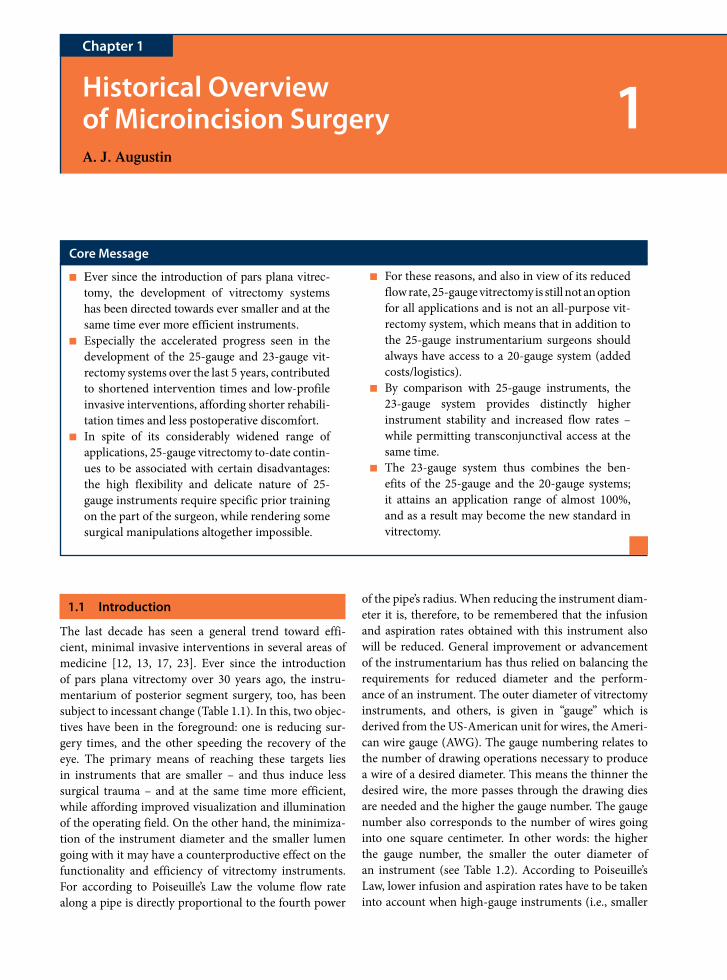
■ Ever since the introduction of pars plana vitrec-tomy, the development of vitrectomy systems has been directed towards ever smaller and at the same time ever more efficient instruments.
■ Especially the accelerated progress seen in the development of the 25-gauge and 23-gauge vit-rectomy systems over the last 5 years, contributed to shortened intervention times and low-profile invasive interventions, affording shorter rehabili-tation times and less postoperative discomfort.
■ In spite of its considerably widened range of applications, 25-gauge vitrectomy to-date contin-ues to be associated with certain disadvantages: the high flexibility and delicate nature of 25-gauge instruments require specific prior training on the part of the surgeon, while rendering some surgical manipulations altogether impossible.
■ For these reasons, and also in view of its reduced flow rate, 25-gauge vitrectomy is still not an option for all applications and is not an all- purpose vit-rectomy system, which means that in addition to the 25-gauge instrumentarium surgeons should always have access to a 20-gauge system (added costs/logistics).
■ By comparison with 25-gauge instruments, the 23-gauge system provides distinctly higher instrument stability and increased flow rates – while permitting transconjunctival access at the same time.
■ The 23-gauge system thus combines the ben-efits of the 25-gauge and the 20-gauge systems; it attains an application range of almost 100%, and as a result may become the new standard in vitrectomy.
1.1 Introduction
The last decade has seen a general trend toward effi-cient, minimal invasive interventions in several areas of medicine [12, 13, 17, 23]. Ever since the introduction of pars plana vitrectomy over 30 years ago, the instru-mentarium of posterior segment surgery, too, has been subject to incessant change (Table 1.1). In this, two objec-tives have been in the foreground: one is reducing sur-gery times, and the other speeding the recovery of the eye. The primary means of reaching these targets lies in instruments that are smaller – and thus induce less surgical trauma – and at the same time more efficient, while affording improved visualization and illumination of the operating field. On the other hand, the minimiza-tion of the instrument diameter and the smaller lumen going with it may have a counterproductive effect on the functionality and efficiency of vitrectomy instruments. For according to Poiseuille’s Law the volume flow rate along a pipe is directly proportional to the fourth power
of the pipe’s radius. When reducing the instrument diam-eter it is, therefore, to be remembered that the infusion and aspiration rates obtained with this instrument also will be reduced. General improvement or advancement of the instrumentarium has thus relied on balancing the requirements for reduced diameter and the perform-ance of an instrument. The outer diameter of vitrectomy instruments, and others, is given in “gauge” which is derived from the US-American unit for wires, the Ameri-can wire gauge (AWG). The gauge numbering relates to the number of drawing operations necessary to produce a wire of a desired diameter. This means the thinner the desired wire, the more passes through the drawing dies are needed and the higher the gauge number. The gauge number also corresponds to the number of wires going into one square centimeter. In other words: the higher the gauge number, the smaller the outer diameter of an instrument (see Table 1.2). According to Poiseuille’s Law, lower infusion and aspiration rates have to be taken into account when high-gauge instruments (i.e., smaller

2 1 Historical Overview of Microincision Surgery
1
instrument diameters) are being used and this, in turn, may affect their functionality and efficiency.
Ever smaller instrument diameters have been designed since the early days of “pars plana vitrectomy” in the 1970s, when Machemer, closely followed by Klöti, relocated the access for vitreous removal to the pars plana area to pre-serve the crystalline lens [16, 24]. While Machemer et al. [25] started out by developing a 17-gauge instrument,
i.e., the vitreous infusion suction cutter (VISC), that still needed a 2.3-mm sclerotomy port, two decisive advance-ments were introduced as early as in 1974: for the first time it was now possible to reduce instrument diameters to a marked degree by separating the infusion system and the cutting system. The 20-gauge vitrectomy system (0.9-mm diameter) was born, with infusion, vitrectome and illumination being introduced into the pars plana via three separate ports. Interestingly, splinting to protect the scleral incisions and facilitate instrument change had been propagated already in those days – this concept has been reintroduced, and today is applied to the microtro-car cannulas of the 25-gauge and 23-gauge systems [16]. While the 20-gauge vitrectomy system still is considered the “gold standard” of pars plana vitrectomy [30], the last 5 years, in particular, have seen fast-paced innovation in the field of the posterior segment instrumentarium toward smaller, more efficient 25-gauge and 23-gauge vitrectomy systems, which nowadays are routinely used in everyday clinical practice. Figure 1.1 compares the sizes of the vitrectomy cannulas of the three different sys-tems (20-, 23-, and 25-gauge). An extensive overview of the clinical studies on safety and efficiency, as well as an assessment of the limits and potential of these innovative vitrectomy systems, has recently been published [1].
Table 1.1. Milestones of pars plana vitrectomy
Early 1970s Machemer and Klöti both have the idea of shifting the site of vitrectomy access to the pars plana area in an effort to preserve the lens. “Birth of pars plana vitrectomy”
1974 O’Malley and Heintz introduce the 20-gauge vitrectomy system, today’s “gold standard”
1974 Klöti recommends the use of guiding cannulas — the precursors of microcannulas — for ease of and added eye protection during instrument change.
1990 de Juan and Hickingbotham present first 25-gauge vitrectomy set, and recommend its use in pediatric interventions in particular
1995 At the ARVO meeting, Singh et al. present a 23-gauge vitrectomy system whose use, however, clearly is restricted to specific office-based interventions
2002 Fujii et al. present first fully integrated 25-gauge system, consisting of microtrocar cannulas, vitrectome and infusion, and demonstrate its safety and efficiency, especially in “simple” vitrectomies
2004/2005 Eckardt introduces first fully integrated 23-gauge vitrectomy system, and demonstrates its safety and efficiency
Up to 2007 Ongoing enhancement of instrumentation and ranges of application for the 25-gauge and 23-gauge systems
Table 1.2. Cannula gauge numbers and corresponding outer diameters
Gauge Outer diameter (mm)
17-G 2.3
19-G 1.1
20-G 0.9
23-G 0.6
25-G 0.5
Fig. 1.1 Comparison of 20-gauge, 23-gauge, and 25-gauge vitrectomy cannulas for size. From Augustin AJ, Offermann J (2007) Möglichkeiten und Grenzen der innovativen Vitrek-tomiessysteme, eine Übersicht. Klin Monatsbl Augenheilkd 224:707–715

Summary for the Clinician
■ The outer diameter of the instruments is given in gauge numbers, in a seemingly counterintui-tive fashion: the higher the gauge number, the smaller the diameter.
■ The 20-gauge vitrectomy system has been con-sidered the “gold standard” since 1974; the last 5 years, in particular, have seen fast-paced innova-tion in the field of the posterior segment instru-mentarium, directed toward smaller, more efficient 25-gauge and 23-gauge vitrectomy systems.
■ However, when reducing the instrument diam-eter, it is to be remembered that flow rates also will be reduced (Poiseuille’s Law), which may affect the functionality and performance of the instruments.
■ It needs to be ensured, therefore, that the per-formance of instruments with reduced diameters at least equals that of the well-established 20-gauge system.
1.2 Pros and Cons of 25-Gauge Vitrectomy Systems
De Juan and Hickingbotham developed a 25-gauge instrument set for pediatric use already in 1990, since the “conventional” 20-gauge vitreous cutters had proven to be big and lacking in precision, especially in chil-dren [7, 42]. This first 25-gauge instrument set, which consisted of just a pneumatic vitrectome, scissors, and a manipulator for membrane removal, at first was used mainly in pediatric surgery, to allow for higher preci-sion and permit controlled operation even in difficult maneuvers in that particular field [6]. De Juan and Hick-ingbotham explicitly stated in their publication that due to its reduced aspiration rate the 25-gauge vitrectome was to be used solely in selected, delicate cases requir-ing particularly precise and careful intervention. It was 12 years later, when eventually a complete 25-gauge vitrectomy system was introduced by Fuji et al. [10] which consisted of microtrocar cannulas, affording ease and safety of instrument introduction and withdrawal, as well as an array of integrated 25-gauge instruments. Due to their small diameter (0.5 mm), 25-gauge can-nulas allow transconjunctival introduction, thus avoid-ing the time-consuming preparation of the conjunctiva that is required in conventional 20-gauge sclerotomies. Using a trocar with forceps, the conjunctiva, in this
procedure, is pulled back a little prior to inserting the cannula, and this displacement provides a slight stagger-ing of the wounds in the sclera and conjunctiva in rela-tion to each other. In 25-gauge vitrectomy, the trocar is introduced perpendicularly to the sclera, i.e., it is directed to the center of the eye. This does not, in fact, create a two-step self-sealing wound. But since the conjunctiva will slip back to its more anterior position, where it is bound to cover the sclerotomy and probably provides a temporary tamponade to the opening – and also in view of the small sclerotomy diameter – no suturing should be required.
As already described, Poiseuille’s Law (see above) indi-cated that the infusion and aspiration rates obtained by the 25-gauge system would be distinctly lower than those of the 20-gauge system. This was confirmed by Fuji et al. [10] in their first evaluation study of the 25-gauge system, where they established markedly reduced infusion and aspiration rates as against the 20-gauge system. To ensure sufficient aspiration rates also for the 25-gauge system, high vacuum settings (500 mmHg) should be used with this system, together with high cutting rates (1,500 cpm), so that optimum tissue fragmentation is guaranteed and “plugging” of the 25-gauge vitrectome is prevented. While the 25-gauge vitrectomy system principally is considered safe and efficient, opinions on the integrity of the suture-less 25-gauge sclerotomies are at variance [11, 15, 18, 19, 35, 36, 43]. Only recently, a modified incision technique was recommended to improve the integrity of sutureless 25-gauge sclerotomies [22, 37]. No significant difference has been identified between 25-gauge vitrectomy and 20-gauge vitrectomy as regards postoperative complications [4, 15, 26–28, 35, 38]. Sutureless sclerotomies have been at the center of constant concern as regards an increased risk of postoperative endophthalmitis following 25-gauge vitrectomy; however, to date but this has never been estab-lished [11, 15, 19, 21, 35, 43]. Interestingly, similar appre-hensions have been expressed in connection with clear cornea incisions in cataract surgery, but despite some case reports [29, 40] again no increased rate of endophthalmitis could be confirmed.
Due to the limited instrumentarium and high flex-ibility of the instruments available in the first years, use of the 25-gauge system initially was restricted to “simple vitrectomies” such as removal of epiretinal membranes or macula surgery, procedures which in the opinion of some surgeons did not involve peripheral work or the removal of major vitreous portions [11, 15, 19]. It is controversially discussed and, in fact, even consid-ered a main limitation of 25-gauge technology whether the deliberately accepted incompleteness of vitreous removal is at all sufficient in these indications, or may
1.2 Pros and Cons of 25-Gauge Vitrectomy Systems 3

4 1 Historical Overview of Microincision Surgery
1
even represent an increased hazard potential. More involved pathologies, demanding extensive removal of the vitreous, initially required the use of 20-gauge vit-rectomy systems. Over recent years, widening the array of instruments (e.g., forceps, picks, and other manipu-lating devices, endolaser), better illumination systems, and specific improvement of the instruments – i.e., by increasing instrument stiffness – has contributed to finally broadening the application range of 25-gauge vitrectomy [31]. The use of silicone oil also was long considered a contraindication of the 25-gauge sys-tem [33], but meanwhile has been made possible by a 25/20-gauge hybrid system for silicone oil infusion [3]. While nowadays silicone oil may be infused through a 25-gauge sclerotomy, this process clearly prolongs surgery times.
Apart from this expanded range of applications, the increasing experience of surgeons as well as commercial aspects certainly contribute to the fact that 25-gauge tech-nology to some extent is now being used in complicated findings such as proliferative vitreoretinopathy, diabetic retinopathy, and retinal detachment, i.e., in pathologies that require the removal of the peripheral vitreous or the complicated removal of membranes [33]. As before, however, 25-gauge vitrectomy is not suitable for all indi-cations, and in these cases the surgeon must be able to resort to the 20-gauge system. This “dual equipment” is, of course, an additional and not trifling cost factor. On a whole, opinions on the applicability of the 25-gauge system vary widely: in a survey the “American Society of Retina Specialists” carried out in 2006, numerous surgeons (26.8%) stated that the 25-gauge system was a viable means in 26–50% of their cases [32]. On the other hand, almost as many surgeons (24%) feel the 25-gauge system can be used in only 11–25% of cases, while a fur-ther 22% of surgeons are of the opinion that they can use it in 51–75% of their cases. However, these data cannot be applied to Germany or Europe without difficulty, since in the US-American system the majority of procedures are carried out on an outpatient basis, and rapid patient recovery may rank higher than other aspects as compared with Germany/Europe.
25-gauge systems presently are available from two manufacturers (Alcon Laboratories, Ft. Worth, TX, USA; Bausch & Lomb, Rochester, NY, USA) and dif-fer essentially in two aspects [31]. While the trocar of the first-generation 25-gauge system by Bausch & Lomb was developed on the principle of a hollow nee-dle, which sometimes makes it difficult to introduce, the trocar of the Alcon 25-gauge system is based on a modified V-shaped stiletto blade, so that only a small amount of force is necessary for insertion [3]. The
Bausch & Lomb vitrectome is operated electrically, while the Alcon vitrectome is pneumatically driven. Its low weight and resulting ease of handling is considered another advantage of the Alcon system [3].
There are two decisive benefits invariably mentioned in connection with the 25-gauge system, viz. faster patient rehabilitation and shorter surgery times. As a subjective assessment, most surgeons register more rapid patient rehabilitation or “less postoperative ocular trauma” fol-lowing the application of a 25-gauge system [11, 15, 19, 35, 43]. It should be remembered, though, that short-ened surgery times are primarily due to the fact that opening and closing the eye globe are considerably less time- consuming when a 25-gauge system is used, while vitreous removal is likely to take a little longer, because of the small lumen of the instrument [10]. In complicated procedures, requiring the extensive removal of vitre-ous, overall intervention time may actually be prolonged when using the 25-gauge system versus the 20-gauge system [11, 19].
In spite of its essentially positive aspects, certain limitations to the 25-gauge system persist to date. For instance, the higher flexibility and delicateness that is associated with 25-gauge instruments, as against the considerably more stable 20-gauge (and 23-gauge) instruments, make specific surgeon training a prereq-uisite [3, 10, 15, 19]. Moreover, high instrument flex-ibility renders certain surgical maneuvers impossible or feasible only subject to limitations [15, 20]. While, for example, the 20-gauge instrument may be used to rotate the eye for a better view of the periphery, this is hardly, if at all, possible with a 25-gauge instrument [20]. This is bound to create problems for the removal of peripheral vitreous. Introduction of the microcannulas also has been known to be difficult in some cases. Some reports describe detachment of a 25-gauge cannula from the trocar, as well as damage and bending of the cannula during introduction, so that inserting and retrieving the delicate 25-gauge instruments presented distinctly more problems and sometimes resulted in warping of the instrument [2, 20].
The disadvantages of the 25-gauge described may not necessarily entail serious complications, but may be the cause of time-consuming procedures. The time saved in opening and closing the eye may subsequently have to be “wasted,” for instance, on more intricate surgical manip-ulations, that may become necessary because of instru-ment flexibility, or on intraoperative problems such as workflow that are complicated by the loss or damage of cannulas and instruments, or because extensive removal of the vitreous with a 25-gauge systems makes consider-ably higher demands on time.

1.3 23-Gauge Vitrectomy Systems: The Future Gold Standard?
The accelerated efforts seen over the last 3 years in the development of a 23-gauge system designed to unite the benefits of the 20-gauge and the 25-gauge system were mainly driven by the limitations described for the 25-gauge system. Singh et al. [39] had, in fact, introduced a first electronic 23-gauge vitrectome as early as 1995, which they later complemented by a 23-gauge infusion system. This, however, was not a complete 23-gauge system provid-ing a wide array of instruments, but just a portable system whose use was meant exclusively for vitreous biopsies and minor office-based interventions – as carried out mainly in the USA [5, 14]. Almost 10 years passed before a fully integrated 23-gauge vitrectomy system for routine clinical use had been designed: in 2005 Eckardt [8] in cooperation with DORC (The Netherlands) eventually introduced a complete 23-gauge instrumentarium and demonstrated its safety and efficiency in a first evaluation study. Pres-ently, 23-gauge systems are available on a larger scale from four manufacturers: Alcon Laboratories (Ft. Worth, TX, USA; Figs. 1.2 and 1.3), DORC (The Netherlands), Oertli
Summary for the Clinician
■ A complete 25-gauge vitrectomy system is first introduced in 2002 (Fujii et al. [10]).
■ Due to small diameters this affords sutureless transconjunctival access and thus shorter surgery times (mainly because opening and closing the globe is less time-consuming), and faster patient rehabilitation.
■ High vacuum settings (500 mmHg) and at the same time high cutting rates (1,500 cpm) are necessary to avoid the “plugging” of a 25-gauge vitrectome.
■ 25-gauge is judged to be principally safe and efficient; complications do not occur more fre-quently than with 20-gauge vitrectomy.
■ Application range initially was limited to “sim-ple vitrectomies”; in the meantime this has been expanded thanks, for instance, to the wider array of instruments.
■ Certain surgical maneuvers are impossible or feasible only subject to limitations; not applica-ble for thorough and extensive vitreous removal.
■ Additional 20-gauge system may be necessary (dual instrumentarium/added cost).
■ Switching to 25-gauge requires a specific training period (due to the higher flexibility and vulner-ability of the instruments).
Fig. 1.2 23-gauge vitrectomy system: flute needle (a), soft-tip cannula (b), vitrectome (c), microtrocar (d), endoillumination probe (e), and endolaser probe (f). From Augustin AJ, Offer-mann J (2007) Möglichkeiten und Grenzen der innovativen Vit-rektomiessysteme, eine Übersicht. Klin Monatsbl Augenheilkd 224:707–715
Fig. 1.3 23-gauge instruments: end-gripping forceps (a), ILM forceps (b), disposable curved scissors and resterilizable hand-piece (c) with end-gripping forceps tip (d), ILM forceps tip (e), and curved scissors tip (f). From Augustin AJ, Offermann J (2007) Möglichkeiten und Grenzen der innovativen Vitrek-tomiessysteme, eine Übersicht. Klin Monatsbl Augenheilkd 224:707–715
1.3 23-Gauge Vitrectomy Systems: The Future Gold Standard? 5

6 1 Historical Overview of Microincision Surgery
1
(Switzerland), and Geuder AG (Heidelberg, Germany). Figures 1.2 and 1.3 show a 23-gauge vitrectomy system as well as a 23-gauge instrument portfolio. 23-gauge instru-ments combine considerably higher stiffness and stability than 25-gauge instruments, with a diameter that is smaller than that of 20-gauge instruments; this permits them to be introduced into the eye through transconjunctival sutureless sclerotomies [8]. Unlike the 25-gauge trocars, 23-gauge trocars are not introduced perpendicular to the scleral surface, but at an angle, and instrumentation is brought to a vertical position in subsequent steps. This type of two-step access is designed to facilitate postopera-tive closure of the sclerotomies by intraocular pressure, ensuring higher integrity of wound closure than with 25-gauge sclerotomies. As early as 2005, Eckardt [8] was able to demonstrate that all 23-gauge sclerotomies were self-sealing and tight. Another very interesting and plau-sible method under anatomical physiological aspects was recently proposed by Rizzo et al., who suggested turning the blade by 30° or a little more. This wound configuration considers the course of the collagen fibers, which ensures even better wound closure [34]. Since 23-gauge instru-ments can be said to be similar to 20-gauge instruments for stiffness and stability, the training period for a surgeon when switching to 23-gauge is much shorter than with 25-gauge instruments, and might more aptly be termed a familiarizing phase. In addition, distinctly higher infusion and aspiration rates could safely be expected with the 23-gauge system than are obtained with the 25-gauge system (Poiseuille’s Law, see above), so that careful and extensive vitreous removal – which should continue to be the stand-ard routine – would pose no problem when using the 23-gauge system. This is corroborated by the fact that, at a vacuum level of 600 mmHg and depending on the cutting rate, the same or even higher flow rates are obtained than with the 20-gauge system (Table 1.3). Thanks to higher flow rates plus increased instrument stability, the 23-gauge system may be employed in simple as well as in complicated
vitrectomies, and thus is suitable for a wider application range than the 25-gauge systems. The application range of 23-gauge vitrectomy is almost identical to that of the 20-gauge system, while surgery times are shortened and interventions are less invasive [41]; it follows, therefore, that it does combine the benefits of the 25-gauge and 20-gauge systems. First experience suggests that solely the use of silicone oil may lead to sclerotomy leakage, so that in these cases sutures are required for wound closure. First experiments have been carried out investigating “dual-diameter” devices (23-gauge for infusion and illumina-tion, 20-gauge for the working channel). While there is, of course, the advantage that surgeons can continue to use available 20-gauge systems, the potential benefits of the 23-gauge system (more rapid rehabilitation, no opening of the conjunctiva) are not turned to maximum profit.
The preliminary results and experience obtained with 23-gauge systems are extremely promising in principle, and with few exceptions 23-gauge vitrectomy may well be able to replace 20-gauge vitrectomy in the near future [9].
Table 1.3. Comparison of different vitrectomes (20-, 23-, and 25-gauge) for cutting rates, inherent stability, as well as flow rates at given aspiration and cutting rates
20-gauge 23-gauge 25-gauge
Max. cutting rate (cpm) 2,500 2,500 1,500Inherent stability (g/4 mm) 130 35 14
Flow rates (ml min–1) at given
vacuum levels and zero cuts
18 (150 mmHg) 23 (600 mmHg) 10 (600 mmHg)
Flow rates (ml min–1) at given
vacuum levels and max. cutting rate
9 (150 mmHg) 9 (600 mmHg) 5 (600 mmHg)
When working with the 23-gauge and 25-gauge systems, high suction levels are required to obtain sufficient flow rates. However, flow
rates with the 25-gauge systems continue to remain distinctly lower (even at high suction levels) than those of the two other systems.
Summary for the Clinician
■ The first complete 23-gauge vitrectomy system was introduced in 2005 and is judged to be safe and efficient (Eckardt).
■ It also affords transconjunctival sutureless access, and thus shortened surgery times and faster patient rehabilitation.
■ At a vacuum level of 600 mmHg, the same or even higher flow rates are obtained than with the 20-gauge system.
■ Unlike 25-gauge vitrectomy, switching to 23-gauge systems merely requires a “familiarizing” phase, since instrument characteristics are similar to those of 20-gauge instruments (stiffness, robustness).

References
1. Augustin AJ, Offermann I (2007) Scope and limitations of innovative vitrectomy systems. Klin Monatsbl Augen-heilkd 224:707–715
2. Byeon SH, Chu YK, Lee SC et al (2006) Problems associated with the transconjunctival sutureless vitrectomy system during and after surgery. Ophthalmologica 220:259–265
3. Charles S (2005) Tips and tricks for 25-gauge sutureless vitrectomy. Rev Ophthalmol. http://www.revophth.com/index.asp?page=1_670.htm
4. De Bustros S, Thomson JT, Michels RG et al (1988) Vitrec-tomy for idiopathic epiretinal membranes causing macular pucker. Br J Ophthalmol 72:692–695
5. De Juan E (2003) Sutureless vitrectomy surgery. Author reply. Ophthalmology 110:2427
6. De Juan E, Hickingbotham D (1990) Refinements in microinstrumentation for vitreous surgery. Am J Ophthal-mol 109:218–220
7. De Juan E, Machemer R (1987) Retinopathy of prematu-rity. Surgical technique. Retina 7:63
8. Eckardt C (2005) Transconjunctival sutureless 23-gauge vitrectomy. Retina 25:208–211
9. Eckardt C (2006) Further experience with 23-gauge transcon-junctival vitrectomy. Retina Today. http://www.retinatoday.com/html Pages/1106/RT1106_CF_Eckhardt.h
10. Fuji GY, de Juan E, Humayun MS et al (2002) A new 25-gauge instrument system for transconjunctival sutureless vitrectomy surgery. Ophthalmology 109:1807–1813
11. Fuji GY, de Juan E, Humayun MS et al (2002) Initial experi-ence using the transconjunctival sutureless vitrectomy system for vitreoretinal surgery. Ophthalmology 109:1814–1820
12. Goldstein DJ, Oz MC (1999) Current status and future directions of minimally invasive cardiac surgery. Curr Opin Cardiol 14:419–425
13. Harell AG, Heniford BT (2005) Minimally invasive abdominal surgery: lux et veritas past present and future. Am J Surg 190:239–243
14. Hilton GF, Josephberg RG, Halperin LS et al (2002) Office-based sutureless transconjunctival Pars Plana Vitrectomy. Retina 22:725–732
15. Ibarra M, Hermel M, Prenner JL, Hassan TS (2005) Longer-term outcomes of transconjunctival sutureless 25-gauge vitrectomy. Am J Ophthalmol 139:831–836
16. Klöti R, Vitrektomie I (1973) Ein neues Instrument für die hintere Vitrektomie. Graefes Arch Clin Exp Ophthalmol 187:161
17. Koh CH, Janik GM (1999) Laparoscopic microsurgery: current and future status. Curr Opin Obstet Gynecol 11:401–407
18. Korobelnik JF, Devin F, Tadayoni R et al (2005) Safety of 25-gauge 3 ports pars plana vitrectomy. ARVO 2005; Invest Ophthalmol Vis Sci 46:5462
19. Lakhanpal RR, Humayun MS, de Juan E et al (2005) Out-comes of 140 consecutive cases of 25-gauge transconjuncti-val surgery for poaterior segment disease. Ophthalmology 112:817–824
20. Lam DSC, Yuen CYF, Tam BSM et al (2003) Sutureless vitrectomy surgery. Ophthalmology 110:2428–2429
21. Lommatzsch A, Spital G, Trieschmann M, Pauleikoff D (2008) Langzeitergebnisse nach pars-plana Vitrektomie in 25-Gauge Technik. [Long-term results after pars plana vitrectomy with 25-gauge technique.] Ophthalmologe 105:445–451 [Epub ahead of print 4 Oct 2007]
22. López-Guajardo L, Pareja-Esteban J, Teus-Guezala MA (2006) Oblique sclerotomy technique for prevention of incompetent wound closure in transconjunctival 25-gauge vitrectomy. Am J Ophthalmol 141(6):1154–1156
23. Lundell L (2000) Anti-reflux surgery in laparoscopic era. Baillieres Best Pract Res Clin Gastroenterol 18:272–277
24. Machemer R, Buettner H, Norton EWD, Parel JM (1971) Vitrectomy: a pars plana approach. Trans Am Acad Oph-thalmol Otolaryngol 75:813
25. Machemer R, Parel JM, Buettner H (1972) A new concept for vitreous surgery. 1. Instrumentation. Am J Ophthalmol 73:1
26. Mango CW, Gupta A, Chen CS et al (2005) 25-gauge trans-conjunctival safety: post-operative complications. ARVO 2005; Invest Ophthalmol Vis Sci 46:E-Abstract 5459
27. Margherio RR, Cox MS Jr, Trese MT et al (1985) Removal of epimacular membranes. Ophthalmology 92:1075–1083
28. McDonald HR, Verre WP, Aaberg TM (1986) Surgical management of idiopathic epiretinal membranes. Ophthal-mology 93:978–983
29. Miller KM, Glasgow BJ (1993) Bacterial endophthalmitis following sutureless cataract surgery. Arch Ophthalmol 111:377–379
30. O’Malley C, Heintz RM (1972) Vitrectomy via the pars plana – a new instrument system. Trans Pac Coast Otooph-thalmol Soc Annu Meet 53:121–137
31. Packo K (2004) Early experience reveals benefits of 25-gauge technology. Rev Ophthalmol. http://www.revophth.com/index.asp?page=1_563.htm
References 7
Summary for the Clinician
■ The system has a very wide application range, therefore no additional vitrectomy system is necessary.
■ The 23-gauge system combines the benefits of the 25-gauge and 20-gauge systems: it provides short-ened surgery times and less invasive interventions, while at the same time its application range is almost identical to that of the 20-gauge system.
■ 23-gauge vitrectomy has the potential to become the future “gold standard.”

8 1 Historical Overview of Microincision Surgery
1
32. PAT Survey (2006) http://www.asrs.org/pat/2006/header.php?s=54
33. Riemann CD, Miller DM, Foster RE, Petersen MR (2007) Outcomes of transconjunctival sutureless 25-gauge vitrec-tomy with silicone oil infusion. Retina 27:296–303
34. Rizzo S, Genovesi-Ebert F (2007) Angled incision techniques for 25-g and 23-g surgery. Euretina, 17–20 May 2007
35. Rizzo S, Genovesi-Ebert F, Murri S et al (2006) 25-gauge, sutureless vitrectomy and standard 20-gauge pars plana vitrectomy in idiopathic epiretinal membrane surgery: a comparative pilot study. Graefes Arch Clin Exp Ophthalmol 244:472–479
36. Rizzo S, Belting C, Cresti F, Genovesi-Ebert F (2007) Suture-less 25-gauge vitrectomy for idiopathic macular hole repair. Graefes Arch Clin Exp Ophthalmol 245(10):1437–1440 [Epub 15 March 2007]
37. Rizzo S, Genovesi-Ebert F, Vento A, Miniaci S, Cresti F, Palla M (2007) Modified incision in 25-gauge vitrectomy in the creation of a tunneled airtight sclerotomy: an ultrabiomicroscopic study. Graefes Arch Clin Exp Oph-thalmol 245(9):1281–1288 [Epub 21 February 2007]
38. Scartozzi R, Bessa AS, Gupta OP, Regillo CD (2007) Intra-operative sclerotomy-related retinal breaks for macula sur-gery, 20- vs. 25-gauge vitrectomy systems. Am J Ophthalmol 143:155–156
39. Singh S, Josephberg RJ, Zaidman GW (1996) Office-based diagnostic pars plana vitrectomy. Invest Ophthalmol Vis Sci 37:402
40. Stonecipher KG, Parmley VC, Jensen H, Rowsey JJ (1991) Infectious endophthalmitis following sutureless cataract surgery. Arch Ophthalmol 109:1562–1563
41. Tewari A, Shah GK (2006) 23-gauge: the further evolu-tion of vitrectomy. Rev Ophthalmol. http://www.revophth.com/index.asp?page=1_984.htm
42. Trese M (1986) Two hand dissection technique during closed vitrectomy for retinopathy of prematurity. Am J Ophthalmol 101:251
43. Yanyali A, Celik E, Horozoglu F et al (2006) 25-gauge transconjunctival sutureless pars plana vitrectomy. Eur J Ophthalmol 16:141–147

3.6 Tool and Visualization Tradeoffs 9
Core Message
25-Gauge Instrumentation: Engineering Challenges and Tradeoffs 2A.C. Barnes, C.M. DeBoer, P.R. Bhadri, O. Magalhaes Jr., R.M. Kerns, M.T. McCormick, L.P. Chong, M.S. Humayun
Chapter 2
■ 25-gauge instrumentation has reduced the surgical incision size. This reduction in size has made vit-reoretinal procedures not only sutureless but, more importantly, made the procedures less invasive and potentially safer.
■ The sutureless 25-gauge pars plana vitrectomy reduces the postoperative inflammation at sclerotomy sites, thus reducing patient discom-fort after surgery and hastening postoperative recovery.
■ The majority of experienced vitreoretinal surgeons have now been exposed at some level to 25-gauge instrumentation, and many use it on a routine basis. However, only a few surgeons have experience with the engineering development challenges and trade-offs associated with small-diameter instrumentation.
■ This chapter will explore some of the key areas of the design and functioning of small-diameter instruments, so that surgeons may better under-stand their performance.
2.1 Introduction
25-gauge instrumentation refers to the body of devices designed specifically to work in conjunction with the 25-gauge Entry Site Alignment system (ESA) or micro-cannula system. The ESA system is the key to 25-gauge instrumentation, and allows the surgeon pars plana access to the vitreous chamber without having to perform con-junctival peritomy (i.e., transconjunctival access), and the ability to remove the system without the need for sutures [1]. The main components used with the ESA system include: a fiber optic light pipe, vitreous cutter, and a range of manipulation and task-specific instruments (Fig. 2.1).
The main advantage of 25-gauge instrumentation — and that which creates engineering challenges — is the dimensional constraint of instruments 0.5 mm in diam-eter. Compared to 20-gauge, 25-gauge instruments have 70% less cross-sectional area to recreate the functionality surgeons expect (Fig. 2.2). This chapter will explore how the size of 25-gauge instrumentation affects mechani-cal properties, such as fluid dynamics and stiffness, and optical properties associated with illumination. As engineering solutions to these challenges continue to be developed, transconjunctival 25-gauge instrumentation will continue to improve as well.
Currently, there are at least four major brands of 25-gauge instrumentation Bausch and Lomb Inc. (St.
Louis, MO, USA), Alcon Laboratories, Inc (Fort Worth, TX, USA), Dutch Ophthalmic, USA (Kingston, NH, USA), Synergetics Inc. (O’Fallon, MO, USA), as well as others) (Fig. 2.3). There are differences between these systems that affect their performance, and some of these will be highlighted. But, as designs evolve and new instrumentation is launched, the comparative landscape is constantly in flux. The goal of this chapter is to provide information on how the design parameters of 25-gauge instruments affect their performance, so that surgeons are better prepared to evaluate and operate current and future instrumentation. For example, 23-gauge instru-mentation systems for use with tunneling scleral incisions are just beginning to be evaluated and compared to 20 and 25-gauge instrumentation [9]. Journal articles, con-ference presentations, and manufacturer information highlighting various comparisons may lead to confusion. Hopefully, this chapter will allow a better understanding of the underlying scientific reasons behind any performance differences.
2.2 Microcannula System
The 25-gauge Entry Site Alignment system (ESA) is the primary component of 25-gauge instrumentation. The ESA system establishes the pars plana transconjunctival