sacculospora felinovii, a novel arbuscular...
TRANSCRIPT
ORIGINAL ARTICLE
Sacculospora felinovii, a novel arbuscular mycorrhizal fungalspecies (Glomeromycota) from dunes on the west coast of India
Andy Willis1,2 & Janusz Błaszkowski3 & Tanvi Prabhu1& Gerard Chwat3 &
Anna Góralska3 & Burla Sashidhar4 & Phil Harris2 & James D’Souza1 &
Jyoti Vaingankar1 & Alok Adholeya4
Received: 12 January 2016 /Revised: 11 June 2016 /Accepted: 14 June 2016 /Published online: 27 June 2016# German Mycological Society and Springer-Verlag Berlin Heidelberg 2016
Abstract During an arbuscular mycorrhiza fungal sporesurvey on a primary coastal sand-dune system in Goaon the west coast of India, entrophosporoid spores tight-ly covered with a dense hyphal mantle were recovered.When intact, the spores, at first sight, seemed to beidentical in morphology to those of Sacculosporabaltica (originally described as Entrophospora baltica)extracted from Polish maritime sand dunes and, to date,the sole member of the recently described genusSacculospora in the new family Sacculosporaceae, phy-lum Glomeromycota. Later detailed morphological stud-ies indicated that both fungi produce two-walled sporesbut the structure and phenotypic features of componentsof the outer spore wall in the novel fungus differ con-siderably from those of S. baltica. Differences betweenthe fungi were subsequently confirmed in the phyloge-netic analysis of SSU–ITS–LSU nrDNA sequences.Consequently, we describe the novel species asSacculospora felinovii sp. nov.
Keywords Entrophosporoid spore .Molecular phylogeny .
Morphology . Tropical coastal dunes
Introduction
Of the ca. 280 described arbuscular mycorrhizal fungal(AMF) species of the phylum Glomeromycota C. Walker &A. Schüßler, only five form spores inside the neck of asporiferous saccule (Błaszkowski 2012). From one side, theneck is continuous with a mycorrhizal extraradical hypha andfrom the other side, the neck passes into a globose to ellipsoi-dal saccule. Contents from both structures are used in sporegenesis. In fully developed specimens, the saccule is usuallyempty and frequently detached from spores and the neck wallis continuous with the outermost spore wall layer, forming thespore surface (Błaszkowski 2012). This mode of spore syn-thesis was first described by Ames and Schneider (1979) in aspecies originally named Glomus infrequens I.R. Hall (Hall1977). Ames and Schneider (1979) subsequently transferredG. infrequens to a newly erected genus Entrophospora R.N.Ames & R.W. Schneid., with E. infrequens (I.R. Hall) R.N.Ames & R.W. Schneid. as type species. The other four speciesaccommodated in Entrophospora were E. baltica Błaszk.,Madej & Tadych, E. colombiana Spain & N.C. Schenck,E. kentinensis C.G. Wu & Y.S. Liu and E. schenckii Sieverd.& S. Toro (Schenck et al. 1984; Sieverding and Toro 1987;Wu et al. 1995; Błaszkowski et al. 1998). Such a mode ofspore formation and the spores themselves have been namedentrophosporoid (Goto and Maia 2006).
From observed differences in the subcellular structure ofspores and the phenotypic and histochemical characters ofcomponents of spore walls of Entrophospora species,Sieverding and Oehl (2006) transferred three of the speciesto two newly erected genera, IntrasporaOehl& Sieverd., with
Section Editor: Marco Thines
* Andy [email protected]
1 Department of Botany, Goa University, Taleigao Plateau, Goa 403206, India
2 Centre for Agroecology, Water and Resilience, Coventry University,Priory St., Coventry CV1 5FB, UK
3 Department of Ecology, Protection and Shaping of Environment,West Pomeranian University of Technology in Szczecin,Słowackiego 17, 71434 Szczecin, Poland
4 The Energy and Resources Institute (TERI), Darbari Seth Block,India Habitat Centre, New Delhi 110 003, India
Mycol Progress (2016) 15:791–798DOI 10.1007/s11557-016-1208-6
I. schenckii (Sieverd. & S. Toro) Oehl & Sieverd. (formerlyE. schenckii) in the family Archaeosporaceae J.B. Morton &D. Redecker, and Kuklospora Oehl & Sieverd., withK. colombiana (Spain & N.C. Schenck) Oehl & Sieverd. (for-merly E. colombiana) and K. kentinensis (C.G. Wu & Y.S.Liu) Oehl & Sieverd. (formerly E. kentinensis) in the familyAcaulosporaceae J.B. Morton & Benny. Kaonongbua et al.(2010) and Schüßler and Walker (2010) rejected Kuklosporaand Intraspora, respectively, later substantiated by the phylo-genetic analyses of Krüger et al. (2012). Oehl et al. (2011)concluded from phylogenetic analyses of nrDNA sequencesthat E. infrequens and E. baltica differ at the family level and,therefore, erected Sacculosporaceae Oehl et al. fam. nov.,with Sacculospora Oehl et al. gen. nov. and S. baltica(Błaszk., Madej & Tadych) Oehl et al. comb. nov.
To date, the genus Sacculospora has been represented byS. baltica alone. Morphologically, S. baltica differs clearlyfrom all other known species forming entrophosporoid spores.The most conspicuous structure is the hyphal mantle tightlycovering spores. The spore subcellular structure, which iscomplex and difficult to define, further distinguishes the spe-cies. According to Błaszkowski et al. (1998), S. baltica sporescontain two spore walls: an outer wall with four layers and aninner three-layered wall. Oehl et al. (2011) stated that layer 1of the inner wall sensu Błaszkowski et al. (1998) clearly sep-arates from the other layers of the wall and, therefore,redefined the subcellular structure of S. baltica spores asthree-walled. However, neither Błaszkowski et al. (1998) norOehl et al. (2011) performed ontogenetic studies of the spe-cies, characterising the subcellular structure of S. balticaspores based only on the spatial grouping of its componentsin crushed specimens. Importantly, the spatial arrangement ofspore subcellular components in AMF may be influenced bythe vigour of spore crushing (Błaszkowski, pers. observ.).Study of the ontogeny of S. baltica spores is severely hinderedfor two reasons. First, the fungus is difficult to grow in culture.In the literature, there is no report that the species has beengrown in single-species cultures and numerous efforts to ob-tain such cultures by J. Błaszkowski failed (Błaszkowski et al.1998; Błaszkowski 2012). The ontogeny of any AMF may beknown with certainty only by the examination of specimensoriginating from single-species cultures. Second, the disclo-sure of sequences of differentiation and organisation of thespore subcellular components may be difficult, or impossible,because of the obscuring influence of the dense and colouredhyphal mantle.
Whilst investigating AMF spore density and diversity onan interrupted belt transect across a primary dune system onthe coast of Goa, India (73°40′33″ E, 14°53′54″ N), AMFspore specimens were found that seemed identical to thoseof S. baltica. Detailed morphological studies of the spore sub-cellular structure, however, and the phenotypic and histo-chemical characters of components of spore walls of the
specimens, indicated that traits in the outer spore wall differed.Unsure if the differences represented intraspecific variabilityof one organism, a comparison was made using molecularmethods. The molecular analyses confirmed the previous con-clusions resulting from morphological observations that thenovel fungal species differs considerably from S. baltica.Consequently, the fungus is described as Sacculosporafelinovii sp. nov.
Materials and methods
Extraction of spores, establishment and growth of trapand single-species cultures
Spores were extracted by the wet-sieving and decanting meth-od (Gerdemann and Nicolson 1963) from field-collected sam-ples of rhizosphere soil of the perennial C4 Zoysia matrella(L.) Merr. (Poaceae) and incorporated into pot trap cultures,12 cm diam., 9 cm high (1018 cm3). The pots were maintainedoutdoors at temperatures of 18–30 °C for 10 months at GoaUniversity. Host plants were Z. matrella, rhizomes transferredfrom the dunes, along with stem cuttings of Solenostemonscutellarioides (L.) R.Br. (Lamiaceae), cultured in unsterilisedZ. matrella rhizosphere soil. Plants were watered 2–3 times aweek. No nutrients were applied during the growing period.Subsequently, spores (ca. 200) extracted from the trap culturewere placed in pots at the base of holes into which stem cut-tings of S. scutellarioides were inserted. The medium wassterilised (180 °C for 12 h × 3) dune sand. The plants wereraised in a ‘closed’ greenhouse (i.e. no ventilation, reducingrisk of outside contamination) at Goa University. Hoaglandssolution minus P was applied fortnightly. After 8 months,single-species spore cultures were achieved and harvested.This two-stage method follows that of Dr. B. F. Rodrigues,Goa University (unpublished), demonstrating successfulsingle-species culture where, all too often, novel species cul-ture fails. Nevertheless, only one of two initial cultures spor-ulated and only one of two subsequent cultures has sporulatedbut in less abundance. The morphological and histochemicalfeatures of mycorrhizal structures revealed following stainingin Trypan blue (Phillips and Hayman 1970) testified that theyrepresent one species of AMF. In addition, in the cultures, noother spore morphotypes were found among the characteristicspores of the new species.
Microscopy and nomenclature
The morphological features of spores and the phenotypic andhistochemical characters of spore wall layers were determinedafter examination of at least 100 spores mounted in water,lactic acid, polyvinyl alcohol/lactic acid/glycerol (PVLG;Omar et al. 1979) and a mixture of PVLG and Melzer’s
792 Mycol Progress (2016) 15:791–798
reagent (1:1, v/v). The preparation of spores and mycorrhizalstructures for study and photography is described byBłaszkowski (2012) and Błaszkowski et al. (2012).Descriptions of spore wall layers follow definitions fromBłaszkowski (2012), Stürmer and Morton (1997) andWalker (1983). Colour names are from Kornerup andWanscher (1983). Nomenclature of fungi and the authors offungal names are from the Index Fungorum websitehttp://www.indexfungorum.org/AuthorsOfFungalNames.htm. Voucher specimens were mounted in PVLG and amixture of PVLG and Melzer’s reagent (1:1, v/v) on slidesand deposited at ETH Zurich, Switzerland (Z + ZT; holotype),the Botany Department, Goa University, the Department ofEcology, Protection and Shaping of Environment (DEPSE),West Pomeranian University of Technology, Szczecin, and inthe herbarium at Oregon State University (OSC) in Corvallis,Oregon, USA (isotypes).
DNA extraction, polymerase chain reaction, cloningand DNA sequencing
Crude DNA was extracted from eight single spores.Procedures prior to polymerase chain reactions (PCRs), theconditions and primers used in the PCRs to obtain SSU–ITS–LSU nrDNA sequences, and cloning and sequencingwere as those described in Błaszkowski et al. (2013). Thesequences were deposited in GenBank (KX345938–KX345943).
Sequence alignment and phylogenetic analyses
Comparisons made between sequences of the novel fungusand those listed after BLAST enquiries indicated that the spe-cies belongs in the Glomeromycota. The closest relatives arefirstly S. baltica, and then a number of Acaulospora spp. andPacispora scintillans (S.L. Rose & Trappe) Sieverd. & Oehl.A set was, therefore, established comprising six sequenceseach of the new species and S. baltica and three to four se-quences each of seven Acaulospora spp., including twoforming entrophosporoid spores, i.e. A. colombiana andA. kentinensis. Pacispora franciscana Sieverd. & Oehl andP. scintillans served as the outgroup taxa. The sequences cov-ered the SSU–ITS–LSU nrDNA segment in all species exceptfor two LSU sequences of A. colombiana, two SSU sequencesof P. franciscana and one SSU sequence of P. scintillans. Thesequences were aligned and analysed phylogenetically by themethod described and justified by Błaszkowski et al. (2015a,b). In Bayesian (BI) phylogenetic analysis, GTR +G and two-parameter Markov (Mk2 Lewis) models were applied for theestablished nucleotide partitions and indel matrices, respec-tively. GTR + G was estimated as the best-fit model byModeltest 3.7 (Posada and Buckley 2004). Maximum likeli-hood (ML) analysis was carried out with the raxmlGUI
(Silvestro and Michalak 2012) implementation of RAxML(Stamatakis 2006), with GTRGAMMA for DNA and the de-fault set for binary (indel) characters. The generated phyloge-netic trees were visualised and edited in MEGA6 (Tamuraet al. 2013).
Results
General data and phylogeny
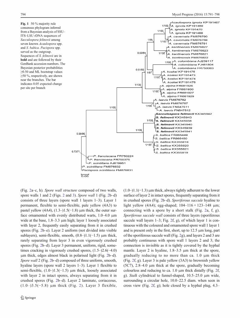
The established set of SSU–ITS–LSU nrDNA sequences, in-cluding those of the putative new species, had a length of 1929characters, 100 % of which were informative. The six PCR-generated new-species sequences showed only 1 % variabili-ty. Bayesian and ML phylogenetic analyses of the sequenceset fully supported the morphological indication that the fun-gus is a glomeromycotean species not previously described,and the closest relative is S. baltica (Fig. 1). The clade with thenew species described below as S. felinovii and the clade withS. baltica, as well as the node linking them, received full (1.0and 100 %) supports. The node linking the Sacculosporaceaeand the Acaulosporaceae clades was also fully supported inboth analyses. The trees generated following the BI and MLanalyses had identical topologies.
Taxonomy
Sacculospora felinovii Willis, Błaszk., T. Prabhu, Chwat,Góralska, Sashidhar, Harris, J. D’Souza, Vaingankar &Adholeya, sp. nov. Figs. 2 and 3
MycoBank No. MB 817258Holotype: ZT Myc 55704 (Z + ZT), isotypes: 3517–3530
(DEPSE), and OSC 154006, OSC 154007 (OSC).The holotype and isotype spore specimens come from
single-species culture with S. scutellarioides as host plantgrown at Goa University, India. The spores were collectedon 14/04/2015 by Ms. Prabhu.
Etymology The specific epithet is respectfully given in hon-our of Prof. Bernard Felinov Rodrigues, Botany Department,Goa University, India, who has dedicated more than 25 yearsto the study of AMF.
Sporocarps unknown. Spores are entrophosporoid arisinginside the neck of a sporiferous saccule, occurring singly insoil, covered with (less often the saccules) a hyphal mantle(Fig. 2a); pale yellow (4A3) to greyish orange (5B5); globoseto subglobose (100–)171(−230) μm diam; rarely ovoid (140–170 × 190–210 μm). Mantle hyaline to brownish yellow(5C8), 15–50 μm thick when seen in cross view, composedof hyaline to pale yellow (4A3) tightly interwoven hyphae;hyphae straight or twisted with numerous branches inclined atdifferent angles, 4.0–8.0 μm wide, walls 1.3–2.3 μm thick
Mycol Progress (2016) 15:791–798 793
(Fig. 2a–c, h). Spore wall structure composed of two walls,spore walls 1 and 2 (Figs. 2 and 3). Spore wall 1 (Fig. 2b–d)consists of three layers (spore wall 1 layers 1–3). Layer 1permanent, flexible to semi-flexible, pale yellow (4A3) topastel yellow (4A4), (1.3–)1.5(−1.8) μm thick, the outer sur-face ornamented with evenly distributed warts, 1.0–4.0 μmwide at the base, 1.8–3.3 μm high; layer 1 loosely associatedwith layer 2, frequently easily separating from it in crushedspores (Fig. 2b–e). Layer 2 uniform (not divided into visiblesublayers), semi-flexible, smooth, (0.8–)1.1(−1.5) μm thick,rarely separating from layer 3 in even vigorously crushedspores (Fig. 2b–d). Layer 3 permanent, uniform, rigid, some-times cracking in vigorously crushed spores, (1.5–)2.6(−4.0)μm thick, edges almost black in polarised light (Fig. 2b–d).Spore wall 2 (Fig. 2b–d) composed of three uniform, smooth,hyaline layers (spore wall 2 layers 1–3). Layer 1 flexible tosemi-flexible, (1.0–)1.3(−1.5) μm thick, loosely associatedwith layer 2 in intact spores, always separating from it incrushed spores (Fig. 2b–d). Layer 2 laminate, coriaceous,(1.0–)3.5(−5.8) μm thick (Fig. 2). Layer 3 flexible,
(1.0–)1.1(−1.3) μm thick, always tightly adherent to the lowersurface of layer 2 in intact spores, frequently separating from itin crushed spores (Fig. 2b–d). Sporiferous saccule hyaline tolight yellow (4A4); egg-shaped; 104–116 × 123–148 μm;connecting with a spore by a short stalk (Fig. 2a, f, g).Sporiferous saccule wall consists of three layers (sporiferoussaccule wall layers 1–3; Fig. 2f, g), of which layer 1 is con-tinuous with the coloured and ornamented spore wall 1 layer 1and is present only in the first, short, up to 12.5 μm long, partof the sporiferous saccule wall (Fig. 2g), and layers 2 and 3 areprobably continuous with spore wall 1 layers 2 and 3; theconnection is invisible as it is tightly covered by the hyphalmantle. Layer 2 is hyaline, 1.8–3.5 μm thick at the spore,gradually reducing to no more than ca. 1.0 μm thick(Fig. 2f, g). Layer 3 is pale yellow (3A3) to brownish yellow(5C7), 2.8–4.0 μm thick at the spore, gradually becomingcolourless and reducing to ca. 1.0 μm thick distally (Fig. 2f,g). Stalk cylindrical to funnel-shaped, 10.5–25.0 μm wide,surrounding a circular hole, 10.0–22.5 diam. when seen incross view (Fig. 2f, g); hole closed by a hyphal plug, 4.3–
Fig. 1 50 % majority ruleconsensus phylogram inferredfrom a Bayesian analysis of SSU–ITS–LSU rDNA sequences ofSacculospora felinovii amongseven known Acaulospora spp.and S. baltica. Pacispora spp.served as the outgroup.Sequences of S. felinovii are inbold and are followed by theirGenBank accession numbers. TheBayesian posterior probabilities≥0.50 and ML bootstrap values≥50 %, respectively, are shownnear the branches. The barindicates 0.05 expected changeper site per branch
794 Mycol Progress (2016) 15:791–798
7.8 μm thick (Fig. 2g). It is likely the plug is formed in thefinal stage of spore development. Connection to myceliumwas not visible. Germination shield not observed to date.Spores and hyphal mantles do not react in Melzer’s reagent(Fig. 2d).
Mycorrhizal associations In single-species cultures withS. scutellarioides as host plant, S. felinovii formed mycorrhizawith arbuscules and intra- and extraradical hyphae (Fig. 3a, b).No vesicles were found. All the structures stained very faintlyin 0.1 % Trypan blue and, hence, were difficult to visualise butthere probably was a patchy distribution along the examinedroot fragments.
Distribution and habitat In the field, S. felinovii waslikely associated with roots of Z. matrella. No molecu-lar analysis on DNA extracted from roots was
performed to confirm this supposition however. Sporeswere most abundant (ca. 35 ± 7 100 g−1 soil) in theregions of the transect where Z. matrella was the dom-inant species, 101–138 m from the strand. BLASTsearches revealed no sequence with similarity ≥97 % toSSU–ITS–LSU sequences of S. felinovii.
Discussion
Sacculospora baltica has, so far, been the only member of therecently newly erected monospecific genus Sacculosporaand the family Sacculosporaceae (Oehl et al. 2011).Intact S. felinovii entrophosporoid spores, always tightlysurrounded by a dense hyphal mantle (Fig. 2a–c, h), areindistinguishable from those of S. baltica (Błaszkowskiet al. 1998; Błaszkowski 2012). However, the unique
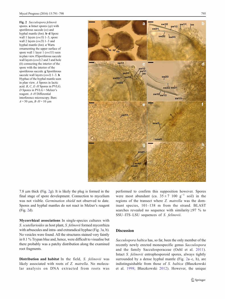
Fig. 2 Sacculospora felinoviispores. a Intact spores (sp) withsporiferous saccule (ss) andhyphal mantle (hm). b–d Sporewall 1 layers (sw1l) 1–3, sporewall 2 layers (sw2l) 1–3 andhyphal mantle (hm). e Wartsornamenting the upper surface ofspore wall 1 layer 1 (sw1l1) seenin plan view. f Sporiferous sacculewall layers (sswl) 2 and 3 and hole(h) connecting the interior of thespore with the interior of thesporiferous saccule. g Sporiferoussaccule wall layers (sswl) 1–3. hHyphae of the hyphal mantle seenin plan view. A Spores in lacticacid. B, C, E–H Spores in PVLG.D Spores in PVLG+Melzer’sreagent. A–H Differentialinterference microscopy. Bars:A = 50 μm, B–H = 10 μm
Mycol Progress (2016) 15:791–798 795
feature of S. felinovii is its spore wall 1 layer 3 that hasbirefringent properties in polarised light, where theedges of the wall, despite being colourless, turn almostblack (Fig. 2b–d). Further, it is comparatively thick,fragile and is covered with a thin layer (spore wall 1layer 2). This layer rarely separates from the upper sur-face of spore wall 1 layer 3 in even vigorously crushedspores (Fig. 2b–d).
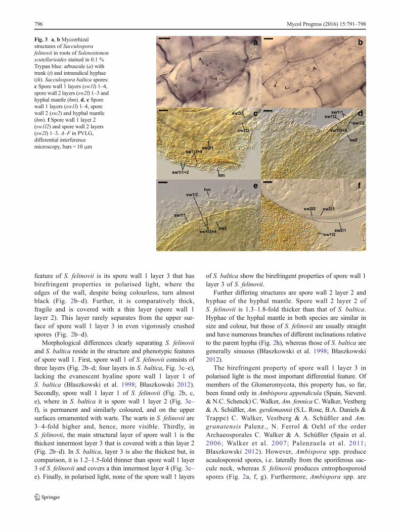
Morphological differences clearly separating S. felinoviiand S. baltica reside in the structure and phenotypic featuresof spore wall 1. First, spore wall 1 of S. felinovii consists ofthree layers (Fig. 2b–d; four layers in S. baltica, Fig. 3c–e),lacking the evanescent hyaline spore wall 1 layer 1 ofS. baltica (Błaszkowski et al. 1998; Błaszkowski 2012).Secondly, spore wall 1 layer 1 of S. felinovii (Fig. 2b, c,e), where in S. baltica it is spore wall 1 layer 2 (Fig. 3c–f), is permanent and similarly coloured, and on the uppersurfaces ornamented with warts. The warts in S. felinovii are3–4-fold higher and, hence, more visible. Thirdly, inS. felinovii, the main structural layer of spore wall 1 is thethickest innermost layer 3 that is covered with a thin layer 2(Fig. 2b–d). In S. baltica, layer 3 is also the thickest but, incomparison, it is 1.2–1.5-fold thinner than spore wall 1 layer3 of S. felinovii and covers a thin innermost layer 4 (Fig. 3c–e). Finally, in polarised light, none of the spore wall 1 layers
of S. baltica show the birefringent properties of spore wall 1layer 3 of S. felinovii.
Further differing structures are spore wall 2 layer 2 andhyphae of the hyphal mantle. Spore wall 2 layer 2 ofS. felinovii is 1.3–1.8-fold thicker than that of S. baltica.Hyphae of the hyphal mantle in both species are similar insize and colour, but those of S. felinovii are usually straightand have numerous branches of different inclinations relativeto the parent hypha (Fig. 2h), whereas those of S. baltica aregenerally sinuous (Błaszkowski et al. 1998; Błaszkowski2012).
The birefringent property of spore wall 1 layer 3 inpolarised light is the most important differential feature. Ofmembers of the Glomeromycota, this property has, so far,been found only in Ambispora appendicula (Spain, Sieverd.& N.C. Schenck) C. Walker, Am. fennica C. Walker, Vestberg& A. Schüßler, Am. gerdemannii (S.L. Rose, B.A. Daniels &Trappe) C. Walker, Vestberg & A. Schüßler and Am.granatensis Palenz., N. Ferrol & Oehl of the orderArchaeosporales C. Walker & A. Schüßler (Spain et al.2006; Walker et al. 2007; Palenzuela et al. 2011;Błaszkowski 2012). However, Ambispora spp. produceacaulosporoid spores, i.e. laterally from the sporiferous sac-cule neck, whereas S. felinovii produces entrophosporoidspores (Fig. 2a, f, g). Furthermore, Ambispora spp. are
Fig. 3 a, b Mycorrhizalstructures of Sacculosporafelinovii in roots of Solenostemonscutellarioides stained in 0.1 %Trypan blue: arbuscule (a) withtrunk (t) and intraradical hyphae(ih). Sacculospora baltica spores:c Spore wall 1 layers (sw1l) 1–4,spore wall 2 layers (sw2l) 1–3 andhyphal mantle (hm). d, e Sporewall 1 layers (sw1l) 1–4, sporewall 2 (sw2) and hyphal mantle(hm). f Spore wall 1 layer 2(sw1l2) and spore wall 2 layers(sw2l) 1–3. A–F in PVLG,differential interferencemicroscopy, bars = 10 μm
796 Mycol Progress (2016) 15:791–798
genetically very distant from the Diversisporales C. Walker &A. Schüßler, to which S. felinovii belongs.
Finally, the uniqueness of S. felinovii is shown in the phy-logenetic analyses of SSU–ITS–LSU nrDNA sequences(Fig. 1). The SSU–ITS–LSU nrDNA sequences of S. felinoviidiffered by an average of 10.1 % from those of S. baltica.
The mycorrhizal structures of S. felinovii grown in single-species culture stained exceptionally faintly (Fig. 3a, b) andmany may not have stained at all in 0.1 % Trypan blue.Unfortunately, comparison cannot be made with S. balticawhere, as mentioned above, pure culture has not been attained.Therefore, currently, it is possible to only conjecture that thefaint or no staining is a family-level synapomorphy, as is likelyin members of Ambisporaceae, Archaeosporaceae andParaglomeraceae (Morton and Redecker 2001; Spain et al.2006; http://invam.wvu.edu/).
The literature and our observations, and the lack in publicdatabases of uncultured AMF sequences with a similarityof ≤97 % to those of S. baltica and S. felinovii, suggest thatthese species occur rarely in the world. However, it is likelythey are widely distributed on the Earth and are adapted todifferent soil and climatic conditions, despite S. felinovii be-ing, so far, identified only in one dune site of the west coast ofIndia. In Poland, S. baltica was found in dunes of the BalticSea but also in inland dunes of the Błędowska Desert and inthe Tatra Mountains, ca. 600 and 700 km away from the sea,respectively (Tadych and Błaszkowski 2000; Błaszkowskiet al. 2002; Zubek et al. 2008; Błaszkowski 2012). In addition,S. baltica was recorded at several Alpine elevations inSwitzerland and in forest ecosystems near Valdina in southernChile (Sieverding and Oehl 2006; Oehl et al. 2011). The rarerecords of both fungi may also result from there being fewmycologists dealing with the morphology of AMF and weak-nesses of molecular methods applied in recognition ofintraradical AMF (Oehl et al. 2010; Wetzel et al. 2014).
Acknowledgments The study was supported, in part, by the PolishNational Centre of Science, grant nos. 2012/05/B/NZ8/00498 and 2012/07/N/NZ8/02363.Ms. J. Vaingankar was supported by the Department ofScience and Technology, New Delhi, India.
References
Ames RN, Schneider RW (1979) Entrophospora, a new genus in theEndogonaceae. Mycotaxon 8:347–352
Błaszkowski J (2012) Glomeromycota. W. Szafer Institute of Botany,Polish Academy of Sciences, Kraków
Błaszkowski J, Madej T, Tadych M (1998) Entrophospora baltica sp.nov. and Glomus fuegianum, two species in the Glomales fromPoland. Mycotaxon 68:165–184
Błaszkowski J, Tadych M, Madej T (2002) Arbuscular mycorrhizal fungi(Glomales, Zygomycota) of the Błędowska Desert, Poland. ActaSoc Bot Pol 71:71–85. doi:10.5586/asbp.2002.008
Błaszkowski J, Kovács GM, Gáspár BK, Balázs TK, Buscot F, Ryszka P(2012) The arbuscular mycorrhizal Paraglomus majewskii sp. nov.represents a new distinct basal lineage in Paraglomeraceae(Glomeromycota). Mycologia 104:148–156. doi:10.3852/10-430
Błaszkowski J, Chwat G, Kovács GM, Gáspár BK, Ryszka P, OrłowskaE, Pagano MC, Araújo FS, Wubet T, Buscot F (2013) Septoglomusfuscum and S. furcatum, two new species of arbuscular mycorrhizalfungi (Glomeromycota). Mycologia 105:670–680. doi:10.3852/12-127
Błaszkowski J, Chwat G, Góralska A (2015a) Acaulospora ignota andClaroideoglomus hanlinii, two new species of arbuscular mycorrhi-zal fungi (Glomeromycota) from Brazil and Cuba. Mycol Prog 14:18. doi:10.1007/s11557-015-1042-2
Błaszkowski J, Chwat G, Góralska A, Ryszka P, Kovács GM (2015b)Two new genera, Dominikia and Kamienskia, and D. disticha sp.nov. in Glomeromycota. Nova Hedwigia 100:225–238. doi:10.1127/nova_hedwigia/2014/0216
Gerdemann JW, Nicolson TH (1963) Spores of mycorrhizal Endogonespecies extracted from soil by wet sieving and decanting. Trans BritMycol Soc 46:235–244
Goto BT, Maia LC (2006) Glomerospores: a new denomination for thespores of Glomeromycota, a group molecularly distinct from theZygomycota. Mycotaxon 96:29–132
Hall IR (1977) Species and mycorrhizal infections of New ZealandEndogonaceae. Trans Br Mycol Soc 68:341–356. doi:10.1016/S0007-1536(77)80186-1
Kaonongbua W, Morton JB, Bever JD (2010) Taxonomic revision trans-ferring species inKuklospora to Acaulospora (Glomeromycota) anda description of Acaulospora colliculosa sp. nov. from field collect-ed spores. Mycologia 102(6):1497–1509. doi:10.3852/10-011
Kornerup A, Wanscher JH (1983) Methuen handbook of colour, 3rd edn.Eyre Methuen, London
Krüger M, Krüger C, Walker C, Stockinger H, Schüßler A (2012)Phylogenetic reference data for systematics and phylotaxonomy ofarbuscular mycorrhizal fungi from phylum to species level. NewPhytol 193:970–984. doi:10.1111/j.1469-8137.2011.03962.x
Morton JB, Redecker D (2001) Two new families of Glomales,Archaeosporaceae and Paraglomaceae, with two new generaArchaeospora and Paraglomus, based on concordant molecularand morphological characters. Mycologia 93:181–195.doi:10.2307/3761615
Oehl F, Laczko E, Bogenrieder A, Stahr K, Bösch R, van der Heijden M,Sieverding E (2010) Soil type and land use intensity determine thecomposition of arbuscular mycorrhizal fungal communities. SoilBiol Biochem 42:724–738. doi:10.1016/j.soilbio.2010.01.006
Oehl F, da Silva GA, Sánchez-Castro I, Goto BT, Maia LC, Vieira HEE,Barea J-M, Sieverding E, Palenzuela J (2011) Revision ofGlomeromycetes with entrophosporoid and glomoid spore forma-tion with three new genera. Mycotaxon 117:297–316. doi:10.5248/117.297
Omar MB, Bolland L, Heather WA (1979) A permanent mounting me-dium for fungi. Bull Br Mycol Soc 13:31–32. doi:10.1016/S0007-1528(79)80038-3
Palenzuela J, Barea JM, Ferrol N, Oehl F (2011) Ambispora granatensis,a new arbuscular mycorrhizal fungus, associated with Asparagusofficinalis in Andalucía (Spain). Mycologia 103:333–340.doi:10.3852/09-146
Phillips JM, Hayman DS (1970) Improved procedures for clearing rootsand staining parasitic and vesicular-arbuscular mycorrhizal fungi forrapid assessment of infection. Trans Brit Mycol Soc 55:158–161
Posada D, Buckley TR (2004) Model selection and model averaging inphylogenetics: advantages of Akaike information criterion andBayesian approaches over likelihood ratio tests. Syst Biol 53:793–808. doi:10.1080/10635150490522304
Schenck NC, Spain JL, Sieverding E, Howeler RH (1984) Several newand unreported vesicular-arbuscular mycorrhizal fungi
Mycol Progress (2016) 15:791–798 797
(Endogonaceae) from Colombia. Mycologia 76:685–699.doi:10.2307/3793226
Schüßler A, Walker C (2010) The Glomeromycota: a species list withnew families and new genera. www.arbuscular-mycorrhiza.net/Schuessler&Walker2010_Glomeromycota.pdf
Sieverding E, Oehl F (2006) Revision of Entrophospora and descriptionof Kuklospora and Intraspora, two new genera in the arbuscularmycorrhizal Glomeromycetes. J Appl Bot Food Qual 80:69–81
Sieverding E, Toro S (1987) Entrophospora schenckii: a new species inthe Endogonaceae from Colombia. Mycotaxon 28:209–214
Silvestro D, Michalak I (2012) raxmlGUI: a graphical front-end forRAxML. Org Divers Evol 12:335–337. doi:10.1007/s13127-011-0056-0
Spain JL, Sieverding E, Oehl F (2006) Appendicispora: a new genus inthe arbuscular mycorrhiza-forming Glomeromycetes, with a discus-sion of the genus Archaeospora. Mycotaxon 97:163–182
Stamatakis A (2006) RAxML-VI-HPC: maximum likelihood-based phy-logenetic analyses with thousands of taxa and mixed models.Bioinformatics 22:2688–2690. doi:10.1093/bioinformatics/btl446
Stürmer SL, Morton JB (1997) Developmental patterns defining morpho-logical characters in spores of four species in Glomus. Mycologia89:72–81. doi:10.2307/3761174
Tadych M, Błaszkowski J (2000) Arbuscular fungi and mycorrhizae(Glomales) of the Słowiński National Park, Poland. Mycotaxon74:463–483
Tamura K, Stecher G, Peterson D, Filipski A, Kumar S (2013) MEGA6:Molecular Evolutionary Genetics Analysis Version 6.0. Mol BiolEvol 30:2725–2729. doi:10.1093/molbev/mst197
Walker C (1983) Taxonomic concepts in the Endogonaceae; sporewall characteristics in species descriptions. Mycotaxon 18:443–455
Walker C, Vestberg M, Demircik F, Stockinger H, Saito M, Sawaki H,Nishmura I, Schüßler A (2007) Molecular phylogeny and new taxain the Archaeosporales (Glomeromycota): Ambispora fennica gen.sp. nov., Ambisporaceae fam. nov., and emendation ofArchaeospora and Archaeosporaceae. Mycol Res 111:137–153.doi:10.1016/j.mycres.2006.11.008
Wetzel K, Silva G, Matczinski U, Oehl F, Fester T (2014) Superior dif-ferentiation of arbuscular mycorrhizal fungal communities from tilland no-till plots by morphological spore identification when com-pared to T-RFLP. Soil Biol Biochem 72:88–96. doi:10.1016/j.soilbio.2014.01.033
Wu C-H, Liu Y-S, Huang Y-L, Wang Y-P, Chao C-C (1995)Glomales of Taiwan: V. Glomus chimonobambusae andEntrophospora kentinensis, spp. novum. Mycotaxon 53:283–294
Zubek S, Turnau K, Błaszkowski J (2008) Arbuscular mycorrhiza ofendemic and endangered plants from the Tatra Mts. Acta Soc BotPol 77:149–156. doi:10.5586/asbp.2008.019
798 Mycol Progress (2016) 15:791–798