safety, pharmacokinetics, and pharmacodynamics of amg 102, a
TRANSCRIPT
Published OnlineFirst January 12, 2010; DOI: 10.1158/1078-0432.CCR-09-1365
Cancer Therapy: Clinical Clinical
CancerResearch
Safety, Pharmacokinetics, and Pharmacodynamics of AMG102, a Fully Human Hepatocyte Growth Factor–NeutralizingMonoclonal Antibody, in a First-in-Human Studyof Patients with Advanced Solid TumorsMichael S. Gordon1, Christopher J. Sweeney2, David S. Mendelson1, S. Gail Eckhardt3, Abraham Anderson4,Darrin M. Beaupre4, Daniel Branstetter4, Teresa L. Burgess4, Angela Coxon4, Hongjie Deng4,Paula Kaplan-Lefko4, Ian M. Leitch4, Kelly S. Oliner4, Lucy Yan4, Min Zhu4, and Lia Gore3
Abstract
Authors' AArizona; 2In3UniversityInc., Thous
Note: SuppResearch O
Corresponna, 9055 Ea860-5000; F
doi: 10.115
©2010 Am
www.aacr
Do
Purpose: The aims were to assess the safety, pharmacokinetics, maximum tolerated dose, and antitu-mor activity of AMG 102, a fully human hepatocyte growth factor/scatter factor (HGF/SF)–neutralizingmonoclonal antibody, in patients with solid tumors.Experimental Design: Patients (N = 40) with refractory advanced solid tumors were enrolled into six
sequential dose-escalation cohorts (0.5, 1, 3, 5, 10, or 20 mg/kg AMG 102 i.v. every 2 weeks) and a dose-expansion cohort (20 mg/kg AMG 102 every 2 weeks). Safety, anti–AMG 102 antibody formation, phar-macokinetics, tumor response, and exploratory biomarkers were assessed.Results: AMG 102 was well tolerated up to the planned maximum dose of 20 mg/kg, and the maxi-
mum tolerated dose was not reached. Treatment-related adverse events were generally mild and includedfatigue (13%), constipation (8%), nausea (8%), vomiting (5%), anorexia (5%), myalgia (5%), and hy-pertension (5%). Two patients experienced dose-limiting toxicities: one patient (0.5 mg/kg cohort) expe-rienced grade 3 hypoxia and grade 3 dyspnea and one patient (1 mg/kg cohort) experienced grade 3 uppergastrointestinal hemorrhage. No anti–AMG 102 antibodies were detected, and AMG 102 had linear phar-macokinetics within the dose range investigated. Sixteen of 23 (70%) evaluable patients had a best re-sponse of stable disease with progression-free survival ranging from 7.9 to 40 weeks. Circulating levelsof the biomarker HGF/SF (bound and unbound) increased in a dose-dependent manner, whereas solublec-Met concentrations were generally similar across doses.Conclusions: AMG 102 is safe and well tolerated, has a favorable pharmacokinetic profile, and will
be further investigated as a monotherapy and in combination with other agents. Clin Cancer Res; 16(2);
699–710. ©2010 AACR.
Hepatocyte growth factor/scatter factor (HGF/SF) andits receptor c-Met regulate multiple cellular functions, in-cluding proliferation, survival, motility, and morphogene-sis, particularly during wound healing and embryogenesis(1–4). Aberrant tumor expression of HGF/SF or c-Met hasbeen linked to poor prognosis in multiple malignancies,including breast cancer, osteosarcoma, colorectal carcino-ma, and glioma (5–9). HGF/SF-dependent autocrine and
ffiliations: 1Premiere Oncology of Arizona, Scottsdale,diana University School of Medicine, Indianapolis, Indiana;of Colorado Cancer Center, Aurora, Colorado; and 4Amgenand Oaks, California
lementary data for this article are available at Clinical Cancernline (http://clincancerres.aacrjournals.org/).
ding Author: Michael S. Gordon, Premiere Oncology of Arizo-st Del Camino, Suite 100, Scottsdale, AZ 85258. Phone: 480-ax: 480-882-0130; E-mail: [email protected].
8/1078-0432.CCR-09-1365
erican Association for Cancer Research.
journals.org
Researcon April clincancerres.aacrjournals.org wnloaded from
paracrine growth loops (6), overexpression of c-Met (7), ac-tivating mutations in c-Met (10–13), and amplifications ofthe c-Met gene resulting in the overexpression of c-Met(14–16) have also beenobserved in several tumor types. Tak-en together, these observations have made the HGF/SF–c-Met axis an important target for therapeutic intervention(17). In preclinical studies, HGF/SF antagonists inhibitedtumor xenograft growth as well as in vitro tumor cell sur-vival, migration, and invasion (18, 19). Several approachestargeting c-Met (small-molecule and peptide inhibitors,RNA-directed inhibitors, andmonoclonal antibodies) havealso shown antitumor activity in preclinical studies (20).AMG 102 is a fully human HGF/SF-neutralizing mono-
clonal antibody (IgG2) that specifically targets HGF/SF. Incynomolgus monkeys, AMG 102 exhibited linear pharma-cokinetics following a single dose, and there were no treat-ment-related cardiovascular, respiratory, or central nervoussystem toxicities (21). In addition, AMG 102 was well tol-erated up to a dose of 150 mg/kg once weekly for up to 9months in this species (22). The antitumor activity and
699
h. 15, 2019. © 2010 American Association for Cancer
Translational Relevance
In recent years, there has been increased interest inthe development of biological agents that target hu-man growth factors or their associated signaling com-ponents. The hepatocyte growth factor/scatter factor(HGF/SF)–c-Met axis has been implicated in thegrowth, survival, and migration of some human can-cers. Clinical trials investigating monoclonal antibo-dies and small-molecule inhibitors directed at theHGF/SF–c-Met axis are currently under way. The re-cently characterized fully human IgG2 monoclonal an-tibody AMG 102 is unique among these because itspecifically targets only HGF/SF. In this first-in-humanstudy, AMG 102 was generally well tolerated at thedoses tested and resulted in stable disease and tumorreductions in some patients. The findings presented inthis study, therefore, warrant further investigation ofAMG 102 for the treatment of solid tumors.
Gordon et al.
700
Published OnlineFirst January 12, 2010; DOI: 10.1158/1078-0432.CCR-09-1365
mechanism of action of AMG 102 have been evaluated inpreclinical studies. AMG 102 inhibited tumor growth, in-duced tumor regressions, increased apoptosis, and decreasedcell proliferation in human xenograft models of cancer (18).The objectives of this phase I, first-in-human, open-labelstudy were to assess the safety, tolerability, pharmacokin-etics, and maximum tolerated dose (MTD) of AMG 102in adult patients with refractory advanced solid tumors. Cir-culating HGF/SF and soluble c-Met were also evaluated aspotential biomarkers of response both in clinical samplesand in a preclinical tumor xenograft model.
Materials and Methods
Eligibility criteriaMen and women ages ≥18 y were included in the study if
they had a pathologically documented and definitively diag-nosed advanced solid tumor refractory to standard treatmentor for which no curative therapy was available. Patients hadan Eastern Cooperative Oncology Group performance statusof ≤2 or, for patients with a primary brain tumor, EasternCooperative Oncology Group performance status of 0 or 1.Exclusion criteria included hematologic malignancies;
unresolved toxicities from prior anticancer therapy; un-treated or symptomatic brain metastases; myocardial in-farction within the previous 6 mo; any conditionaffecting cardiac function (e.g., unstable angina or conges-tive heart failure) of New York Heart Association class >II;uncontrolled hypertension (diastolic blood pressure >90mm Hg; systolic blood pressure >160 mm Hg); cardiac ar-rhythmia; recent venous thrombosis (including deep veinthrombosis or pulmonary embolism within 1 y of study);history of upper gastrointestinal hemorrhage, peptic ulcerdisease, or bleeding diathesis; iron deficiency anemia; anti-tumor treatment within 3 wk of study day 1 (or antibody
Clin Cancer Res; 16(2) January 15, 2010
Researcon April clincancerres.aacrjournals.org Downloaded from
therapy within 8 wk of study day 1); high-dose chemo-therapy requiring hematopoietic progenitor cell supportwithin 24 mo of study day 1; requirement for daily non-steroidal anti-inflammatory drugs or corticosteroids (<325mg aspirin was acceptable); absolute neutrophil count<1.5 × 10⁹/L (without granulocyte colony-stimulating fac-tor support within 2 wk of study day 1); platelet count<100 × 109/L (without transfusion within 2 wk of studyday 1); hemoglobin <10 g/dL (without transfusion within4 wk of study day 1); prothrombin time or partial throm-boplastin time >1.5× the upper limits of normal (ULN);serum creatinine >1.5× ULN (unless 24-h creatinine clear-ance ≥60 mL/min); aspartate aminotransferase or alanineaminotransferase >2.5× ULN; aspartate aminotransferaseor alanine aminotransferase >5× ULN in the presence ofliver metastasis; total bilirubin >1.5× ULN; proteinuria>1+ on urine dipstick or >30 mg/dL; known positive testfor HIV, hepatitis C virus antibody, or hepatitis B virus sur-face antigen; positive test for hepatitis B virus core anti-body in the presence of a negative test for hepatitis Bvirus surface antibody; or documented cytomegalovirusor EBV infection within 6 mo of study day 1.Institutional Review Board approval was obtained for
all study procedures, and all study procedures were donein accordance with the Declaration of Helsinki. Each pa-tient provided written informed consent before enroll-ment in the study.
Study designThis was a first-in-human open-label study with a dose-
escalation and a dose-expansion component. The princi-pal aims were to determine the incidence of adverseevents, clinically significant changes in vital signs and clin-ical laboratory tests, the presence of anti–AMG 102 anti-bodies, pharmacokinetics, MTD, and tumor responseand to conduct an assessment of exploratory biomarkers.AMG 102 was administered as an i.v. infusion over ap-
proximately 30 to 60 min. In the dose-escalation phase,patients were enrolled sequentially into six dose cohortsreceiving a single dose of AMG 102 (0.5, 1, 3, 5, 10, or20 mg/kg; four patients per cohort). The maximumplanned dose was 20 mg/kg. Following the first dose, pa-tients entered a 4-wk treatment-free period to evaluatesafety and pharmacokinetics. If no dose-limiting toxicity(DLT) was observed during the 4-wk period, AMG 102 ad-ministration was resumed at the same dose level every2 wk and continued until patients experienced a DLT, anunacceptable adverse event, disease progression, or volun-tary withdrawal. DLTs were defined as any treatment-relat-ed grade 3 or 4 hematologic or nonhematologic toxicityaccording to Common Terminology Criteria for AdverseEvents, version 3.0. If no DLT was observed among the ini-tial four patients during the first 4 wk of treatment, pa-tients could be enrolled at the next dose level. If apatient experienced a DLT, up to two additional patientswere added to that cohort. The MTD was defined as thehighest dose level with DLTs in <33% of the patients en-rolled in the cohort. After the completion of the dose
Clinical Cancer Research
h. 15, 2019. © 2010 American Association for Cancer
Safety of AMG 102 in Patients with Advanced Solid Tumors
Published OnlineFirst January 12, 2010; DOI: 10.1158/1078-0432.CCR-09-1365
escalation, additional patients were to be enrolled into adose expansion at the MTD, or the maximum planneddose (20 mg/kg every 2 wk) if the MTD was not reached,to further explore safety, tolerability, and pharmacokinet-ics and to support biomarker development. Nine patientswere enrolled in the expansion cohort.
Evaluation of safety and formation of anti–AMG 102antibodiesAdverse events (graded according to the Common Termi-
nology Criteria for Adverse Events) were recorded for all pa-tients who received one or more dose of AMG 102. Duringdose escalation and dose expansion, serum for anti–AMG102 antibodies was collected before dose, before day 29(second dose), before day 57 (fourth dose; dose escalationonly), at each scheduled tumor assessment (every 8wkwith-in 3 d before dosing), and at 4 and 8 wk following the lastdose of AMG 102. Anti–AMG 102-binding antibodies wereassayed using an electrochemiluminescence-based immu-noassay (see Supplementary Materials and Methods).
PharmacokineticsDuring the dose-escalation phase, serum samples were
collected immediately before the first dose; at 30 min (dur-ing infusion) and 60min (immediately after infusion); at 2,8, 24, 48, and 96 h after dosing; at days 8, 15, and 29 (beforethe second dose); and at day 43 (before the third dose). Be-ginning on day 57, serum samples were collected beforedosing (before the fourth dose) and at 0.5, 1, 2, 8, 24, 48,and 96 h after dosing. Serum was also collected before dos-ing at every dosing visit thereafter and at 4 and 8 wk follow-ing the last dose of AMG 102. During the dose expansion,serumwas collected at baseline, at 1 h (immediately follow-ing infusion), at 2 and 24 h, at the time of each scheduledtumor assessment (every 8 wk within 3 d before dosing),and at 4 and 8 wk following the last dose of AMG 102.Pharmacokinetic and exposure parameters of AMG 102
were estimated, including maximum observed serum con-centration (Cmax), time to Cmax (tmax), area under the con-centration versus time curve in a dosing interval τ (AUCτ;τ = 2 wk), systemic clearance, terminal elimination half-life (t1/2), and accumulation ratio (AR). Noncompartmen-tal analysis was used for pharmacokinetic assessmentusing WinNonlin Professional software version 5.1 (Phar-sight Corp.). In the pharmacokinetic analysis, meanand SD of pharmacokinetic parameters were calculatedfor each dose level and for the first and fourth doses.The t1/2 was estimated using the data collected in the sin-gle-dose study phase (sampled up to 4 wk). The AR wascalculated based on the ratio of the AUCτ after the firstand fourth doses. Dose linearity was assessed using one-way ANOVA.
Serum AMG 102 measurementSerum concentrations of AMG 102 were determined by
an AMG 102–specific ELISA (see Supplementary Materialsand Methods) using a recombinant human HGF/SF (cap-ture reagent; Amgen Inc.) and a biotinylated polyclonal
www.aacrjournals.org
Researcon April clincancerres.aacrjournals.org Downloaded from
rabbit anti–AMG 102 antibody (Amgen Inc.) as previouslydescribed (21).
Preclinical biomarker measurementMethods used tomeasure total circulating humanHGF/SF
and human soluble c-Met inmice bearing U-87MG humanglioblastoma xenografts are described in SupplementaryMaterials and Methods.
Clinical biomarker measurementPlasma soluble c-Met and total HGF/SF. During dose es-
calation, plasma samples for the assessment of HGF/SFand soluble c-Met were collected before dosing; at 60min (immediately after infusion); at 48 h; at days 8, 15,29, and 57; and at 4 wk following the last dose of AMG102. During the dose expansion, plasma samples werecollected before dosing, before subsequent doses ofAMG 102, and at 4 wk following the last dose of AMG102. Plasma HGF/SF was measured using a quantitativesandwich ELISA kit that detects free and antibody-boundHGF/SF (including both pro-HGF/SF and mature HGF/SF;R&D Systems, Inc.). Color development was measuredusing a SpectraMax plate reader (Molecular Devices). AnAmgen Molecular Sciences data analysis tool was used tocalculate study sample and quality control sample HGF/SFconcentrations using a linear regression model based onthe eight-point standard curve. Standards consisted ofHGF/SF protein reconstituted in assay diluent.Plasma soluble c-Met was measured by a Meso Scale
Discovery (MSD) electrochemiluminescence assay with abiotinylated, affinity-purified c-Met ectodomain-specificcapture antibody (R&D Systems) and a MSD-conjugatedantibody against recombinant human c-Met extracellulardomain (MSD; ref. 23). A MSD Sector Imager 6000 wasused to measure electrochemiluminescence, and un-knowns were quantified by interpolation from the stan-dard curve on each sample plate.Tumor c-Met expression. Archival tumor samples were
stained for cytoplasmic and membrane c-Met and scoredon a scale of 0 to 4 (0 = no stain, 4 =maximal stain). Photo-micrographs of c-Met immunohistochemical staining weregenerated as 8-bit RGB color images using a Nikon E600microscope, Nikon DXM1200 digital camera, andMetaVueversion 6.26 image software (MDS Analytical Technolo-gies). Images were adjusted for brightness and convertedto CMYK format using Adobe Photoshop, version 7.0.1(Adobe Systems, Inc.). Complete staining methods are de-scribed in Supplementary Materials and Methods.
Evaluation of tumor responseTo evaluate tumor responses, tumor imaging was done
by computed tomography (CT) or magnetic resonance im-aging (MRI) per Response Evaluation Criteria in Solid Tu-mors (RECIST; ref. 24). Positron emission tomography(PET) scanning with the 18F-fluorodeoxyglucose tracer(18F-FDG PET/CT) was included as an exploratory aim, al-though not used to judge patient response. During thedose-escalation phase, 18F-FDG PET/CT scans were done
Clin Cancer Res; 16(2) January 15, 2010 701
h. 15, 2019. © 2010 American Association for Cancer
Gordon et al.
702
Published OnlineFirst January 12, 2010; DOI: 10.1158/1078-0432.CCR-09-1365
within 7 d before the first dose and at days 8 (±3 d) and 36(±3 d), excluding patients with a primary brain tumor dueto high baseline glucose uptake with 18F-FDG in the brain.During the dose expansion, evaluations using 18F-FDGPET/CT were done before dosing at day 1 (within 7 d beforedosing) and once within 3 d of the third AMG 102 admin-istration (excluding patients with a primary brain tumor).A metabolic response was defined as a 25% change thresh-old in total maximum standardized uptake value betweenbaseline and day 8 or baseline and day 36 (25).CT/MRI evaluations were done during initial patient
screening and before dosing (within 3 d before dosing) ev-ery 8 wk thereafter.
Statistical analysisDescriptive statistics were provided for selected demo-
graphic, safety, pharmacokinetics, preliminary efficacy,and biomarker data by dose and time as appropriate. Todetermine if levels of HGF/SF were dependent on the doseof AMG 102 or time on treatment (visits from day 1 to day57) and to determine if there was an interaction betweendose and time, log HGF/SF concentrations were analyzedusing a linear mixed model (adjusted for baseline levels),with dose, time, and the interaction between dose andtime as fixed effects and patient as a random effect.
Results
Patient demographics and disposition. Forty patients withrefractory advanced solid tumors were enrolled in thestudy between December 16, 2004 and August 1, 2007.The last patient completed the study on August 29,2007. Patient demographics and baseline characteristicsare summarized in Table 1. Patients had a variety of re-lapsed and/or refractory cancers, including renal cell(13%), breast (10%), and ovarian (10%) cancer. Thirty-one patients received AMG 102 in the dose escalation:0.5 mg/kg (n = 7), 1 mg/kg (n = 7), 3 mg/kg (n = 4),5 mg/kg (n = 4), 10 mg/kg (n = 5), and 20 mg/kg (n =4). Nine additional patients received 20 mg/kg AMG 102in the dose-expansion portion, resulting in 13 total pa-tients who received 20 mg/kg AMG 102 during the study.Patients were on study for 14 to 334 days. Reasons for dis-continuing the study included disease progression (n =29), discontinued to pursue alternative therapy (includingpatients who discontinued despite maintaining stable dis-ease; n = 4), adverse event (n = 3), ineligibility (n = 1),death resulting from progressive disease (n = 2), and con-sent withdrawn (n = 1).DLT and MTD. AMG 102 seemed to be well tolerated at
all evaluated doses. Two patients experienced DLT andwere discontinued from the study, receiving no additionalAMG 102 (Table 2). In the 0.5 mg/kg cohort, a patientwith non–small cell lung cancer experienced grade 3 treat-ment-related hypoxia and treatment-related dyspnea exac-erbation following one dose (onset at day 2) of AMG 102.The patient received therapy for exacerbation of chronicobstructive pulmonary disease and was removed from
Clin Cancer Res; 16(2) January 15, 2010
Researcon April clincancerres.aacrjournals.org Downloaded from
the study at day 14. In the 1 mg/kg cohort, a patient withpancreatic cancer experienced a grade 3 treatment-relatedupper gastrointestinal hemorrhage following one dose (on-set day 17) of AMG 102. The patient received lansoprazole,
h. 15
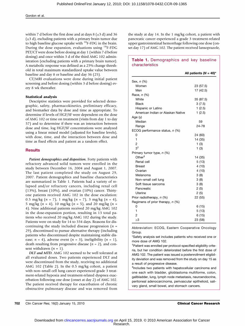
Table 1. Demographics and key baselinecharacteristics
A
Clin
, 2019. © 2010 American Associ
ll patients (N = 40)*
Sex, n (%)
Women 23 (57.5) Men 17 (42.5)Race, n (%)
White 35 (87.5) Black 3 (7.5) Hispanic or Latino 1 (2.5) American Indian or Alaskan Native 1 (2.5)Age (y)
Median 59 Range 24-78ECOG performance status, n (%)
0 24 (60) 1 14 (35) 2 1 (3) 3† 1 (3)Primary tumor type, n (%)
Other‡ 14 (35) Renal cell 5 (13) Breast 4 (10) Ovarian 4 (10) Melanoma 3 (8) Non–small cell lung 3 (8) Soft tissue sarcoma 3 (8) Pancreatic 2 (5) Uterine 2 (5)Prior radiotherapy, n (%)
22 (55) Regimens of prior therapy, n (%)0
6 (15) 1 5 (13) 2 6 (15) ≥3 23 (58)Abbreviation: ECOG, Eastern Cooperative OncologyGroup.*Safety analysis set includes patients who received one ormore dose of AMG 102.†Patient was enrolled per protocol-specified eligibility crite-ria, but her condition deteriorated before the first dose ofAMG 102. The patient was issued a postenrollment eligibil-ity deviation and was removed from the study on day 15 asa result of progressive disease.‡Includes two patients with hepatocellular carcinoma andone each with bladder, glioblastoma multiforme, colon,gallbladder, lung, lymph node metastasis, neuroendocrine,peritoneal adenocarcinoma, perivascular epithelioid, sali-vary gland, small bowel, and stomach cancers.
ical Cancer Research
ation for Cancer
Safety of AMG 102 in Patients with Advanced Solid Tumors
Published OnlineFirst January 12, 2010; DOI: 10.1158/1078-0432.CCR-09-1365
and the condition resolved 3 days after discontinuationof AMG 102. As a result of the DLTs, two additional pa-tients were enrolled in the 0.5 mg/kg and in the 1 mg/kgcohorts. Additionally, two patients (one each in the0.5 mg/kg and 1 mg/kg cohorts) who withdrew for rea-sons other than DLT were replaced, resulting in a totalenrollment of seven patients in each of these cohorts.No further DLTs were observed in either cohort, and doseescalation continued to the 20 mg/kg dose. The MTD wasnot reached. Nine additional patients were enrolledin the dose expansion and received 20 mg/kg AMG102 every 2 weeks.
www.aacrjournals.org
Researcon April clincancerres.aacrjournals.org Downloaded from
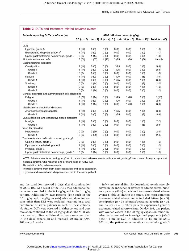
Safety and tolerability. No dose-related trends were ob-served in the incidence or severity of adverse events. Nine-teen patients (48%) experienced treatment-related adverseevents (Table 2) during the study. The most commontreatment-related adverse events included fatigue (n = 5),constipation (n = 3), anorexia/decreased appetite (n = 3),and nausea (n = 3). Three patients experienced grade 3treatment-related adverse events. On day 203, one patientwith ovarian cancer in the 20 mg/kg expansion cohort in-advertently received an investigational peptibody (AMG386; ∼4 mg/kg i.v.) in addition to 15 mg/kg AMG102 i.v.; the patient subsequently experienced a grade 3
Table 2. DLTs and treatment-related adverse events
Patients reporting DLTs or AEs, n (%)
AMG 102 dose cohort (mg/kg)0.5 (n = 7)
1 (n = 7)h1
3 (n = 4)
. 5, 2019.
5 (n = 4)
© 2010 A
10 (n = 5)
Clin Canc
merican A
20 (n = 13)*
er Res; 16(2)
ssociation fo
Total (N = 40)
DLTs
Hypoxia, grade 3† 1 (14) 0 (0) 0 (0) 0 (0) 0 (0) 0 (0) 1 (3) Exacerbated dyspnea, grade 3† 1 (14) 0 (0) 0 (0) 0 (0) 0 (0) 0 (0) 1 (3) Upper gastrointestinal hemorrhage, grade 3 0 (0) 1 (14) 0 (0) 0 (0) 0 (0) 0 (0) 1 (3)All treatment-related AEs
5 (71) 4 (57) 1 (25) 3 (75) 1 (20) 5 (38) 19 (48) Gastrointestinal disordersConstipation
1 (14) 0 (0) 0 (0) 1(25) 0 (0) 1 (8) 3 (8) Grade 1 1 (14) 0 (0) 0 (0) 1 (25) 0 (0) 0 (0) 2 (5) Grade 2 0 (0) 0 (0) 0 (0) 0 (0) 0 (0) 1 (8) 1 (3)Nausea
1 (14) 0 (0) 0 (0) 1 (25) 0 (0) 1 (8) 3 (8) Grade 1 1 (14) 0 (0) 0 (0) 1 (25) 0 (0) 1 (8) 3 (8)Vomiting
0 (0) 1 (14) 0 (0) 0 (0) 0 (0) 1 (8) 2 (5) Grade 1 0 (0) 0 (0) 0 (0) 0 (0) 0 (0) 1 (8) 1 (3) Grade 2 0 (0) 1 (14) 0 (0) 0 (0) 0 (0) 0 (0) 1 (3)General disorders and administration site conditions
Fatigue 2 (29) 1 (14) 0 (0) 1 (25) 1 (20) 0 (0) 5 (13)Grade 1
1 (14) 0 (0) 0 (0) 1 (25) 0 (0) 0 (0) 2 (5) Grade 2 1 (14) 1 (14) 0 (0) 0 (0) 1 (20) 0 (0) 3 (8)Metabolism and nutrition disorders
Anorexia/decreased appetite 1 (14) 0 (0) 0 (0) 1 (25) 0 (0) 1 (8) 3 (8)Grade 1
1 (14) 0 (0) 0 (0) 1 (25) 0 (0) 1 (8) 3 (8) Musculoskeletal and connective tissue disordersMyalgia
1 (14) 0 (0) 0 (0) 0 (0) 0 (0) 1 (8) 2 (5) Grade 1 1 (14) 0 (0) 0 (0) 0 (0) 0 (0) 1 (8) 2 (5)Vascular disorders
Hypotension 0 (0) 2 (29) 0 (0) 0 (0) 0 (0) 0 (0) 2 (5)Grade 1
0 (0) 2 (29) 0 (0) 0 (0) 0 (0) 0 (0) 2 (5) Treatment-related AEs with a worst grade ≥3Colonic fistula, grade 3
0 (0) 0 (0) 0 (0) 0 (0) 0 (0) 1 (8) 1 (3) Dyspnea exacerbated, grade 3 1 (14) 0 (0) 0 (0) 0 (0) 0 (0) 0 (0) 1 (3) Hypoxia, grade 3 1 (14) 0 (0) 0 (0) 0 (0) 0 (0) 0 (0) 1 (3) Upper gastrointestinal hemorrhage, grade 3 0 (0) 1 (14) 0 (0) 0 (0) 0 (0) 0 (0) 1 (3)NOTE: Adverse events occurring in ≥5% of patients and adverse events with a worst grade ≥3 are shown. Safety analysis setincludes patients who received one or more dose of AMG 102.Abbreviation: AEs, adverse events.*Includes patients from both dose escalation and dose expansion.†Hypoxia and exacerbated dyspnea occurred in the same patient.
January 15, 2010 703
r Cancer
Gordon et al.
704
Published OnlineFirst January 12, 2010; DOI: 10.1158/1078-0432.CCR-09-1365
colonic fistula on day 236. The adverse event was deter-mined to be treatment related, as it could not be ruledout that the possibility of receiving both AMG 102 andAMG 386 resulted in the fistula of the colon. The patientwas hospitalized, discontinued AMG 102, and was re-moved from the study.Thirty-nine patients (98%) experienced treatment-
emergent adverse events during the study, 20 (50%)of whom experienced at least one grade 3 adverse event(Supplementary Table S1). The most common treatment-emergent adverse events (occurring in ≥10 patients) werefatigue (43%), nausea (40%), vomiting (33%), anorexia/decreased appetite (33%), peripheral edema (30%),and dyspnea (28%). Fourteen (35%) patients experiencedserious treatment-emergent adverse events. Seriousadverse events observed included pneumonia (n = 3), dys-pnea (n = 2), hypotension (n = 2), and vomiting (n = 2),which were not considered related to treatment withAMG 102.In total, five patients had adverse events that resulted in
discontinuation of AMG 102 administration. In additionto the two patients who discontinued AMG 102 due toDLTs and the patient who discontinued AMG 102 as a re-sult of a colonic fistula, one patient (1 mg/kg cohort) haddeep vein thrombosis not considered related to studytreatment that resulted in discontinuation of AMG 102,and one patient (1 mg/kg cohort) discontinued AMG102 as a result of retroperitoneal sarcoma (patient diedas a result of progressive disease). In addition to the pa-tient with retroperitoneal sarcoma, one patient died dueto progressive non–small cell lung cancer (20 mg/kg co-hort). Thirteen patients had one or more adverse event(treatment emergent or treatment related) resulting in hos-pitalization. No patients had detectable anti–AMG 102-binding antibodies.Pharmacokinetics. Pharmacokinetic parameters are
shown in Table 3, and pharmacokinetic profiles aredepicted in Fig. 1. Pharmacokinetic parameters were esti-mated from combined data from patients in the dose-escalation (n = 31) and dose-expansion (n = 9) phases.The Cmax was observed mainly around the end of the1-hour infusion, and the serum concentration of AMG102 declined biphasically. The Cmax and AUCτ, which wereassessed by one-way ANOVA, increased linearly with dose.The estimated clearance and t1/2 were similar across doses,suggesting that AMG 102 exhibits linear pharmacokineticsin the dose range of 0.5 to 20 mg/kg. At the doses tested,the estimated mean clearance range was 0.104 to 0.176mL/h/kg, and the t1/2 range was 14.5 to 22 days. The meanclearance and t1/2 of all dose levels were 0.141 mL/h/kgand 18 days, respectively. Following multiple-dose admin-istration, accumulation of AMG 102 exposure wasobserved under the biweekly regimen. The trough concen-tration (C336h) increased ∼2-fold after the fourth dosecompared with the first dose, and the AR based on theAUC ranged from 1.30 to 3.86.Preclinical biomarkers. In mice bearing U-87 MG human
glioblastoma xenografts, total plasma HGF/SF levels in-
Clin Cancer Res; 16(2) January 15, 2010
Researcon April clincancerres.aacrjournals.org Downloaded from
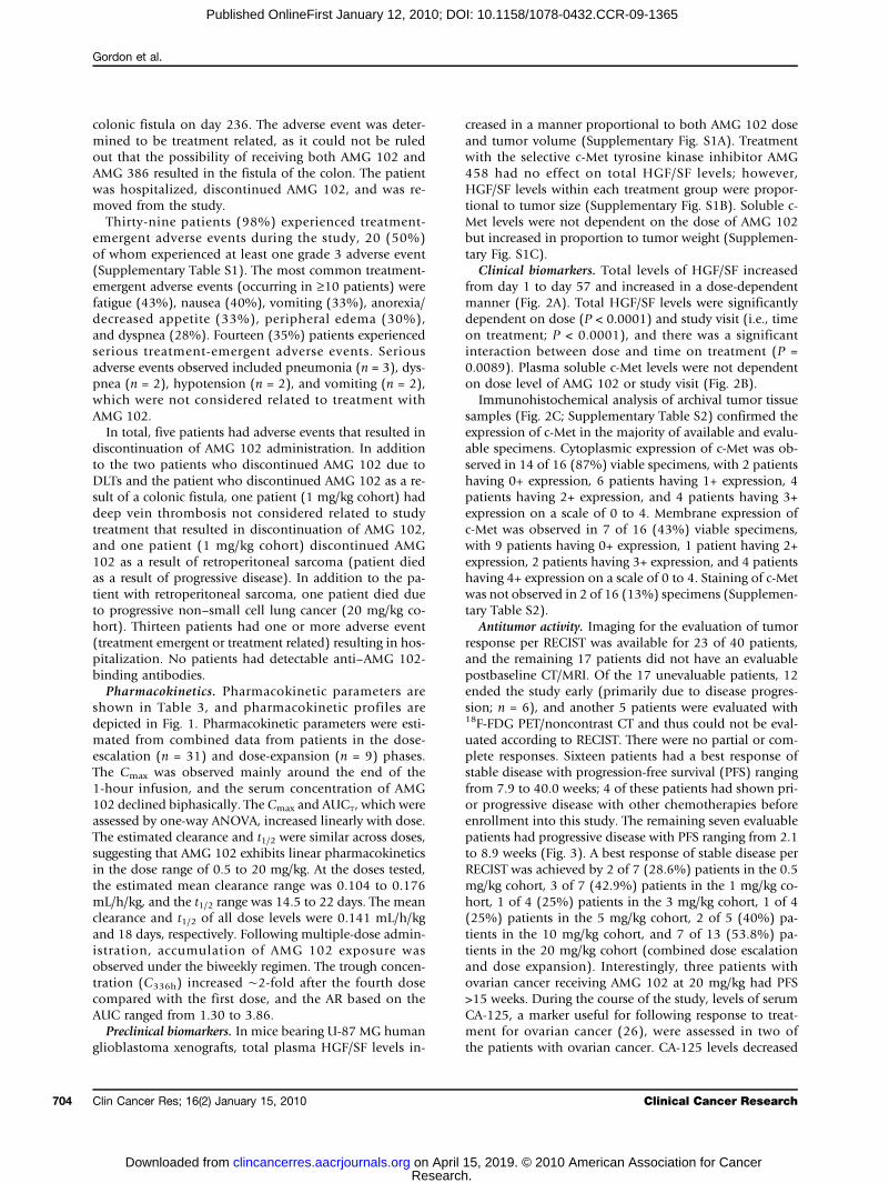
creased in a manner proportional to both AMG 102 doseand tumor volume (Supplementary Fig. S1A). Treatmentwith the selective c-Met tyrosine kinase inhibitor AMG458 had no effect on total HGF/SF levels; however,HGF/SF levels within each treatment group were propor-tional to tumor size (Supplementary Fig. S1B). Soluble c-Met levels were not dependent on the dose of AMG 102but increased in proportion to tumor weight (Supplemen-tary Fig. S1C).Clinical biomarkers. Total levels of HGF/SF increased
from day 1 to day 57 and increased in a dose-dependentmanner (Fig. 2A). Total HGF/SF levels were significantlydependent on dose (P < 0.0001) and study visit (i.e., timeon treatment; P < 0.0001), and there was a significantinteraction between dose and time on treatment (P =0.0089). Plasma soluble c-Met levels were not dependenton dose level of AMG 102 or study visit (Fig. 2B).Immunohistochemical analysis of archival tumor tissue
samples (Fig. 2C; Supplementary Table S2) confirmed theexpression of c-Met in the majority of available and evalu-able specimens. Cytoplasmic expression of c-Met was ob-served in 14 of 16 (87%) viable specimens, with 2 patientshaving 0+ expression, 6 patients having 1+ expression, 4patients having 2+ expression, and 4 patients having 3+expression on a scale of 0 to 4. Membrane expression ofc-Met was observed in 7 of 16 (43%) viable specimens,with 9 patients having 0+ expression, 1 patient having 2+expression, 2 patients having 3+ expression, and 4 patientshaving 4+ expression on a scale of 0 to 4. Staining of c-Metwas not observed in 2 of 16 (13%) specimens (Supplemen-tary Table S2).Antitumor activity. Imaging for the evaluation of tumor
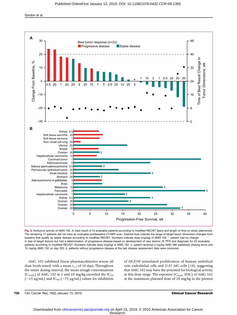
response per RECIST was available for 23 of 40 patients,and the remaining 17 patients did not have an evaluablepostbaseline CT/MRI. Of the 17 unevaluable patients, 12ended the study early (primarily due to disease progres-sion; n = 6), and another 5 patients were evaluated with18F-FDG PET/noncontrast CT and thus could not be eval-uated according to RECIST. There were no partial or com-plete responses. Sixteen patients had a best response ofstable disease with progression-free survival (PFS) rangingfrom 7.9 to 40.0 weeks; 4 of these patients had shown pri-or progressive disease with other chemotherapies beforeenrollment into this study. The remaining seven evaluablepatients had progressive disease with PFS ranging from 2.1to 8.9 weeks (Fig. 3). A best response of stable disease perRECIST was achieved by 2 of 7 (28.6%) patients in the 0.5mg/kg cohort, 3 of 7 (42.9%) patients in the 1 mg/kg co-hort, 1 of 4 (25%) patients in the 3 mg/kg cohort, 1 of 4(25%) patients in the 5 mg/kg cohort, 2 of 5 (40%) pa-tients in the 10 mg/kg cohort, and 7 of 13 (53.8%) pa-tients in the 20 mg/kg cohort (combined dose escalationand dose expansion). Interestingly, three patients withovarian cancer receiving AMG 102 at 20 mg/kg had PFS>15 weeks. During the course of the study, levels of serumCA-125, a marker useful for following response to treat-ment for ovarian cancer (26), were assessed in two ofthe patients with ovarian cancer. CA-125 levels decreased
Clinical Cancer Research
h. 15, 2019. © 2010 American Association for Cancer

Safety of AMG 102 in Patients with Advanced Solid Tumors
Published OnlineFirst January 12, 2010; DOI: 10.1158/1078-0432.CCR-09-1365
from 728 units/mL at baseline to 545 units/mL on day114 in the first patient (22). In the second patient, CA-125 levels decreased from 450 units/mL at baseline to260 units/mL (day 171) and then to 109 units/mL (day228). However, it should be noted that the second patientincorrectly received an investigational peptibody, AMG386, in addition to AMG 102 as a consequence of a dosingerror on day 203.Changes in the best-result sum of longest diameters of
target lesions ranged from −19.0% to 24.0% (Fig. 3). Re-duction in tumor dimensions was associated with the fol-lowing primary tumor types: ovarian (n = 3), brain (n = 1),kidney (n = 1), hepatocellular carcinoma (n = 1), melano-
www.aacrjournals.org
Researcon April clincancerres.aacrjournals.org Downloaded from
ma (n = 1), and pancreatic (n = 1). All eight patients whohad a reduction in tumor dimensions achieved stable dis-ease. 18F-FDG PET/noncontrast CT revealed that 2 of 34evaluable patients had metabolic responses: one patientwith abdominal sarcoma had a response (−30%) at studyday 8 but subsequently withdrew from the study on day21, and one patient with appendiceal carcinoid had a re-sponse (−27%) at day 36.
Discussion
In this first-in-human study of AMG 102, a HGF/SF-specific neutralizing antibody, the MTD was not reached.
Table 3. AMG 102 pharmacokinetic parameters
AMG 102 dose cohort (mg/kg)
0.5 (n = 1-7)
1 (n = 2-7) 3 (n = 1-4)h1
5 (n = 1-4)
. 5, 2019. © 2010
10 (n = 2-5)
Clin Cancer Res;
American Associat
20 (n = 3-13)*
Dose
1 4 1 4 1 4 1 4 1 4 116(2) Januar
ion for Can
4
tmax (h)
n 7 2 7 3 4 2 4 1 5 2 13 3 Median 1.1 1.5 1.18 2.58 1.02 28.1 1.5 1 1 1.58 2 2 Range 1-8.42 1-2 1-170 1-8 1-24.5 8-48.1 1-2 1-1 1-2.85 0.5-2.65 0.917-24 1-2.62Cmax (μg/mL)
n 7 2 7 3 4 2 4 1 5 2 13 3 Mean 15.4 18.7 18.1 38.2 95.1 118 77.2 192 234 372 443 695 SD 3.24 NC 5.29 6.46 44.8 NC 14.6 NC 76.2 NC 115 184C336h (μg/mL)
n 7 1 6 2 4 1 4 1 4 2 12 3 Mean 3.99 6.32 5.54 12.6 24.3 45.9 25.1 65.3 73.5 151 140 356 SD 1.66 NC 1.74 NC 9.88 NC 5.64 NC 26.3 NC 45.7 125t1/2 (d)
n 7 NC 7 NC 4 NC 4 NC 5 NC 4 NC Mean 15.2 NC 14.5 NC 17.5 NC 20.0 NC 22.0 NC 21.6 NC SD 10.0 NC 6.8 NC 3.81 NC 7.88 NC 11.3 NC 15.2 NCAUCτ (h·μg/mL)
n 7 1 6 2 4 1 4 1 4 2 12 3 Mean 2,320 3,520 2,690 6,920 11,800 17,700 12,500 42,100 36,000 55,900 75,900 147,000 SD 462 NC 553 NC 4,160 NC 2,340 NC 13,400 NC 18,900 35,700AUC0-inf (h·μg/mL)
n 5 NC 5 NC 3 NC 4 NC 4 NC 4 NC Mean 5,600 NC 6,630 NC 26,800 NC 30,900 NC 91,300 NC 168,000 NC SD 2,800 NC 1,950 NC 1,190 NC 10,500 NC 16,400 NC 112,000 NCCL (mL/h/kg)
n 5 NC 5 NC 3 NC 4 NC 4 NC 4 NC Mean 0.104 NC 0.162 NC 0.135 NC 0.176 NC 0.112 NC 0.158 NC SD 0.0378 NC 0.0497 NC 0.783 NC 0.0566 NC 0.0197 NC 0.0858 NCAR
n NC 1 NC 2 NC 1 NC 1 NC 2 NC 3 Mean NC 1.43 NC 2.39 NC 1.30 NC 3.86 NC 2.25 NC 2.45 SD NC NC NC NC NC NC NC NC NC NC NC 0.316Abbreviations: AUC0-inf, AUC from time 0 to infinity; CL, systemic clearance; C336h, observed plasma concentration at 336 h afterdose; NC, not calculated; τ, 336 h with once-every-2-wk regimen.*Cohort (20 mg/kg) includes samples from the dose escalation and dose expansion.
y 15, 2010 705
cer

Gordon et al.
706
Published OnlineFirst January 12, 2010; DOI: 10.1158/1078-0432.CCR-09-1365
However, AMG102was generally well tolerated at doses upto the planned maximum dose (20 mg/kg) in heavily pre-treated adult patients with refractory solid tumors. AMG102–related adverse events were generally mild or moder-ate in severity, with the most frequently occurring (>5%)events being fatigue, constipation, and nausea. A total oftwo patients in the lowest dose cohorts (0.5 and 1 mg/kg)experiencedDLTs. There were no other hematologic or non-hematologic DLTs observed in this trial. Indeed, there wereno dose-related trends in the incidence of adverse events upto the highest dose tested (20 mg/kg). There were twodeaths during the study resulting from disease progression.These were deemed to be unrelated to study treatment.Recent studies have investigated the toxicity of small-
molecule inhibitors of c-Met, some of which target kinasesin addition to the c-Met protein. For example, in phase Iand II studies, administration of XL880, an orally availablesmall-molecule inhibitor of c-Met and vascular endothelialgrowth factor receptor 2, has been associated with mild tomoderate hypertension, nausea, anorexia, vomiting, fa-tigue, and liver function abnormalities (27–30). Treatmentwith ARQ 197, a selective, non–ATP-competitive small-molecule inhibitor of c-Met, has been associated with fa-tigue, nausea, vomiting, diarrhea, and anorexia (30, 31),and treatment with XL184, a small-molecule inhibitor ofc-Met, vascular endothelial growth factor receptor 2, Kit,
Clin Cancer Res; 16(2) January 15, 2010
Researcon April clincancerres.aacrjournals.org Downloaded from
Ret, Flt3, and Tie-2, has been associated with palmar/plan-tar erythema, mucositis, elevations of alanine aminotrans-ferase and lipase, diarrhea, and hypopigmentation of thehair (32). Fatigue, nausea, vomiting, and anorexia were al-so observed during AMG 102 administration; however, hy-pertension and significant liver enzyme abnormalities werenot observed. This may be due to the selectivity of AMG102 for the c-Met ligand HGF/SF.Although antitumor activity was not a primary objective
of this study, a substantial proportion of the patients withrefractory solid tumors who received AMG 102 in this studyhad stable disease, and a degree of tumor regressionwas ob-served in some patients. Of those evaluable for postbaselinetumor responses per RECIST, 16 of 23 (70%) achieved sta-ble disease as a best response, 4 of whom had shown dis-ease progression with chemotherapeutic interventionbefore entry into this trial. Three patients with ovarian can-cer receiving AMG 102 at 20 mg/kg remained in the studywith stable disease for a prolonged period (PFS >15 weeks)and showed reductions in tumor size. In addition, two ofthese patients with ovarian cancer had reductions in the tu-mor marker CA-125 (26). Although tumor c-Met data werenot available for these two patients, this is an interesting ob-servation because overexpression of c-Met among patientswith advanced ovarian cancer has been associated with de-creased overall survival (33).
Fig. 1. Serum concentration and dose linearity of AMG 102. A, serum concentration versus time profiles. Cmax (B) and AUC (C) were estimated by non-compartmental analysis and plotted against dose with linear regression. *, 20 mg/kg cohort includes samples from the dose escalation and dose expansion.
Clinical Cancer Research
h. 15, 2019. © 2010 American Association for Cancer
Safety of AMG 102 in Patients with Advanced Solid Tumors
Published OnlineFirst January 12, 2010; DOI: 10.1158/1078-0432.CCR-09-1365
Fig. 2. Analysis of HGF/SF and c-Met as biomarkers. A, effect of AMG 102 on total plasma HGF/SF in patients with advanced solid tumors. B, effect ofAMG 102 on plasma soluble c-Met in patients with advanced solid tumors. C, immunohistochemical staining of c-Met in a renal cell carcinoma,graded as 3 for cytoplasmic staining and 4 for membrane staining, and hepatocellular carcinoma, graded as 3 for cytoplasmic staining and 0 for membranestaining, using a relative scale of 0 to 4 (0 = no stain, 4 = maximal stain).
Clin Cancer Res; 16(2) January 15, 2010www.aacrjournals.org 707
Research. on April 15, 2019. © 2010 American Association for Cancerclincancerres.aacrjournals.org Downloaded from
Gordon et al.
708
Published OnlineFirst January 12, 2010; DOI: 10.1158/1078-0432.CCR-09-1365
AMG 102 exhibited linear pharmacokinetics across alldose levels tested, with a mean t1/2 of 18 days. Throughoutthe entire dosing interval, the mean trough concentrations(C336h) of AMG 102 at 3 and 10 mg/kg exceeded the IC50
(∼15 μg/mL) and IC90 (∼75 μg/mL) values for inhibition
Clin Cancer Res; 16(2) January 15, 2010
Researcon April clincancerres.aacrjournals.org Downloaded from
of HGF/SF-stimulated proliferation of human umbilicalvein endothelial cells and U-87 MG cells (18), suggestingthat AMG 102may have the potential for biological activityat this dose range. The exposure (Cmax, AUC) of AMG 102at the maximum planned dose of 20 mg/kg in the present
Fig. 3. Antitumor activity of AMG 102. A, best result of 23 evaluable patients according to modified RECIST (bars) and length of time on study (diamonds).The remaining 17 patients did not have an evaluable postbaseline CT/MRI scan. Dashed lines indicate the range of target lesion dimension changes frombaseline that qualify as stable disease according to modified RECIST. Numbers indicate dose (mg/kg) of AMG 102. *, patient had no changein size of target lesions but had a determination of progressive disease based on development of new lesions. B, PFS and diagnosis for 23 evaluablepatients according to modified RECIST. Numbers indicate dose (mg/kg) of AMG 102. †, patient received 3 mg/kg AMG 386 peptibody (dosing error) and15 mg/kg AMG 102 on day 203; ‡, patients without progressive disease at the last disease assessment date were censored.
Clinical Cancer Research
h. 15, 2019. © 2010 American Association for Cancer
Safety of AMG 102 in Patients with Advanced Solid Tumors
Published OnlineFirst January 12, 2010; DOI: 10.1158/1078-0432.CCR-09-1365
study was at least 6-fold lower than the no observed ad-verse event level dose (150 mg/kg) in the 40-week toxicitystudy in nonhuman primates, indicating that there was asufficient safety margin in exposure. Although a MTDwas not reached, a preliminary population pharmacokinet-ic model (22) developed with the pharmacokinetic datacollected from this study suggested that dosing regimensof ≥10 mg/kg every 2 weeks, 15 mg/kg every 3 weeks,and 20 mg/kg every 4 weeks should maintain serumAMG 102 trough concentrations above the IC90 value inthe human umbilical vein cell and U-87 MG cell prolifera-tion assays described above in >90% of patients. Therefore,these regimens were chosen for ongoing phase 2 studies.Biomarkers are important for identifying cancer subtypes,
assessing metastatic potential, providing prognostic infor-mation, and predicting responses to therapeutic agents. Toexplore the potential of HGF/SF as a clinical biomarker, wefirst examined the effect of AMG 102 treatment on plasmatotal HGF/SF levels in a U-87 MG glioblastoma xenograftmodel. In this model, the total plasma level of HGF/SF(sum of free HGF/SF and HGF/SF bound to AMG 102) in-creased in proportion to AMG 102 dose and tumor size.Because the c-Met tyrosine kinase inhibitor AMG 458 hadno effect on total HGF/SF levels, it is unlikely that the ob-served increases in HGF/SF levels were due to negativefeedback from inhibition of c-Met. We hypothesize that in-creases in HGF/SF may be the result of AMG 102 stabiliza-tion of HGF/SF, resulting in increased HGF/SF half-lifeand/or HGF/SF redistribution from tissues (including tu-mors). At this time, we do not know why levels of solublec-Met did not change after treatment with AMG 102.The clinical biomarker data were similar to the preclin-
ical data: in patients, plasma HGF/SF concentrations in-creased in a dose- as well as a time-dependent manner,and levels of soluble c-Met did not seem to be dependenton either dose or time on treatment. In addition, c-Met wasexpressed in themajority of available patient tumor samples.Although both HGF/SF and c-Met are well-documentedprognostic markers of disease progression and survival(34–40), additional studies are required to determinewhether these markers will be useful for predicting the effi-cacy of HGF/SF–c-Met axis–targeted therapies.Overall, AMG 102 showed acceptable safety as a mono-
therapy at i.v. doses up to20mg/kgwhen administered every2 weeks to patients with advanced solid tumors. Because ofits favorable safety and pharmacokinetic profile and uniquespecificity to HGF/SF, the only known ligand of the c-Met
www.aacrjournals.org
Researcon April clincancerres.aacrjournals.org Downloaded from
receptor, there is a potential for incorporation of AMG 102into combination therapy regimens both with cytotoxic che-motherapy and with other targeted agents. In a recent pre-clinical study, AMG 102 enhanced the efficacy oftemozolomide or docetaxel in inhibiting growth of U-87MG cells in vitro and U-87 MG tumor xenografts in vivo(41). In an ongoing phase Ib study, AMG 102 in combina-tion with bevacizumab or motesanib (a small-molecule in-hibitor of vascular endothelial growth factor receptors) led todurable stable disease in some patients (42). The most fre-quently occurring treatment-related adverse eventswere nau-sea and fatigue. Studies are ongoing to determine the clinicalactivity of AMG 102 both as monotherapy and in combina-tion with other therapeutic regimens in several tumor types,including recurrent glioblastoma multiforme (43).
Disclosure of Potential Conflicts of Interest
A. Anderson, D.M. Beaupre, D. Branstetter, T.L. Burgess,A. Coxon, H. Deng, P. Kaplan-Lefko, I.M. Leitch, K.S.Oliner, L. Yan, and M. Zhu: employees/ownership interest,Amgen Inc. L. Gore: commercial research support,Merck andCo., Inc. S.G. Eckhardt: consultant, Amgen Inc. The authorsacknowledge Benjamin Scott, Ph.D., who provided assis-tance in writing this manuscript with funding by AmgenInc. The other authors declare no conflicts of interest.
Acknowledgments
We thank Jianfeng Lu, Mario Bejarano, and Mark Ma forpharmacokinetic assessment and sample analysis; NoelleErbeck, Charity Scripture, Poornima Shubhakar, and SusanGlover for clinical support; JimGould for datamanagement;Yao Zhuang for antibody analysis; Yuying Hwang forimaging support; Ronals Korn for 18F-FDG PET/CTanalysis; Jennifer Malella, Yun Lan, and Gwyneth Van forbiomarker sample analysis; Karen Rex and Jan Sun forpreclinical xenograft studies; Benjamin Scott, Ph.D., whosework was funded by Amgen Inc., for assistance in writingthis manuscript; the research staff at each participatinginstitution; and the patients who took part in this study.The costs of publication of this article were defrayed in
part by the payment of page charges. This article musttherefore be hereby marked advertisement in accordancewith 18 U.S.C. Section 1734 solely to indicate this fact.Received 5/28/09; revised 9/28/09; accepted 10/22/09;
published OnlineFirst 1/12/10.
References
1. Stoker M, Gherardi E, Perryman M, Gray J. Scatter factor is a fibro-blast-derived modulator of epithelial cell mobility. Nature 1987;327:239–42.
2. Zarnegar R, Michalopoulos G. Purification and biological character-ization of human hepatopoietin A, a polypeptide growth factor forhepatocytes. Cancer Res 1989;49:3314–20.
3. Montesano R, Matsumoto K, Nakamura T, Orci L. Identification of afibroblast-derived epithelial morphogen as hepatocyte growth factor.Cell 1991;67:901–8.
4. Nakamura T, Nishizawa T, Hagiya M, et al. Molecular cloning andexpression of human hepatocyte growth factor. Nature 1989;342:440–3.
5. Birchmeier C, Birchmeier W, Gherardi E, Vande Woude GF. Met, me-tastasis, motility and more. Nat Rev Mol Cell Biol 2003;4:915–25.
6. Ferracini R, Di Renzo MF, Scotlandi K, et al. The Met/HGF receptor isover-expressed in human osteosarcomas and is activated by either aparacrine or an autocrine circuit. Oncogene 1995;10:739–49.
7. Jin L, Fuchs A, Schnitt SJ, et al. Expression of scatter factor and
Clin Cancer Res; 16(2) January 15, 2010 709
h. 15, 2019. © 2010 American Association for Cancer
Gordon et al.
710
Published OnlineFirst January 12, 2010; DOI: 10.1158/1078-0432.CCR-09-1365
c-met receptor in benign and malignant breast tissue. Cancer1997;79:749–60.
8. Fukuura T, Miki C, Inoue T, Matsumoto K, Suzuki H. Serum hepato-cyte growth factor as an index of disease status of patients with co-lorectal carcinoma. Br J Cancer 1998;78:454–9.
9. Koochekpour S, Jeffers M, Rulong S, et al. Met and hepatocytegrowth factor/scatter factor expression in human gliomas. CancerRes 1997;57:5391–8.
10. Schmidt L, Junker K, Nakaigawa N, et al. Novel mutations of the METproto-oncogene in papillary renal carcinomas. Oncogene 1999;18:2343–50.
11. Miller M, Ginalski K, Lesyng B, Nakaigawa N, Schmidt L, Zbar B.Structural basis of oncogenic activation caused by point mutationsin the kinase domain of the MET proto-oncogene: modeling studies.Proteins 2001;44:32–43.
12. Di Renzo MF, Olivero M, Martone T, et al. Somatic mutations of theMET oncogene are selected during metastatic spread of humanHNSC carcinomas. Oncogene 2000;19:1547–55.
13. Olivero M, Valente G, Bardelli A, et al. Novel mutation in the ATP-binding site of the MET oncogene tyrosine kinase in a HPRCC family.Int J Cancer 1999;82:640–3.
14. Di Renzo MF, Olivero M, Giacomini A, et al. Overexpression and am-plification of the met/HGF receptor gene during the progression ofcolorectal cancer. Clin Cancer Res 1995;1:147–54.
15. Nakajima M, Sawada H, Yamada Y, et al. The prognostic significanceof amplification and overexpression of c-met and c-erb B-2 in humangastric carcinomas. Cancer 1999;85:1894–902.
16. Kijima Y, Hokita S, Yoshinaka H, et al. Amplification and overexpres-sion of c-met gene in Epstein-Barr virus-associated gastric carcino-mas. Oncology 2002;62:60–5.
17. Comoglio PM, Giordano S, Trusolino L. Drug development of METinhibitors: targeting oncogene addiction and expedience. Nat RevDrug Discov 2008;7:504–16.
18. Burgess T, Coxon A, Meyer S, et al. Fully human monoclonal antibo-dies to hepatocyte growth factor with therapeutic potential againsthepatocyte growth factor/c-Met-dependent human tumors. CancerRes 2006;66:1721–9.
19. Martin TA, Parr C, Davies G, et al. Growth and angiogenesis of hu-man breast cancer in a nude mouse tumour model is reduced byNK4, a HGF/SF antagonist. Carcinogenesis 2003;24:1317–23.
20. Sattler M, Salgia R. c-Met and hepatocyte growth factor: potential asnovel targets in cancer therapy. Curr Oncol Rep 2007;9:102–8.
21. Kakkar T, Ma M, Zhuang Y, Patton A, Hu Z, Mounho B. Pharma-cokinetics and safety of a fully human hepatocyte growth factorantibody, AMG 102, in cynomolgus monkeys. Pharm Res 2007;24:1910–8.
22. Data on file. Thousand Oaks (CA): Amgen Inc. 2008.23. Athauda G, Giubellino A, Coleman JA, et al. c-Met ectodomain shed-
ding rate correlates with malignant potential. Clin Cancer Res 2006;12:4154–62.
24. Therasse P, Arbuck SG, Eisenhauer EA, et al. European Organizationfor Research and Treatment of CancerNational Cancer Institute ofthe United StatesNational Cancer Institute of Canada. New guide-lines to evaluate the response to treatment in solid tumors. J NatlCancer Inst 2000;92:205–16.
25. Young H, Baum R, Cremerius U, et al. European Organization for Re-search and Treatment of Cancer (EORTC) PET Study Group. Mea-surement of clinical and subclinical tumour response using [18F]-fluorodeoxyglucose and positron emission tomography: review and1999 EORTC recommendations. Eur J Cancer 1999;35:1773–82.
26. Hogdall E. Cancer antigen 125 and prognosis. Curr Opin Obstet Gy-necol 2008;20:4–8.
27. Jhawer MP, Kindler HL, Wainberg ZA, et al. Preliminary activity ofXL880, a dual MET/VEGFR2 inhibitor, in MET amplified poorly differ-entiated gastric cancer (PDGC): interim results of a multicenter phaseII study [abstract]. J Clin Oncol 2008;26:4572.
Clin Cancer Res; 16(2) January 15, 2010
Researcon April clincancerres.aacrjournals.org Downloaded from
28. Ross RW, Srinivasan R, Vaishampayan U, et al. A phase 2 study ofthe dual MET/VEGFR2 inhibitor XL880 in patients (pts) with papillaryrenal carcinoma (PRC) [abstract]. In: AACR-NCI-EORTC InternationalConference: Molecular Targets and Cancer Therapeutics, November14-18, 2007, San Francisco (CA). 2007.
29. Shapiro GI, Heath E, Malburg L, et al. A phase I dose-escalationstudy of the safety, pharmacokinetics (PK) and pharmacodynamicsof XL880, a VEGFR and MET kinase inhibitor, administered daily topatients (pts) with advanced malignancies [abstract]. In: AACR-NCI-EORTC International Conference: Molecular Targets and Can-cer Therapeutics, November 14-18, 2007, San Francisco (CA)2007.
30. Yap TA, Harris D, Barriuso J, et al. Phase I trial to determine the doserange for the c-Met inhibitor ARQ 197 that inhibits c-Met and FAKphosphorylation, when administered by an oral twice-a-day schedule[abstract]. J Clin Oncol 2008;26:3584.
31. Rosen L, Senzer N, Nemunaitis J, et al. A phase I dose escalationstudy and signs of anti-metastatic activity of ARQ 197, A selectivec-Met inhibitor [abstract]. In: AACR-NCI-EORTC International Con-ference: Molecular Targets and Cancer Therapeutics, November14-18, 2007, San Francisco (CA). 2007.
32. Salgia R, Sherman S, Hong DS, et al. A phase I study of XL184, aRET, VEGFR2, and MET kinase inhibitor, in patients (pts) with ad-vanced malignancies, including pts with medullary thyroid cancer(MTC) [abstract]. J Clin Oncol 2008;26:3522.
33. Sawada K, Radjabi AR, Shinomiya N, et al. c-Met overexpression is aprognostic factor in ovarian cancer and an effective target for inhibi-tion of peritoneal dissemination and invasion. Cancer Res 2007;67:1670–9.
34. Ghoussoub RA, Dillon DA, D'Aquila T, Rimm EB, Fearon ER, RimmDL. Expression of c-met is a strong independent prognostic factor inbreast carcinoma. Cancer 1998;82:1513–20.
35. Kang JY, Dolled-Filhart M, Ocal IT, et al. Tissue microarray analysisof hepatocyte growth factor/Met pathway components reveals a rolefor Met, matriptase, and hepatocyte growth factor activator inhibitor1 in the progression of node-negative breast cancer. Cancer Res2003;63:1101–5.
36. Ren Y, Cao B, Law S, et al. Hepatocyte growth factor promotes can-cer cell migration and angiogenic factors expression: a prognosticmarker of human esophageal squamous cell carcinomas. Clin Can-cer Res 2005;11:6190–7.
37. Srinivasan R, Choueiri TK, Vaishampayan U, et al. A phase II study ofthe dual MET/VEGFR2 inhibitor XL880 in patients (pts) with papillaryrenal carcinoma (PRC) [abstract]. J Clin Oncol 2008;26:5103.
38. Toi M, Taniguchi T, Ueno T, et al. Significance of circulating hepato-cyte growth factor level as a prognostic indicator in primary breastcancer. Clin Cancer Res 1998;4:659–64.
39. Tuynman JB, Lagarde SM, Ten Kate FJ, Richel DJ, van Lanschot JJ.Met expression is an independent prognostic risk factor in patientswith oesophageal adenocarcinoma. Br J Cancer 2008;98:1102–8.
40. Pena C, Shan M, Wilhelm S, Lathia C. Hepatocyte growth factor(HGF) is a prognostic biomarker for overall survival and a pharmaco-dynamic biomarker of sorafenib response in the SHARP phase III trial[abstract 460O]. Ann Oncol 2008;19:viii153–65.
41. Jun HT, Sun J, Rex K, et al. AMG 102, a fully human anti-hepatocytegrowth factor/scatter factor neutralizing antibody, enhances the effi-cacy of temozolomide or docetaxel in U-87 MG cells and xenografts.Clin Cancer Res 2007;13:6735–42.
42. Rosen P, Sweeney C, Park D, et al. AMG 102, an HGF/SF antagonist,in combination with anti-angiogenesis targeted therapies in adult pa-tients with advanced solid tumors [abstract]. J Clin Oncol 2008;26:3570.
43. Reardon D, Cloughsey T, Raizer J, et al. Phase II study of AMG 102,a fully human neutralizing antibody against hepatocyte growth fac-tor/scatter factor, in patients with recurrent glioblastoma multiforme[abstract]. J Clin Oncol 2008;26:2051.
Clinical Cancer Research
h. 15, 2019. © 2010 American Association for Cancer

2010;16:699-710. Published OnlineFirst January 12, 2010.Clin Cancer Res Michael S. Gordon, Christopher J. Sweeney, David S. Mendelson, et al. with Advanced Solid TumorsMonoclonal Antibody, in a First-in-Human Study of Patients
Neutralizing−102, a Fully Human Hepatocyte Growth Factor Safety, Pharmacokinetics, and Pharmacodynamics of AMG
Updated version
10.1158/1078-0432.CCR-09-1365doi:
Access the most recent version of this article at:
Material
Supplementary
http://clincancerres.aacrjournals.org/content/suppl/2010/01/18/1078-0432.CCR-09-1365.DC1
Access the most recent supplemental material at:
Cited articles
http://clincancerres.aacrjournals.org/content/16/2/699.full#ref-list-1
This article cites 39 articles, 11 of which you can access for free at:
Citing articles
http://clincancerres.aacrjournals.org/content/16/2/699.full#related-urls
This article has been cited by 23 HighWire-hosted articles. Access the articles at:
E-mail alerts related to this article or journal.Sign up to receive free email-alerts
Subscriptions
Reprints and
To order reprints of this article or to subscribe to the journal, contact the AACR Publications
Permissions
Rightslink site. Click on "Request Permissions" which will take you to the Copyright Clearance Center's (CCC)
.http://clincancerres.aacrjournals.org/content/16/2/699To request permission to re-use all or part of this article, use this link
Research. on April 15, 2019. © 2010 American Association for Cancerclincancerres.aacrjournals.org Downloaded from
Published OnlineFirst January 12, 2010; DOI: 10.1158/1078-0432.CCR-09-1365