salicylic acid activates a 48-kd map kinase in tobacco
TRANSCRIPT
The Plant Cell, Vol. 9, 809-824, May 1997 O 1997 American Society of Plant Physiologists
Salicylic Acid Activates a 48-kD MAP Kinase in Tobacco
Shuqun Zhang and Daniel F. Klessig' Waksman lnstitute and Department of Molecular Biology and Biochemistry, Rutgers, The State University of New Jersey, P.O. Box 759, Piscataway, New Jersey 08855-0759
The involvement of phosphorylalion/dephosphorylation in the salicylic acid (SA) signal transduction pathway leading to pathogenesis-related gene induction has previously been demonstrated using kinase and phosphatase inhibitors. Here, we show that in tobacco suspension cells, SA induced a rapid and transient activation of a 48-kD kinase that uses my- elin basic protein as a substrate. This kinase is called the p48 SIP kinase (for ZA-jnduced protein kinase). Biologically active analogs of SA, which induce pathogenesis-related genes and enhanced resistance,also activated this kinase, whereas inactive analogs did not. Phosphorylation of a tyrosine residue(s) in the SIP kinase was associated with its ac- tivation. The SIP kinase was purified to homogeneity from SA-treated tobacco suspension culture cells. The purified SIP kinase is strongly phosphorylated on a tyrosine residue(s), and treatment with either protein tyrosine or serine/thre- onine phosphatases abolished its activity. Using primers corresponding to the sequences of interna1 tryptic peptides, we cloned the SIP kinase gene. Analysis of the SIP kinase sequence indicates that it belongs to the MAP kinase family and that it is distinct from the other plant MAP kinases previously implicated in stress responses, suggesting that dif- ferent members of the MAP kinase family are activated by different stresses.
INTRODUCTION
Activation of tobacco defense responses by tobacco mo- saic virus (TMV) infection includes both local .resistance, manifested 'as necrotic lesion formation resulting from host cell death at the site of infection (hypersensitive response), and systemic resistance induced in the surrounding and dis- tal uninfected parts of the plant (systemic acquired resis- tance). Numerous studies have demonstrated that salicylic acid (SA) is an endogenous signal for the activation of sev- era1 plant defense responses, including the synthesis of pathogenesis-related (PR) proteins (Ryals et al., 1994, 1996; Dempsey and Klessig, 1995). However, many of the compo- nents that transduce the signal between SA and PR genes remain to be elucidated.
One of the major pathways by which extracellular stimuli are transduced into intracellular responses is the MAP ki- nase signaling cascade (Herskowitz, 1995; Seger and Krebs, 1995; Vojtek and Cooper,' 1995; Kyriakis and Avruch, 1996; Hirt, 1997). The basic module of a MAP kinase cas- cade is a specific set of three functionally interlinked ki- nases. Each of the three tiers of kinases contains several members. This multiplicity contributes to the specificity of the transmitted signal (Cano and Mahadevan, 1995; Seger and Krebs, 1995). ln'addition, the MAP kinase cascade ex- hibits a very steep stimulus-response curve, indicating that small changes in the leve1 of stimulus can turn a pathway on
To wh,om correspondence should be addressed. E-mail klesig Qmbcl.rutgers.edu; fax 908-445-5735.
or off (Huang and Ferrell, 1996). Therefore, the MAP kinase cascade can function as a molecular switch.
MAP kinase is activated by the dual phosphorylation of threonine and tyrosine residues, in a TXY motif located be- tween subdomains VI1 and Vlll of the kinase catalytic do- main, where X can be Glu (E), Pro (P), or Gly (G). Based on the sequence of this phosphorylation motif, three subfami- lies of MAP kinases have been defined. The first subfamily consists of the extracellular signal-regulated protein kinases (ERKs) 1 and 2, which contain a TEY phosphorylation se- quence. These proteins were the first members of the mam- malian MAP kinase family to be cloned, and they are activated by diverse mitogens that stimulate cell' division (Boulton et al., 1990; Seger and Krebs, 1995). The second subfamily consists of the Jun N-terminal kinasehtress - activated protein kinases (JNWSAPK), which contain a TPY phosphorylation sequence. The p38 and HOGl -homolo: gous kinases comprise the third subfamily; they contain ,a TGY phosphorylation sequence. Both JNWSAPK and p38/ HOGl subfamilies are involved in the responses to differ- ent stress signals (Herskowitz, 1995; Kyriakis and Avruch, 1996).
The MAP kinase family is present in a diverse array of or- ganisms, including mammals, XeTopus, Drosophila, yeast, Dictyostelium, and plants. By using polymerase c>ah reac- tion (PCR)-based homology cloning, a variety of MAP'kinase genes has been isolated from several plant species. Thesq in- clude MMKl (also known as MsERKl and Msk7), MMK2, MMK3, and MMK4 from aifalfa (Duerr et al., 1993i'Jonak et al.,

810 The Plant Cell
1993, 1995, 1996), Ntf3, Ntf4, and NffG from tobacco (Wilson et al., 1993, 1995), AtMfK7 to AtMPK7 from Arabidopsis (Mizoguchi et al., 1993, 1994), D5 (also known as PsMAfK) from pea (Stafstrom et al., 1993; Popping et al., 1996), Aspk9 (also known as AsMAf 7) from oat (Huttly and Phillips, 1995), and fMEK7 from petunia (Decroocq-Ferrant et al., 1995).
An increasing body of evidence suggests that MAP kinases play important signaling roles in plants (Jonak et al., 1994; Nishihama et al., 1995; Stone and Walker, 1995; Hirt, 1997). Similar to the classical mammalian MAP kinases ERK1 and ERK2, MMKI, AtMPK1, and AtMPK2 are thought to be in- volved in cell proliferation (Jonak et al., 1993; Mizoguchi et al., 1994). Severa1 other MAP kinases may play roles in plant stress responses. In alfalfa, the MMK4 kinase has been di- rectly linked with cold, drought, and mechanical stresses (Bogre et al., 1996, 1997; Jonak et al., 1996). In addition, transcripts for the Arabidopsis AtMPK3 kinase accumulate after touch, cold, dehydration, and salinity stresses (Mizoguchi et al., 1996). Similarly, mRNA levels of WPK, a MAP kinase homolog, accumulate after wounding (Seo et al., 1995). WPK may encode a 46-kD MAP kinase activity that is rapidly acti- vated by wounding or cutting (Se0 et al., 1995; Usami et al., 1995). However, because MAP kinases are activated post- translationally by phosphorylation, further analyses are re- quired to determine whether these elevated transcript levels are responsible for the increases in kinase activity.
In addition, stresses or phytohormones have been shown to induce severa1 kinase activities that are believed to be MAP kinases because they preferentially phosphorylate the myelin basic protein (MBP) and are themselves phosphory- lated on a tyrosine residue(s) during activation. These in- clude the tobacco cutting (wounding)-activated p46 kinase (Se0 et al., 1995; Usami et al., 1995), the funga1 elicitor-acti- vated p47 kinase from tobacco (Suzuki and Shinshi, 1995), and the abscisic acid-activated kinase from barley (Knetsch et al., 1996). In this study, we report the identification and characterization of a 48-kD kinase that is called SIP kinase (for SA-jnduced protein kinase). Based on sequence analy- ses of both the firified protein and the encoding gene, this kinase is a new member of the MAP kinase gene family. The p48 SIP kinase was rapidly and transiently activated in to- bacco suspension cells by SA and may be involved in the defense response of tobacco to TMV infection. Purification of the SIP kinase has enabled us to rigorously establish that the cloned gene encodes the activated kinase and to con- duct a primary characterization of plant MAP kinases.
R ESU LTS
SA and Its Biologically Active Analogs Activate a 48-kD Protein Kinase
Previous studies have demonstrated that in tobacco, both protein phosphorylation and dephosphorylation are required
for the activation of the hypersensitive response and PR gene expression in response to TMV infection or SA treatment (Dunigan and Madlener, 1995; Conrath et al., 1997). To search for the kinase(s) involved in the pathway, we used an in-gel kinase activity assay. Various kinase substrates, includ- ing casein, MBP, and histone, were imbedded in the separat- ing gel, and kinase activities were tested under different reaction conditions. Treatment of tobacco suspension cells with 250 pM SA activated a 48-kD kinase that efficiently used MBP as an artificial substrate (Figure lA), whereas the same kinase was not activated by the addition of water (Figure 1 B). This kinase is called the p48 SIP kinase. Activation of the p48 SIP kinase was very rapid and transient, peaking within 5 min and returning to basal level in -45 min, and then de- creasing further thereafter (Figures I A and 1 B). The p48 SIP kinase activity in SA-treated cells increased from 15- to 25- fold in different experiments. When casein was used as the substrate, no increases in kinase activities were detected in extracts from SA-treated cells (Figure 1C). The minimum concentration of SA required to activate this p48 kinase was 100 pM under our experimental conditions, with maximum activity obtained at 500 pM (Figure 1D). In all subsequent experiments, 250 pM SA was used because it gave near maximum activation of the SIP kinase.
To assess the biological relevance of SA induction of the p48 SIP kinase, we tested analogs of SA. Biologically active analogs of SA, such as 4-chloroSA (4-CSA) and 5-CSA, which induce enhanced disease resistance in tobacco and f R gene expression in both tobacco plants and tobacco suspension cells (Conrath et al., 1995), activated the SIP ki- nase to extents similar to that of SA (Figure 2). However, both induced a more sustained activation of the SIP kinase than did SA (data not shown). In contrast, the biologically in- active analogs 3-hydroxybenzoic acid (3-HBA) and 4-HBA were poor activators of this kinase (Figure 2).
Activation of the p48 SIP Kinase by SA 1s Associated with Phosphorylation of a Tyrosine Residue(s) on the Kinase
The use of MBP as a preferred substrate suggested that the p48 SIP kinase might be a MAP kinase. Another hallmark of MAP kinases is their activation via dual phosphorylation of tyrosine and threonine residues by MAP kinase kinases (Seger and Krebs, 1995). To determine whether the SIP ki- nase was similarly activated, extracts from SA-treated cells were subjected to immunoblot analysis, using the phospho- tyrosine-specific monoclonal antibody 4G1 O (Figure 3A). lncreases in the amount of a phosphotyrosine-containing 48-kD polypeptide were associated with the SA-mediated activation of the p48 SIP kinase. Furthermore, the transient activation of the p48 kinase after SA treatment (Figure IA) correlated with a transient increase in the level of a phos- photyrosine-containing 48-kD protein (Figure 3A). These re- sults suggest that the p48 SIP kinase is a MAP kinase.

SA-Activated MAP Kinase 811
mm
204-121-82-
50.2-
34.2-28.1-
19.4-
o in o o10 i- i- CM CO
in o o o oo CM eo *t co<0 ^ T- CM -
o o o omin ~ ^ O IO O O U> O CM CO «t COmm o in Y- i- CM n ^ <o •?- i- CM *t
204-121-82-
50.2-
34.2-28.1-
19.4-
B
20 40 60 80
Time (min)100 120 200 400 600 800 1000
Concentration of SA (|iM)Figure 1. SA Activates a 48-kD Kinase That Preferentially Uses MBP as a Substrate.
(A) Tobacco suspension culture cells were treated with either 250 nM SA or an equal volume of water as a control. Aliquots of the culture weretaken at the indicated times, and kinase activity in total cell extracts was tested with an in-gel kinase activity assay using MBP as a substrate.Only the result from SA-treated cells is shown.(B) The p48 SIP kinase activities (•, SA treated; O, water treated) were quantitated using a Phosphorlmager, and the relative activities were plot-ted against time. Kinase activities were normalized to the level present at the zero time point, which was given a value of 1.(C) The kinase activities in the same extracts from SA- or water-treated cells were tested using casein as a substrate. Only the result from SA-treated cells is shown.(D) The dose response of p48 SIP kinase activation in tobacco suspension cells was plotted. Cells were treated with different concentrations ofSA. Aliquots of culture were taken at 0 (before treatment) and 5 min, and p48 SIP kinase activities were assayed using MBP as a substrate. Datapresented are fold of induction relative to the basal kinase activity at time zero.The position of the p48 SIP kinase is indicated by an asterisk in (A) and in Figures 2, 3A and 3B, 5B, and 6A to 6C. In (A) and (C), molecular massmarkers at left are given in kilodaltons. Data presented in Figures 1 to 5 and 8 are from one of at least three independent experiments with similarresults; the results presented in Figures 6 and 7 were replicated at least once.
To confirm these results as well as demonstrate the speci-ficity of the 4G10 monoclonal antibody for phosphotyrosine,we coupled immunoprecipitation of phosphotyrosine-contain-ing proteins with the in-gel kinase activity assay. Phosphoty-rosine-containing polypeptides were first immunoprecipitatedfrom extracts of SA-treated cells and then subjected to the
in-gel kinase activity assay (Figure 3B). Again, the immuno-precipitated 48-kD kinase activity (Figure 3B, lanes 1 to 3)correlated with the p48 SIP kinase activity in the cell ex-tracts (Figure 1 A) as well as with the amount of a phosphoty-rosine-containing 48-kD protein detected by immunoblotanalysis (Figure 3A). To confirm that the antibody specifically

812 The Plant Cell
COS>rt
co m m Qo o i i <\ito ^ in co * ^
m l " 0 5 0 5 0 5 0 5 0 ~ 5 0 5204-121-82-
50.2-
34.2-28.1-
19.4-
Figure 2. Only Biologically Active Analogs of SA Activate the p48SIP Kinase.
Cells were treated with water (H20), 250 ^M SA, or its analogs4-CSA, 5-CSA, 3-HBA, and 4-HBA. Aliquots of culture were taken at0 (before treatment) and 5 min. Total cell extracts were prepared,and kinase activities were assayed using MBP as a substrate. Mo-lecular mass markers at left are given in kilodaltons.
recognized a phosphorylated tyrosine residue in the SIP ki-nase, we used phosphoserine, phosphothreonine, andphosphotyrosine as competitors in the immunoprecipitationexperiments described above (Figure 3B, lanes 4 to 6). Onlyphosphotyrosine inhibited precipitation of the p48 SIP ki-nase, confirming the specificity of the antibody.
Purification of the Tobacco p48 SIP Kinase
The tobacco p48 SIP kinase was purified to apparent homo-geneity by (NH4)2SO4 fractionation, ultracentrifugation, andsix column chromatography steps (see Methods for details).Its purification from SA-treated tobacco suspension cells issummarized in Table 1, and typical elution profiles of totalprotein (/4280 nm) and kinase activity (counts per minute, withMBP as the substrate) for the six chromatography steps areshown in Figure 4. The 0 to 30% (NH4)2SO4 fraction con-tained almost all of the p48 SIP kinase activity. Several ki-nases with similar molecular mass but much lower activity,as determined by the in-gel kinase activity assay, werepresent in 30 to 75% fractions from both SA-treated andcontrol water-treated cell extracts (data not shown). The 0to 30% (NH4)2SO4 fraction was initially fractionated on aQ-Sepharose anion exchange column (Figure 4A). The majorpeak of kinase activity that eluted from this column corre-sponded to the p48 SIP kinase, based on its absence in theprotein preparation obtained from control tobacco cellstreated with water (Figure 5A). Its identity was confirmed bythe in-gel kinase activity assay that revealed a major kinase of48 kD in the Q-Sepharose fractions with high kinase activity(Figure 5B). In all of the subsequent chromatographic steps,
only a single major kinase activity peak was detected by thein-solution assay, using MBP as the substrate (Figure 4).
Analysis of the pooled fractions from each purificationstep by SDS-PAGE revealed that the p48 SIP kinase waspurified to apparent homogeneity after the final gel filtrationstep (Figure 6A, lane 10). More than 25,000-fold purificationwas required to obtain enzyme of this purity (Table 1). Thisresult indicated that the p48 SIP kinase was ~0.004% of thetotal soluble protein. The final purified enzyme had a molec-ular mass of 48 kD (Figures 6A and 6B) and a specific activ-ity of 340 nmol min~1 mg~1, which is higher than purified
B1- V- U.0> £ >.
Q. CL OL
SA SA SA
01030 101010min 0 10 30204-121-82-
50.2-
34.2-28.1-
19.4-
1 2 3 1 2 3 4 5 6Figure 3. Activation of the p48 SIP Kinase by SA Is Associated withPhosphorylation of Tyrosine Residue(s) in the Kinase.
(A) Extracts prepared from cells sampled before treatment (0 min) orcells treated with 250 .M SA for 10 or 30 min were separated on a10% SDS-polyacrylamide gel. Proteins were then transferred to ni-trocellulose membranes, and phosphotyrosine-containing proteinswere detected by immunoblot analysis, using the phosphotyrosine-specific monoclonal antibody 4G10. Molecular mass markers indi-cated at left are given in kilodaltons.(B) Phosphorylation of tyrosine residues of the p48 SIP kinase wereconfirmed, and the specificity of the phosphotyrosine monoclonalantibody 4G10 was tested by immunoprecipitation coupled with thein-gel kinase activity assay. Phosphotyrosine-containing proteins intotal cell extracts were immunoprecipitated with 4G10 in the ab-sence of a competitor (lanes 1 to 3) or in the presence of 1 mMphosphoserine (+P-Ser, lane 4), phosphothreonine (+P-Thr, lane 5),or phosphotyrosine (+P-Tyr, lane 6). Kinase activities in the com-plexes were determined by using the in-gel kinase assay. When the4G10 antibody was omitted from the precipitation reaction, no ki-nase activity was precipitated from the protein extracts (data notshown).

SA-Activated MAP Kinase 813
Table 1. Purification of the p48 SIP Kinase from Tobacco Suspension Culture Cells
Fraction
~ _______ _____
Protein Total Activity Specific Activity Recovery Purification “1 (pmol min-I) (pmol min-I mg-I) (%) (fold)
Total extracta 30% (NH4),S04 130,OOOg Q-Sepharose Phenyl-Sepharose HP MonoQ HR 515 MBP-Sepharose Poly-L-lysine-agarose Superdex 200 HR 10130
1,600 149 116 12 1.482 0.572 0.065b 0.030b 0.005b
20,943 17,082 16,582 13,950 8,072 7,231 6,870 5,094 1,698
13 115 143
1,163 5,447
12,642 105,692 169,800 339,600
1 O0 82 79 67 39 35 33 24 8
1 9
11 89
41 6 965
8,068 12,962 25,924
aThe starting total extract was prepared from 200 g of cells treated with SA for 5 min. , bThe amount of protein was estimated by comparison with known standards on a Coomassie blue-stained SDS-polyacrylamide gel.
MAP kinases obtained from most other sources (Childs and Mak, 1993a, 1993b). To ensure that the 48-kD band de- tected on the Coomassie blue-stained SDS-polyacrylamide gels corresponded to the p48 SIP kinase, severa1 precau- tions were taken. In addition to demonstrating that the p48 SIP kinase activity is present only in protein preparations from SA-treated tobacco cells, we followed the purification of both the 48-kD band (detected by Coomassie blue stain- ing of SDS-polyacrylamide gels) and the p48 SIP kinase (monitored by the in-gel activity assay) through the last three steps of purification (data not shown). Their copurification strongly argues that they are the same protein. Moreover, because the activation of MAP kinases requires phosphory- lation of a tyrosine residue in the TXY motif between subdo- mains VI1 and Vlll of the kinase catalytic domain, the presence of a phosphorylated tyrosine residue in the 48-kD protein would provide further evidence that it is a MAP ki- nase. lmmunoblot analysis of pooled fractions from each purification step, using an anti-phosphotyrosine monoclonal antibody, indicated that the 48-kD polypeptide was heavily phosphorylated on a tyrosine residue(s) (Figure 6C). Further- more, the enrichment of tyrosine-phosphorylated 48-kD protein correlated with the purification of p48 SIP kinase ac- tivity. Thus, we conclude that the purified 48-kD protein is the p48 SIP kinase described above.
Characterization of the p48 SIP Kinase
Because of the tremendous loss of kinase activity during the concentration process and gel filtration chromatography step, the partially purified p48 SIP kinase from the poly-~- lysine-agarose chromatography step was used for charac- terization. This partially purified p48 SIP kinase preparation did not contain other contaminant kinases and therefore al- lowed the characterization of the p48 SIP kinase by the in- solution kinase assay. All further characterization of the p48
SIP kinase, described in Figures 7 and 8, used this partially purified enzyme.
The native molecular mass, as calculated from gel filtration fast protein liquid chromotography, was -50 kD (Figure 4F). This is in good agreement with the 48 kD estimated by using SDS-PAGE. This result indicates that the p48 SIP kinase is a monomeric enzyme, as are all the other MAP kinases.
The partially purified p48 SIP kinase had a very broad op- timum pH (data not shown). Even at a pH of 5.5 or 10, it re- tained >50% of its activity. The enzyme exhibited an absolute requirement for Mgz+; Mnz+ could not substitute for Mg2+ (data not shown). Kinase activity was strongly stim- ulated by MgCI, up to a total concentration of 2 mM and then decreased slightly as the concentration of MgCIz in- creased. Stimulation by Mgz+ in excess of the amount nec- essary for the formation of a stable Mgz+-ATP complex suggests an additional function for this metal ion. The puri- fied p48 SIP kinase could not use Mg2+-GTP as a phos- phate (data not shown). The absolute K, and V,, of the enzyme for Mg2+-ATP was estimated to be 24 p M and 0.39 pmol min-’ mg-l, respectively. Of the proteins tested for their ability to serve as in vitro substrates, only MBP proved to be a good phosphate acceptor; histone was weakly phosphorylated (Figure 7A). The extremely weak phosphory- lation of casein detected in the in-solution assay, using a phosphocellulose filter (Figure 7A), may have been due to impurities in the casein preparation because no phosphory- lated band could be detected when the same reaction mix- tures were subjected to SDS-PAGE (data not shown). The absolute K, for MBP was 0.19 mg/mL. Phosphoamino acid analysis using two-dimensional thin-layer electrophoresis demonstrated that only threonine residues were phosphor- ylated on MBP (Figure 7B). In contrast, phosphorylation of histone occurred on both serine and threonine residues (data not shown).
The SIP kinase was very sensitive to the general kinase in- hibitors K252a and staurosporine, with an IC,, of 12 and 60

814 The Plant Cell
A B C
3.0
0 2.5 ao (Y - 2.0 o) o E 1.5 m
n
E
g 1.0
5 0.0
fn 9 0.5
O 50 100 150 200 250 : Volume (mL)
D - 0.10 ,.--. E
Volume (mL)
200 0.37
0.27
120
80
40
0.17
0.07
O -0.03 1
500
400
300
200
100
O
1.98
1.58
1.18
0.78
0.38
-0.02
-1 500
::: -- 200
100
O
.' _ _ _ _ _ _ _ _ _ _ _ _ ---
O 10 20 30 40 50 60 70 Y
Volume (mL) Volume (mL)
E F
S O 10 20 30 40 50 60 70" O 7 14 21 28 -9 Volume (mL) Volume (mL)
Figure 4. Elution Profiles of Protein Concentration (A28o ",,,) and Kinase Activity from Each Chromatography Step.
(A) Q-Sepharose anion exchange column. (B) Phenyl-Sepharose HP hydrophobic interaction column. (C) MonoQ HR 5/5 anion exchange fast protein liquid chromatography column. (D) MBP-SepharoSe affinity column. (E) Poly-L-lysine-agarose column. (F) Superdex 200 HR 10/30 gel filtration fast protein liquid chromotography column. The position of the molecular mass markers in kilodaltons is indicated at the top. The dashed lines indicate the NaCl gradient profiles (as described in Methods), except in (B), where it represents the ethylene glycol gradient. The kinase activity (O) was determined by the in-solution kinase assay, with MBP as substrate.
nM, respectively. In contrast, a variety of other kinase inhibi- tors, including the calcium-dependent protein kinase inhibi- tor EGTA, the tyrosine kinase inhibitor genistein, the protein kinase A peptide inhibitor TTYADFIASGRTGRRNAIHD, and the protein kinase C peptide inhibitor RFARKALRQKN- VHEVKN, had no effect on its activity.
Phosphorylation of Both Tyrosine and Serinenhreonine Residues Are Required for p48 SIP Kinase Activity
To assess whether phosphotylation is required for p48 SIP ki- nase activation, we treated the kinase with either the tyrosine- specific protein phosphatase YOP or the serinehhreonine- specific protein phosphatase PP1. Both phosphatases inacti- vated the SIP kinase (Figure 8). Furthermore, the inactivation
of the SIP kinase by PP1 and YOP, respectively, could be prevented by the addition of okadaic acid, a PPl inhibitor, or Na,V04, a tyrosine phosphatase inhibitor. These results confirm that the inactivation of the purified SIP kinase was the result of the presence of these phosphatases.
The p48 SIP Kinase 1s a Member of the MAP Kinase Family
The purified p48 SIP kinase shares severa1 characteristics with MAP kinases, including phosphorylation of a tyrosine residue upon activation, preference for MBP as substrate, a monomeric structure, and a molecular mass within the range of 38 to 55 kD. To confirm that it is a MAP kinase, partia1 amino acid sequence was obtained by microsequencing of

SA-Activated MAP Kinase 815
several internal tryptic peptides (Figure 9). The sequencesfrom two of the peptides were then used to design primers,and a fragment of the SIP kinase gene was cloned by reversetranscription-PCR. A full-length cDNA clone, which containsall of the peptide sequences obtained by microsequencingof the purified protein (Figure 9), was obtained by screeningof a tobacco cDNA library. Analysis of the SIP kinasesequence shows that it contains all 11 conserved kinasesubdomains found in serine/threonine kinases (Hanks andHunter, 1995) and has the MAP kinase signature phosphory-lation motif TXY preceding subdomain VIII (Figure 9). TheSIP kinase shares 93% amino acid sequence identity withNtf4 (Figure 9), a tobacco MAP kinase of unknown functionpreviously isolated by PCR-based homology cloning (Wilsonet al., 1995). However, the nucleotide sequence identity of
these two genes is only 74%. Although the SIP kinase isdefinitely a MAP kinase, it differs from all other cloned plantMAP kinases at its N terminus. Furthermore, in conservedsubdomain VIII of the kinase catalytic domain, the SIP ki-nase contains a proline in place of alanine, which is con-served in all of the other MAP kinases cloned in plants aswell as in yeast and mammals.
The SIP kinase is clearly distinct from WIPK, anothermember of the tobacco MAP kinase family that has been im-plicated in the wounding response (Seo et al., 1995). Theyshare only 73% amino acid sequence identity and 51 % nu-cleotide sequence identity. These results suggest that differ-ent MAP kinases are involved in different stress responsesin plants.
DISCUSSION
•P- 120
B
---100
0 5 10 15 20 25 30 35 40 45
Number of Fraction
kD
48-
[ Number of fractionE
19 20 21 22 23 24
Figure 5. The Major MBP Kinase Peak Eluted from the Q-SepharoseColumn Corresponds to the p48 SIP Kinase.
(A) After the ultracentrifugation step, protein samples prepared si-multaneously from either SA-treated cells (•) or control water-treated cells (O) were loaded onto a HiTrapQ column connected to afast protein liquid chromatography system. The column was theneluted with a NaCI gradient, depicted by the dashed lines. The ki-nase activity was determined by the in-solution kinase assay, withMBP as substrate.(B) Selected fractions from the HiTrapQ column chromatography ofthe protein sample from SA-treated cells were assayed by the in-gelkinase method, with MBP as the substrate. The mass of the SIP ki-nase in kilodaltons is indicated at left.
Numerous studies have demonstrated that SA is an endoge-nous signal for the activation of several plant defense re-sponses, including the synthesis of PR proteins (Klessig andMalamy, 1994; Ryals et al., 1994, 1996). However, many ofthe components in the SA signal transduction pathway re-main to be elucidated. In this study, we demonstrate that a48-kD protein kinase was rapidly activated by SA treatmentof tobacco suspension cells (Figure 1). Several lines of evi-dence suggest that this p48 SIP kinase belongs to the MAPkinase family. Activation of the SIP kinase was associatedwith phosphorylation of a tyrosine residue(s) based on bothimmunoblotting analysis with a phosphotyrosine-specificantibody and a coupled immunoprecipitation-in-gel kinaseactivity assay (Figure 3). In addition, the SIP kinase preferen-tially phosphorylated MBP (Figure 1), was unable to useGTP as a phosphate donor, and lacked a calcium require-ment for activity (data not shown). Finally, the size of this SIPkinase (48 kD) was in the range of known members of theMAP kinase family from many organisms (38 to 55 kD; Segerand Krebs, 1995).
To confirm that the p48 SIP kinase is indeed a MAP ki-nase, we purified it and cloned its encoding gene. The SIPkinase sequence exhibits high homology to other clonedMAP kinases, particularly the tobacco Ntf4 MAP kinase (Fig-ure 9). Furthermore, SIP kinase contains the TEY phosphory-lation sequence and conserved kinase catalytic domainassociated with serine/threonine kinases. However, based onthe presence of a unique N-terminal sequence and a prolinein place of alanine in the conserved kinase subdomain VIII,SIP kinase appears to be a new member of the MAP kinasefamily. All other MAP kinases cloned from plants as well asfrom yeast and mammals have an alanine at this position.
The activation of p48 SIP kinase by SA was rapid andtransient, with activity returning to basal levels within 45 minafter stimulation. A second addition of SA at up to 4 hr afterthe initial treatment did not reactivate the SIP kinase, sug-gesting that the activation pathway was desensitized (data

816 The Plant Cell
B
o> t?* 2
0)
Q.
i£Jo
0)oCT
kD o o o w 2 jc- I- co i- O Q.
co0) O)V) (02 4>
X a ^a ~O i l *
o m QS S a.
orT*
O§T-
CCX
CM
ITS(0
Xo
3</>
- • 8.S ^V Q
g"S^
0)(0o(5
a>
O0) CO
CO GC0) O) TW (0 •*•o
a JM 8I a £ x« « -T -SHito 5 o D
§5 2
200
100-
50-
-H
20-
1 2 3 4 5 6 7 8 9 1 0 1 2 3 4 5 6 7 8 9Figure 6. Analyses of Protein Composition, the Size of Purified SIP Kinase, and Phosphotyrosine-Containing Polypeptides after Different Purifi-cation Steps.
(A) For protein composition analysis, an aliquot of protein sample from each step of the purification was separated on a 10% SDS-polyacryl-amide gel and stained with Coomassie blue. Lane 1 contains the 10-kD size maker (Gibco BRL); lane 2, crude extract (20 pig); lane 3, 0 to 30%(NH4)2SO4 fraction (10 jj.g); lane 4, supernatant after 130,000g ultracentrifugation (10 p.g); lane 5, pooled fractions from the Q-Sepharose column(5 (jig); lane 6, pooled fractions from the phenyl-Sepharose HP column (5 p.g); lane 7, pooled fractions from the MonoQ HR 5/5 column (2.5 jxg);lane 8, pooled fractions from the MBP-Sepharose affinity column (1 (xg); lane 9, pooled fraction from poly-L-lysine-agarose (1 i g); and lane 10,pooled fractions from the Superdex 200 HR 10/30 column (0.5 n.g). The position of molecular mass markers in kilodaltons is indicated at left.(B) To determine the size of the purified enzyme, 1 unit was loaded onto a 10% SDS-polyacrylamide gel embedded with MBP, and the kinaseactivity was detected by using an in-gel assay.(C) For analysis of phosphotyrosine-containing proteins, samples from each purification step were subjected to SDS-PAGE, blotted, and probedwith anti-phosphotyrosine monoclonal antibody 4G10. Lane 1 contains crude extract (5 (xg); lane 2, 0 to 30% (NH4)2S04 fraction (5 jxg); lane 3,supernatant after 130,000g ultracentrifugation (5 n.g); lane 4, pooled fractions from the Q-Sepharose column (5 g); lane 5, pooled fractions fromthe phenyl-Sepharose HP column (1 jxg); lane 6, pooled fractions from the MonoQ HR 5/5 column (0.5 jj.g); lane 7, pooled fractions from theMBP-Sepharose affinity column (5 units, ~47 ng); lane 8, pooled fractions from poly-L-lysine-agarose (5 units, ~29 ng); and lane 9, pooled frac-tions from the Superdex 200 HR 10/30 column (5 units, ~15 ng).
not shown). A negative regulatory loop is likely responsiblefor the transient nature of the activation and the subsequentrefractory period. In mammalian systems, inactivation ofMAP kinases is performed by dual specificity protein phos-phatases (MAP kinase phosphatases), which simultaneouslydephosphorylate both the threonine and tyrosine residues of
MAP kinases with high efficiency (Keyse, 1995; Groom et al.,1996). Transcription of these MAP kinase phosphatases isquickly activated by the MAP kinase signaling pathway(Bokemeyer et al., 1996).
It has previously been demonstrated that protein phos-phorylation/dephosphorylation events are correlated with
SA-Activated MAP Kinase 817
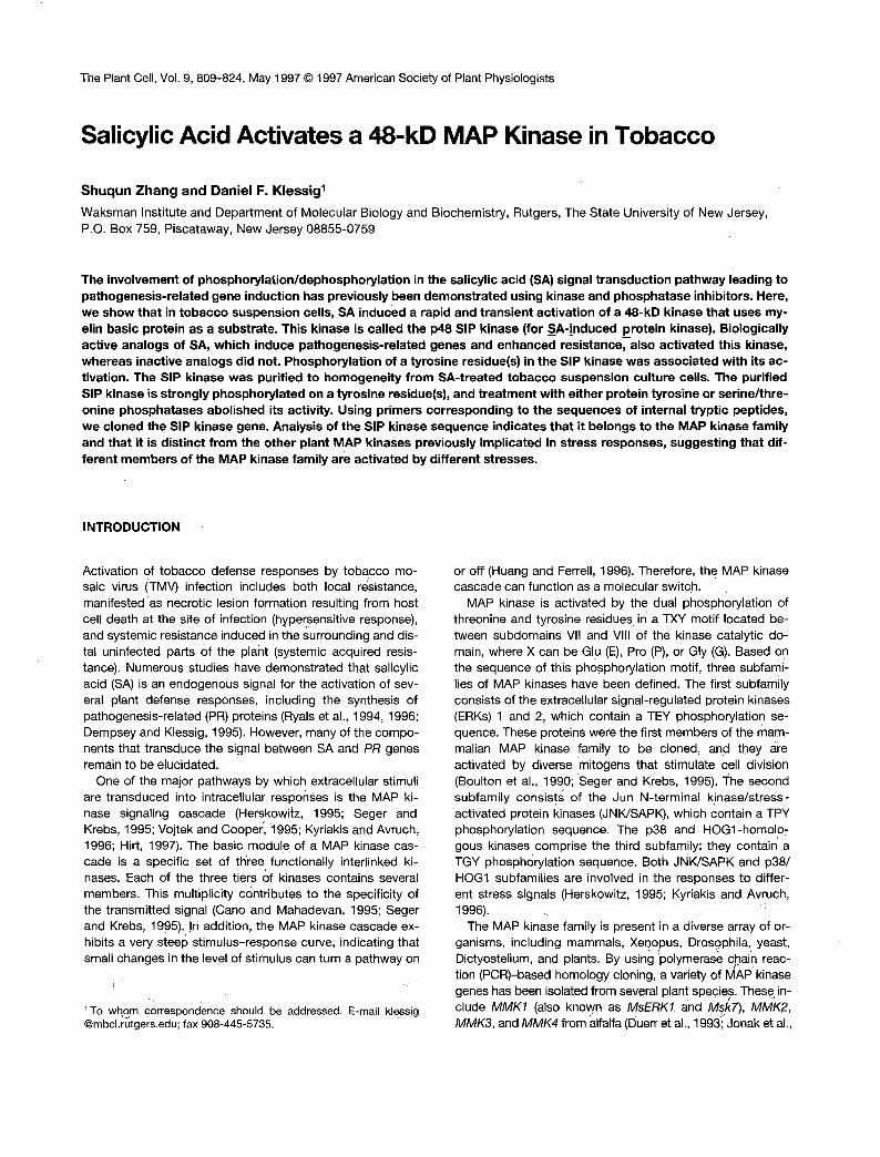
the activation of defense responses in a number of plants(Dietrich et al., 1990; Felix et al., 1991; Viard et al., 1994). Forexample, activation of the potato PR-10a gene requiresphosphorylation of the nuclear factor PBF-1 (Despres et al.,1995), and protein kinase inhibitors blocked both the fungalelicitor-induced oxidative burst and the H2O2-mediated acti-
I".*. 60'
> 40'
0> 20
3 010 1 2 3 4 5
Protein Concentration (mg/mL)
B
OP-Tyr
inCO
Q.
pH1.9 -*———
Figure 7. Analyses of the in Vitro Protein Substrate Preference andthe Amino Acid Phosphorylated in MBP by the p48 SIP Kinase.
(A) Substrate preference was determined using ~0.5 units of puri-fied enzyme for each reaction. The assay conditions were as de-scribed in Methods, except that various concentrations of MBP (•),histone (A; type III-SS), and casein (•) were used.(B) To identify the phosphorylated amino acids, MBP was phospho-rylated in the presence of -y-32P-ATP with 2 units of purified SIP ki-nase under standard assay conditions. After acid hydrolysis, thedried pellet was dissolved in 10 H.L of a phosphoamino acid stan-dard (1 mg/mL each of L-phosphoserine [P-Ser], L-phosphothreo-nine [P-Thr], and L-phosphotyrosine [P-Tyr]). The phosphoaminoacids were separated by two-dimensional high-voltage thin-layerelectrophoresis, as described by Sefton (1996). The circles indicatethe positions of phosphoserine and phosphotyrosine as visualizedby ninhydrin staining. The labeled amino acids were detected by au-toradiography that matched phosphothreonine visualized by ninhy-drin staining. The (+) indicates the origin.
1 2 3 4 5 6 7 8 9 10
Figure 8. Inactivation of Purified p48 SIP Kinase by PhosphataseTreatment.
Approximately 20 units of the SIP kinase was treated with either theserine/threonine-specific phosphatase PP1 (0.1 units) or the ty-rosine-specific protein phosphatase YOP (0.5 units) for 10 min (bars2 to 5) or 30 min (bars 7 to 10) at 30°C in the presence or absence ofa phosphatase inhibitor. Bars 1 and 6 represent the control withoutphosphatase or the phosphatase inhibitor; bars 2 and 7, PP1; bars 3and 8, PP1 plus okadaic acid (1 ^M); bars 4 and 9, YOP; bars 5 and10, YOP plus Na3VO4 (1 mM). Before assaying for SIP kinase activ-ity, the phosphatase inhibitors were added to all of the reactions tothe same final concentrations. Each bar represents the mean ofthree replicates ± SE.
vation of defense genes in soybean suspension cells (Levineet al., 1994). In addition, the formation of TMV-induced le-sions in tobacco was prevented by the phosphatase inhibitorokadaic acid (Dunigan and Madlener, 1995). Similarly, phos-phatase inhibitors abolished the SA-mediated induction ofPR-1 gene expression in tobacco, whereas kinase inhibitorsinduced PR-1 gene expression (Conrath et al., 1997).
Because the SIP kinase is activated by SA, which plays animportant role in signaling defense responses, it is temptingto speculate that the SIP kinase is also involved in the acti-vation of these defense responses. Interestingly, a 48-kD ki-nase that preferentially used MBP as an artificial substratewas activated by both SA treatment and TMV infection of to-bacco leaves (data not shown). We suspect that this SA/TMV-activated kinase and the SIP kinase from suspensioncells are the same. However, further analyses are requiredto determine the identity of these proteins as well as theirassociation with the SA signal transduction pathway. Ourhypothesis that SIP kinase plays a role in SA-mediated PR-1gene induction seems to conflict with the finding that phos-phatase inhibitors block SA induction of PR-1 genes (Conrathet al., 1997). However, there is ample precedence for activa-tion of the same pathway by both phosphorylation and de-phosphorylation events (Hunter, 1995). A particularly relevantexample is the tyrosine kinase c-Src, which activates a MAPkinase cascade (Zheng and Pallen, 1994). c-Src itself is acti-vated by a tyrosine phosphatase (Sun and Tonks, 1994).
818 The Plant Cell
SIPK (100%) Ntfl ( 93%) Msk7 ( 89%) WIPK ( 73%) MMK4 ( 75%) AtMPK3 ( 74%) Ntf6 ( 66%) Ntf3 ( 5 6 % )
P e p t i d e 1 : NIFEVTAK MDGSGQ-QTDTMMSDAGAEQPPTAPQPVAGMDNIPATLSHGGRFIQ~IFGNIFE~AKYKPPIL
.E.G.APPA . . V....A------PAP.QM.IE....V........................... M
MARVNQN.VAEF . . VQT . . . Q.V . . . V. . . L . . . . . . . R. . . M MNTGGGQYTDF .. VDT...Q..S.D...SL..I.S..R...I
MATPVEPPNG1RTP.KHY--.SMWQSL..IDT..V.-.K
. . . PAH- . . . . V . . . . AGQ . . APPS. . . . . I................................. M
MADANMGAGGGQFPDF.SV.T . . . QYV.FD . . . . F..I.T..R...M
MENETNEKLEIKGIPT.E.KWE . . VL . . F....S..I...Q
SIPK Ntfl Msk? WIPK MMKI AtMPK3 Ntf6 Ntf3
SIPK Ntf4 Msk7 WIPK MMK4 AtMPK3 Ntf6 Ntf3
SIPK Ntf4 Msk7 WIPK MMK4 AtMPK3 Ntf 6 Ntf3
SIPK Ntf4 Msk7 . WIPK MMK4 AtMPK3 Ntf6 Ntf3
SIPK Ntfl Msk7 WIPK MMK4 AtMPK3 Ntf6 Ntf 3
PIGKGAYGIVCSALNSETIENVAIKKIANAFDNKIDAKRTLREIKLLRHMDHENIVAIRDIIPPPQREAF . . . . . . . . . N.H . . . . . . . . . . . . . . . . . . . . . . . . . . . . . . . . . . . . . . . . . . . . . . . . . . . . . H. . . . N.H .. v ...........................
. . . R . . . . . . . . V..T.LN.M .. V........IYM.............. L.
... R . . . . . . . . L..T..N.L..V.........HM..........
. . . R . . . . . . . . V.DT . . N.L . . M.........HM..............L.....I....W...L.R Q.
.V.R . . . . M..C.T....K.E......G...E.R.............S.......IK.K..VR..D.. E.
. . . R . . . . . . . . SV.R .. N.K . . . . . . N. .. E.R . . . L. . . . . L. . . . . LR . . . VI.LK.VMM.1H.R S. I I1 IIr IV
P e g t i d e 2 : DLKPSNLLLNAN NDWIAYELMDTDLHQIIRSNQGLSEEHCQYFLYQILRGLKYIHSANVLHRDLKPSNLLLNANCDLKICD . . . . . . . . . . . . . . . . . . . . . . . . . . . . . . . . . . . . . . . . . . . . . . . . . . . . . . . . . . . . . . . . ...................... A. .......................
. . . . D . . . . . M..L . . . . . . . . . . . . TT.. . . . . . N..D R.. S. . . . ST . . . . . . . s . . . . . . . . . . . . ................................... . . . . . v . . . . . . . . A.TDD . . . . . . . . L . . . . . . ............................ K. . . LV . . . . . . . . . . . . K.S.T . . ND . . . . . . F.L . . . . . . L . . . . I. . . . . . . G...I.......
V VI
. . . . . . . . . . . . . .
* P e p t i d e 3 : WYRPPELLLN P e p t i d e 4 : KPLFPGR
FGLARVTSETD-FMTEYWTRWYRPPELLLNSSDYTAAIDVWSVGCIFMELMDRKPLFPGRDHVH ... . . . . . . . . . . A .......................... A . . . . . . . . . . . . . . . . . . . . . . . . . . . . . . . . . . . . . . . . . . . . L
. . . . . PNI.NE-N . . . . . . . . . . . A ........................... N.....G.K.....I.. L
. . . . . P.M.S.-.. M. .L M. .L .G..
-
. . . . . TS.GK.Q . . . . . . . . . . . . A . . . . . CCDN.GTS . . . . . . . . . . A..LG...V...TECLN..K.. vrrr I X - V I 1
I P e p t i d . 9 5 íi 6 : AIDLVEKMLTFDPR
MELIGTPSEAEMEFL-NENAKRYIRQLPLYR PTA VEGALAH . . . . . . . . . . . . . . . . . . . . . . . . . . . . . . .A. . .D.. .. . . . . . . . . . DDLG .E. . .D.. . . T .. L...T..DLG..Q..D.........QHP..QLA.V....N.L.....D.......T......E.. D. T . . L...TD.DVGLVK.DD.R.......Q.P..PLNRV......L.....D....I..T......E.... T..L...T.SDLG.TH..D... .NFP . . PLAKL.S . . N.M . . . . . DR . . . . . . N......Q.. N. 1A.L.S.EDSDLG .. RSD . . R K . VKH .. RVP.HP.SQ . . . D.S.L.L . . A.R . . V...AK.....D.. N. INIL.SQR.ED1 . . 1D.PK.RK . . KS . . YSPGTP.SRLY .. A..L....LQR..V...SK..S.IE..Q.
X xr
PYLNSLHDISDEPICMTPFSFDFEQHALTEEQMKELIYRESLAFNPEYQHM . . . T . . . . . . . . . V. . . . . ................................ . . . T . . . . . . . . . .................................... Q ...AK... AG . . . . . PV . . . . . . . . QGIG . . . I.DM ..Q. A.SL. . . . A ... EK . . . VA ...... E....E...QH.D...I..M....A..L....A Q ..AK... PN . . . . . QK . . . . E...QP.D...I..M..Q.AI.L.. T.G .F.I . . . E.NE . . V.DS . . N.....AS.S.DDI....WN.A.K. D.NTMK ..MSP.Y.PNTD.PAQV.INL.IDED-.G..TIR.MMWS.I.EYH..AATAAMEWL
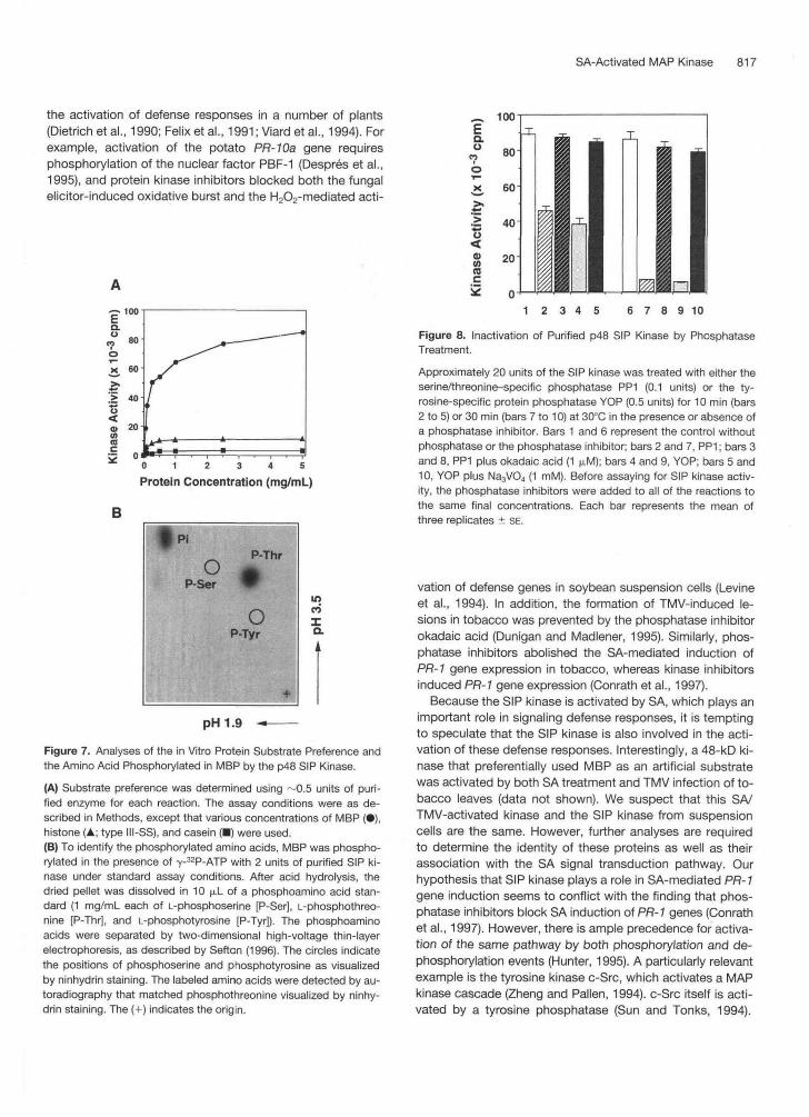
Figure 9. Alignment of the Deduced Amino Acid Sequence of the SIP Kinase (SIPK) with Other Members of the Tobacco MAP Kinase Family as Well as Stress-Related MAP Kinases from Other Plants.
The amino acid sequences of Ntf3, Ntf4, Ntf6 (Wilson et al., 1993, 1995) and WIPK (Se0 et al., 1995) from tobacco, MsK7 (Duerr et al., 1993; Jonak et al., 1993) and MMK4 (Jonak et al., 1996) from alfalfa, and AtMPK3 (Mizoguchi et al., 1993) from Arabidopsis were deduced from cDNA sequences. Numbers within parentheses indicate the percentage of identity to the SIP kinase. Dots represent amino acid residues that match the SIP kinase, and dashes indicate gaps introduced to maximize alignment. The conserved TEY phosphorylation motif for MAP kinase is under- lined. The unique proline residue in the conserved kinase subdomain Vll l of SIP kinase is marked with an asterisk. Roman numerals indicate the 11 major conserved subdomains of serinekhreonine protein kinases. The six peptide sequences obtained by microsequencing are shown on top of the SIP kinase sequence. The boundary between peptides 5 and 6 is indicated by a vertical line. The GenBank accession number for the SIP kinase cDNA is U94192.
SA-Activated MAP Kinase 819
Whereas protein tyrosine kinases and phosphatases have not been found in plants, severa1 receptor-like serinehhreo- nine kinases have been identified, including those encoded by the disease resistance genes Pto (Martin et al., 1993; Scofield et al., 1996; Tang et al., 1996) and Xa27 (Song et al., 1995). In addition, one of the receptor-like serinehhreo- nine kinases, RLK5 of Arabidopsis, has an associated phos- phatase, KAPP (Stone et al., 1994). It has been suggested that the receptor-like serine/threonine kinase-phosphatase pairs mimic the receptor tyrosine kinase-phosphatase pairs in animals (Braun and Walker, 1996).
The availability of the purified SIP kinase has allowed the primary characterization of a plant MAP kinase both in its active form and in the absence of other contaminating activ- ities. The purified protein was strongly phosphorylated on a tyrosine residue(s). Moreover, both tyrosine-specific protein phosphatase and serinehhreonine-specific protein phos- phatase treatments inactivated the kinase activity, suggest- ing that phosphorylation of these residues was required for SIP kinase activation. The purified p48 SIP kinase had a specific activity of 340 nmol min-l mg-l, with MBP as its phosphate acceptor. This is at least 40- and 200-fold higher than the specific activities of purified bovine brain p42 and p44 MAP kinases, respectively (Childs and Mak, 1993a). These large differences may be due in part to the efficient activation of the SIP kinase by SA and/or the ability of MBP to serve as a better substrate for the SIP kinase. In addition, the high specific activity of the SIP kinase may reflect the purification strategy, which allowed purification of the SIP kinase in 3 days. Rapid purification is critical because the phosphatase inhibitors included in the buffer were unable to completely inhibit all of the phosphatases that can inactivate the SIP kinase.
The purified SIP kinase exhibited an absolute requirement for Mgz+ and could barely use Mn2+-ATP as a phosphate do- nor. This is in contrast to the cloned tobacco MAP kinases Ntf3, Ntf4, and Ntf6 (wilson et al., 1995). For these three re- combinant proteins, Mn2+ was a better cofactor than Mgz+ in both MBP phosphorylation and autophosphorylation reac- tions. It is possible that the SIP kinase and the other tobacco MAP kinases, Ntf3, Ntf4, and Ntf6, inherently prefer different metal ions. Alternatively, the basal and activated forms of these kinases may utilize different metal cofactors. Which of these explanations is correct cannot be determined at present because the fusion proteins expressed in Escherichia coli were largely in their inactive state because of the lack of up- stream activating components, whereas the SIP kinase was purified in an active state. The purified p48 SIP kinase strongly phosphorylated MBP, a preferred substrate of MAP kinases. Histone was weakly phosphorylated, and casein is not a sub- strate for the SIP kinase. Similar substrate preferences have been reported for the tobacco Ntf3, Ntf4, and Ntf6 MAP ki- nases expressed in E. coli (wilson et al. 1995) and the murine and sea star MAP kinases (Rossomando et al., 1991).
Two other stress-activated kinases that exhibit character- istics associated with MAP kinases have been described in
tobacco. These include the p47 funga1 elicitor-activated ki- nase (Suzuki and Shinshi, 1995) and the p46 cutting-acti- vated kinase (Usami et al., 1995). Because purification of either of these proteins has yet to be reported, they cannot be directly compared with the purified p48 SIP kinase. Thus, it is unclear whether they differ from the SIP kinase in enzy- matic properties, such as metal cofactor requirement, Km and V,,, values for different substrates, or inhibitor sensitiv- ity. These properties could be used to determine whether they are the same or different MAP kinases. A gene whose transcript is induced by wounding (WIPK) has been isolated from tobacco and proposed to encode the p46 wounding- activated kinase (Se0 et al., 1995). If this indeed is the case, then the wounding-activated kinase (which may be the same as the cutting-activated kinase) is different from the SIP kinase, based on a comparison of their amino acid se- quences (Figure 9).
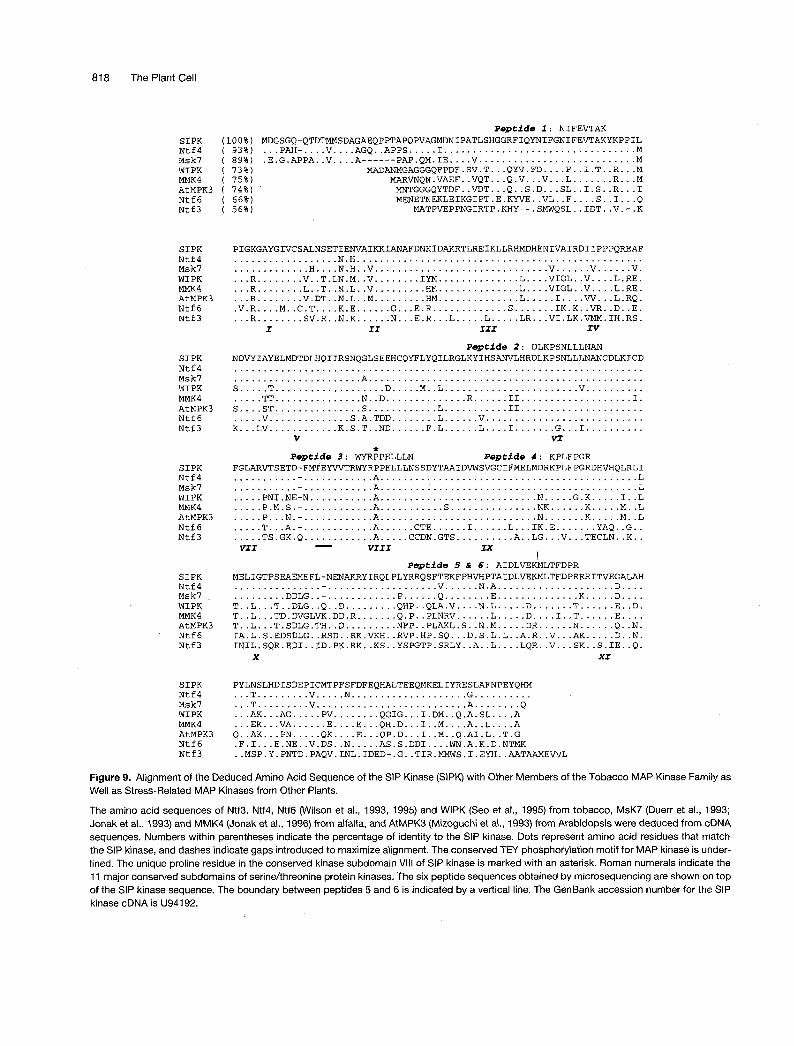
Analysis of the phylogenetic tree constructed for all cloned plant MAP kinases suggests that these kinases can be divided into two major groups (Figure 10). One group is represented by the 2,4-D-responsive AtMPKl and AtMPK2 of Arabidopsis. These kinases may be functionally similar to the mammalian ERKl and ERK2, which are involved in the cell proliferation in response to mitogens (Mizoguchi et al.,
I I
MSk? PsMAPK Ntf4 SIPK AtMPK6 AtMPK3 WIPK MMK4 AsMAPl AtMPK4 MMK2 AtMPKS Ntf6 AtMPKl AtMPK2 Ntf3 PMEKl AtMPK7
Figure 10. Relationship between All Cloned Plant MAP Kinase Members.
The phylogenetic tree shown as a dendrogram was created by the Clustal method (MegaAlign program; DNAStar, Madison, WI). The deduced amino acid sequences are from cDNA clones of Msk7 (also termed MsfRK7 or MMKI; Duerr et al., 1993; Jonak et al., 1993), AfMPKl to AfMPK7 (Mizoguchi et al., 1993), PsMAPK (also called D5; Stafstrom et al., 1993; Popping et al., 1996), Nff3 (wilson et al., 1993), AsMAPl (Huttly and Phillips, 1995), PMEK7 (Decroocq- Ferrant et al., 1995), MMK2 (Jonak et al., 1995), WPK (Se0 et al., 1995), Nff4 and Nff6 (wilson et al., 1995), and MMK4 (Jonak et al., 1996) and for the SIP kinase (SIPK; this study).

820 The Plant Cell
1994). The other group can be further divided into three sub- groups. The subgroup represented by AtMPK3 of Arabidop- sis, WlPK of tobacco, and MMK4 of alfalfa appears to be activated by a variety of stresses, such as wounding, touch, cold, drought, and salinity (Se0 et al., 1995; Bogre et al., 1996, 1997; Jonak et al., 1996; Mizoguchi et al., 1996). Inter- estingly, this group is also characterized by increases in their mRNA levels in response to the same stresses. The second subgroup includes Ntf4 and the SIP kinase from to- bacco. Its members have N-terminal extensions of -20 amino acids and, based on results described in this report, may also respond to stresses such as those caused by in- fection or SA. The function of the third subgroup is currently unknown. However, MMK2 of alfalfa can complement the yeast MPKl (Jonak et al., 1995), which is required for yeast cell wall integrity at high temperature (Herskowitz, 1995).
In yeast and mammals, multiple MAP kinase families have been identified that participate in different signaling path- ways (Cano and Mahadevan, 1995; Herskowitz, 1995). In yeast, for example, at least six independent MAP kinase pathways have been identified (Herskowitz, 1995; Ruis and Schüller, 1995). In human cells, the JNK subfamily of MAP kinases alone consists of at least 10 isoforms that are pro- duced by alternative splicing of transcripts encoded by the JNK7, JNK2, and JNK3 genes. The differing abilities of these isoforms to interact with the transcription factors ATF2, Jun, and Elk-1 may provide a mechanism by which various stresses cause the specific activation of different target genes (Gupta et al., 1996). Consistent with the picture emerging from studies with yeast and mammals, our study provides further evidence that different members of the plant MAP kinase family are activated by different stimuli.
METHODS
Treatment of Tobacco Cell Suspension Culture
The cell suspension culture was derived from callus tissue from leaves of Nicotiana tabacum cv Xanthi and grown in Murashige and Skoog medium (Gibco BRL, Gaithersburg, MD) supplemented with 1 mg/L a-naphthaleneacetic acid, 0.1 mg/L 2,4-D, 0.1 mg/L benzylad- enine, and 3% sucrose. Log phase cells were used -3 days after a 1:IO dilution. Treatment with salicylic acid (SA) and its analogs was done in the original flasks in the dark to avoid any stresses associ- ated with transfer. At various times, 10 mL of cells (-0.2 to 0.3 g fresh weight of cells) was harvested by filtration. The cells were quickly frozen in liquid nitrogen and stored at -80°C until analysis.
Preparation of Protein Extracts
To prepare extracts from treated cells, cells (-0.2 g) with 2 volumes (w/v) of extraction buffer (100 mM Hepes, pH 7.5,5 mM EDTA, 5 mM EGTA, 10 mM DTT, 10 mM Na,VO,, 10 mM NaF, 50 mM P-glycero- phosphate, 1 mM phenylmethylsulfonyl fluoride, 5 p,g/mL antipain, 5 pg/mL aprotinin, 5 p,g/mL leupeptin, 10% glycerol, 7.5% polyvi-
nylpolypyrrolidone) were sonicated twice for 15 sec each, with a W-375 Sonicator (Heat System-Ultrasonics, Inc., Farmingdale, NY) fitted with a microprobe at setting 4 and 80% duty cycle in a 1.5-mL microcentrifuge tube. After centrifugation at 13,000 rpm for 20 min, supernatants were transferred into clean tubes, quickly frozen in liq- uid nitrogen, and stored at -80°C.
For purification of the p48 SIP kinase (for SA-jnduced protein ki- nase), -200 g of cells treated for 5 min with 250 pM SAwas soni- cated in 1.5 volumes of extraction buffer (w/v) until all cells were disrupted. After centrifugation at 23,0009 for 30 min, the supernatant was transferred into clean tubes, quickly frozen in liquid nitrogen, and stored at -80°C.
Protein Concentration Assay
Unless otherwise stated, the concentration of protein extracts was de- termined using the Bio-Rad protein assay kit with BSA as a standard.
In-Gel Kinase Activity Assay
The in-gel kinase assay was performed as described previously (Zhang et al., 1993). Extracts containing 10 pg of protein were elec- trophoresed on 10% SDS-polyacrylamide gels embedded with 1 mg/mL casein, 1 mg/mL histone (type III-SS; Sigma), or 0.25 mg/mL of myelin basic protein (MBP) in the separating gel as a substrate for the kinase. After electrophoresis, SDS was removed by washing the gel with washing buffer (25 mM Tris, pH 7.5, 0.5 mM DTT, 0.1 mM Na3V0,, 5 mM NaF, 0.5 mg/mL BSA, 0.1 % Triton X-100 [v/v]) three times, each for 30 min at room temperature. The kinases were allowed to renature in 25 mM Tris, pH 7.5, 1 mM DTT, 0.1 mM Na3V0,, and 5 mM NaF at 4°C overnight with three changes of buffer. The gel w.@ then incubated at room temperature in a 30-mL reaction buffer (25 Tris, pH 7.5,2 mM EGTA, 12 mM MgCI,, 1 mM DTT, 0.1 mM Na3V0,) with 200 nM ATP plus 50 +Ci Y-~~P-ATP (3000 Ci/mmol) for 60 min. The reaction was stopped by transferring the gel into 5% trichloroace- tic acid (TCA) (w/v)/l% NaPPi (w/v). The unincorporated Y-~~P-ATP was removed by washing in the same solution for at least 6 hr with five changes. The gel was dried onto Whatman 3MM paper and exposed to Kodak XAR-5 film. Prestained size markers (Bio-Rad) were used to calculate the size of kinases. Quantitation of the relative kinase activ- ities was done using a Phosphorlmager (Molecular Dynamics Inc., Sunnyvale, CA).
e
lmmunoblot Analysis with the Anti-Phosphotyrosine Antibody
Protein extracts or pooled fractions from each purification step were separated on 10% SDS-polyacrylamide gels, and the proteins were transferred to nitrocellulose membrane by semidry electroblotting. The membrane was blocked for 2 hr in TBS buffer (20 mM Tris, pH 7.5, 150 mM NaCI, 0.1% Tween 20, 0.1 mM Na3VO4) with 10% BSA at room temperature and then incubated with the phosphotyrosine-specific monoclonal antibody 4G1 O (Upstate Biotechnology Incorporated, Lake Placid, NY) in TBS buffer with BSA for 1 hr. After the blot was in- cubated with horseradish peroxidase-conjugated secondary antibody, the complexes were visualized using an enhanced chemiluminescence kit (Du Pont), following the manufacturer’s instructions.

SA-Activated MAP Kinase 821
lmmunoprecipitation Kinass Activity Assay
Protein extract (50 pg) with or without phosphoamino acid (1 mM fi- nal concentration) competitor was incubated with the 4G1 O antibody in immunoprecipitation buffer (20 mM Tris, pH 7.5, 150 mM NaCI, 1 mM EDTA, 1 mM EGTA, 1 mM Na,VO,, 1 mM NaF, 10 mM p-glyc- erophosphate, 2 pg/mL antipain, 2 pg/mL aprotinin, 2 pg/mL leu- peptin, 0.5% Triton X-100, 0.5% Nonidet P-40) at 4°C for 4 hr on a rocker. About 25 pL packed volume of protein A-agarose was added, and the incubation was continued for another 2 hr. Agarose bead-protein complexes were pelleted by brief centrifugation. After washing with immunoprecipitation buffer three times, 1 x SDS sam- ple buffer was added and boiled for 3 min. After centrifugation, the supernatant fraction was electrophoresed on 10% SDS-polyacryl- amide gel, and the in-gel kinase assay was performed.
Purification of the p48 SIP Kinase
All purification procedures were performed in a cold room or a 4°C chamber. Protein extract from -200 g of SA-treated cells was brought to 30% (NH4),S04 saturation. After stirring slowly for 30 min, the precipitant was collected by centrifugation at 23,OOOg for 1 O min. The pellets were then dissolved in a total of 30 mL of buffer A (25 mM Tris, pH 7.5, 1 mM EGTA, 10 mM p-glycerophosphate, 0.1 mM Na,V04, 1 mM DTT, 5% glycerol) plus 1 mM phenylmethylsulfonyl fluoride and 5 pg/mL each of antipain, aprotinin, and leupeptin. After centrifugation at 130,OOOg for 1 hr, the SI 30 supernatant was dia- lyzed against 1 liter of buffer A for 4 hr in a cold room and loaded onto a 10-mL Q-Sepharose anion exchange column (two tandemly connected 5-mL HiTrapQ columns; Pharmacia) equilibrated with buffer A plus 50 mM NaCl (Figure 4A). After washing with 30 mL of buffer A containing 50 mM NaCI, the column was eluted with a 150- mL linear gradient of 50 to 400 mM NaCl in buffer A. The p48 SIP ki- nase activity eluted at -250 mM.
The fractions containing the highest kinase activity (peak fractions) were pooled, adjusted to a final concentration of 300 mM NaCI, and loaded onto a 15-mL phenyl-Sepharose HP hydrophobic interaction column (1.6 x 7.5 cm; Pharmacia) equilibrated with buffer A plus 300 mM NaCl (Figure 4B). The column was washed with 50 mL of buffer A plus 300 mM NaCl and eluted with a 100-mL linear gradient of O to 60% ethylene glycol and 300 to O mM NaCl in buffer A. The active fractions (eluting at -40% ethylene glycol) were pooled and diluted with an equal volume of buffer A and then loaded onto a MonoQ HR 5/5 fast protein liquid chromatography anion exchange column (Pharmacia) equilibrated with buffer A plus 1 O0 mM NaCl (Figure 4C). After washing with 5 mL of buffer A plus 100 mM NaCI, the column was eluted with a 30" gradient of 1 O0 to 400 mM NaCl in buffer A. The peak fractions containing the p48 SIP kinase ("3 mL) were pooled, adjusted to a final concentration of 10 mM MgCI,, and di- luted with an equal volume of buffer B (25 mM Tris, pH 7.5, 10 mM MgCI,, 1 mM EGTA, 1 mM DTT, 10 mM p-glycerophosphate, 0.1 mM Na3V04, 0.02% Triton X-100).
The above-mentioned sample was then loaded onto an MBP- Sepharose affinity column (1 mL; prepared by coupling MBP to N-hydroxysuccinimideactivated Sepharose HP; Pharmacia) equili- brated with buffer B plus 50 mM NaCl (Figure 4D). The column was then step eluted with 5 mL each of buffer B plus 100, 500, or 1 O00 mM NaCI. The peak fractions containing the p48 SIP kinase were pooled (-2.5 mL), diluted with 3 volumes of buffer B, and loaded onto a 3.5-mL poly-L-lysine-agarose column (0.9 x 5.5 cm; Sigma). After
washing with buffer B plus 50 mM NaCI, the column was eluted with a 50-mL gradient of 50 to 800 mM NaCl in buffer B (Figure 4E). The active fractions were pooled and concentrated with a Centricon filter (1 0,000 molecular weight cut-off Amicon, Beverly, MA).
To further purify the p48 SIP kinase, the above-mentioned concen- trated sample was loaded onto a Superdex 200 HR 10/30 fast pro- tein liquid chromatography gel filtration column (Pharmacia) equilibrated with buffer B plus 250 mM NaCI, and the coJumn was eluted with the same buffer at 0.5 mL/min (Figure 4F). For the mea- surement of the native molecular weight, the purified p48 SIP kinase was mixed with gel filtration size standards (Bio-Rad) and loaded onto the Superdex 200 HR 10/30 column. The retention time of the SIP kinase activity peak was used to calculate its molecular weight. The purified or partially purified SIP kinase after the poly-L-lysine agarose step was stored at -20°C in buffer B plus 50% glycerol and was found to be stable for severa1 months.
Assay of Kinase Activity in Solution
Unless specifically indicated, assays were performed at room tem- perature for 20 min in a final volume of 15 pL containing 0.5 mg/mL MBP, 50 pM Y-~~P-ATP (3000 to 6000 cpm/pmol), 25 mM Tris, pH 7.5,5 mM MgCI2, 1 mM EGTA, 1 mM DTT, and enzyme. The reaction was terminated by the addition of an equal volume of 150 mM H3P04; 20 pL of the mixture was then spotted onto an eight-well phosphocellulose filter strip (Pierce, Rockford, IL). After washing ex- tensively with 150 mM H3P04, the phosphoprotein was eluted with 0.4 mL of 1 N NaOH, and the radioactivity was determined by liquid scintillation counting. Total counts per minute in the reaction mixture was also determined to calculate the specific activity of the enzyme preparation. One unit of p48 SIP kinase was defined as the amount of enzyme that can catalyze the transfer of 1 pmol of phosphate from ATP to MBP in 1 min.
ldentification of Phosphorylated Amino Acids in the Substrates
The procedure used to identify phosphorylated amino acids was identical to that described earlier (Zhang et al., 1993), except two- dimensional thin-layer electrophoresis was used. Protein substrates were labeled by phosphorylation in the presence of y3*P-ATP and precipitated by using 10% (w/v) TCA. After washing with 10% (w/v) TCA and acetone, the pellets were hydrolyzed in 6 N HCI for 2 hr at 11 O"C, dried in an evaporator (Speed-Vac; Savant Instruments, Inc., Farmingdale, NY), and then dissolved in 1 O pL of a phosphoamino ac- ids standard (1 mg/mL each of L-phosphoserine, L-phosphothreonine, and L-phosphotyrosine). The phosphoamino acids were separated by two-dimensional high-voltage thin-layer electrophoresis, as described by Sefton (1996). The position of the standards was visualized by nin- hydrin (0.2% [w/v] in acetone), and the labeled amino acids were de- tected by autoradiography.
Treatment of the p48 SIP Kinase with Phosphatases
The phosphatase inhibitors present in the purified p48 SIP kinase preparation were first removed by dialysis against buffer B lacking p-glycerophosphate and Na3V04 in a 0.5-mL 10K dialysis cassette (Slide-A-Lyzer; Pierce). For treatment with serine/threonine protein phosphatase PP1 (Calbiochem, San Diego, CA), MnCI,, DTT, and BSA were added to aliquots of the SIP kinase preparation to final

822 The Plant Cell
concentrations of 200 pM, 5 mM, and 100 kg/mL, respectively. Then 0.1 unit of PP1 was added, and the reaction mixture was incubated at 30°C in the presence or absence of 1 pM of the phosphatase in- hibitor okadaic acid (Calbiochem). For treatment with the tyrosine- specific protein phosphatase YOP (Calbiochem), NaCI, DTT, and BSA were added to purified SIP kinase to final concentrations of 150 mM, 5 mM, and 100 kg/mL, respectively. Then 0.5 unit of YOP was added, and the reaction was incubated at 30°C in the presence or absence of 1 mM of the tyrosine phosphatase inhibitor Na3V04. Be- fore assaying for the SIP kinase activity, the phosphatase inhibitors were brought to the same final concentration.
Microsequencing of Interna1 Tryptic Peptides
The protein in the active fractions eluted from the poly-L-lysine col- umn was concentrated and precipitated with acetone. The pellets were then dissolved in SDS-Laemmli sample buffer and separated on a 10% SDS-polyacrylamide gel. After Coomassie Brilliant Blue R 250 staining, the 48-kD band was excised and sent to the W.M. Keck Foundation Biotechnology Resource Laboratory (Yale University, New Haven, CT) for amino acid composition and sequence analyses. Briefly, for sequence analysis, the p48 SIP kinase was subjected to in-gel digestion with trypsin, and the resultant peptides were sepa- rated by reverse phase HPLC. After determination of molecular mass and purity by using matrix-assisted laser desorption ionization (MALDI) mass spectrometry, selected peptides were sequenced.
Cloning of the Tobacco SIP Kinase Gene
Two primers, 5’-AAYATHTTYGARGTNACNGC-3’ and 5’-CKNC- CNGGRAANARNGGYTT-3’ (where H is A, T, and C; K is T and G; N is A, T, C, and G; R is A and G; and Y is T and C), which correspond to peptide 1 and peptide 4 (Figure 9), respectively, were used to polymerase chain reaction (PCR) amplify the cDNA that was reverse transcribed from poly(A) RNA prepared from tobacco cell suspension culture. The reverse transcription-PCR product of ~ 6 0 0 bp was cloned into pGEM-T vector (Promega) and sequenced. A clone whose deduced amino acid sequence matched the interna1 peptide 2 and peptide 3 (Figure 9) was labeled with LT-~~P-~CTP and used to screen a tobacco cDNA library under high stringency. Briefly, nylon membranes (Duralon-UV; Stratagene, La Jolla, CA), each with 5 x 10⁴ plaque-forming units, were hybridized at 42°C for 20 hr in solu- tion containing 20 mM Pipes, pH 6.5, 0.8 M NaCI, 50% formamide, 1 % (w/v) SDS, 100 pg/mL denatured salmon sperm DNA, plus 106 cpm/mL of probe. After hybridization, the filters were washed once with 1 x SSC (0.15 M NaCI, 0.015 M sodium citrate), O.l%(w/v) SDS at room temperature, and three times, each for 15 min, with 0.1 x SSC, 0.1 %(w/v) SDS at 55°C. More than 10 positive clones were ob- tained by screening 4 0 6 plaque-forming units. Both strands of the clone containing the longest insert were sequenced using the Se- quenase 2.0 kit (Amersham).
ACKNOWLEDGMENTS
The amino acid analysis of the p48 SIP kinase was performed by the W.M. Keck Foundation Biotechnology Resource Laboratory at Yale University. We gratefully acknowledge D’Maris Dempsey for assis-
tance preparing this manuscript. We thank members of the laboratory for their critical reading of the manuscript. This work was supported by Grant No. MCB-9310371 from the National Science Foundation.
Received February 24, 1997; accepted February 27,1997.
REFERENCES
Bogre, L., Ligterink, W., Heberle-Bors, E., and Hirt, H. (1996). Mechanosensors in plants. Nature 383, 489-490.
Bogre, L., Ligterink, W., Meskiene, I., Barker, P.J., Heberle-Bors, E., Huskisson, N.S., and Hirt, H. (1997). Wounding induces the rapid and transient activation of a specific MAP kinase pathway. Plant Cell 9, 75-83.
Bokemeyer, D., Sorokin, A., Yan, M., Ahn, N.G., Templeton, D.J., and Dunn, M.J. (1 996). lnduction of mitogen-activated protein kinase phosphatase 1 by the stress-activated protein kinase sig- naling pathway but not by extracellular signal-regulated- kinase in fibroblasts. J. Biol. Chem. 271, 639-642.
Boulton, T.G., Yancopoulos, G.D., Gregory, J.S., Slaughter, C., Moomaw, C., Hsu, J., and Cobb, M.H. (1990). An insulin-stimu- lated protein kinase similar to yeast kinases involved in cell cycle control. Science 249, 64-67.
Braun, D.M., and Walker, J.C. (1996). Plant transmembrane recep- tors: New pieces in the signaling puzzle. Trends Biochem. Sci. 21,
Cano, E., and Mahadevan, L.C. (1 995). Parallel signal processing among mammalian MAPKs. Trends Biochem. Sci. 20, 117-122.
Childs, T.J., and Mak, A.S. (1993a). MAP kinases from bovine brain: Purification and characterization. Biochem. Cell Biol. 71, 544-555.
Childs, T.J., and Mak, A.S. (1 993b). Smooth-muscle mitogen-acti- vated protein (MAP) kinase: Purification and characterization, and the phosphorylation of caldesmon. Biochem J. 296,745-751.
Conrath, U., Chen, Z., Ricigliano, J.R., and Klessig, D.F. (1995). Two inducers of plant defense responses, 2,6-dichloroisonicotinic acid and salicylic acid, inhibit catalase activity in tobacco. Proc. Natl. Acad. Sci. USA 92, 7143-7147.
Conrath, U., Silva, H., and Klessig, D.F. (1997). Protein dephos- phorylation mediates salicylic acid-induced expression of PR-7 genes in tobacco. Plant J. 11,747-757.
Decroocq-Ferrant, V., Decroocq, S., Van Went, J., Schmidt, E., and Kreis, M. (1995). A homologue of the MAP/ERK family of protein kinase genes is expressed in vegetative and in female reproductive organs of Petunia hybrida. Plant MOI. Biol. 27,334-350.
Dempsey, D.A., and Klessig, D.F. (1995). Signals in plant disease resistance. Bull. Inst. Pasteur 93, 167-186.
Després, C., Subramaniam, R., Matton, D.P., and Brisson, N. (1 995). The activation of the potato PR-lOa gene requires the phos- phorylation of the nuclear factor PBF-1. Plant Cell7, 589-598.
Dietrich, A., Mayer, J.E., and Hahlbrock, K. (1 990). Funga1 elicitor triggers rapid, transient, and specific protein phosphorylation in parsley cell suspension cultures. J. Biol. Chem. 265, 6360-6368.
70-73.

SA-Activated MAP Kinase 823
Duerr, B., Gawienowski, M., Ropp, T., and Jacobs, T. (1993). MsERK1: A mitogen-activated protein kinase from a flowering plant. Plant Cell 5, 87-96.
Dunigan, D.D., and Madlener, C. (1 995). Serinekhreonine protein phosphatase is required for tobacco mosaic virus-mediated pro- grammed cell death. Virology 207, 460-466.
Felix, G., Grosskopf, D.G., Regenass, M., and Boller, T. (1991). Rapid changes of protein phosphorylation are involved in trans- duction of the elicitor signal in plant cells. Proc. Natl. Acad. Sci. USA 88,8831-8834.
Groom, L.A., Sneddon, A.A., Alessi, D.R., Dowd, S., and Keyse, S.M. (1996). Differential regulation of the MAP, SAP, and RWp38 kinases by Pystl , a novel cytosolic dual-specificity phosphatase.
Gupta, S., Barrett, T., Whitmarsh, A.J., Cavanagh, J., Sluss, H.K., Dérijard, B., and Davis, R.J. (1996). Selective interaction of JNK protein kinase isoforms with transcription factors. EMBO J. 15,
Hanks, S.K., and Hunter, T. (1995). The eukaryotic protein kinase superfamily: Kinase (catalytic) domain structure and classification.
Herskowitz, 1. (1995). MAP kinase pathways in yeast: For mating and more. Cell80, 187-197.
Hirt, H. (1997). Multiple roles of MAP kinases in plant signal trans- duction. Trends Plant Sci. 2, 11-15.
Huang, C.-Y.F., and Ferrell, J.E., Jr. (1996). Ultrasensitivity in the mitogen-activated protein kinase cascade. Proc. Natl. Acad. Sci.
Hunter, T. (1995). Protein kinases and phosphatases: The yin and yang of protein phosphorylation and signaling. Cell 80, 225-236.
Huttly, A.K., and Phillips, A.L. (1 995). Gibberellin-regulated expres- sion in oat aleurone cells of two kinases that show homology to MAP kinase and a ribosomal protein kinase. Plant MOI. Biol. 27,
Jonak, C., Páy, A., Bogre, L., Hirt, H., and Heberle-Bors, E. (1993). The plant homologue of MAP kinase is expressed in a cell cycle-dependent and organ-specific manner. Plant J. 3,611-61 7.
Jonak, C., Heberle-Bors, E., and Hirt, H. (1994). MAP kinases: Uni- versal multipurpose signaling tools. Plant MOI. Biol. 24,407-41 6.
Jonak, C., Kiegerl, S., Lloyd, C., Chan, J., and Hirt, H. (1995). MMK2, a novel alfalfa MAP kinase, specifically complements the yeast MPKl function. MOI. Gen. Genet. 248, 686-694.
Jonak, C., Kiegerl, S., Ligterink, W., Barker, P.J., Huskisson, N.S., and Hirt, H. (1996). Stress signaling in plants: A mitogen- activated protein kinase pathway is activated by cold and drought. Proc. Natl. Acad. Sci. USA 93, 11274-1 1279.
Keyse, S.M. (1995). An emerging family of dual specificity MAP kinase phosphatases. Biochim. Biophys. Acta 1265, 152-1 60.
Klessig, D.F., and Malamy, J. (1994). The salicylic acid signal in plants. Plant MOI. Biol. 26, 1439-1458.
Knetsch, M.L.W., Wang, M., Snaar-Jagalska, B.E., and Heimovaara- Dijkstra, S. (1 996). Abscisic acid induces mitogen-activated pro- tein kinase activation in barley aleurone protoplasts. Plant Cell 8,
Kyriakis, J.M., and Avruch, J. (1996). Protein kinase cascades acti- vated by stress and inflammatory cytokines. Bioessays 18, 567-577.
EMBO J. 15,3621-3632.
2760-2770.
FASEB J. 9,576-596.
USA 93,10078-10083.
1043-1 052.
1061-1067.
Levine, A., Tenhaken, R., Dixon, R., and Lamb, C. (1994). H,O, from the oxidative burst orchestrates the plant hypersensitive dis- ease resistance response. Cell79, 583-593.
Martin, G.B., Brommonschenkel, S.H., Chunwongse, J., Frary, A., Ganal, M.W., Spivey, R., Wu, T., Earle, E.D., and Tanksley, S.D. (1993). Map-based cloning of a protein kinase gene confer-,.- ring disease resistance in tomato. Science 262, 1432-1436.
Mizoguchi, T., Hayashida, N., Yamaguchi-Shinozaki, K., Kamada, H., and Shinozaki, K. (1993). ATMPKs: A gene family of plant MAP kinases in Arabidopsis thaliana. FEBS Lett. 336,440-444.
Mizoguchi, T., Gotoh, Y., Nishida, E., Yamaguchi-Shinozaki, K., Hayashida, N., Iwasaki, T., Kamada, H., and Shinozaki, K. (1994). Characterization of two cDNAs that encode MAP kinase homologues in Arabidopsis fhaliana and analysis of the possible role of auxin in activating such kinase activities in cultured cells. Plant J. 5, 111:122.
Mizoguchi, T., Irie, K., Hirayama, T., Hayashida, N., Yamaguchi- Shinozaki, K., Matsumota, K., and Shinozaki, K. (1996). A gene encoding a mitogen-activated protein kinase kinase kinase is induced simultaneously with genes for. a mitogen-activated pro- tein kinase and an S6 ribosomal protein kinase by touch, cold, and water stress in Arabidopsis thaliana. Proc. Natl. Acad. Sci.
Nishihama, R., Banno, H., Shibata, W., Hirano, K., Nakashima, M., Usami, S., and Machida, Y. (1995). Plant homologues of compo- nents of MAPK (mitogen-activated protein kinase) signal pathways in yeast and animal cells. Plant Cell Physiol. 36, 749-757.
Popping, B., Gibbons, T., and Watson, M.D. (1996). The Pisum sativum MAP kinase homologue (PsMAPK) rescues the Saccharo- myces cerevisiae hOg7 deletion mutant under conditions of high osmotic stress. Plant MOI. Biol. 31, 355-363.
Rossomando, A.J., Sanghera, J.S., Marsden, L.A., Weber, M.J., Pelech, S.L., and Sturgill, T.W. (1 991). Biochemical characteriza- tion of a family of serinekhreonine protein kinases regulated by tyrosine and serine/threonine phosphorylation. J. Biol. Chem. 266,
Ruis, H., and Schiiller, C. (1995). Stress signaling in'yeast. Bioes- says 17,959-965.
Ryals, J.A., Uknes, S., and Ward, E. (1994). Systemic acquired resistance. Plant Physiol. 104, 11 09-1 11 2.
Ryals, J.A., Neuenschwander, U.H., Willits, M.G., Molina, A., Steiner, H.-Y., and Hunt, M.D. (1996). Systemic acquired resis- tance. Plant Cell8, 1809-1 81 9.
Scofield, S.R., Tobias, C.M., Rathjen, J.P., Chang, J.H., Lavelle, D.T., Michelmore, R.W., and Staskawicz, B.J. (1 996). Molecular basis of gene-for gene specificity in bacterial speck disease- of tomato. Science 274,2063-2065.
Sefton, B.M. (1996). Phosphoamino acid analysis. In Current Proto- cols in Molecular Biology, F.M. Ausubel, R. Brent, R.E. Kingston, D.D. Moore, J.G. Seidman, J.A. Smith, and K. Struhl, eds (New York: John Wiley and Sons), pp. 18.3.1-18.3.8.
Seger, R., and Krebs, E.G. (1995). The MAPK signaling cascade.
USA 93,765-769.
20270-20275.
FASEB J. 9,726-735.
Seo, S., Okamoto, M., Seto, H., Ishizuka, K., Sano, H., and Ohashi, Y. (1995). Tobacco MAP kinase: A possible mediator in wound signal transduction pathways. Science 270,1988-1992.

824 The Plant Cell
Song, W.-Y., Wang, G.-L., Chen, L.-L., Kim, H.-S., Pi, L.-Y., Holsten, T., Gardner, J., Wang, B., Zhai, W.-X., Zhu, L.-H., Fauquet, C., and Ronald, P. (1995). A receptor kinase-like pro- tein encoded by the rice disease resistance gene Xa27. Science
Stafstrom, J.P., Altschuler, M., and Anderson, D.H. (1 993). Molecular cloning and expression of a MAP kinase homologue from pea. Plant MOI. Biol. 22, 83-90.
Stone, J.M., and Walker, J.C. (1995). Plant protein kinase families and signal transduction. Plant Physiol. 108, 451-457.
Stone, J.M., Collinge, M.A., Smith, R.D., Horn, M.A., and Walker, J.C. (1994). lnteraction of a protein phosphatase with an Arabidopsis serine-threonine receptor kinase. Science 266, 793-795.
Sun, H., and Tonks, N.K. (1994). The coordinated action of protein tyrosine phosphatases and kinases in cell signaling. Trends Bio- chem. Sci. 19,480-485.
Suzuki, K., and Shinshi, H. (1995). Transient activation and tyrosine phosphorylation of a protein kinase in tobacco cells treated with a funga1 elicitor. Plant Cell 7 , 639-647.
Tang, X., Frederick, R.D., Zhou, J., Halterman, D.A., Jia, Y., and Martin, G.B. (1996). lnitiation of plant disease resistance by physi- cal interaction of AvrPto and Pto kinase. Science 274,2060-2063.
270, 1804-1806.
Usami, S., Banno, H., Ito, Y., Nishihama, R., and Machida, Y. (1 995). Cutting activates a 46-kilodalton protein kinase in plants. Proc. Natl. Acad. Sci. USA 92, 8660-8664.
Viard, M.-P., Martin, F., Pugin, A., Ricci, P., and Blein, J.-P. (1994). Protein phosphorylation is induced in tobacco cells by the elicitor cryptogein. Plant Physiol. 104, 1245-1 249.
Vojtek, A.B., and Cooper, J.A. (1995). Rho family member: Activa- tors of MAP kinase cascades. Cell82, 527-529.
Wilson, C., Eller, N., Gartner, A., Vicente, O., and Heberle-Bors, E. (1993). lsolation and characterization of a tobacco cDNA clone encoding a putative MAP kinase. Plant MOI. Biol. 23, 543-551.
Wilson, C., Anglmayer, R., Vicente, O., and Heberle-Bors, E. (1 995). Molecular cloning, functional expression in Escherichia coli, and characterization of multiple mitogen-activated protein kinases from tobacco. Eur. J. Biochem. 233, 249-257.
Zhang, S., Jin, C.-D., and Roux, S.J. (1993). Casein kinase Il-type protein kinase from pea cytoplasm and its inactivation by alkaline phosphatase in vitro. Plant Physiol. 103, 955-962.
Zheng, X.M., and Pallen, C.J. (1 994). Expression of receptor-like protein tyrosine phosphatase 01 in rat embryo fibroblasts activates mitogen-activated protein kinase and c-Jun. J. Biol. Chem. 269, 23302-23309.