sample case 2
TRANSCRIPT

+
ABOI/ID Part II Case Presentation
– Template
Patient #4
2013

+Case # 4
Type of Case:
Case 4: Anterior maxilla with implant support that includes
one or more root form implant with a minimum diameter of
3.0 mm.

+Implant Surgery
Date of Initial implant surgery:
3/8/2010
Number of implants placed and where:
2 implants in position of # 8 and # 9
Did this case require pre-implant placement grafting of any kind?
Yes, pre-implant placement anterior maxilla ridge augmentation
(7/16/2009)

+
Date of final prosthesis insertion:
1/20/2011
Type of restoration:
Implant Fixed Partial Denture - Cemented Prosthesis
Opposing dentition:
Natural dentition
Current status:
Satisfactory function

+Patient Medical History
ASA Classification: ASA I
Patient’s mental status: Philosophical
Relevant past/and current medical history:
Car accident and facial trauma 8 years ago (patient was 17.5yr).
Anterior maxillary avulsed incisors #7,8,9,10, followed by replantation
( longer than 2 hours extra-alveolar time), splinting and subsequent
root canal treatment.
Medications: none
Allergies: none

+Dental History
Missing teeth: 3rd molars
Periodontal status: AAP Classification: Class I
Generalized PPD 2-3 mm; Localized attachment loss; Recession: ~1
mm on # 3,12,14; Plaque levels: Low; Bleeding on probing: 10%;
Class III mobility: # 8, 9.
Occlusion/ Angle Classification:
Canine Relationship: Right Side Class I, Left Side Class II
Molar Relationship: Right Side Class I, Left Side Class II

+Pre-Surgical X-Ray:Panoramic 2009

+Pre-Surgical X-Ray: 2009

+ Pre-Surgical X-Ray:
CBCT crossections # 7,8,9,10
#7 #8 #9 #10

+Social History
Smoking: rare, once a week one cigarette
Alcohol: 3-4 beers a month
Drug/substance abuse: none

+Treatment Planning
Surgical Plan:
STEP 1: Extractions and ridge augmentation (7/16/2009)
STEP 2: Implant placement surgery (3/8/2010)
STEP 3: Uncovery and adjunctive procedures (8/16/2010)

+Prosthetic Plan
Prosthetic plan:
Data collection, radiographic examination, CBCT
Endodontic Consultation
Orthodontic Consultation, TMD Examination
Diagnostic wax pattern
Provisional restorations
Final prosthesis (1/20/2011)

+Informed Consent (insert)

+Informed Consent (insert)
+Alternative treatment plans
discussed with patient
Alternative treatments discussed:
I. Fixed Implant Restoration
II. Fixed Partial Denture
III. Removable Partial Denture

+Bone Graft Surgery
Operative report: Aug 18th 2009
Pre-operative Diagnosis:
Inflammatory external root resorption and ankylosis as replacement
resorption for teeth #7,8,9. Tooth #10 is ankylosed, shows stable
condition and diagnosed with fair endodontic prognosis. Multiple
corono-radicular fractures of teeth #8, 9.
Surgical procedure:
Extractions and anterior maxillary ridge augmentation
( prophylaxis was performed 2 weeks prior surgery)

+Bone Graft Surgery
Extractions and anterior maxilla ridge augmentation :
7/16/2009
BP: 123/71, pulse 68, O2 saturation monitored by pulse oximetry: 98-
99%. 2 grams of Amoxicillin administrated to the patient on the
morning of surgery.
Patient was asked to rinse with Peridex (oral rinse containing 0.12%
chlorhexidine gluconate) for 3 min and his face was swabbed with
betadine solution. Patient was fully draped for surgery and local
anesthetic 4 carpules Lido 2% 1/100000 epinephrine was
administrated by infiltration buccal and palatal in the maxillary
anterior sextant. An intrasulcular incision was done with 15c blade
around teeth # 7,8,9,10 and vertical releasing incisions made distal to
each lateral incisor. Fractured crowns #8,9 were removed. Full
thickness mucoperiosteal flap was reflected.

+Pre-operative clinical view

+Pre-operative clinical view

+Bone Graft Surgery

+Bone Graft Surgery

+Bone Graft Surgery cont…
Upon inspection of area of #7,8,9 the following were noted:
dehiscence and bony fenestrations, ankylosed and loose root
fragments, granulation tissue and brown –black discoloration from
bacterial by-products due to external root resorption process.
Removal of all root fragments and granulation tissue was done by
curettage and rotary instrumentation. Radiographic confirmation of
root fragments removal was done. Root resection was decided for
tooth #7 leaving only 3 mm of retain root that was sealed with MTA.
Decontamination was done using tetracycline powder mixed with
saline on cotton yarns for 30 sec and then rinsed abundantly with
saline water. More curettage of the granulation tissue was carried out
where needed. Decortications of the underline bone were made with a
small bur.

+Intra-operative radiographs

+Bone Graft Surgery

+Bone Graft Surgery cont…
With the use of a bone scraper autogenous bone shavings were
harvested from the anterior nasal spine and were mixed with hydrated
allograft Puros small particles (Zimmer dental) and xenograft Bio-Oss®
(Geistlich) small particles. The bone mixture was applied in side and
outside of the buccal plate. Two Bio-Gide® (Geistlich) collagen
membranes 25 x 25mm were layer over the grafted area. A total of five
buccal and palatal titanium tacks were use to fixate and stabilize the
membrane. To enhance soft tissue healing GEM 21S® (Osteohealth ®)
was used.
Periosteal releasing incisions done by blunt dissection with scissors.
Tension free primary closure was obtained using vertical and horizontal
mattress suture and interrupted sutures. Gore-Tex 5.0 and chromic gut
suture 5.0 were used. End BP: 135/80. pulse 69, O2 saturation 99%.

+Bone Graft Surgery

+Bone Graft Surgery

+Bone Graft Surgery
PO instructions reviewed with patient in oral and written form. Patient
tolerated the procedure well.
Periapical radiographs taken.

+ Post Surgical radiographs:

+Post-Operative Care
What were your post-operative instructions for this patient?
Patient was instructed to continue gentle rinsing with Peridex for the
following 2 weeks. Also gentle rinsing with lukewarm salt water will aid
the healing process(add one half teaspoon of salt to a 6oz glass of
water). Avoid the use of a water-pick tooth brush. Vigorous rinsing of
the mouth prolongs bleeding by removing the clotting blood, so when
rinsing your mouth, do it gently. No drinking through straws. The use of
a straw creates negative pressure in the mouth and will tend to loosen
the sutures. Patient was given a soft brush to use.
Patient was advice to continue taking Amoxicillin TID until done 21
capsules. Motrin 800mg was prescribed for pain as needed. Smoking
is to be avoided for the time of healing (increases the heat in the
surgical site and significantly lowers the body’s ability to heal the site).
Avoid alcohol with post operative medications.

+Post-Operative Care
Patient was instructed to apply the ice pack that has been given for a
period of 20 minutes on and 20 minutes off during the day for the next
two days. The application of ice to the outside of the face over the
surgical area will minimize swelling.
Patient was told following surgery to restrict diet to liquid diet foods for
the first 2 weeks and soft diet for the following 2 months. Also avoid
chewing on the surgical site until the tissue is completely healed. Is
better to remove the Essix retainer when having meals. Soft foods
such as Jell-O, pudding, mashed potatoes, scrambled eggs and soups
are suggested. If having difficulty chewing, try blenderized foods or
diet supplements such as Carnation Instant Breakfast and Ensure.

+Maintenance
What is your maintenance protocol?
24 hour phone call; 2 weeks suture removal. Follow the diet
instructions for the first 2 months. Hygiene procedures by brushing ,
flossing , and rinsing as usual. Provisional appliance given.
List this patients maintenance history:
24 hour phone call; 2 weeks suture removal. Follow the diet
instructions for the first 2 months. Hygiene procedures by brushing ,
flossing , and rinsing as usual.
An Essix retainer free of tissue contact was fabricated and renewed
when needed.

+ Suture removal at 2 weeks
postoperative check

+ Suture removal at 2 weeks
postoperative check

+ Suture removal at 2 weeks
Essix retainer as provisional

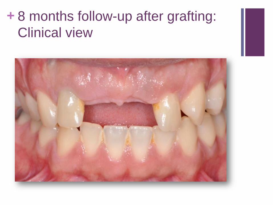
+ 8 months follow-up after grafting:
Clinical view
+ 8 months follow-up after grafting:
Clinical view

+ 8 months follow-up after grafting:
CBCT cross-sections
#8 #9

+Informed Consent (insert)

+Informed Consent (insert)

+ Implant Surgery #8 and #9
Operative report of implant surgery:
March 8th 2010
BP: 123/71, pulse 69, O2 saturation 98-99%.
2 grams of Amoxicillin administrated to the patient on the morning of
surgery. Patient was asked to rinse with Peridex (oral rinse containing
0.12% chlorhexidine gluconate) for 3 min and his face was swabbed
with betadine solution. Patient was fully draped for surgery and local
anesthetic 2 carpules Lido 2% 1/100000 epinephrine was administrated
by infiltration buccal and palatal in the maxillary anterior sextant. An
intrasulcular incision around teeth #7 and #10 and crestal incision in the
anterior edentulous area was done using15c blade. Full thickness
mucoperiosteal flap reflected and with the use of a surgical template
the osteotomy for implant placement in position of 8 and 9 was started.
Straumann Surgical kit was used for the placement of two Bone Level
SLA Active implants

+Implant Surgery
•with a diameter and length of: 3.3 mm x 14 mm.
•During sequential osteotomy preparation bone quality on site was
evaluated as 2-3. No countersink drill used. Insertion torque 20 and 25
N/cm. Implants were primary stable and placed 3-4mm from the
predetermined cervical margin of the surgical template. Healing
abutments of Ø3.6mm by height 3.5mm placed. Hydrated 0.5cc
xenograft Bio-Oss® (Geistlich) small particles applied over the buccal
plate and healing abutments. A Bio-Gide® (Geistlich) collagen
membrane was stabilized with titanium tacks. Primary closure obtained
with vicryl 5.0 suture. End BP: 138/73, pulse 70, O2 saturation
monitored by pulse oximetry: 98-99%.
•The Essix retainer was adjusted free of contact with the surgical site.
PO instructions reviewed with patient in oral and written form. Patient
tolerated the procedure well.

+Implant Surgery x-rays

+Post-Operative Care
What were your post-operative instructions for this patient?
Patient was told to continue gentle rinsing with Peridex for the
following 2 weeks. Also gentle rinsing with lukewarm salt water will aid
the healing process(add one half teaspoon of salt to a 6oz glass of
water). Avoid the use of a water-pick tooth brush. Vigorous rinsing of
the mouth prolongs bleeding by removing the clotting blood. No
drinking through straws. The use of a straw creates negative pressure
in the mouth and will tend to loosen the sutures. Patient was given a
soft brush to use.
Patient was advice to continue taking Amoxicillin TID until done 21
capsules. Motrin 800mg was prescribed for pain as needed. Smoking
is to be avoided for the time of healing (increases the heat in the
surgical site and significantly lowers the body’s ability to heal the site).
Avoid alcohol with post operative medications.

+Post-Operative Care cont…
Patient was instructed to apply the ice pack that has been given for a
period of 20 minutes on and 20 minutes off during the day for the next
two days. The application of ice to the outside of the face over the
surgical area will minimize swelling.
Patient was told following surgery to restrict diet to liquid diet foods for
the first 2 weeks and soft diet for the following 2 months. Also to avoid
chewing on the surgical site until the tissue is completely healed. Is
better to remove the Essix retainer when having meals. Soft foods
such as Jell-O, pudding, mashed potatoes, scrambled eggs and soups
are suggested. If having difficulty chewing, try blenderized foods or
diet supplements such as Carnation Instant Breakfast and Ensure.

+Maintenance
What is your maintenance protocol?
24 hour phone call; 2 weeks suture removal. Follow the diet
instructions for the first 2 months. Hygiene procedures by brushing ,
flossing , and rinsing as usual.
Periapical radiographs taken once a month to monitor healing.
List this patients maintenance history:
24 hour phone call; 2 weeks suture removal. Follow the diet
instructions for the first 2 months. Hygiene procedures by brushing ,
flossing , and rinsing as usual.
An Essix retainer free of tissue contact was fabricated.
Periapical radiographs taken once a month to monitor healing.

+Prosthetic Restoration
What type of restoration was placed?
Fixed partial denture with retainers as central incisors #8, #9 and a
distal cantilever on lateral incisor #7. (Due to financial constrains and
willing to accommodate esthetics, 2 implant supported three unit fixed
partial denture was decided with a cantilever on lateral incisor
position).
Explain:
Implant uncovery procedure (15th 2010). Implant level impression.
Extraction of #7 with socket seal procedure.
Fabrication and placement of provisional titanium abutments and
provisional resin fixed partial denture. Hygiene procedures by brushing
, flossing underneath the cantilever area, and rinsing as usual.

+Prosthetic Restoration
Explain:
Maxillary implant level impression with the replica of the transmucosal
path that will be mounted in a semi-adjustable articulator
Fabrication of the custom abutments and try in.
Fabrication of the metal substructure of the IFPD and try in.
Biscuit try in and final adjustments.
Delivery of custom abutments and fixed partial denture along with an
occlusal guard.

+Implant uncovery procedure

+Implant uncovery procedure

+Impression copings x-ray

+Extraction of #7 with socket seal
procedure

+Extraction of #7 with socket seal
procedure

+Extraction of #7 with socket seal
procedure

+Extraction of #7 with socket seal
procedure

+1 week post-uncover surgery

+Provisional Abutments # 8,9 at
1 month post uncovery

+6 weeks post uncover surgery

+Open tray impression copings with
replica of transmucosal path

+Impression copings seating
radiographs

+Open Tray Impression Copings
splinted
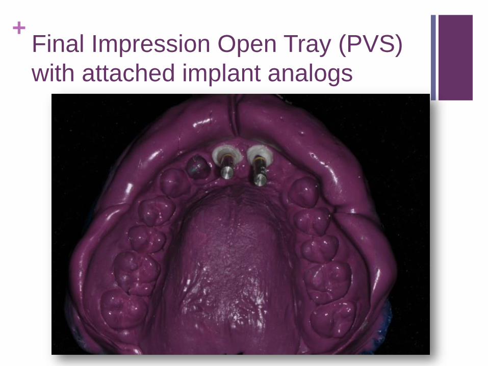
+Final Impression Open Tray (PVS)
with attached implant analogs

+Maxillary Working Cast

+Gold Custom Abutments # 8,9

+Gold Custom Abutments # 8,9
x-ray

+Substructure Fixed partial Denture

+Substructure Fixed partial Denture
x-ray

+Immediate post prosthetic
placement x-ray(insert)
#8 #9

+Occlusal view of maxillary arch
(insert)

+Occlusal view of mandibular arch
(insert)

+Frontal view in maximum
intercuspation position (insert)

+Left side (insert)

+Right side (insert): replace photo

+One year post prosthetic placement
x-ray (insert)

+Revision (if necessary)
No revision surgery was necessary for this patient.