sample orthopaedic surgery questions & critiques - nccpa
TRANSCRIPT
Sample Orthopaedic Questions & Critiques
©NCCPA. 2011-2015. All rights reserved
Sample Orthopaedic Surgery Questions & Critiques The sample NCCPA items and item critiques are provided to help PAs better understand how
exam questions are developed and should be answered for NCCPA’s Orthopaedic Surgery CAQ
exam.
Question 1
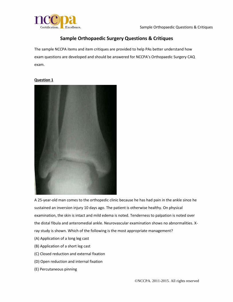
A 25-year-old man comes to the orthopedic clinic because he has had pain in the ankle since he
sustained an inversion injury 10 days ago. The patient is otherwise healthy. On physical
examination, the skin is intact and mild edema is noted. Tenderness to palpation is noted over
the distal fibula and anteromedial ankle. Neurovascular examination shows no abnormalities. X-
ray study is shown. Which of the following is the most appropriate management?
(A) Application of a long leg cast
(B) Application of a short leg cast
(C) Closed reduction and external fixation
(D) Open reduction and internal fixation
(E) Percutaneous pinning
Sample Orthopaedic Questions & Critiques
©NCCPA. 2011-2015. All rights reserved
Content Area: Fractures/Dislocations (22%)
Critique
This question assesses the examinee’s ability to correctly interpret an x-ray study to determine
the diagnosis and then select the most appropriate management. The correct answer is Option
(D), open reduction and internal fixation. The x-ray study shows oblique fracture of the lateral
malleolus, widening of the medial clear space, and lateral displacement of the talus. On the basis
of these findings, the diagnosis is supination external rotation, type IV (Weber B2), which is an
unstable injury to the ankle that requires operative intervention.
Option (A), application of a long leg cast, is plausible but incorrect because closed reduction must
be performed first. Additionally, even with adequate closed reduction and cast immobilization,
outcomes with surgery are superior. Option (B), application of a short leg cast, is incorrect
because this method of immobilization allows internal and external rotation of the leg and is,
therefore, not suitable management of this patient’s injury. Option (C), closed reduction and
external fixation, is incorrect because although this intervention is useful for fractures of the
tibial plafond or for ankle arthrodesis, it is not appropriate for the injury described. Option (E),
percutaneous pinning, is incorrect because this intervention is not as appropriate as fixation with
a plate and screws.
Question 2
A 19-year-old woman is brought to the emergency department by ambulance after she
sustained an injury to the right knee while rollerblading. The patient says she felt sudden, severe
pain in the knee when she turned a corner quickly. She fell to the ground and was unable to
bear weight on the right leg. Physical examination shows swelling and deformity of the right
knee as well as inability to fully extend and straighten the right lower extremity. X-ray studies
show dislocation of the patella. In addition to administration of analgesics, which of the
following is the most appropriate management?
(A) Arthroscopic lateral release
(B) Arthroscopic medial plication
(C) Closed reduction of the patella
Sample Orthopaedic Questions & Critiques
©NCCPA. 2011-2015. All rights reserved
(D) Open reduction of the patella
(E) Tibial tubercle medialization
Content Area: Fractures/Dislocations (22%)
Critique
This question tests the examinee’s ability to select the most appropriate management of a
patient with a known diagnosis. The correct answer is Option (C), closed reduction of the patella.
The patient’s age, gender, and athletic activity are all predisposing factors of this injury, and the
x-ray study confirms the diagnosis. Prompt reduction of a dislocated patella is the most
appropriate management because the longer the patella remains dislocated, the more damage
is done to the medial retinaculum and the medial ligamentous structure. In addition, risks of
closed reduction are minimal compared with other surgical options.
Option (A), arthroscopic lateral release, is incorrect because this procedure is not indicated for
management of acute patellofemoral instability. It is a more suitable intervention for patients
with retinacular tightness and pain. Option (B), arthroscopic medial plication, is incorrect
because this procedure is used to manage chronic patellofemoral instability. Option (D), open
reduction of the patella, is incorrect because surgery is rarely required for management of
dislocation of the patella. Open reduction may be needed, but this is only in circumstances in
which closed reduction is repeatedly unsuccessful or when a clear mechanical obstruction to
reduction is evident. Option (E), tibial tubercle medialization, is incorrect because this procedure
is used to correct patellofemoral alignment in patients with patellofemoral instability due to
factors such as increased Q angle.
Question 3
A 40-year-old woman comes to the emergency department because she has pain in the right
arm two hours after she fell in her home. Physical examination shows swelling and deformity of
the right arm. The patient is unable to dorsiflex the wrist. X-ray studies show a spiral midshaft
fracture of the humerus. Which of the following nerves is most likely affected by this fracture?
(A) Axillary
Sample Orthopaedic Questions & Critiques
©NCCPA. 2011-2015. All rights reserved
(B) Median
(C) Musculocutaneous
(D) Radial
(E) Ulnar
Content Area: Fractures/Dislocations (22%)
Critique
This question tests the examinee’s knowledge of anatomy and the ability to correlate this
knowledge with findings on physical examination and x-ray studies. The correct answer is Option
(D), radial. The radial nerve courses posterior to the middle third of the humeral shaft and is
prone to injury with fractures of the midshaft of the humerus. Sensory distribution of the radial
nerve includes the first dorsal web space of the hand, and motor innervation includes the dorsal
forearm extensor muscles, including those of the wrist.
Option (A), axillary, is incorrect because this nerve is located posterior to the humeral neck. It
provides sensation over the deltoid region and innervates the deltoid muscle. Option (B), median,
is incorrect because this nerve courses through the medial aspect of the arm between the biceps
brachii and brachialis muscles. It provides sensation to the radial aspect of the palm and fingers
as well as the distal dorsal surfaces of the thumb, index finger, long finger, and the radial aspect
of the ring finger. Motor innervation within the hand includes the muscles of the thenar
eminence. Option (C), musculocutaneous, is incorrect because this nerve does not innervate any
muscles involved in motor function of the wrist or hand. Option (E), ulnar, is incorrect because
this nerve courses through the arm relatively parallel to the median nerve and is not in proximity
to the humerus at the level of the fracture. It provides sensation to the ulnar aspect of the dorsal
and palmar surfaces of the hand and innervates the flexor carpi ulnaris, which provides wrist
flexion and ulnar deviation. The patient described is unable to dorsiflex the wrist, which is not
related to the ulnar nerve.
Sample Orthopaedic Questions & Critiques
©NCCPA. 2011-2015. All rights reserved
Question 4
A 16-year-old girl who plays tennis on her high school team is brought to the emergency
department by her parents because she has had pain and worsening swelling of her right knee
since she sustained an injury during a match four hours ago. The patient says she was playing on
a hard court and fell directly onto her knee. She was able to complete the match. The pain is
localized to the anterior aspect of the knee and is dull in nature. The patient rates the pain as 6
on a 10-point scale. Physical examination of the right knee shows a fluctuant mass (5×3 cm) over
the patella. Full range of motion is noted, and muscle strength is 5/5. Result of apprehension
test is negative. Which of the following is the most likely diagnosis?
(A) Dislocation of the patella
(B) Fracture of the patella
(C) Patellar tendinitis
(D) Prepatellar bursitis
(E) Sprain of the patellar ligament
Content Area: Sprains and Soft-Tissue Pathology (23%)
Critique
This question tests the examinee’s ability to discriminate among various types of injury involving
the patella to determine the most likely diagnosis. The correct answer is Option (D), prepatellar
bursitis. The history and physical examination findings of a direct blow to the anterior aspect of
the knee followed by localized pain, swelling, a fluctuant mass, normal to near-normal range of
motion (depending on size of the mass), and normal muscle strength are characteristic of
prepatellar bursitis.
Option (A), dislocation of the patella, is incorrect because the patient does not exhibit
characteristics of this condition, such as the knee acutely giving way, intense pain, rapid swelling,
and deformity. Additionally, findings on range of motion and muscle strength testing would not
be expected to be normal if dislocation of the patella were present. Also, negative result of
apprehension test effectively excludes dislocation of the patella as a possible diagnosis. Option
(B), fracture of the patella, is plausible but incorrect in the patient described for several reasons.
Sample Orthopaedic Questions & Critiques
©NCCPA. 2011-2015. All rights reserved
Pain from fracture is typically intense and would most likely preclude the patient from
completing her tennis match. Additionally, in a patient with fracture of the patella, physical
examination is unlikely to show full range of motion and muscle strength is likely to be
diminished because of pain. Option (C), patellar tendinitis, is incorrect because this condition
typically is the result of an overuse injury from repetitive overloading of the extensor mechanism
of the knee. The physical examination findings in the patient described are not consistent with
patellar tendinitis. Option (E), sprain of the patellar ligament, is incorrect because this injury
would cause pain and swelling localized to the patellar tendon and decreased muscle strength
because of pain. Complete rupture of the patellar ligament would manifest as inability to extend
the knee.
Question 5
A 20-year-old man who plays baseball on his college team comes to the clinic because he has
had pain in the right elbow for the past three weeks. The pain began approximately one week
after the beginning of the baseball season. Physical examination of the elbow shows full range
of motion in flexion, extension, supination, and pronation. No pain is elicited on varus or valgus
stress of the elbow. Dorsiflexion of the right hand against resistance immediately elicits pain
that is localized to the lateral aspect of the elbow. Which of the following is the most likely
diagnosis?
(A) Biceps tendinitis
(B) Fracture of the radial head
(C) Lateral epicondylitis
(D) Olecranon bursitis
(E) Sprain of the lateral collateral ligament
Content Area: Sprains and Soft-Tissue Pathology (23%)
Critique
This question tests the examinee’s ability to recognize signs and symptoms of a musculoskeletal
disorder to determine the most likely diagnosis. The correct answer is Option (C), lateral
epicondylitis. Overuse or repetitive motion activities involving wrist extension and/or supination
Sample Orthopaedic Questions & Critiques
©NCCPA. 2011-2015. All rights reserved
are common causes of lateral epicondylitis. Clinical manifestations usually include pain in the
lateral aspect of the elbow and the dorsal aspect of the forearm that is exacerbated by use.
Physical examination usually shows maximal point tenderness over the lateral epicondyle and/or
the area overlying the extensor carpi radialis brevis muscle. Extension or supination of the wrist
against resistance typically elicits pain.
Option (A), biceps tendinitis, is incorrect because this condition involves inflammation of the long
head of the biceps tendon, which causes pain in the anterior aspect of the shoulder. Option (B),
fracture of the radial head, is incorrect because the patient has no history of substantive trauma.
In addition, the physical examination findings of full range of motion in all planes and
provocation of pain on dorsiflexion of the wrist against resistance point away from this diagnosis
as a possibility. Option (D), olecranon bursitis, is incorrect because this condition involves
inflammation of the bursa overlying the olecranon process and none of the physical examination
findings in the patient described are suggestive of this condition. Option (E), sprain of the lateral
collateral ligament, is incorrect because the physical examination finding of no pain elicited on
varus or valgus stress of the elbow excludes this condition as the most likely diagnosis.
Additionally, activities that involve overhead throwing, such as baseball in the patient described,
are more likely to involve the medial collateral ligament rather than the lateral collateral
ligament.
Question 6
A 54-year-old man with a history of metastatic lung cancer comes to the office because he had
sudden onset of pain in the lower back 24 hours ago. Which of the following findings in this
patient differentiates lumbar disk herniation from cauda equina syndrome as the cause of his
pain?
(A) Anesthesia of the saddle region
(B) Bilateral weakness of the legs
(C) Impotence
(D) Pain radiating to one buttock
(E) Urinary incontinence
Sample Orthopaedic Questions & Critiques
©NCCPA. 2011-2015. All rights reserved
Content Area: Spine (11%)
Critique
This question tests the examinee’s ability to discriminate between clinical characteristics of
lumbar disk herniation and cauda equina syndrome. The correct answer is Option (D), pain
radiating to one buttock. Most lumbar disk herniations are posterolateral, and 90% to 95% of
compressive radiculopathies occur at the level of L4-L5 and L5-S1. Pain associated with disk
disease is usually localized to the lower back and gluteal region and commonly radiates down
the leg, particularly below the knee. Therefore, pain radiating to one buttock differentiates
lumbar disk herniation from cauda equina syndrome.
Cauda equina syndrome is typically associated with significant neurologic disability and is caused
by an intraspinal lesion caudal to the conus medullaris that impacts two or more of the 18 nerve
roots comprising the cauda equina. Clinical manifestations most often include bilateral leg
weakness in multiple root distributions (L3-S1); bowel, bladder, and sexual dysfunction; and/or
perineal sensory loss (S2-S4). Causes of cauda equina syndrome include neural tube defects,
infection or inflammation, trauma, spinal stenosis, or mass lesions (e.g., tumor, ruptured disk).
Therefore, Option (A), anesthesia of the saddle region, Option (B), bilateral weakness of the legs,
Option (C), impotence, and Option (E), urinary incontinence, are incorrect because they are
characteristic of cauda equina syndrome and do not support the diagnosis of lumbar disk
herniation.
Question 7
A 32-year-old man comes to the clinic because he has had pain in the back for the past 24 hours.
The patient says he first noticed the pain when he awoke in the morning and had difficulty
getting out of bed. He had been playing flag football the day before the pain began but did not
sustain any injuries during the game. Acetaminophen has provided only minimal relief of the
patient's pain. On physical examination, pain is elicited on palpation of the back on the left,
lateral to the region of L2-L5. Full range of motion is noted in vertebral flexion, extension, lateral
Sample Orthopaedic Questions & Critiques
©NCCPA. 2011-2015. All rights reserved
rotation, and lateral bending, with some hesitancy because of pain on the left side. Which of the
following is the most appropriate initial step?
(A) Anti-inflammatory and muscle relaxant therapy
(B) CT scan of the lumbar spine
(C) Epidural injection of a corticosteroid
(D) MRI of the lumbar spine
(E) Strict bed rest and application of moist heat to the lower back
Content Area: Spine (11%)
Critique
This question tests the examinee’s ability to recognize signs and symptoms of a common
musculoskeletal disorder and then determine the most appropriate initial step. The correct
answer is Option (A), anti-inflammatory and muscle relaxant therapy. In high-performing or
“weekend” athletes, the most common causes of pain in the lower back are
musculoligamentous sprains and strains. Typical signs and symptoms include pain and muscle
spasm localized over the posterior paraspinous muscles. Range of motion may be decreased
because of pain. Pain in the midback as well as neurologic symptoms, which are suggestive of
structural deformities, should be absent. During the acute phase, the most appropriate
management is therapy with anti-inflammatory drugs and muscle relaxants.
Option (B), CT scan of the lumbar spine, is incorrect because there is no clinical evidence of
structural deformity (e.g., fracture) or neurologic symptoms. Option (C), epidural injection of a
corticosteroid, is incorrect because although this therapy has been shown to be effective in
reducing radicular pain in patients with disk herniation, it is not indicated in the treatment of
acute strain or sprain of the back. Option (D), MRI of the lumbar spine, is incorrect because
there is no clinical evidence of structural deformity or neurologic complaint (e.g., radiculopathy).
Option (E), strict bed rest and application of moist heat to the lower back, is plausible but
incorrect because most studies show that patients have a more rapid functional recovery if they
maintain some level of activity, even during the acute phase.
Sample Orthopaedic Questions & Critiques
©NCCPA. 2011-2015. All rights reserved
Question 8
A 63-year-old man is referred to the office by his primary care provider because he has pain in
the right knee that has been worsening over the past two years. He usually plays tennis several
times per week, but recently the pain has made it difficult for him to continue this routine.
Conservative treatment measures such as courses of nonsteroidal anti-inflammatory drugs
and injections of cortisone have failed to relieve the patient's pain. Current physical examination
of the right knee shows moderate effusion and tenderness along the medial joint line. Result of
McMurray test is positive. MRI of the knee shows a 3-cm defect of the articular cartilage of the
medial femoral condyle. Weight-bearing x-ray studies of the right knee show no narrowing of
the joint spaces. Which of the following is the most appropriate management?
(A) Application of a medial unloader knee brace
(B) Arthroscopy with microfracture of the articular cartilage defect
(C) Osteochondral grafting of the articular cartilage defect
(D) Total arthroplasty of the knee
(E) Unicompartmental joint replacement
Content Area: Total Joint (15%)
Critique
This question tests the examinee’s ability to recognize signs and symptoms of a common
musculoskeletal disorder and interpret imaging studies to determine the appropriate
management. The correct answer is Option (B), arthroscopy with microfracture of the articular
cartilage defect. Recent studies have shown that microfracture of a defect that is 3 cm or smaller
restores pain-free activity in 80% to 90% of patients. Therefore, arthroscopy with microfracture is
the most appropriate treatment of the patient described.
Option (A), application of a medial unloader knee brace, Option (D), total arthroplasty of the
knee, and Option (E), unicompartmental joint replacement, are incorrect because the x-ray
studies do not show joint space narrowing. Option (C), osteochondral grafting of the articular
cartilage defect, is incorrect because this intervention has not been proven to be effective in the
knee joint of patients in this age group.

Sample Orthopaedic Questions & Critiques
©NCCPA. 2011-2015. All rights reserved
Question 9
A 35-year-old man comes to the primary care office because he has had pain and swelling of the
right knee for the past three days. Also, for the past two days, he has felt feverish. The patient is
able to ambulate, but walking exacerbates the pain in his knee. Temperature is 39.3°C (102.7°F).
On physical examination, the right knee is red, warm to touch, and tender. A large effusion is
noted. Which of the following diagnostic studies of the knee is the most appropriate initial step?
(A) Arthrocentesis
(B) Arthroscopy
(C) Bone scan
(D) CT scan
(E) MRI
Content Area: Infectious Disease (8%)
Critique
This question tests the examinee’s ability to select the most appropriate study to determine the
diagnosis. The correct answer is Option (A), arthrocentesis. The clinical presentation described is
characteristic of septic joint. Arthrocentesis for aspiration and analysis of joint fluid is the only
diagnostic study that will specify the diagnosis of septic joint.
Option (B), arthroscopy, is incorrect because this study is not the initial step in diagnosis. Option
(C), bone scan, is incorrect because this study is appropriate to nonspecifically localize areas of
inflammation but cannot be used to distinguish infectious from sterile processes. Option (D), CT
scan, and Option (E), MRI, are incorrect because these studies are more sensitive for diagnosing
osteomyelitis and periarticular abscesses.
Question 10
A 49-year-old man comes to the sports medicine office because he has pain in the right hip and
thigh that has been worsening since he fell while working in his yard two weeks ago. Physical
examination shows a healing puncture wound over the proximal aspect of the thigh. Erythema
and warmth are noted over the lateral aspect of the right hip and the proximal aspect of the
Sample Orthopaedic Questions & Critiques
©NCCPA. 2011-2015. All rights reserved
right thigh. Full range of motion of the hip is noted, and distal sensation and pulses are intact.
On laboratory studies, erythrocyte sedimentation rate is 30 mm/hr. Results of complete blood
cell count are within normal limits. X-ray studies of the hip show a slightly raised periosteum in
the proximal femoral shaft. Which of the following additional diagnostic studies is most
appropriate?
(A) CT scan
(B) Indium 111 bone scan
(C) MRI
(D) Technetium 99m bone scan
(E) Ultrasonography
Content Area: Infectious Disease (8%)
Critique
This question tests the examinee’s ability to review a detailed clinical scenario, including history
and physical examination findings, interpret laboratory values, evaluate x-ray study findings, and
then determine the most appropriate additional study to establish the diagnosis. The correct
answer is Option (C), MRI. The clinical presentation is characteristic of osteomyelitis, and MRI is
the most appropriate study to confirm this diagnosis because it shows marrow edema and
periosteal elevation.
Option (A), CT scan, is incorrect because this study is not sensitive for acute osteomyelitis. Option
(B), indium 111 bone scan, and Option (D), technetium 99m bone scan, are incorrect because
although these studies might show increased metabolic activity in patients with osteomyelitis,
this finding is not distinguishable from post-traumatic injury, cancer, or postoperative findings.
Option (E), ultrasonography, is incorrect because this study can only show fluid collection next to
bone, which is not distinguishable from a traumatic response.
Question 11
A male neonate who was delivered vaginally at term one hour ago has a deformity of the right
foot. On physical examination, plantar flexion of the ankle, inversion of the subtalar joint, and
Sample Orthopaedic Questions & Critiques
©NCCPA. 2011-2015. All rights reserved
medial subluxation of the talocalcaneal and calcaneocuboid joints are noted. The position of the
foot cannot be passively corrected. Which of the following disorders is the most likely diagnosis?
(A) Calcaneovalgus
(B) Congenital clubfoot
(C) Metatarsus adductus
(D) Pes planus
(E) Tarsal coalition
Content Area: Pediatric (14%)
Critique
This question tests the examinee’s knowledge of anatomy and the ability to correlate this
knowledge with physical examination findings to determine the diagnosis. The correct answer is
Option (B), congenital clubfoot. The physical examination findings described are characteristic of
congenital clubfoot.
Option (A), calcaneovalgus, is incorrect because this condition involves the foot in dorsiflexion,
not plantar flexion. Option (C), metatarsus adductus, is incorrect because this condition is
characterized by deformity that can be passively corrected. Option (D), pes planus, is incorrect
because in patients with this condition, the foot is flexible. Option (E), tarsal coalition, is incorrect
because this condition typically presents during the second decade of life but can present as early
as 3 years of age, when the tarsal bones begin to ossify.
Question 12
A 6-year-old boy is brought to the office by his parents because he has had pain in the right hip
with weight-bearing as well as obvious limping for the past week. The patient's parents say they
have noticed the child favoring his right leg during the past few weeks. He has not had any
recent illness or injury to the leg. Medical history includes no chronic disease conditions.
Physical examination shows tenderness on passive internal rotation of the hip joint and mild
diffuse atrophy of the right thigh musculature. X-ray studies of the hip and femur show no
abnormalities. Which of the following studies is the most appropriate next step?
Sample Orthopaedic Questions & Critiques
©NCCPA. 2011-2015. All rights reserved
(A) Aspiration of the hip
(B) Bone scan
(C) CT scan
(D) MRI
(E) Ultrasonography
Content Area: Pediatric (14%)
Critique
This question tests the examinee’s ability to review a detailed clinical scenario, including history
and physical examination findings, evaluate x-ray study findings, and then determine the most
appropriate additional study to establish the diagnosis. The correct answer is Option D, MRI. The
clinical presentation is characteristic of Legg-Calvé-Perthes disease, and MRI is the most sensitive
study for staging of this condition.
Option (A), aspiration of the hip, is incorrect because the patient has no history of current or
recent illness and, therefore, septic joint is very low on the differential diagnosis list. Option (B),
bone scan, is incorrect because it can only confirm the presence of avascular necrosis and not the
extent of involvement of the femoral head. Option (C), CT scan, is incorrect because although this
study is used to diagnosis Legg-Calvé-Perthes disease, it is not as sensitive as MRI. Option (E),
ultrasonography, is incorrect because this study can only confirm the presence of a joint effusion,
which is a nonspecific finding when confirming a suspected diagnosis of Legg-Calvé-Perthes
disease.
Question 13
A 12-year-old boy is brought to the office by his mother because he has had intermittent pain in
the right hip during the past two weeks. The patient ambulates with difficulty. He has not had
fever, chills, malaise, recent illness, or trauma to the hip. The patient is obese but otherwise
healthy. On physical examination, vague pain in the groin is elicited on range of motion of the
right hip. The most appropriate next step is x-ray studies to rule out which of the following
conditions?
Sample Orthopaedic Questions & Critiques
©NCCPA. 2011-2015. All rights reserved
(A) Femoral acetabular impingement syndrome
(B) Legg-Calvé-Perthes disease
(C) Septic arthritis
(D) Slipped capital femoral epiphysis
(E) Tear of the labrum
Content Area: Pediatric (14%)
Critique
This question tests the examinee’s ability to discriminate between various types of conditions
involving the hip joint in a pediatric patient and determine which condition is most likely to be
ruled out by x-ray studies. The correct answer is Option (D), slipped capital femoral epiphysis.
The history and physical examination findings are characteristic of slipped capital femoral
epiphysis, including obesity, limp, and pain with range of motion of the joint.
Option (A), femoral acetabular impingement syndrome, is incorrect because the most
appropriate study to rule out this condition is MRI, not x-ray studies. Option (B), Legg-Calvé-
Perthes disease, is plausible but incorrect in this patient considering his age because the mean
age of onset of Legg-Calvé-Perthes disease is 7 years and x-ray studies do not rule this condition
out. Option (C), septic arthritis, is plausible but incorrect because the patient has no history of
acute illness and x-ray studies do not rule this condition out. Option (E), tear of the labrum, is
incorrect because the patient has no history of injury and because MRI is the best diagnostic
study to evaluate tear of the labrum.
Question 14
A 19-year-old man who is a long-distance runner is referred to the office by his primary care
provider because he has had dull, aching pain in his right thigh after running during the past nine
months. Six months ago, the patient's primary care provider prescribed ibuprofen, which
relieves the pain only temporarily. The patient has no history of specific injury. Physical
examination shows no abnormalities of the right lower extremity. X-ray studies of the right
Sample Orthopaedic Questions & Critiques
©NCCPA. 2011-2015. All rights reserved
femur show cortical thickening of the distal one-third of the shaft with a central nidus measuring
approximately 8 mm in diameter. Which of the following is the most likely diagnosis?
(A) Aneurysmal bone cyst
(B) Enchondroma
(C) Osteoblastoma
(D) Osteochondroma
(E) Osteoid osteoma
Content Area: Benign and Malignant Bone Tumors (4%)
Critique
This question tests the examinee’s ability to review a detailed clinical scenario, including history
and physical examination findings, evaluate x-ray study findings, and then determine the most
likely diagnosis. The correct answer is Option (E), osteoid osteoma. The x-ray finding of cortical
thickening with a central nidus is characteristic of osteoid osteoma.
Option (A), aneurysmal bone cyst, is incorrect because although the femur is the most common
site of involvement of aneurysmal bone cyst, it usually presents as a large lytic lesion. Option (B),
enchondroma, is incorrect because although the distal femur is a potential location for this
lesion, it is characterized as a lytic area filled with a calcified matrix. Option (C), osteoblastoma,
is plausible but incorrect because although it is closely related to osteoid osteoma, the two are
distinguished by the size of the nidus: larger than 2 cm represents osteoblastoma, and 1 cm or
less represents osteoid osteoma. In addition, the pain of osteoblastoma is less likely to be
relieved by nonsteroidal anti-inflammatory drug therapy. Option (D), osteochondroma, is
incorrect because this lesion arises from the growth plate on the metaphyseal side and results in
an exostosis that points away from the joint of origin.
Question 15
A 72-year-old man comes to the office for follow-up examination eight weeks after he
underwent total arthroplasty of the right hip. The patient's rehabilitation had been progressing
fairly well until approximately five days ago, when worsening pain developed in the hip. The
Sample Orthopaedic Questions & Critiques
©NCCPA. 2011-2015. All rights reserved
patient says the pain is aggravated by walking and persists during sleeping hours even after he
takes acetaminophen. Infection of the prosthetic joint is suspected. Which of the following is the
most likely causative organism?
(A) Coagulase-negative staphylococcus
(B) Escherichia coli
(C) Group A beta-hemolytic streptococcus
(D) Haemophilus influenzae
(E) Pseudomonas aeruginosa
Content Area: Total Joint (15%)
Critique
This question tests the examinee’s ability to apply scientific concepts to a clinical scenario by
discriminating between multiple possible bacterial species to determine the most likely causative
organism of an orthopedic infection. The correct answer is Option (A), coagulase-negative
staphylococcus. The most commonly cultured microorganism (30% to 43%) from prosthetic joint
infections is coagulase-negative staphylococcus. The likelihood of infection with other organisms
depends on perioperative or contiguous factors, hematogenous seeding from distant infections,
and/or other comorbid diseases. The scenario described does not include any of these factors.
Option (B), Escherichia coli, and Option (E), Pseudomonas aeruginosa, are incorrect because they
are gram-negative bacilli, which are uncommon causes of prosthetic joint infection. Gram-
negative bacilli account for 3% to 6% of prosthetic joint infections. Option (C), group A beta-
hemolytic streptococcus, is incorrect because streptococci account for only 9% to 10% of
prosthetic joint infections. Option (D), Haemophilus influenzae, is incorrect because it is a very
uncommon cause of prosthetic joint infection. Its absolute burden is unknown.