sant abstract leaflet - lung health berlin 2018|lungh ... · abstract leaflet . 2 ... were randomly...
TRANSCRIPT

1
sant
ABSTRACT LEAFLET
2
P01. [109] Respiratory Symptoms And Ventilatory Functions In Women Exposed To Domestic
Cooking Fuel In A Rural Community In Lagos State, South West, Nigeria
Olayinka Olufunke Adeyeye1; Yetunde Kuyinu1; Tunmise Bamisile2; Adejunmo Olusola2
1lagos State College Of Medicine Ikeja, Lagos, Nigeria; 2lagos State University Teaching Hospital Ikeja, Lagos, Nigeria
Background
Cooking with biomass fuel is common in Nigeria and this has been associated with chronic obstructive pulmonary disease in women. This study was conducted to determine prevalence of respiratory symptoms and ventilatory indices of women in relation to the type of fuel used for domestic cooking.
Methodology
A descriptive cross- sectional study was conducted in Agbowa, a rural settlement in Lagos State, South West Nigeria. This is largely a farming community and with no industrial presence. Consented non-smoking women with no history of exposure to passive smoke or occupational exposure to fume were randomly selected and interviewed with a respiratory questionnaire. Spirometry was performed using the portable spirotract intuitive version IV.
Result
450 women participated in the study, 374 (83.1) were suitable for analysis. The mean age of respondents 36.57±12.41. The mean duration of cooking was 22.40 ± 13.02 years. Biomass fuel, Non biomass fuel, and mixed fuel was used by 52(13.9) 159(42.5) and 163(43.6) respectively. Those who used biomass fuel were older, cooked for much longer period and with higher exposure index. The prevalence of chronic bronchitis was 6.9%. Cough, phlegm production and chest tightness occur more frequently in women exposed to biomass fuel P <0.05). Sneezing was the most frequently experienced symptom reported while cooking 23(6.15%). This was more prevalent in those women cooking with biomass fuel or those using mixed fuel. (P<0.05). Exposure to biomass fuel was associated with lower mean ventilatory indices for all the parameter measured. (FEV1, FVC, and PEFR). The use of biomass fuel for cooking was associated with an abnormality on spirometry. Conclusion
The use of biomass fuel for cooking was associated with abnormality on spirometry. Intervention at providing a cleaner source of fuel and public enlightenment to educate and empower women about cooking with clean fuel is therefore recommended.

3
P02. [112] Epidemiological and clinical aspects of pulmonary exacerbations in cystic fibrosis - our experience in Clinical County Hospital of Constanta
Adelina Anton1,5; Doina Ecaterina Tofolean1,5; Laura Mazilu2,5; Petronela Fildan4,5; Elena Dantes4,5; Viviana Cuzic3,5
1Department of Pneumology and Internal Medicine, Clinical County Hospital of Constanta, Constanta, Romania; 2Department of Oncology, Clinical County Hospital of Constanta, Constanta, Romania; 3Department of Pediatrics, Clinical County Hospital of Constanta, Constanta, Romania; 4Clinical Hospital of Pneumophtisiology, Constanta, Romania; 5Faculty of Medicine, "Ovidius" University, Constanta, Romania
Significant improvement in survival of patients with cystic fibrosis in last decades, and , with this, clinical polymorphism of the disease continues to evolve. An acute pulmonary exacerbation is the primary clinical event in the course of this disease and a major cause of morbidity. Since there is no consensus diagnostic criteria, pulmonary exacerbations were defined as acute worsening of respiratory symptoms requiring antibiotic therapy.
The aim of this review is to characterize acute pulmonary exacerbations, providing a summary of the clinical and microbiological particularities.
To this end, we performed an ambispective study of patients with cystic fibrosis followed up in Constanta Center over a period of 2 years and 9 months and we defined exacerbations using European Consensus Group’s definition . The study was conducted on 15 patients, 9 children and 6 adults, aged from birth to 44 years old, that were stratified into two groups, based on age at diagnosis, group A (pediatric patients) and group B (adult patients). Clinical presentation, genotype, pulmonary function, rate and microbiology of exacerbations were compared between groups.
Patients with more frequent exacerbations were more likely to be in group A. For patients in group A increased cough frequency, decline in weight and fever were the most frequent symptoms found during exacerbations while for patients in group B, change in sputum volume or color, increased dyspnea and cough frequency were the most common . Among patients capable of expectorating sputum, Pseudomonas aeruginosa was the most frequent bacterial pathogen identified. A correlation between genotype and rate or severity of exacerbations could not be established. Pulmonary function assessments were unavailable for group A. In group B , decline of pulmonary function increased with age and exacerbation rates.
Despite advances in treatment, pulmonary exacerbations remain common in both adults and children, therefore understanding these events could provide an important insight into the course, prognosis and complications of the disease.

4
P03. [57] Amiko® : the cornerstone role of adherence in obstructive diseases
Diego Bagnasco1; Fulvio Braido1
1IRCCS - San Martino - IST , Allergy & Respiratory Diseases Clinic, Genoa, Italy
Asthma and COPD are the chronic respiratory diseases with the highest incidence and prevalence. One of the critical point of their management is the adherence to long term-treatment. The weak or bad utilization of prescribed therapy is related to patient’s symptoms but also to health care burden. More in details poor medication use is associated to more frequent exacerbations, need to rescue medications prescriptions, unscheduled GP od specialist visit, up to hospitalization. For theabove mentioned reasons seems useful a better attention to correct administration of prescribed therapy, but also to the recording of the real adherence to the treatment. Some methods having the aim of monitoring adherence have been developed, with mixed results. Amiko® , commercialised by Amiko Digital Health Limited (Salisbury House 31, Finsbury Circus, London UK) and developed by the R&D subsidiary Amiko Srl (Piazzale Aquileia 6, Milano Italy – R&D labs- Milano Italy), is developed as an add-on to be positioned on drugs’ devices. The technology of Amiko® permits not only to register the real number of administrated doses, but also the used inhalation technique, the inspiratory effort and the orientation of the inhaler. All the data are, in real time, transmitted to a smartphone, tablet or compatible PC and stored.
The potential of Amiko® are really many, it can offer many insights for the best individual patient management starting from the objective adherence, going on with data about inhalation duration, orientation of the device and peak inspiratory flow (PIF). The management of these data can help clinician to better control patients’ care and, if necessary, to adopt strategies ( such as training, education etc) aimed to improve adherence.
A study to observe all these fact is now understudy in our clinic, associating Amiko® to a range of currently available dry powder inhalers (DPI) (Ellipta®, Spiromax® and Nexthaler®).

5
P04. [107] Bronchiectasis and Gastroesofageal Reflux – How important is this connection
Gertruda Capova; Hans-Werner Duchna
1Hochgebirgsklinik, Davos, Switzerland Background Bronchiectasis (BE) is the chronic disease of the lower airways. BE is characterised by permanent widening of the bronchi and abnormal bronchial wall thickening. These irreversible changes are result of longstanding airway inflammation. The prevalence increases with age. BE may be under-diagnosed. Increasing knowledge and awareness about the disease and improved diagnostic possibilities ( CT or MRI) may increase the reported prevalence. BE is also final pathway of a number of disorders. Today the diagnosis of bronchiectasis is simple and straightforward. We see a great importance in seeking the underlying causes of the disease, because it will lead to the right treatment and management. Methods
Patients at the clinic are with a variety of pulmonary diseases - COPD , moderate AB, and difficult –to- treat and severe asthma, ABPA, Immotile-cilia-syndrome (PCD), Recurrent Pneumonia, Cough. Where despite treatment with an intensive therapy was not possible to achieve stability of the disease we did the 24-hours pH metry. For a period of six months were examined 36 patients. 14 Patients with difficult to treat AB (38,8%), 4 with ABPA (11,1%), 12 with COPD (33,3%), 2 with Immotile-cilia sy (5,5%), 4 with Cough (11,1%).
Results
We confirmed the gastroesophageal reflux in all patients (100%). The Reflux in patients with difficult-to-treat and severe asthma and ABPA was serious, with the duration more then 12 hours, presented during the day and night.
Conlusion
The confirmation oft he Reflux in patients with not stabile disease was important fort the future therapeutical management oft he disease. With the PPI medication and anti-reflux regime together with treatment of underlying disease based on the guidlines, was achieved stability of the disease. In the patients group with cough were subsequently confirmed BE by CT. In conclusion to our analysis and also to answer, how important ist the connection with the reflux the future analysis and monitoring are needed. Conflict of interest I declare no conflict of interest

6
P05. [110] Real-life study with escalation of the basic treatment of uncontrolled asthma
Alena Davidouskaya1; Dzimitry Ruzanau2; Syarhey Myatelsky3; Tatyana Baranouskaya1
1Belarusian Medical Academy of Postgraduate Education, Minsk, Belarus; 2Gomel State Medical University, Gomel, Belarus; 3Main military clinical center, Minsk, Belarus
Background
Further progress in the results of pharmacotherapy BA was in need of new therapeutic solutions. So, in 2014, was registered a new indication for tiotropium - uncontrolled BA in patients receiving adequate doses of inhaled corticosteroids (ICS), including in combination with long acting beta2-agonists (LABA). Randomized studies demonstrate that patients with uncontrolled asthma using tiotropium improves spirometry and decreases the risk of exacerbations.
Aims
To explore the possibilities and dynamics of achieving control of BA in the escalation basic therapy of tiotropium inhalation solution in a real - life study.
Methods
Prospective, interventional, multicenter study was conducted at four clinical centers of the Republic of Belarus. The study included 34 patients older than 18 years with uncontrolled BA in patients receiving ICS (dose equivalent to ≥ 1000 µg/day beclomethasone propionate) including in combination with LABA. From the study it was not excluded the patients older than 60 years, patients with comorbidity, including chronic disease, nocturnal asthma, BA on the background of obesity and patients with asthma-COPD overlap syndrome, smokers. All patients were assigned to inhalation solution tiotropium bromide in a daily dose of 5 microgram. Initial and during follow-up were evaluated results: the Asthma Control Test (ACT), Asthma Control Questionnaire (ACQ), FEV1, number of doses of short-acting beta2-agonists (SABA) per day, the number of exacerbations. Control points: the second and fourth week of observation, then monthly. Results
Add tiotropium to previous therapy significantly reduced the frequency of asthma symptoms after the first two weeks of therapy. The questionnaire ACQ score decreased on average from 1.62 to 0.87 points; the need for SABA from 1.9 to 0.5 per day; increased the maximum value FEV1by142 ml; decreased the total number of BA exacerbations by 24% (p < 0.04) for the 12 months of observation.
Conclusions
The inclusion of tiotropium inhalation solution in the basic therapy of BA significantly increases the chances of achieving control of asthma, including patients with complex BA.

7
P06. [96] Efficacy of non-invasive ventilation (NIV) in patients with acute and chronic respiratory failure
Valentina Dimitrova1; Diana Petkova1; Velin Stratev1; Tanya Dobreva1
1University Hospital St. Marina, Clinic of Pulmonology, Varna, Bulgaria
Introduction
NIV is a contemporary and reliable method for treatment of patients with acute and chronic hypercapnic respiratory failure.
Aim
To analyze the frequency of usage, NIV regimen modalities and mean duration of NIV in patients with respiratory failure.
Patients and methods
We performed a retrospective study of patients on NIV, hospitalized due to exacerbation of respiratory failure in intensive care unit of pulmonary clinic at MHAT “St. Marina” for 3 years period (2013-2015). We analyzed 203 patients with mean age 63.17±12.58 years (52.2% males). All the patients received medication treatment and NIV was started according to international criteria and after admission in ICU.
Results
Treatment with NIV received 170 (83.7%) of the patients with chronic hypercapnic respiratory failure and 33 (16.3%) of the patients with acute respiratory failure. 68(33.5%) of the patients receiving NIV had underlying COPD, 48 (23.65%) had obesity hypoventilation syndrome and 31 (15.27%) had overlap between this conditions. We used pressure control ventilation (PCV) in 62.1 % of the cases, pressure support ventilation (PSP) in 24.1% and AVAPS in 13.8 % of the cases. NIV succeeded significant decrease of РаСО2 from mean 8.38 кРа range (3-14 кРа) to 7.41 кРа range (3-12 кРа) (р<0.0001) and significant increase of SaO2% from mean 56.76% range (19-88%) in the first hour of NIV, to 80.27% range (43-96%) (p<0.0001) on the fourth day of treatment. Mean duration of NIV was 4.47±3.4 days and it correlated positively with the PaCO2 values (r=0.356, p=0.023). In 25 (12.3%) of the patients NIV was stopped due to treatment failure and invasive ventilation was introduced.
Conclusion
NIV is an efficacious and useful treatment method in patients with acute and chronic hypercapnic respiratory failure.
8
P07. [106] The study of correlation between bronchial and alveolar NO level and clinical and biological characteristics of children with asthma
Hanh Do-Thi1; Thuy Nguyen-Thi-Dieu2; Sy Duong-Quy3,4,5
1Department of Immunology, Allergology, and Rheumatology. National Hospital of Pediatrics, Hanoi, Viet Nam; 2Hanoi University of Medicine, Hanoi, Viet Nam; 3Lam Dong Medical College, Dalat, Viet Nam; 4Cochin Hospital. Paris Descartes University, Paris, France; 5Division of Pulmonary, Allergy and Critical Care Medicine. Penn State College of Medicine, Pennsylvania, USA
Introduction
Asthma is a common disease with various phenotypes, characterized predominantly by chronic inflammation. Exhaled nitric oxide (NO) is currently used as a biomarker of airway inflammation in patients with asthma. However, the role of bronchial and alveolar NO (FENO and CANO) measurement in asthma phenotype has not been clearly demonstrated.
Objectives
To study the concentrations of FENO and CANO in Vietnamese children with asthma and their correlation with clinical, functional, and biological characteristics in these patients.
Methods
It was a prospective and descriptive study. One hundred and fifty-five children with asthma and 30 healthy control subjects were included. FENO, CANO, spirometry, blood eosinophil counts, and IgE quantifying were done for each study subject.
Results
The mean age of asthmatic children was 10 (6-15) years, with 65.3% of male. The concentration of FENO in children with asthma was 21 (1-113) ppb vs 8 (3-24) ppb in healthy controls (p<0.05). The concentration of CANO in asthmatic children was 5.4 (1.0-37.1) ppb vs 2.8 (1.0-10.9) ppb in healthy subjects (p <0.05). In asthmatic children with naive corticosteroid therapy, the concentrations of FENO in patients with mild severe asthma were significantly higher than who with severe asthma (p<0.05). There were no significant differences between FENO, CANO, and clinical features (p>0.05). There were weak correlations between FENO and FEV1 in atopic patients. In asthmatic children with corticosteroid treatment, there were no significant correlations between exhaled NO (FENO and CANO), atopy status, age of asthma onset, number of acute asthma exacerbations, level of asthma control, lung functional parameters (FEV1, FEV1/FVC, FEF25-75, and PEFR), blood eosinophil counts, and IgE levels.
Conclusion
Exhaled NO (FENO and CANO) measuring is useful for characterizing asthma phenotypes in children over 5 years old.
Best Poster
Winner

9
P08. [92] Asthma control and medication use during pregnancy, is a specialized Asthma & pregnancy outpatient clinic of added value?
Katrien Eger1; Saar van Nederveen-Bendien1; Sarah van Oord1; Harry Heijerman1
1Haga Teaching Hospital, The Hague, Netherlands
Background/aims
Asthma is the most common chronic disease in pregnancy. During pregnancy it deserves special attention because uncontrolled asthma is a potential hazard for the unborn child and is associated with adverse perinatal outcomes as preterm birth and low birth weight. Pregnant patients with unstable asthma could benefit by outpatient consultation of a specialized multidisciplinary team. This study aims to evaluate the asthma management by our Asthma & Pregnancy outpatient clinic.
Methods
In this retrospective cohort all pregnant women referred to the Asthma & Pregnancy outpatient clinic (Haga Teaching Hospital, The Netherlands) from 2014 until 2016 were registered. At first consultation spirometric testing with reversibility, fractional exhaled nitric oxide (FeNO) measurement, asthma control questionnaire (ACQ), blood eosinophils and allergy testing were performed. Depending on asthma symptoms, medication was intensified. Education was given about medication and the importance of asthma control. Inhalation technique was observed by a pulmonary care nurse.
Results
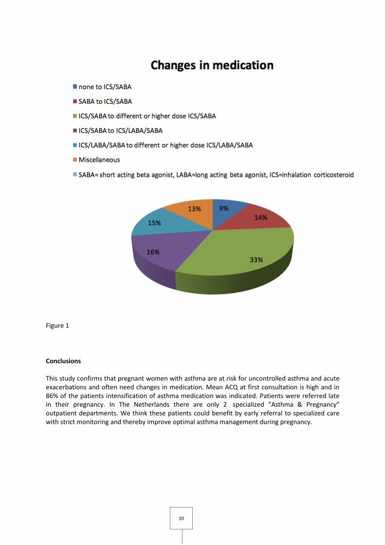
56 pregnant women were referred at a mean gestational age of 20 weeks. At first consultation mean FEV1 was 94% predicted and mean ACQ was 2.1. In 86% of patients there was at least one change in inhalation medication, see figure 1. In 9 patients 11 acute exacerbations occurred, 6 required hospitalization. In 84% of patients FeNO was measured (mean 24.9, range 5-158) and in 53% of patients blood eosinophils (mean 0,25, range 0-1,8).
Best Poster
Winner
10
Figure 1
Conclusions This study confirms that pregnant women with asthma are at risk for uncontrolled asthma and acute exacerbations and often need changes in medication. Mean ACQ at first consultation is high and in 86% of the patients intensification of asthma medication was indicated. Patients were referred late in their pregnancy. In The Netherlands there are only 2 specialized “Asthma & Pregnancy” outpatient departments. We think these patients could benefit by early referral to specialized care with strict monitoring and thereby improve optimal asthma management during pregnancy.
11
P09. [78] Uncovering the link between asthma and obesity in children. BMI fails as a fat mass marker in this context
George Guibas1,2; Yianis Manios2; Paraskeyi Xepapadaki3; George Moschonis2; Nikos Douladiris3; Christine Mavrogianni2; Nikolaos Papadopoulos1,3
1University Of Manchester, Manchester, United Kingdom; 2Charokopeion University, Athens, Greece; 3University of Athens, Athens, Greece
Background
In adults, an obesity/asthma link is well-defined; however, any such link is unclear in children. This obscurity may arise due to Body Mass Index (BMI) drawbacks as a paediatric fat mass marker. We thus hypothesized that obesity predisposes to asthma throughout the entire paediatric age range, and that the conflicting evidence reflects BMI limitations. We investigated this in children of different ages, using several markers of adiposity.
Methods
Wheeze ever/in the last 12 months (current) and physician-diagnosed asthma were reported by parents of children of two large cross-sectional studies: 1626 children aged 2-5 (the Genesis Study) and 2015 children aged 9-13 (the Healthy Growth Study). Anthropometric measurements (BMI, waist/hip circumference, biceps/triceps/subscapular/suprailiac skinfold thickness) were conducted in both cohorts; bioelectric impedance analysis (BIA) was conducted only in preadolescents.
Results
In children aged 2-5, asthma was positively associated with conicity index, waist/hip circumference, waist-to-height ratio, skinfold thickness, and skinfold-derived percentage fat mass (P < 0.05) but, importantly, not BMI or BMI-defined overweight/obesity, after adjusting for numerous confounders in logistic regression models. In children aged 9-13, asthma was positively associated with conicity index, waist circumference, waist-to-height ratio, skinfold thickness, skinfold-derived percentage fat mass, BIA-derived percentage fat mass, and BMI/BMI-defined overweight/obesity (P < 0.05). Current/ever wheeze was not consistently associated with fat mass.
Conclusions
We present two large studies which indicate that adiposity is a risk factor for asthma in both 2-5 and 9-13 age spans. We have also shown for the first time that BMI is unable to uncover this association in preschool children and is therefore inefficient for epidemiologic studies of asthma in this age range; this is likely due to to age-related adipose tissue fluctuations. Conversely, BMI reliably uncovered the adiposity-asthma link in preadolescent children. The conflicting findings reported so far could be due to the wide use of BMI to assess obesity-asthma associations.
The authors declare no COI
Both studies have been approved by Charokopeion University.

12
P10. [79] Differential capacity of Rhinovirus and Influenza Virus to cause asthma exacerbations; clinical characterization of the acute viral conditions on an asthmatic background.
George Guibas1,2; Maria Tsolia2; Ioannis Christodoulou2; Foteini Stripeli2; Zoi Sakkou2; Nikolaos Papadopoulos1,2
1University Of Manchester, Manchester, United Kingdom; 2University of Athens, Athens, Greece
Background
Rhinovirus (RV) infection is an established trigger of asthma exacerbations, whereas evidence of such capacity of influenza virus (IFV) is not as consistent. In this context, we hypothesized that IFV infection may cause a condition that is different from RV, in that it is less prone to asthma exacerbations. We opted to test this hypothesis by evaluating the associations between IFV/RV positivity and clinical characteristics in children hospitalized for a flu-like illness.
Methods
The participants were 1207 children, 6-months to 13-years old, hospitalized for respiratory symptoms/fever. The information collected included demographics, medical history, symptoms/physical findings/diagnosis at presentation and treatment. Nasal secretions were tested via PCR for IFV/RV. Associations were evaluated with logistic regression models controlled for several confounders.
Results
RV positivity was associated with an asthma-like presentation, including increased wheeze/effort of breathing/diagnosis of an asthma exacerbation, and decreased fever/vomiting. Conversely, IFV+ children presented with decreased wheeze/effort of breathing/diagnosis of an asthma exacerbation, while they had more often fever. In the subgroup of children with an asthma history, both viruses induced wheeze that required asthma medication, however, IFV was uniquely associated with a more generalised and severe clinical presentation including fever, rales, intercostal muscle retractions and lymphadenopathy. These symptoms were not seen in RV+ asthmatics, who had less fever and more cough.
Conclusions
In children with respiratory symptoms/fever, RV but not IFV is associated with wheeze and an asthma-like presentation. In the subgroup of children with asthma history, IFV causes a more generalised and severe disease which, however, when compared to the RV-induced condition lacks salient clinical characteristics of an asthma exacerbation. This clinical distinction in the acute infections caused by these viruses highlights the remarkable potential of RV to induce asthma attacks, and provides a clearer characterization of these viral conditions on an asthmatic background.
The authors declare no COI.
This study was approved by the University of Athens.

13
P11. [67] Improving asthma care: a retrospective audit in a major teaching hospital
Rachel Henery1,2; Navya Bezawada1,2; Ratna Alluri2
1University of Aberdeen, Aberdeen, United Kingdom; 2Aberdeen Royal Infirmary, Aberdeen, United Kingdom
Introduction Asthma is a common, treatable condition that remains a major cause of hospitalisation and death in children and young adults. Worryingly, the National Review of Asthma Deaths (NRAD) found that asthma-related deaths in the UK have not reduced since 2000, and in 2015 there was a 10 year high in mortality. The NRAD report highlighted several aspects of asthma care that could be improved, particularly related to patient education and recognition of severity. In order to improve our own practice, we audited NICE and British Thoracic Society quality standards in all asthma admissions to a major teaching hospital. Methods This was a retrospective audit and data was obtained from the health records department on patients admitted to the respiratory ward with an acute asthma exacerbation from January to March 2016. Results In the audit cohort of 22 patients, severity was documented in only 27.2% of cases. Although 86.4% of patients had a peak expiratory flow (PEF) documented on admission, only 40.9% had PEF documented on discharge. None of the patients admitted were provided with a personal asthma action plan (PAAP). 72.7% of patients were followed up post-discharge, the majority of whom (81.3%) were seen in a respiratory clinic. The remainder were followed up in general practice. For those patients followed up in secondary care, the average follow-up time was 11.3 weeks; this was significantly higher than the average planned follow up time of 6 weeks. Conclusions This audit highlighted that most aspects of acute asthma management are falling short of current standards. Although the data has to be interpreted with caution given the small sample size, it is worrying that a significant proportion of patients did not have severity assessed on admission, whilst others were discharged with no follow up. Furthermore, no patients received a PAAP. These issues are being addressed with the introduction of a standardised asthma care checklist and PAAP, with ongoing education of medical staff.

14
P12. [80] Identifying a novel therapeutic strategy for asthma: aiding airway epithelial cell repair
Thomas Iosifidis1,2; Erin Gill3; Amy Lee3; Alysia Buckley4,5; Erika Sutanto4,6; Kak-Ming Ling1,4; Luke Garratt1,4; Kevin Looi1,4; Elizabeth Kicic-Starcevich4,6; Kelly Martinovich1,4; Nicole Shaw1,4; Samuel Montgomery1,4; Francis Lannigan1,7; Paul Rigby5; Robert Hancock3; Darryl Knight8,9,10; Stephen Stick1,2,4,6; Anthony Kicic1,2,4,6
1School of Paediatrics & Child Health, The University of Western Australia, Perth, Australia; 2Centre for Cell Therapy & Regenerative Medicine, Perth, Australia; 3Centre for Microbial Diseases and Immunity Research, University of British Columbia, Vancouver, Canada; 4Telethon Kids Institute, Perth, Australia; 5Centre for Microscopy, Characterisation & Analysis, Perth, Australia; 6Department of Respiratory Medicine, Princess Margaret Hospital for Children, Perth, Australia; 7School of Medicine, Notre Dame University, Fremantle, Australia; 8School of Biomedical Sciences & Pharmacy, University of Newcastle, Newcastle, Australia; 9Priority Research Centre for Asthma & Respiratory Disease, Hunter Medical Research Institute, Newcastle, Australia; 10Department of Anesthesiology, Pharmacology & Therapeutics, University of British Columbia, Vancouver, Canada
Introduction/Aims
Recent advances have implicated the airway epithelium as a key driver in asthma pathogenesis, partly due to its dysregulated response to damage. Novel therapies focusing on protecting and repairing vulnerable airways, particularly in early life, could transform asthma treatment by preventing disease development/progression. (1) To analyse primary airway epithelial cells (pAEC) migration patterns post-wounding in-vitro. (2) To identify mechanisms and potential therapeutic targets enhancing wound repair processes.
Methods
pAEC were obtained from non-asthmatic and asthmatic paediatric airways. Scratch wounds were performed on pAEC monolayers to assess repair and imaged every 30 minutes (IncuCyte ZOOM®, Essen Bioscience). Migration trajectories of leading-edge pAEC were analysed using ImageJ. Integrin gene and protein expression were investigated by qPCR and In-Cell™ Western, respectively. Total RNA was sequenced (Illumina Hi-Seq2500) and differential gene-expression analysis was performed (DESeq2 and Ingenuity Systems(QIAGEN)).
Results
Response to wounding in asthmatic children was deficient and lacked specificity with significantly lower mean migration distance(non-asthma and asthma; 256.9±7.1 and 152.3±8.6μm (mean±SEM)), velocity (0.42±0.02 and 0.22±0.01μm/min), directionality (91.3±0.1 and 61.1±0.1%) and forward migration index (95.3±0.1 and 64.6±0.1%). A major regulator of cell migration, i.e. integrin α5β1, was investigated in pAEC. Lower gene (α5, 5.9-fold, p<0.0001;β1, 1.5-fold, p<0.05). and protein (α5, 2.8-fold, p<0.05; β1, 3.1-fold, p<0.05) levels in pAEC from asthmatic children (n≥11) compared to control (n≥23). RNA-seq analysis identified 1,153 differentially expressed genes in asthmatic children (n=6) relative to control (n=4) with overrepresented gene ontologies related to integrins and extracellular matrix. Drug database screening identified several clinically safe drugs that are now being examined for drug repurposing potential to restore integrin expression and aid wound repair.

15
Conclusion
These novel experiments demonstrate abnormal migration behaviour of asthmatic airway epithelium post-wounding. Some mechanisms controlling this disease phenomenon were identified like decreased integrin α5β1, and multiple transcriptional mechanisms were dysregulated in asthmatics, some of which are targetable by existing drugs. Supporting airway epithelial repair and barrier integrity may be a novel therapeutic avenue for asthma.

16
P13. [90] Pulmonary emphysema subtypes in patients with association COPD and pulmonary tuberculosis: influence influence on pulmonary function and tubercular structural changes of lung by computer tomography
Larisa Kiryukhina1; Leonid Mikhailov1; Pavel Gavrilov1; Olga Volodich1; Maria Pavlova1; Liudmila Archakova1,2; Piotr Yablonskii1,2
1Federal State Institution Saint-Petersburg Research Institute of Phthisiopulmonology, Ministry of Health of Russia, Saint-Petersburg, Russian Federation; 2Saint-Petersburg State University, Saint-Petersburg, Russian Federation
Background
Most patients with pulmonary tuberculosis (PT) are current or ex-smokers. Many of them began to smoke in childhood and often have co-morbidity as COPD in age before 45 years. Emphysematous changes (EC) in patients with association COPD and PT is often under estimated. Clinical characteristics of emphysema subtypes on computer tomography (CT) are not well defined.
Aim
To assess emphysema subtypes on CT in patients with COPD and PT and influence of emphysema on pulmonary function and specific structural changes.
Methods
Patients with PT receiving treatment at the Saint-Petersburg Research Institute of Phthisiopulmonology (2014-2016 years) were divided into 2 groups: 1 group - PT and COPD (n=23), 2 group - PT without COPD (n=93). All patients were observed using CT and lung function testing (LFT): CT with the additional program for Nodule Analysis and Lung Volume Analysis (AQUILION PRIME, TOSHIBA) and spirometry, body plethysmography, diffusing capacity (MasterScreen Body Diffusion, VIASYS Healthcare).
Results
Expressiveness of EC was higher in patients with COPD, the mixed (26%) and panlobular (17%) subtypes were prevailed. The greatest volume of EC was in patients with panlobular subtype. In 1st gr. the widespread specific changes (more than 3 segments) were more often compared to patients 2nd gr. (83 and 44%,p<0,05), more volumes of tubercular damage (p<0,05) and volume of EC (61 and 30%,p<0,05). Correlation of EC volume and LFT parameters (p<0,05): 1 gr. FEV1/FVC r=-0,62, RV/TLC r=0,46, DLCO r=-0,59; 2 gr. FEV1 r=-0,69.
Conclusions
In patients with PT and COPD the mixed and panlobular subtypes were prevailed and specific destruction was significantly more than in patients without COPD. Increase in EC volume led to airways disorders in all patients and to increase of lung hyperinflation and decrease of diffusing capacity in patients with PT and COPD.

17
P14. [91] Plasmacytoid dendritic cells drive acute exacerbations of asthma
Ourania Koltsida1,2; Aikaterini Chairakaki2; Maria Saridaki2; Katerina Pyrillou2; Marios Mouratis2; Ross Walton3; Nathan Bartlett3; Athanasios Stavropoulos2; Nikoletta Rovina1; Sebastian Johnston3; Evangelos Andreakos2
121st Department of Respiratory Medicine, Medical School, National Kapodistrian University of Athens, 'Sotiria' Regional Chest Diseases Hospital, Athens, Greece; 21Department of Immunology, Center for Clinical, Experimental Surgery and Translational Research, Biomedical Research Foundation of the Academy of Athens, Athens, Greece; 33Airway Disease Infection Section, National Heart and Lung Institute, London, United Kingdom
Background
Although acute exacerbations, mostly triggered by rhinoviruses (RVs), account for the majority of hospitalizations and life-threatening situations in asthma, there is still very little known about the pathophysiological mechanisms involved.
Objective
We sought to investigate the role of plasmacytoid DCs (pDCs) in asthma exacerbations and unwind potential mechanisms involved.
Methods
Patients with asthma under stable disease or acute exacerbations, and healthy individuals, were studied for pDC presence in their sputum and their association with various leukocytic populations, cytokines and disease parameters. Animal models of asthma and virus-induced asthma exacerbations were further used to dissect the functional role of pDCs in the disease process.
Results
pDCs were markedly increased in the sputum of patients with stable asthma and acute exacerbations. Moreover, increasing pDC numbers were directly linked to the severity of type 2 inflammation, deterioration of lung function and risk for asthmatic attacks. In animal models of allergic asthma and RV-induced exacerbations, pDCs were shown to be key mediators of the immuno-inflammatory response driving asthma; they were recruited to the lung during inflammation and migrated to the lymph nodes to boost Th2-mediated effector responses. Accordingly, pDC depletion post-allergen challenge or during RV infection abrogated disease exacerbation. Central to this was interleukin 25 (IL-25) which conditioned pDCs for pro-inflammatory activation and migration.
Conclusion
Our studies uncover a previously unsuspected role of pDCs in asthma exacerbations with major implications in disease diagnosis, prognosis and monitoring. They also propose the therapeutic targeting of pDCs and IL-25 for the treatment of asthma attacks.
Best Poster
Winner

18
P15. [105] The profile of leucocytes, CD3+, CD4+, CD8+ cells, and cytokine concentration in peripheral blood of children with acute asthma exacerbation
Huong Le-Thi-Thu1; Thuy Nguyen-Thi-Dieu2; Sy Duong-Quy3,4,5
1Department of Immunology, Allergology, and Rheumatology. National Hospital of Pediatrics, Hanoi, Viet Nam; 2Hanoi University of Medicine, Hanoi, Viet Nam; 3Lam Dong Medical College, Dalat, Viet Nam; 4Cochin Hospital. Paris Descartes University, Paris, France; 5Division of Pulmonary, Allergy and Critical Care Medicine. Penn State College of Medicine, Pennsylvania, USA
Introduction
Acute asthma exacerbation (AAE) is a common cause of visiting the emergency department and increases the risk of mortality in asthmatic children. The study of predicted biomarkers for a high risk of severe AAE is a crucial objective. Our study was planned to demonstrate the leucocyte profile and cytokine concentration in peripheral blood of children with AAE.
Methods
It was a descriptive and prospective study. The severity of AAE was assessed by PAS (pediatric asthma score). Peripheral bloods were collected for quantification of CD3+, CD4+, and CD8+ by flow-cytometry and cytokine by flowcytometry-assisted immunoassay.
Results
127 children with AAE were included. The percentage of subjects with decreased CD3+, CD4+, CD8+ cells in severe AAE was significantly higher than in light to moderate AAE (P<0.05). The concentrations of IL-2, IL-8, IL-12, and IL-4 in subjects with rhinovirus infection were significantly higher. IL-4 and IL-6 concentrations during and after AAE were significantly lower than control (P<0.001; P<0.5). IL-5 and GM-CSF concentrations during AAE were significantly higher than control (P<0.001; P<0.05).
Conclusions
The decrease of CD3+, CD4+, CD8+ cells and IL-4 and IL-6 combined with the increase of IL-5 and GM-CSF are associated with a high risk of AAE in children with asthma.

19
P16. [70] Smoking Increases the Risk for Death From Pneumonia
Adamantia Liapikou1; Katerina Dimakou2; Michael Toumbis3
1Conslultand Respiratory Medicine,Sotiria Hospital, Athens, Greece; 2Director of 5th Respiratory Department,Sotiria Hospital, Athens, Greece; 3Director of 6th Respiratory Department,Sotiria Hospital, Athens, Greece
Background
Active smoking increases the risk of developing community-acquired pneumonia (CAP) although its impact on mortality in CAP outcomes remains unclear. The aim of this study was to investigate the influence of smoking status on CAP mortality.
Method
We performed a prospective, observational cohort study in hospitalized patients with CAP. Patients were classified according to their smoking habits and compared the severity and evolution of pneumonia in both groups.
Results
The study group consisted of 740 patients with CAP: 335 current smokers (45.2%), 85 ex-smokers (11.5%) and 320 nonsmokers (43.2%). In the group of non smokers there were more women (61%, p<0,001), with higher incidence of neurological disease (26%,p=0,001). Current smokers with CAP were younger (59 ys vs. 73 ys vs.65 ys, p<0,001), men, with fewer incidence of cardiac diseases and diabetes. Εx-smokers had more often comorbidities (85% vs.68% vs.66%, p<0,003), especially cardiac, pulmonary disease and diabetes than the two other groups and presented with more severe pneumonia ; 35% of them belonging to CURB-65≥3 classes. Current smokers although had lower CURB-65 scores, had more often severe respiratory failure, tachycardia and ICU admission at diagnosis. Overall mortality at 30 days was 7.7%: 4.5% in current smokers vs. 10 % in nonsmokers and 11.8% in ex-smokers (p=0,032). In multivariable analysis current smoking (OR, 2,37; p= .015) , CURB65 (OR, 2.2; p <.001) and PAFI <250(OR, 3,2 ,p<0,001) were risk factors for 30-day mortality of CAP.
Conclusion
Current smokers with CAP often develop severe disease with respiratory failure and require hospitalization at a younger age. Smoking increases the risk of 30-day mortality from CAP.

20
P17. [58] The role of bronchodilation on airway mechanical stress, lung hyperinflation and NO production in stable COPD
Dejan Radovanovic1; Marco Contoli2; Susanna Mascetti2; Matteo Pecchiari3; Alberto Papi2; Stefano Centanni4; Pierachille Santus4
1School of Respiratory Diseases, University of Milan, Milano, Italy; 2Research Centre on Asthma and COPD, Section of Internal and Cardio-Respiratory Medicine, University of Ferrara, Ferrara, Italy; 3Department of Physiopathology and Transplantation, University of Milan, Milano, Italy; 4Department of Health Sciences, University of Milan, Milano, Italy
Background
Patients with severe chronic obstructive pulmonary disease (COPD) experience cyclic opening and closure of the airways even at tidal volume (Pecchiari M et al. Respir Physiol Neurobiol 2016). The effect of this mechanical stress on lung inflammation and its reversibility are still poorly understood. The aim of our study is to investigate the acute effect of long acting beta-agonists (LABAs) on lung inflammation and its relationship with lung function in patients with moderate to very severe COPD.
Methods
This was a phase IV, multicenter, randomized, interventional, double blind, crossover study. Stable patients with COPD and a forced expiratory volume in 1 second (FEV1)<60%predicted were consecutively enrolled. Following a pharmacological washout, patients underwent body plethysmography, diffusing capacity for carbon monoxide (DLCO), multi-flow exhaled fraction of nitric oxide (FeNO) at 50, 150 and 350 ml/s, single breath nitrogen washout test (SBN2) at baseline and 30, 60 and 180 minutes after randomization to either formoterol 12 mcg Modulite (FF) or salmeterol 50 mcg (S) metered-dose inhaler. 72 hours after, patients were crossed-over. The distal airway/alveolar NO (Calv) was calculated with the slope-intercept method (Tsoukias N et al. JAP 1998). Tissue factor (KCO) and ventilation inhomogeneity (VA/TLC) were derived from DLCO.
Results
The analysis included 45 patients (Table 1). FF and S similarly improved plethysmographic parameters (P<0.05 in all instances, ANOVA). Compared to baseline, FeNO at 350 ml/s and SBN2 phase III slope decreased equally after FF and S (P<0.05 in all instances, ANOVA). Calv was reduced at all time-points (Figure 1A). Calv was correlated with KCO (r=-0.43, P<0.001) and residual volume to total lung capacity ratio (r=0.38, P=0.013). ΔCalv was correlated with baseline VA/TLC (r=0.48, P=0.001), ΔKCO (Figure 1B) and changes in functional residual capacity (r=0.33, P=0.03).
Conclusions
In COPD, the degree of lung inflammation at rest is related to the functional impairment of small airways. In acute conditions, LABAs are able to reduce peripheral FeNO improving hyperinflation and ventilation inhomogeneity.
Rising Star

21

22
23
P18. [95] Leukotrienes affect airway hyperreactivity and pulmonary inflammation in a sex-dependent manner in an experimental mouse model of asthma
Antonietta Rossi1; Fiorentina Roviezzo1; Rosalinda Sorrentino2; Aldo Pinto2; Giuseppe Cirino1; Oliver Werz3; Bruno D' Agostino4; Lidia Sautebin1
1Department of Pharmacy, University of Naples Federico II, Naples, Italy; 2Department of Pharmacy, University of Salerno, Fisciano, Italy; 3Chair of Pharmaceutical/Medicinal Chemistry, Institute of Pharmacy, Friedrich-Schiller-University, Jena, Germany; 4Department of Experimental Medicine, Second University of Naples, Naples, Italy
Background
Asthma is a chronic airway inflammatory disease with higher incidence in female than male patients (1). Recently, sex disparities in leukotriene (LT) production have been shown in experimental models of inflammation (2-4).
Aim
Because of the key role of LTs in asthma, the goal of this study was to evaluate sex difference in LT production following allergen sensitization and its correlation with airway hyperreactivity and pulmonary inflammation in a mouse model of asthma. Moreover sex bias in the efficacy of LT-modifying drugs was also evaluated.
Methods
Female and male BALB/c mice were sensitized on days 0 and 7 by subcutaneous injection of ovalbumin (OVA) adsorbed onto Al(OH)3 and killed at different time points to evaluate bronchial reactivity, pulmonary LT, IL-13, PGD2 levels and lung inflammation. In a separate set of experiments mice were treated with montelukast sodium (1 mg/kg 30 min before OVA injection and three time at week) and zileuton (35 mg/kg 30 min before OVA injection and at 14 days).
Results
Female OVA-sensitized mice presented higher bronchial reactivity than male mice (3, 8, 14 and 21 days). These data well-correlated with the pulmonary levels of LTC4 and LTB4 which were higher in female than male mice. Similarly, female OVA-sensitized mice displayed a higher pulmonary IL-13 and PGD2 levels and lung inflammation compared to male mice. Moreover montelukast and zileuton inhibited OVA-induced bronchial hyperreactivity only in female. This effect was coupled to a reduction of LTC4 levels in lungs from sensitized-female mice.
Conclusions
Our data reveal significant sex differences in the biosynthesis of LTs in mouse model of asthma, associated with differential inflammatory responses. Therefore, these results add significant insights into the understanding of basic sex differences in asthma, with possible therapeutic implications for a sex-tailored treatment of related disorders.
1. Almqvist C (2008) Allergy 63, 47. 2. Pergola C (2008) Proc Natl Acad Sci 105, 19881. 3. Pergola C (2011) FASEB J. 25, 3377. 4. Rossi A (2014) Pharmacological Research 87, 1.

24
P19. [97] Toll-Like Receptor 4 is required for S1P-induced airway smooth muscle hyperresponsiveness and lung inflammation
Fiorentina Roviezzo1; Rosalinda Sorrentino2; Michela Terlizzi2; Maria Antonietta Riemma1; Antonietta Rossi1; Giuseppe Spaziano3; Aldo Pinto2; Bruno D'Agostino3; Giuseppe Cirino1
1Department of Pharmacy, University of Naples Federico II, Naples, Italy; 2Department of Pharmacy (DIFARMA), University of Salerno, Salerno, Italy; 3Department of Experimental Medicine L. Donatelli, Second University of Naples, Naples, Italy
Background
Subcutaneous administration of sphingosine-1-phosphate (S1P) reproduces a disease closely mimicking the cardinal features of severe asthma in humans, including airway hypereactivity, pulmonary eosinophil inflammation, high circulating levels of IgE and a predominance of CD4T cell-derived IL-4
Aims
Because TLR4 has been widely described as involved in the pathogenesis of asthma, the aim of this study was to address its potential role in S1P-mediated airway hyperreactivity and inflammation.
Methods
Female BALB/c or C3H/HeJ (TLR-4 defective) mice received on days 0 and 7 subcutaneous injection of LPS(0.1µg), S1P(10ng) or LPS plus S1P. In another set of experiment BALB/c mice received 30 minutes prior to S1P the purified anti-TLR4 (10 µg).
Results
Both LPS and S1P administration to BALB/c mice induced a significant increase in carbachol-induced contractions. In addition, a further increase was observed following co-administration of S1P and LPS. Conversely, both S1P and LPS administration to C3H/HeJ mice did not induce any effect on bronchial tone. In addition, S1P administration to BALB/c mice induced pulmonary TLR-4 up-regulation. Accordingly, pre-treatment of BALB/c mice with TLR-4 neutralizing antibody abrogated both S1P induced bronchial hyper-reactivity and pulmonary inflammation. This was accompanied by a marked reduction in S1P-induced pulmonary IL-13 expression, while IgE and Il-4 were not altered. Flow cytometry analysis evidenced a significant increase of dendritic cell in the lung and a decrease in MLN following S1P treatment in BALB/c mice, that were not affected by TLR4 blocking. In perfect tune, TLR4 blocking did not interfere with S1P-induced dendritic cell maturation. Finally, co-immunoprecipitation of S1P1 and TLR4 was observed in the lung following S1P challenge.
Conclusions
The concerted effects, induced by S1P1 and TLR4 signalling, might be required in synergy to optimally induce effector T cell immunity to a harmless antigen.

25
P20. [101] Biomarkers for Precision medicine in Chronic Obstructive Pulmonary Disease Patients:Is It Right Way To Go?
Alizaman Sadigov1; Ibrahim Isayev1; Jabrail Mammadov1
1Medical University,Pulmonary Medicine Department, Baku, Azerbaijan; 2Medical University,Pulmonary Medicine Department, Baku, Azerbaijan; 3Medical University, Pulmonary Medicine Department, Baku, Azerbaijan
Background
Precision medicine can be implemented using a variety of different methods and tools and the most promising of which are biomarker-based.There are emerging areas in which biomarkers may guide therapeutic choices for patients with chronic obstructive pulmonary disease(COPD).
Objective
The aim of our study was to assess the peripheral blood level of eosinophils and serum level of periostin as target for precision medicine in patients with acute exacerbations of COPD.
Methods
78 patients with AECOPD who admitted to pulmonary medicine department were investigated by using pulmonary function test,serum level of C-reactive protein(CRP),peripheral blood eosinophils count, serum level of periostin , arterial blood gas assessement.
Results
36 patients with eosinophilc exacerbations of COPD (peripheral blood eosinophils≥2%) and 42 patients with non-eosinophilic exacerbations of COPD (peripheral blood level of eosinophils ≤2%) have received adequate treatment according to GOLD guidelines.In patients with eosinophilic exacerbations the serum level of periostin was significantly higher than in patients group with non -eosinophilic exacerbations of COPD(69.75±23.93 ng/ml vs 24.45±11.52 ng/ml;p<0.001).In patients with non-eosinophilic exacerbations of COPD the serum level of CRP was significantly higher compared with patients with eosinophilic exacerbations of the disease(67.85 ±19.65mg/ml vs 21.32±12.94 mg/ml;p<0.001).In patients with non-eosinophilic exacerbations of COPD the rate of community-acquired pneumonia was greater compared with patients eosinophilic exacerbations of COPD(15[34%] vs 2[5%];p<0.005). ICU admission due to hypercapnic respiratory failure was more often in patients group with non-eosinophilic exacerbations of COPD(11[24%] vs 1[2%];p<0.001).In patients with eosinophilic exacerbations of COPD admission of systemic corticosteroids(SCs) lead to the significantly improvement of lung function and clinical features of the disease exacerbations withour admission of antibiotics.The length of inhospital stay was greater inpatients with noneosinophilic exacerbations of COPD compared with eosinophilic exacerbations of the disease ecacerbations(8.0[4.0-22 days] vs 5.0[2.0-15 days]'p<0.05).
Conclusions
For patients with acute exacerbations of COPD the peripheral eosinophil counts and serum periostin level may enable separation of patients who are more likely to respond to corticosteroids versus those who would benefit from non-steroid based therapies.

26
P21. [114] Risk factors for infectious exacerbations in Chronic Obstrutive Pulmonary Disease GOLD
3 and 4
Maria Santos1; Margarida Aguiar1; Sofia Furtado1
1Hospital Beatriz Ângelo, Loures, Portugal
Introduction
Chronic Obstructive Pulmonary Disease (COPD) has an estimated prevalence of 14.2% in adult population above 40 years old, in Lisbon. Infectious exacerbations of COPD affect disease progression and strongly influence quality of life.
Objective
Investigate potential risk factors for infectious exacerbations in COPD GOLD 3 and 4 patients.
Methods
Patients with COPD and severe or very severe airflow limitation in pulmonary function tests performed in 2014 were selected. They were characterized with regard to demographics, smoking habits, functional evaluation and comorbidities. These patients were followed up for a year and data on infectious exacerbations was collected. Potential risk factors were assessed.
Results
57 patients were included: 83% were male and mean age was 65.3 ± 9.1 years. One third were current smokers and mean pack-years was 59.6 ± 29.1. The most common respiratory comorbidities were bronchiectasis (39%) and obstructive sleep apnea (21%). Other comorbidities were hypertension (67%), anxiety/ depression (37%), heart failure (28%) and type 2 diabetes (21%). 54% had at least one exacerbation in the previous year.
During follow-up, mean exacerbation rate was 1.4. Bronchiectasis and at least one exacerbation in the previous year were associated with more exacerbations (1.7 vs 1.2, p value 0.09 and 1.9 vs 0.9, p value 0.007, respectively). We also found a correlation between the number of exacerbations and FEV1 % predicted (r=-0.39), residual volume % predicted (r=0.31) and diffusing capacity corrected for the alveolar volume (r=-0.31). Patients with anxiety or depression had a lower mean FEV1 (36.4 vs 39.8, p value 0.09), a higher proportion of current smokers (0.52 vs 0.19, p value 0.004) and exacerbated more (2.19 vs 0.94, p value 0.002).
Conclusion
Poorer functional evaluation, bronchiectasis and exacerbations in the previous year are associated with a higher risk of infectious exacerbations in COPD GOLD 3 and 4 patients. Patients with anxiety or depression smoke more and have worse functional status which might contribute to a higher rate of infectious exacerbations.
27
P22. [113] Exercise intolerance in COPD patients: is it all about the lung?
Maria Santos1; Susana Clemente1; Filipa Todo-Bom1; Sílvia Rodrigues1; Liliana Gomes1; Jorge Rosa1;
Inês Alves1; Ana Campos1; Sofia Furtado1
1Hospital Beatriz Ângelo, Loures, Portugal
Introduction
In Chronic Obstructive Pulmonary Disease (COPD), exercise intolerance may be caused not only by ventilatory limitation but also by other factors such as cardiovascular limitation, muscular dysfunction, nutritional or psychological factors. Cardiopulmonary exercise testing is considered the gold standard exercise test for evaluating the causes of exercise intolerance.
Objective
To identify exercise limiting factors in COPD patients.
Methods
A retrospective analysis of cardiopulmonary exercise tests performed between May 2012 and June 2016 in patients with COPD diagnosis was done. A cycle ergometer was used, and an incremental protocol until maximal exercise tolerated by the patient or ending by medical order was chosen.
Results
96 tests were included. Patients mean age was 62.9 ± 11.1 years and 80% were male. 56% were current and 44% were previous smokers. Cardiopulmonary exercise test was performed in these patients in order to prescribe adequate pulmonary rehabilitation programs (66%), evaluate surgical risk (23%) and in patients with unexplained exercise intolerance (11%). Mean maximum work was 68.2 ± 36.6 watts (51.6 ± 28.5%), mean oxygen consumption was 16.7 ± 5.5 mL/kg/min (61.1 ± 21.3 %), mean anaerobic threshold was 15.0 ± 10.9 mL/kg/min (50.6 ± 18.4%), mean respiratory reserve was 22.2 ± 19.7%, mean respiratory rate was 33.3 ± 7.6 breaths per minute, mean cardiac reserve was 35.7 ± 18.5 beats per minute, mean oxygen pulse was 79.4 ± 25.9 % of maximum predicted and mean VE/ VCO2 was 35.3 ± 8.1. Exercise limiting factors identified were: obstructive disease (61%), cardiovascular disease (17%), deconditioning (17%), obesity (1%) and anemia (1%).
Conclusion
The main exercise limiting factor in these COPD patients was their pulmonary disease. Nonetheless, a relevant proportion of these patients (36%) had other exercise limiting factors. Identifying other exercise limiting factors may lead to the appropriate treatment, reducing dyspnea and exercise intolerance.
28
P23. [83] A case of pulmonary cystecircosis. Differential diagnosis of pulmonary nodules
Velin Stratev1; Tanya Dobreva1; Boyan Balev2; Valentina Dimitrova1; Margarita Gospodinova3; Diana Petkova1
1UMHAT "St. Marina" Clinic of pulmonary diseases, Varna, Bulgaria; 2UMHAT "St. Marina" Clinic of image diagnostics, Varna, Bulgaria; 3UMHAT "St. Marina" Clinic of infectious diseases, Varna, Bulgaria
Introduction
The differential diagnosis of multiple pulmonary nodules is wide and includes congenital, acquired, infectious and non-infectious granulomatoses, malignomes and others. Extra-pulmonary symptoms and findings help for their discrimination.
Aim
We describe a case of multiple pulmonary nodules and brain involvement of nodules with characteristic image morphology.
Patients and methods
An 81 years old male, who breeds swine and cattle presented at the pulmonary clinic with acute onset of fever, cough and neurological symptoms. At admission he was with stable vital signs and normal reflexes, there were crackles at lung basis on auscultation. During hospital stay the patient developed neurological symptoms and his condition deteriorated rapidly. We performed biochemical and hematological tests, urine analysis, fecal probes, serological tests for toxoplasmosis, echinococcosis, cystecircosis and liquor analysis. We performed CXR, CT of the lung and andomen, MRI of central nervous system.
Results
The serological tests were negative for parasites. All of the image techniques showed characteristic features of scolices, which are pathognomonic for cystecircosis. The unique morphology is not seen in other diseases with multiple nodules.
Conclusion
Cystecircosis is an infectious disease of the larva stage of the measles-Т.solium. The disseminated form is rare. Unlike brain involvement (neurocystecircosis), lung involvement is extremely rare. The presence of one absolute criterion according to Del Brutto’s scale (cystic lesions with scolices) is sufficient for the diagnosis. Enzyme and serological tests may remain negative when scolices are dead or in the presence of calcifications.

29
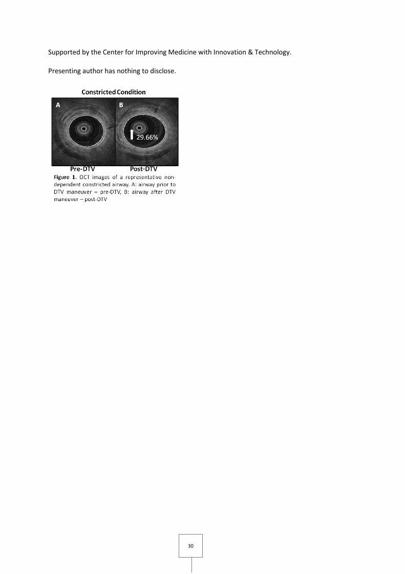
P24. [68] Using Optical Coherence Tomography to evaluate airway dynamics in vivo
Margit Szabari1,4; Vanessa J. Kelly1; Matthew Applegate1; David C. Adams1,4; Lida P. Hariri3,4; Chunmin Chee1,4; Khay Tan1,4; Robert S. Harris1; Tilo Winkler2; Melissa J. Suter1,4
1Department of Medicine, Pulmonary and Critical Care Unit, Massachusetts General Hospital and Harvard Medical School, Boston, USA; 2Department of Anesthesia, Critical Care and Pain Medicine, Massachusetts General Hospital and Harvard Medical School, Boston, USA; 3Department of Pathology, Massachusetts General Hospital and Harvard Medical School, Boston, USA; 4Wellman Center for Photomedicine, Massachusetts General Hospital and Harvard Medical School, Boston, USA
Background/Aims
Airway hyperresponsiveness is a hallmark feature of asthma. To better understand this condition, it is essential to visualize airway behavior in vivo. Optical coherence tomography (OCT) is a high-resolution imaging modality that can be used to provide real-time visualization of airway dynamics in vivo. Our aim was to use OCT to investigate the structure and function of airways in the healthy and constricted lung in both dependent and non-dependent airway regions during mechanical ventilation.
Methods
N=3 sheep were anesthetized and mechanically ventilated. A total of 6 dependent and 6 non-dependent airways were imaged at baseline and following Methacholine administration (Mch: 1 mg/hr). OCT imaging was used to acquire cross-sectional images during Regular Tidal Breathing (RTB) (tidal volume (TV): 10 ml/kg) and in a response to one minute of Double TV ventilation (DTV). The airway-areas during the breathing cycle and pre- and post-DTV maneuvers were calculated from the OCT images using semi-automated image processing algorithms.
Results
Dependent airway-area increased more than non-dependent airway-area during RTB in the healthy airways (p<0.001). This difference was most prominent in the expiratory phase of the breathing cycle (maximum change in dependent vs. non-dependent airway-area: +13.8% vs. +3.9%). In the constricted airways this difference disappeared during RTB. DTV maneuver had no effect on post-DTV airway-area in healthy airways in both dependent and non-dependent regions. However, the constricted post-DTV airway-area increased compared to the pre-DTV airway-area by +15.5% in the dependent and by +44% in the non-dependent regions (Figure 1).
Conclusions
OCT provided unprecedented insight into the behavior of airway structure and function of a sheep asthma model. Under normal healthy conditions during RTB dependent airways were more distensible than non-dependent airways. This behavior was reversed after the DTV maneuver during airway constriction, which may be explained by the dependent lung area receiving a higher local Mch dose.
Rising Star
30
Supported by the Center for Improving Medicine with Innovation & Technology.
Presenting author has nothing to disclose.
31
P25. [81] Thyroid hormone inhibits pulmonary fibrosis through enhancement of mitochondrial function in alveolar epithelial cells
Argyrios Tzouvelekis1,2; Yu Guoying2; Aidinis Vassilis1; Herazo-Maya Jose2; Kaminski Naftali2
1Biomedical Sciences Research Center Alexander Fleming, ATHENS, Greece; 2Yale School of Medicine, New Havenn, USA
Background
Idiopathic Pulmonary Fibrosis (IPF) lungs are characterized by increased numbers of damaged mitochondria and impaired mitophagy. Thyroid hormone (TH) is critical for maintenance of cellular and mitochondrial homeostasis during stress responses, but its role in lung fibrosis is unknown.
Methods and Results
Reanalysis of the Lung Genomic Research Consortium (LGRC) dataset revealed that DIO2 that converts thyroxine to triiodothyronine, was among the most significantly increased genes in IPF lungs compared to controls. DIO2 expression and activity were significantly increased in IPF lungs compared to controls. Genetic ablation of DIO2 (DIO2-knockout mice) enhanced bleomycin-induced lung fibrosis as assessed by significant increases in: a) hydroxyproline content, b) COL1A1 and COL3A1, c) Masson Trichrome staining. Aerosolized TH delivery reversed bleomycin-induced fibrosis, as assessed by significant reductions in: a) hydroxyproline and b) Masson Trichrome staining. Bleomycin injury caused mitochondrial dysfunction in primary murine alveolar type II epithelial cells (AECIIs) while TH therapy reversed these abnormalities as assessed by extracellular flux and electron microscopy analysis. Aerosolized TH therapy induced regulators of mitochondrial function, PPARGC1A and PINK1. TH treatment did not blunt fibrosis in PPARGC1A or PINK1 knockout mice, suggesting that TH anti-fibrotic effects are mediated through PPARGC1A and PINK1.
Conclusions
TH treatment is protective against fibrotic lung injury through restoration of AECIIs mitochondrial homeostasis and may represent an effective therapy for IPF.

32
P26. [117] Efficacy and safety of the direct switch from various previous treatments to glycopyrronium or indacaterol/glycopyrronium in patients with moderate copd: the crystal study
Kostikas Konstantinos2; Claus Vogelmeier1; Maryam Aalamian-Mattheis2; Tim Greulich3; Jose M Marin3; Walter Castellani4; Thomas Similowski5; Vincent Ninane6; Stephen Lane8; Xavier Nunez9; Francesco Patalano2; Andreas Clemens2;
1University Medical Center Giessen and Marburg, Marburg, Germany; 2Novartis Pharma AG, Basel, Switzerland; 3Respiratory Medicine, Hospital Universitario Miguel Servet, Zaragoza, Spain; 4Department of Respiratory Physiopathology, Palagi Hospital, Florence, Italy; 5Respiratory Medicine and Intensive Care, Pitié - Salpêtrière Hospital, Paris, France; 6CHU Saint-Pierre - Service de Pneumologie, Brussells, Belgium; 7Athens Chest Hospital "Sotiria", Athens, Greece; 8Adelaide & Meath Hospital, Dublin, Ireland; 9TFS Develop, Barcelona, Spain
Introduction and Objectives
In contrast to clinical trials, changes to new therapies in clinical practice occur without any washout period. The CRYSTAL study was designed to mimic clinical practice. Patients with symptomatic, non-frequently exacerbating, moderate COPD treated with various drugs were directly switched to glycopyrronium 50 μg (GLY) or indacaterol/glycopyrronium 110/50 μg (IND/GLY). Lung function and symptoms were evaluated.
Methods
CRYSTAL was a prospective, multicentre, 12-week, randomised, pragmatic, open-label trial. Patients were recruited into 4 Groups according to previous medication and symptoms (mMRC) and randomised to a direct switch to GLY or IND/GLY vs. continuation of baseline therapy (3:1). Co-primary objectives were superiority of GLY vs. previous SABA and/or SAMA, non-inferiority of GLY vs. previous LABA or LAMA, and superiority of IND/GLY vs. LABA, LAMA and LABA+ICS regarding trough FEV1 and transition dyspnoea index (TDI) at Week 12. Due to slow recruitment, Groups 1 and 2 were prematurely discontinued at the time of completion of Groups 3 and 4.
Results
Of the 4,389 patients randomised, 2,159 patients received IND/GLY or continued their previous treatment. IND/GLY provided superior improvement in trough FEV1 at Week 12 vs. LABA+ICS (treatment difference (Δ)=71 mL, p<0.0001) and LABA or LAMA (Δ=101 mL, p<0.0001). IND/GLY also improved TDI vs. LABA+ICS (Δ=1.10 units, p<0.0001) and vs. LABA or LAMA (Δ=1.26 units, p<0.0001). Significantly more patients on IND/GLY reached the minimally clinically important difference (MCID) of 100 mL for trough FEV1 and 1 point for TDI vs. comparators. In the Groups 1 and 2 that were underpowered due to sample size, GLY was superior to previous SABA and/or SAMA and was non-inferior to previous LABA or LAMA on trough FEV1 and TDI. GLY and IND/GLY were well tolerated.
Conclusions
In the pragmatic CRYSTAL trial, IND/GLY demonstrated superior improvement in lung function (trough FEV1) and dyspnoea (TDI) after 12 weeks, in symptomatic patients with moderate COPD and a history of up to 1 exacerbation in the previous year, after direct switch from previous treatment with either LABA+ICS or with a LABA or LAMA.

33
P27. [102] Efficacy and safety of formoterol fumarate in acute exacerbation of chronic obstructive pulmonary disease
Dejan Zujovic1; Cvijanovic Dane2
1Municipal Institute for Lung Diseases and Tuberculosis, Belgrade, Serbia; 2Phillips Healthcare Serbia, Belgrade, Serbia
Background
Formoterol fumarate is a long-acting β2-agonist with rapid onset, used for the standard treatment of stable chronic obstructive pulmonary disease (COPD). Its quick and sustainable bronchodilation is the reason for which this medicine is taken into consideration as a possible treatment for COPD exacerbations. Up to date the efficacy and safety of its use in the treatment of COPD exacerbations has not been compared with repeated nebulizations of fenoterol/ipratropium bromide.
Aim
To compare the efficacy of repeated inhalations of formoterol fumarate via Turbuhaler with repeated nebulizations of fenoterol/ipratropium bromide, as expressed by the number of days needed to reach clinical improvement, lung function values and sO2. The safety of the treatment protocols has been analysed by electrocardiography and pulse oximetry.
Method
In the prospective trial 120 patients were randomized into two groups and treated with inhalations of 9 mcg formoterol every 30 minutes until the total delivered dose of 27 mcg was reached, or repeated nebulizations of 1ml fenoterol/ipratropium bromide until the total dose of 3ml was reached. All patients received systemic parenteral corticosteroids, oral antibiotics and, in case of manifested respiratory insufficiency, oxigenotherapy. Lung function values, sO2, heart rate and possible heart rhythm disturbances were monitored at the beginning and in the end of the trial. The clinical improvement was deemed the reaching of the best personal lung function value in the patient’s medical records and the stabilization of the clinical condition.
Results
The treatment with formoterol fumarate resulted in clinical improvement after 4.23±1.18 days on average, which was significantly (p<0.0001) better than in case of repeated nebulizations, and it showed equally effective improvement of some of lung function values (FVC, FEV1 and PEF), sO2, and more effective (p ˂ 0.0001) improvement peak inspirium flow (PIF) and heart rate(HR). Both treatment protocols exhibited good safety profile.
Conclusion
Formoterol fumarate may be used as an effective treatment of COPD exacerbation, with good safety profile.
Keywords
Formoterol fumarate; fenoterol/ipratropium bromide; AECOPD

34
The opportunities to support the young generation, such as
Rising Stars
Travel Grants
Best Poster Awards
Have been made possible thanks to

35