saqs for dentistry - pastestsaqs for dentistry third edition kathleen f m fan phd, mbbs, bds, fdsrcs...
TRANSCRIPT
SAQs for Dentistry Third Edition
Kathleen F M Fan PhD, MBBS, BDS,
FDSRCS (Eng), FRCS (Ed), FRCS (OMFS)Consultant Oral and Maxillofacial Surgeon,
Honorary Senior Lecturer
King’s College Hospital, London
Judith Jones BDS, MSc, FDSRCS (Eng),
PhD, FDS (OS), FHEAReader / Honorary Consultant, Department of Oral and
Maxillofacial Surgery, Queen Mary University of London,
Barts and the London School of Medicine and Dentistry,
Institute of Dentistry
Contents
List of Contributors vi
Introduction vii
1 Child Dental Health and Orthodontics 1
2 Restorative Dentistry 49
3 Oral Surgery 159
4 Oral Medicine 213
5 Oral Pathology 253
6 Oral Radiography/Radiology 283
7 Human Disease and Therapeutics 319
8 General Dentistry 383
Index 439
3Oral Surgery

161ORAL SURGERY
3.1 Local anaesthetics
(a) You plan to extract a lower left first permanent molar tooth on a fit
and healthy 34-year-old patient using 25% lidocaine with adrenaline
1:80 000. You plan to carry out an inferior dental/alveolar block,
but what other nerves will you need to anaesthetise for the
extraction to be carried out, and which injections will you give to
achieve this?
(b) Once you have given your injections how will you test each nerve to
see whether it is anaesthetised?
(c) What are the techniques for giving an inferior dental/alveolar block?
Please give an advantage and disadvantage for each of the
techniques.
(d) The patient is still feeling discomfort when you try to elevate the
tooth. What alternative techniques or anaesthetic agents could you
try?

162 ORAL SURGERY
Answer 3.1
(a) An inferior dental (alveolar) block (IDB) of the nerves will anaesthetise the
pulp of the tooth to be extracted. Which technique is used (see section c)
for an IDB will determine whether you need to use other injection
techniques, eg with certain high IDBs the long buccal nerve is blocked at
the same time as the inferior dental/alveolar nerve. Hence, if not already
anaesthetised, the long buccal nerve will need to be anaesthetised,
because this supplies the buccal tissues adjacent to the tooth.
You will also need to anaesthetise the lingual nerve because this supplies
the lingual tissues adjacent to the tooth, and can be given at the same
time as the IDB.
(b) To test that the various injection techniques have been successful, you will
need to probe in diEerent areas. Probing in the buccal gingival sulcus of
the lower first permanent molar to be extracted will test whether the long
buccal nerve has been anaesthetised. Probing in the lingual gingival
sulcus of the lower first permanent molar to be extracted will test
whether the lingual nerve has been anaesthetised. Hence it is necessary
to probe at another site to determine whether your IDB has been
successful. As the buccal mucosa anterior to the mental foramen will be
anaesthetised in a successful IDB, this area can be probed to determine
whether the inferior dental/alveolar nerve has been successfully
anaesthetised. However, care must be taken not to do this too close to
the midine, because there is crossover supply from fibres on the
contralateral side and a false-negative result may occur.
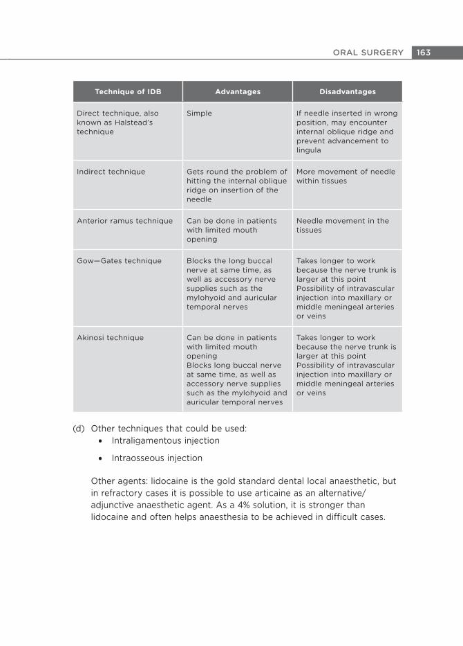
(c) IDB techniques are shown below.
163ORAL SURGERY
Technique of IDB Advantages Disadvantages
Direct technique, also known as Halstead’s technique
Simple If needle inserted in wrong position, may encounter internal oblique ridge and prevent advancement to lingula
Indirect technique Gets round the problem of hitting the internal oblique ridge on insertion of the needle
More movement of needle within tissues
Anterior ramus technique Can be done in patients with limited mouth opening
Needle movement in the tissues
Gow—Gates technique Blocks the long buccal nerve at same time, as well as accessory nerve supplies such as the mylohyoid and auricular temporal nerves
Takes longer to work because the nerve trunk is larger at this point Possibility of intravascular injection into maxillary or middle meningeal arteries or veins
Akinosi technique Can be done in patients with limited mouth openingBlocks long buccal nerve at same time, as well as accessory nerve supplies such as the mylohyoid and auricular temporal nerves
Takes longer to work because the nerve trunk is larger at this pointPossibility of intravascular injection into maxillary or middle meningeal arteries or veins
(d) Other techniques that could be used:
Intraligamentous injection
Intraosseous injection
Other agents: lidocaine is the gold standard dental local anaesthetic, but
in refractory cases it is possible to use articaine as an alternative/
adjunctive anaesthetic agent. As a 4% solution, it is stronger than
lidocaine and often helps anaesthesia to be achieved in diQcult cases.
164 ORAL SURGERY
3.2 (a) You are seeing a patient who needs to have a tooth surgically
removed in your practice. One of the principles of flap design is
that vital structures should be avoided. Name two vital structures
that you should avoid when carrying out surgical tooth removal in
the maxilla and the mandible.
(b) What are the other principles to which you should adhere when
designing a mucoperiosteal flap for surgical tooth extraction?
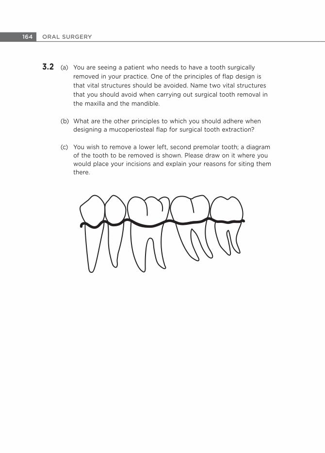
(c) You wish to remove a lower left, second premolar tooth; a diagram
of the tooth to be removed is shown. Please draw on it where you
would place your incisions and explain your reasons for siting them
there.

165ORAL SURGERY
(d) After the procedure you wish to suture the wound. What functions
do sutures perform?
(e) Name two di7erent sutures that could be used to suture an
intraoral wound and an advantage of each.
166 ORAL SURGERY
Answer 3.2
(a)
Maxilla: greater palatine artery; nasopalatine nerves and arteries
Mandible: lingual nerve and mental nerve
(b) Mucoperisoteal flaps that are raised when surgically removing a tooth
need to:
Provide adequate access to the surgical site
Retain a good blood supply to the mucoperiosteal flap, so the base
must be broader than the apex, unless the flap includes a decent-
sized artery within the flap
Avoid vital structures such as local nerves and blood vessels
Have their margins placed on sound bone and not over the area
where you are removing bone
Be able to be extended if necessary
Be able to be closed appropriately at the end of the operation
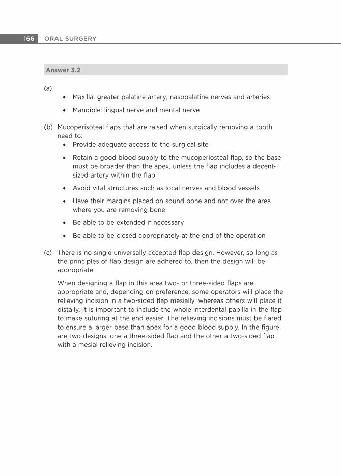
(c) There is no single universally accepted flap design. However, so long as
the principles of flap design are adhered to, then the design will be
appropriate.
When designing a flap in this area two- or three-sided flaps are
appropriate and, depending on preference, some operators will place the
relieving incision in a two-sided flap mesially, whereas others will place it
distally. It is important to include the whole interdental papilla in the flap
to make suturing at the end easier. The relieving incisions must be flared
to ensure a larger base than apex for a good blood supply. In the figure
are two designs: one a three-sided flap and the other a two-sided flap
with a mesial relieving incision.
167ORAL SURGERY
(d) Sutures primarily approximate and hold the wound margins in the
appropriate place to enable them to heal. The smaller the space between
the two wound margins, the quicker the wound will heal. Sutures will help
in holding the mucoperiosteal flap over bone, which will reduce the risk of
it becoming non-vital. Sutures also help haemostasis.
(e)
Non-resorbable:
braided: black silk — soft and easy to knot
monofilament: Prolene — hygienic
Resorbable:
braided: polyglactin (Vicryl) or Polysorb, which is a glycolide/
lactide co-polymer — soft, easy to knot, resorbs so patient does
not need to have sutures removed
monofilament: poliglecaprone 25 (Monocryl) — hygienic,
resorbable but slow resorption

168 ORAL SURGERY
3.3 (a) A fit and healthy patient presents to your surgery complaining of
recurrent episodes of pain and swelling of the gum in the region of
an impacted, lower right wisdom tooth. What is the most likely
diagnosis?
(b) A radiograph of the tooth involved is shown here. How would you
describe the position of the tooth?
169ORAL SURGERY
(c) What is the relationship of the inferior dental canal to the tooth, as
judged by this radiographic view? What are the likely implications
of this appearance and how would you proceed?
(d) Coronectomy: what is the rationale behind a coronectomy and what
are the complications of carrying it out?

170 ORAL SURGERY
Answer 3.3
(a) Recurrent pericoronitis
(b) Mesioangularly impacted and partially erupted
(c) The inferior dental canal crosses the root of the tooth and there is a
radiolucent band across the root in this area. There is also loss of the
superior cortical outline of the inferior dental canal as it crosses the tooth.
This is likely to represent an intimate relationship between the inferior
dental nerve and the roots of the tooth, which means that, if the tooth
were to be removed, the patient would be at higher risk of damage to the
inferior dental canal.
In an ideal world a cone-beam CT (CBCT) scan would be the next step
because this would provide a three-dimensional view of the area and
provide a definitive answer as to the true relationship between the root
and the nerve. If there is an intimate association or if no CBCT scan is
available, then to minimise damage to the nerve the treatment options
are:
To leave the tooth in situ and treat each episode of pericoronitis as
and when it occurs
To remove the tooth in its entirety but accept that it has a higher
than average risk of causing damage to the inferior dental nerve
To carry out a coronectomy
(d) A coronectomy is a procedure in which the crown of the tooth is removed
and the vital roots are retained. The rationale is that not touching the
roots will limit damage to the inferior dental nerve, and removing the
crown will allow the mucosa to be sutured across to the lingual side,
closing the wound primarily and thereby preventing any further episodes
of pericoronitis.
The possible complications are infection from or migration of the retained
root. In some instances the root becomes mobile when the crown is
sectioned and removed, and a mobile root cannot be left in situ so it is
necessary to surgically remove the whole tooth.

171ORAL SURGERY
3.4 (a) Which patients should be referred to a specialist for urgent
assessment according to the 2005 National Institute for Health and
Care Excellence (NICE) guidelines on urgent referrals for suspected
oral cancer?
(b) As a general dental practitioner, to whom would you refer a patient
for management if you suspected that they had a squamous cell
carcinoma of the oral cavity?
(c) What treatment modalities are commonly used for treating
squamous cell carcinoma of the oral cavity?
(d) What do you understand by the term palliative care?

172 ORAL SURGERY
Answer 3.4
(a) Any patient with:
Unexplained red and white patches (including suspected lichen
planus) of the oral mucosa that are painful or bleeding or swollen.
Note: a non-urgent referral should be made in the absence of these,
ie not painful, bleeding or swollen.
Unexplained ulceration of the oral mucosa persisting for more than 3
weeks
Any adult patient with
Unexplained tooth mobility persisting for more than 3 weeks
An unexplained lump in the neck which has recently appeared or a
lump which has not been diagnosed before that has changed over a
period of 3—6 weeks
(b) An oral and maxillofacial surgery consultant who manages oncology
patients within a cancer centre would be the best person to manage the
patient as the surgeon is part of a multidisciplinary team that can oCer
the patient holistic care.
Oral medicine and oral surgery consultants will see patients referred for
suspected squamous cell carcinomas (SCCs), and may arrange for
biopsies to be performed but as they are not able to oCer the patient
definitive surgical treatment. Therefore, it would be ideal for the patient to
be referred to the person who would be able to diagnose and manage
that lesion from the start.
(c)
Surgery
Radiotherapy
Chemotherapy
Combination of any of the above
(d) According to the World Health Organization (2003), ‘palliative care is an
approach that improves the quality of life of patients and their families
facing the problems associated with life-threatening illness, through the
prevention and the relief of suCering by means of early identification and
impeccable assessment and treatment of pain and other symptoms,
physical, psychosocial and spiritual’.

173ORAL SURGERY
3.5 (a) What are bisphosphonates?
(b) You are a general dental practitioner who has a patient who is
about to commence treatment with bisphosphonates. How would
you manage them?
(c) You have a patient who has been on oral bisphosphonates for 5
years and requires a dental extraction. Describe how you would
manage this patient.
174 ORAL SURGERY
Answer 3.5
(a) Bisphosphonate are pyrophosphate analogues that inhibit resorption of
bone. Their proposed mechanism of action includes:
Reduction of bone turnover
Inhibition of osteoclast activity
(b) Patients about to commence treatment with bisphosphonates should have
been informed of the risk and benefits of the chosen drug by the
prescribing physician including the risk of medication-related
osteonecrosis of the jaw (MRONJ), which was previously known as
bisphosphonate-related osteonecrosis of the jaw (BRONJ). The patient
ideally should have a dental assessment prior to commencement of the
drugs. This is especially important if the patient is to be given high-dose
iv bisphosphonates.
Patients should be informed of the importance of maintaining a high
standard of dental health following treatment with bisphosphonate
drugs, and the consequences of not doing so.
Dental hard and soft tissues must be examined for any disease. Any
active infection must be treated before commencement of the
treatment.
Any prosthesis must be carefully examined, as mucosal injury and
breakdown is the second most commonly identified risk factor for
MRONJ.
Teeth which are in an acceptable condition but unlikely to be
retained in the long term need careful consideration as future
exodontia is a risk factor for MRONJ.
Any teeth of dubious prognosis must also be removed.
Patients should be advised on oral hygiene and preventive measures
to minimise risk of dental disease.
Patients must be educated about the signs and symptoms of MRONJ
and to seek advice if they have concerns.
Patients must be made dentally fit before commencement of the
drug treatment.

175ORAL SURGERY
(c) Current evidence would suggest that those at serious risk of MRONJ are
likely to have been on iv bisphosphonates for more than 12 months or at
least 36 months of oral bisphosphonates. Prevention is the best option
and it is generally recommended that high-risk procedures, eg extractions,
should be avoided and instead root canal treatment should be considered,
even when it is not possible to restore the crown of the tooth to a
functional form.
There are some variations in the guidelines for exodontia from diDerent
countries, eg oral as oppose to parental (iv) bisphosphonates, and thus
it is worthwhile checking your up-to-date local guidelines, in particular,
Scottish Dental Clinical EDectiveness Programme
(www.scottish.dental.org.uk).
The common steps usually followed are:
1 Preoperatively:
Rinse with chlorhexidine mouthwash
Prophylactic antibiotics (although this is not universally adopted
by all clinicians, hence the need to consult with local guidelines)
2 Conservative surgical technique (atraumatic)
3 Primary closure of soft tissue where possible, without stripping
periosteum
4 Postoperatively:
Chlorhexidine mouthwash for 2 weeks or until mucosa has healed
Antibiotics for 5 days (again this is not universally adopted — see
above)
5 Keep the patient under review until the socket has healed
Do not attempt further extractions in other sextants of the mouth until
the first socket has healed.
176 ORAL SURGERY
3.6 (a) In order to diagnose MRONJ, certain criteria must be met. What are
they?
(b) Apart from bisphosphonate drugs, what other types of drugs are
associated with MRONJ? Give an example of each type, and list the
conditions for which they are prescribed.
(c) In which conditions might a patient be prescribed bisphosphonate
medication?
(d) What are the common routes of administration of bisphosphonate
medication?
(e) Which patients are most at risk of getting MRONJ?
(f) Name some local risk factors.
177ORAL SURGERY
Answer 3.6
(a)
The patient must be taking or have taken anti-resorptive or anti-
angiogenic medication.
The patient must have exposed bone or bone that can be probed
through an intraoral or extraoral fistula in the maxillofacial region
that has persisted for more than 8 weeks.
There must be a history of radiotherapy to the jaws
There must be no obvious metastatic disease to the jaws (see
Ruggiero SL, Dodson TB, Fantasia J, et al. American Association of
Oral and Maxillofacial Surgeons. Journal of Oral & Maxillofacial
Surgery 2014; 72:1938—56)
(b) Bisphosphonates, along with other anti-resorptive medications such as
denosumab are associated with MRONJ. The other group of drugs that
are implicated is the anti-angiogenesis drugs such as bevacizumab and
sunitinib.
(c) Anti-resorptive drugs are taken for:
Osteoporosis: both prevention and treatment
Prevention of skeletal fractures in susceptible individuals
Paget’s disease
Osteogenesis imperfecta
Metastatic bone disease (usually in connection with breast or
prostate carcinoma)
Bisphosphonates are also used in the management of patients with
multiple myeloma, although other anti-resorptives such as
denosumab are not
Anti-angiogenesis drugs are taken for renal cell carcinoma and
gastric tumours
(d) Oral and iv.
(e) Patients who have been on high-dose potent medication by an iv route,
usually for the management of malignancies.

178 ORAL SURGERY
(f)
Mandibular extractions
All dentoalveolar surgery
Periodontitis, presence of oral abscesses or infection
Poor oral hygiene
Denture-related trauma
Thin mucosal coverage, eg lingual tori
179ORAL SURGERY
3.7 A fit and healthy 25-year-old patient attends your dental practice
with a 2-day history of a painful, loose left mandibular first
permanent molar after he was hit in the face with the cricket ball.
(a) What key questions you would ask the patient?
(b) What radiological investigations if any would you carry out after
examination?
(c) Following your examination and investigations, you are concerned
that the mobile tooth is a result of a fractured mandible. How would
you proceed?
(d) If there was a mandibular fracture which radiological view(s) would
demonstrate it?
(e) What treatment is likely to be required in this case?

180 ORAL SURGERY
Answer 3.7
(a) You would take the history and examination as usual to ascertain the
current complaint, the history of the complaint, the patient’s medical,
dental and social history. However, in this type of injury, in particular, you
would also want to know:
The circumstances surrounding the incident
Any loss of consciousness or any other injuries
If his occlusion is deranged
If there is any altered sensation in the distribution of the inferior
alveolar/dental nerve
The state of the tooth before the incident, eg pain and mobility
(b) A panoramic radiograph to obtain an overview of the dentition and
mandible. If there is insuCcient detail of the region of the lower left
mandibular first molar tooth then a periapical radiograph may be
warranted to determine whether there is a fracture in the tooth or to
determine the periodontal status of the tooth.
(c) Immediately refer the patient to the nearest oral and maxillofacial surgery
department for further assessment and management.
(d)
A dental panoramic radiograph and another view at another angle,
usually a posterior-anterior view of the mandible (PA mandible).
An alternative would be oblique lateral views of the mandible and PA
mandible, but the oblique lateral views are often inferior to a
panoramic radiograph.
Cone-beam computed tomography (CT) or standard CT would also
provide good information regarding the fracture but is not indicated
in simple fractures due to the higher radiation dose relative to a
dental panoramic radiograph and PA mandible.
(e) It is likely that the fracture is displaced as the patient feels movement in
the lower left first molar. Hence he requires surgical treatment in the form
of open reduction and internal fixation of the fractured mandible. For a
body of mandible fracture this is often accessed via an intraoral approach.

181ORAL SURGERY
3.8 (a) A fit and healthy 10-year-old child fell while playing on his micro-
scooter and is brought into your surgery with evidence of injury to
his maxillary anterior teeth. Your worry is that the child may have
sustained an alveolar or dento-alveolar fracture. What are the
diBerences between these two terms?
(b) What features would lead you to suspect that the child had
sustained a dento-alveolar fracture?
(c) What investigations would you carry out and what findings would
you expect?
(d) Assuming the child is co-operative and there are no other injuries,
how would you manage the dento-alveolar fracture?
(e) What post-treatment instructions would you give the patient and
his parents?
182 ORAL SURGERY
Answer 3.8
(a) A fracture of the alveolar process may or may not involve the alveolar
socket. A dento-alveolar fracture would involve fracture of the alveolar
process and the socket.
(b)
Teeth related to the fractured dento-alveolar segment are typically
all mobile and move as a unit.
An occlusal change will often be present due to the displacement of
the entire segment.
The teeth of the a<ected segment are often tender to percussion.
(c)
Vitality testing of all the involved teeth — this is usually negative.
Radiographs — usually two views are recommended for identification
of fractures. Ideally, these should be at right angles to one another
for better identification of fracture lines but in practice the views are
usually taken with the X-ray tube head in two di<erent positions. In
the anterior region the options would be periapical views and an
upper standard occlusal. A panoramic or a cone-beam CT may also
be useful. Radiographic findings suggestive of a dento-alveolar
fracture may present as:
A radiolucent line between the fragments. However, the
vertical line of the fracture may be diFcult to see as it may
run along the periodontal ligament space. The horizontal line
may be located apical at the apex or coronal to the apex.
An alteration in the outline shape of the root and discontinuity
of the periodontal ligament
An associated fracture(s) of the roots of the teeth.
(d) After gaining consent you would:
1 Administer local analgesia
2 Reposition the displaced segment with digital pressure applied both
labially and palatally or with forceps if necessary
3 Stabilise the fractured segment for 4 weeks with flexible splinting,
such as:
An acid etch splint with composite with or without a wire
183ORAL SURGERY
Orthodontic brackets on the teeth and splinting with a flexible
sectional archwire
Preformed trauma arch bars
(e)
Soft diet for 1 week
Explain that good oral hygiene is essential for healing of the tissue
and that chlorhexidine mouthwash may be beneficial
Explain the need for longer-term follow-up, as there is the risk of:
Pulp necrosis
Ankylosis
Resorption associated with infection
Bone loss
Loss of tooth
Current suggested guidelines for follow-up
Splint removal and clinical and radiographic control after 4 weeks
Clinical and radiographic control after 6—8 weeks, 4 months, 6
months, 1 year and yearly for 5 years
For further information, please see www.dentaltraumaguide.org
3.9 (a) What does the term pericoronitis mean? Which teeth are most
commonly aRected by it?
(b) What are the signs and symptoms of pericoronitis?
(c) How do you treat acute pericoronitis?

184 ORAL SURGERY
Answer 3.9
(a) Pericoronitis means infection of the tissue surrounding the crown of a
tooth. The lower third molars are most commonly a7ected.
(b) Depends on the severity of the infection:
Mild — swelling of soft tissue around the crown of the tooth, bad
taste, pain
Moderate — lymphadenopathy, trismus, extraoral swelling
Severe — fever, malaise, spreading infection and abscess formation
(c) Treatment depends on the severity of the infection. Management of mild
infection includes:
Oral hygiene instructions such as cleaning around the tooth and
operculum with chlorhexidine or hot salty water
Debridement of the area around the tooth and under the operculum
Relief of trauma from opposing tooth — grind cusps or extraction of
the tooth
Analgesics
Antibiotics (metronidazole)
Severe infection may need hospitalisation, intravenous antibiotics, removal
of the lower third molar and/or incision and drainage.
185ORAL SURGERY
3.10 (a) What does the acronym NICE stand for?
(b) NICE guidelines gives specific indications for removal of wisdom
teeth. List five such indications.
(c) What features on a radiograph would suggest that a wisdom tooth
is associated with the inferior dental nerve?
(d) What specific information must be given to a patient prior to
removal of an impacted lower wisdom tooth, which you would not
give if you were removing an upper wisdom tooth?

186 ORAL SURGERY
Answer 3.10
(a) National Institute for Health and Care Excellence
(b) Surgical removal of impacted third molars should be limited to patients
with evidence of pathology such as (any five of the following):
Caries
Non-treatable pulpal and/or periapical pathology
Cellulitis
Abscess and osteomyelitis
Internal and external resorption of the tooth or adjacent tooth
Fracture of tooth
Tooth/teeth impeding surgery or reconstructive jaw surgery
Tooth is within the field of tumour resection
(c) Loss, deviation or narrowing of the ‘tramlines’ of the inferior dental canal,
and a radiolucent band across the root of the tooth.
(d) Information specific to lower wisdom teeth: numbness/tingling or altered
sensation of the lower lip, chin and tongue which may be temporary or
permanent. This information needs to be given to the patient because of
the possibility of damage to the inferior dental nerve or the lingual nerve
during the procedure.