sars-like wiv1-cov poised for human...
TRANSCRIPT

SARS-like WIV1-CoV poised for human emergenceVineet D. Menacherya, Boyd L. Yount Jr.a, Amy C. Simsa, Kari Debbinka,b, Sudhakar S. Agnihothramc, Lisa E. Gralinskia,Rachel L. Grahama, Trevor Scobeya, Jessica A. Plantea, Scott R. Royala, Jesica Swanstroma, Timothy P. Sheahana,Raymond J. Picklesc,d, Davide Cortie,f,g, Scott H. Randelld, Antonio Lanzavecchiae,f, Wayne A. Marascoh,and Ralph S. Barica,c,1
aDepartment of Epidemiology, University of North Carolina at Chapel Hill, Chapel Hill, NC 27599; bDepartment of Microbiology and Immunology, Universityof North Carolina at Chapel Hill, Chapel Hill, NC 27599; cDivision of Microbiology, National Center for Toxicological Research, Food and DrugAdministration, Jefferson, AR 72079; dDepartment of Cell Biology and Physiology and Marsico Lung Institute/Cystic Fibrosis Center, University of NorthCarolina at Chapel Hill, Chapel Hill, NC 27599; eInstitute for Research in Biomedicine, Bellinzona, Switzerland; fInstitute of Microbiology, EidgenössischeTechnische Hochschule Zurich, Zurich, Switzerland; gHumabs BioMed SA, Bellinzona, Switzerland; and hDepartment of Cancer Immunology and AIDS,Dana-Farber Cancer Institute–Department of Medicine, Harvard Medical School, Boston MA 02215
Edited by Peter Palese, Icahn School of Medicine at Mount Sinai, New York, NY, and approved January 6, 2016 (received for review September 4, 2015)
Outbreaks from zoonotic sources represent a threat to bothhuman disease as well as the global economy. Despite a wealth ofmetagenomics studies, methods to leverage these datasets to identifyfuture threats are underdeveloped. In this study, we describe anapproach that combines existing metagenomics data with reversegenetics to engineer reagents to evaluate emergence and pathogenicpotential of circulating zoonotic viruses. Focusing on the severe acuterespiratory syndrome (SARS)-like viruses, the results indicate that theWIV1-coronavirus (CoV) cluster has the ability to directly infect andmay undergo limited transmission in human populations. However,in vivo attenuation suggests additional adaptation is required forepidemic disease. Importantly, available SARS monoclonal antibodiesoffered success in limiting viral infection absent from availablevaccine approaches. Together, the data highlight the utility of aplatform to identify and prioritize prepandemic strains harbored inanimal reservoirs and document the threat posed by WIV1-CoV foremergence in human populations.
SARS | CoV | emergence | Spike | WIV1
Although previously associated with upper respiratory infec-tions, the emergence of severe acute respiratory coronavirus
(SARS-CoV) in 2002–2003, and more recently, Middle Eastrespiratory syndrome (MERS)-CoV underscores the threat ofcross-species transmission leading to virulent pandemic viral in-fections (1, 2). Whereas prevailing research suggests that SARS-CoV emerged from viruses in the Chinese horseshoe bat, identi-fying a progenitor strain that used human angiotensin convertingenzyme 2 (ACE2) had proven elusive (3, 4). However, recentmetagenomics studies isolated several SARS-like virus sequencesthat share ≥90% genome-wide homology and represented theclosest sequences to the epidemic strains (5, 6). Importantly, re-searchers also isolated replication competent virus; WIV1-CoV,part of the Rs3306 cluster, could use ACE2 orthologs and medi-ated low-level replication in human cells (5). Overall, the evidenceindicates that SARS-CoV likely emerged from Chinese horseshoebats and that similar viruses are still harbored in these populations.The identification of WIV1-CoV and its capacity to use ACE2
orthologs offers a warning for possible reemergence and pro-vides an opportunity to prepare for a future CoV outbreak. Toachieve this goal, a new platform is required to translate meta-genomics findings; the approach must generate critical di-agnostic reagents, define emergence potential of novel strains,and determine efficacy of current therapeutics. Building on thispremise, we developed a framework to examine circulating CoVsusing reverse genetic systems to construct full-length and chi-meric viruses. The results indicate that viruses using WIV1-CoVspike are poised to emerge in human populations due to efficientreplication in primary human airway epithelial cell cultures.However, additional adaptation, potentially independent of thespike protein receptor-binding domain, is required for patho-genesis and epidemic disease. Importantly, monoclonal antibody
strategies against SARS were effective against WIV1-CoV spikeunlike available vaccine approaches. Together, the results highlightthe utility of developing platforms to evaluate circulating zoonoticviruses as threats for future emergence and epidemic potential.
ResultsThe discovery of SARS-like virus clusters that bridge the gapbetween the epidemic strains and related precursor CoV strainHKU3 virus provided the best evidence for emergence of SARS-CoV from Chinese horseshoe bats (5). Comparing the receptorbinding domain (RBD), SARS-CoV Urbani and WIV1 sharehomology at 11 of the 14 contact residues with human ACE2(Fig. 1A); importantly, the three amino acid changes representrelatively conservative substitution not predicted to ablatebinding (Fig. 1B). Therefore, exploring WIV1 strains allows ex-amination of emergence, pathogenesis potential, and adap-tation requirements. Using the SARS-CoV infectious clone as atemplate (7), we designed and synthesized a full-length infectiousclone of WIV1-CoV consisting of six plasmids that could beenzymatically cut, ligated together, and electroporated into cellsto rescue replication competent progeny virions (Fig. S1A). Inaddition to the full-length clone, we also produced WIV1-CoV
Significance
The emergence of severe acute respiratory syndrome coronavirus(SARS-CoV) and Middle East respiratory syndrome (MERS)-CoVhighlights the continued risk of cross-species transmission leadingto epidemic disease. This manuscript describes efforts to extendsurveillance beyond sequence analysis, constructing chimeric andfull-length zoonotic coronaviruses to evaluate emergence poten-tial. Focusing on SARS-like virus sequences isolated from Chinesehorseshoe bats, the results indicate a significant threat posed byWIV1-CoV. Both full-length and chimeric WIV1-CoV readily repli-cated efficiently in human airway cultures and in vivo, suggestingcapability of direct transmission to humans. In addition, whilemonoclonal antibody treatments prove effective, the SARS-basedvaccine approach failed to confer protection. Together, the studyindicates an ongoing threat posed by WIV1-related viruses andthe need for continued study and surveillance.
Author contributions: V.D.M., B.L.Y., and R.S.B. designed research; V.D.M., B.L.Y., A.C.S.,S.S.A., L.E.G., T.S., J.A.P., S.R.R., J.S., and T.P.S. performed research; V.D.M., B.L.Y., R.J.P.,D.C., S.H.R., A.L., and W.A.M. contributed new reagents/analytic tools; V.D.M., A.C.S.,K.D., R.L.G., and R.S.B. analyzed data; and V.D.M. and R.S.B. wrote the paper.
The authors declare no conflict of interest.
This article is a PNAS Direct Submission.
Freely available online through the PNAS open access option.
See Commentary on page 2812.1To whom correspondence should be addressed. Email: [email protected].
This article contains supporting information online at www.pnas.org/lookup/suppl/doi:10.1073/pnas.1517719113/-/DCSupplemental.
3048–3053 | PNAS | March 15, 2016 | vol. 113 | no. 11 www.pnas.org/cgi/doi/10.1073/pnas.1517719113
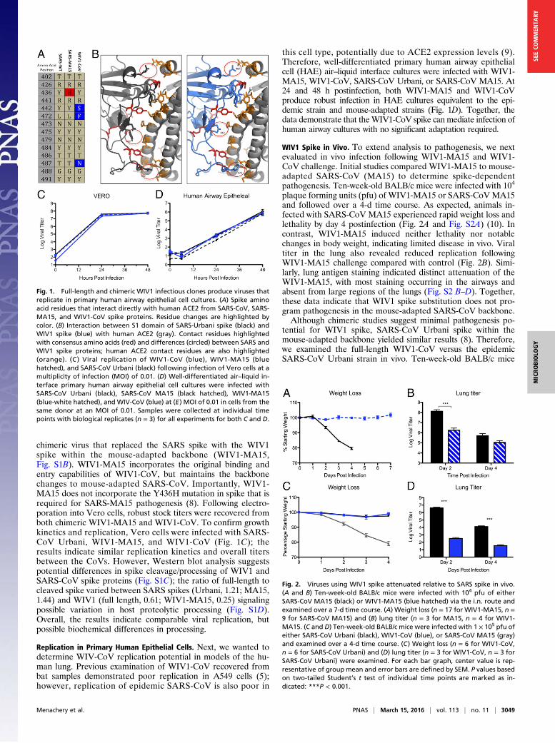
chimeric virus that replaced the SARS spike with the WIV1spike within the mouse-adapted backbone (WIV1-MA15,Fig. S1B). WIV1-MA15 incorporates the original binding andentry capabilities of WIV1-CoV, but maintains the backbonechanges to mouse-adapted SARS-CoV. Importantly, WIV1-MA15 does not incorporate the Y436H mutation in spike that isrequired for SARS-MA15 pathogenesis (8). Following electro-poration into Vero cells, robust stock titers were recovered fromboth chimeric WIV1-MA15 and WIV1-CoV. To confirm growthkinetics and replication, Vero cells were infected with SARS-CoV Urbani, WIV1-MA15, and WIV1-CoV (Fig. 1C); theresults indicate similar replication kinetics and overall titersbetween the CoVs. However, Western blot analysis suggestspotential differences in spike cleavage/processing of WIV1 andSARS-CoV spike proteins (Fig. S1C); the ratio of full-length tocleaved spike varied between SARS spikes (Urbani, 1.21; MA15,1.44) and WIV1 (full length, 0.61; WIV1-MA15, 0.25) signalingpossible variation in host proteolytic processing (Fig. S1D).Overall, the results indicate comparable viral replication, butpossible biochemical differences in processing.
Replication in Primary Human Epithelial Cells. Next, we wanted todetermine WIV-CoV replication potential in models of the hu-man lung. Previous examination of WIV1-CoV recovered frombat samples demonstrated poor replication in A549 cells (5);however, replication of epidemic SARS-CoV is also poor in
this cell type, potentially due to ACE2 expression levels (9).Therefore, well-differentiated primary human airway epithelialcell (HAE) air–liquid interface cultures were infected with WIV1-MA15, WIV1-CoV, SARS-CoV Urbani, or SARS-CoV MA15. At24 and 48 h postinfection, both WIV1-MA15 and WIV1-CoVproduce robust infection in HAE cultures equivalent to the epi-demic strain and mouse-adapted strains (Fig. 1D). Together, thedata demonstrate that theWIV1-CoV spike can mediate infection ofhuman airway cultures with no significant adaptation required.
WIV1 Spike in Vivo. To extend analysis to pathogenesis, we nextevaluated in vivo infection following WIV1-MA15 and WIV1-CoV challenge. Initial studies compared WIV1-MA15 to mouse-adapted SARS-CoV (MA15) to determine spike-dependentpathogenesis. Ten-week-old BALB/c mice were infected with 104
plaque forming units (pfu) of WIV1-MA15 or SARS-CoVMA15and followed over a 4-d time course. As expected, animals in-fected with SARS-CoV MA15 experienced rapid weight loss andlethality by day 4 postinfection (Fig. 2A and Fig. S2A) (10). Incontrast, WIV1-MA15 induced neither lethality nor notablechanges in body weight, indicating limited disease in vivo. Viraltiter in the lung also revealed reduced replication followingWIV1-MA15 challenge compared with control (Fig. 2B). Simi-larly, lung antigen staining indicated distinct attenuation of theWIV1-MA15, with most staining occurring in the airways andabsent from large regions of the lungs (Fig. S2 B–D). Together,these data indicate that WIV1 spike substitution does not pro-gram pathogenesis in the mouse-adapted SARS-CoV backbone.Although chimeric studies suggest minimal pathogenesis po-
tential for WIV1 spike, SARS-CoV Urbani spike within themouse-adapted backbone yielded similar results (8). Therefore,we examined the full-length WIV1-CoV versus the epidemicSARS-CoV Urbani strain in vivo. Ten-week-old BALB/c mice
Fig. 1. Full-length and chimeric WIV1 infectious clones produce viruses thatreplicate in primary human airway epithelial cell cultures. (A) Spike aminoacid residues that interact directly with human ACE2 from SARS-CoV, SARS-MA15, and WIV1-CoV spike proteins. Residue changes are highlighted bycolor. (B) Interaction between S1 domain of SARS-Urbani spike (black) andWIV1 spike (blue) with human ACE2 (gray). Contact residues highlightedwith consensus amino acids (red) and differences (circled) between SARS andWIV1 spike proteins; human ACE2 contact residues are also highlighted(orange). (C ) Viral replication of WIV1-CoV (blue), WIV1-MA15 (bluehatched), and SARS-CoV Urbani (black) following infection of Vero cells at amultiplicity of infection (MOI) of 0.01. (D) Well-differentiated air–liquid in-terface primary human airway epithelial cell cultures were infected withSARS-CoV Urbani (black), SARS-CoV MA15 (black hatched), WIV1-MA15(blue-white hatched), and WIV-CoV (blue) at (E) MOI of 0.01 in cells from thesame donor at an MOI of 0.01. Samples were collected at individual timepoints with biological replicates (n = 3) for all experiments for both C and D.
Fig. 2. Viruses using WIV1 spike attenuated relative to SARS spike in vivo.(A and B) Ten-week-old BALB/c mice were infected with 104 pfu of eitherSARS-CoV MA15 (black) or WIV1-MA15 (blue hatched) via the i.n. route andexamined over a 7-d time course. (A) Weight loss (n = 17 for WIV1-MA15, n =9 for SARS-CoV MA15) and (B) lung titer (n = 3 for MA15, n = 4 for WIV1-MA15. (C and D) Ten-week-old BALB/c mice were infected with 1 × 10⁵ pfu ofeither SARS-CoV Urbani (black), WIV1-CoV (blue), or SARS-CoV MA15 (gray)and examined over a 4-d time course. (C) Weight loss (n = 6 for WIV1-CoV,n = 6 for SARS-CoV Urbani) and (D) lung titer (n = 3 for WIV1-CoV, n = 3 forSARS-CoV Urbani) were examined. For each bar graph, center value is rep-resentative of group mean and error bars are defined by SEM. P values basedon two-tailed Student’s t test of individual time points are marked as in-dicated: ***P < 0.001.
Menachery et al. PNAS | March 15, 2016 | vol. 113 | no. 11 | 3049
MICRO
BIOLO
GY
SEECO
MMEN
TARY
were infected with 105 pfu of WIV1-CoV or SARS-CoV Urbaniand followed over a 4-d time course. As expected, neither in-fection condition resulted in significant weight loss comparedwith MA15 (Fig. 2C). However, viral replication was significantlyattenuated for WIV1-CoV compared with SARS-CoV Urbani(Fig. 2D); at both days 2 and 4 postinfection, WIV1-CoV titer wasreduced nearly 10,000- and 1,000-fold, respectively. Similarly, onlyminor antigen staining was observed following WIV1-CoV in-fection, contrasting antigen staining throughout the parenchyma2-d post–SARS-CoV Urbani infection (Fig. S2 E and F). To-gether, the data indicate significant attenuation of WIV1-CoVrelative to the epidemic SARS-CoV in wild-type mice.
WIV1-CoV in Human ACE2 Expressing Mice.Whereas studies in wild-type mice provide insight into pathogenesis potential, the ab-sence of clinical disease in the epidemic strains of SARS-CoVsuggests that the mouse model may not be adequate to accesshuman disease potential. To test a model more relevant to hu-mans, we generated a mouse that expresses human ACE2 re-ceptor under control of HFH4, a lung ciliated epithelial cellpromoter (11). However, whereas robust expression was ob-served in the lung, other tissues including brain, liver, kidney,and gastrointestinal tract had varying levels of human ACE2expression, indicating greater tissue distribution of HFH4-mediated expression than initially expected (Fig. S3A). In addi-tion, examination of individual HFH4-ACE2–expressing progenyrevealed the occasional absence of the human ACE2 gene,suggesting possible selection against human receptor (Fig. S3B).Therefore, PCR-positive, 10- to 20-wk-old HFH4-ACE2–expressing mice were infected with 105 pfu of WIV1-CoV orSARS-CoV Urbani and then followed for a 7-d time course todetermine pathogenesis. The results indicated that WIV1-CoVinfection was augmented, but remained attenuated relative toSARS-CoV Urbani in the presence of human ACE2. FollowingSARS-CoV Urbani challenge, HFH4-hACE2–expressing micelost no weight, but then, experienced rapid weight loss and deathbetween days 4 and 5 (Fig. 3A and Fig. S3C). In contrast, WIV1-CoV produce minimal changes in weight loss until late timeswhere animals fell into distinct categories either losing less thanor more than 10% of their body weight. Whereas day-2 lung ti-ters were still attenuated relative to SARS-CoV Urbani, titers forWIV1-CoV were 100-fold higher in the presence of human ACE2compared with wild-type BALB/c, with no similar augmentationobserved with the epidemic SARS-CoV strain (Fig. 3B). Based onpilot studies and previous studies with ACE2 transgenic animals(12), mice experiencing rapid weight loss were predicted to havelethal encephalitis and were humanely killed and harvested forlung and brain titer if weight loss approached >20% of startingbody weight. All HFH4-ACE2 mice infected with SARS-CoVUrbani lost >20% body weight and maintained robust replica-tion in the lung and brain following infection (Fig. 3 C and D).Similarly, mice with >10% weight loss following WIV1-CoVinfection produced robust viral replication in the brain, but sig-nificantly lower titers in the lung. In contrast, mice that main-tained minimal weight loss (<10%) following WIV1-CoVinfection after 7 d had minimal titers in both the lung and brain,suggesting a sufficient adaptive immune response was generatedto clear virus and survive infection. Together, the data indicatethat WIV1-CoV maintains attenuation relative to SARS-CoVUrbani despite the availability of human ACE2. In addition,augmented replication suggests that WIV1-CoV may bind thehuman ACE2 receptor more efficiently that the mouse ACE2,indicating potential inadequacies in the current mouse models ofSARS pathogenesis.
Therapeutics Against WIV1 Emergence. Having established a po-tential threat based on replication in primary human cells andpreference for the human ACE2 receptor in vivo, we next sought
to determine if monoclonal antibody therapies could be used tolessen disease similar to ZMApp for Ebola (13). We first tested aSARS-CoV monoclonal derived via phage display and antibodyescape (Fm6) (14) and found both wild-type SARS-CoV Urbaniand WIV1-MA15 were strongly neutralized at low antibodyconcentrations (Fig. 4A). Similarly, a panel of monoclonal anti-bodies derived from B cells from SARS-CoV–infected patientsalso prevented virus infection via WIV1-CoV spike (15, 16).Both antibodies 230.15 and 227.14 robustly inhibited WIV1-MA15 replication with kinetics similar to or exceeding SARS-CoV Urbani (Fig. 4 B and C). In contrast, antibody 109.8, whichmaps outside the receptor binding domain, produced only mar-ginal neutralization of WIV1-MA15 (Fig. 4D). Whereas theresidue associated with prior escape mutants was conservedat position 332, the adjacent residue had a significant change(K332T) in WIV1-CoV, possibly contributing to reduced efficacyof this antibody.To further extend these findings, in vivo studies with antibody
227.14 were initiated in HFH4-ACE2–expressing mice. One daybefore infection, HFH4-ACE2–expressing mice were injected with200 μg of antibody 227.14 or PBS control as previously described(17); mice were subsequently challenged with either SARS-CoVUrbani or WIV1-CoV and monitored for 7 d. The results indicatethat antibody 227.14 protected mice from both lethal SARS-CoVUrbani and WIV-CoV challenge (Fig. 4E); in addition, lung titersrevealed no detectable virus in either SARS-CoV or WIV1-CoV–infected HFH4-ACE2–expressing mouse lungs following antibodytreatment (Fig. 4F). Together, the in vitro and in vivo data in-dicate that a mixture of broadly neutralizing antibodies againstSARS-CoV would likely provide significant protection if WIV1-CoV–like viruses successfully transmitted to humans.
Vaccine Efficacy Limited Against WIV1 Spike. Previously, whole vi-rion SARS-CoV inactivated by both formalin and UV irradiation(double inactivated virus, DIV) was demonstrated as a po-tential vaccination candidate based on robust neutralizationand protection following homologous SARS-CoV challenge inyoung mice (18). However, both aged animal and heterologous
Fig. 3. WIV1-Cov still attenuated despite human ACE2 expression in vivo.(A) Ten- to twenty-week-old HFH4 ACE2-expressing mice were infected with105 pfu of SARS-CoV Urbani (black) or WIV1-CoV (blue) and examined over a7-d time course for (A) survival and (B) day-2 lung titer (n = 3 for WIV1-CoV,n = 3 for SARS-CoV Urbani). (C and D) Upon reaching thresholds for humanesacrifice (>20% weight loss) or 7 d postinfection (DPI), endpoint titers weredetermined in the (C) lung and (D) brain following infection. P values basedon two-tailed Student’s t test of individual time points are marked as in-dicated: *P < 0.05.
3050 | www.pnas.org/cgi/doi/10.1073/pnas.1517719113 Menachery et al.

challenge studies revealed incomplete protection, increased im-mune pathology, and eosinophilia, indicating the possibility ofadverse affects following DIV vaccination (19). To determineif heterologous challenge with WIV1-CoV spike produced asimilar affect, 1-y-old BALB/c mice were vaccinated and boostedwith DIV or PBS mock control. Mice were then challenged 6 wkpostinitial vaccination with WIV1-MA15 and examined over a4-d time course. Similar to previous experiments, mice infectedwith WIV1-MA15 had only marginal weight loss and showed noclinical signs of disease with either vaccination group (Fig. 5A).However, viral replication at day 4 was not significantly reducedin DIV-vaccinated groups compared with control (Fig. 5B). Inaddition, plaque reduction neutralization titers from the serumof aged DIV-vaccinated mice indicated no neutralization of WIV1-MA15, suggesting inadequate protection (Fig. 5C). Importantly,examination of histopathology revealed increased eosinophiliain DIV-vaccinated mice compared with PBS controls, indicatingthe potential for immune induced pathology due to vaccination.Together, the data indicate that DIV vaccination would not pro-vide significant protection and may cause adverse effects in thecontext of WIV1-CoV spike-mediated outbreak.
DiscussionThe recent outbreaks of Ebola, influenza, and MERS-CoV un-derscore the threat posed by viruses emerging from zoonoticsources. Coupled with air travel and uneven public health in-frastructures, it is critical to develop approaches to mitigate theseand future outbreaks. In this paper, we outline a platform thatleverages metagenomics data, synthetic genome design, trans-genic mouse models, and therapeutic human antibodies toidentify and treat potential prepandemic viruses. Focusing onSARS-like CoVs, the approach indicates that viruses using theWIV1-CoV spike protein are capable of infecting HAE culturesdirectly without further spike adaptation. Whereas in vivo dataindicate attenuation relative to SARS-CoV, the augmentedreplication in the presence of human ACE2 in vivo suggests thatthe virus has significant pathogenic potential not captured bycurrent small animal models. Importantly, therapeutic treatmentwith monoclonal antibodies suggests a Zmapp-based approachwould be effective against a WIV1-CoV spike-mediated out-break. However, failure of SARS DIV vaccine to induce pro-tection highlights the need for continued development ofadditional therapeutics. Overall, the characterization of WIV1-CoV and its pathogenic potential highlight the utility of thisplatform in evaluating currently circulating zoonotic viruses.Primary human airway epithelial cell cultures derived from
human donors and grown at an air–liquid interface represent the
Fig. 4. SARS-CoV monoclonal antibodies have robust neutralization againstWIV1 spike-mediated infection. Neutralization efficacy was evaluated usingpercent neutralization assays against SARS-CoV Urbani (black) or WIV1-MA15(blue) with a panel of monoclonal antibodies: (A) fm6, (B) 230.15, (C) 227.15,and (D) 109.8, all originally generated against epidemic SARS-CoV. Each datapoint is representative of two or more independent neutralization wells. (E andF) Twenty- to twenty-four-week-old HFH4 ACE2-expressing mice were injectedwith 200 μg of anti-SARS human antibody 227.15 (hatched line) or mock (solidline) 1 d before infection with 1 × 10̂ 5 pfu of SARS-CoV Urbani (black) or WIV1-CoV (blue) and examined over a 7-d time course for (E) survival (n = 3 for bothantibody-treated groups and mock PBS control WIV1-CoV, n = 2 for mock-treated SARS-CoV Urbani), (F) day-2 lung titer (n = 3 for all groups). ND signifiesno titers detected. For each bar graph, center value is representative of groupmean and error bars are defined by SEM.
Fig. 5. Double-inactivated whole SARS-CoV vaccine fails to protect agedanimals from chimeric WIV1-CoV infection. Twelve-month-old mice werevaccinated and boosted with DIV (dotted line) or PBS (solid line) and infected21 d postboost with 104 pfu of WIV1-MA15 via the i.n. route. (A) Weight lossfollowing WIV1-MA15 challenge and (B) viral replication in the lung 4 DPI.(C) Neutralization of WIV1-MA15 (blue) with serum from aged, DIV-vacci-nated mice. (D–H) Histopathology lung sections stained for H&E from DIV-and mock-vaccinated mice. (D) Eosinophil score (scale 0–4) following DIV ormock vaccination 4 DPI. (E and F) Representative H&E lung sections for(E) mock- and (F) DIV-vaccinated mice infected with WIV-MA15. Red arrowsindicate individual eosinophil locations. P values based on two-tailed Stu-dent’s t test of individual time points are marked as indicated: **P < 0.01.
Menachery et al. PNAS | March 15, 2016 | vol. 113 | no. 11 | 3051
MICRO
BIOLO
GY
SEECO
MMEN
TARY

closest model to the human lung. Therefore, the ability of bothWIV1-CoV and WIV1-MA15 to grow equivalently to the epi-demic SARS-CoV in these cultures is a major concern foremergence. However, pathogenesis studies in mice suggest thatfurther adaptation may be required for epidemic disease. Com-pared with SARS equivalents, both full-length and chimericWIV1 viruses had significant attenuation even with the presenceof human ACE2 in the mouse model. Together, the data suggestthat despite using ACE2 and robust replication in primaryhuman airway epithelial cultures, WIV1-CoV likely maintainsdeficits that impact pathogenesis in mice; therefore, WIV1-mediated infection may have diminished epidemic potential inhumans relative to SARS-CoV.A number of factors may contribute to reduced mouse path-
ogenesis observed following WIV1-CoV spike-mediated in-fection. In the context of both the SARS-CoV and MERS-CoVoutbreaks, focus had been primarily directed to spike bindingas the key component of emergence and pandemic potential.Supported by adaption at Y436H in mouse-adapted SARS spike(10), improved binding to host receptor cannot be discounted asa crucial component in emergence. This fact is supported byimproved replication of WIV1-CoV in mice expressing humanACE2 compared with control (Fig. 2D versus Fig. 3B). However,in vivo attenuation of WIV1-CoV relative to SARS-CoV Urbanidespite efficient infection in primary human airway culturessuggests that additional factors contribute to epidemic emer-gence. One possibility is that adaptation outside of spike proteinmay lead to emergence via altered host–virus interactions.Whereas WIV1-MA15 was attenuated relative to SARS-MA15in vivo, overall titers in the lung were similar to the epidemicSARS-CoV Urbani in BALB/c mice (Figs. 3 and 4). These datasuggest that CoV backbone changes may account for or com-pensate for deficits in WIV1-CoV replication compared withSARS-CoV Urbani in vivo. Another possible factor accounting forattenuation is changes to spike that are independent of receptorbinding. Whereas the receptor binding domain had garnered themost interest, changes in the remaining portion of S1 as well as theS2 portion of spike may also play a critical role in facilitating CoVinfection, transmission, and/or pathogenesis (20). Differences inthese regions of spike may yield increased protease targeting,enhanced spike cleavage, and/or expanded tropism leading tomore robust infection for the epidemic SARS strains. Globally,the in vivo results suggest that any of these areas may have con-tributed to SARS-CoV emergence. However, even these inter-pretations must be tempered due to the robust differencesbetween mouse and human models; further studies in nonhumanprimates are required to confirm these results and derive furtherinsight into CoV emergence from zoonotic sources.Despite the differences in the backbone genome sequences,
therapeutics developed against SARS-CoV provide some mea-sure of protection in the context of a future outbreak. Testing thefour most broadly neutralizing SARS-CoV antibodies revealedeffective control of WIV1-MA15 at relatively low concentrationsof antibody (14, 15, 21). For two of the four antibodies tested(Fm6 and 230.15), WIV1-CoV spike-expressing virus was neu-tralized equivalently or better than SARS-CoV Urbani. Simi-larly, only minimal differences at the low end of the neutrali-zation curve were noted for antibody 227.14. Whereas antibody109.8 produced only marginal neutralization of WIV1-MA15,the overall antibody neutralization data argue that multivalentmonoclonal antibody approaches could limit a WIV1-CoVspike-mediated outbreak. As such, a “ZMapp”-based approachcould have great potential in stemming or preventing a futureSARS-CoV–like outbreak.In contrast to the success of monoclonal antibodies, vaccine
failure indicated further development and refinement are nec-essary. The development of a DIV SARS-CoV vaccine wasbuoyed as a possible means to control SARS-CoV outbreaks
based on robust neutralization and protection in young mice(18). However, studies with DIV in aged animals revealed in-complete protection, significant immune pathology, and eo-sinophilia (19). Despite these prior results, the efficacy ofmonoclonal antibody treatments made further testing of DIVseemingly worthwhile against WIV1-CoV spike-mediated in-fection. However, the results remained the same, as vaccinationof aged mice resulted in no protection from WIV1-MA15replication in vivo (Fig. 5). Importantly, increased immunepathology and observed eosinophilia indicate that broad-basedvaccination efforts against SARS-CoV–like viruses must con-sider heterologous viruses as well as failure due to senescence inthe aged host. A number of novel platforms including VenezuelanEquine Encephalitis Virus Replicon Particle (VRP) and live-attenuated vaccine approaches show great promise in theseareas, but require further testing and development before de-ployment in an outbreak setting (22, 23).Overall, the results from these studies highlight the utility of a
platform that leverages metagenomics findings and reversegenetics to identify prepandemic threats. For SARS-like WIV1-CoV, the data can inform surveillance programs, improve di-agnostic reagents, and facilitate effective treatments to mitigatefuture emergence events. However, building new and chimericreagents must be carefully weighed against potential gain-of-function (GOF) concerns. Whereas not generally expected toincrease pathogenicity, studies that build reagents based onviruses from animal sources cannot exclude the possibility ofincreased virulence or altered immunogenicity that promoteescape from current countermeasures. As such, the potential of athreat, real or perceived, may cause similar exploratory studies tobe limited out of an “abundance of caution.” Importantly, thegovernment pause on GOF studies may have already impactedthe scope and direction of these studies. Whereas previousadaption of the epidemic SARS-CoV strain provided insightsinto species-specific changes, bat-derived WIV1 adaptationmay identify elements critical for pathogenesis and transitionfrom reservoir to human host; targets include viral proteins thatinteract with host machinery or host immunity like NSP1, en-velope, or ORF6 (22, 24, 25). Similarly, WIV1-CoV could beused to drive improved therapeutics, including escape mutantsfor improved monoclonal antibodies or more broadly neutral-izing vaccine approaches. However, it remains unclear from thecurrent policies and GOF environment if these types of studieswill be permissible. Although limits and standards for thesetypes of experiments must be established, erring on the side ofcaution is not without its own risks and balancing the benefitsof these types of studies must also be weighed against thepotential hazards.Using a novel platform to translate metagenomics findings, the
WIV1-CoV cluster has been identified as a threat for futureemergence in human populations due to robust replication inprimary human airway epithelial cell cultures. However, basedon in vivo mouse data, additional adaptations will likely be re-quired to produce epidemic disease. Notably, whereas currentantibody-based therapies hold great promise in treating WIV1-CoV spike-mediated infection, failure of SARS-CoV vaccinationapproaches presents a major challenge for any efforts to protectagainst future emergent viruses. Together, the data illustrate theutility of the platform and highlight the need to build andmaintain preparations for future emergence events.
Materials and MethodsViruses, Cells, and Infection. Wild-type and chimeric CoVs were cultured onVero E6 cells, grown in DMEM (Gibco) and 5% fetal clone serum (HyClone)along with anti/anti (Gibco). Growth curves in Vero and primary humanairway epithelial cells were performed as previously described (26, 27). Hu-man lungs were procured under University of North Carolina at Chapel Hill(UNC) Institutional Review Board-approved protocols.
3052 | www.pnas.org/cgi/doi/10.1073/pnas.1517719113 Menachery et al.

Construction of Chimeric SARS-Like Viruses. Both wild-type and chimeric WIV-CoV infectious clones were designed using published sequences and basedon the SARS-CoV infectious clone (10). Synthetic construction of chimericmutant and full-length WIV1-CoV were approved by the UNC InstitutionalBiosafety Committee and the Dual Use Research of Concern Committee.
Ethics Statement. The study was carried out in accordance with the recom-mendations for care and use of animals by the Office of Laboratory AnimalWelfare (OLAW), National Institutes of Health. The Institutional Animal Careand Use Committee (IACUC) of University of North Carolina (UNC permit no.A-3410-01) approved the animal study protocol (IACUC no. 13–033).
Mice and in Vivo Infection. Female 10-wk- and 12-mo-old Balb/cAnNHsDmice ordered from the Harlan Labs were infected as previously described(23). For vaccination, young and aged mice were vaccinated and boostedby footpad injection with a 20-μL volume of either 0.2 μg of double-inactivated SARS-CoV vaccine (DIV) with alum or mock PBS as previouslydescribed (19).
Generation and Infection of ACE2 Tissue-Specific Transgenic Mice. Transgenicmice with airway-targeted overexpression of human ACE2 were generatedby microinjection of fertilized C3H × C57BL/6 (C3B6) F1 hybrid oocytes withan expression cassette consisting of the HFH4/FOXJ1 lung ciliated epithelialcell-specific promoter elements and the coding region of ACE2 cDNA in apTG1 vector (11) (UNC Animal Model Core). Founder mice were crossed toC3B6, producing human ACE2-transgenic mice that were each previouslytested for transgene expression as described in SI Materials and Methods.ACE2-transgenic mice given i.p. injection of 200 μg of human Ab S227.14 orPBS control (0.20 mL total volume, five to six mice per group) 1 d beforeinfection as previously described (17).
Histological Analysis. Lung tissues for histological analysis were fixed inbuffered formalin phosphate 10% (4–5% wt/wt formaldehyde) (Fisher#SF100-20) for at least 7 d, tissues were embedded in paraffin, and 5-μmsections were prepared by the UNC histopathology core facility as previousdescribed (23). Images were captured using an Olympus BX41 microscopewith an Olympus DP71 camera.
Virus Neutralization Assays. Plaque reduction neutralization titer assays werepreformed with previously characterized antibodies against SARS-CoV aspreviously described (14, 15, 21). Briefly, neutralizing antibodies or serumwere serially diluted twofold and incubated with 100 pfu of the different virusstrains for 1 h at 37 °C. The virus and antibodies were then added to a six-wellplate with 5 ×10⁵ Vero E6 cells per well with n ≥ 2. After a 1-h incubation at37 °C, cells were overlaid with 3 mL of 0.8% agarose in media. Plates wereincubated for 2 d at 37 °C and then stained with neutral red for 3 h, andplaques were counted. The percentage of plaque reduction was calculated as[1 − (no. of plaques with antibody/no. of plaques without antibody)] × 100.
Statistical Analysis. All experiments were conducted contrasting two experi-mental groups (either two viruses or vaccinated and unvaccinated cohorts).Therefore, significant differences in viral titer and histology scoring were de-termined by a two-tailed Student’s t test at individual time points. Data werenormally distributed in each group being compared and had similar variance.
Biosafety and Biosecurity. Reported studies were initiated after the Universityof North Carolina Institutional Biosafety Committee approved the experi-mental protocol: project title: Generating infectious clones of Bat SARS-likeCoVs; lab safety plan ID: 20145741; schedule G ID: 12279. These studies wereinitiated before the US Government Deliberative Process Research FundingPause on Selected Gain of Function Research Involving Influenza, MERS, andSARS Viruses (www.phe.gov/s3/dualuse/Documents/gain-of-function.pdf),and the current paper has been reviewed by the funding agency, the Na-tional Institutes of Health (NIH). Continuation of these studies has beenrequested and approved by the NIH.
ACKNOWLEDGMENTS. We thank Dr. Zhengli-Li Shi of the Wuhan Instituteof Virology for access to bat CoV sequences and plasmid of WIV1-CoV spikeprotein. Research was supported by the National Institute of Allergy andInfectious Disease and the National Institute of Aging of the NIH underAwards U19AI109761 and U19AI107810 (to R.S.B.), AI1085524 (to W.A.M.),and F32AI102561 and K99AG049092 (to V.D.M.). Human airway epithelialcell cultures were supported by the National Institute of Diabetes and Di-gestive and Kidney Disease under Award NIH DK065988 (to S.H.R.). Supportfor the generation of the mice expressing human ACE2 was provided by NIHGrants AI076159 and AI079521 (to A.C.S.).
1. Peiris JS, Guan Y, Yuen KY (2004) Severe acute respiratory syndrome. Nat Med10(12, Suppl):S88–S97.
2. Al-Tawfiq JA, et al. (2014) Surveillance for emerging respiratory viruses. Lancet InfectDis 14(10):992–1000.
3. Graham RL, Baric RS (2010) Recombination, reservoirs, and the modular spike:Mechanisms of coronavirus cross-species transmission. J Virol 84(7):3134–3146.
4. Graham RL, Donaldson EF, Baric RS (2013) A decade after SARS: Strategies for con-trolling emerging coronaviruses. Nat Rev Microbiol 11(12):836–848.
5. Ge XY, et al. (2013) Isolation and characterization of a bat SARS-like coronavirus thatuses the ACE2 receptor. Nature 503(7477):535–538.
6. He B, et al. (2014) Identification of diverse alphacoronaviruses and genomic charac-terization of a novel severe acute respiratory syndrome-like coronavirus from bats inChina. J Virol 88(12):7070–7082.
7. Yount B, et al. (2003) Reverse genetics with a full-length infectious cDNA of severeacute respiratory syndrome coronavirus. Proc Natl Acad Sci USA 100(22):12995–13000.
8. Frieman M, et al. (2012) Molecular determinants of severe acute respiratory syndromecoronavirus pathogenesis and virulence in young and aged mouse models of humandisease. J Virol 86(2):884–897.
9. Gillim-Ross L, et al. (2004) Discovery of novel human and animal cells infected by thesevere acute respiratory syndrome coronavirus by replication-specific multiplex re-verse transcription-PCR. J Clin Microbiol 42(7):3196–3206.
10. Roberts A, et al. (2007) A mouse-adapted SARS-coronavirus causes disease and mor-tality in BALB/c mice. PLoS Pathog 3(1):e5.
11. Ostrowski LE, Hutchins JR, Zakel K, O’Neal WK (2003) Targeting expression of atransgene to the airway surface epithelium using a ciliated cell-specific promoter.MolTher 8(4):637–645.
12. Netland J, Meyerholz DK, Moore S, Cassell M, Perlman S (2008) Severe acute respiratorysyndrome coronavirus infection causes neuronal death in the absence of encephalitis inmice transgenic for human ACE2. J Virol 82(15):7264–7275.
13. Qiu X, et al. (2014) Reversion of advanced Ebola virus disease in nonhuman primateswith ZMapp. Nature 514(7520):47–53.
14. Sui J, et al. (2008) Broadening of neutralization activity to directly block a dominantantibody-driven SARS-coronavirus evolution pathway. PLoS Pathog 4(11):e1000197.
15. Rockx B, et al. (2010) Escape from human monoclonal antibody neutralization affectsin vitro and in vivo fitness of severe acute respiratory syndrome coronavirus. J InfectDis 201(6):946–955.
16. Traggiai E, et al. (2004) An efficient method to make human monoclonal antibodies frommemory B cells: Potent neutralization of SARS coronavirus. Nat Med 10(8):871–875.
17. Zhu Z, et al. (2007) Potent cross-reactive neutralization of SARS coronavirus isolates byhuman monoclonal antibodies. Proc Natl Acad Sci USA 104(29):12123–12128.
18. Spruth M, et al. (2006) A double-inactivated whole virus candidate SARS coronavirusvaccine stimulates neutralising and protective antibody responses. Vaccine 24(5):652–661.
19. Bolles M, et al. (2011) A double-inactivated severe acute respiratory syndrome corona-virus vaccine provides incomplete protection in mice and induces increased eosinophilicproinflammatory pulmonary response upon challenge. J Virol 85(23):12201–12215.
20. McRoy WC, Baric RS (2008) Amino acid substitutions in the S2 subunit of mousehepatitis virus variant V51 encode determinants of host range expansion. J Virol82(3):1414–1424.
21. Sui J, et al. (2014) Effects of human anti-spike protein receptor binding domain an-tibodies on severe acute respiratory syndrome coronavirus neutralization escape andfitness. J Virol 88(23):13769–13780.
22. DeDiego ML, et al. (2014) Coronavirus virulence genes with main focus on SARS-CoVenvelope gene. Virus Res 194:124–137.
23. Agnihothram S, et al. (2014) A mouse model for Betacoronavirus subgroup 2c using abat coronavirus strain HKU5 variant. MBio 5(2):e00047–e14.
24. Narayanan K, Ramirez SI, Lokugamage KG, Makino S (2015) Coronavirus non-structural protein 1: Common and distinct functions in the regulation of host and viralgene expression. Virus Res 202:89–100.
25. Bolles M, Donaldson E, Baric R (2011) SARS-CoV and emergent coronaviruses: Viraldeterminants of interspecies transmission. Curr Opin Virol 1(6):624–634.
26. Sheahan T, Rockx B, Donaldson E, Corti D, Baric R (2008) Pathways of cross-speciestransmission of synthetically reconstructed zoonotic severe acute respiratory syn-drome coronavirus. J Virol 82(17):8721–8732.
27. Sims AC, et al. (2013) Release of severe acute respiratory syndrome coronavirus nu-clear import block enhances host transcription in human lung cells. J Virol 87(7):3885–3902.
Menachery et al. PNAS | March 15, 2016 | vol. 113 | no. 11 | 3053
MICRO
BIOLO
GY
SEECO
MMEN
TARY