scans te 2014
DESCRIPTION
SCANSTETRANSCRIPT
DIAGNOSIS OF ACS DOCENTE: DR. TINOCA AMILCAR
CURSO:MEDICINA INTERNA II, SEMINARIO
ALUMNO: WALTER ESPINOZA CIERTO

DIAGNOSIS OF NSTE-ACS
2014 AHA/ACC Guideline for the Management of Patients With Non–ST-Elevation Acute Coronary Syndromes

STEMI
2014 AHA/ACC Guideline for the Management of Patients With Non–ST-Elevation Acute Coronary Syndromes
.
DIAGNOSIS OF NSTE-ACS
Definition of Terms
ST-segment elevation (ST elevation) or new left bundle-branch block on the electrocardiogram
Spectrum of conditions compatible acute myocardial ischemia infarction due to an abrupt reduction in coronary blood flow SCA
The absence of persistent ST elevation NSTE-ACS

2014 AHA/ACC Guideline for the Management of Patients With Non–ST-Elevation Acute Coronary Syndromes
DIAGNOSIS OF NSTE-ACS

2014 AHA/ACC Guideline for the Management of Patients With Non–ST-Elevation Acute Coronary Syndromes
DIAGNOSIS OF NSTE-ACS
Initial Evaluation and Management
Class 1. Patients with suspected ACS should be risk stratified based on the likelihood of ACS and adverse outcome(s) to decide on the need for hospitalization and assist in the selection of treatment options. (Level of Evidence: B)
1. What is the likelihood that the symptoms and signs represent ACS? 2. What is the likelihood of adverse clinical outcome
Common risk assessment tools include: The TIMI risk score , the GRACE risk score ,the PURSUIT risk score , , and the NCDR-ACTION registry .
1. Clinical Assessment and Initial Evaluation: Recommendation

2014 AHA/ACC Guideline for the Management of Patients With Non–ST-Elevation Acute Coronary Syndromes
DIAGNOSIS OF NSTE-ACS
SCORE TIMIEdad de 65 ≥ años o mayor 1Al menos 3 factores de riesgo para enfermedad coronaria
2
Antecedente de Estenosis coronaria ≥ 50%, 3Infradesnivel del segmento ST en el electrocardiograma de ingreso.
4
Dos o más episodios de angina en las 24 horas previas
5
Uso de aspirina en los 7 días previos 6Elevación de enzimas cardíaca 7
PUNTAJE RIESGO
0-2 BAJO
3-4 INTERMEDIO
5-7 ALTO
GRACE RISK SCORE
CATEGORIA DE RIESGO PUNTAJE MORTALIDAD HOSPITALARIA
BAJO ≤108 ≤1%
INTERMEDIO 109-140 1-3%
ALTO >140 >3%
CATEGORIA DE RIESGO PUNTAJE MORTALIDAD A 6 MESES
BAJO ≤88 <3%
INTERMEDIO 89-118 3-8%
ALTO >118 >8%

2014 AHA/ACC Guideline for the Management of Patients With Non–ST-Elevation Acute Coronary Syndromes
DIAGNOSIS OF NSTE-ACS
Differential diagnosis of NSTE-ACS includes:
- Nonischemic cardiovascular causes of chest pain ( aortic dissection, expanding aortic aneurysm, pericarditis, pulmonary embolism)
- Noncardiovascular causes of chest, back, or upper abdominal discomfort include: Pulmonary causes ( pneumonia, pleuritis, pneumothorax) Gastrointestinal causes (gastroesophageal reflux, esophageal spasm, peptic ulcer, pancreatitis, biliary disease) Musculoskeletal cause (costochondritis, cervical radiculopathy)
2.-Diagnosis of NSTE-ACS

2014 AHA/ACC Guideline for the Management of Patients With Non–ST-Elevation Acute Coronary Syndromes
DIAGNOSIS OF NSTE-ACS
HISTORY
NSTE-ACS most commonly presents as a pressure-type chest pain that typically occurs at rest or with minimal exertion lasting ≥10 minutes
Patients with NSTE-ACS may also present with diaphoresis, dyspnea, nausea, abdominal pain, or syncope.
Unexplained new onset or increased exertional dyspnea is the most common angina equivalent
Less common presentations include nausea and vomiting, diaphoresis, unexplained fatigue, and syncope
Factors that increase the probability of NSTE-ACS are older age, male sex, positive family history of CAD, and the presence of peripheral arterial disease, diabetes mellitus, renal insufficiency, prior MI, and prior coronary revascularization
Psychiatric disorders (somatoform disorders, panic attack, anxiety disorders) are noncardiac causes of chest pain that can mimic ACS .
Atypical symptoms
Epigastric pain, indigestion, stabbing or pleuritic pain, and increasing dyspnea in the absence of chest pain.
Older patients (≥75 years of age) women, in patients with diabetes mellitus, impaired renal function, and dementia

2014 AHA/ACC Guideline for the Management of Patients With Non–ST-Elevation Acute Coronary Syndromes
DIAGNOSIS OF NSTE-ACS
Patients with NSTE-ACS may have typical or atypical anginal symptoms:
but episodes are more severe and prolonged, may occur at rest, or may be precipitated by less exertion than the patient previously experienced.
Some patients have no chest pain but present solely with dyspnea or with arm, shoulder, back, jaw, neck, epigastric, or ear discomfort
History: Angina Symptoms and Angina Equivalents
Features not characteristic of myocardial ischemia include
Pleuritic pain (sharp or knifelike pain provoked by respiration or cough) Primary or sole location of discomfort in the middle or lower abdomen Pain localized by the tip of 1 finger, particularly at the LV apex or costochondral junction Pain reproduced with movement or palpation of the chest wall or arms Brief episodes of pain lasting a few seconds or less Pain that is of maximal intensity at onset Pain that radiates into the lower extremities
Although typical characteristics increase the probability of CAD, atypical features do not exclude ACS
The relief of chest pain with nitroglycerin is not predictive of ACS
2014 AHA/ACC Guideline for the Management of Patients With Non–ST-Elevation Acute Coronary Syndromes
DIAGNOSIS OF NSTE-ACS
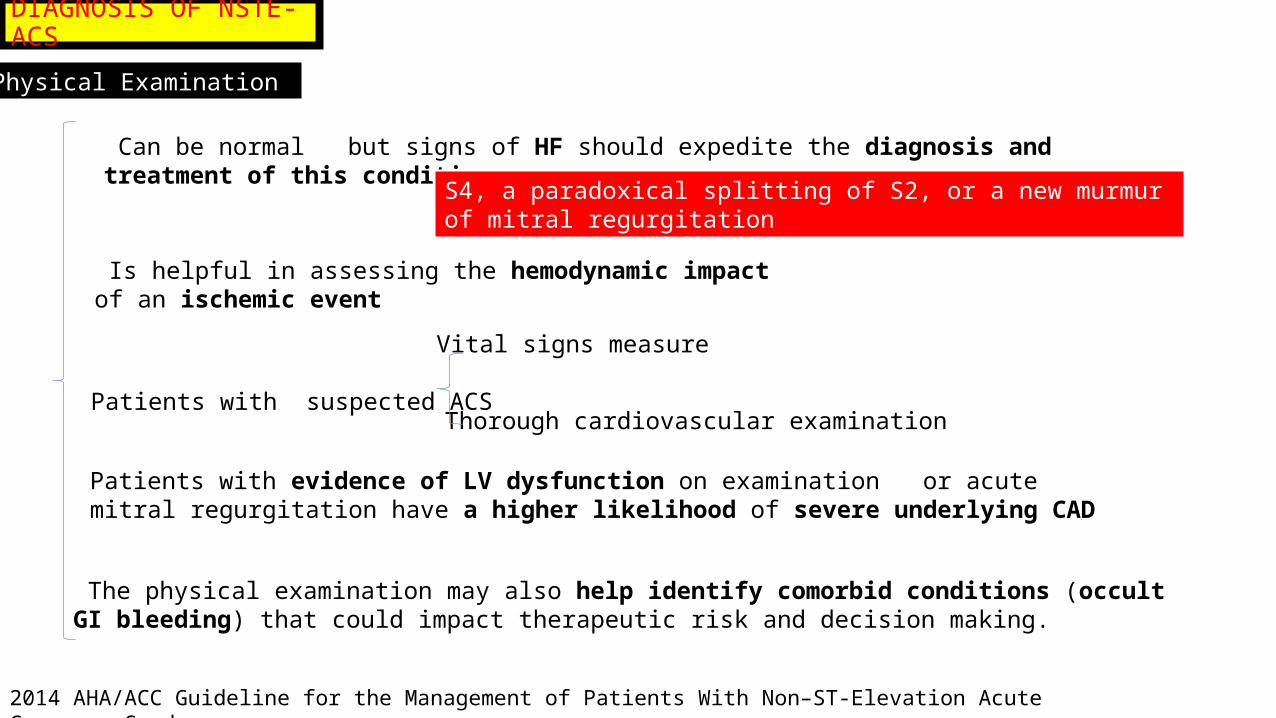
Physical Examination
Is helpful in assessing the hemodynamic impact of an ischemic event
Patients with evidence of LV dysfunction on examination or acute mitral regurgitation have a higher likelihood of severe underlying CAD
Vital signs measure
Thorough cardiovascular examinationPatients with suspected ACS
The physical examination may also help identify comorbid conditions (occult GI bleeding) that could impact therapeutic risk and decision making.
Can be normal but signs of HF should expedite the diagnosis and treatment of this condition
S4, a paradoxical splitting of S2, or a new murmur of mitral regurgitation
2014 AHA/ACC Guideline for the Management of Patients With Non–ST-Elevation Acute Coronary Syndromes
DIAGNOSIS OF NSTE-ACS
PHYSICAL EXAMINATIO
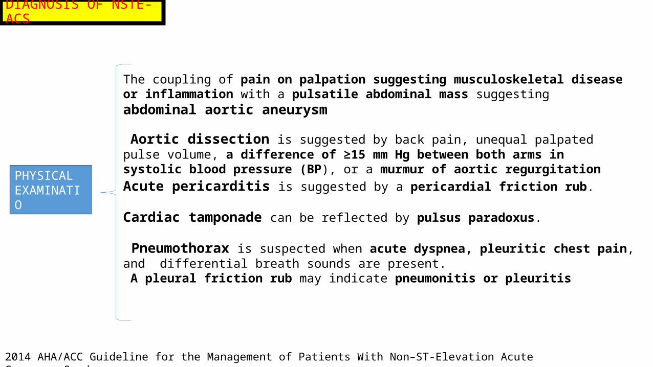
The coupling of pain on palpation suggesting musculoskeletal disease or inflammation with a pulsatile abdominal mass suggesting abdominal aortic aneurysm
Aortic dissection is suggested by back pain, unequal palpated pulse volume, a difference of ≥15 mm Hg between both arms in systolic blood pressure (BP), or a murmur of aortic regurgitation
Acute pericarditis is suggested by a pericardial friction rub.
Cardiac tamponade can be reflected by pulsus paradoxus.
Pneumothorax is suspected when acute dyspnea, pleuritic chest pain, and differential breath sounds are present. A pleural friction rub may indicate pneumonitis or pleuritis

2014 AHA/ACC Guideline for the Management of Patients With Non–ST-Elevation Acute Coronary Syndromes
DIAGNOSIS OF NSTE-ACS
ELECTROCARDIOGRAM
A12-lead ECG should be performed and interpreted within 10 minutes of the patient’s arrival at an emergency facility to assess for cardiac ischemia or injury
Changes on ECG include ST depression, transient ST elevation, or new T-wave inversion .
Persistent ST elevation or anterior ST depression indicative of true posterior MI should be treated according to the STEMI CPG .
The ECG can be relatively normal or initially nondiagnostic; if this is the case, the ECG should be repeated (at 15- to 30- minute intervals during the first hour), especially if symptoms recur . A normal ECG does not exclude ACS and occurs in 1% to 6% of such patient
A normal ECG may also be associated with left circumflex or right coronary artery occlusions, which can be electrically silent (in which case posterior electrocardiographic leads [V7 to V9] may be helpful)
Left ventricular (LV) hypertrophy, bundle-branch blocks with repolarization abnormalities, and ventricular pacing may mask signs of ischemia/injury .
Right-sided leads (V3R to V4R) are typically performed in the case of inferior STEMI to detect evidence of right ventricular infarction

2014 AHA/ACC Guideline for the Management of Patients With Non–ST-Elevation Acute Coronary Syndromes
DIAGNOSIS OF NSTE-ACS
Electrocardiogram
ST depression (especially horizontal ) is highly suggestive of NSTE-ACS
Transient ST changes (≥0.5 mm [0.05 mV]) during symptoms at rest
Marked symmetrical precordial T-wave inversion (≥2 mm [0.2 mV]) suggests acute ischemia
Nonspecific ST-T changes (usually defined as ST deviation of <0.5 mm [0.05 mV] or T-wave inversion of <2 mm [0.2 mV]) are less helpful diagnostically.
Significant Q waves are less helpful
Isolated Q waves in lead 3 are a normal finding
A completely normal ECG in a patient with chest pain does not exclude ACS
Fibrinolytic therapy is contraindicated for patients with ACS without ST elevation, except for those with electrocardiographic evidence of true posterior MI (ST elevation in posterior chest leads [V7 to V9]

2014 AHA/ACC Guideline for the Management of Patients With Non–ST-Elevation Acute Coronary Syndromes
DIAGNOSIS OF NSTE-ACS
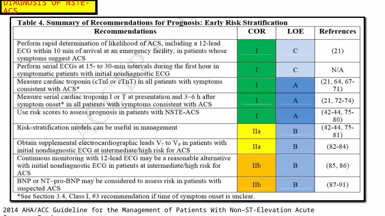
Prognosis—Early Risk Stratification: Recommendations
Class I 1. In patients with chest pain or other symptoms suggestive of ACS, a 12-lead ECG should be performed and evaluated for ischemic changes within 10 minutes of the patient’s arrival at an emergency facility . (Level of Evidence: C)
2.If the initial ECG is not diagnostic but the patient remains symptomatic and there is a high clinical suspicion for ACS, serial ECGs ( 15- to 30-minute intervals during the first hour) should be performed to detect ischemic changes. (Level of Evidence: C) 3. Serial cardiac troponin I or T levels (when a contemporary assay is used) should be obtained at presentation and 3 to 6 hours after symptom onset (Class I, #3 recommendation if time of symptom onset is unclear) in all patients who present with symptoms consistent with ACS to identify a rising and/or falling pattern of values . (Level of Evidence: A)
4. Additional troponin levels should be obtained beyond 6 hours after symptom onset ( Class I, #3 recommendation if time of symptom onset is unclear) in patients with normal troponin levels on serial examination when changes on ECG and/or clinical presentation confer an intermediate or high index of suspicion for ACS . (Level of Evidence: A)
5. Risk scores should be used to assess prognosis in patients with NSTE-ACS (42-44, 75-80). (Level of Evidence: A)
2014 AHA/ACC Guideline for the Management of Patients With Non–ST-Elevation Acute Coronary Syndromes
DIAGNOSIS OF NSTE-ACS
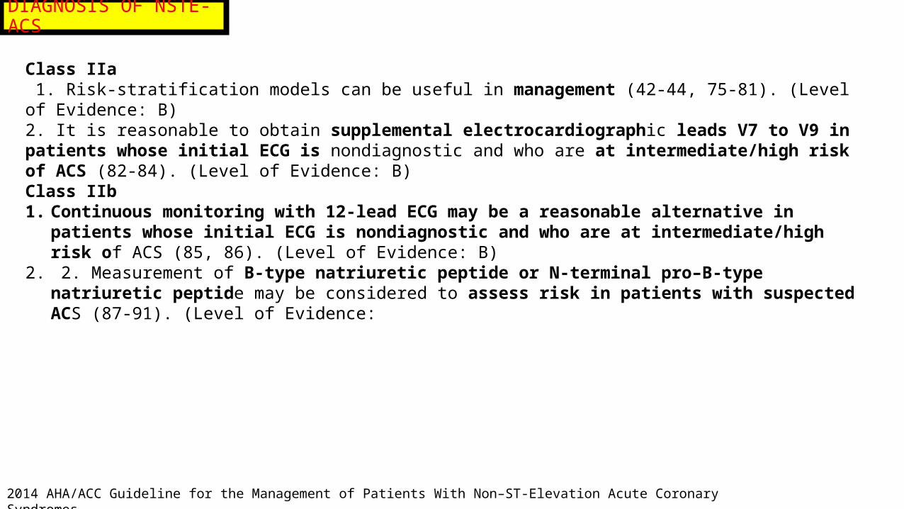
Class IIa 1. Risk-stratification models can be useful in management (42-44, 75-81). (Level of Evidence: B) 2. It is reasonable to obtain supplemental electrocardiographic leads V7 to V9 in patients whose initial ECG is nondiagnostic and who are at intermediate/high risk of ACS (82-84). (Level of Evidence: B) Class IIb 1. Continuous monitoring with 12-lead ECG may be a reasonable alternative in patients whose initial ECG is
nondiagnostic and who are at intermediate/high risk of ACS (85, 86). (Level of Evidence: B) 2. 2. Measurement of B-type natriuretic peptide or N-terminal pro–B-type natriuretic peptide may be considered to
assess risk in patients with suspected ACS (87-91). (Level of Evidence:
2014 AHA/ACC Guideline for the Management of Patients With Non–ST-Elevation Acute Coronary Syndromes
DIAGNOSIS OF NSTE-ACS

2014 AHA/ACC Guideline for the Management of Patients With Non–ST-Elevation Acute Coronary Syndromes
DIAGNOSIS OF NSTE-ACS
High concentrations of troponins in the myocardium
Virtual absence of troponins in nonmyocardial tissue
High-release ratio into the systemic circulation
Rapid release into the blood in proportion to the extent of myocardial injuryThe ability to quantify values with reproducible, inexpensive,rapid, and easily applied assay
Cardiac troponins are the mainstay for diagnosis of ACS and for risk stratification in patients with ACS
cardiac troponin I
and cardiac troponin T
MI
A rising and/or falling pattern of troponin with ≥1 value above the 99th percentile of the upper reference level and evidence for serial increases or decreases in the levels of troponins
AND
Emerging high-sensitivity troponin assays
Cardiac Biomarkers and the Universal Definition of MI: Recommendations

2014 AHA/ACC Guideline for the Management of Patients With Non–ST-Elevation Acute Coronary Syndromes
DIAGNOSIS OF NSTE-ACS
Cardiac Troponins
For the diagnosis of acute myocardial necrosis, it is important to determine not only the peak troponin value but also serial changes:
1. A troponin value above the 99th percentile of the upper reference level is required. Additionally, evidence for a serial increase or decrease ≥20% is required if the initial value is elevated .
2. For any troponin values below or close to the 99th percentile, evidence for acute myocardial necrosis is indicated by a change of ≥3 standard deviations of the variation around the initial value as determined by the individual laboratory .
3. Clinical laboratory reports should indicate whether significant changes in cardiac troponin values for the particular assay have occurred.
Absolute changes VS relative changes
high-sensitivity cardiac troponin T levelTroponins are elevated in MI as early as 2 to 4 hours after symptom onset

2014 AHA/ACC Guideline for the Management of Patients With Non–ST-Elevation Acute Coronary Syndromes
DIAGNOSIS OF NSTE-ACS
Cardiac Troponins
In the vast majority of patients with symptoms suggestive of ACS, MI can be excluded or confirmed within 6 hours, because very few patients present immediately after symptom onset.
In high-risk patients, measurements after 6 hours may be required to identify ACS.
Solitary elevations of troponin cannot be assumed to be due to MI, because troponin elevations can be due to tachyarrhythmia, hypotension or hypertension, cardiac trauma, respiratory failure, acute neurological diseases, and drug toxicity (including cancer chemotherapy)
Chronic elevations can result from structural cardiac abnormalities such as LV hypertrophy or ventricular dilatation and are also common in patients with renal insufficiency
Patients with end-stage renal disease and no clinical evidence of ACS frequently have elevations of cardiac troponin
Troponin elevations may persist for up to 14 days or occasionally longer
References suggest that an increase of >20% of previous troponin levels or an absolute increase of high-sensitivity cardiac troponin T values ( >7 ng/L over 2 hours) may indicate reinfarction

2014 AHA/ACC Guideline for the Management of Patients With Non–ST-Elevation Acute Coronary Syndromes
DIAGNOSIS OF NSTE-ACS
Cardiac Troponins
During pregnancy, troponin values are within the normal range in the absence of cardiovascular morbidities
CK-MB and Myoglobin Compared With Troponin
Previously, CK-MB was used for early evidence of myocardial injur
With the availability of cardiac troponin, CK-MB, myoglobin, and other diagnostic biomarkers are no longer necessary
CK-MB may be used to estimate MI size.

2014 AHA/ACC Guideline for the Management of Patients With Non–ST-Elevation Acute Coronary Syndromes
DIAGNOSIS OF NSTE-ACS
Cardiac Biomarkers and the Universal Definition of MI: Recommendations Cardiac Biomarkers and the Universal Definition of MI: Recommendations

2014 AHA/ACC Guideline for the Management of Patients With Non–ST-Elevation Acute Coronary Syndromes
DIAGNOSIS OF NSTE-ACS
IMAGING
Transesophageal echocardiography can identify a proximal aortic dissection
Chest roentgenogram
Potential pulmonary causes
May show a widened mediastinum in patients with aortic dissection
Computed tomography (CT) of the chest with intravenous contrast can help exclude pulmonary embolism and aortic dissection
Transthoracic echocardiography can identify a pericardial effusion and tamponade physiology and may also be useful to detect regional wall motion abnormalities
In low-risk patients with chest pain, coronary CT angiography can result in a more rapid, more cost-effective diagnosis than stress myocardial perfusion imaging

Guía de práctica clínica de la ESC 2013 para el manejo del infarto agudo de miocardio en pacientes con elevación del segmento ST
DIAGNOSTICO DE INFARTO AGUDO DE MIOCARDIO CON ELEVACIÓN DEL SEGMENTO ST ( IAMCEST)

Guía de práctica clínica de la ESC 2013 para el manejo del infarto agudo de miocardio en pacientes con elevación del segmento ST
DIAGNOSTICO DE IMACEST
Definición de infarto agudo de miocardio
«infarto agudo de miocardio» debe usarse cuando haya evidencia de necrosis miocárdica en un contexto clínico consistente con isquemia miocárdica.

Guía de práctica clínica de la ESC 2013 para el manejo del infarto agudo de miocardio en pacientes con elevación del segmento ST
DIAGNOSTICO DE IMACEST
Diagnóstico inicial
El manejo del IAM —incluido el diagnóstico y el tratamiento— empieza en el lugar donde se produce el primer contacto médico .
En primer lugar debe hacerse el diagnóstico de trabajo de infarto de miocardio
Historia de dolor torácico de 20 min de duración o más que no responde a la nitroglicerina
Historia de cardiopatía isquémica y la irradiación del dolor hacia el cuello, la mandíbula o el brazo izquierdo.
síntomas menos típicosNáuseas/ vómitos, disnea, fatiga, palpitaciones o síncope.
Mujeres, diabéticos o pacientes ancianos Angiografía aguda

Guía de práctica clínica de la ESC 2013 para el manejo del infarto agudo de miocardio en pacientes con elevación del segmento ST
DIAGNOSTICO DE IMACEST
La monitorización ECG debe iniciarse lo antes posible en todos los pacientes con sospecha de IAMCEST
Típicamente se debe encontrar una elevación del segmento ST en el IAM, medido en el punto J, en 2 derivaciones contiguas y debe ser ≥ 0,25 mV en varones de menos de 40 años de edad, ≥ 0,2 mV en varones de más de 40 años o ≥ 0,15 mV en mujeres en las derivaciones V2-V3 o ≥ 0,1 mV en otras derivaciones (en ausencia de hipertrofia del ventrículo izquierdo [VI] o bloqueo de rama
En pacientes con infarto de miocardio inferior es recomendable registrar derivaciones precordiales derechas (V3R y V4R)

Guía de práctica clínica de la ESC 2013 para el manejo del infarto agudo de miocardio en pacientes con elevación del segmento ST
DIAGNOSTICO DE IMACEST
La depresión del segmento ST en las derivaciones V1-V3 sugiere isquemia miocárdica, sobre todo cuando la onda T terminal es positiva y se puede confirmar por una elevación ST concomitante ≥ 0,1 mV registrada en las derivaciones V7-V9
El diagnóstico ECG puede ser más difícil en algunos casos Manejo inmediato

Guía de práctica clínica de la ESC 2013 para el manejo del infarto agudo de miocardio en pacientes con elevación del segmento ST
DIAGNOSTICO DE IMACEST
Bloqueo de rama
La presencia de elevación ST concordante (es decir, en derivaciones con desviaciones QRS positivas) parece ser uno de los mejores indicadores de infarto de miocardio en curso con una arteria ocluid
Un ECG previo puede ayudar a determinar si el bloqueo de rama es nuevo
Un test de troponina positivo en el punto de atención, 1-2 h después del inicio de los síntomas en pacientes con bloqueo de rama de origen incierto
Guía de práctica clínica de la ESC 2013 para el manejo del infarto agudo de miocardio en pacientes con elevación del segmento ST
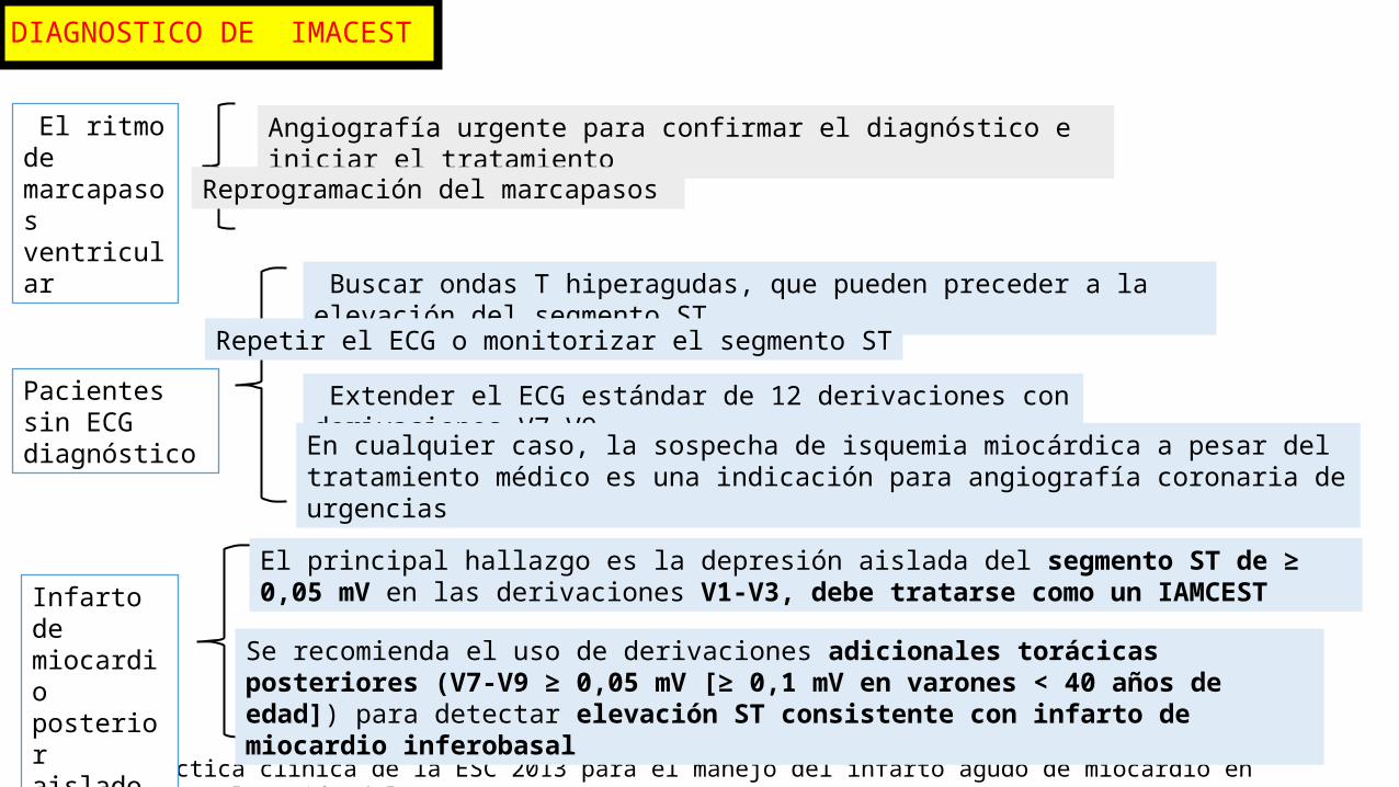
DIAGNOSTICO DE IMACEST
El ritmo de marcapasos ventricular
Angiografía urgente para confirmar el diagnóstico e iniciar el tratamiento
Reprogramación del marcapasos
Pacientes sin ECG diagnóstico
Buscar ondas T hiperagudas, que pueden preceder a la elevación del segmento ST
Repetir el ECG o monitorizar el segmento ST
Extender el ECG estándar de 12 derivaciones con derivaciones V7-V9
En cualquier caso, la sospecha de isquemia miocárdica a pesar del tratamiento médico es una indicación para angiografía coronaria de urgencias
Infarto de miocardio posterior aislado
El principal hallazgo es la depresión aislada del segmento ST de ≥ 0,05 mV en las derivaciones V1-V3, debe tratarse como un IAMCEST
Se recomienda el uso de derivaciones adicionales torácicas posteriores (V7-V9 ≥ 0,05 mV [≥ 0,1 mV en varones < 40 años de edad]) para detectar elevación ST consistente con infarto de miocardio inferobasal

Guía de práctica clínica de la ESC 2013 para el manejo del infarto agudo de miocardio en pacientes con elevación del segmento ST
DIAGNOSTICO DE IMACEST
Obstrucción de la descendente anterior-elevación de la derivación aVR ST y depresión ST inferolateral.
La presencia de depresión ST > 0,1 mV en 8 o más derivaciones de superficie, unido a elevación ST en aVR o V1 en ausencia de otros cambios en el ECG, sugiere isquemia debida a obstrucción de la coronaria izquierda principal o multivaso, especialmente si el paciente presenta compromiso hemodinámico.
En pacientes con sospecha de isquemia miocárdica y elevación del segmento ST o bloqueo de rama izquierda nueva o presuntamente nueva, se debe iniciar terapia de reperfusión lo antes posible.
En la fase aguda se realiza de forma rutinaria la extracción de sangre para marcadores séricos, pero no se debe esperar a los resultados para iniciar el tratamiento de reperfusión.
Si hay dudas sobre la posibilidad de que haya un infarto de miocardio en evolución, la prueba de imagen de urgencias permite la indicación de una terapia de reperfusión a tiempo en estos pacientes
La ausencia de anomalías de la motilidad de la pared excluye un infarto de miocardio importante.

Guía de práctica clínica de la ESC 2013 para el manejo del infarto agudo de miocardio en pacientes con elevación del segmento ST
DIAGNOSTICO DE IMACEST
La miocardiopatía inducida por estrés (tako-tsubo) es un síndrome recientemente reconocido que puede ser difícil de diferenciar del IAMCEST

Guía de práctica clínica de la ESC 2013 para el manejo del infarto agudo de miocardio en pacientes con elevación del segmento ST
DIAGNOSTICO DE INFARTO AGUDO DE MIOCARDIO CON ELEVACIÓN DEL SEGMENTO ST