scatter corrections for cone beam optical ct - iopscience
TRANSCRIPT

Journal of Physics Conference Series
OPEN ACCESS
Scatter corrections for cone beam optical CTTo cite this article Tim Olding et al 2009 J Phys Conf Ser 164 012031
View the article online for updates and enhancements
You may also likeEvaluation of Kinetic and ThermodynamicParameters of Rizatriptan Reduction UsingNovel NanobiosensorSaemeh Mohammadi Deylamani andAkbar Islamnezhad
-
Synergy Effect between BSA and Co2+ toBoost ChemiluminescenceJing Li Huanhuan Xing and Erkang Wang
-
Increasing the accumulation of aptamerAS1411 and verapamil conjugated silvernanoparticles in tumor cells to enhancethe radiosensitivity of gliomaJing Zhao Dongdong Li Jun Ma et al
-
This content was downloaded from IP address 42988259 on 06022022 at 1334
Scatter Corrections for Cone Beam Optical CT
Tim Olding1 Oliver Holmes1 L John Schreiner23 1Department of Physics Queenrsquos University
2Medical Physics Department Cancer Centre of Southeastern Ontario
3Departments of Physics and Oncology Queenrsquos University Kingston ON Canada
TimOldingkrcconca
Abstract Cone beam optical computed tomography (OptCT) employing the VISTA scanner (Modus Medical London ON) has been shown to have significant promise for fast three dimensional imaging of polymer gel dosimeters One distinct challenge with this approach arises from the combination of the cone beam geometry a diffuse light source and the scattering polymer gel media which all contribute scatter signal that perturbs the accuracy of the scanner Beam stop array (BSA) beam pass array (BPA) and anti-scatter polarizer correction methodologies have been employed to remove scatter signal from OptCT data These approaches are investigated through the use of well-characterized phantom scattering solutions and irradiated polymer gel dosimeters BSA corrected scatter solutions show good agreement in attenuation coefficient with the optically absorbing dye solutions with considerable reduction of scatter-induced cupping artifact at high scattering concentrations The application of BSA scatter corrections to a polymer gel dosimeter lead to an overall improvement in the number of pixel satisfying the (3 3mm) gamma value criteria from 78 to 015
1 Introduction The effects of multiple scatter on the measurement integrity of optical computed tomography (OptCT) for polymer gel dosimetry is an important issue that remains to be addressed From preliminary work in characterizing the VISTA OptCT scanner [12] it appears that optical scatter effects are not unlike in-scatter effects seen in x-ray CT [3] Different pre-processing and post-processing schemes have been employed in cone beam x-ray CT with varying degrees of success [4] One strategy employs anti-scatter grids to reject X-ray scatter signal prior to image acquisition [56] Another approach is the beam stop method where an array of opaque disks is added at the beam input side of the scanner [78] Scatter data is then acquired and interpolated to form scatter maps that are subtracted from the image projections prior to tomographic reconstruction This method has been used in the calculation of stray light values for optical absorption in patent blue violet dye solutions [9] but has yet to be applied to optically scattering media In this work we describe the application of anti-scatter polarizers a beam stop array (BSA) and a beam pass array (BPA) toward the removal of scatter signal from OptCT data Scatter corrections for standard well-characterized scattering solutions (described in an accompanying paper in these proceedings) and polymer gel dosimeters are investigated
5th International Conference on Radiotherapy Gel Dosimetry (DOSGEL 2008) IOP PublishingJournal of Physics Conference Series 164 (2009) 012031 doi1010881742-65961641012031
ccopy 2009 IOP Publishing Ltd 1
2 Materials and Methods Standard scattering solutions based on Duramax B-1000 colloidal latex binder and incorporating 12 wt propylene glycol have been prepared in 1 L polyethylene terephthalate (PET) jars The solutions were evaluated using the VISTA cone beam OptCT scanner and with (a) a 4mm diameter dot BSA inserted at the front face of the aquarium (see Figure 1) (b) polarizing films placed at the front and back faces of the scanner aquarium or (c) a 2mm diameter pinhole BPA inserted at the front face of the aquarium Reference and data scans were taken using 633 nm LED illumination and a 1024x768 pixel CCD camera (410 projections over 360o 15 minutes per scan) Image reconstruction was completed using Feldkamp backprojection with a Hamming filter to a 05 mm voxel size
N-isopropylacrylamide (NIPAM) based polymer gels were prepared according to Senden et al[10] (50 C 4T) Gels were poured into 1 L polyethylene teraphthalate (PET) bottles for imaging and irradiated using a T780 Cobalt-60 tomotherapy benchtop (MDS Nordion Kanata Canada) approximately 24 hours post-manufacture using 1x1cm2 pencil beams OptCT imaging of the polymer gel dosimeters was completed using the BSA method
3 Results and Discussion Both anti-scatter polarizers and BSA corrections yield an improvement in the overall linearity of the VISTA OptCT scanner through rejection of multiple scatter from the signal (Figure 1) However the anti-scatter polarizer method did not remove the cupping artifact observed at higher solution concentrations despite the appearance of linearity This method also introduces additional optical artifacts in the projection images that compromise the measurement integrity in some locations
In comparison the BSA method extends the range of scanner measurement integrity with significant cupping artifact reduction (Figure 2) As the beam stop array reduces the overall primary
0
100
200
0 05 1
Attenuation Coefficient (cm-1)
Vis
ta C
T
Uncorrected Scatter Solutions
BSA Corrected
Anti-Scatter Polarizers
Dye Solutions
Linear Fit (BSA Corrected)
Figure 1 A 4mm diameter dot BSA was inserted at the front of the aquarium (top left) and average scattered light values determined at the centre of the blockers These values were used to develop interpolated scatter maps (bottom left) which are subtracted from the reference and data projections prior to reconstruction Mean reconstructed VISTA CT numbers (right) are determined from a 80 mm diameter 80 mm high cylindrical region of interest (ROI) along the central axis of the PET jar Attenuation coefficients are determined from spectrophotometer measurement of the same solution Error bars are smaller than symbol size
5th International Conference on Radiotherapy Gel Dosimetry (DOSGEL 2008) IOP PublishingJournal of Physics Conference Series 164 (2009) 012031 doi1010881742-65961641012031
2
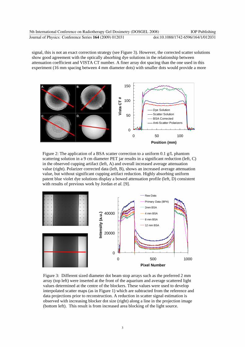
signal this is not an exact correction strategy (see Figure 3) However the corrected scatter solutions show good agreement with the optically absorbing dye solutions in the relationship between attenuation coefficient and VISTA CT number A finer array dot spacing than the one used in this experiment (16 mm spacing between 4 mm diameter dots) with smaller dots would provide a more
0
50
100
150
0 50 100
Position (mm)
Vis
ta C
T
Dye SolutionScatter Solution
BSA CorrectedAnti-Scatter Polarizers
Figure 2 The application of a BSA scatter correction to a uniform 01 gL phantom scattering solution in a 9 cm diameter PET jar results in a significant reduction (left C) in the observed cupping artifact (left A) and overall increased average attenuation value (right) Polarizer corrected data (left B) shows an increased average attenuation value but without significant cupping artifact reduction Highly absorbing uniform patent blue violet dye solutions display a bowed attenuation profile (left D) consistent with results of previous work by Jordan et al [9]
0
20000
40000
0 500 1000
Pixel Number
Inte
nsi
ty (
au
)
Raw Data
Primary Data (BPA)
2mm BSA
4 mm BSA
8 mm BSA
12 mm BSA
Figure 3 Different sized diameter dot beam stop arrays such as the preferred 2 mm array (top left) were inserted at the front of the aquarium and average scattered light values determined at the centre of the blockers These values were used to develop interpolated scatter maps (as in Figure 1) which are subtracted from the reference and data projections prior to reconstruction A reduction in scatter signal estimation is observed with increasing blocker dot size (right) along a line in the projection image (bottom left) This result is from increased area blocking of the light source
5th International Conference on Radiotherapy Gel Dosimetry (DOSGEL 2008) IOP PublishingJournal of Physics Conference Series 164 (2009) 012031 doi1010881742-65961641012031
3
accurate scatter map correction near the jar edges and at the central axis with the additional benefit of reduced primary signal loss from the beam stop array (Figure 3) The 4 mm BSA blocks close to 5 of the light source intensity whereas an equivalent 2 mm BSA would block only 1
Application of the BSA scatter correction method to polymer gels irradiated with a pencil beam calibration pattern leads to considerable improvement in the dosimetry (Figure 4) The middle third of the gel dosimeter is particularly susceptible to scatter artifacts In a comparison of the calculated reference and OptCT measured dose distributions 2D gamma values greater than 1 may be observed in 78 of the pencil beam calibration pattern pixels within this region This can be reduced to less than 02 pixel failure with a BSA scatter correction even in gels irradiated with highly scattering pencil beams
Figure 4 A 4T polymer gel dosimeter irradiated with a 500 cGy intersecting pencil beam pattern (left) shows significant pixel failure (78) for 3 3mm 2D gamma criteria (centre) A BSA scatter correction applied to the OptCT data reduces pixel failure to 015 (right)
0
20000
40000
60000
0 500 1000
Position (Pixels)
Inte
nsi
ty (
au
)
Raw Data
BPA Scatter Map
BSA Scatter Map
BPA Primary Map + BPAScatter Map
Figure 5 Evaluation of the light intensity along a line in the projection image of a 4T polymer gel dosimeter irradiated with a 400 cGy test prostate plan (OptCT projection top left) The BPA method (bottom left) yields a closer approximation (right) of the primary to scatter signal than the BSA method
5th International Conference on Radiotherapy Gel Dosimetry (DOSGEL 2008) IOP PublishingJournal of Physics Conference Series 164 (2009) 012031 doi1010881742-65961641012031
4
OptCT projection data from polymer gels irradiated with a larger scatter volume test prostrate plan is shown in Figure 5 As previously stated the BSA method underestimates the scatter signal due to blocking of primary signal from the light source An alternative method of scatter correction utilizes a beam pass array which essentially consists of a grid of 2 mm diameter pinholes In this method the primary OptCT signal is estimated from the pinhole points then subtracted from the raw OptCT projection (primary + scatter) to obtain the scatter signal at the pinhole point The scatter values at the pinhole points are then interpolated using a bicubic spline to form a scatter map as in Figure 1 While the BPA method does not suffer from inaccuracy due to blocking of primary signal it is significantly perturbed by refraction effects and minor imperfections in the gel dosimeters at different projection angles leading to streak artifacts in the reconstructed image Further results are forthcoming on the development of adaptive BSA and BPA scatter maps correcting individual projections prior to image reconstruction
4 Conclusions Beam stop array and beam pass array corrections have exciting potential for multiple scatter corrections of cone beam OptCT in polymer gel dosimetry While BSA scatter corrections are limited in cone beam x-ray CT by the realities of increased x-ray dose and acquisition time the benefits to cone beam OptCT are well worth the time afforded to such corrections Irregularities in the reference scan (ie the base level of the dosimeter) are well-addressed by such corrections and adaptive scatter maps correcting individual projection images prior to reconstruction have promise to provide further correction to artifacts introduced in highly scattering irradiated dosimeters
5 Acknowledgements Research funding has been provided by the Ontario Research and Development Fund with the Cancer Centre of Southeastern Ontario (CCSEO) and the Canadian Institutes of Health Research (CIHR)
6 References [1] DeJean P Senden R MacAuley K Rogers M and Schreiner L J 2001 Initial experience with a
commercial cone beam optical CT unit for polymer gel dosimetry I amp II J Phys Conference Ser 56 179-186
[2] Sarabipour S Bosi S Hill B and Baldock C 2006 A preliminary study of the measurement of slice-width dose profiles (SWDP) on diagnostic x-ray CT scanners using PAGAT polymer gel dosimeters with optical CT read-out J Phys Conference Ser 56 280-282
[3] Joseph P M and Spital R D 1982 The effects of scatter in x-ray computed tomography MedPhys 9 464-472
[4] Rinkel J Gerfault L Esteve F and Dinten J-M 2007 A new method for x-ray scatter correction first assessment on a cone-beam CT experimental setup Phys Med Biol 52 4633-4652
[5] Endo et al 2001 Effect of scattered radiation on image noise in cone beam CT Med Phys 28 469-474
[6] Siewardsen J H Moseley D J Bakhtiar B Richard S and Jaffray D A 2004 The influence of antiscatter grids on soft-tissue detectability in cone-beam computed tomography with flat-panel detectors Med Phys 31 3506-3520
[7] Siewardsen J H and Jaffray D A 2001 Cone-beam computed tomography with a flat-panel imager magnitude and effects of x-ray scatter Med Phys 28 220-231
[8] Ning R and Tang X 2004 X-ray scatter correction algorithm for cone beam CT imaging Med Phys 31 1195-1202
[9] Jordan K and Battista J 2006 Linearity and image uniformity of the VistaTM optical cone beam scanner J Phys Conference Ser 56 217-220
[10] Senden R J DeJean P MacAuley K and Schreiner L J 2006 Polymer gel dosimeters with reduced toxicity a preliminary investigation of the NMR and optical dose-response using different monomers Phys Med Biol 51 3301-3314
5th International Conference on Radiotherapy Gel Dosimetry (DOSGEL 2008) IOP PublishingJournal of Physics Conference Series 164 (2009) 012031 doi1010881742-65961641012031
5

Scatter Corrections for Cone Beam Optical CT
Tim Olding1 Oliver Holmes1 L John Schreiner23 1Department of Physics Queenrsquos University
2Medical Physics Department Cancer Centre of Southeastern Ontario
3Departments of Physics and Oncology Queenrsquos University Kingston ON Canada
TimOldingkrcconca
Abstract Cone beam optical computed tomography (OptCT) employing the VISTA scanner (Modus Medical London ON) has been shown to have significant promise for fast three dimensional imaging of polymer gel dosimeters One distinct challenge with this approach arises from the combination of the cone beam geometry a diffuse light source and the scattering polymer gel media which all contribute scatter signal that perturbs the accuracy of the scanner Beam stop array (BSA) beam pass array (BPA) and anti-scatter polarizer correction methodologies have been employed to remove scatter signal from OptCT data These approaches are investigated through the use of well-characterized phantom scattering solutions and irradiated polymer gel dosimeters BSA corrected scatter solutions show good agreement in attenuation coefficient with the optically absorbing dye solutions with considerable reduction of scatter-induced cupping artifact at high scattering concentrations The application of BSA scatter corrections to a polymer gel dosimeter lead to an overall improvement in the number of pixel satisfying the (3 3mm) gamma value criteria from 78 to 015
1 Introduction The effects of multiple scatter on the measurement integrity of optical computed tomography (OptCT) for polymer gel dosimetry is an important issue that remains to be addressed From preliminary work in characterizing the VISTA OptCT scanner [12] it appears that optical scatter effects are not unlike in-scatter effects seen in x-ray CT [3] Different pre-processing and post-processing schemes have been employed in cone beam x-ray CT with varying degrees of success [4] One strategy employs anti-scatter grids to reject X-ray scatter signal prior to image acquisition [56] Another approach is the beam stop method where an array of opaque disks is added at the beam input side of the scanner [78] Scatter data is then acquired and interpolated to form scatter maps that are subtracted from the image projections prior to tomographic reconstruction This method has been used in the calculation of stray light values for optical absorption in patent blue violet dye solutions [9] but has yet to be applied to optically scattering media In this work we describe the application of anti-scatter polarizers a beam stop array (BSA) and a beam pass array (BPA) toward the removal of scatter signal from OptCT data Scatter corrections for standard well-characterized scattering solutions (described in an accompanying paper in these proceedings) and polymer gel dosimeters are investigated
5th International Conference on Radiotherapy Gel Dosimetry (DOSGEL 2008) IOP PublishingJournal of Physics Conference Series 164 (2009) 012031 doi1010881742-65961641012031
ccopy 2009 IOP Publishing Ltd 1
2 Materials and Methods Standard scattering solutions based on Duramax B-1000 colloidal latex binder and incorporating 12 wt propylene glycol have been prepared in 1 L polyethylene terephthalate (PET) jars The solutions were evaluated using the VISTA cone beam OptCT scanner and with (a) a 4mm diameter dot BSA inserted at the front face of the aquarium (see Figure 1) (b) polarizing films placed at the front and back faces of the scanner aquarium or (c) a 2mm diameter pinhole BPA inserted at the front face of the aquarium Reference and data scans were taken using 633 nm LED illumination and a 1024x768 pixel CCD camera (410 projections over 360o 15 minutes per scan) Image reconstruction was completed using Feldkamp backprojection with a Hamming filter to a 05 mm voxel size
N-isopropylacrylamide (NIPAM) based polymer gels were prepared according to Senden et al[10] (50 C 4T) Gels were poured into 1 L polyethylene teraphthalate (PET) bottles for imaging and irradiated using a T780 Cobalt-60 tomotherapy benchtop (MDS Nordion Kanata Canada) approximately 24 hours post-manufacture using 1x1cm2 pencil beams OptCT imaging of the polymer gel dosimeters was completed using the BSA method
3 Results and Discussion Both anti-scatter polarizers and BSA corrections yield an improvement in the overall linearity of the VISTA OptCT scanner through rejection of multiple scatter from the signal (Figure 1) However the anti-scatter polarizer method did not remove the cupping artifact observed at higher solution concentrations despite the appearance of linearity This method also introduces additional optical artifacts in the projection images that compromise the measurement integrity in some locations
In comparison the BSA method extends the range of scanner measurement integrity with significant cupping artifact reduction (Figure 2) As the beam stop array reduces the overall primary
0
100
200
0 05 1
Attenuation Coefficient (cm-1)
Vis
ta C
T
Uncorrected Scatter Solutions
BSA Corrected
Anti-Scatter Polarizers
Dye Solutions
Linear Fit (BSA Corrected)
Figure 1 A 4mm diameter dot BSA was inserted at the front of the aquarium (top left) and average scattered light values determined at the centre of the blockers These values were used to develop interpolated scatter maps (bottom left) which are subtracted from the reference and data projections prior to reconstruction Mean reconstructed VISTA CT numbers (right) are determined from a 80 mm diameter 80 mm high cylindrical region of interest (ROI) along the central axis of the PET jar Attenuation coefficients are determined from spectrophotometer measurement of the same solution Error bars are smaller than symbol size
5th International Conference on Radiotherapy Gel Dosimetry (DOSGEL 2008) IOP PublishingJournal of Physics Conference Series 164 (2009) 012031 doi1010881742-65961641012031
2
signal this is not an exact correction strategy (see Figure 3) However the corrected scatter solutions show good agreement with the optically absorbing dye solutions in the relationship between attenuation coefficient and VISTA CT number A finer array dot spacing than the one used in this experiment (16 mm spacing between 4 mm diameter dots) with smaller dots would provide a more
0
50
100
150
0 50 100
Position (mm)
Vis
ta C
T
Dye SolutionScatter Solution
BSA CorrectedAnti-Scatter Polarizers
Figure 2 The application of a BSA scatter correction to a uniform 01 gL phantom scattering solution in a 9 cm diameter PET jar results in a significant reduction (left C) in the observed cupping artifact (left A) and overall increased average attenuation value (right) Polarizer corrected data (left B) shows an increased average attenuation value but without significant cupping artifact reduction Highly absorbing uniform patent blue violet dye solutions display a bowed attenuation profile (left D) consistent with results of previous work by Jordan et al [9]
0
20000
40000
0 500 1000
Pixel Number
Inte
nsi
ty (
au
)
Raw Data
Primary Data (BPA)
2mm BSA
4 mm BSA
8 mm BSA
12 mm BSA
Figure 3 Different sized diameter dot beam stop arrays such as the preferred 2 mm array (top left) were inserted at the front of the aquarium and average scattered light values determined at the centre of the blockers These values were used to develop interpolated scatter maps (as in Figure 1) which are subtracted from the reference and data projections prior to reconstruction A reduction in scatter signal estimation is observed with increasing blocker dot size (right) along a line in the projection image (bottom left) This result is from increased area blocking of the light source
5th International Conference on Radiotherapy Gel Dosimetry (DOSGEL 2008) IOP PublishingJournal of Physics Conference Series 164 (2009) 012031 doi1010881742-65961641012031
3
accurate scatter map correction near the jar edges and at the central axis with the additional benefit of reduced primary signal loss from the beam stop array (Figure 3) The 4 mm BSA blocks close to 5 of the light source intensity whereas an equivalent 2 mm BSA would block only 1
Application of the BSA scatter correction method to polymer gels irradiated with a pencil beam calibration pattern leads to considerable improvement in the dosimetry (Figure 4) The middle third of the gel dosimeter is particularly susceptible to scatter artifacts In a comparison of the calculated reference and OptCT measured dose distributions 2D gamma values greater than 1 may be observed in 78 of the pencil beam calibration pattern pixels within this region This can be reduced to less than 02 pixel failure with a BSA scatter correction even in gels irradiated with highly scattering pencil beams
Figure 4 A 4T polymer gel dosimeter irradiated with a 500 cGy intersecting pencil beam pattern (left) shows significant pixel failure (78) for 3 3mm 2D gamma criteria (centre) A BSA scatter correction applied to the OptCT data reduces pixel failure to 015 (right)
0
20000
40000
60000
0 500 1000
Position (Pixels)
Inte
nsi
ty (
au
)
Raw Data
BPA Scatter Map
BSA Scatter Map
BPA Primary Map + BPAScatter Map
Figure 5 Evaluation of the light intensity along a line in the projection image of a 4T polymer gel dosimeter irradiated with a 400 cGy test prostate plan (OptCT projection top left) The BPA method (bottom left) yields a closer approximation (right) of the primary to scatter signal than the BSA method
5th International Conference on Radiotherapy Gel Dosimetry (DOSGEL 2008) IOP PublishingJournal of Physics Conference Series 164 (2009) 012031 doi1010881742-65961641012031
4
OptCT projection data from polymer gels irradiated with a larger scatter volume test prostrate plan is shown in Figure 5 As previously stated the BSA method underestimates the scatter signal due to blocking of primary signal from the light source An alternative method of scatter correction utilizes a beam pass array which essentially consists of a grid of 2 mm diameter pinholes In this method the primary OptCT signal is estimated from the pinhole points then subtracted from the raw OptCT projection (primary + scatter) to obtain the scatter signal at the pinhole point The scatter values at the pinhole points are then interpolated using a bicubic spline to form a scatter map as in Figure 1 While the BPA method does not suffer from inaccuracy due to blocking of primary signal it is significantly perturbed by refraction effects and minor imperfections in the gel dosimeters at different projection angles leading to streak artifacts in the reconstructed image Further results are forthcoming on the development of adaptive BSA and BPA scatter maps correcting individual projections prior to image reconstruction
4 Conclusions Beam stop array and beam pass array corrections have exciting potential for multiple scatter corrections of cone beam OptCT in polymer gel dosimetry While BSA scatter corrections are limited in cone beam x-ray CT by the realities of increased x-ray dose and acquisition time the benefits to cone beam OptCT are well worth the time afforded to such corrections Irregularities in the reference scan (ie the base level of the dosimeter) are well-addressed by such corrections and adaptive scatter maps correcting individual projection images prior to reconstruction have promise to provide further correction to artifacts introduced in highly scattering irradiated dosimeters
5 Acknowledgements Research funding has been provided by the Ontario Research and Development Fund with the Cancer Centre of Southeastern Ontario (CCSEO) and the Canadian Institutes of Health Research (CIHR)
6 References [1] DeJean P Senden R MacAuley K Rogers M and Schreiner L J 2001 Initial experience with a
commercial cone beam optical CT unit for polymer gel dosimetry I amp II J Phys Conference Ser 56 179-186
[2] Sarabipour S Bosi S Hill B and Baldock C 2006 A preliminary study of the measurement of slice-width dose profiles (SWDP) on diagnostic x-ray CT scanners using PAGAT polymer gel dosimeters with optical CT read-out J Phys Conference Ser 56 280-282
[3] Joseph P M and Spital R D 1982 The effects of scatter in x-ray computed tomography MedPhys 9 464-472
[4] Rinkel J Gerfault L Esteve F and Dinten J-M 2007 A new method for x-ray scatter correction first assessment on a cone-beam CT experimental setup Phys Med Biol 52 4633-4652
[5] Endo et al 2001 Effect of scattered radiation on image noise in cone beam CT Med Phys 28 469-474
[6] Siewardsen J H Moseley D J Bakhtiar B Richard S and Jaffray D A 2004 The influence of antiscatter grids on soft-tissue detectability in cone-beam computed tomography with flat-panel detectors Med Phys 31 3506-3520
[7] Siewardsen J H and Jaffray D A 2001 Cone-beam computed tomography with a flat-panel imager magnitude and effects of x-ray scatter Med Phys 28 220-231
[8] Ning R and Tang X 2004 X-ray scatter correction algorithm for cone beam CT imaging Med Phys 31 1195-1202
[9] Jordan K and Battista J 2006 Linearity and image uniformity of the VistaTM optical cone beam scanner J Phys Conference Ser 56 217-220
[10] Senden R J DeJean P MacAuley K and Schreiner L J 2006 Polymer gel dosimeters with reduced toxicity a preliminary investigation of the NMR and optical dose-response using different monomers Phys Med Biol 51 3301-3314
5th International Conference on Radiotherapy Gel Dosimetry (DOSGEL 2008) IOP PublishingJournal of Physics Conference Series 164 (2009) 012031 doi1010881742-65961641012031
5