scientific committee on consumer...
TRANSCRIPT

SCCP/0867/05
EUROPEAN COMMISSION HEALTH & CONSUMER PROTECTION DIRECTORATE-GENERAL Directorate C - Public Health and Risk Assessment C7 - Risk assessment
SCIENTIFIC COMMITTEE ON CONSUMER PRODUCTS
SCCP
Opinion on
para-Aminophenol
COLIPA n° A16
Adopted by the SCCP during the 3rd plenary meeting of 15 March 2005

SCCP/0867/05 Opinion on para-Aminophenol
2
TABLE OF CONTENTS
1. BACKGROUND......................................................................................................... 3
2. TERMS OF REFERENCE.......................................................................................... 3
3. OPINION .................................................................................................................... 3
4. CONCLUSION ......................................................................................................... 40
5. MINORITY OPINION ............................................................................................. 40
6. REFERENCES.......................................................................................................... 41
7. ACKNOWLEDGEMENTS ...................................................................................... 46

SCCP/0867/05 Opinion on para-Aminophenol
3
1. BACKGROUND The adaptation to technical progress of the Annexes to Council Directive 76/768/EEC of 27 July 1976 on the approximation of the laws of the Member States relating to cosmetic products. 2. TERMS OF REFERENCE The SCCP is requested to answer the following questions: • Is para-Aminophenol safe for use in cosmetic products? • Does the SCCP propose any restrictions or conditions for its use in cosmetic products? 3. OPINION 3.1. Chemical and Physical Specifications 3.1.1. Chemical identity 3.1.1.1. Primary name and/or INCI name para-Aminophenol (INCI name) 3.1.1.2. Chemical names 1-hydroxy-4-aminobenzene (IUPAC) phenol, 4-amino- phenol, p-amino- p-hydroxyaniline 4-hydroxyaniline 4-amino-1-hydroxybenzene 4-hydroxy-1-aminobenzene 1-amino-4-hydroxybenzene p-aminophenol 4-aminophenol 4-hydroxybenzenamine p-hydroxyphenylamine 4-hydroxyphenylamine 3.1.1.3. Trade names and abbreviations Activol; Azol; BASF Ursol P Base; Benzofur P; Certinal; Cetal; Citol; Durafur Brown RB; Fouramine P; Fourrine 84; Fourrine P Base; Furro P Base; Imexine OB (Hydrochloride); Nako Brown R; PAP; Paranol; Pelagol Grey P Base; Pelagol P Base; Renal AC; Rodinal; Tertral P Base; Unal; Ursol P; Ursol P Base; Zoba Brown P Base COLIPA n° A16 Colour index: CI 76550 (Oxidation base 6)
CI 76551 (Oxidation base 6a — Hydrochloride)

SCCP/0867/05 Opinion on para-Aminophenol
4
3.1.1.4. CAS / EINECS number CAS : 123-30-8 (free base) 51-78-5 (hydrochloride) EINECS : 204-616-2 (free base) 200-122-6 (hydrochloride) 3.1.1.5. Structural formula
NH2
OH
3.1.1.6. Empirical formula Formula : C6H7NO 3.1.2. Physical form White, odourless crystals or powder, turning brownish on exposure to air/humidity (commercial product usually pink) 3.1.3. Molecular weight Molecular weight : 109.13 3.1.4. Purity, composition and substance codes Batch and analytical methods not specified in Section 1 “General” of Submission IV. Purity
titre : ≥ 97.5% water content : not assessed or not reported ash content : not assessed or not reported
Potential impurities and reaction intermediates : not assessed or not reported Solvent residues : not assessed or not reported Other : not assessed or not reported 3.1.5. Impurities / accompanying contaminants
SCCP/0867/05 Opinion on para-Aminophenol
5
/ 3.1.6. Solubility Water : 0.60-0.65% (at 24-25 °C) Ethanol (abs) : 4.5% (at 0 °C) Receptor fluid (PBS, at pH 7.4) : / 3.1.7. Partition coefficient (Log Pow) Log Pow : 0.04 (at pH 7.4) 3.1.8. Additional physical and chemical specifications Organoleptic properties : / Melting point : 189-190 °C (commercial product, 186 °C) Boiling point : 110 °C (at 0.3 mmHg) Flash point : / Vapour press. : 0.0053 Pa (at 25 °C) Density : / Viscosity : / pKa : / Refractive index : / Stability : Stable for months when pure; stable for hours in an aqueous
medium at room temperature or for days at 4 °C, as specifically assessed
HPLC procedure and features provided. HPTLC and IR, UV-Vis, MS, and NMR spectral characteristics also available for identification purposes. General comments on analytical and physico-chemical characterisation * para-Aminophenol purity was reported in Section 1 “General” of Submission IV (2001)
with no clear identification of the batch. There was no consistent measure of analysis of purity between batches.
* When reported, chemical purity was usually obtained from potentiometric titration. The more reliable chromatographic purity was not available in the dossier.
* In general, the analytical characterisation was inadequate, this resulting in a lack of information on several purity parameters.
* The physico-chemical profile of the substance, including solubility, was not properly characterised in the dossier. Due to para-aminophenol long history, supplementary data were derived from the literature.
* Batch number and/or chemical purity were not stated in some toxicity study reports. 3.2. Function and uses

SCCP/0867/05 Opinion on para-Aminophenol
6
para-Aminophenol is a hair dye precursor. It is incorporated in oxidative hair dye formulations and in the bottle on the market at a maximum concentration of 1.8% and is typically mixed in a 1:1 ratio with an oxidative agent thereby reaching a concentration of 0.9% for in use application. 3.3. Toxicological Evaluation 3.3.1. Acute toxicity 3.3.1.1. Acute oral toxicity Oral median lethal dose values of para-aminophenol (PAP) previously reported in Submission I were: Rats 671 mg/kg (Lloyd, 1977) 1270 mg/kg (Lloyd, 1977) Ref. C26 375 mg/kg (Ind. Bio-Test Lab. Inc., 1975) Ref. C23; Mice 1550 mg/kg (Segré, 1977), Ref. C43. In a review report, an acute oral toxicity study in rats where the lethal median dose in male rats was 393 mg/kg and that for females was 1139 mg/kg. It also reports an oral lethal median dose in rabbits of between 4 and 10 g/kg.
Ref.: A2 A 5% suspension of PAP in a 3% starch solution was administered orally to albino rats. Deaths were reported within 48 hours of compound administration. A median lethal dose of 0.5 g/kg was established for PAP. Methemoglobin concentration was less than 10% of the total haemoglobin concentration.
Ref.: A1 3.3.1.2. Acute dermal toxicity The dermal median lethal dose for cats was calculated to be 37 mg/kg.
Ref.: C29 The medial lethal dose in mice was calculated to be 470 mg/kg.
Ref.: A2 The dermal median lethal dose for rabbits was calculated to be 10,000 mg/kg.
Ref.: C23 A separate study reported a dermal median lethal dose of >8000 mg/kg in rabbits. In rats, a dermal lethal median dose of >5000 mg/kg has been reported.
Ref.: A2

SCCP/0867/05 Opinion on para-Aminophenol
7
3.3.1.3. Acute inhalation toxicity The median lethal dose for rats (1 hour exposure) was calculated to be >0.005 mg/l.
Ref.: C23 In separate studies, median lethal doses of >5.91 mg/l (1.3 hour exposure) and >3.42 mg/l (4 hour exposure) were calculated for rats.
Ref.: A2 3.3.2 Irritation and corrosivity 3.3.2.1. Skin irritation A 2.5% aqueous solution of PAP containing 0.05% sodium sulphite was mildly irritant when applied to the skin of New Zealand White (NZW) rabbits. The primary irritation index was estimated to be 0.2 out of a total score of 8.
Ref.: C26 Three protocols for evaluating the skin irritation of PAP were compared. 0.5 g of PAP as powder was applied to clipped skin of 6 New Zealand White rabbits under occlusive and semiocclusive patches [occlusive - Official French Cosmetic (OFC) method and Association Française de Normalisation (AFNOR) method; semiocclusive OECD method]. The occlusive patches were applied on both scarified and intact sites. Patches were removed at 4 hours (AFNOR, OECD) or at 23 hours (OFC). Macroscopic changes were recorded up to 42 hours (OFC, AFNOR) or 78 hours (OECD). The calculated primary cutaneous irritation indices (PCI) ranged from 0 (OECD) to 0.21 (OFC); these were well below the 0.5 threshold value of both the OFC and the AFNOR for irritation.
Ref.: A1 Conclusion PAP showed irritancy on normal skin above a concentration of 2.5 %. 3.3.2.2. Mucous membrane irritation A 2.5% aqueous solution of PAP containing 0.05% sodium sulphite was not considered to be irritant when instilled into the rabbit eye and then rinsed with water after ten seconds. Mild conjunctival irritation was observed in two animals.
Ref.: C26 100 mg/kg of PAP in the form of dry powder were instilled into one eye of each of six albino rabbits. The Ocular Irritation Index was estimated to be 17/110 after 24 hours, 4.5/110 after 48 hours, and 0/110 after 72 hours.
Ref.: C23 100 mg of PAP were instilled into the eyes of 9 New Zealand White rabbits. The eyes of 3 rabbits were rinsed after a 20 second exposure. The Ocular Irritation Index was estimated to be 6/110. Barely perceptible to mild conjunctival erythema, chemosis and secretion were observed;

SCCP/0867/05 Opinion on para-Aminophenol
8
these signs improved over time. PAP was considered to be non-irritant under the conditions of this study.
Ref.: A2 Conclusion PAP was classified as slightly irritant to mucous membranes. 3.3.3. Skin sensitisation Animal studies In an open epicutaneous test in Pirbright white guinea-pigs, PAP at a concentration of 3% was applied on the flank six days per week for three weeks. Two weeks later, a challenge application was performed on the opposite untreated flank of the animals. A single application of the test compound produced an allergic reaction at the challenge application site in 4 of 20 animals.
Ref.: C42 Using 24-hour occlusive patches, induction of Hartley albino guinea pigs with 2% PAP in petrolatum was performed on the flank (scapular area). Four patches were applied on alternate days. Following a 14-day waiting period, animals were challenged with a concentration of 2%, 1%, 0.5%, or 0.1% PAP on the opposite flank using a Finn chamber (closed technique). Dose-dependent elicitation was observed: 9/10 animals were positive at 2%, 6/10 animals at 1%, 5/10 animals at 0.5%, and 3/10 animals at 0.1%.
Ref.: C25 A 3% preparation of PAP in "Schultz Hamburg vehicle II" did not evoke a sensitization reaction in Hartley albino guinea pigs after induction using 18 applications of 0.5 ml of the test compound followed by a 0.5 ml challenge dose administered on the opposite flank two weeks later. No study details were available to the SCCP.
Ref.: A2 The potential for cross sensitization between the industrial allergen 2-amino-4-chloro-phenol (ACP) and PAP was studied in guinea pigs (strain not indicated). Fifteen guinea pigs were injected intracutaneously with ACP and then challenged with ACP and PAP. No reaction to PAP was elicited by challenge with 1.0% PAP for up to six weeks.
Ref.: A1 In a comparative study using guinea pigs (strain not indicated) and humans, 1% PAP in Vaseline produced no sensitization in the guinea pigs tested versus 36% of humans tested. 0.1% PAP in Vaseline produced no sensitization in the guinea pigs compared with 14% of the humans tested.
Ref.: A2 In a comparative study using two methods of induction, Hartley guinea pigs were tested for sensitization with PAP (>99%). For the first method, Freund’s Adjuvant was injected into the foot pad of the hind paw and 0.18 mmol/l PAP was administered topically twice (over two days). For the second method, a preparation containing a 1:1 ratio of Freund’s Adjuvant and 0.18 mmol/l PAP in distilled water was injected into the foot pad of the hind paw. After a 16-day waiting period, both groups of animals were challenged with a dose of 0.09 mmol/l in the lumbar

SCCP/0867/05 Opinion on para-Aminophenol
9
region. Animals tested under the first method of induction exhibited no sensitization reactions, while 40% of those tested under the second method of induction were positive for sensitization.
Ref.: A2 A photosensitization test with 10% PAP in 80% DAE (40% dimethylacetamide, 30% acetone and 30% ethanol) was performed in Hartley guinea pigs. 5% musk ambrette was used as the positive control. The induction phase was 3 weeks long. During the first week, it consisted of the topical administration of 4 daily doses of 0.1 ml of the PAP solution followed by irradiation with ½ MED of UVA. Scoring was performed 24 hours after each dose. During weeks 2 and 3, it consisted of the topical administration of 4 daily doses of 0.1 ml of the PAP solution followed by irradiation with 1 MED of UVB. On the first and third days of the second and third weeks of induction, 0.1 ml Freund’s Complete adjuvant was injected intradermally surrounding the topical application site. Challenge was performed two weeks after the final induction dose was given, using the same application site and using 0.1 ml of 5% PAP for 3 consecutive days. One part of the application site was irradiated with ½ MED UVA, one part with ½ MED UVB, and one part received no irradiation. Sites were scored 24 hours after treatment. Rechallenge consisted of a single application of a dye containing 7.5% PAP 9 days after the initial challenge. Sites were irradiated and scored 24 hours after application. No photosensitization was observed.
Ref.: A1 Comment The studies were not performed according to standard methods. Human studies Various tests reported in previous submissions have demonstrated cross-reactivity between PAP and other aromatic amines in humans. The following reports on human sensitization to PAP appeared in the 1995 were compiled in 1995 (BCI, 1995, ref. A2): • Among 60 patients from a dermatology clinic who were tested with 1% PAP, 7 (12%) were
positive. • Between 1973 and 1977, 4600 patients were tested for sensitization to benzidine. Of the
5.02% who were positive, 16.4% also had positive reactions to para-amino compounds. However, only 1% of the patients (n=46) had a positive reaction to PAP.
• Between 1974 and 1984, 32 professional hairdressers with hand dermatitis due to use of hair dyes were patch tested for sensitization to these products. Twenty-two subjects had a positive reaction to hair dyes and 25% of these were positive when tested with PAP.
• 408 patients with eczema were patch tested for reactions against PAP. In response to the application of 1% PAP in Vaseline, 3% of the patients were positive.
• Of 13 female cosmetologists with hand, face, and/or axillary dermatitis, 4 were patch tested with a concentration of 1% PAP in Vaseline using standards approved by the International Contact Dermatitis Research Group. Of these, one person tested positive for sensitization with PAP.
• Two groups of hairdressers were tested for sensitization to para-phenylenediamine (PPD). 32 were negative for sensitization and 7 were positive. When the same subjects were tested for

SCCP/0867/05 Opinion on para-Aminophenol
10
sensitization to PAP, the 32 who were negative with PPD were also negative with PAP. One of the 7 who was positive with PPD was also positive with PAP.
Conclusion PAP is a sensitizer, but did not show photosensitizing effects. 3.3.4. Dermal / percutaneous absorption In vitro studies PAP was among several para-substituted phenols for which percutaneous penetration was evaluated in vitro in full thickness hairless mouse skin. 4 µg/cm² PAP in acetone were applied to a surface measuring 3.1 cm². After evaporation of the solvent, the diffusion chamber was perfused with phosphate-buffered normal saline for a period of 48 hours. Transport of 72 ± 6% of the applied dose across the skin was attained after 24 hours, with the time of maximum flux occurring three hours after compound administration.
Ref.: Col. 73 The in vitro percutaneous penetration and metabolism of 14C-labeled PAP in ethanol/water or acetone was studied in rat and human skin. 10% of the dose of 20 mg/ml PAP was absorbed through dermatomed rat skin when applied as an infinite dose over a 24-hour period. Skin stripping (removal of the stratum corneum) increased the absorption to 40% over 24 hours. In human skin, absorption resulting from the application of 20 mg/ml PAP was measured as 0.5 – 6.0% over 24 hours. Skin stripping increased absorption up to 27% of the dose administered. Results pertaining to the metabolism of PAP by the skin during percutaneous penetration were inconclusive.
Ref.: Col 74 The percutaneous absorption of two test formulations containing 14C-labeled PAP was examined in vitro using human abdominal skin. One formulation (A) contained 0.84% 14C-labeled PAP as well as other primary intermediates and couplers used to produce a reddish-brown shade. The other (B) contained 14C-labeled PAP as the sole dye constituent. Formulation A was mixed with an equal volume of hydrogen peroxide; formulation B was mixed with an equal amount of water. The final concentration of dye was 0.42% in each formulation. 20 mg/cm² of the test compounds were applied to heat-separated epidermal membranes for 30 minutes, after which the application sites were rinsed with water. Samples of receptor fluid were taken at 1, 2, 4, 6, 8, 24, 30, and 48 hours following dye application. For Formulation A, mass balance recovery after 48 hours consisted of 84.62 ± 2.9% of the applied dose in the 30-minute rinsate, 1.60 ± 0.26% in solubilized skin samples, and 0.14 ± 0.04% in the receptor fluid. For Formulation B, mass balance recovery after 48 hours consisted of 91.69 ± 2.95% of the applied dose in the 30-minute rinsate, 2.60 ± 0.62% in solubilized skin samples, and 0.46 ± 0.11% in the receptor fluid. Mean cumulative absorption results were as follows:
Formulation
Mean Cumulative Absorption ± S.E. (µg/cm2)
Applied Dose Absorbed (%)
Dose: 20 mg/cm2
24 hours 48 hours 24 hours 48 hours
A 0.12 ± 0.04 0.13 ± 0.03 0.13 ± 0.04 0.14 ± 0.04

SCCP/0867/05 Opinion on para-Aminophenol
11
B 0.32 ± 0.09 0.46 ± 0.14 0.32 ± 0.07 0.46 ± 0.11 Under these conditions, the penetration of 14C-labeled PAP in a formulation containing other primary intermediates and couplers and mixed with an equal volume of developer was approximately 3.5x lower than in a formulation containing 14C-labelled PAP as the sole dye constituent and in the absence of developer.
Ref.: Col. 75 In vivo studies Sprague-Dawley rats of both sexes were treated with a single dermal application of either an aqueous solution (8%) or one of several hair dye formulations (1%, 2%, or 3% PAP mixed with 6% hydrogen peroxide) containing 14C-labelled PAP for 30 minutes, followed by rinsing of the application site. The doses applied were 0.3 ml for the aqueous solution and 45, 90, and 135 mg/kg PAP for the 1%, 2%, and 3% hair dye solutions, respectively. The shaved application site measured 9 cm², except in one group of animals receiving the 2% hair dye formulation where the skin was not shaved and the application site measured 16 cm². Additionally, a single dose of 37.5 mg/kg of a 1% aqueous solution of PAP was administered either orally or subcutaneously to Sprague-Dawley rats. The urine and faeces of all animals were collected over a period of 72 hours. The animals were then killed, and urine, faeces and organs were evaluated for the presence of 14C-labelled PAP using scintillography. Results The levels of absorption observed were as follows: 12.5 µg/cm² (0.627%) for the aqueous solution, 0.8 µg/cm² (0.08%) for 1% PAP, 5.4 µg/cm² (0.27% for 2% PAP), and 2.7µg/cm² (0.09%) for 3% PAP. The absorption observed at unshaved application sites was lower (0.103% with 2% PAP). 95% of the radioactivity was found in the rinsing waters. The maximum amount of PAP in organs was found 35 minutes after compound administration. Excretion was primarily via the urine regardless of the route of administration.
Ref.: C21 Female hairless Wistar rats were administered a single dose of 0.14, 0.69, or 3.44 µM/cm² of a 0.75% solution of 14C-labelled PAP mixed with an equal volume of 20 vol. hydrogen peroxide on the skin of the back. The application site measured 10 cm². After 30 minutes of exposure, the application site was rinsed. Urine, faeces, skin and viscera were evaluated for PAP content over/after 4 days. Results the amounts of PAP absorbed were 15.9 nM/cm², 52.04 nM/cm², and 58.4 nM/cm². In another set of experiments, penetration was measured for 3.44 µM/cm² of 14C -labelled PAP alone, 14C -labelled PAP admixed with a non-radioactive coupler and the 14C -labelled indamine structure (a benzoquinoneneimine) produced from the reaction between PAP and its coupler. At this concentration, the level of 14C -labelled PAP detected in the skin was approximately the same as that of 14C -labelled PAP applied with the nonradiolabeled coupler. Penetration of the 14C -labelled benzoquinoneneimine was ~17x less than that of 14C -labelled PAP alone. The investigator concluded that indamine structures do not effectively cross the skin barrier.
Ref.: C46

SCCP/0867/05 Opinion on para-Aminophenol
12
The percutaneous absorption of PAP was evaluated in a study using 5 human volunteers. 2 µg/cm² of 14C-labelled PAP in 95% ethanol was applied to the bend of the forearm over a total surface area of 2.5 cm². The application time was not specified. Urine from each subject was collected over a seven day period and the radioactivity recovered was quantified. The dermal absorption of PAP was thus determined to be 13% of the applied dose. In a subsequent study performed by the same investigators using similar dosing and urine collection regimens, penetration was determined to be 6-8%.
Ref.: A2 3.3.5. Repeated dose toxicity 3.3.5.1. Repeated Dose (13 days) oral Wistar rats were fed 55 mg/kg/day PAP in the diet for 13 days. No compound-related toxicity was observed.
Ref.: A2 3.3.5.2. Sub-chronic (90 days) oral / dermal toxicity Oral PAP was administered to male and female Wistar rats by oral gavage at a dose of 20 mg/kg/day for 12 weeks. The control group received a 1% carboxymethylcellulose solution only. No death, clinical signs, or histopathological findings could be attributed to compound administration.
Ref.: C18 A 13-week study on the dietary toxicity of PAP was conducted in Sprague-Dawley rats. The doses administered were 0, 47, 133, and 467 mg/kg. At 467 mg/kg, both sexes were observed to have reduced body weight gain and food consumption. Female rats had significantly lower haemoglobin and erythrocyte counts and increased corpuscular haemoglobin levels, but these changes were without clinical significance. Increased relative liver, kidney, and brain weights in both sexes and increased relative pituitary and thyroid weights in females were attributed to compound administration. Nephrosis was observed in all groups, including controls, but the incidence and severity of this change increased in a dose-dependent fashion.
Ref.: A2 The same doses (0, 47, 133, and 467 mg/kg) were administered daily in the diet to groups of Sprague-Dawley rats for a period of 6 months. To evaluate reversibility, 20 additional male rats were administered the dose of 467 mg/kg/day over a 20 week period and then observed for 7 weeks without treatment. Control, high dose and recovery animals were evaluated. A significant decrease in body weight gain was observed at 467 mg/kg/day as in the three month study; the males that had been allowed a reversibility period also had decreased body weight gain. There were no significant clinical pathology changes. Increased relative liver, kidney, and brain weights were attributed to compound administration; these changes were not observed in rats allowed a reversibility period. Nephrosis, characterized by eosinophilic droplets in renal tubules, was diagnosed in both sexes at 467 mg/kg/day (including recovery males). The severity of this finding was dose-dependent, as in the three month study.

SCCP/0867/05 Opinion on para-Aminophenol
13
Ref.: A2 Doses of 10, 30, or 100 mg/kg/day of PAP were administered by gavage to Sprague-Dawley rats daily for 13 weeks. Compound-related findings consisted of dose-related nephrosis at 30 and 100 mg/kg/day in both sexes. Lower body weight gain (not dose-related in severity) was observed in females at all doses. The dose of 10 mg/kg/day was determined to be near the No Toxic Effect Level.
Ref.: Col. 69 Dermal Three hair dye formulations containing 0.04%, 0.2% or 1.0% PAP and mixed with 6% hydrogen peroxide were applied topically to the clipped skin of rabbits twice weekly for 13 weeks. Rabbits in three independent control groups were clipped as were treated animals, but no dyes were applied. No compound-related findings were observed. Scattered variations in clinical pathology parameters were observed, but none were considered to be of toxicological significance.
Ref.: C7 3.3.5.3. Chronic (> 12 months) toxicity PAP dissolved in corn oil was fed to 12 Sprague-Dawley rats at a concentration of 0.087% (approximately 43.5 mg/kg) in the diet for 9 months. No compound-related histopathological effects were observed.
Ref.: C30 The chronic toxicological and carcinogenic potential following skin painting in Swiss Webster mice was evaluated for three oxidative formulations of PAP (see also Section Carcinogenicity).
Ref.: C8 Chronic dermal toxicity was evaluated in a multi-generation reproduction study in Charles River CD rats (see Section Reproductive Toxicology). No compound-related dermal toxicity was observed.
Ref.: A1
SCCP/0867/05 Opinion on para-Aminophenol
14
3.3.6. Mutagenicity / Genotoxicity 3.3.6.1. Mutagenicity / Genotoxicity in vitro 1. Bacterial Reverse Mutation Test The test is considered unacceptable for evaluation due to the following reasons: • purity and batch not given; • the test has not been performed in the presence of activation systems, no concurrent positive
control was included; • the assays were not conducted in compliance with GLP or OECD guidelines; • the data are issued from a published paper and not from a testing report.
Ref.: 81 2. In Vitro Mammalian Cell Gene Mutation Test The test is considered unacceptable for evaluation due to the following reasons: • purity and batch not given; • no concurrent positive control was included; • the assays were not conducted in compliance with GLP or OECD guidelines; • the data are issued from a published paper (for which the objective was to indicate that the
CHO assay test performed in suspension rather than monolayer cultures is also valid) and not from a testing report.
Ref.: 82 3. Chinese hamster ovary and mouse lymphoma cells The test is unacceptable for evaluation due to the following reasons: • purity and batch not given; • no concurrent positive control was included; • the assays were not conducted in compliance with GLP or OECD guidelines; • the data are issued from a published paper and not from a testing report. The paper aims to give explanation with regards to the positive effects (clastogenicity for example and mutation) observed in L5178Y cells at the TK locus and not in CHO at the HGPRT locus. The nature of the gene itself and the lack of mutant recovery in CHO might be involved in the negative results observed so far.
Ref.: 83
SCCP/0867/05 Opinion on para-Aminophenol
15
4. Somatic and germ cell lines of Drosophila melanogaster Sex linked recessive lethal mutation (SLRLT) and somatic mutation and recombination test (SMART) have been used. SLRLT gave negative results. SMART gave positive results The test is unacceptable for evaluation due to the following reasons: • purity and batch not given; • no concurrent positive control was included; • the assays were not conducted in compliance with GLP or OECD guidelines; • the data are issued from a published paper and not from a testing report.
Ref.: 84 5. Gene mutations in Muta Mouse transgenic mice liver 4-Aminophenol is considered as a promutagen converted into labile mutagen metabolites in the liver. This preliminary study use an oral dose (50 % of the MTD – PAP: 214.5 mg/kg). ENU intraperitonneally Liver sampled 7 days post treatment: Mutation frequencies in the lack gene: Ctrl 32.3 10-6
ENU 78.2 10-6
PAP 25.1 10-6 The test is unacceptable for evaluation due to the following reasons: • purity and batch not given; • no concurrent positive control was included; • the assays were not conducted in compliance with GLP guidelines; • the data are issued from a published paper and not from a testing report. Studies 1 to 5 are of secondary importance but provide supportive evidence for a genotoxic potential of para-Aminophenol. 6. In Vitro Mammalian Chromosomal Aberration Test Guideline : OECD 473 Species/strain : Human lymphocytes (from 1 healthy donor) Replicates : Duplicate cultures, 2 independent experiments Test substance : p-aminophenol Batch No : 9040158 Purity : Not Stated Concentrations : Test #1 without S9 mix 48 h after the initiation of cultures -20 h continuous treatment: 13.0, 10.0, 25.0 µg/ml with S9 mix 48 h after the initiation of cultures -3 h treatment – 17 h harvest:

SCCP/0867/05 Opinion on para-Aminophenol
16
960.4, 1372, 1960 µg/ml Test # 2
48 h after the initiation of cultures -44 h continuous treatment: 13.0, 10.0, 25.0 µg/ml (not analyzed due to the positive results observed
in the earlier sampling time) with S9 mix 48 h after the initiation of cultures -3 h treatment – 41 h harvest: 960.4, 1372, 1960 µg/ml. (not analyzed due to the positive results
observed in the earlier sampling time) GLP : in compliance p-Aminophenol has been investigated for induction of chromosomal aberrations in human lymphocytes withdrawn from one single volunteer. The test concentrations were established from a preliminary toxicity study. Liver S9 fraction from Aroclor 1254-induced rats was used as the exogenous metabolic activation system.
Results Structural chromosome aberrations Exp # 1 Without S9 mix * A highly statistically significant (p< 0.001) and biologically relevant increase in the number of aberrant cells (mainly with chromatid breaks) was observed as compared to the corresponding solvent control for all doses. Exp # 1 With S9 mix * A highly statistically significant (p< 0.001) and biologically relevant increase in the number of aberrant cells (mainly with chromatid breaks) was observed as compared to the corresponding solvent control for all doses. Numerical chromosome aberrations Exp # 1 with or Without S9 mix The frequency of numerical aberrant cells was within the historical negative control range for all doses. Conclusions p-Aminophenol is considered positive for its clastogenic potential in human lymphocytes in the presence or the absence of activation system under the conditions of the test.
Ref.: 87

SCCP/0867/05 Opinion on para-Aminophenol
17
3.3.6.2. Mutagenicity/Genotoxicity in vivo Unscheduled DNA Synthesis (UDS) Test with Mammalian Liver Cells in vivo Guideline : / Species/strain : Wistar rat Group size : 5 male rats Test substance : p-Aminophenol Batch No : 9040158 Purity : Not Stated Dose levels : Maximum Tolerated Dose (MTD): The top dose of p-Aminophenol,
suspended in corn oil, was chosen on the basis of clinical signs and toxic reactions of the treated rats ; the top dose has been chosen to be 1425 mg/kg bw ( ± 80 % of the LD50)
A single oral dose was given to a group of male rats at dose levels of 1425 and 285 mg/kg. Two sampling times were selected: 4 h & 12 h post-treatment.
Exposure time : 4 h and 12 hours: all dose groups GLP : In compliance p-Aminophenol has been investigated for induction of unscheduled DNA synthesis in rats hepatocytes at 2 doses 1425 and 285 mg/kg bw. While OECD guidelines are not cited, the positive controls (AAF and DMN) are in accordance with the actual OECD guidelines and UDS analyzed by autoradiography. 3 males were used per dose/time sampling. Results • Treatment with p-Aminophenol at doses of 1425 & 285 mg/kg yielded group mean NNG values less than 0 for both experiment time and caused no significant increases, as compared to control, in the mean nuclear grain counts. • The percentage of cells in repair did not significantly differ from the control group. Conclusions • Data indicate that single oral gavage treatment of male rats dosed with 285 & 1425 mg/kg of p-Aminophenol did not induced increased unscheduled DNA synthesis in hepatocytes isolated approximately 4 or 12 hours after dosing. • Under the experimental conditions, it is concluded that p-Aminophenol did not induce DNA damage in rat liver cells that can be repaired by excision repair.
Ref.: 86 Mammalian Erythrocyte Micronucleus Test Guideline : OECD 474 (1983) Species : Swiss OF1 mice Group sizes : 5 males and 5 females Material : p-Aminophenol in carboxymethylcellulose
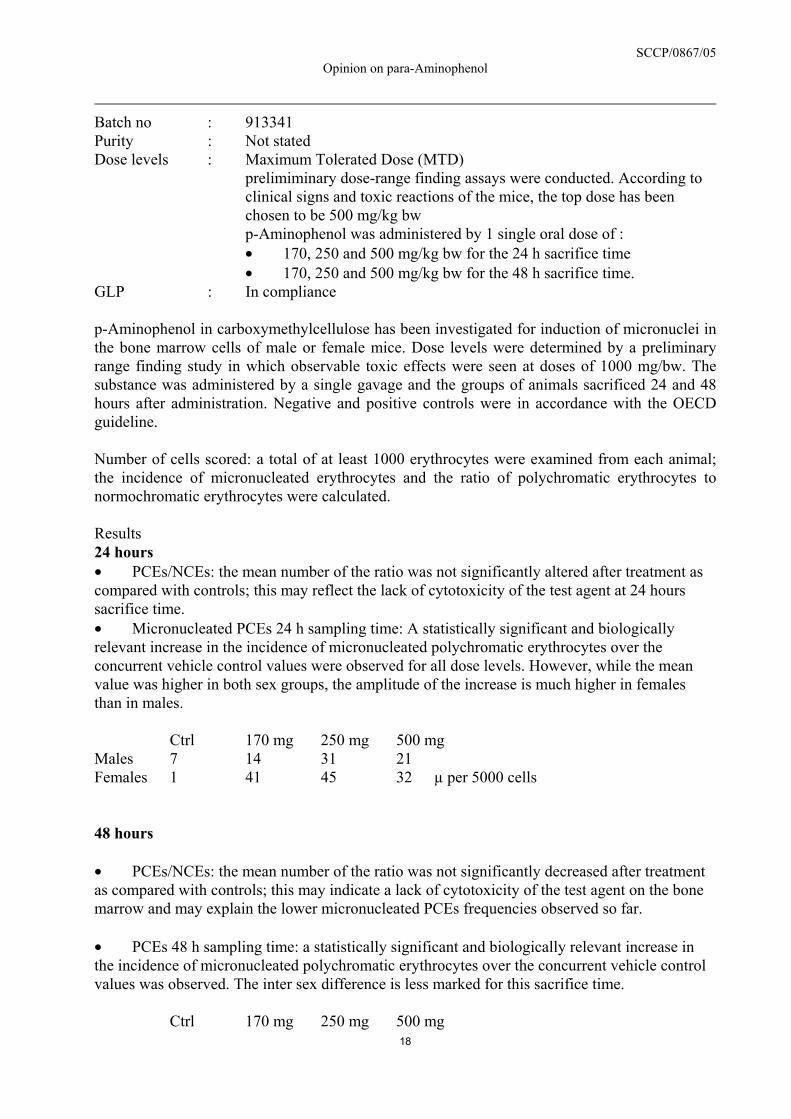
SCCP/0867/05 Opinion on para-Aminophenol
18
Batch no : 913341 Purity : Not stated Dose levels : Maximum Tolerated Dose (MTD) prelimiminary dose-range finding assays were conducted. According to
clinical signs and toxic reactions of the mice, the top dose has been chosen to be 500 mg/kg bw
p-Aminophenol was administered by 1 single oral dose of : • 170, 250 and 500 mg/kg bw for the 24 h sacrifice time • 170, 250 and 500 mg/kg bw for the 48 h sacrifice time.
GLP : In compliance p-Aminophenol in carboxymethylcellulose has been investigated for induction of micronuclei in the bone marrow cells of male or female mice. Dose levels were determined by a preliminary range finding study in which observable toxic effects were seen at doses of 1000 mg/bw. The substance was administered by a single gavage and the groups of animals sacrificed 24 and 48 hours after administration. Negative and positive controls were in accordance with the OECD guideline. Number of cells scored: a total of at least 1000 erythrocytes were examined from each animal; the incidence of micronucleated erythrocytes and the ratio of polychromatic erythrocytes to normochromatic erythrocytes were calculated. Results 24 hours • PCEs/NCEs: the mean number of the ratio was not significantly altered after treatment as compared with controls; this may reflect the lack of cytotoxicity of the test agent at 24 hours sacrifice time. • Micronucleated PCEs 24 h sampling time: A statistically significant and biologically relevant increase in the incidence of micronucleated polychromatic erythrocytes over the concurrent vehicle control values were observed for all dose levels. However, while the mean value was higher in both sex groups, the amplitude of the increase is much higher in females than in males.
Ctrl 170 mg 250 mg 500 mg Males 7 14 31 21 Females 1 41 45 32 µ per 5000 cells 48 hours • PCEs/NCEs: the mean number of the ratio was not significantly decreased after treatment as compared with controls; this may indicate a lack of cytotoxicity of the test agent on the bone marrow and may explain the lower micronucleated PCEs frequencies observed so far. • PCEs 48 h sampling time: a statistically significant and biologically relevant increase in the incidence of micronucleated polychromatic erythrocytes over the concurrent vehicle control values was observed. The inter sex difference is less marked for this sacrifice time.
Ctrl 170 mg 250 mg 500 mg

SCCP/0867/05 Opinion on para-Aminophenol
19
Males 7 4 6 10 Females 3 6 7 11 µ per 5000 cells Conclusions • Under the conditions of the test it can be concluded that p-Aminophenol at doses at which some signs of clinical toxicity were recorded, induces statistically significant increase in the frequency of micronucleated PCEs. • Therefore, p-Aminophenol is considered clastogenic and/or aneugenic in this mouse bone marrow micronucleus test.
Ref.: 88 In vivo Micronucleus Test using mouse hepatocytes The test indicates in vivo induction of micronuclei in liver cells and thus provides supportive evidence for a genotoxic potential of para-Aminophenol in vivo. However, it is considered unacceptable for evaluation due to the following reasons: • purity and batch not given; • no concurrent positive control was included; • the assays were not conducted in compliance with GLP or OECD guidelines • the data are issued from a published paper and not from a testing report. The paper aims to give insight on the absence of micronucleus formation in bone marrow when labile and/or short lived intermediates are produced via hepatic metabolism. It uses compounds rather to demonstrate the applicability of this technique using hepatocytes than to determine the safety of a given compound.
Ref.: 89 in vivo CD1 mouse splenocyte micronucleus test Positive results have been found with a maximum 14 days after treatment, providing supportive evidence for a genotoxic potential of para-Aminophenol in vivo. However, the test is considered unacceptable for evaluation due to the following reasons: purity and batch not given, no concurrent positive control was included, the assays were not conducted in compliance with GLP or OECD guidelines. The data are issued from a published paper and not from a testing report. The paper aims to give insight on the absence of micronucleus formation in bone marrow when labile and/or short lived intermediates are produced via hepatic metabolism. It uses compounds rather to demonstrate the applicability of this technique using splenocytes than to determine the safety of a given compound.
Ref.: 90 5. Micronucleus test by oral route (gavage) in rats for 13 weeks.

SCCP/0867/05 Opinion on para-Aminophenol
20
Guideline : OECD 408 Species : Sprague Dawley rats Group sizes : 10 males and 10 females Material : p-Aminophenol in 0.5 % carboxymethylcellulose Batch no : 2070155 Purity : Not stated Dose levels : Maximum Tolerated Dose (MTD) The doses were determined by the sponsor
p-Aminophenol was administered daily by oral gavage at doses of 12 and 30 mg/kg bw for 13 weeks.
GLP : In compliance p-Aminophenol in carboxymethylcellulose has been investigated for induction of micronuclei in the bone marrow cells of male and female rats. Dose levels were determined by the sponsor. The substance was administered by a single intragastric gavage during 13 weeks. No mention is made regarding concurrent negative and positive controls. Number of cells scored: a total of at least 1000 erythrocytes were examined from each animal; the incidence of micronucleated erythrocytes and the ratio of polychromatic erythrocytes to normochromatic erythrocytes were calculated. Results • PCEs/NCEs: the mean number of the ratio was not significantly altered after 13 weeks treatment as compared to controls. • Micronucleated PCEs: no statistically significant and biologically relevant increase in the incidence of micronucleated polychromatic erythrocytes over the concurrent vehicle control values were observed in both sexes given 12 or 30 mg/kg bw. Conclusions • Under the conditions of the test it can be concluded that p-Aminophenol at doses at which no signs of clinical toxicity were recorded, does not induce a statistically significant increase in the frequency of micronucleated PCEs. • Therefore, p-Aminophenol is not considered clastogenic and/or aneugenic in this rat bone-marrow micronucleus test.
Ref.: 91 Since this protocol only allows the application of a lower dose compared with a single application, it may be less suited to detect the mutagenic potential of a test substance. However, the results might be useful in the context of risk estimation. Mammalian Bone Marrow Chromosomal Aberration Test

SCCP/0867/05 Opinion on para-Aminophenol
21
Guideline : 475 Species : Wistar rats Group sizes : 5 males and 5 females Material : p-Aminophenol in 0.5 % carboxymethylcellulose Batch no : S190611; Lot 99 B 197 Purity : 99.9 % Dose levels : Maximum Tolerated Dose (MTD)
The MTD was determined by preliminary studies; it has been calculated to be 800 mg/kg bw p-Aminophenol was administered by 1 single oral dose of 200, 400 and 800 mg/kg bw. Animals were scarified 24 hours post treatment for all dosage groups while only the top dose has been selected for a 48h post treatment evaluation.
GLP : In compliance p-Aminophenol in carboxymethylcellulose has been investigated for induction of chromosomal aberrations in the bone marrow cells of male or female rats. Dose levels were determined by a preliminary dose range finding study. The substance was administered by a single intragastric gavage. Sacrifice was performed 24 h (all groups) or 48 h (top dose group) after administration. Number of cells scored: a total of at least 100 metaphases were examined from each animal; only cells with a modal number of chromosomes (n = 42 ± 2) have been taken into account. Mitotic Index has been determined on 1000 cells. Results • Under the conditions of the test it can be concluded that p-Aminophenol at doses at which signs of clinical toxicity and some deaths were recorded, does not induce a biologically relevant increase in the frequency of aberrant cells. • The statistically significant increase observed at 800 mg/kg at 24 h harvest time might be considered as devoid of biological significance due to the fact that the total frequency of aberrant metaphases is similar to that seen in general. The low baseline frequency (0.1%) may be the reason for the statistical significance observed. Conclusions This test revealed an equivocal result. An isolated positive effect at the highest dose which also induced toxicity and lethality was reported. The biological significance of this effect has to be questioned.
Ref.: 92 3.3.7. Carcinogenicity In vitro cell transformation

SCCP/0867/05 Opinion on para-Aminophenol
22
p-Aminophenol has induced morphological transformation of Syrian hamster embryo cells (A), but did not transform the C3H/10T1/2 mice cell line (B). Animal studies Guideline : OECD Guideline 451 Species/strain : Sprague-Dawley rats Test substance : p-aminophenol suspended in 0.5 w/w aqueous carboxymethylcellulose Batch : 2070155, purity stated 99% ± 0.5% Concentrations : 2, 5, 12 and 30 mg/kg/d administered daily by gavage GLP : in compliance Sprague-Dawley rats (approximately 6 weeks old), groups of 50 males and 50 females received p-aminophenol daily by gavage, at dose levels of 0, 2, 5, 12 and 30 mg/kg/d for at least 101 weeks. The dose levels selected on the basis of results of a previous 13-week toxicity study where daily oral doses of 30 and 100 mg/kg/d produced minimal to marked tubular nephrosis. The dose level of 30 mg/kg/d was considered to be the MTD. The test substance was administered as a suspension in 0.5% w/w aqueous carboxymethylcellulose. The study was scheduled for at least 104 weeks, however, since the survival in the control groups was only 26% in males and 32% in females in week 101, it was decided to terminate the study and all surviving animals were killed. Representative organs were weighed and the animal submitted to microscopic necropsy observations. No major difference from the controls was noted in the mean bodyweight of treated males and females or in the survival except for a lower survival of the high dosed females (20% compared to 32% at week 101). Orange coloured urine was noted from week 10 in almost all the animals given 30 mg/kg/d. The number of animals with more than one primary neoplasm and the number of benign and malignant tumours were comparatively similar in all groups including the control group, except for a marginal increase in the number of malignant lymphoma in males given 30 mg/kg/d (3 cases of heterog. mal. lymphoma compared to 1 in the control group and 1 in the low dose group). It was concluded that the test substance showed neither a carcinogenic potential nor an effect on the incident of spontaneously occurring tumours at any dose level.
Ref.: Col. 93 Dermal study A two-generation reproduction study, which included a chronic toxicity-carcinogenicity study, was conducted with Sprague-Dawley rats receiving topical applications of six oxidative hair- colouring formulations. p-Aminophenol (1.0%) was present together with 2,5-diaminotoluene sulphate (6.0%) in one of the formulations tested. Each formulation was mixed with an equal volume of 6% hydrogen peroxide prior to application. In the reproduction study, p-aminophenol at a concentration of 1% in the formulation was applied twice weekly throughout the growth, mating, gestation and lactation phases of the F0 parents to the weaning of the F1a and F2b litters. Weanlings selected from the F1a litters were the subjects for the lifetime carcinogenesis study. For 24 months, they received topical administrations of the formulation containing p-aminophenol twice weekly.

SCCP/0867/05 Opinion on para-Aminophenol
23
Five animals/sex/group were killed at 12 months; the remainder of the animals were killed at 24 months. All were necropsied; their tissues were subjected to histological evaluation. No compound-related increases in neoplasms were observed. It is noted that 2,4-diaminoanisole (2 and 4%) were also negative in the experiments.
Ref: Col. 72 Mice Swiss Webster mice (8 – 10 weeks old), groups of 50 males and 50 females, were painted weekly for 21 or 23 months. The study consisted of 12 treatment groups and 3 negative control groups. Nine oxidative hair dye formulations and 3 semipermanent hair dye formulations were studied. The 9 oxidative hair dye formulations were mixed with an equal volume of 6% hydrogen peroxide just before use and applied within 15 min after mixing. A 0.05 ml sample was used for application. p-Aminophenol was present in 3 of the oxidative hair dye formulations in concentrations of 0.04, 0.2 and 1.0%. Animals found dead or sacrificed in moribund conditions or at termination of the study were necropsied and evaluated histopathologically. Comparison of incidence of tumours and of non-tumour pathology among the various treatment and control groups revealed no biologically significant differences. The authors state that toxicological and carcinogenic effects were not induced by the hair dye formulations. It is noted that 2,4-diaminoanisole (2% and 4%) was also negative in the experiments.
Ref.: Col. 8 The composition of the formulations used was given in a separate publication.
Ref.: Col. 7 Swiss mice (8 weeks old), groups of 60 males and 60 females, were painted weekly in the case of 2 oxidative and 3 times weekly in the case of 12 non-oxidative hair dye formulations for 20 months. Aliquots of 0.05 ml were delivered to an area of skin in the interscapular region. The mice were shaved 24 hours before treatment as needed. Two control groups were shaved only and received no treatments. The oxidative dye solutions were mixed with an equal volume of 6% H2O2 just prior to application. One of the oxidative hair dye formulations contained 1.5% p-aminophenol. A gross necropsy was performed on all mice. The application of hair dyes did not have an adverse effect on average body weight gains or survival of any group. Body weights were not depressed more than 10% in any group compared with the controls. No unusual tumours developed in any of the groups. Significant increases in malignant lymphomas over those in control group 2 (12%; 7/60) were observed in 3 treated groups of females (p-aminophenol group [32%; 19/60], a non-oxidative hair dye group [30%; 18/60], a non-oxidative hair dye group [38%; 23/60]. The authors state that the observed increases were possibly due to a low control value in control group 2. The percentage of animals with malignant lymphomas in control group 1 was 22% [13/60]. Moreover, the average in 3 previous control groups was 33%. Otherwise, no increased tumour frequencies were found. It is noted that both the oxidative hair dye formulations contained p-phenylenediamine and one contained 4-amino-2-nitrophenol. Two of the non-oxidative hair dye formulation contained Disperse Blue 1.
Ref: Jacobs et al., 1984 Comments

SCCP/0867/05 Opinion on para-Aminophenol
24
An oral rat study with p-aminophenol has been performed according to OECD Guideline No. 451. The mortality in the control group was very high. The study was negative. One dermal study with p-aminophenol has been performed with rats and 2 dermal studies have been performed with mice. They were all negative. It should be noted that several hair dye formulations were tested in the dermal studies and some of the formulations contained substances classified as carcinogenic either by EU or the German MAK commission (2,4-diaminoanisole, EU carcinogen category 2; Disperse Blue 1, EU carcinogen category 2; p-phenylenediamine, MAK commission carcinogen category 3B; 4-amino-2-nitrophenol, MAK commission carcinogen category 3B). This may indicate that the sensitivity of the dermal carcinogenicity studies has not been sufficient to identify possible carcinogenic effects of hair dye formulations. Charles River rats from the first litter of a multigeneration reproduction study received topical applications of formulations containing 0.04, 0.2 or 1.0% PAP mixed with 6% hydrogen peroxide twice weekly for two years. No compound related changes were observed.
Ref.: C48 Additional references:
Jacobs MM, Burnett CM, Penicnak AJ, Herrera JA, Morris WE, Shubik P, Apaja M, Granroth G. Evaluation of the toxicity and carcinogenicity of hair dyes in Swiss mice. Drug Chem Toxicol 7(6): 573-586, 1984.
A Pienta R, Kawalek J. Transformation of hamster embryo cells by aromatic amines. Natl Cancer Inst Monogr 58: 243-251, 1981.
B Patierno SR, Lehman L, Henderson BE, Landolph JR. Study of the ability of phenacetin , acetaminophen, and aspirin to induce cytoxicity, mutation, and morphological transformation in C3H/10/T1/2 clone 8 mouse embryo cells. Cancer Res 49: 1038-1044, 1989.
3.3.8. Reproductive toxicity In Vitro Reproductive Toxicology In an in vitro/in vivo Hen’s egg test, 20 ng to 50 mg of PAP dissolved in egg albumen were onto the egg’s chorion allantois membrane during preincubation or on the fifth day of incubation. All nonviable embryos and hatched chicks were examined for gross abnormalities and other signs of toxicity. PAP induced dose-related mortality, with a median lethal dose of 18.6 mg/egg (~170 ppm) on day 1 and 10.5 mg/egg (~20 ppm) on day 5. Developmental retardation and significant variations in blood chemistry were observed up to 25 mg/egg. Hatched chicks had dose-dependent increases in absolute and relative heart weights. The No Effect Level for the study was considered to be between 1 and 5 ppm.
Ref.: C27 Peri/postnatal reproductive toxicity

SCCP/0867/05 Opinion on para-Aminophenol
25
A single dose of 0, 100, 333, 667, or 1000 mg/kg PAP was administered by oral gavage to pregnant female Sprague-Dawley rats on day 11 of gestation. Maternal body weight was significantly reduced after 24 and 72 hours in animals receiving 667 or 1000 mg/kg PAP. Associated with this maternal toxicity were perinatal loss at 1000 mg/kg, decreased pup weight on days 1 and 6 postpartum, and tail abnormalities and/or paralysis of the hind limbs in 50% of the pups at 667 mg/kg and 37.5% of the surviving pups at 1000 mg/kg.
Ref.: Col. 71 3.3.8.1. Two generation reproduction toxicity Multi-generation reproduction toxicity A multi-generation reproduction study was performed using three oxidative hair dye formulations, one of which was a mixture of 1.0% PAP and 0.7% meta-aminophenol (MAP). Freshly prepared formulations were mixed with equal volumes of 6% hydrogen peroxide and applied twice weekly to the clipped back and neck areas of groups of 40 male and 40 female Charles River CD rats. The initial dose was 0.2 ml per application; this was increased incrementally by 0.1 ml weekly to a dose of 0.5 ml per application. Three control groups were clipped regularly but received no other treatment. Treatment was administered through the growth, mating, gestation and lactation periods through the weaning of the F1B, F2B, and F3C litters of the respective generations. Selected animals from the F1B and F2B litters were used as parents for the F2 and F3 generations, respectively. There were no compound-related findings observed in any generation of the study.
Ref.: C48 In a combination reproduction/chronic toxicity/carcinogenicity study in Sprague-Dawley rats, PAP mixed with hydrogen peroxide (final concentration: 1% PAP) was applied topically twice weekly throughout the growth, mating, gestation and lactation phases of the F0 parents to the weaning of the F1a and F2b litters. Fertility, gestation, foetal viability indices, and foetal body weights were evaluated and compared with controls. No adverse effects on fertility of males or females, or on gestation, lactation or weaning indices were observed.
Ref.: Col. 72 3.3.8.2. Teratogenicity Groups of pregnant female Charles River CD rats were treated with topical applications of three hair dye formulations containing 0.04%, 0.2%, or 1% of PAP on days 1, 4, 7, 10, 13, 16, and 19 of gestation. The formulations were mixed 1:1 with 6% hydrogen peroxide just prior to application to mimic normal use. No compound-related effects were observed in this study.
Ref.: C7 PAP dissolved in distilled water was administered by gavage at doses of 0 (vehicle control), 25, 85, or 250 mg/kg/day to pregnant female Sprague-Dawley rats from day 6 to day 15 of gestation. Vitamin A (15 mg/kg/day) was administered by gavage as a positive control. A reduction in body weight gain was associated with skeletal malformations, anophthalmia and hydrocephalus at 250 mg/kg/day. Reduced body weight gain was also observed at 85 mg/kg/day, but no

SCCP/0867/05 Opinion on para-Aminophenol
26
embryotoxic or teratogenic effects were observed at this dose level. The No Effect Level of the study was 25 mg/kg/day.
Ref.: C38 Syrian Golden hamsters were treated with PAP in acidified isotonic saline on day 8 of gestation using three different routes of administration. Animals treated using intraperitoneal injection received a single dose of 100, 150, or 200 mg/kg body weight. Those treated using intravenous injection received a single dose of 100, 150, 200, or 250 mg/kg body weight. Hamsters treated using oral gavage were administered 100 or 200 mg/kg bw. Control animals received saline alone. A comparison was made between the administration of a fresh PAP solution and solutions made 1, 2, or 4 weeks prior to administration. No difference was observed related to the age of the solution administered. Animals treated using intraperitoneal or intravenous injection were observed to have a significant increase in the frequency of litters with one or more malformed foetuses. No developmental anomalies were observed in litters from animals treated orally.
Ref.: C41 25 female Sprague-Dawley rats received concentrations of 0.07, 0.2, or 0.7% PAP in the diet for 13 weeks (corresponding to a daily intake of approximately 35, 100 or 350 mg/kg/day). They were then mated with untreated males. Pregnant females were once again fed the PAP-containing diet until day 20 of gestation, when they were killed. On day 0 of gestation, the body weights of animals in the 0.2 and 0.7% dose groups were lower than those of controls. From day 0 to day 20 of gestation, animals in the 0.7% dose group had significantly reduced body weight gain. Dose-related postimplantation loss was observed at 0.2 and 0.7%; this was significant at the dose level of 0.7%. No teratologic effects were observed, but some skeletal variations secondary to maternal toxicity were seen at 0.2 and 0.7% PAP.
Ref.: Col. 70 Conclusion on developmental and reproductive toxicity Despite not all studies are in compliance with acknowledged methodology it can be stated that PAP showed no effects on fertility or on gestation, embryonic development, lactation or weanling indices in experimental animals. Teratogenic effects were observed in one study, but only at maternal toxic doses. 3.3.9. Toxicokinetics In vitro metabolism The rates of secretion of PAP and its sulphate and glucuronide conjugates were determined in cultures of rat hepatocytes, using aniline and PAP as substrates. When hepatocytes were incubated with inorganic sulphate and 1 mM aniline, secretion of PAP or its conjugates was linear over a period of two hours. With PAP as the substrate (concentration not indicated), free PAP disappeared from the medium almost completely within 30 minutes. The secretion of conjugates was linear for 30 minutes only. There was a characteristic lag in glucuronide secretion.
Ref.: C16

SCCP/0867/05 Opinion on para-Aminophenol
27
Glucuronidation in cultured human skin epithelial cells, human skin fibroblasts, or homogenates from these cells was examined using 0.2 - 0.5 mM PAP mixed with ascorbic acid (to prevent auto-oxidation). Epithelial cells and the homogenate from these cells glucuronidated PAP at a rate of about 5 nmol/mg cell protein per hour. Skin fibroblasts did not glucuronidate PAP.
Ref.: C 40 A clonal strain of rat hepatoma cells (MH1C1) was used to study the metabolism of PAP in vitro. Concentrations of PAP ranging from 0.25 mM to 0.75 mM were incubated with hepatoma cells for up to 5 hours. The PAP-glucuronide rate vs. time curve did not reach linearity due to a lag which increased with increasing substrate concentrations. The highest rate of PAP-glucuronide formation was observed at a concentration of 0.5 mM, with a decrease observed at higher concentrations. This indicated the possibility of substrate inhibition of the glucuronidation process.
Ref.: A1 The metabolism of PAP in pulmonary and renal microsomes from the rat, rabbit and mouse was evaluated in the presence of another aniline metabolite. Results indicated that PAP (concentration not indicated) was not metabolized by these tissue preparations from any species.
Ref.: Col. 76 The metabolic reactivity (measured by covalent binding) of 60 µM PAP to microsomal proteins in human, rat, and mouse livers was compared to that of 1mM paracetamol (APAP) in vitro. PAP was observed to have higher intrinsic reactivity than APAP, regardless of the species considered. Biotransformation of both substances occurs via the cytochrome P450 system, with the exception of only 50% involvement of P450 in the transformation of PAP in the mouse and no involvement of P450 in the human biotransformation of this compound. Based on the results of this study, the sensitivity to the potential electrophile generation and alkylation of PAP is greatest in the mouse, intermediate in the rat, and least in the human.
Ref.: Col 77 The in vitro metabolism of 100 µM PAP by phase I enzymes in the presence of human, rat and mouse hepatic microsomes was evaluated. Incubation with glutathione was performed to identify electrophilic metabolites. Six principal metabolites were isolated – two were common to all three species, one was common to the rat and the mouse and one was shared by the rat and the human. One metabolite was specific to the rat and one to the mouse. Quantitatively, the biotransformation of PAP over a 15 minute period was most extensive in the human, intermediate in the rat, and least extensive in the mouse. The metabolites specific to the rat and mouse were electrophilic in nature, while those shared by all three species and the one common to the rat and human were not.
Ref.: Col. 78 The interspecies comparison of the in vitro metabolism of 100 µM PAP in the presence of human, rat, and mouse hepatocytes was evaluated. Four metabolites, common to all three species, were identified as glucuronides and sulphates of PAP and of paracetamol (APAP). A fifth metabolite, APAP, was identified only in human hepatocytes. These metabolites demonstrate that PAP is metabolized by phase II enzyme reactions. Biotransformation was observed to be most extensive in mouse hepatocytes, and less extensive, but comparable, in rat and human hepatocytes. Covalent binding (evidence of phase I metabolism) occurred in the same
SCCP/0867/05 Opinion on para-Aminophenol
28
pattern among the three species evaluated. Mouse hepatocytes were observed to have a deficiency in sulfotransferase activity compared to rat and human hepatocytes, as well as a greater capacity to form reactive electrophilic metabolites.
Ref.: Col. 79 The metabolism of 100 µM PAP was studied in vitro using the reconstructed human skin model, Episkin. The results indicated that the epidermis transforms PAP into paracetamol (APAP) via N-acetylation. No other metabolites were found. The biotransformation was maximal after 24 hours of contact. The formation of reactive electrophiles, measured by the level of covalent binding to tissue proteins, was low (0.3 ± 0.02 nmol/mg); PAP and APAP were equally involved in covalent binding.
Ref.: Col. 80 PAP and 14C-PAP were tested in the Episkin model of reconstructed human epidermis at a concentration of 100 µM for a 24-hour period determine the capacity of the epidermis to metabolize the compound. Episkin kits were obtained from a commercial supplier. After exposure of the cultures to the test compound, the culture medium was decanted and protected from auto-oxidation by the addition of ascorbic acid. The surface of the skin cultures was rinsed in PBS containing ascorbic acid. The medium, PBS rinse, epidermal cells and their collagen matrices were then frozen in liquid nitrogen and preserved until they were to be analyzed. Aliquots of culture medium were prepared for evaluation of the level of radioactivity and chromatographic analysis. The collagen matrices were extracted with methanol, and these extracts were evaluated for radioactivity. The epidermal cells were homogenized in PBS; aliquots were taken for chromatographic analysis and for determination of the levels of radioactivity, of protein inextractable radioactivity and covalent binding. The PBS rinse was also evaluated for radioactivity. The results indicated that the epidermis quantitatively transforms PAP into paracetamol (APAP) via N-acetylation (a phase II reaction). Virtually all the PAP applied to the skin culture (98%) was biotransformed. Only APAP was identified as a metabolite; no other metabolites were found. The biotransformation was virtually complete after 24 hours of contact. The formation of reactive electrophiles, measured by the level of covalent binding to tissue proteins, was low (0.3 ± 0.02 nmol/mg); indicating that Episkin has limited capacity to metabolize PAP using phase I enzymes. From the results of this study, it was concluded that exposure of Episkin to PAP results in complete biotransformation to APAP, leaving a negligible amount available for eventual penetration of the epidermis.
Ref.: Col. 80 In vivo metabolism Doses of 91 or 182 mg/kg aqueous PAP with Arabica gum were administered once orally to mice, and blood levels of PAP and its metabolites were evaluated 0.5, 1.0, 1.5, 2.0, and 2.5 hours after dosing. The following results were obtained:

SCCP/0867/05 Opinion on para-Aminophenol
29
Time after Dosing (hours) Dose (mg/kg) PAP
(µg/ml) 0.5 1.0 1.5 2.0 2.5
free 10 6 3 1 0.5 91 conjugate 14 9 4 3 0.5 total 24 15 7 4 1 free 18 10 6 3 1 182 conjugate 28 27 13 6 2 total 46 37 19 9 3 The nature of the conjugate was not identified.
Ref.: A2 Rabbits (2-3 kg in weight) were administered 1 g/animal PAP in aqueous solution by gavage, and urine levels of PAP and its metabolites were examined. The collection time(s) were not specified. 2% of the PAP was excreted unchanged. Metabolites consisted of 25% acetaminophenol, 8% aminophenylsulphate, 4% acetaminophenylsulphate, 45% aminophenylglucuronide, and 16% acetaminophenylglucuronide. Acetaminophenol and its conjugates were major metabolites of PAP. 100% of the administered compound was accounted for.
Ref.: A2 Rats (200 g) received a single dose of either 200 mg/kg glucuronic acid or 200 mg/kg glucuronamide by gavage, followed one hour later by a single gavage dose of 300 mg/kg PAP. Urine collected 24 hours later contained increased PAP and glucuronidated PAP compared to controls. Glucuronamide was found to be a more effective conjugating agent than was glucuronic acid.
Ref.: A2 In a human metabolism study, one subject received 200 mg and two others received 500 mg PAP-hydrochloride. The following results were obtained:
Subject PAP
(dose in mg)
PAP in Blood (mg/100 ml)
Unconjugated N-acetyl-PAP Total 0.5 hours 1 hour 0.5 hours 1 hour 0.5 hours 1 hour
1 200 0 0 – – 0.41 – 2 500 0.09 0.012 0.56 0.45 0.84 0.96 3 500 0.12 0.016 0.56 0.26 0.98 0.69
The route of administration was not indicated.
Ref.: A2 In vivo-metabolism, rat

SCCP/0867/05 Opinion on para-Aminophenol
30
The blood and plasma pharmacokinetics and metabolites of 14C-PAP were studied in vivo after a single cutaneous administration (15 mg/ml 14C-PAP) to female Wistar rats. Twelve (12) rats received the test substance in a single topical administration of 12.5 mg/kg (18.5 MBq/kg; 5 mg/cm²) over 12.5% of the body surface area for a total exposure period of 24 hours. During exposure, the application site was covered with an occlusive plastic film that was held in place using light bandages. Blood samples were collected from two rats per time point at 0.5, 1, 2, 4, 8, and 24 hours during the exposure period. Plasma samples were obtained from each blood sample. Blood and plasma samples were analyzed for total radioactivity, and selected plasma samples were analyzed for metabolic patterns by HPLC/UV radioactivity determination. Metabolites were identified by comparison of peak retention times with those of known standards before and after enzyme hydrolysis. The radioactivity in the plasma and blood samples increased from time zero to the Cmax (498 and 313 ng-eq/g, respectively) at 4 hours after application of PAP. It then decreased until the last quantifiable time point at 24 hours (the end of the exposure period), when it was found to be 139 ng-eq/g for plasma and 106 ng-eq/g for blood. The principal pharmacokinetic parameters (calculated using non-compartmental methods) are presented below:
Sample t1/2z
(h) λz
(1/h) Cmax
(ng-eq/g) tmax
(h) AUC0-24h
(ng-eq h/g) AUC0-∞
(ng-eq h/g) Plasma 5.95 0.0626 498 4 7038 9271 Blood 4.79 0.0523 313 4 4567 6782
The parent compound, PAP, was undetectable in all plasma samples. Three peaks were detected in the plasma, corresponding to APAP, the glucuronic acid conjugate of APAP (shortest retention time) and the sulphate conjugate of APAP (longest retention time) (see table below). The limit of quantification was 1.04 ng-eq/g in plasma and 0.99 ng-eq/g in blood.
Time (h)
Animal No. PAP (%)
Glucuro-APAP (%)
APAP (%)
Sulpho-APAP (%)
2 W20855 - - 30.0 70.0 W20856 - - 30.0 70.0 4 W20853 - - 45.0 55.0 W20854 - 13.4 27.6 59.0 8 W20851 - 17.7 35.4 46.9 W20852 - 11.7 39.8 48.5
From these results, it was concluded that PAP was not present in the plasma of rats during 24 hours of continuous exposure. All detectable radioactivity consisted of APAP and/or its metabolites. In vivo metabolism, mini-pig The blood and plasma pharmacokinetics and metabolites of 14C-PAP were studied in vivo after a single cutaneous administration (15 mg/ml 14C-PAP) to a single female Göttingen minipig. The pig received the test substance in a single topical administration of 4.7 mg/kg (18.5 MBq/kg; 5 mg/cm²) over 12.5% of the body surface area for a total exposure period of 24 hours. During exposure, the application site was covered with an occlusive plastic film that was held in place by self-adhesive bandages and a body stocking fitted with a collar. Blood samples were collected

SCCP/0867/05 Opinion on para-Aminophenol
31
pretreatment and at 0.5, 1, 2, 4, 6, 8, 12 and 24 hours during the exposure period. Additional samples were taken at 48 and 72 hours after application. Plasma was obtained from each blood sample. Blood and plasma samples were analyzed for total radioactivity, and selected plasma samples were analyzed for metabolic patterns by HPLC/UV radioactivity determination. Metabolites were identified by comparison of peak retention times with those of known standards. The radioactivity in the plasma and blood samples increased from time zero to the Cmax (11.70 and 9.23 ng-eq/g, respectively) at 12 hours after application of PAP. It then decreased until the last quantifiable time point at 72 hours post-application, when it was found to be 2.24 ng-eq/g for plasma and 2.80 ng-eq/g for blood. The limit of quantification was >1.78 ng-eq/g (7.0 Bq/g) in plasma and >1.25 ng-eq/g (4.9 Bq/g) in blood. The principal pharmacokinetic parameters (calculated using non-compartmental methods) are presented below:
Sample t1/2z
(h) λz
(1/h) Cmax
(ng-eq/g) tmax
(h) AUC0-24h
(ng-eq h/g) AUC0-∞
(ng-eq h/g) Plasma 31.3 0.021 11.70 12 389 490 Blood 53.6 0.013 9.23 12 350 566
A minor secondary peak, of unknown origin, was noted at 2 hours in both plasma and blood. The levels of radioactivity in the plasma were very low, and thus only a single interpretable metabolic pattern for the 6 hour time point could be obtained. The one peak that was observed upon analysis for metabolites in the plasma was identified as APAP. The parent compound, PAP, was undetectable in the plasma. From these results, it was concluded that PAP was not present in the plasma of the pig during 24 hours of continuous exposure. Only its metabolite, APAP could be detected.
Ref.: Col. 98 Conclusion The safety of a topically applied substance may be assessed by two principal methods: comparison of the calculated penetrated amount (systemic exposure dose; SED) with a NOAEL from a repeated-dose animal toxicity study yielding a nominal “margin of safety”. The accepted empirical margin of safety is generally 100-fold (Ref.: KM4). comparison of toxicokinetic, quantitative exposure data (plasma levels, area under curve) from animal toxicology studies with respective human data (Ref. KM5). Although the first approach permits an approximate estimation of a potential risk of a topically applied substance, it includes a number of uncertainty factors, such as statistical uncertainty of no-effect levels, unknown oral bioavailability of the test substance, unknown relation of percutaneous penetration to the actual systemic exposure of the organism or different inter-species pharmacokinetic / metabolic parameters. Therefore, the second, more precise approach is preferred and generally selected for the safety evaluation of human drugs and other substances for which human and animal kinetic and systemic exposure data are available (Ref KM5, Ref. KM8). The available evidence for PAP suggests that the substance is quantitatively metabolised in the skin to paracetamol (APAP) and that topical application of PAP results in systemic exposure to APAP and/or its metabolites, but not to PAP.

SCCP/0867/05 Opinion on para-Aminophenol
32
APAP is a widely used antipyretic and analgesic drug, inclusive for pediatric use. Given that human pharmacokinetic data for APAP are available in the literature it seems reasonable to compare systemic exposure data from topically applied PAP to known values of APAP, in order to arrive at a toxicokinetic-based margin of safety. As previously described, 14C-PAP was topically applied to 12.5% of the body surface of rats and a pig. The resulting kinetics of the blood/plasma concentration in rats (14C-equivalents) is shown in the following figure: Figure 1. Blood concentration (14C-equivalents) in rats treated topically (12.5% of the body surface) for 24 hours with a single dose 12.5 mg/kg 14C-PAP. The area under the curve from 0 to 12 hours reflecting the quantitative systemic exposure of the organism to APAP and/or its metabolites during this time period was calculated to be AUC0-12hrs = 1.09 µg/ml x hours (Ref. KM3).
0 4 8 12 16 20 240.05
0.10
0.15
0.20
(hours after application)
PAP
14C
EQ
s. µ
g/m
l

SCCP/0867/05 Opinion on para-Aminophenol
33
The following figure shows the corresponding value for the pig treated topically with 14C-PAP: Figure 2. Blood concentration (14C-equivalents) in a minipig treated topically (12.5% of its body surface) for 24 hours with a single dose 4.7 mg/kg 14C-PAP.
The area under the curve from 0 to 12 hours reflecting the quantitative systemic exposure of the organism to APAP during this time period was calculated to be AUC0-12hrs = 0.07 µg/ml x hours (Ref. KM3).
0 4 8 1 2 1 6 2 0 2 4 2 8 3 2 3 6 4 0 4 4 4 80
2
4
6
8
1 0
1 2
(h o u rs a fte r a p p lica tio n )
PAP
14C
-EQ
s. (n
g/m
l)

SCCP/0867/05 Opinion on para-Aminophenol
34
Blood levels of paracetamol (APAP) in humans after oral administration are well documented. Typical values are shown in the following figure: Figure 3. Blood concentrations in human volunteers after oral administration of a single dose of 500 mg APAP (2). The area under the curve from 0 to 12 hours reflects the quantitative systemic exposure of the human organism during this time period to APAP and was calculated to be AUC0-12hrs = 28.8 µg/ml x hours (Ref. KM2, Ref. KM3). Oral administration of 500 milligrams of APAP is recognised to be a safe human dose (Ref. KM1). APAP has been shown to be non-genotoxic in humans who received the maximum therapeutic dose of 3x1000 mg over 8 hours (Ref. KM9). Given that topically applied PAP results in systemic exposure to APAP and/or its metabolites only in both humans and animals, the systemic exposure seen in animal studies may be compared to the systemic human exposure after a single oral dose of the drug APAP (= AUCman): • On the basis of the blood values found in rats, the resulting margin would be the AUCman /
AUCrat, i.e. 28.8 (man) / 1.09 (rat) = 26-fold. • On the basis of the blood values in the pig, the margin would be the AUCman / AUCpig, i.e.
28.8 (man) / 0.07 (pig) = 410-fold. The recommended human therapeutic regimen of APAP consists of multiple oral doses, i.e. 500 mg every four to six hours (Ref. KM1). On the basis of a therapeutic regimen of four doses of 500 mg APAP within 24 hours, the actual safety margins may be estimated to be several times higher than the nominal values calculated above. A systemic exposure ratio of 25 (animal vs. human exposure) is considered to represent a very high margin of safety for human drugs: a dose resulting in a 25-fold systemic exposure in
0 2 4 6 8 1 0 1 20 .0
2 .5
5 .0
7 .5
1 0 .0
H o u rs a fte r a d m in is tra t io n
(µg
/ ml)

SCCP/0867/05 Opinion on para-Aminophenol
35
animals when compared to the therapeutic human exposure is considered to be a maximum tolerated dose (Ref. KM6). In the recent EU report on the harmonisation of risk assessment procedures, an inter-species ratio of 10-fold (4.0-fold for kinetics, 2.5-fold for dynamics) was suggested to be an adequate margin of safety, since partitioning of xenobiotic chemicals between blood and tissues is not a major cause of inter-species differences or human variability (Ref. KM8). In summary, the above toxicokinetic-based safety margins (26- to 400-fold) suggest a very high degree of safety. In addition, the topical exposure conditions used in the rat and pig studies were far more severe than those expected for humans during a hair dyeing process. For example, occlusion alone is known to increase percutaneous penetration in animals or man by 2- to 10-fold, respectively (Ref. KM7). Other conditions in the animal studies which may be expected to increase the systemic availability of PAP include the exposure time (24 hours), the large body surface area treated (12.5%) and the absence of hair on the treated skin surface of the animals. Exposure parameters are compared in the following table: Table 1.: Comparison of the in vivo systemic exposure to APAP in humans (following topical
exposure to PAP during hair dyeing) with that found in the pig and rat after topical exposure to PAP.
PARAMETER MAN
(hair dyeing process) PIG
(topical toxicokinetic / metabolism study)
RAT (topical toxicokinetic /
metabolism study) APPLIED DOSE of PAP (mg/kg) <0.26 4.7 12.5 TREATED SURFACE (cm²) 800 (scalp) 500 30 % of BODY SURFACE AREA 4.5 12.5 12.5 EXPOSURE TIME (hrs)
0.5 24 24
OCCLUSION no yes yes ABSORPTION TO HAIR yes noa noa SYSTEMIC EXPOSURE APAP, AUC0-12h, (µg/ml x hours)
negligible 0.07 1.09
a 14C-PAP was applied to shaved skin Therefore, actual human systemic exposure during a hair dyeing process may be estimated to be only a fraction of the values measured in the animal models. Given that the toxicokinetic-based safety margins were very large even under the exaggerated exposure conditions used in the rat and pig studies and taking into account that human exposure to PAP during a hair dyeing process is intermittent (max. every four to six weeks), the in vitro and in vivo experimental evidence cited above suggest that human exposure to PAP or APAP when applied to the head in an oxidative hair dye is negligible. 3.3.10. Photo-induced toxicity / 3.3.11. Human data
SCCP/0867/05 Opinion on para-Aminophenol
36
/ 3.3.12. Special investigations Induction of Methemoglobinemia Human foetal and adult haemoglobin samples were incubated in 0.01 M Bis-Tris buffer in the presence of 0.5, 1, or 2M PAP per mole of haemoglobin tetramer. Under the conditions of this study, human foetal haemoglobin was more susceptible to methemoglobin formation than was adult methemoglobin.
Ref.: C52 PAP (0.5 mM in 0.2 M phosphate buffer) was found to produce methemoglobin more rapidly in purified human haemoglobin than in washed red blood cells. The investigators stated their belief that PAP binds covalently to haemoglobin.
Ref.: C15 Japanese quail received a single intraperitoneal injection of 1, 5, 10, 25, or 50 mg/kg PAP dissolved in water. The highest observed concentration of methemoglobin (9%) was observed after administration of 50 mg/kg PAP; no PAP was detectable in the blood after 30 minutes.
Ref.: C3 Several studies have examined the formation of methemoglobin after administration of PAP. A median lethal dose value of 470 mg/kg was obtained in a subcutaneous study in mice. A subcutaneous dose of 37 mg/kg PAP caused severe clinical signs and methemoglobinemia in cats, with death occurring within 30 minutes. The same dose caused similar observations in dogs, but no death; in rabbits, this dose produced no methemoglobin formation. In a separate study in cats, subcutaneous administration of 6 mg/kg induced a level of 37.5% methemoglobin in the blood 3.5 hours after exposure. Normal and glucose-6-phosphate (G-6-P)–deficient human erythrocytes (35% suspension in isotonic saline, pH 7.4) were incubated with 0.001 M PAP for two hours at 37° C. While the concentration of glutathione was not altered in G-6-P–deficient cells, red blood cell fragility and methemoglobinemia were significantly increased. PAP was added to freshly drawn human blood in varying concentrations and the amount of methemoglobin generated was measured. At 0.1 and 0.5 mg%, no methemoglobin was detected. At 1 mg%, one blood sample was found to contain 3% methemoglobin, but another contained none. At 5 and 10 mg%, between 13.2% and 21.2% methemoglobin was found in all samples.
Ref.: A2 Melanocytotoxicity C57B1/6J black mice (7-days or 5-weeks of age) were injected with a single dose of PAP to determine whether or not it would induce depigmentation. PAP dissolved in water was injected subcutaneously at a dose of 400 mg/kg or intraperitoneally at a dose of 50, 100, 200, or 400 mg/kg. Dose-related depigmentation was observed in mice injected intraperitoneally. Melanocytotoxicity was observed at 12, 24, 48, and 96 hours post administration in histopathologic preparations from mice injected subcutaneously.
Ref.: C5

SCCP/0867/05 Opinion on para-Aminophenol
37
Nephrotoxicity Male Fischer 344 rats were administered subcutaneous injections of 25, 50, 100, or 200 mg/kg in water. Urine was collected for 24 hours after compound administration. Animals were killed 24 or 48 hours after compound administration and blood was collected for analysis for alanine aminotransferase (ALT) and blood urea nitrogen (BUN). The kidneys were collected and thin cortical slices were incubated in the presence of p-Aminohippurate (PAH) and 14C Tetraethylammonium (TEA). The accumulation of these substances was evaluated. Changes in renal function were observed: BUN was detected and accumulation of PAH and TEA was reduced in renal slices. No effect on hepatic function was noted.
Ref.: C32, C33 The nephrotoxicity of PAP was evaluated in male Fischer 344 rats. Three groups of rats received 0, 150, or 300 mg/kg PAP via intraperitoneal (IP) injection 30 minutes after pretreatment with bis (p-nitro-phenyl) phosphate or water (100 mg/kg) IP. Urine was collected for 24 hours, after which time the rats were killed. Blood was collected for measurement of blood urea nitrogen, kidneys were prepared for histopathology, and urine was evaluated for metabolites. PAP caused tubular epithelial necrosis of the kidneys and changes in renal function. It was excreted in the urine as free or conjugated PAP or as acetaminophen.
Ref.: C 34 The influence of metabolic variation on PAP toxicity was examined in homozygous and heterozygous male Gunn rats as well as albino rats. 0.5 or 1 mM/kg was administered intravenously to rats (tail vein), with controls receiving an injection of normal saline. Rats were killed 48 hours after compound administration. Tubular necrosis was observed in all treated rats, with more severe lesions occurring in the Gunn rats.
Ref.: C6 Morphological and functional studies were performed with PAP in male Sprague-Dawley rats. In the first study, a single intravenous injection of 400 mg/kg PAP in saline was administered and rats were killed 30, 60, or 90 minutes, or 2 hours after this administration. At necropsy, the kidneys were dark and swollen. Histopathology of the kidneys revealed damage to the proximal tubules, which on electron microscopy was shown to be cytoplasmic oedema with a reduction in the number of organelles. In subsequent studies, renal tubular pressure was measured and urine was collected after infusion of 3.5 mM PAP into the jugular vein. An isolated kidney perfusion test was also performed. In these studies, the appearance of brown material in the urine preceded a rise in tubular pressure that occurred 35 to 45 minutes after infusion. Renal blood flow did not change. Mean arterial blood pressure fell immediately after injection but stabilized thereafter.
Ref.: C13 In further investigations of nephrotoxicity in rats (strain not specified), intraperitoneal injections of 100 to 400 mg/kg PAP caused functional renal changes that corresponded to damage to the straight portion of the proximal tubules. A dose-dependent reduction in renal, but not hepatic, glutathione levels was observed, and covalent binding of PAP was stronger for renal proteins than for hepatic proteins.
Ref.: Col. 95 Oxidation of PAP has been shown to be an important factor in its ability to induce nephrotoxicity (see Appendix, p. 62). And ascorbic acid, an antioxidant, has been shown to

SCCP/0867/05 Opinion on para-Aminophenol
38
decrease glutathione depletion and cell death in suspensions of rabbit renal tubular epithelial cells incubated with 0.5 or 1 mM PAP (see Appendix, p. 62), as well as in rats dosed simultaneously with ascorbic acid and [ring 3H]-PAP at a 3:1 molar excess.
Ref.: Col. 94 Additional studies on the nephrotoxicity of PAP have shown that this compound causes inhibition of mitochondrial respiration, ATPase activity, oxidative phosphorylation, and renal microsomal glucose-6-phosphate activity.
Ref.: C9, C10, C11, C12, C19 Hepato-toxicity Hepatotoxicity and nephrotoxicity due to PAP administration were investigated in male C57BL/6 mice and Sprague-Dawley rats. A single intraperitoneal dose of PAP (dose range: 100 – 700 mg/kg) was administered and blood samples were collected for evaluation of serum levels of glutamic pyruvate transaminase (GPT), sorbitol dehydrogenase (SDH), blood urea nitrogen (BUN) and creatinine (Cr). Liver and kidney tissue samples were collected for histopathologic examination. Rats were dosed up to 400 mg/kg (lethal dose); mice were dosed up to 700 mg/kg. Significant changes in renal function were observed in both species at 12 hours after PAP administration, with rats being affected at doses as low as 200 mg/kg while mice were affected at doses of 650 – 700 mg/kg. Significant changes in liver function were also observed at 12 hours after exposure to PAP, but only in mice at doses of 400 – 700 mg/kg. Rats displayed no alterations in liver enzymes, even at the lethal dose of 400 mg/kg. Skin Depigmentation An aqueous solution of 0.5% PAP was applied topically to the forearms of 13 volunteers (10 black and 3 white) over a three week period to evaluate the potential for skin depigmentation and or discoloration. 0.15 ml of the test solution was applied to the same site for 3 consecutive days during the first week and for 4 consecutive days during the second and third weeks. Sites were washed by the subjects 1 hour after application of the solution. Three days after the 11th application, the subjects’ arms were evaluated for changes in appearance. There was no evidence of change in the skin of the white subjects. In two black subjects, there was slight darkening of the skin. Slight to moderate skin irritation was also reported in one subject after three to seven applications of PAP.
Ref.: A1 Immuno-suppression The potential for immunosuppressive activity due to exposure to PAP was evaluated in NMRI mice. These animals were given four subcutaneous injections of ¼ the maximal tolerated dose. Sheep red blood cells (2 x 10⁸) were administered with the first dose of PAP. On the fifth day of the study, a plaque test and a hemagglutination test were performed. No compound-related effects were observed.
Ref.: C17 Miscellaneous Studies

SCCP/0867/05 Opinion on para-Aminophenol
39
The effect of PAP on the activity of the enzymes delta-aminolevulinic acid synthetase and ferrochelatase in homogenized rat liver was evaluated. After exposure to 0.001 M PAP, there was a 33% reduction in δ-ALAS activity and a 15% increase in ferrochelatase activity.
Ref.: A2 PAP was observed to have antimitotic activity in the mouse small intestine, spleen and thymus after a single intraperitoneal injection of 0.05 mg PAP/mouse.
Ref.: A2 The in vitro cytotoxicity of PAP was investigated in two cell lines. In 3T3-L1 cells derived from mouse embryo fibroblasts, concentrations tested ranged from 0-1000 µg/ml. A median inhibitory dose of 28 µl/ml was established. PAP was ranked fifth in cytotoxic potential among the 30 substances evaluated. In V79 cells from Chinese hamsters, a median lethal concentration was established as >1000 µg/ml after a 4-hour incubation and as 6 µg/ml after 3 days of incubation. PAP was ranked fourth in cytotoxic potential among the 27 substances evaluated.
Ref.: A2 3.3.13. Safety evaluation (including calculation of the MoS) Not applicable 3.3.14. Discussion The conclusion is an overall view not only on the new submission IV and V and their amendments, but also refers to references from previous submissions I, II and III. Regarding the possible recent classification of PAP within CMR categories in class 3 it was necessary to repeat older studies or to perform entirely new ones; these were reported in submissions IV and V and especially laid down in an appendix, covering references Col. 69-96. PAP showed a low acute toxicity in several species and different application sites. In subchronic and chronic experiments the target organs were the kidneys and the liver. An NOAEL was found in a 90 day oral study in the rat at 10 mg/kg/bw/day and 30 mg/kg/bw/day were administered in a long term (110 d) study without harmful effects. No irritancy was seen on the skin following OECD/AFOR methodology up to a concentration of 2.5 % PAP aqueous solution. On mucous membranes PAP was classified as slightly irritant. Tests for sensitization revealed such a potential; no photosensitization. Within the teratologic sector no signs of embryo – or foeto-toxicity were seen at non-maternal toxic doses; the respective NOAEL is 25 mg/kg/bw/day. p-Aminophenol is genotoxic in vitro and in vivo. It induces clastogenic effects under standard test conditions. These effects occur at high dose in the presence of toxic effects. The biological significance of these effects and their relevance for exposed humans has to be discussed. There is evidence that p-aminophenol is metabolized in the skin to acetaminophen (Paracetamol). Paracetamol has also been shown to be clastogenic in vitro and in vivo in standard genotoxicity tests. However, the potential mechanisms of paracetamol-induced

SCCP/0867/05 Opinion on para-Aminophenol
40
genotoxicity seem to involve dose thresholds (Bergman et al., 1996). If this can be assumed for exposure to p-aminophenol too, there should be no mutagenic risk for humans under normal use conditions. This view is supported by the negative 13 weeks micronucleus test and the negative carcinogenicity study. No evidence of a carcinogenic potential was found for both orally or dermal routes of application. PAP is metabolized via sulfation, glucuronidation and N-acetylation. The latter metabolic pathway leads to the formation of APAP (Paracetamol), and is of particular importance in the safety assessment of PAP. An in vitro study using the reconstructed human skin model Episkin® demonstrated that human epidermis quantitatively transforms PAP into APAP. These results corroborate the findings of an in vivo human study where a 2- to 6-fold ratio of APAP to PAP was found in the blood 0.5 to 1 hour after PAP administration. The metabolism of PAP to APAP and its conjugates was also found in an in vivo study in rats, rabbits and guinea pigs. 4. CONCLUSION The SCCP is of the opinion that the information submitted is inadequate to assess the safe use of the substance. Before any further consideration, the following information is required: • adequate analytical characterisation as well as the physico-chemical profile, including
information on solubility, purity and impurities; • data on the genotoxicity/mutagenicity following the relevant SCCNFP opinions and in
accordance with the Notes of Guidance. 5. MINORITY OPINION Not applicable
SCCP/0867/05 Opinion on para-Aminophenol
41
6. REFERENCES For practical reasons the references and their enumeration are given with a letter-prefix: A Toxicological summaries and overviews (submission IV and V) B Journal articles and books (submission IV and V) C References from previous submissions KM Kinetics and metabolism; journal articles and special investigations Col. Ongoing enumeration through all COLIPA submissions A1 CIR Expert Panel (1988), Final Report of the Safety Assessment of p-Aminophenol, m-
Aminophenol, and o-Aminophenol, J. Am. Coll. Toxicol. 7 (3), 279-333 A2 Berufsgenossenschaft der chemischen Industrie (1995), Toxikologische Bewertung – p-
Aminophenol, BCI, Heidelberg, Germany B1 Bergman, K. et al (1996). The genotoxicity and carcinogenicity of paracetamol: a
regulatory (re)view. Mutation Research 349, pp. 263-288 B2 Cole, J. et al (1990). Gene Mutation Assays in Cultured Mammalian Cells, in Basic
Mutagenicity Tests: UKEMS Recommended Procedures, D. Kirkland, ed. Cambridge University Press, Cambridge. p. 89
B3 Hori, M. et al (1989) Classification of Percutaneous Penetration Enhancers: A Conceptual Diagram, in Percutaneous Absorption, R. L. Bronaugh and H. I. Maibach, eds. Marcel Dekker, Inc., New York. p. 197
B4 Insel, P.A. (1996) Analgesic-Antipyretic and Antiinflammatory Agents and Drugs, in Goodman and Gilman’s The Pharmacological Basis of Therapeutics, J. G. Hardman and L. E. Limbird, eds.-in-chief. McGraw Hill, New York. pp. 631-633
B5 Maibach, H.I. and Wolfram, L.J. (1981) Percutaneous penetration of hair dyes. J. Soc. Cosmet. Chem. 32, p. 223-229
B6 Parkinson, A. (1996) Biotransformation of Xenobiotics, in Cassarett and Doull’s Toxicology: The Basic Science of Poisons, Amdur, M. et al, eds. McGraw Hill, New York. p. 173
C1 Amacher, D.E. and Turner, G. N. (1982) Mutagenic evaluation of carcinogens and
non-carcinogens in the L5178Y/TK assay utilizing postmitochondrial fractions (S9) from normal rat liver. Mut. Research 97, pp. 49-65
C2 Andersson, B. et al (1982) Prostaglandin synthetase-catalyzed activation of phenacetin metabolites to genotoxin products. Molecular Pharmacology 22, p. 479-485
C3 Blaauboer, B.J. et al (1980) Effects of phenylhydroxylamine and aminophenols in Japanese Quail in vivo. Xenobiotica 10 (7/8), pp. 495-498
C4 Blijleven, W. G. H. (1983) Evaluation of compound p-aminophenol in the sex-linked recessive lethal test with Drosophila melanogaster. Department of Radiation Genetics and Chemical Mutagenesis – University of Leiden – The Netherlands Report 31 August 1983
C5 Boekelheide, K. et al (1980) Melanocytotoxicity and the mechanism of activation of gamma-L-Glutaminyl-4-hydroxybenzene. J. Investigative Dermatology 75 (4), p. 322-327

SCCP/0867/05 Opinion on para-Aminophenol
42
C6 Briggs, D. et al (1982) The influence of metabolic variation on analgesic nephrotoxicity. Experiments with the Gunn rat. Pathology 14, pp. 349-353
C7 Burnett et al (1976) Teratology and percutaneous toxicity studies on hair dyes. J. of Toxicol. and Environ. Health 1, p. 1027-1040
C8 Burnett et al (1980) Evaluation of the toxicity and carcinogenicity of hair dyes. J. of Toxicol. and Environ. Health 6, pp. 247-257
C9 Calder, I.C. et al (1979) The nephrotoxicity of p-aminophenol: II – The effect of metabolic inhibitors and inducers. Chem. Biol. Interactions 27, pp. 245-254
C10 Crowe, C.A. et al (1977) An experimental model of analgesic-induced renal damage. Some effects of p-Aminophenol on rat kidney mitochondria. Xenobiotica 7 (6), p. 345-356
C11 Crowe, C.A. et al (1977) Loss of kidney microsomal glucose-6-phosphatase activity following acute administration of p-Aminophenol. Biochem Pharmaco 26, pp. 2069-2071
C12 Crowe, C.A. et al (1979) The Nephrotoxicity of p-Aminophenol: I – The effect on microsomal cytochromes, glutathione and covalent binding in kidney and liver. Chem. Biol. Interactions 27, pp. 235-243
C13 Davis, J. M. et al (1983) Early functional and morphological changes in renal tubular necrosis due to p-aminophenol. Kidney International 24, pp 740-747
C14 De Flora et al (1984) Genotoxic activity and potency of 135 compounds in the AMES reversion test and in a bacterial DNA-repair test. Mut. Research 133, pp. 161-198
C15 Eckert, K.G. and Eyer, P (1983) Differences in the reactions of isomeric ortho- and para-aminophenols with haemoglobin. Biochemical Pharmacology 32 (6), pp. 1019-1027
C16 Evelo, C. T. A. (1984) Kinetics of the formation and secretion of the aniline metabolite 4-aminophenol and its conjugate by isolated rat hepatocytes. Xenobiotica 174 (5), p. 409-416
C17 Gericke (1977) Bericht über die Prufüng auf Immunosuppressive Wirkung von elf Sulstanzen der Hans Schwarzkopf GmbH. Krebsforschungslabor Hoechst AG Frankfurt/M Report 4 January 1977
C18 Gloxhuber, et al (1979) p-Amino-phenol. Subakute orale Toxzität an Ratten bei einer Versuchsdauer von 12 Wochen. HENKEL Kommanditgesellschaft auf Aktien Hauptabteilung Toxikologie – Düsseldorf. Report of 5th June 1979
C19 Green, C.R. et al (1969) Kidney lesions induced in rats by p-Aminophenol. Brit. Med. J. 1, pp. 162-164
C20 Hayward, N. K. (1982) Inhibition of DNA synthesis and alteration to DNA structure by the phenacetin analog p-aminophenol. Biochemical Pharmacology 31 (7), pp. 1425-1429
C21 Hofer, H. et al (1984) Studie zur resorption und toxikokonetik von p-aminophenol. Institut für Biologie, Seibersdorf, AUSTRIA. Report OEFZS Ber. N° AO486 – BL448/84, February 1984
C22 Hossack et al (1977) Examination of the potential mutagenicity of hair dye constituents using the micronucleus test. Experientia 33, pp. 377-378
C23 Ind. Bio-Test Lab. Inc (1975) Toxicity data: “p-Amino-phenol” C24 Kirchner, G. and Bayer, U. (1982) Genotoxic activity of the aminophenols as evidenced
by the induction of sister chromatid exchange. Human Toxicol. 1, pp. 387-392 C25 Kleniewska, D. and Maibach, H. (1980) Allergenicity of Aminobenzene Compounds:
Structure/Function Relationships. Dermatosen 28 (1), pp. 11-13

SCCP/0867/05 Opinion on para-Aminophenol
43
C26 Lloyd, et al (1977) Assessment of the Acute Toxicity and Potential Irritancy of Hair Dye Constituents. Fd. Cosmet. Toxicol. 15, pp. 607-610
C27 Luepke, N P (1982) Embryotoxicity-testing of cosmetic ingredients by HET. IFSSC 12th International Congress – Paris, 13/17 September 1982 (Vol. II, pp. 231-248) and Toxicity-testing of hair dye ingredients and antidandruff agents by HET (Hen’s Egg Test) Hair and Aesthetic Medicine – Rome 24/26 March 1983, pp. 296-297
C28 Mamber, S. W. (1984) Evaluation of the Escherichia coli K12 Inductest for detection of potential chemical carcinogens Mut. Research 130, pp. 141-151
C29 Merck Index (1968) p-Amino-phenol. 8th Edition, p. 59 C30 Miller, et al (1948) The carcinogenicity of certain derivatives of p-
Dimethylaminoazobenzene in the rat. J. Exp. Med. 87, pp. 139-156 C31 Mitra et al (1971) Effect of some phenolic compounds on chromosomes of bone
marrow cells in mice. Indian J. Med. Res. 59, pp. 1442-1447 C32 Newton, J. F. et al (1982) Nephrotoxicity of p-Aminophenol, a metabolite of
acetominophen, in the Fischer 344 rat. Toxicol. and Applied Pharmacol. 65, pp. 336-344
C33 Newton, J. F. et al (1983) Acetominophen nephrotoxicity in the rat. II. Strain differences in nephrotoxicity and metabolism of p-aminophenol, a metabolite of acetaminophen. Toxicol. and Applied Pharmacol. 69, pp. 307-318
C34 Newton, J. F. et al (1985) The role of p-aminophenol in acetaminophen-induced nephrotoxicity – effect of bis(p-nitrophenyl)phosphate on acetaminophen and p-aminophenol nephrotoxicity and metabolism in Fischer 344 rats. Toxicol. and Applied Pharmacol. 81, pp. 416-430
C35 Nishioka (1975) Data presented to the Japanese Study Group of Environmental Mutagens
C36 Nishioka (1976) Detection of carcinogenicity of color cosmetics in bacterial test systems Mut. Research 38 (5), p. 345
C37 Oberly, T.J. et al (1984) An evaluation of the L5178Y TK+/- mouse lymphoma forward mutation assay using 42 chemicals. Mut. Research 125, pp. 291-306
C38 Osterburg, I. (1982) p-aminophenol: Oral (Gavage) Teratology Study in the Rat. Hazleton Laboratories Deutschland GmbH, D-Münster, Report n° 67-213/8 – June 1982
C39 Probst, G.S. (1981) Chemically-induced unscheduled DNA synthesis in primary rat hepatocyte cultures: a comparison with bacterial mutagenicity using 218 compounds. Environmental Mutagenesis 3, pp. 11-32
C40 Rugstad, H. E. and Dybing, E. (1975) Glucuronidation in cultures of human skin epithelial cells. Europ. J. Clin. Invest. 5, pp. 133-137
C41 Rutowski, J.V. and Ferm, V.H. (1982) Comparison of the Teratogenic Effects of the Isomeric Forms of Aminophenol in the Syrian Golden Hamster. Toxicol and Applied Pharmacol 63, pp. 264-269
C42 Schultz (1976) Vergleichende Untersuchungen über das Sensibilisierungsvermögen verschiedener Haarfarbstoffe. Universitäts – Hautklinik – D-Hamburg, Report 24.5.1976
C43 Segré (1977) Rapport concernant les essais de toxicité aigüe chez la souris. Universität di Siena (Italy), Instituto di Farmacologia. Report of 1st February 1977
C44 Takehisa, S. and Kanaya, N. (1982) SCE Induction in human lymphocytes by combined treatment with aniline and norharman. Mut. Research 101, pp. 165-172
C45 Topham, J. C. (1980) The detection of carcinogen-induced sperm head abnormalities in mice. Mut. Research 69, pp. 149-155

SCCP/0867/05 Opinion on para-Aminophenol
44
C46 Tsomi, V. and Kalopissis, G. (1982) Cutaneous penetration of some hairdyes in the hairless rat. Toxicological European Research 4 (3), pp. 119-127
C47 Turanitz, K. et al (1983) Investigations on the effect of repeated hair dyeing on sister chromatid exchanges. Fd. Chem. Toxic. 21 (6), pp. 791-793
C48 Wazeter, F. X. et al (1977) Multigeneration Reproduction Study in Rats. International Research and Development Corporation, Michigan, U.S.A. Report November 1977
C49 Wild et al (1980) Comparative results of short-term in vitro and in vivo mutagenicity tests obtained with selected environmental chemicals, in Short-Term Systems for Detecting Carcinogens, Norpoth and Garner, eds., New York, pp. 170-178
C50 Wild, D. et al (1981) Mutagenic activity of aminophenols and diphenols, and relations with chemical structure. Mut. Research 85, p. 456
C51 Wilmer, J. L. et al (1981) Sister-chromatid exchanges induction and cell cycle inhibition by aniline and its metabolites in human fibroblasts. Environ Mutagenesis 3, pp. 627-638
C52 Wind, M. and Stern, A. (1977) Comparison of human and adult hemoglobin: aminophenol-induced methemoglobin formation. Experientia 33 (5.2.1977), pp. 1500-1501
KM1 Paracetamol, Martindale, The complete drug reference, p. 72-75, Pharmaceutical Press,
London, 1999. KM2 Sack W, Luckow V, Guserle R, Weber E. Untersuchungen der relativen
Bioverfügbarkeit von Paracetamol nach Gabe von festen und flüssigen oralen Zubereitungen sowie rektaler Applikationsformen. Arzneimittelforschung 39 (1), Nr. 6, p. 719-724 (1989)
KM3 AUC0-12hrs values were calculated using GraphPad Prism Version 3.0 Software, GraphPad Software Inc, San Diego, CA, US, 2000.
KM4 EU. Scientific Committee on Cosmetic Products and Non-Food Products Intended for Consumers. Notes of Guidance for Testing of Cosmetic Ingredients for their Safety Evaluation. Adopted October 24, 2000.
KM5 ICH Topic S 3 A: Toxicokinetics: A Guidance for Assessing Exposure in Toxicology Studies. International Conference on Harmonisation. 1 June 1995 and ICH Topic S 1 C: Addendum on the Limit Dose Related to: Dose Selection for Carcinogenicity Studies of Pharmaceuticals, Step 4, Consensus Guidelines, 16 July 1997.
KM6 Contrera JF, Jacobs AC, Prasanna HR, Mehta M, Schmidt WJ and DeGeorge J. A Systemic Exposure-Based Alternative to the Maximum Tolerated Dose for Carcinogenicity Studies of Human Therapeutics. J. Am. Coll. Toxicol. 14 (1), p. 1-10 (1995).
KM7 Zhai H and Maibach HI. Effects of Skin Occlusion on Percutaneous Absorption: an Overview. Skin Pharmacol. Appl. Skin Physiol. 14, p. 1-10 (2001).
KM8 European Commission. Health & Consumer Protection. First Report on the Harmonisation of Risk Assessment Procedures in the Scientific Committees advising the European Commission in the area of human and environmental health, p. 96-105. 26-27 October, 2000. Published December 12, 2000.
KM9 Kirkland DJ, Dresp JH, Marshall RR, Baumeister M, Gerloff C, Gocke E. Normal chromosome aberration frequencies in peripheral lymphocytes of healthy human volunteers exposed to a maximum daily dose of paracetamol in a double blind trial. Mutation Research, 279, p. 182-194 (1992).

SCCP/0867/05 Opinion on para-Aminophenol
45
Col. 69 Centre International de Toxicologie – Evreux, France. 13-Week Toxicity Study by
Oral Route (Gavage) in Rats. Report No. 11328 TCR (CIES 1 94002) Col. 70 Burnett, C. M. et al. The Toxicity of p-Aminophenol in the Sprague-Dawley Rat:
Effects on Growth, Reproduction and Fœtal Development. Fd. Chem. Toxic. 27(10), 691-698 (1989)
Col. 71 Kavlock, R. J. Structure-Activity Relationships in the Developmental Toxicity of Substituted Phenols: in vivo Effects. Teratology 41, 43-59 (1990)
Col. 72 Burnett, C. M. et al. Multigeneration Reproduction and Carcinogenicity Studies in Sprague-Dawley Rats Exposed to Oxidative Hair-Colouring Formulations Containing p-Phenylenediamine and Other Aromatic Amines. Fd. Chem. Toxic. 26(5), 467-474 (1988)
Col. 73 Hinz, R. J. et al. Percutaneous Penetration of para-Substituted Phenols in Vitro Fundamental and Applied Toxicology 17, 575-583 (1991)
Col. 74 University of Newcastle upon Tyne – Newcastle, UK; Faith M., Williams MA, PhD Studies of in vitro Percutaneous Absorption and Metabolism of p-Aminophenol. Clairol Report No. 97087 (1/1999)
Col. 75 Bristol-Myers Squibb Worldwide Beauty Care Research and Development –Stamford, CT, USA. In vitro Human Skin Penetration of 4-Amino[U-14C]phenol. Study No. 97014 (6/1997)
Col. 76 Litterst, C.L. et al. Comparison of in vitro Drug Metabolism by Lung, Liver, and Kidney of Several Common Laboratory Species. Drug Metabolism and Disposition 3 (4), 259-265 (1975)
Col. 77 L’Oréal – Aulnay-sous-Bois, France. Etude de l’Activation in vitro du p-Aminophenol en Métabolites Réactifs par la Mesure de la Fixation Covalente aux Protéines Microsomales Hépatiques Humaines, de Rat et de Souris. Comparaison Avec le Paracetamol. Report No. 60 TMM 97001 (10/1997)
Col. 78 L’Oréal – Aulnay-sous-Bois, France. p-Aminophenol – Etude in vitro de Son Métabolisme de Phase I en Presence de Microsomes Hépatiques de Rat, Souris, et Humains. Comparaison Interespèces. Report No. 60-97002 TMM (5/1998)
Col. 79 L’Oréal – Aulnay-sous-Bois, France. p-Aminophenol – Etude in vitro de Son Métabolisme en Presence d’Hépatocytes de Rat, Souris, et Humains. Comparaison Interespèces. Report No. 60-98001 TMM (2/1999)
Col. 80 L’Oréal – Aulnay-sous-Bois, France. p-Aminophenol – Etude in vitro de Son Métabolisme par L’Epiderme Reconstruit Humain Episkin. Report No. 60-98002 TMM (5/1999)
Col. 81 Yoshida, R. et al. Mutagenicity of p-Aminophenol in E. coli WP2uvrA/pKM101 and its Relevance to Oxidative DNA Damage. Mutation Research 415, 139-150 (1998)
Col. 82 Oberly, T. J. et al. An Evaluation of the CHO/HGPRT Mutation Assay Involving Suspension Cultures and Soft Agar Cloning: Results for 33 Chemicals. Environmental and Molecular Mutagenesis 16, 260-271 (1990)
Col. 83 Majeska, J. B. et al. Genotoxic Effects of p-Aminophenol in Chinese Hamster Ovary and Mouse Lymphoma Cells: Results of a Multiple Endpoint Test. Environmental and Molecular Mutagenesis 23, 163-170 (1995)
Col. 84 Eiche, A. et al. Genotoxicity of p-Aminophenol in Somatic and Germ Line Cells of Drosophila melanogaster. Mutation Research 240, 87-92 (1990)
Col. 85 Thybaud, V. et al. Lack of Induction of Gene Mutation in Muta Mouse Transgenic Mice (sic) Liver Seven Days After Treatment by Two Promutagens, 4-aminobiphenyl and 4-aminophenol (Abstract). Mutation Research 360, 286 (1996)

SCCP/0867/05 Opinion on para-Aminophenol
46
Col. 86 Microtest, York, UK. Study To Evaluate the Potential of p-Aminophenol To Induce Unscheduled DNA Synthesis in Rat Liver Using an in vivo/in vitro Procedure. Report N°: ILUREBRP.029 (9/1989)
Col. 87 Microtest, York, UK. Study To Evaluate the Chromosome Damaging Potential of p-Aminophenol By Its Effects on Cultured Human Lymphocytes Using an in vitro Cytogenetics Assay. Report N°: 1HLRREBRP.029 (1/1990)
Col. 88 Centre International de Toxicologie – Evreux, France. p-Aminophenol – Micronucleus Test by the Oral Route in Mice. Report No. 7757 MAS (CIAUL 91053) (5/1992)
Col. 89 Cliet, I. et al. In vivo Micronucleus Test Using Mouse Hepatocytes. Mutation Research 216, 321-326 (1989)
Col. 90 Benning, V. et al. Validation of the in vivo CD1 Mouse Splenocyte Micronucleus Test. Mutagenesis 9 (3), 199-204 (1994)
Col. 91 Centre International de Toxicologie – Evreux, France. P-Aminophenol – Micronucleus Test by Oral Route (Gavage) in Rats for 13 Weeks. Report No. 12269 TCR (95/2/022) (8/1995)
Col. 92 RCC-CCR – Leppsteinswiesen, Germany. Chromosome Aberration Assay in Bone Marrow Cells of the Rat with Paraminophenol. Report No. 655000 (5/2000)
Col. 93 Centre International de Toxicologie – Evreux, France. P-Aminophenol – Potential Carcinogenic Effects by Oral Route (Gavage) in Rats. Report No. 11902 TCR (95/2/023) (1/1998)
Col. 94 Fowler, L. M. et al. Effect of Ascorbic Acid, Acivicin and Probenecid on the Nephrotoxicity of 4- Aminophenol in the Fischer 344 Rat. Arch Toxicol 67, 613-621 (1993)
Col. 95 Lock, E. A. et al. Studies on the Mechanism of 4-Aminophenol-Induced Toxicity to Renal Proximal Tubules. Human and Experimental Toxicology 12, 383-388 (1993)
Col. 96 Song, H. and T.S. Chen. Mouse and Rat Differences in p-Aminophenol ToxicityToxic Substance Mechanisms 14, 307-325 (1995)
7. ACKNOWLEDGEMENTS Members of the working group are acknowledged for their valuable contribution to this opinion. The members of the working group are: Prof. R. Dubakiene Prof. T. Platzek (chairman) Prof. C.L. Galli Dr. S.C. Rastogi Prof. V. Kapoulas Prof. T. Sanner Prof. F. Kemper (rapporteur) Prof. G. Speit Prof. J.-P. Marty Dr. I.R. White