scientific discussion 1. introduction · scientific discussion ... finished product and the...
TRANSCRIPT
1/42 EMEA 2005
SCIENTIFIC DISCUSSION This module reflects the initial scientific discussion for the approval of Herceptin. This scientific discussion has been updated until 30 November 2004. For information on changes after this date please refer to module 8B. 1. Introduction Herceptin contains the active substance trastuzumab (anti-p185, rhuMab HER2), which is a humanised monoclonal antibody that binds to the HER2 protein. HER-2 is a transmembrane spanning receptor-like protein, which is structurally related to the epidermal growth factor receptor and has been shown to inhibit the proliferation of human tumor cells that overexpress HER2 both in vitro and in vivo. Herceptin is presented as a white to pale yellow lyophilised powder for concentrate for solution for infusion. Herceptin is indicated for the treatment of patients with metastatic breast cancer whose tumours overexpress HER2: a) As monotherapy for the treatment of those patients who have received at least two chemotherapy regimens for their metastatic disease. Prior chemotherapy must have included at least an anthracycline and a taxane unless patients are unsuitable for these treatments. Hormone receptor positive patients must also have failed hormonal therapy, unless patients are unsuitable for these treatments. b) In combination with paclitaxel for the treatment of those patients who have not received chemotherapy for their metastatic disease and for whom an anthracycline is not suitable. Herceptin should only be used in patients whose tumours have HER2 overexpression at a 3+ level as determined by immunohistochemistry. The recommended dosage scheme consists of a trastuzumab loading (4mg/kg body weight) and subsequent weekly doses of 2-mg/kg body weights. It should be administered until progression of disease. 2. Chemical, pharmaceutical and biological aspects Documents were filed according to the Notice to Applicants. During the approval procedure, the applicant performed the validation of a new manufacturing site for the active substance at Genentech, Vacaville, and USA. In addition, due to the non acceptance of the submitted multidose finished product formulation which originally contained benzyl alcohol after reconstitution, which is not in compliance with the Ph. Eur. the applicant changed the manufacturing procedure, the fill size of the finished product and the manufacturing site of the finished product from Genentech, USA to Roche, Basel. These changes resulted in a new set of data on the active substance and finished product provided with the response. A separate solvent is no longer part of the drug product. The manufacturing sites of Vacaville (active substance) and of Hoffman La-Roche Basel (finished product) were inspected following a CPMP request and found in general compliance with EC-GMP (Inspection report is annexed to this assessment report).
2/42 EMEA 2005
Composition Trastuzumab is formulated as a lyophilised powder and each vial is designed to deliver 150 mg trastuzumab. The finished product also includes 3.36 mg L-Histidine HCl, 2.16 mg L-Histidine, 136.2 mg trehalose, dihydrate, and 0.6 mg polysorbate 20. The sterile solution is filled aseptically into 15 ml Type I borosilicate glass vials with 20 mm lyophilised stoppers and lyophilised using validated methods. The lyophilised vial (finished product) is reconstituted with 7.2 ml of sterile water for injections (not supplied) to yield a single-dose formulation at 21-mg/ml trastuzumab, at pH of approximately 6.0. A volume overage of 4% ensures that the labelled dose of 150 mg can be withdrawn from each vial. The reconstituted HERCEPTIN is a colourless to pale yellow transparent solution and should be essentially free of visible particulates. The required volume is determined on the basis of a loading dose of 4 mg trastuzumab /kg body weight, or a subsequent weekly dose of 2 mg trastuzumab/kg body weight. The appropriate amount of solution should be withdrawn from the vial and added to an infusion bag containing 250 ml of 0.9% sodium chloride solution. The single dose vial of 150 mg was used for clinical trials outside the US. However in the dossier originally submitted Herceptin was presented as a multidose formulation of 440 mg trastuzumab to be reconstituted with 20 ml of Bacteriostatic Water for Injection, containing 1.1% benzyl alcohol to yield a multi dose formulation at 21 mg/ml trastuzumab. As the use of preservative was contrary to the Ph. Eur. requirements the applicant following a CPMP request changed to 150 mg single dose vials to be reconstituted with sterile water for injections without preservative. Product development and finished product Method of preparation The applicant developed a new final dosage form to obtain 150 mg as a single dose preparation without the need for a preservative in the reconstitution solution. In contrast to the originally submitted dossier, filling and lyophilization is now performed at the Roche, Basel, and facility. With the response, the applicant provided data on three pilot scale batches of Herceptin 150 mg which were completed by a second response including data on the validation of the full-scale manufacture on three full-scale batches produced at manufacturing scale. Trastuzumab bulk drug substance for storage is aseptically filled into 120 L stainless steel tanks at Vacaville, USA, during prefiltration and stored at ≤ -20°C prior to use. The manufacture of Herceptin starts at Roche, Basel, with thawing of bulk material, pooling of up to three bulk lots and aseptically filtration. After filling under aseptically conditions the material is lyophilised. Production and control of active substance. Trastuzumab was generated by the immunisation of Balb/c mice with cells expressing HER-2 on their surface and partially purified membranes containing p185 HER-2 according to standard hybridoma techniques. Hybridomas were either screened by an ELISA utilising immobilised p185-HER-2 protein, an assay detecting HER-2 mediated growth inhibition of SK-BR-3 cells or a nude mice breast cancer xenograph model, resulting in muMAb 4D5. The humanisation of muMAb 4D5 was performed according to standard procedures after the determination of the primary sequence of the VH+L chain regions of muMAb 4D5.The resulting constructs were designed to express the human Fc γ1 isotype to maximally support CDC and ADCC. The resulting antibody of the humanisation huMAb 4D5-8, which expressed maximal amount of the humanised antibody, is reported to bind to ECD of HER-2 about 3-fold more tightly than muMAb 4D5. The active substance trastuzumab is produced in recombinant Chinese Hamster Ovary cells using a serum free medium. The MCB, WCB and End of Production Cells were characterised sufficiently. MCB andWCB were adapted to growth in serum free medium. Manufacturing process of the active ingredient starts with thawing and expansion of cells from the MCB or the WCB derived from the MCB. Cells are expanded using a seed train and fermenters from 80 liters up to 12 000 liters.
3/42 EMEA 2005
After harvesting different chromatographic steps are used for purification. With affinity chromatography (Protein A) unwanted protein and potential endotoxin contaminants can be removed. Cation ion exchange chromatography removes antibody aggregates and fragments and CHO impurities. Anion ion exchange chromatography is intended to separate DNA, endotoxin, and retrovirus, if present. With hydrophobic interaction chromatography antibody aggregates, fragments and CHO proteins can be removed. After formulation and filtration into freeze/thaw stainless steel tanks the formulated bulk can be stored at 2-80C and/or frozen and stored at –200C or lower until further processing to finished product takes place. Process validation (active substance and finished product) The critical steps of the manufacture of the finished product have been validated using pilot scale and full-scale batches: influence of the mixing parameters during pooling, protein yield, homogeneity during filling, simulation of an interruption during filling, homogeneity during filling tested after lyophilization, homogeneity of drying, evaluation of the lyophilization cycle. In addition, adequate in-process controls have been established and analysis of three full-scale finished product batches shows consistency of the manufacturing process. As a follow-up measure, the data on in-process and release controls for two further batches will be provided For active substance, process validation studies were presented to demonstrate the removal of host-related DNA, Chinese Hamster ovary cell proteins (CHOP) and non-host-related impurities. Lifetime of purification columns and hold points during the purification process were validated. Data from the validated release assays for five lots of bulk active ingredient produced at Vacaville were presented and compared to the ranges of these assays specified for trastuzumab. Consistency of the drug substance was assessed using test methods and specifications as described in the MAA in section II.C.1.1 All results were within the specification limits and within the range of the lots produced at the previous site, South San Francisco. Comparing the cell culture process of Vacaville and the previous manufacturing site assessed production culture performance. All results were within the ranges of the results of the previous production. Recovery performance was assessed by comparing recovery yields of the Vacaville lots with the lots produced at the previous site and the yields of every production step were within the range of the known results. In-process controls for the Vacaville lots showed results within the specified limits. Impurity profiles were obtained by testing for host cell proteins, host cell DNA, and residual Protein A at various intermediate stages in the process All results were within the ranges of the lots produced at the previous site. Stability studies were performed after storage for 1 month at 37°C. Changes observed were within the range of the changes of material manufactured at the previous site. Further stability data are required as follow-up measure for the bulk product to reflect the anticipated storage time and conditions used during full production. Viral validation Five production steps were investigated in order to demonstrate the virus safety of trastuzumab: (1) Protein A Chromatography, (2) incubation at low pH (<3.7), (3) Cation exchange Chromatography, (4) Anion exchange Chromatography and (5) Hydrophobic Interaction Chromatography. Xenotropic murine leukemia virus (X-MuLV), MVM, and SV40 were used for the validation studies. X-MuLV is a model for type A and C retroviruses, which contaminate the cell culture. It was used for the evaluation of all five steps. No other enveloped virus was utilised. The Protein A Chromatography was additionally investigated with MVM and the anion exchange chromatography with MVM and SV40. Although the virus safety of trastuzumab relies especially on the virus removal capacity of the chromatographic steps, three of the four chromatographies were only tested with one or two viruses. Further data were provided for the Protein-A and anion exchange chromatographic steps in order to clarify the underlying mechanism for the removal of model viruses through partitioning and to demonstrate the scaled-down conditions in relation to that of the production scale. Furthermore, the
4/42 EMEA 2005
efficacy of the anion exchange column to remove viruses after 50 cycles of use and the effect of sanitization of columns onto virus inactivation have been shown adequately. Product characterisation The drug substance is characterised by a number of modern analytical techniques to determine chemical-physical and biological parameters. For non-compendial methods validation data were presented. Trastuzumab manufactured either by the early development cell line, which was used in phase I and phase II clinical trials, or by the to-be-marketed cell line was compared intensively. It was demonstrated that the drug substances manufactured with both processes are equivalent, except for the absence of the polymorphism at heavy chain residue 376. Primary reference material, lot HER1097-3 is characterised by a number of tests. For the previous manufacturing site, batch analysis data of bulk active ingredient of four qualification lots and 24 production lots were submitted. For the new manufacturing site, sufficient data obtained from manufacture of 5 batches of bulk active ingredient were submitted to demonstrate comparability of batches of bulk active ingredient produced at the previous and new sites. The finished product is characterised by a number of validated control tests. Comparability of the single dose vial versus multidose vial was demonstrated. Similarities were the same strength, filled from the same formulated bulk, same final product composition, and same glass quality. Differences: were stoppers laminated with a fluoro resin film instead of siliconized, new vial size, additional; sterile filtration, adjusted lyophilization cycle to the smaller vial size, Roche Basel site for filling and lyophilization. One lot of reference material has been manufactured and adequate characterisation data were provided. Finished product testing A comprehensive assay control system was developed to ensure that the product meets rigorous standards of quality and batch-to-batch consistency. The quality control of recombinant proteins requires a careful selection of multiple assays that are complementary for the evaluation of identity, purity, potency, strength, and stability. In the case of a recombinant protein such as trastuzumab, the degradation pattern is complex and no single method can address all of the modes of degradation. Thus, a series of individual assays are used to detect subtle molecular changes. Testing for purity and molecular consistency in production of trastuzumab is primarily performed on the Bulk for Storage. This step in the process was chosen because, at this point, all protein purification operations have been completed, and one bulk, or part of it, may be combined with other bulks, or parts of other bulks, prior to production of the Final Vial. Consideration has been given to molecular characterisation information, process validation results, compendial requirements, and assay validation results in devising the control systems. The action limits and specifications are consistent with the manufacturing history and clinical experience. Assay validation reports for the non-compendial release tests for Bulk for Storage and Finished Product were provided and found adequate. Complete re-testing is performed at Roche, Grenzach, Germany. During the approval procedure, samples of 3 batches of the finished product were tested experimentally at the laboratory of the Rapporteur. The results meet the finished product specification. There were, however, some methodological issues identified on the potency assays, which will be clarified by submission of an updated, SOP as a follow-up measure. Stability of the active ingredient Three months real time studies were performed using a variety of storage conditions to assess the impact of performing freeze/thaw cycles, liquid storage at 2°C-8°C, and frozen storage at < -20°C.

5/42 EMEA 2005
While the data originally provided have been obtained for bulk active ingredient manufactured at the previous site, results of an ongoing study for the material produced at Vacaville will be provided as a follow-up measure in order to reflect the anticipated storage time and conditions used for bulk active ingredient produced at the new site. Stability of the finished product Results of real time studies to determine the stability of Herceptin in the to-be-marketed configuration were provided with the application for the product produced at the sites of Genentech. Accordingly, the drug product has been reconstituted with Bacteriostatic Water for Injection, containing 1.1 % benzyl alcohol. Stability was monitored at the recommended storage condition of 2° - 8°C as well as at 30°C. Samples were tested according to defined protocols and assayed using stability-indicating methods. In addition, studies were conducted to determine the effect of intense light, ambient temperature handling and shipping, handling and manipulation of reconstituted Herceptin for multiple uses, as well as to assess finished product stability after dilution into 0.9% sodium chloride or 5% dextrose in either polyvinyl chloride or polyolefin IV bags. In addition, the stability of the and subsequently stored at 2° - 8°C, was examined. The results of these studies provided adequate reassurance on the stability of the finished product. However, since the manufacturing site and the formulation of the finished product have been changed to Basel and the 150 mg single dose, respectively, a new stability study was necessary to perform. Stability data of three pilot scale batches of 150 mg vials covering 6 months were provided. Supportive data were provided for 36 months from 150 mg vials manufactured at Genentech for clinical trials. As a follow-up measure, results of an ongoing study to demonstrate stability of full-scale finished product batches produced at Roche, Basel. • Discussion on chemical, pharmaceutical and biological aspects The first list of questions raised 4 major objections regarding the lack of data about the intended manufacturing site, the use of a multi-dose formulation containing 1% benzylalcohol as preservative which was in contrary to the Ph. Eur. requirements, the need of further information on the assay performed to test potency and residual DNA content. As part of the response, the complete data on the viral safety of the manufacturing procedure were submitted for the first time since originally brief summarising reports were only available. In addition, a large number of questions and points for clarification was raised. A second list of question resulted from the assessment of the response of the applicant. The questions were mainly related to issues which needed further clarification on the performance of the virus validation studies. Five issues mainly resulting from the fact that the manufacturing sites for the active substance and the finished product were established newly and the final product dosage from were changed from multi-dose to single-dose were accepted to be handled as follow-up measures. These relate to the need of submitting updated stability results and to the need of updating the SOP of the potency assay. 3. Toxico-pharmacological aspects Herceptin is directed against HER2 (human epidermal growth factor receptor 2 protein), which is part of a family of membrane-bound phosphoglycoproteins with tyrosine kinase activity. HER2 is encoded by the proto-oncogene c-erb B-2, the human homologue of the rat neu oncogene. The proteins coded by the oncogens, the oncoproteins, are all involved in the signalling cascades that control cell proliferation and differentiation. The principal relationship of the v-erbB2 oncogene and its associated protein (the receptor for a growth factor) with cancer concerns overproduction of the receptor with the consequence that the affected cell becomes unusually sensitive to mitogenic stimulation by normal (small) amounts of growth factor. Overexpression of the endogenous receptor protein can occur by genomic amplification or by a mutation in the ‘protein-enhancer control region’ of the cellular c-erbB2 proto-oncogene, which can result in increased transcription and subsequently, increased protein formation.
6/42 EMEA 2005
HER2 overexpression, observed in approximately 30% of human breast tumors, is a prognostic factor of poor survival. Pharmacodynamics In vitro studies Trastuzumab inhibited proliferation of HER2 overexpressing cells and induced loss of intrinsic resistance of cells that overexpress HER2 to the cytotoxic effects of TNFα. Furthermore, reduction in synthesis of cellular components affecting cell adhesion and the metastatic potential of tumour cells and suppression of production of vascular endothelial growth factor was observed upon treatment with trastuzumab. Based on evidence from a variety of cell lines, antibody-coated cells are also susceptible to cytotoxic damage through binding with the Fcγ RIII (CD16) receptor on effector cells, NK cells and monocytes, but not neutrophiles. Although trastuzumab has been shown to bind to HER2 on several breast adenocarcinoma cell lines and activate the complement cascade, no complement-mediated tumour-cell lysis occurred, probably due to the presence of regulatory proteins such as CD35, CD46 or CD55. Treatment of cells overexpressing HER2 (eg SK-BR-3, MCF7) with muMAb 4D5, the murine parent antibody, or trastuzumab significantly reduced the expression of the HER2 receptor in the cell surface (up to 50% over 5 days). Although trastuzumab or muMAb 4D5 seem to increase tyrosine autophosphorylation, and cause other agonist effects that may have the potential to stimulate the growth of HER2 overexpressing tumour cells, downstream signalling pathways appear not to be affected. In cross-reactivity studies with frozen human or Cynomolgus monkey’s tissues, trastuzumab and muMAb 4D5 showed similar patterns of immunoreactivity. They both were reactive in normal tissues with membrane staining in a subset of epithelial cells including squamous epithelium of exocervix, skin, esophagus, urothelium of the bladder and tonsil. Epithelial cells of different organs showed positive membrane staining in breast acinar and ductal cells, endocervical glands, esophageal glands, epithelial cells of the renal tubules and epithelial cells lining the gastro-intestinal tract including pancreas and salivary glands. The erbB2 receptor is currently being cloned from cynomolgus monkey tissue. Preliminary results (sequencing of one clone) at the DNA level indicate a very high degree of homology. The nonlinear PK observed at lower doses of trastuzumab in monkey is consistent with specific, saturable binding. The tissue cross reactivity and nonclinical PK studies and the demonstrated specificity of muMAb4D5 for HER2, support the conclusion that Herceptin (trastuzumab, GN1450) recognised monkey HER2. High sequence homology of human ErbB2 with Macaque fasicularis, and compatible/parallel binding patterns in human and Macaque mulatta tissue screens will indicate monkey is a good tox species. In vivo studies Pharmacodynamic effects relating to the proposed indications were studied in nude mouse models, which have been transplanted with human breast tumour xenografts. The murine parent antibody of trastuzumab (muMAb 4D5) and cisplatin/carboplatin alone or in combination did interfere with tumor growth, leading to a greatly reduced tumour size in comparison to untreated animals. The combination muMAb 4D5 and cisplatin did not lead to a significant improvement over one of the components alone. Combination studies Using both in-vitro and in-vivo approaches, the anti-tumour potential of trastuzumab in combination with a variety of established therapeutic agents has been assessed in SK-BR-3, HER2-transfected MCF7 and BT-474 cell lines. Synergistic effects were observed in cell culture with cisplatin, thiotepa and etoposide and additive interactions with doxorubicin, paclitaxel, methotrexate and vinblastine.
7/42 EMEA 2005
Combinations with doxorubicin, paclitaxel, cyclophosphamide, methotrexate, etoposide and vinblastine were most effective in vivo. The combination with paclitaxel produced the greatest tumour regression in vivo with the BT-474 cell line. Pharmacokinetics Since trastuzumab is a humanised MAb, significant species differences in pharmacokinetics are to be expected: rodent p185neu (corresponding receptor protein to human p185HER2) is not recognised, whereas in non-human primates trastuzumab recognises a receptor (as yet uncharacterised) in epithelial cells. However, unlike humans these primate species do not overexpress p185HER2 or produce shed antigen. Several studies on the pharmacokinetic profiles of trastuzumab after a single administration revealed a terminal half-life ranging from approx. 2,8 to 14 days determined in mice, rhesus and cynomolgus monkeys. The presence of free extracellular domains (ECD) of HER-2 in the serum of cynomolgus resulted in an increased clearance and thus a shorter half life of trastuzumab. ECD clearance was also decreased in the presence of trastuzumab in both the mouse and the monkey indicating that ECD can be maintained in circulation when complexed with trastuzumab.. In single-dose studies in mice Cmax was 16.0, 250, 2250 µg/ml for the doses of 1, 10, 100 mg/kg respectively. The dose response in terms of Cmax or AUC in the rhesus monkey was non-linear, with a pattern of supraproprotional increases in AUC in relation to dose. The terminal half-life in the mouse (11-39 days) was considerably longer than that in the rhesus monkey (6 days for 0.5 mg/kg dose). In repeated-dose studies in rhesus and cynomolgus monkeys over 4-26 weeks involving doses of 1-25 mg/kg once or twice weekly, clearance was reasonably similar in all groups (0.17-0.33 ml/h/kg) with terminal half-lives ranging from 3 to 14 days. However, non-linear kinetics were evidenced at doses approximately lower than 2 mg/kg, while dose independent (dose proportional) kinetics were obeyed above this dose. The distribution and fate of 125I-labelled trastuzumab were compared with those of similarly labelled huIgG1 in tumour-bearing beige-nude athymic mice. Through tissue and blood analysis, and whole-body autoradiography, it was shown that the disposition of the specific (trasyuzumab) and non-specific IgG1 Abs were similar in blood and non-tumour tissues. On the other hand uptake of radioactivity was localised in tumour tissue for 125I-labelled trastuzumab and not IgG1, and was shown to be saturable. Peak tumour uptake occurred 24-48 hours after administration and ranged from 22-66% dose/g of tissue. The corresponding tumour-to-serum radioactivity ratios ranged from 1.07 to 4.34. Extrapolation of these results to humans is compromised by the fact that the animals used do not express human p185HER2 on normal tissues. A study was undertaken in groups of female rhesus monkeys to investigate kinetic interactions between trastuzumab and a range of conventional anti-tumour drugs (Taxol, Adriamycin, Adriamycin/Cytoxan combination). The kinetic parameters of the various chemotherapeutics were essentially unaffected by the presence of trastuzumab and vice versa, except in the case of the combination with paclitaxel where the Cmax for trastuzumab was doubled and the clearance halved, terminal half-life being unaffected. In intravenous embryo-fetal development studies in cynomolgus monkeys after repeated administration, the fetal serum levels were 10-33% of the respective maternal concentrations. Trastuzumab was detected in the milk of Cynomolgus monkeys and in their neonates. Toxicology Single Dose Toxicity Single-dose acute studies were undertaken using iv bolus administration in mice (M+F) at 0, 9.4, 47 and 94 mg/kg and in rhesus monkeys (M+F) at 0, 4.7, 23.5 and 47 mg/kg. The absence of toxicity of several different preparations and formulations of trastuzumab could be demonstrated, as measured by

8/42 EMEA 2005
standard parameters like food consumption, body weight, antibody formation, clinical chemistry and macro- and microscopic examination of standard organs/tissues. The no-observable-effect-level (NOEL) was determined as 94 and 47 mg/kg in mice and monkeys, respectively. Repeated-Dose Toxicity The repeated-dose toxicity evaluation of trastuzumab is based on a four-week study in rhesus monkeys and 12- and 26-week studies in cynomolgus monkeys. In all three studies there was a minimal toxic response, with the only noteworthy observations concerning injection-site trauma in the rhesus monkey. Neutralising antibodies were detected from weeks 5-26 in one low-dose female cynomolgus monkey. This represents an incidence of 1/84 animals in repeated-dose studies in which antibodies to trastuzumab were detected. Death of a mid-dose female in the 26-week study (not considered treatment-related) was considered connected to presence of large thoracic mass found at necropsy. A study of the administration of trastuzumab together with Taxol, Adriamycin, or Cytoxan/Adriamycin in rhesus monkeys did not elicit significant findings on parameters like mortality and clinical observations, body weights, electrocardiograms, clinical pathology including hematology, serum chemistry and urinalysis. Reproduction Toxicity Owing to the lack of suitability of the species used conventionally (rat, rabbit), studies were undertaken in the cynomolgus monkey. Reproductive function: No effects on menstrual cycles or sex hormone profiles Embryotoxicity: No maternal toxicity, embryotoxicity or teratogenicity Peri-/Post-natal toxicity: No maternal, foetal or neonatal toxicity Although several mortalities occurred in treated pregnant cynomolgus monkeys, results of necropsies and other follow-up studies indicated that the deaths were probably unrelated to treatment, being characteristic of mortalities commonly observed in cynomolgus monkey colonies. Anti-trastuzumab antibodies The induction of antibodies against trastuzumab was a rare event in the monkeys. It is recognised that the sensitivity of the detection system of anti-trastuzumab antibodies could be compromised by the presence of trastuzumab in the serum samples of the monkeys. Mutagenic Potential The genotoxic potential of trastuzumab has been investigated both in vitro and in vivo. In-vitro studies included Ames test in Salmonella typhimurium (strains TA 98, 100, 1535 and 1537), E. coli assays (strains WP2pKM101 and WP2uvrApKM101), and a chromosome aberration assay in human peripheral lymphocytes. Concentrations up to 5 mg/ml were employed in both assays. The in-vivo test was a mouse micronucleus assay involving single iv injection of trastuzumab at 29.5, 59 and 118 mg/kg. All tests gave clearly negative results. Local tolerance No local irritation was observed when trastuzumab and trastuzumab excipient were given by single bolus iv injection into the rabbit ear vein. Cardiotoxicity Preclinical studies have been undertaken in an attempt to elucidate the mechanism for the enhanced cardiotoxicity observed in some clinical-trial patients receiving trastuzumab in combination with an anthracycline-based cytotoxic such as doxorubicin. Tissue cross reactivity studies with trastuzumab in monkey and human tissue did not reveal localisation to heart tissue. Single-dose studies in rhesus monkeys with the trastuzumab-doxorubicin combination (both at 1.5 mg/kg) had previously shown no evidence for cardiac effects.
9/42 EMEA 2005
Enhanced cardiotoxicity was not observed in a rat model of doxorubicin cardiotoxicity following addition of a surrogate antibody specific for rat c-erbB2. Potential models of anthracycline-induced cardiotoxicity in mice and dogs using trastuzamab were unsuitable due to species specificity of trastuzumab and in consideration of potential immunogenic responses to a humanised protein. Possible anthracycline models in the monkey were considered unsuitable based on ill-defined dose requirements to produce cardiotoxicity. Discussion The toxico-pharmacological properties of trastuzumab were thoroughly examined in Part III of the dossier, which comprises more than 50 studies, of which the large majority was of very good quality. All preclinical safety studies appeared to be well designed, and conducted in concordance with appropriate guidelines and in compliance with GLP. The list of questions included different topics as i.e. Adriamycin-induced cardiotoxicity, affinity of Herceptin for monkey’s HER-2, activation of breast cancer cells into invasiveness, signal transduction, formation of anti-trastuzumab antibodies, xenograft models and technical questions. The questions were either answered by the submission of additional documentation in the form of literature, references within the dossier, re-evaluation of data or by submission of new data. All but one of the answers was considered acceptable. Further mechanistic studies on the mode of action and impact of trastuzumab on the enhanced cardiotoxicity are being performed for which the results will be submitted on an ongoing basis. SPC sections relevant to preclinical data (particularly Sections 4.6, and 5.3) were discussed and changed during the procedure. 4. Clinical aspects HER-2 over-expression has been linked with a poorer outcome in patients with breast cancer. Consequently, HER-2 over-expressing breast cancer presents an ideal opportunity to exploit the concept of “targeted” cancer therapy. A strategy to antagonise the abnormal function of over-expressed HER-2 was therefore developed. Murine monoclonal antibodies were produced against the extracellular domain of the HER-2 protein. One such antibody (muMAb 4D5) was found to markedly inhibit the proliferation of human tumour cells over-expressing HER-2. This effect was mediated through the binding of muMAb 4D5 to the HER-2 receptor. Efficacy was observed in non-clinical in vivo studies using the antibody alone, and in combination with cytotoxic chemotherapy. Since chronic administration of murine monoclonal antibodies to humans is limited by immune responses to the non-human protein, the antibody was “humanised” (i.e. the regions of muMAb 4D5 that determine anti-HER-2 binding specificity were engineered into the framework of a generic human antibody. The resulting antibody, rhuMAb HER-2 (trastuzumab), binds specifically to the HER-2 protein extracellular domain with high affinity.
10/42 EMEA 2005
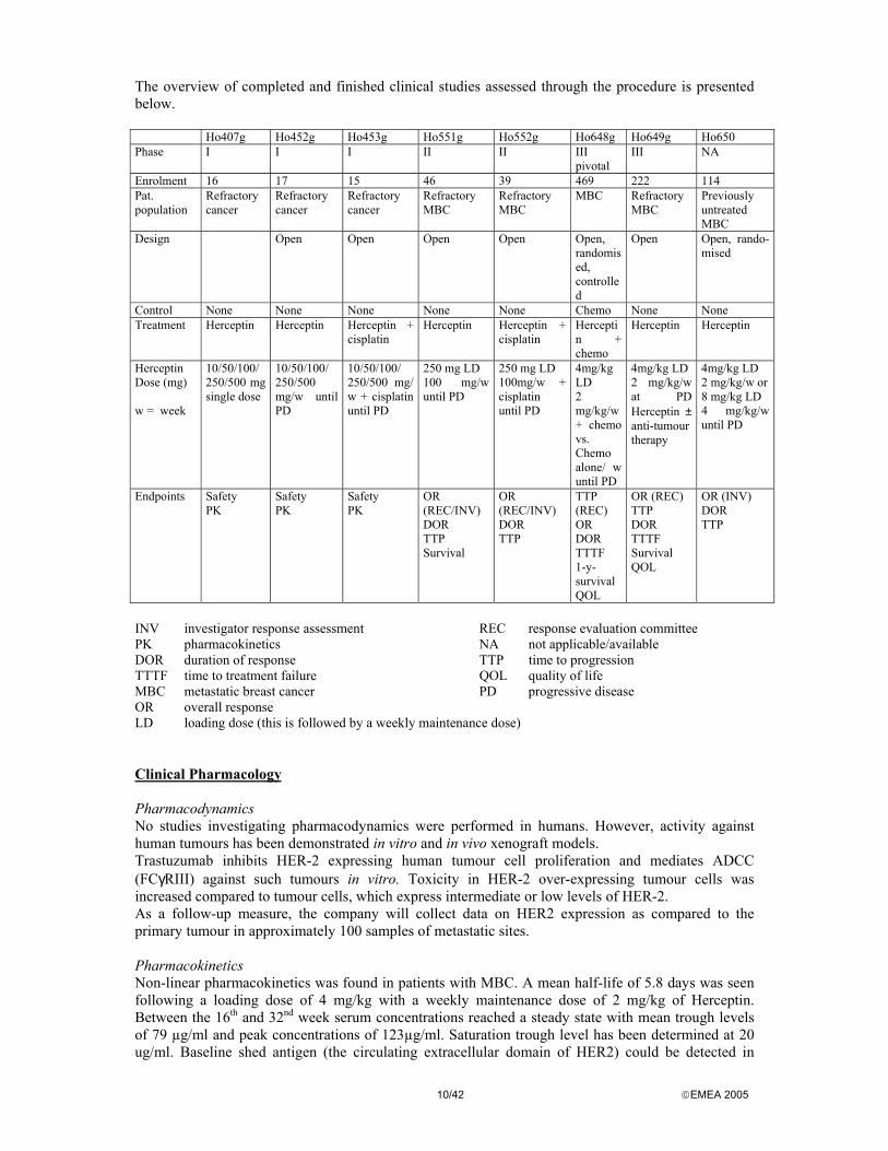
The overview of completed and finished clinical studies assessed through the procedure is presented below. Ho407g Ho452g Ho453g Ho551g Ho552g Ho648g Ho649g Ho650 Phase I I I II II III
pivotal III NA
Enrolment 16 17 15 46 39 469 222 114 Pat. population
Refractory cancer
Refractory cancer
Refractory cancer
Refractory MBC
Refractory MBC
MBC Refractory MBC
Previously untreated MBC
Design Open Open Open Open Open, randomised, controlled
Open Open, rando-mised
Control None None None None None Chemo None None Treatment Herceptin Herceptin Herceptin +
cisplatin Herceptin Herceptin +
cisplatin Herceptin + chemo
Herceptin Herceptin
Herceptin Dose (mg) w = week
10/50/100/ 250/500 mg single dose
10/50/100/ 250/500 mg/w until PD
10/50/100/ 250/500 mg/ w + cisplatin until PD
250 mg LD 100 mg/w until PD
250 mg LD 100mg/w + cisplatin until PD
4mg/kg LD 2 mg/kg/w + chemo vs. Chemo alone/ w until PD
4mg/kg LD 2 mg/kg/w at PD Herceptin ± anti-tumour therapy
4mg/kg LD 2 mg/kg/w or 8 mg/kg LD 4 mg/kg/w until PD
Endpoints Safety PK
Safety PK
Safety PK
OR (REC/INV) DOR TTP Survival
OR (REC/INV) DOR TTP
TTP (REC) OR DOR TTTF 1-y-survival QOL
OR (REC) TTP DOR TTTF Survival QOL
OR (INV) DOR TTP
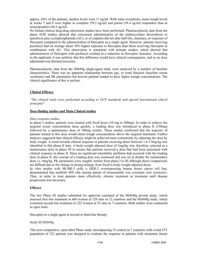
INV investigator response assessment REC response evaluation committee PK pharmacokinetics NA not applicable/available DOR duration of response TTP time to progression TTTF time to treatment failure QOL quality of life MBC metastatic breast cancer PD progressive disease OR overall response LD loading dose (this is followed by a weekly maintenance dose) Clinical Pharmacology Pharmacodynamics No studies investigating pharmacodynamics were performed in humans. However, activity against human tumours has been demonstrated in vitro and in vivo xenograft models. Trastuzumab inhibits HER-2 expressing human tumour cell proliferation and mediates ADCC (FCγRIII) against such tumours in vitro. Toxicity in HER-2 over-expressing tumour cells was increased compared to tumour cells, which express intermediate or low levels of HER-2. As a follow-up measure, the company will collect data on HER2 expression as compared to the primary tumour in approximately 100 samples of metastatic sites. Pharmacokinetics Non-linear pharmacokinetics was found in patients with MBC. A mean half-life of 5.8 days was seen following a loading dose of 4 mg/kg with a weekly maintenance dose of 2 mg/kg of Herceptin. Between the 16th and 32nd week serum concentrations reached a steady state with mean trough levels of 79 µg/ml and peak concentrations of 123µg/ml. Saturation trough level has been determined at 20 ug/ml. Baseline shed antigen (the circulating extracellular domain of HER2) could be detected in
11/42 EMEA 2005
approx. 64% of the patients, median levels were 11 ng/ml. With some exceptions, mean trough levels at weeks 7 and 8 were higher in complete (70.3 ug/ml) and partial (58.4 ug/ml) responders than in nonresponders (44.3 ug/ml). No formal clinical drug-drug interaction studies have been performed. Pharmacokinetic data from the phase II/III studies showed that concurrent administration of the anthracyclines doxorubicin or epirubicin plus cyclophosphamide (AC), or of cisplatin did not alter half-life, clearance, or exposure of Herceptin compared to the administration of Herceptin as a single agent. However, patients receiving paclitaxel had on average about 30% higher exposure to Herceptin than those receiving Herceptin in combination with AC. This observation is consistent with primate studies, which showed that administration of Herceptin with paclitaxel resulted in a reduction in Herceptin clearance. According to the applicant, it was unlikely that this difference would have clinical consequences, and so no dose adjustment was deemed necessary. Pharmacokinetic data from the H0649g single-agent study were analysed by a number of baseline characteristics. There was no apparent relationship between age, or renal function (baseline serum creatinine) and PK parameters but heavier patients tended to have higher trough concentrations. The clinical significance of this is unclear. Clinical Efficacy “The clinical trials were performed according to GCP standards and agreed international ethical principles” Dose-finding studies and Main Clinical studies Dose response studies In phase I studies, patients were treated with fixed doses (10 mg to 500mg). In order to achieve the targeted serum concentration more quickly, a loading dose was introduced in phase II (250mg) followed by a maintenance dose of 100mg weekly. These studies confirmed that the majority of patients treated at this dose would attain trough concentrations above the targeted minimum. Further analyses suggested that clinical efficacy might be achieved more consistently by adjusting the dose by body weight. A trend towards clinical response in patients receiving doses between 1.6-1.9mg/kg was identified in this phase II data. A body-weight adjusted dose of 2mg/kg was, therefore, selected as a maintenance dose in phase III to ensure that patients received a dose that had been associated with clinical response in phase II. Since no significant tolerability problems had occurred with the loading dose in phase II, the concept of a loading dose was continued and was set at double the maintenance dose i.e. 4mg/kg. PK parameters were roughly similar from phase I to III although direct comparisons are difficult due to the change in dosing strategy from fixed to body-weight adjusted doses. In vitro studies with SK-BR-3 cells, a HER-2 overexpressing human breast cancer cell line, demonstrated that muMAb 4D5 (the murine parent of trastuzumab) was cytostatic (not cytotoxic). Thus, in order to treat patients more effectively, chronic treatment or treatment until disease progression was necessary. Efficacy The two Phase III studies submitted for approval consisted of the HO648g pivotal study, which assessed first line treatment in 469 women at 120 sites in 12 countries and the HO649g study, which evaluated second line treatment in 222 women at 55 sites in 7 countries. Both studies were conducted as open trials. Herceptin as a single agent in second or third-line therapy Study III HO649g This non-comparative, open-label Phase study encompassing 55 centres in 7 countries with a total ITT population of 222 patients was designed to evaluate the response in patients with metastatic breast
12/42 EMEA 2005
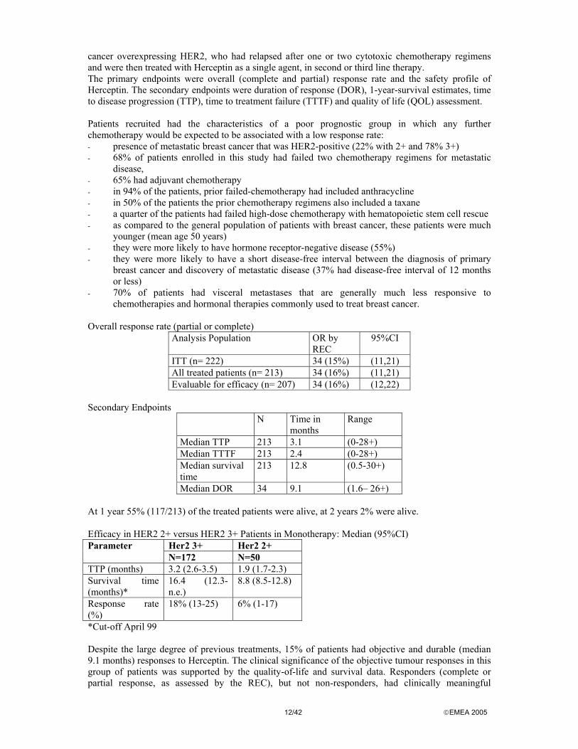
cancer overexpressing HER2, who had relapsed after one or two cytotoxic chemotherapy regimens and were then treated with Herceptin as a single agent, in second or third line therapy. The primary endpoints were overall (complete and partial) response rate and the safety profile of Herceptin. The secondary endpoints were duration of response (DOR), 1-year-survival estimates, time to disease progression (TTP), time to treatment failure (TTTF) and quality of life (QOL) assessment. Patients recruited had the characteristics of a poor prognostic group in which any further chemotherapy would be expected to be associated with a low response rate: - presence of metastatic breast cancer that was HER2-positive (22% with 2+ and 78% 3+) - 68% of patients enrolled in this study had failed two chemotherapy regimens for metastatic
disease, - 65% had adjuvant chemotherapy - in 94% of the patients, prior failed-chemotherapy had included anthracycline - in 50% of the patients the prior chemotherapy regimens also included a taxane - a quarter of the patients had failed high-dose chemotherapy with hematopoietic stem cell rescue - as compared to the general population of patients with breast cancer, these patients were much
younger (mean age 50 years) - they were more likely to have hormone receptor-negative disease (55%) - they were more likely to have a short disease-free interval between the diagnosis of primary
breast cancer and discovery of metastatic disease (37% had disease-free interval of 12 months or less)
- 70% of patients had visceral metastases that are generally much less responsive to chemotherapies and hormonal therapies commonly used to treat breast cancer.
Overall response rate (partial or complete)
Analysis Population OR by REC
95%CI
ITT (n= 222) 34 (15%) (11,21) All treated patients (n= 213) 34 (16%) (11,21) Evaluable for efficacy (n= 207) 34 (16%) (12,22)
Secondary Endpoints
N Time in months
Range
Median TTP 213 3.1 (0-28+) Median TTTF 213 2.4 (0-28+) Median survival time
213 12.8 (0.5-30+)
Median DOR 34 9.1 (1.6– 26+) At 1 year 55% (117/213) of the treated patients were alive, at 2 years 2% were alive. Efficacy in HER2 2+ versus HER2 3+ Patients in Monotherapy: Median (95%CI)
Her2 3+ Her2 2+ Parameter N=172 N=50
TTP (months) 3.2 (2.6-3.5) 1.9 (1.7-2.3) Survival time (months)*
16.4 (12.3-n.e.)
8.8 (8.5-12.8)
Response rate (%)
18% (13-25) 6% (1-17)
*Cut-off April 99 Despite the large degree of previous treatments, 15% of patients had objective and durable (median 9.1 months) responses to Herceptin. The clinical significance of the objective tumour responses in this group of patients was supported by the quality-of-life and survival data. Responders (complete or partial response, as assessed by the REC), but not non-responders, had clinically meaningful
13/42 EMEA 2005
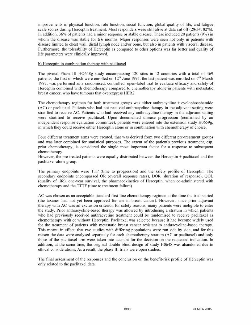
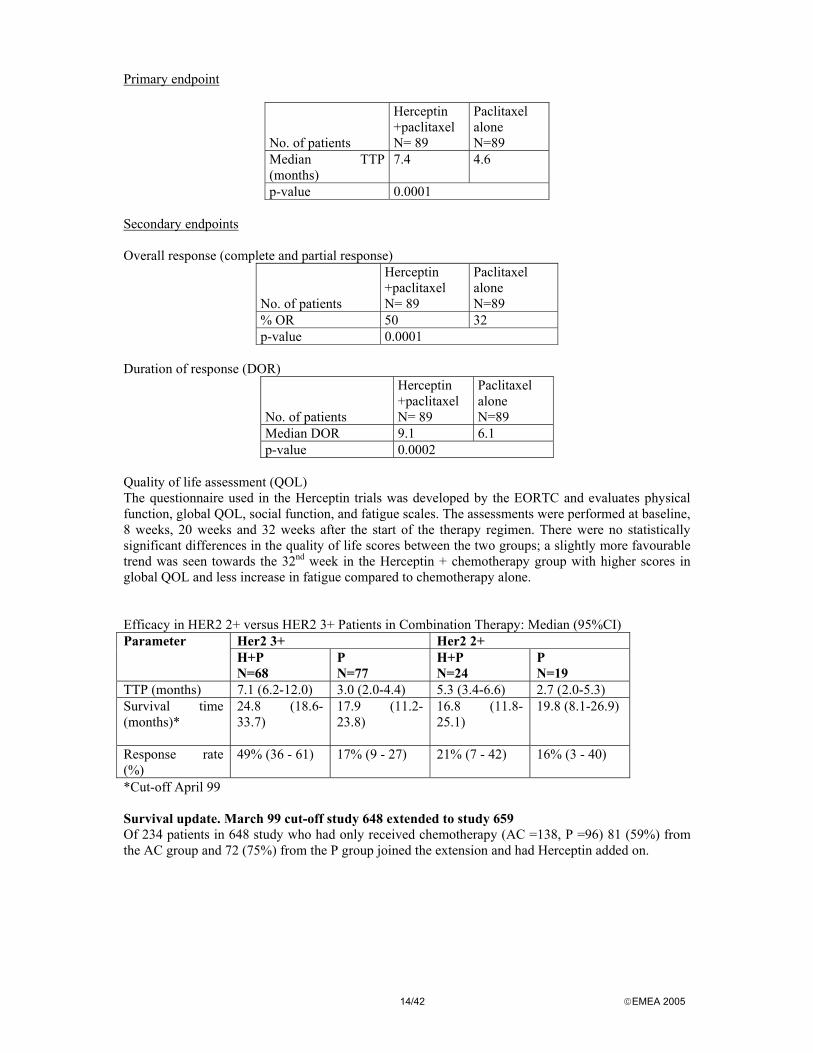
improvements in physical function, role function, social function, global quality of life, and fatigue scale scores during Herceptin treatment. Most responders were still alive at data cut off (28/34; 82%). In addition, 36% of patients had a minor response or stable disease. These included 20 patients (9%) in whom the disease was stable for ≥ 6 months. Major responses were seen not only in patients with disease limited to chest wall, distal lymph node and/or bone, but also in patients with visceral disease. Furthermore, the tolerability of Herceptin as compared to other options was far better and quality of life parameters were clinically improved. b) Herceptin in combination therapy with paclitaxel The pivotal Phase III HO648g study encompassing 120 sites in 12 countries with a total of 469 patients, the first of which were enrolled on 12th June 1995, the last patient was enrolled on 7th March 1997, was performed as a randomised, controlled, open-label trial to evaluate efficacy and safety of Herceptin combined with chemotherapy compared to chemotherapy alone in patients with metastatic breast cancer, who have tumours that overexpress HER2. The chemotherapy regimen for both treatment groups was either anthracycline + cyclophosphamide (AC) or paclitaxel. Patients who had not received anthracycline therapy in the adjuvant setting were stratified to receive AC. Patients who had received any anthracycline therapy in the adjuvant setting were stratified to receive paclitaxel. Upon documented disease progression (confirmed by an independent response evaluation committee), patients were entered into the extension study H0659g, in which they could receive either Herceptin alone or in combination with chemotherapy of choice. Four different treatment arms were created, that was derived from two different pre-treatment groups and was later combined for statistical purposes. The extent of the patient's previous treatment, esp. prior chemotherapy, is considered the single most important factor for a response to subsequent chemotherapy. However, the pre-treated patients were equally distributed between the Herceptin + paclitaxel and the paclitaxel-alone group. The primary endpoints were TTP (time to progression) and the safety profile of Herceptin. The secondary endpoints encompassed OR (overall response rates), DOR (duration of response), QOL (quality of life), one-year survival, the pharmacokinetics of Herceptin, when co-administered with chemotherapy and the TTTF (time to treatment failure). AC was chosen as an acceptable standard first-line chemotherapy regimen at the time the trial started (the taxanes had not yet been approved for use in breast cancer). However, since prior adjuvant therapy with AC was an exclusion criterion for safety reasons, many patients were ineligible to enter the study. Prior anthracycline-based therapy was allowed by introducing a stratum in which patients who had previously received anthracycline treatment could be randomised to receive paclitaxel as chemotherapy with or without Herceptin. Paclitaxel was selected because it had become widely used for the treatment of patients with metastatic breast cancer resistant to anthracycline-based therapy. This meant, in effect, that two studies with differing populations were run side by side, and for this reason the data were analysed separately for each chemotherapy stratum (AC or paclitaxel) and only those of the paclitaxel arm were taken into account for the decision on the requested indication. In addition, at the same time, the original double blind design of study H0648 was abandoned due to ethical considerations. As a result, the phase III trials were open studies. The final assessment of the responses and the conclusion on the benefit-risk profile of Herceptin was only related to the paclitaxel data.
14/42 EMEA 2005
Primary endpoint
No. of patients
Herceptin +paclitaxel N= 89
Paclitaxel alone N=89
Median TTP (months)
7.4 4.6
p-value 0.0001 Secondary endpoints Overall response (complete and partial response)
No. of patients
Herceptin +paclitaxel N= 89
Paclitaxel alone N=89
% OR 50 32 p-value 0.0001
Duration of response (DOR)
No. of patients
Herceptin +paclitaxel N= 89
Paclitaxel alone N=89
Median DOR 9.1 6.1 p-value 0.0002
Quality of life assessment (QOL) The questionnaire used in the Herceptin trials was developed by the EORTC and evaluates physical function, global QOL, social function, and fatigue scales. The assessments were performed at baseline, 8 weeks, 20 weeks and 32 weeks after the start of the therapy regimen. There were no statistically significant differences in the quality of life scores between the two groups; a slightly more favourable trend was seen towards the 32nd week in the Herceptin + chemotherapy group with higher scores in global QOL and less increase in fatigue compared to chemotherapy alone. Efficacy in HER2 2+ versus HER2 3+ Patients in Combination Therapy: Median (95%CI)
Her2 3+ Her2 2+ Parameter H+P N=68
P N=77
H+P N=24
P N=19
TTP (months) 7.1 (6.2-12.0) 3.0 (2.0-4.4) 5.3 (3.4-6.6) 2.7 (2.0-5.3) Survival time (months)*
24.8 (18.6-33.7)
17.9 (11.2-23.8)
16.8 (11.8-25.1)
19.8 (8.1-26.9)
Response rate (%)
49% (36 - 61) 17% (9 - 27) 21% (7 - 42) 16% (3 - 40)
*Cut-off April 99 Survival update. March 99 cut-off study 648 extended to study 659 Of 234 patients in 648 study who had only received chemotherapy (AC =138, P =96) 81 (59%) from the AC group and 72 (75%) from the P group joined the extension and had Herceptin added on.
15/42 EMEA 2005
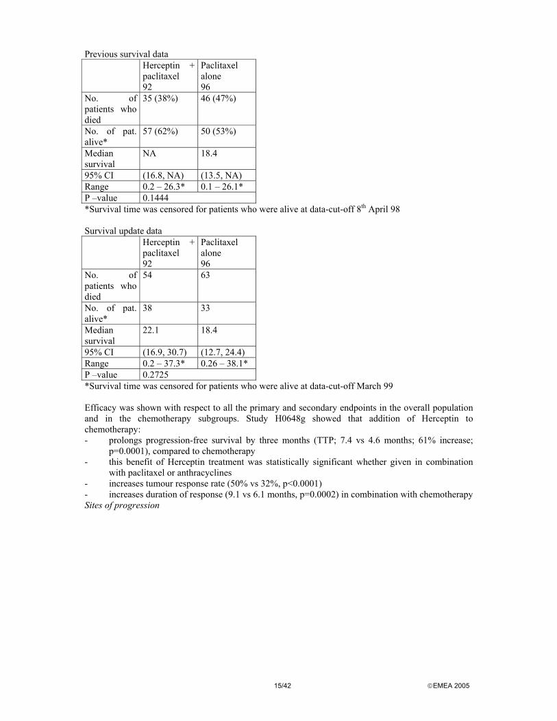
Previous survival data Herceptin +
paclitaxel 92
Paclitaxel alone 96
No. of patients who died
35 (38%) 46 (47%)
No. of pat. alive*
57 (62%) 50 (53%)
Median survival
NA 18.4
95% CI (16.8, NA) (13.5, NA) Range 0.2 – 26.3* 0.1 – 26.1* P –value 0.1444 *Survival time was censored for patients who were alive at data-cut-off 8th April 98 Survival update data Herceptin +
paclitaxel 92
Paclitaxel alone 96
No. of patients who died
54 63
No. of pat. alive*
38 33
Median survival
22.1 18.4
95% CI (16.9, 30.7) (12.7, 24.4) Range 0.2 – 37.3* 0.26 – 38.1* P –value 0.2725 *Survival time was censored for patients who were alive at data-cut-off March 99 Efficacy was shown with respect to all the primary and secondary endpoints in the overall population and in the chemotherapy subgroups. Study H0648g showed that addition of Herceptin to chemotherapy: - prolongs progression-free survival by three months (TTP; 7.4 vs 4.6 months; 61% increase;
p=0.0001), compared to chemotherapy - this benefit of Herceptin treatment was statistically significant whether given in combination
with paclitaxel or anthracyclines - increases tumour response rate (50% vs 32%, p<0.0001) - increases duration of response (9.1 vs 6.1 months, p=0.0002) in combination with chemotherapy Sites of progression
16/42 EMEA 2005
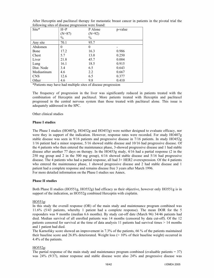
After Herceptin and paclitaxel therapy for metastatic breast cancer in patients in the pivotal trial the following sites of disease progression were found: Site* H+P
(N=87) %
P Alone (N=92) %
p-value
Any site 70.1 95.7 Abdomen 0 0 - Bone 17.2 16.3 0.986 Chest 5.7 13.0 0.250 Liver 21.8 45.7 0.004 Lung 16.1 18.5 0.915 Dist. Node 3.4 6.5 0.643 Mediastinum 4.6 2.2 0.667 CNS 12.6 6.5 0.377 Other 4.6 9.8 0.410 *Patients may have had multiple sites of disease progression The frequency of progression in the liver was significantly reduced in patients treated with the combination of Herceptin and paclitaxel. More patients treated with Herceptin and paclitaxel progressed in the central nervous system than those treated with paclitaxel alone. This issue is adequately addressed in the SPC. Other clinical studies Phase I studies The Phase I studies (HO407g, HO452g and HO453g) were neither designed to evaluate efficacy, nor were they in support of the indication. However, response rates were recorded. For study HO407g stable disease was seen in 9/16 patients and progressive disease in 7/16 patients. In study HO452g 1/16 patient had a minor response, 5/16 showed stable disease and 10/16 had progressive disease. Of the 4 patients who then entered the maintenance phase, 3-showed progressive disease and 1 had stable disease after another 77 days on therapy. In the HO453g study, 4/16 had a partial response (2 in the 250 mg group and 2 in the 500 mg group), 6/16 showed stable disease and 5/16 had progressive disease. The 4 patients who had a partial response, all had 3+ HER2 overexpression. Of the 4 patients who entered the maintenance phase, 1 showed progressive disease and 2 had stable disease and 1 patient had a complete response and remains disease free 3 years after March 1996. For more detailed information on the Phase I studies see Annex. Phase II studies Both Phase II studies (HO551g, HO552g) had efficacy as their objective, however only HO551g is in support of the indication, as HO552g combined Herceptin with cisplatin. HO551g: In this study the overall response (OR) of the main study and maintenance program combined was 11.6% (5/43 patients, whereby 1 patient had a complete response). The mean DOR for the 5 responders was 9 months (median 6.6 months). By study cut-off date (March 96) 34/46 patients had died. Median survival of all enrolled patients was 14 months (censored by data cut-off). Of the 12 patients censored for survival at the time of data analysis 11 patients had survival times > 14 months and 1 patient had died. The Karnofsky score showed an improvement in 7.3% of the patients, 66 % of the patients maintained their baseline score and 26.8% deteriorated. Weight loss (> 10% of their baseline weight) occurred in 4.4% of the patients. HO552g: The partial response of the main study and maintenance program combined (evaluable patients = 37) was 24% (9/37), minor response and stable disease were also 24% and progressive disease was
17/42 EMEA 2005
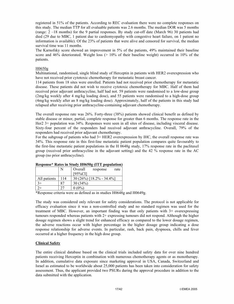
registered in 51% of the patients. According to REC evaluation there were no complete responses on this study. The median TTP for all evaluable patients was 2.6 months. The median DOR was 5 months (range: 2 –18 months) for the 9 partial responses. By study cut-off date (March 96) 30 patients had died (29 due to MBC, 1 patient due to cardiomyopathy with congestive heart failure, on 1 patient no information is available). Of the 23% of patients that were alive and censored for survival, the median survival time was 11 months. The Karnofsky score showed an improvement in 5% of the patients, 49% maintained their baseline score and 46% deteriorated. Weight loss (> 10% of their baseline weight) occurred in 10% of the patients. H0650g Multinational, randomised, single blind study of Herceptin in patients with HER2 overexpression who have not received prior cytotoxic chemotherapy for metastatic breast cancer. 114 patients from 18 sites were enrolled. Patients had not received prior chemotherapy for metastatic disease. These patients did not wish to receive cytotoxic chemotherapy for MBC. Half of them had received prior adjuvant anthracycline, half had not. 59 patients were randomised to a low-dose group (2mg/kg weekly after 4 mg/kg loading dose), and 55 patients were randomised to a high-dose group (4mg/kg weekly after an 8 mg/kg loading dose). Approximately, half of the patients in this study had relapsed after receiving prior anthracycline-containing adjuvant chemotherapy. The overall response rate was 26%. Forty-three (38%) patients showed clinical benefit as defined by stable disease or minor, partial, complete response for greater than 6 months. The response rate in the Her2 3+ population was 34%. Responses were seen in all sites of disease, including visceral disease. Sixty-four percent of the responders had received adjuvant anthracycline. Overall, 79% of the responders had received prior adjuvant chemotherapy. For the subgroup of patients who had 3+ HER2 overexpression by IHC, the overall response rate was 34%. This response rate in this first-line metastatic patient population compares quite favourably to the first-line metastatic patient populations in the H 0648g study, 17% response rate in the paclitaxel group (received prior anthracycline in the adjuvant setting) and the 42 % response rate in the AC group (no prior anthracycline). Response* Rates in Study H0650g (ITT population) N Overall response rate
[95%CI] All patients 114 30 (26%) [18.2% - 34.4%] 3+ 87 30 (34%) 2+ 27 0 (0%)
*Response criteria were as defined as in studies H0648g and H0649g. The study was considered only relevant for safety considerations. The protocol is not applicable for efficacy evaluation since it was a non-controlled study and no standard regimen was used for the treatment of MBC. However, an important finding was that only patients with 3+ overexpressing tumours responded whereas patients with 2+ expressing tumours did not respond. Although the higher dosage regimen shows a slight trend for enhanced efficacy as compared to the lower dosage regimen, the adverse reactions occur with higher percentage in the higher dosage group indicating a dose response relationship for adverse events. In particular, rash, back pain, dyspnoea, chills and fever occurred at a higher frequency in the high dose group. Clinical Safety The entire clinical database based on the clinical trials included safety data for over nine hundred patients receiving Herceptin in combination with numerous chemotherapy agents or as monotherapy. In addition, cumulative data exposure since marketing approval in USA, Canada, Switzerland and Israel as estimated to be worldwide about 25,000 patients has been taken into consideration for safety assessment. Thus, the applicant provided two PSURs during the approval procedure in addition to the data submitted with the application.

18/42 EMEA 2005
While in the clinical trial programme the main safety issue was identified as cardiotoxicity of Herceptin, during the approval procedure additional issues arose, infusion related reactions including some with a fatal outcome, hypersensitivity reactions, including fatal anaphylaxis and pulmonary events including adult respiratory distress syndrome and death. While some of these serious adverse events were observed in clinical trials, some of the events reported in the postmarketing setting were more severe. Thus, the originally suggested SPC needed to be completely amended according to the newly arising issues. Safety in clinical trials All patients who received treatment on study were evaluable for safety. Patients who received Herceptin + chemotherapy (study H0648g) or Herceptin alone (study H0649g) were evaluated for safety weekly with each infusion. In contrast, patients who received chemotherapy alone (study H0648g) were evaluated less frequently. In study H0648g, patients were followed for safety until progressive disease. In the single-agent study H0649g, patients were followed for safety until discontinuation of Herceptin therapy. Herceptin in Combination with Chemotherapy: Study H0648g 469 patients were enrolled into study H0648g, and 464 patients were evaluable for safety (five patients discontinued the study prior to treatment with Herceptin or chemotherapy). 234 patients received Herceptin. The incidence of serious adverse events was greater in the paclitaxel alone subgroup than in the Herceptin + paclitaxel subgroup. Few serious adverse events occurred in >2.5% of patients. The incidence of many adverse events was increased among patients receiving Herceptin. Infusion-associated signs and symptoms: In this study, fever, chills, nausea, pain at the tumor site, vomiting, headache, back pain, and dizziness in association with Herceptin infusion occurred in 25% of patients. Cardiovascular: see below under separate chapter. Infection: There was an increased incidence of adverse events that mapped to the preferred term of infection in both the Herceptin + chemotherapy treatment groups compared with the chemotherapy alone treatment group. Most of these events could be grouped into two categories: upper respiratory tract infection (cold, upper respiratory infection, etc.), which constituted 72% of events, and catheter infections, which constituted 9% of events. The imbalance in the incidence of catheter-related infections among Herceptin-treated patients may be due to the increased frequency of indwelling catheter access with weekly Herceptin infusions. Leukopenia and anemia: The incidence of mild leukopenia and anemia reported as an adverse event was increased with Herceptin treatment (leukopenia 41% vs 26%: anaemia 27% vs 19%). Digestive: An increase in a number of adverse events, including diarrhea and nausea and vomiting was noted in both Herceptin + chemotherapy treatment groups. The events were mostly mild to moderate in severity. Respiratory: An increased incidence of dyspnoea and cough in the Herceptin + chemotherapy treatment groups occurred. Occurrence of leukemia/myelodysplastic syndrome: see below under separate chapter. Other adverse events: A number of other adverse events of uncertain relationship were increased in incidence with Herceptin treatment which were adequately addressed in the SPC. Laboratory Parameters: Routine hematology and serum chemistries were analyzed at baseline and at scheduled intervals at a core laboratory facility. Modest changes were noted in the incidence of neutropenia, anemia, and abnormal liver function test results. Hematological Laboratory Parameters: Hematologic adverse events were transient and occurred during the period of chemotherapy administration. Improvement was noted at week 20, and by week 32, hematologic values were nearly back to baseline. Median hemoglobin values dropped during chemotherapy administration (ie: from baseline to week 8 (–2.6g/dL) and week 20 (–2.4g/dL) but were close to baseline levels by week 32 (change from baseline –1.0g/dL) when patients were no longer receiving chemotherapy. Median absolute neutrophil counts did not vary markedly during the study. The incidence of WHO grade 3 and 4 abnormalities in haemoglobin levels was higher in the Herceptin + chemotherapy groups than in the chemotherapy alone groups (7% vs 1%). Grade 3 or 4

19/42 EMEA 2005
neutropenia was observed more in the Herceptin + paclitaxel group than the paclitaxel alone group, which probably reflects the greater exposure to chemotherapy in the Herceptin-treated patients. Hepatic and Renal Laboratory Parameters: Overall, Grade 3 or 4 laboratory abnormalities were infrequent. Hepatic laboratory abnormalities were observed less frequently among patients receiving Herceptin + chemotherapy than among patients receiving chemotherapy alone. No patient experienced Grade 3 or 4 elevations in laboratory tests measuring renal function (BUN and creatinine). Antibodies: No patients enrolled in study H0648g developed antibodies against Herceptin. Herceptin as a Single Agent - Study H0649g A total of 213 patients were treated and evaluable for safety (received at least one dose of Herceptin) in study H0649g. Patients were to receive weekly 2mg/kg infusions up to first disease progression. Following first disease progression, patients could continue to receive weekly Herceptin infusions of 2mg/kg or could have their dose increased to 4mg/kg. Overall, 77 patients received the higher dose (4 mg/kg IV weekly) of Herceptin either as a single agent or with systemic anti-cancer therapy. Nearly all of these patients reported at least one adverse event during treatment with the higher dose (97%; 75/77), while about a third experienced adverse events considered severe (36%; 28/77). Similar types of events occurred during treatment at the higher dose as those seen prior to first disease progression when patients (with few exceptions) were treated with the lower, 2mg/kg Herceptin dose. The following events commonly occurred (>10% incidence) during high dose Herceptin treatment: dyspepsia, anemia, leukopenia, bone pain, myasthenia, depression and paraesthesia. In general, the adverse events did not substantially differ as compared to study HO648g. One patient had a positive, neutralising antibody to Herceptin. This patient had received nine weekly infusions of Herceptin and had discontinued the study on day 67 due to progressive disease. This finding was not associated with any clinical symptoms. Cardiotoxicity A main safety concern was cardiotoxicity (cardiomyopathy leading to congestive heart failure, CHF). The original dossier contained a retrospective analysis of cardiac adverse events, which was made by a cardiac review and evaluation committee (CREC). For this analysis, the clinical data were searched for patients with cardiac-related AEs using specific criteria for symptoms of heart failure. A full re-assessment of cardiac-related events was performed on using more broader search criteria. The findings were largely in accordance with those of the original CREC evaluation. The data provided by the applicant as part of the response confirmed that during the clinical studies cardiotoxicity was not prospectively measured as an adverse event and that any data only allow retrospective analysis. Therefore, the cardiotoxic potential of Herceptin alone or in combination with chemotherapy, in particular with paclitaxel, demands explicit clarification with regard to symptoms and nature of cardiotoxicity, frequency, mechanism, threshold of toxicity, time and dose response relationship, risk factors other than age, major confounding factors, mechanism of interaction between Herceptin toxicity and chemotherapy toxicity. These data will be submitted through a follow-up measure. Symptoms, nature, and frequency of cardiotoxicity Heart failure (New York Heart Association [NYHA] class II-IV) has been observed in patients receiving Herceptin therapy alone or in combination with paclitaxel following anthracycline (doxorubicin or epirubicin)–containing chemotherapy. This may be moderate to severe and has been associated with death. These symptoms are apparently very similar to anthracycline induced cardiotoxicity.
20/42 EMEA 2005
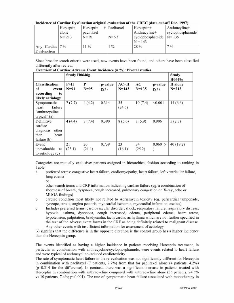
Incidence of Cardiac Dysfunction original evaluation of the CREC (data cut-off Dec. 1997) Herceptin
alone N= 213
Herceptin + paclitaxel N= 91
Paclitaxel N= 93
Herceptin+ Anthracyline+ cyclophosphamide N = 143
Anthracyline+ cyclophosphamide N= 135
Any Cardiac Dysfunction
7 % 11 % 1 % 28 % 7 %
Since broader search criteria were used, new events have been found, and others have been classified differently after review. Overview of Cardiac Adverse Event Incidence (n,%): Pivotal studies Study H0648g Study
H0649g Classification of event according to likely aetiology
P+H N=91
P N=95
p-value (χ2)
AC+H N=143
AC N=135
p-value (χ2)
H alone N=213
Symptomatic heart failure ”anthracycline typical” (a)
7 (7.7) 4 (4.2) 0.314 35 (24.5)
10 (7.4) <0.001 14 (6.6)
Definitive cardiac diagnosis other than heart failure (b)
4 (4.4) 7 (7.4) 0.390 8 (5.6) 8 (5.9) 0.906 5 (2.3)
Event unevaluable as to aetiology (c)
21 (23.1)
20 (21.1)
0.739 23 (16.1)
34 (25.2)
0.060 (-)
40 (19.2)
Categories are mutually exclusive: patients assigned in hierarchical fashion according to ranking in Table. a preferred terms: congestive heart failure, cardiomyopathy, heart failure, left ventricular failure,
lung edema or other search terms and CRF information indicating cardiac failure (eg. a combination of shortness of breath, dyspnoea, cough increased, pulmonary congestion on X-ray, echo or MUGA findings)
b cardiac condition most likely not related to Adriamycin toxicity (eg. pericardial tamponade, syncope, stroke, angina pectoris, myocardial ischemia, myocardial infarction, ascites)
c Includes preferred terms: cardiovascular disorder, shock, respiratory failure, respiratory distress, hypoxia, asthma, dyspnoea, cough increased, edema, peripheral edema, heart arrest, hypotension, palpitation, bradycardia, tachycardia, arrhythmia which are not further specified in the text of the adverse event forms in the CRF as being definitely related to malignant disease. Any other events with insufficient information for assessment of aetiology
(-) signifies that the difference is in the opposite direction ie the control group has a higher incidence than the Herceptin group. The events identified as having a higher incidence in patients receiving Herceptin treatment, in particular in combination with anthracycline/cyclophosphamide, were events related to heart failure and were typical of anthracycline-induced cardiotoxicity. The rate of symptomatic heart failure in the re-evaluation was not significantly different for Herceptin in combination with paclitaxel (7 patients, 7.7%) from that for paclitaxel alone (4 patients, 4.2%) (p=0.314 for the difference). In contrast, there was a significant increase in patients treated with Herceptin in combination with anthracycline compared with anthracycline alone (35 patients, 24.5% vs 10 patients, 7.4%; p<0.001). The rate of symptomatic heart failure associated with monotherapy in
21/42 EMEA 2005
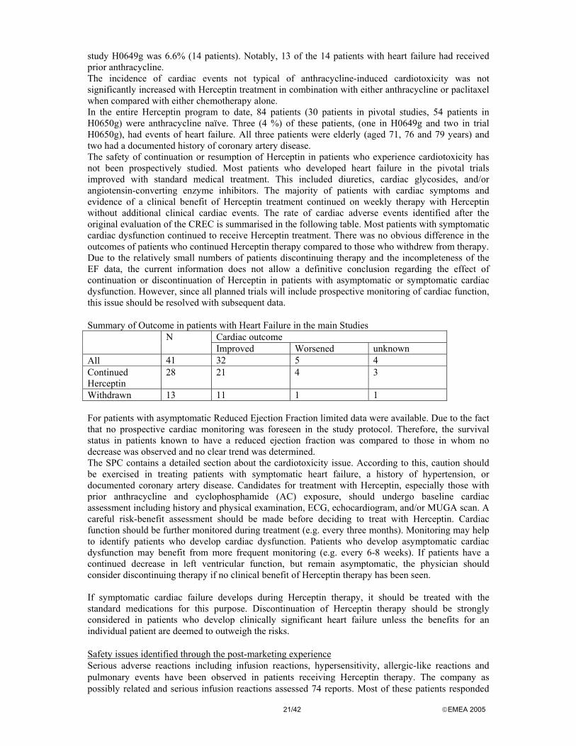
study H0649g was 6.6% (14 patients). Notably, 13 of the 14 patients with heart failure had received prior anthracycline. The incidence of cardiac events not typical of anthracycline-induced cardiotoxicity was not significantly increased with Herceptin treatment in combination with either anthracycline or paclitaxel when compared with either chemotherapy alone. In the entire Herceptin program to date, 84 patients (30 patients in pivotal studies, 54 patients in H0650g) were anthracycline naïve. Three (4 %) of these patients, (one in H0649g and two in trial H0650g), had events of heart failure. All three patients were elderly (aged 71, 76 and 79 years) and two had a documented history of coronary artery disease. The safety of continuation or resumption of Herceptin in patients who experience cardiotoxicity has not been prospectively studied. Most patients who developed heart failure in the pivotal trials improved with standard medical treatment. This included diuretics, cardiac glycosides, and/or angiotensin-converting enzyme inhibitors. The majority of patients with cardiac symptoms and evidence of a clinical benefit of Herceptin treatment continued on weekly therapy with Herceptin without additional clinical cardiac events. The rate of cardiac adverse events identified after the original evaluation of the CREC is summarised in the following table. Most patients with symptomatic cardiac dysfunction continued to receive Herceptin treatment. There was no obvious difference in the outcomes of patients who continued Herceptin therapy compared to those who withdrew from therapy. Due to the relatively small numbers of patients discontinuing therapy and the incompleteness of the EF data, the current information does not allow a definitive conclusion regarding the effect of continuation or discontinuation of Herceptin in patients with asymptomatic or symptomatic cardiac dysfunction. However, since all planned trials will include prospective monitoring of cardiac function, this issue should be resolved with subsequent data. Summary of Outcome in patients with Heart Failure in the main Studies
Cardiac outcome N Improved Worsened unknown
All 41 32 5 4 Continued Herceptin
28 21 4 3
Withdrawn 13 11 1 1 For patients with asymptomatic Reduced Ejection Fraction limited data were available. Due to the fact that no prospective cardiac monitoring was foreseen in the study protocol. Therefore, the survival status in patients known to have a reduced ejection fraction was compared to those in whom no decrease was observed and no clear trend was determined. The SPC contains a detailed section about the cardiotoxicity issue. According to this, caution should be exercised in treating patients with symptomatic heart failure, a history of hypertension, or documented coronary artery disease. Candidates for treatment with Herceptin, especially those with prior anthracycline and cyclophosphamide (AC) exposure, should undergo baseline cardiac assessment including history and physical examination, ECG, echocardiogram, and/or MUGA scan. A careful risk-benefit assessment should be made before deciding to treat with Herceptin. Cardiac function should be further monitored during treatment (e.g. every three months). Monitoring may help to identify patients who develop cardiac dysfunction. Patients who develop asymptomatic cardiac dysfunction may benefit from more frequent monitoring (e.g. every 6-8 weeks). If patients have a continued decrease in left ventricular function, but remain asymptomatic, the physician should consider discontinuing therapy if no clinical benefit of Herceptin therapy has been seen. If symptomatic cardiac failure develops during Herceptin therapy, it should be treated with the standard medications for this purpose. Discontinuation of Herceptin therapy should be strongly considered in patients who develop clinically significant heart failure unless the benefits for an individual patient are deemed to outweigh the risks. Safety issues identified through the post-marketing experience Serious adverse reactions including infusion reactions, hypersensitivity, allergic-like reactions and pulmonary events have been observed in patients receiving Herceptin therapy. The company as possibly related and serious infusion reactions assessed 74 reports. Most of these patients responded
22/42 EMEA 2005
well to supportive treatment and continued to receive Herceptin. 9/74 was reported with fatal outcome and 6 additional deaths. In some of these cases, a conclusive assessment was not possible due to the lack of data. All of these 9 deaths had pre-existing severe, malignancy-related respiratory distress, 7/9 were hospitalised prior to Herceptin infusion. Patients who are experiencing dyspnoea at rest due to complications of advanced malignancy and comorbidities may therefore be at increased risk of a fatal infusion reaction. Therefore, it was required to contraindicate the use of Herceptin in those patients with severe pulmonary compromise with dyspnoea at rest. The severe reactions were usually associated with the first infusion of Herceptin and generally occurred during or immediately following the infusion. For some patients, symptoms progressively worsened and led to further pulmonary complications. Initial improvement followed by clinical deterioration and delayed reactions with rapid clinical deterioration have also been reported. Fatalities have occurred within hours and up to one week following infusion. On very rare occasions, patients have experienced the onset of infusion symptoms or pulmonary symptoms more than six hours after the start of the Herceptin infusion. Patients should be warned of the possibility of such a late onset and should be instructed to contact their physician if these symptoms occur. Since at present the mechanisms of the above mentioned adverse events, risk factors, adequate premedication and tolerability of subsequent Herceptin infusions are unknown, a follow-up measure has been required to investigate these issues further. Infusion reactions, allergic-like reactions and hypersensitivity Serious adverse reactions to Herceptin infusion that have been reported infrequently include dyspnoea, hypotension, wheezing, bronchospasm, tachycardia, reduced oxygen saturation, anaphylaxis, respiratory distress, urticaria and angioedema. The majority of these events occur during or within 2.5 hours of the start of the first infusion. Should an infusion reaction occur the Herceptin infusion should be discontinued and the patient monitored until resolution of any observed symptoms. The majority of patients experienced resolution of symptoms and subsequently received further infusions of Herceptin. Serious reactions have been treated successfully with supportive therapy such as oxygen, beta-agonists, and corticosteroids. In rare cases, these reactions are associated with a clinical course culminating in a fatal outcome. Patients who are experiencing dyspnoea at rest due to complications of advanced malignancy and comorbidities may be at increased risk of a fatal infusion reaction. Therefore, these patients should not be treated with Herceptin. An infusion reaction can clinically resemble an anaphylactic or other allergic reaction. There were single cases of allergic reactions associated with subsequent infusions. It is difficult to differentiate between infusion-related and hypersensitivity reactions due to a similar pattern of symptoms. Pulmonary events Dyspnoea, bronchospasm, asthma and hypoxia can occur as part of an infusion reaction. These are most common with the first infusion and their severity decreases with subsequent infusions. Serious reactions have been treated successfully with supportive therapy such as oxygen, beta-agonists, and corticosteroids. Single cases of pulmonary infiltrates, pneumonia, pulmonary effusion, respiratory distress, acute pulmonary oedema and respiratory insufficiency have been reported rarely. Adult respiratory distress syndrome has been reported rarely with fatal outcome. Patients who are experiencing dyspnoea at rest due to complications of advanced malignancy and comorbidities may be at increased risk of pulmonary events. Therefore, these patients should not be treated by contraindication. Other safety issues Haematological toxicity Haematological toxicity was infrequent following the administration of Herceptin as a single agent, WHO Grade III leucopenia, thrombocytopenia and anaemia occurring in < 1% of patients. No WHO Grade IV toxicities were observed. There was an increase in WHO Grade III or IV haematological toxicity in patients treated with the combination of Herceptin and paclitaxel compared with patients receiving paclitaxel alone (34% versus 21%). This is possibly due to the result of greater cumulative
23/42 EMEA 2005
exposure to paclitaxel in the Herceptin plus paclitaxel arm of the study as time to disease progression is increased in this patient group compared with the group treated with paclitaxel alone. Neuropathy The raw incidence peripheral neuropathy was higher in the Herceptin group plus paclitaxel group than in the paclitaxel alone group. However, when these rates were adjusted according to the length of observation period for the two groups, the frequency of these adverse events is similar between treatment groups. Nevertheless the issue has been addressed in the SPC. Leukaemia and myelodepression The incidence of leukaemia observed in Herceptin trials so far does not exceed that expected in a population of metastatic breast cancer patients who have been treated with chemotherapy. To date, there have been four reports of secondary acute leukemia and one report of chronic myelogenous leukemia (CML) in patients participating in Herceptin clinical trials in advanced breast cancer. All four of these patients received anthracycline plus cyclophosphamide (AC) with Herceptin in either Study H0648g (n=3) or Study H0659g (n=1). These cases were considered in an analysis of incidence per patient year. The fifth case in study H0693g, was diagnosed in January 1999 in a patient receiving Herceptin and vinorelbine who had had prior CMF/anthracycline therapy and occurred after the analysis was performed. This patient had the characteristic 9:22 translocation of CML and was not felt to have drug-related leukemia. When the Dec-97 cut-off (which included the first 3 cases) was used, the incidence rate was 0.81 cases per 100 person-years (95% CI, 0.17 to 2.36). Additional data through November 1998 were available only for patients in Studies H0648g and H0659g. Using a data cutoff date of 15 November 1998 for these two studies, the incidence rate was calculated as 0.76 cases per 100 person-years (95% CI, 0.21 to 1.95). These rates do not exceed expected rates estimated from a modified best evidence synthesis (BES); a systematic, critical evaluation and synthesis of the global literature in metastatic breast cancer. The observed number of four cases in patients receiving Herceptin treatment in combination with chemotherapy falls below the expected range of 9–18 cases predicted by the BES for patients receiving both an alkylating agent and a topoisomerase inhibitor, and falls within the expected range of 2–8 cases predicted for patients treated with just one of these types of agent. Based on this analysis, the current evidence does not support an association between Herceptin and an increased rate of secondary acute leukemia in women treated for metastatic breast cancer. Occurrence of leukemia or myelodysplasia will be observed in the future and data reported as a specific item of following PSURs. The issue is adequately addressed in the SPC. Hepatic and renal toxicity WHO Grade III or IV hepatic toxicity was observed in 12% of patients following administration of Herceptin as single agent. This toxicity was associated with progression of disease in the liver in 60% of these patients. WHO Grade III or IV hepatic toxicity was less frequently observed among patients receiving Herceptin and paclitaxel than among patients receiving paclitaxel (7% compared with 15%). No WHO Grade III or IV renal toxicity was observed in patients treated with Herceptin. Diarrhoea Of patients treated with Herceptin as a single agent, 27% experienced diarrhoea. An increase in the incidence of diarrhoea, primarily mild to moderate in severity, has also been observed in patients receiving Herceptin in combination with paclitaxel compared with patients receiving paclitaxel alone. Infection An increased incidence of infections, primarily mild upper respiratory infections of minor clinical significance or catheter infections, has been observed primarily in patients treated with Herceptin plus paclitaxel compared with patients receiving paclitaxel alone. Anti-Herceptin antibodies All except two patients in the two pivotal trials have been evaluated for antibody production. Human anti- trastuzumab antibodies were detected in one patient, who had no allergic manifestations.
24/42 EMEA 2005
• Discussion on Clinical Efficacy and Safety As a result of the assessment of the clinical data ten major objections were raised related to efficacy and safety of the product. In particular, there were concerns on the insufficient efficacy data, the low level of efficacy observed in the paclitaxel alone arm of the pivotal study, the potential bias induced by the open-label design of the clinical studies, the pharmacodynamic behaviour of the antibody, the cardiotoxic reactions of Herceptin alone and in combination with chemotherapy and the development of peripheral neuropathy. Furthermore, a large number of points for clarification were raised, among them of particular concern was the correlation of the clinical benefit of patients with the level of HER2 overexpression, the impact of the diagnostic determination of HER2 overexpression on the correct clinical grading of patients, the increase of CNS metastases in the Herceptin arms and safety issues such as Herceptin’s potential of inducing leukemia and myelodepression.
In their response, the applicant replied on the major concern of using a 440 mg multidose vial with bacteriostatic water as solvent by providing a proposal to use a 150 mg single dose vial instead. Efficacy and level of HER2 overexpression There was concern over the lack of correlation between the clinical benefit and the HER2 expression level. Since study H0648g was not designed to allow for subgroup analysis of patients expression HER2 at 2+ or 3+ level and no stratification at randomisation on the basis of level of overexpression nor for other potential prognostic factors was performed, analysis was only retrospectively available. Nonetheless, on the basis of the analysis provided by the applicant as part of the response and during the oral presentation, it was concluded that a benefit is only really discernible in the 3+ groups. A significant difference in both the H0648 and H0649 study concerning the primary endpoint TTP is only achieved in the 3+ overexpressing HER2 group. Accordingly, the indication has been limited to patients whose tumours have HER2 overexpression at a 3+ level. Diagnostic methods to determine HER2 The diagnostic methodology used to determine HER2 overexpression in patients before Herceptin treatment is of importance in order to identify the patients who benefit most. In the clinical trial patients were enrolled if they had 2+ or 3+ levels of overexpression, determined by a immunohistochemical method (IHC) performed by one central testing laboratory. The data showed that only laboratories specialised in performing immunohistochemistry should investigate the tumour specimens. This has been adequately addressed in the SPC. Herceptin in combination with paclitaxel Treatment options will only change for a small group of patients, those with HER2 positive tumors. The indication was supported since the real target group for the use of Herceptin can be clearly identified by the use of appropriate diagnostic methods. Therefore, a strong requirement for the SPC recommendation on the use of diagnostic methods has been implemented. However, there were arguments to limit the indication to patients who did receive prior adjuvant and to patients for whom AC is not suitable. There are no data available on those patients with no prior adjuvant AC (approx. 10% of MBC patients). Furthermore, comparative data to AC (AC vs. H+P) are not taken into account for approval decision. Therefore, the wording of the indication under b) combination therapy was amended as follows: „....b) in combination with paclitaxel for the treatment of those patients who have not received chemotherapy for their metastatic disease and for whom AC is not suitable“
25/42 EMEA 2005
Herceptin as a single agent in second- and third-line therapy The applicant has presented a substantial and detailed discussion of the data observed with Herceptin used as single agent in second line therapy as compared to the current knowledge of alternative treatment options. It has been shown that the patients recruited into the study H0649g had the characteristics of a poor prognostic group in which any further chemotherapy would be expected to be associated with a low response rate. Herceptin was well tolerated in the pivotal monotherapy trial, even by patients who had received multiple prior chemotherapy regimens. Despite the poor characteristics, Herceptin monotherapy led to an objective response rate of 14 % in these patients. In addition, 36% of patients had a minor response or stable disease. The monotherapy indication was therefore considered acceptable whereas the wording of the indication was changed in order to limit the use to patients who have received at least two chemotherapy regimens for their metastatic disease (instead of one or more). Prior chemotherapy must have included at least an anthracycline and a taxane unless patients are unsuitable for these treatments. Hormone receptor positive patients must also have failed hormonal therapy, unless patients are unsuitable for these treatments
Safety
Substantial assessment has been performed on the new safety issues which arised within the procedure leading to comprehensive changes of the SPC:
4.4.: mentioning of Infusion related reactions, including fatal outcome Hypersensitivity reactions, including some with fatal outcome Pulmonary events, including ARDS, pleural effusion, pulmonary infiltrates, pulmonary oedema 4.4.: adequate information about the onset, frequency, progression and outcome of AEs (i.e.,
majority during or after first infusion, within 6 hours, some cases may occur later, also after first improvement)
4.4.: adequate information about risk groups 4.4.: adequate information and recommendation about how to administer Herceptin, how to
monitor patients and how to treat AEs 4.4.: recommendation on the need to inform patients about the potential (re)occurrence of events
with onset at a later time point 4.8. was updated entirely according to the assessment taking into account the above mentioned
points for 4.4. Furthermore, three separate chapters on Infusion related reactions, Hypersensitivity reactions, and pulmonary events were included.
4.3. A contraindication in those patients with severe pulmonary compromise with dyspnoea at rest was introduced.
4.2. It was required that patients are monitored for at least 6 hours after the first infusion and for at least 2 hours after subsequent infusions
5. Overall Conclusion and benefit risk assessment The data provided on quality were adequate and demonstrated that Herceptin is manufactured and controlled according to the requirements. The preclinical characterisation of Herceptin has been performed according to the requirements and showed adequate preclinical safety. Five clinical questions were still outstanding to be clarified at an oral presentation in order to justify the proposed Monotherapy indication, the explain the impact of the indication of Herceptin in combination with paclitaxel in First-line Therapy on the use of current standard treatment regimens of metastatic disease, to discuss the correlation between the clinical benefit and the HER2 expression level, further discuss the data presented in the recent PSUR in relation to serious allergic reactions and infusion related reactions and the impact of these data on the risk benefit assessment. From the assessment of the second PSUR two additional questions resulted which were addressed by the applicant at the Oral presentation (overview of the number of anthracycline naive patients with cardiac
26/42 EMEA 2005
events and outcome, re-assessment of those cases from clinical trials where the causality assessment of the drug/event relationship was listed by the investigator as at least possible). As outcome of the Oral presentation, the clinical efficacy and safety has been considered sufficiently demonstrated to recommend approval of Herceptin on clinical grounds. The Committee considered the applied indications acceptable with the suggested changes of the SPC. The company will perform a large number of clinical trials worldwide to investigate combinations with new medications, the adjuvant use of Herceptin, safety and pharmacodynamic issues. In addition, the company committed a number of follow-up measures for further investigation of clinically relevant questions, which are listed separately. Based on the CPMP review of data on quality, safety and efficacy, the CPMP considered by majority decision that the benefit/risk profile of Herceptin in the treatment of patients with metastatic breast cancer whose tumours overexpress HER2 a) as monotherapy for the treatment of those patients who have received at least two chemotherapy regimens for their metastatic disease. Prior chemotherapy must have included at least an anthracycline and a taxane unless patients are unsuitable for these treatments. Hormone receptor positive patients must also have failed hormonal therapy, unless patients are unsuitable for these treatments. b) in combination with paclitaxel for the treatment of those patients who have not received chemotherapy for their metastatic disease for whom an anthracycline is not suitable. Was favourable. 6. Update on pharmacokinetics from Follow-up Measures: Study BO15935 Roche committed to investigate further the PK of trastuzumab in post-authorisation studies. Preliminary PK data derived from Study BO15935 indicated a longer half-life for Herceptin than originally estimated. The interim report of study BO15935 and the re-analysis of the data of single-agent studies and the combined Herceptin+paclitaxel data of the pivotal study HO648 provided with the Marketing Authorisation Application were assessed. Based on the single agent studies H0407g n=16, H0551g n=46 and H0649g n=213 as well as on the data from trial H0648g, n=234 (where Herceptin was co-administered with either + anthracycline/ cyclophosphamide (AC) or paclitaxel) the population PK was re-analysed to assess the effect of pathophysiologic covariates and the potential influence of concomitant chemotherapy (Herceptin + AC or paclitaxel) on the pharmacokinetics of trastuzumab. A non-linear mixed model approach was employed to analyze the data using a linear two-compartment model with zero-order input (infusion) as base model. In a first step a model for pathophysiologic covariates (i.e. demographic factors, laboratory parameters, HER2 overexpression, shed antigen, number of metastatic sites) was built using data from the single agent trials only. First, covariates were added sequentially to the model in order to evaluate the effect of the covariates on clearance and central compartment distribution volume. Having identified those covariates improving the model fit statistically significant the correlation between covariates was evaluated in order to determine which covariates should be incorporated in the ‘full’ model. The full model incorporated the following covariates: total protein, shed antigen, and number of metastatic sites (for clearance) and weight, total bilirubin, alkaline phosphatase, number of metastatic sites, and shed antigen (for volume of distribution). As a non-linear relationship was discovered between shed antigen and clearance respectively volume of distribution, shed antigen was modeled nonlinearly while all other parameters were modelled linearly. Removing each covariate from the full model one at a time in order to identify those covariates that significantly influence the model fit then derived the final model. By means of this approach “number of metastatic sites” and “shed antigen” were identified as the most influential covariates in the model for clearance, and “weight” and “shed antigen” were the influential covariates in the model for volume of distribution.

27/42 EMEA 2005
In a second step the data from the concomitant chemotherapy Study H0648 were added to the dataset and the effect of concomitant chemotherapy on the final model was evaluated. After adjusting for baseline covariates, there was no statistically significant effect of concomitant chemotherapy on clearance or volume of distribution. Therefore, concomitant chemotherapy was not included in the final model. The model and program used to re-evaluate the PK data is considered adequate. The two-compartment model has been determined to be more appropriate to describe the PK data than the one-compartment model used previously. It cannot be excluded that a third compartment is involved, however, with the currently available data the two-compartment model is considered appropriate. In conclusion, the re-assessed data indicate that the half-life is approximately 28.5 days (95% confidence interval, 25.5 –32.8 days). The washout period is up to 20 weeks (95% confidence interval, 18-24 weeks). Steady state pharmacokinetics should be reached by approximately 20 weeks ( 95 % confidence interval, 18 – 24 weeks ). The estimated mean AUC was 578 mg day/L and the estimated mean peak and trough concentrations were 110 mg/L and 66 mg/L, respectively. The mean clearance when a loading dose of 4 mg/kg trastuzumab followed by a subsequent weekly dose of 2 mg/kg was used was 0.225 L/day. The volume of distribution approximated serum volume, 2.95 L. Detectable concentrations of the circulating extracellular domain of the HER2 receptor (shed antigen) are found in the serum of some patients with HER2 overexpressing breast cancers. Determination of shed antigen in baseline serum samples revealed that 64 % (286/447) of patients had detectable shed antigen, which ranged as high as 1880 ng/ml (median = 11 ng/ml). The section 5.2 of the SPC was updated accordingly. A sentence informing the patient of the long washout period of Herceptin was included in the Package Leaflet section 2. Letters to the physicians and a relevant public statement were issued in May 2001 informing the prescribers on the longer half life and wash-out period and the fact that due to this, potential risk of cardiotoxicity needs to be carefully considered when patients are treated or have been treated with Herceptin. 7. Update of Clinical Safety post-authorisation. The statement on cardiotoxicity (SPC section 4.4) and the need for cardiac function monitoring has been emphasised, the warning was strengthened and extended to all candidates for receiving Herceptin, including anthracycline - naive patients. Severe rare pulmonary events are also described under 4.4. as requested by the CPMP following the assessment of the 3rd PSUR, pneumonitis has also been added in respiratory serious adverse reactions (section 4.8) and hypertension has been added as an infusion related syndrome. Following the assessment of the 1st PSUR glomerulopathy was added in 4.8. Section 4.8 “Undesirable effects”, was amended to include pulmonary fibrosis following an update to the Sponsor’s Core Data Sheet as presented in the 5th PSUR. 8. Additional indication: Herceptin in combination with Taxotere An extension of the indication for the combination of Herceptin and docetaxel (Taxotere) as a treatment for patients with HER2-positive MBC was based on data from:. • Study JP16003. This is a clinical pharmacology study in Japanese patients, assessing the
pharmacokinetics of Herceptin and docetaxel in combination. • Study M77001. This controlled, randomized, multi-centre pivotal trial was designed to
investigate efficacy and safety of the combination of Herceptin and docetaxel compared with docetaxel alone.
• Publications from six completed and two ongoing phase II supportive efficacy studies. Safety information from four ongoing multicenter trials on HER2-positive MBC patients treated with the combination of Herceptin and docetaxel.
The main characteristics of these studies are summarised in table 1 below.
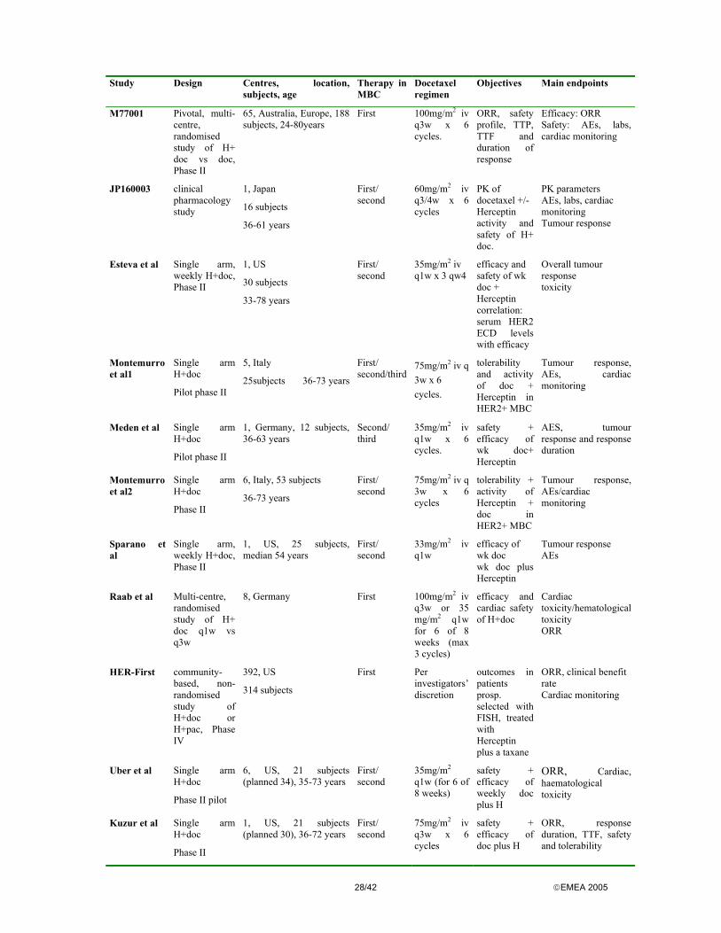
28/42 EMEA 2005
Study Design Centres, location,
subjects, age Therapy in MBC
Docetaxel regimen
Objectives Main endpoints
M77001 Pivotal, multi-centre, randomised study of H+ doc vs doc, Phase II
65, Australia, Europe, 188 subjects, 24-80years
First 100mg/m2 iv q3w x 6 cycles.
ORR, safety profile, TTP, TTF and duration of response
Efficacy: ORR Safety: AEs, labs, cardiac monitoring
JP160003 clinical pharmacology study
1, Japan
16 subjects
36-61 years
First/ second
60mg/m2 iv q3/4w x 6 cycles
PK of docetaxel +/-Herceptin activity and safety of H+ doc.
PK parameters AEs, labs, cardiac monitoring Tumour response
Esteva et al Single arm, weekly H+doc, Phase II
1, US
30 subjects
33-78 years
First/ second
35mg/m2 iv q1w x 3 qw4
efficacy and safety of wk doc + Herceptin correlation: serum HER2 ECD levels with efficacy
Overall tumour response toxicity
Montemurro et al1
Single arm H+doc
Pilot phase II
5, Italy
25subjects 36-73 years
First/ second/third
75mg/m2 iv q 3w x 6 cycles.
tolerability and activity of doc + Herceptin in HER2+ MBC
Tumour response, AEs, cardiac monitoring
Meden et al Single arm H+doc
Pilot phase II
1, Germany, 12 subjects, 36-63 years
Second/ third
35mg/m2 iv q1w x 6 cycles.
safety + efficacy of wk doc+ Herceptin
AES, tumour response and response duration
Montemurro et al2
Single arm H+doc
Phase II
6, Italy, 53 subjects
36-73 years
First/ second
75mg/m2 iv q 3w x 6 cycles
tolerability + activity of Herceptin + doc in HER2+ MBC
Tumour response, AEs/cardiac monitoring
Sparano et al
Single arm, weekly H+doc, Phase II
1, US, 25 subjects, median 54 years
First/ second
33mg/m2 iv q1w
efficacy of wk doc wk doc plus Herceptin
Tumour response AEs
Raab et al Multi-centre, randomised study of H+ doc q1w vs q3w
8, Germany
First 100mg/m2 iv q3w or 35 mg/m2 q1w for 6 of 8 weeks (max 3 cycles)
efficacy and cardiac safety of H+doc
Cardiac toxicity/hematological toxicity ORR
HER-First community-based, non-randomised study of H+doc or H+pac, Phase IV
392, US
314 subjects
First Per investigators’ discretion
outcomes in patients prosp. selected with FISH, treated with Herceptin plus a taxane
ORR, clinical benefit rate Cardiac monitoring
Uber et al Single arm H+doc
Phase II pilot
6, US, 21 subjects (planned 34), 35-73 years
First/ second
35mg/m2 q1w (for 6 of 8 weeks)
safety + efficacy of weekly doc plus H
ORR, Cardiac, haematological toxicity
Kuzur et al Single arm H+doc
Phase II
1, US, 21 subjects (planned 30), 36-72 years
First/ second
75mg/m2 iv q3w x 6 cycles
safety + efficacy of doc plus H
ORR, response duration, TTF, safety and tolerability
29/42 EMEA 2005
Pharmacokinetics The applicant provided (limited) information on the PK of Herceptin in Japanese patients. Using a population PK approach an attempt was made to assess the comparability of the PK in Japanese and non-Japanese patients. Due to limited number of Japanese patients this approach does not allow for a final conclusion. There are no additional PK data dealing with the possible impact of docetaxel on the PK of Herceptin. This is in a way justified by pointing to the similarity of taxanes in general. Pharmacodynamics No specific pharmacodynamic study has been performed. The data gained from the exploratory pharmacodynamic analysis of the pivotal trial M77001 are too limited to draw any conclusion concerning trends between shed HER2 extracellular domain (ECD) and clinical response. Thus it remains open whether ECD concentrations at baseline > 200 ng/mL are predictive of a worse clinical outcome. Regarding the immunogenicity of the combination Herceptin + docetaxel, there is no new information on the incidence of anti-Herceptin antibodies. No patient receiving the combination has been tested, because no unusual immune complex diseases or manifestations have been reported. Clinical efficacy Study M77001 was an open-label, comparative, multicenter, multinational, randomized phase II study, conducted as pivotal trial with the title:“A multicenter, randomized comparative study on the efficacy and safety of Herceptin (trastuzumab) plus docetaxel versus docetaxel alone as first line treatment in patients with HER2-positive metastatic breast cancer”. Eligible patients had to have metastatic breast cancer (MBC) with HER2 overexpression/amplification (IHC3+ and/or FISH positive) who had not previously received chemotherapy except, given as neoadjuvant or adjuvant treatment. All patients were randomised to receive Herceptin in combination with docetaxel or docetaxel alone. The loading dose of 4mg /kg Herceptin was given intravenously over 90 minutes on day 1, followed by 2 mg/kg weekly Herceptin infusions over 30 minutes until disease progression. Patients received an initial dose of docetaxel (100mg/m2) intravenously on the day following the first dose of Herceptin (study day 2). Docetaxel in subsequent cycles (every 3 weeks) was administered 30 minutes after completion of the Herceptin infusion, if the preceding dose of Herceptin was well tolerated. Primary endpoint: • Overall response rate (ORR) in each treatment arm. (Complete response CR plus partial response PR) during the treatment period. The investigator, according to WHO criteria, assessed the tumour response levels. Additionally an independent radiological review (IRR) evaluated the best response unless best response was PD. In case of different assessments reconciliation was performed manually to decide whether the difference was due to over-riding clinical factors (i.e. information not available to the IRR who performed a radiological review only). For the primary analysis, this reconciled data was used (i.e. IRR assessment modified to reflect over-riding clinical data, otherwise the IRR assessment prevailed). The investigator’s assessments were analysed separately and considered secondary. Secondary endpoints: • To characterise the safety profile of docetaxel plus Herceptin and of docetaxel as a single agent in patients with HER2-positive MBC. • To determine the time to progression (TTP), progression-free survival (PFS), time to treatment failure (TTF), time to response, duration of response and overall survival. The analysis of efficacy was primarily based on the full analysis set (FAS) defined by all patients who were randomised and received at least one dose of study medication (including the chemotherapy part). Groups were defined as they were randomised. In addition a per protocol set (PPS) was defined as subset of the FAS, excluding patients fulfilling at least one of the following criteria: 1. Prior chemotherapy treatments specifically listed in the inclusion/exclusion criteria for the
protocol. 2. Failure to receive at least one dose of assigned treatment medication. 3. Patients with LVEF <40% at baseline 4. Patients who fail to meet the tumour assessment criteria specified in the inclusion/exclusion
criteria for the protocol.
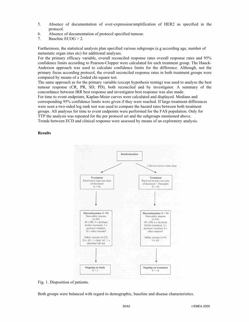
30/42 EMEA 2005
5. Absence of documentation of over-expression/amplification of HER2 as specified in the protocol.
6. Absence of documentation of protocol specified tumour. 7. Baseline ECOG > 2. Furthermore, the statistical analysis plan specified various subgroups (e.g according age, number of metastatic organ sites etc) for additional analyses. For the primary efficacy variable, overall reconciled response rates overall response rates and 95% confidence limits according to Pearson-Clopper were calculated for each treatment group. The Hauck-Anderson approach was used to calculate confidence limits for the difference. Although, not the primary focus according protocol, the overall reconciled response rates in both treatment groups were compared by means of a 2sided chi-square test. The same approach as for the primary variable (except hypothesis testing) was used to analyse the best tumour response (CR, PR, SD, PD), both reconciled and by investigator. A summary of the concordance between IRR best response and investigator best response was also made. For time to event endpoints, Kaplan-Meier curves were calculated and displayed. Medians and corresponding 95% confidence limits were given if they were reached. If large treatment differences were seen a two-sided log rank test was used to compare the hazard rates between both treatment groups. All analyses for time to event endpoints were performed for the FAS population. Only for TTP the analysis was repeated for the per protocol set and the subgroups mentioned above. Trends between ECD and clinical response were assessed by means of an exploratory analysis.
Results
Fig. 1. Disposition of patients.
Both groups were balanced with regard to demographic, baseline and disease characteristics.
31/42 EMEA 2005
The median age in the docetaxel group was 55 years, in the docetaxel + Herceptin group 53 years and 90 and 99% of the patients were Caucasian. In both groups breast cancer histories and treatments were comparable. The median duration of primary disease (time from first diagnosis to diagnosis of metastasis) was 22.6 and 26.6 months in the docetaxel group and in the combination group, respectively. At study entry the median duration of metastatic disease (time from diagnosis of metastasis to entry in the study) was 1.0 months (range 0 – 66.8 months) in the docetaxel arm versus 1.3 months (range 0 – 67.9 months) for the patients in the docetaxel + Herceptin group. Approximately two thirds of patients in both groups had received previous radiotherapy (66% in docetaxel and 64% in docetaxel + Herceptin) and chemotherapy (68% in docetaxel and 71% in docetaxel + Herceptin) as (neo)adjuvant treatment. As part of it, slightly more patients in the docetaxel+Herceptin arm had received prior anthracycline therapy (64% in the Herceptin arm versus 55% in the docetaxel arm). The primary tumours were more frequently estrogen and/or progesterone receptor positive in the docetaxel group (53 patients = 56%) than in the docetaxel + Herceptin group (38 patients = 41%). In both treatment arms the degree of HER2 overexpression was comparable with 87% and 88% of patients in the docetaxel arm and the docetaxel +Herceptin arm, respectively, being tested IHC3+. Overall, 92 % and 96 % had IHC3+ and/or FISH positive disease. In median number of metastases was 4 metastases located at 2 sites with a very slightly higher burden of disease in patients in the docetaxel alone arm as compared to patients in the docetaxel + Herceptin arm (2 – 5 % more patients had lung, liver, bone and soft tissue metastases). Cardiovascular risk factors other than prior anthracycline use were well balanced between the two treatment arms except for smoking (10% versus 20%). The results in terms of efficacy endpoints can be summarised as follows:
Table 2: Overall tumour response and best tumour response (IRR, FAS population) Docetaxel alone
(n=94) Docetaxel plus Herceptin
(n=92) Difference in response rate
Responders 34 (36.2%) 56 (60.9%)
Complete response 2 (2.1%) 6 (6.5%)
24.7% (10.2%,39.2%)
p=0.001 Partial response 32 (34.0%) 50 (54.3%)
Non-responders 60 (63.8%) 36 (39.1%)
Stable disease 39 (41.5%) 25 (27.2%)
Progressive disease 14 (14.9%) 11 (12.0%)
Missing (response not assessed)
7 (7.4%) 0 (0.0%)
Secondary efficacy endpoints (time to response and duration of response) were assessed using the investigator assessed tumour response. Additional survival time, progression-free survival (PFS), time to progression (TTP) and time to failure (TTF) were evaluated. The final analysis was conducted when all patients had been observed for at least 6 months, or had withdrawn or died. At data-cut-off nearly all patients 177/186 (95%) had been followed for at least 1 year (withdrawn or died) and the median duration of follow-up was 12.4 and 14.6 months in the docetaxel respectively docetaxel+Herceptin arm. The results are summarised in the following table:

32/42 EMEA 2005
Table 3: Time related secondary efficacy endpoints (FAS population, months, median and range)
Docetaxel alone (N=94)
Docetaxel plus Herceptin (N=92)
Time to response Duration of response
n=41 2 (1.1-3.8)
4.2 (1.2-10.7)
n=64 1.6 (0.8-7.2)
8.3 (1.6-27.4) Number with PD
Number without PD (censored)Time to progression (TTP) p=0.0001
57 (60.6%) 37 (39.4%)
6.1 (0.2-12.2)
57 (62%) 35 (38%)
10.6 (0.5-29) Number dying
Number surviving (censored)Overall survival (OS) p=0.0002
34 (36.2%) 60 (63.8%) 18.3 (0.2-27)
20 (21.7%) 72 (78.3%)
27.7 (1.5-29.7) Number progressing or dying
Number surviving and disease free (censored)Progression free survival (PFS) p=0.0001
58 (61.7%) 36 (38.3%)
6.1 (0.2-12.2)
58 (63%) 34 (37%)
10.6 (0.5-29)
Number with failure Number without failure
Time to treatment failure (TTF) p=0.0001
79 (84%) 15 (16%) 3.7 (0-9.2)
68 (73.9%) 24 (26.1%)
9.2 (0.2-24.4)
n denotes the number of patients reaching the endpoint, N the total number of patients in the specified treatment group and population Table 4. Efficacy outcomes in Anthracycline pre-treated and Anthracycline naive subgroups Anthracycline pre-treated patients Anthracycline naive patients
Docetaxel alone n=52
Docetaxel + Herceptin
n=59
p-value Docetaxel alone n=42
Docetaxel + Herceptin
n=33
p-value
ORR IRR* (95%CI)
35% (22-49%)
58% (44-70%) 0.015 38% (24-54%)
67% (48-82%) 0.014
Median (range) duration of response (months)
4.2 (1.2-6.9) 8.8 (1.7-21.9) 4.6 (1.9-10.7) 8.2 (1.6-27.4)
Median (range) TTP (months)
5.4 (0.2-11.4) 10.6 (0.5-23.3) 0.0001 6.9 (0.7-12.2) 10.4 (7.6-29) 0.0113
Median (range) survival (months)
21.9 (0.2-27) 25 (4.5-29.7) 0.0198 18.3 (1.3-21.8)
** (1.5-29) 0.0028
* response as assessed by independent radiological review reconciled with investigators assessment (eg where overriding clinical information available) **= median could not be estimated due to extensive censoring The primary and secondary parameters were reanalyzed after excluding the 16 patients whose assessment of objective response was based on clinical assessment only. The results of these analyses (ITT, excluding the 16 patients with assessment by clinical exam only) are given in the table below.
Table 5 Analysis of primary and secondary parameters after excluding 16 patients
Docetaxel alone N=87
Docetaxel +Herceptin N=83
Reconciled response Responders, n (%) 29 (33.3%) 48 (57.8%) P-value (chi square) P=0.001
Investigator response Responders, n (%) 36 (41.4%) 56 (67.5%) P-value (chi square) P=0.0006
Survival Median (months), 95% CI 17.4 (15.6 – 22.6) 27.7 (23.6 – 29.7) P-value (log-rank) P=0.0002
PFS Median (months) 95% CI 5.8 (5.3 – 6.8) 10.4 (7.3 – 12.9) P-value (log-rank) P=0.0001
TTP Median (months), 95% CI 6.1 (5.4 – 6.8) 10.6 (7.6 – 13.5) P-value (log-rank) P=0.0001
TTF Median (months), 95% CI 3.7 (3.6 – 4.2) 9.8 (6.4 – 12.2) P-value (log-rank) P=0.0001
33/42 EMEA 2005
The MAH was required to update the 6-months analysis of the M77001 study to include data up to 12 months after the last patient entered. Table 6 Efficacy Data from the M77001 Study – 12 month Analysis (ITT) Docetaxel alone
n=94 Docetaxel plus Herceptin
n=92 p-value
ORR IRR* 34% (CR/PR 2/30) 61% (CR/PR 6/50) 0.0002 ORR investigator 44% (CR/PR 5/36) 70% (CR/PR 12/52) 0.001 Median (range) duration of response (months)
5.1 (1.2 – 32.1+) 11.4 (1.6 – 34.4+) 0.0011
Median (range) TTP (months)
5.7 (0.2 – 33.6+) 10.6 (0.5 – 36+) 0.0001
Median (range) survival (months)
22.1 (0.2 – 36.2+)
30.5 (5.9 –36+) 0.0062
*Response as assessed by independent radiological review reconciled with investigator assessment (eg where overriding clinical information available) + censored observations
Efficacy data were also presented from six completed and two ongoing phase I/II published studies, as well as from the Japanese clinical pharmacology study (JP16003) and from an ongoing community-based study in the US (HER-First study) They are considered to be supportive for the combination therapy with docetaxel plus Herceptin. All studies had an open-label design. The pivotal, randomised study M77001 and the ongoing community based phase IV study in the US recruited a large patient population, (188 and 314 patients respectively) whereas the remaining phase I/II studies were only conducted with <42 patients. Herceptin was given in the approved dose and regimen (4mg/kg iv initial dose followed by 2mg/kg weekly). Only in one (Esteva et al) study there was a minor variation because every fourth dose of Herceptin was omitted (ie three weekly doses and one week’s rest). The docetaxel regimen varied in dose and dosing interval (q1w and q3w) with a weekly regimen of between 30-35mg/m2 in about half of the studies.
Demographic and baseline characteristics of the supportive studies were comparable and thus the populations similar to that of the pivotal study. However, there were differences with regard to the disease characteristics and pre-study treatment. Across studies women with ICH 2+ tumours and unknown gene amplification status were included. Around 30% of patients had received prior chemotherapy for their metastatic disease compared to none in the pivotal study. Prior anthracycline use varied between 24 and 100% (pivotal study ~64-68%). The average tumour burden was similar across all studies.
The comparison of the efficacy results is based on the investigator,s assessment of the pivotal study data, since the supportive studies did not have independent reviews. The overall response rate in the Herceptin +docetaxel arm of the pivotal study was 70% (44% in the docetaxel alone arm) and lies within the range reported in the other studies (44% to 83% in the completed studies). However, the supportive studies are heterogeneous and differ in their applied docetaxel regimens, the degree of pretreatment and the proportion of patients with HER-2 3+ disease. The lowest reported response study of 44% in an ongoing study might change because of 3 minor ongoing responses. Additionally, in those completed studies evaluating median TTP the data are similar with a range between 8.3 and 9 months compared to 10.6 months in the pivotal study. Clinical Safety
Safety data on the Herceptin plus docetaxel combination is derived from the pivotal study M77001, and from the Japanese clinical pharmacology study JP16003. Interim safety information comes from an ongoing randomised study investigating Herceptin + docetaxel ± capecitabine (MO16149 study). Supplementary information is provided on serious AEs (SAEs) occurring in 2 ongoing studies: one community-based study in the USA (Study H2251n) and one co-operative group study (BCIRG007). Limited safety information is available in the literature from six completed and two ongoing phase I/II studies of efficacy and safety, and from an expanded access program in the UK. In total, data from approximately 700 patients with HER2-positive MBC who received Herceptin in combination with docetaxel in clinical trials are available and more than 80 patients with HER2-positive breast cancer
34/42 EMEA 2005
have been treated with the combination in adjuvant and neoadjuvant settings. Duration of observation ranges up to 29.7 months in the pivotal study, and greater than 20 months in the supporting studies. This assessment will focus mainly on the data gained from the pivotal study.
Adverse events in the pivotal study with an incidence >10% mainly included those usually associated with docetaxel treatment (eg alopecia, asthenia, nausea, diarrhoea, peripheral oedema, vomiting, neuropathy and neutropenia). The incidence of these events was generally slightly higher in the Herceptin plus docetaxel arm than in the control arm. Common adverse events in the Herceptin plus docetaxel arm that occurred infrequently in the docetaxel alone arm included influenza-like illness, and rigors, which are common infusion, related reactions with Herceptin treatment. The slightly higher incidence of diarrhea in the Herceptin plus docetaxel arm (43% versus 36%) is consistent with the addition of Herceptin to docetaxel since both drugs cause some diarrhoea. With regard to the incidence of adverse events by body system the following differences between both treatment arms were observed: AEs related to respiratory system disorders were increased in the Herceptin arm compared to the docetaxel alone arm and included nasal passage, larynx and pharynx disorders like irritation, pain, erythema, epistaxis, rhinorrhoea etc. The incidence of infections in the combination arm was higher (53% vs 40%) and mainly due to an increase in nasopharyngitis (15% vs 6%). Cardiac-related events were more frequent in the Herceptin arm (12% vs 3%) with most of them being tachycardia (6.5% vs 0%) and palpitations (3% vs 1%).
In the pivotal study M77001 up to the cut-off data a total of 54 patients died, 50 due to progressive disease and 4 as a result of adverse events. The deaths of two patients in the docetaxel alone arm (1 sepsis, 1 multi-organ failure) were considered to be related to docetaxel treatment. Two patients in the Herceptin plus docetaxel arm died with cardiac failure in the context of progressive disease. One event was judged to be related to Herceptin, the other one not. In one case, the patient received a novel anthracycline one month after stopping Herceptin and this was felt to have been the primary cause of heart failure. In other clinical studies there were six fatal serious adverse events in patients receiving the Herceptin plus docetaxel combination: 4 events of neutropenic sepsis (2 of them under neoadjuvant therapy), 1 hepatic failure, 1 case with brain metastases. Hepatic failure was related to the treatment with both Herceptin and docetaxel (autopsy pending), 2-neutropenic sepses were considered to be related to docetaxel alone. In 2 ongoing studies MO16419 and BCIRG007 a total of 3 fatal events occurred in the three-drug arms in patients with neutropenia: patients with pulmonary embolism and atypical pneumonia (death related to docetaxel) had received Herceptin + docetaxel +capecitabine, the patient who died due to neutropenic enterocolitis had received Herceptin + docetaxel +platinum salt.
The overall incidence of serious adverse events was as follows: Pivotal study M77001: 104 SAEs in 66 patients Study MO16419: 56 SAEs in 30 patients StudyBCIRG007: 80 SAEs in 42 patients HER-First study: in 19 patients Study M77998: 33 drug-related SAEs in 33 patients In the pivotal study a total of 104 SAEs other than death were reported: 42 in 29/94 (31%) patients in the docetaxel arm and 62 in 37/92 patients (40%) in the docetaxel plus Herceptin arm. The types of SAEs occurring were well balanced between both treatment arms. SAEs occurring in at least 2% patients are summarised in the following table:
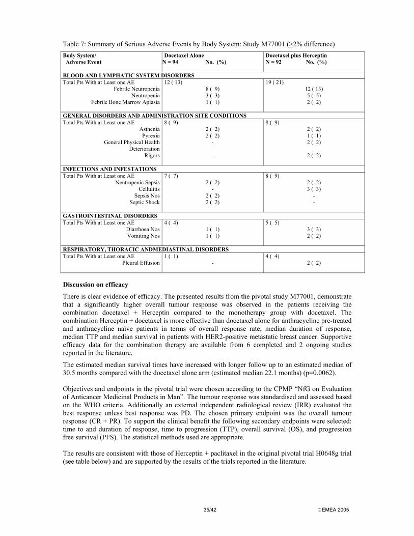
35/42 EMEA 2005
Table 7: Summary of Serious Adverse Events by Body System: Study M77001 (>2% difference) Body System/ Adverse Event
Docetaxel Alone N = 94 No. (%)
Docetaxel plus Herceptin N = 92 No. (%)
BLOOD AND LYMPHATIC SYSTEM DISORDERS Total Pts With at Least one AE
Febrile Neutropenia Neutropenia
Febrile Bone Marrow Aplasia
12 ( 13) 8 ( 9) 3 ( 3) 1 ( 1)
19 ( 21) 12 ( 13) 5 ( 5) 2 ( 2)
GENERAL DISORDERS AND ADMINISTRATION SITE CONDITIONS Total Pts With at Least one AE
Asthenia Pyrexia
General Physical Health Deterioration
Rigors
8 ( 9) 2 ( 2) 2 ( 2)
- -
8 ( 9) 2 ( 2) 1 ( 1) 2 ( 2)
2 ( 2)
INFECTIONS AND INFESTATIONS Total Pts With at Least one AE
Neutropenic Sepsis Cellulitis
Sepsis Nos Septic Shock
7 ( 7) 2 ( 2)
- 2 ( 2) 2 ( 2)
8 ( 9) 2 ( 2) 3 ( 3)
- -
GASTROINTESTINAL DISORDERS Total Pts With at Least one AE
Diarrhoea Nos Vomiting Nos
4 ( 4) 1 ( 1) 1 ( 1)
5 ( 5) 3 ( 3) 2 ( 2)
RESPIRATORY, THORACIC ANDMEDIASTINAL DISORDERS Total Pts With at Least one AE
Pleural Effusion
1 ( 1) -
4 ( 4) 2 ( 2)
Discussion on efficacy
There is clear evidence of efficacy. The presented results from the pivotal study M77001, demonstrate that a significantly higher overall tumour response was observed in the patients receiving the combination docetaxel + Herceptin compared to the monotherapy group with docetaxel. The combination Herceptin + docetaxel is more effective than docetaxel alone for anthracycline pre-treated and anthracycline naïve patients in terms of overall response rate, median duration of response, median TTP and median survival in patients with HER2-positive metastatic breast cancer. Supportive efficacy data for the combination therapy are available from 6 completed and 2 ongoing studies reported in the literature.
The estimated median survival times have increased with longer follow up to an estimated median of 30.5 months compared with the docetaxel alone arm (estimated median 22.1 months) (p=0.0062). Objectives and endpoints in the pivotal trial were chosen according to the CPMP “NfG on Evaluation of Anticancer Medicinal Products in Man”. The tumour response was standardised and assessed based on the WHO criteria. Additionally an external independent radiological review (IRR) evaluated the best response unless best response was PD. The chosen primary endpoint was the overall tumour response (CR + PR). To support the clinical benefit the following secondary endpoints were selected: time to and duration of response, time to progression (TTP), overall survival (OS), and progression free survival (PFS). The statistical methods used are appropriate. The results are consistent with those of Herceptin + paclitaxel in the original pivotal trial H0648g trial (see table below) and are supported by the results of the trials reported in the literature.
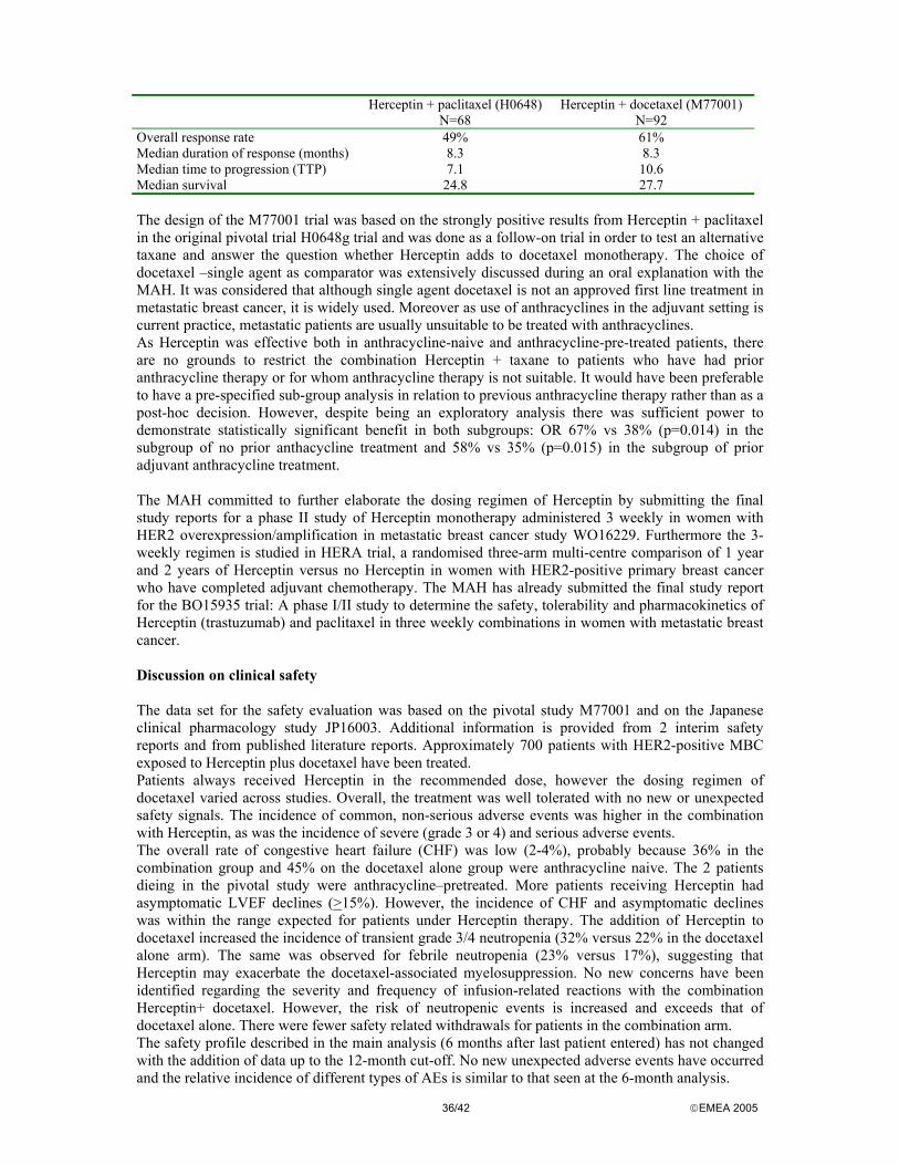
36/42 EMEA 2005
Herceptin + paclitaxel (H0648)
N=68 Herceptin + docetaxel (M77001)
N=92 Overall response rate 49% 61% Median duration of response (months) 8.3 8.3 Median time to progression (TTP) 7.1 10.6 Median survival 24.8 27.7 The design of the M77001 trial was based on the strongly positive results from Herceptin + paclitaxel in the original pivotal trial H0648g trial and was done as a follow-on trial in order to test an alternative taxane and answer the question whether Herceptin adds to docetaxel monotherapy. The choice of docetaxel –single agent as comparator was extensively discussed during an oral explanation with the MAH. It was considered that although single agent docetaxel is not an approved first line treatment in metastatic breast cancer, it is widely used. Moreover as use of anthracyclines in the adjuvant setting is current practice, metastatic patients are usually unsuitable to be treated with anthracyclines. As Herceptin was effective both in anthracycline-naive and anthracycline-pre-treated patients, there are no grounds to restrict the combination Herceptin + taxane to patients who have had prior anthracycline therapy or for whom anthracycline therapy is not suitable. It would have been preferable to have a pre-specified sub-group analysis in relation to previous anthracycline therapy rather than as a post-hoc decision. However, despite being an exploratory analysis there was sufficient power to demonstrate statistically significant benefit in both subgroups: OR 67% vs 38% (p=0.014) in the subgroup of no prior anthacycline treatment and 58% vs 35% (p=0.015) in the subgroup of prior adjuvant anthracycline treatment. The MAH committed to further elaborate the dosing regimen of Herceptin by submitting the final study reports for a phase II study of Herceptin monotherapy administered 3 weekly in women with HER2 overexpression/amplification in metastatic breast cancer study WO16229. Furthermore the 3-weekly regimen is studied in HERA trial, a randomised three-arm multi-centre comparison of 1 year and 2 years of Herceptin versus no Herceptin in women with HER2-positive primary breast cancer who have completed adjuvant chemotherapy. The MAH has already submitted the final study report for the BO15935 trial: A phase I/II study to determine the safety, tolerability and pharmacokinetics of Herceptin (trastuzumab) and paclitaxel in three weekly combinations in women with metastatic breast cancer. Discussion on clinical safety The data set for the safety evaluation was based on the pivotal study M77001 and on the Japanese clinical pharmacology study JP16003. Additional information is provided from 2 interim safety reports and from published literature reports. Approximately 700 patients with HER2-positive MBC exposed to Herceptin plus docetaxel have been treated. Patients always received Herceptin in the recommended dose, however the dosing regimen of docetaxel varied across studies. Overall, the treatment was well tolerated with no new or unexpected safety signals. The incidence of common, non-serious adverse events was higher in the combination with Herceptin, as was the incidence of severe (grade 3 or 4) and serious adverse events. The overall rate of congestive heart failure (CHF) was low (2-4%), probably because 36% in the combination group and 45% on the docetaxel alone group were anthracycline naive. The 2 patients dieing in the pivotal study were anthracycline–pretreated. More patients receiving Herceptin had asymptomatic LVEF declines (>15%). However, the incidence of CHF and asymptomatic declines was within the range expected for patients under Herceptin therapy. The addition of Herceptin to docetaxel increased the incidence of transient grade 3/4 neutropenia (32% versus 22% in the docetaxel alone arm). The same was observed for febrile neutropenia (23% versus 17%), suggesting that Herceptin may exacerbate the docetaxel-associated myelosuppression. No new concerns have been identified regarding the severity and frequency of infusion-related reactions with the combination Herceptin+ docetaxel. However, the risk of neutropenic events is increased and exceeds that of docetaxel alone. There were fewer safety related withdrawals for patients in the combination arm. The safety profile described in the main analysis (6 months after last patient entered) has not changed with the addition of data up to the 12-month cut-off. No new unexpected adverse events have occurred and the relative incidence of different types of AEs is similar to that seen at the 6-month analysis.

37/42 EMEA 2005
The incidence of decreases in LVEF (falls > 15% or absolute value <40%) remains the same. It can be concluded that no new emerging safety concerns could be identified and in principle, the toxicity profile is consistent with that of the two drugs alone. Benefit – Risk assessment Herceptin administered weekly in combination with 3-weekly docetaxel is an efficacious treatment of patients with HER2-positive metastatic breast cancer. The benefit in terms of overall response rate, duration of response, time to progression, and overall survival is comparable with that of the licensed combination Herceptin plus paclitaxel. In principal no new emerging safety signals could be identified. The benefit risk ratio in the indication: in combination with docetaxel for the treatment of those patients, who have not received chemotherapy for there metastatic disease, is therefore positive. 9. Update of the SPC on diagnostic methods to determine HER2 status.
Herceptin, a humanized anti-HER2 antibody is approved for the treatment of MBC patients whose tumours overexpress HER2 as determined by an immunohistochemistry (IHC) diagnostic assay. This overexpression of the HER2 receptor in breast cancer is triggered by amplification of the HER2 gene located on chromosome 17. The amplification leads to increased transcription and consequently to an overexpression of HER2 receptor proteins on the cell surface and is found in 20% to 30% of breast cancer tumours. Only patients with a strong overexpression (IHC score of 3+) are HER2 positive and thus eligible for Herceptin treatment.
The diagnosis of HER2 expression in the pivotal trials was performed using in-house investigational assays. In parallel to the clinical development, a commercial assay was developed by DAKO, the HercepTest® (DakoCytomation). In the meantime diagnostic developments continued and led to the introduction of HER2 testing methodologies based on the detection of HER2 gene amplification which is the initial genetic event that results in HER2 overexpression. Fluorescence in situ hybridisation (FISH) and chromogenic in situ hybridisation (CISH) assays were developed and validated against IHC.
The SPC for Herceptin was updated in order to reflect the recent progress in the diagnostic methods to determine the HER2 status of a patient (previously defined on the basis of an immunohistochemistry (IHC) assay). Fluorescence in situ hybridization (FISH) and chromogenic in situ hybridization (CISH) were included as an alternative to immunohistochemistry (IHC) to assess the eligibility of MBC patients for Herceptin therapy. Methods.
For the individual treatment regimen of a patient with metastatic breast cancer it is essential to determine the HER2 status, because only patients with a strong overexpression (IHC score 3+) that denotes HER2 positivity will benefit from Herceptin therapy. Therefore reliable and robust methodologies for the determination of the HER2 status are required. All assays described below are for usage on paraffin-embedded tumour tissue samples and assess the HER2 status on a cell-by-cell basis.
Immunohistochemistry (IHC) employs antibodies specifically directed against an epitope of the HER2 protein in the tumour tissue, thereby detecting HER2 on the cell surface. HER2 expression in fixed breast tumour samples is recognized by a typical IHC staining pattern of tumour cells and is interpreted semi-quantitatively by the observer, applying a 0 to 3+ scale, where IHC3+ indicates the strongest staining intensity. The advantages of IHC are its wide availability, speed, simplicity and relative low cost.
New methodologies like fluorescence in situ hybridisation (FISH) and chromogenic in situ hybridisation (CISH) detect the genetic event, HER2 gene amplification, which leads to overexpression of HER2 on the cell surface. These DNA-based methodologies directly assess the HER2 gene copy number, and use labelled complementary DNA probes to detect HER2-specific DNA

38/42 EMEA 2005
sequences by hybridisation. Interpretation of the testing results is numeric and more quantitative than IHC. DNA is an inherently more stable target compared to protein as it is less susceptible to degradation. With CISH the HER2 gene is detected using a peroxidase enzyme-labelled probe with a chromogenic detection instead of using a fluorescent (FISH) dye to visualize the HER2 gene copies. One advantage is that a standard light microscope can view CISH staining signals and the histopathology of the specimen can be assessed simultaneously.
To date, two IHC assays, three FISH assays and one CISH assay are commercially available. These are CE marked IVD assays in accordance with Directive 98/79/EC.
IHC assays
- HercepTest (DakoCytomation, Glostrup, Denmark) was developed in 1998, in order to have a diagnostic tool to select patients suitable for Herceptin therapy, as the Clinical Trials Assay (CTA), used in the two pivotal trials for the initial approval of Herceptin, was too impractical for commercialization and widespread clinical use.
- PathWay HER2 assay (for use with the Benchmark® automated System; Ventana Medical Systems Inc., Tucson, USA): was developed in 2000 to aid in assisting the selection of patients for Herceptin therapy whose tumours overexpress HER2.
In Europe, MBC patients are eligible for receiving Herceptin when their tumours express HER2 at an IHC score of 3+ (on a visualisation scale of 0, 1+, 2+, 3+).
FISH assays for detecting HER2 gene amplification
- PathVysion FISH assay (Abbott Laboratories, Abbott Park, USA) uses a 190 Kb DNA probe directly fluorescence labelled with Spectrum Orange. The probe is specific for the HER2 gene locus 17q11.2-q12. In addition to the HER2 specific probe, this assay also includes another DNA probe, which is labelled with Spectrum Green and specific for the centromere region of chromosome 17 (17p11.1-q11.1) known as the CEP17 probe.
HER2 scoring is based on the ratio of the average number of HER2 and CEP17 gene copy signals observed per nucleus with a signal ratio of ��2.0 considered to indicate HER2 amplification.
- HER2 FISH pharmDx™Kit (DakoCytomation, Glostrup, Denmark) employs a ready-to-use FISH probe mix based on a combination of peptide nucleic acid (PNA) and DNA technology. The probe mix consists of a mixture of Texas Redlabelled DNA probes covering a 218kb region including the HER2 gene on chromosome 17, and a mixture of fluorescein-labelled probes targeted at the centromeric region of chromosome 17 (CEN-17). The specific hybridisation to the two targets results in formation of a distinct red fluorescent signal at each HER2 gene locus and a distinct green fluorescent signal at each chromosome 17 centromere. Using a fluorescence microscope equipped with appropriate filters, tumour cells are located and counting of red (HER2) and green (CEN-17) signals is conducted. HER2/CEN-17 signal ratio �� 2.0 indicates HER2 amplification.
- INFORM® HER2/neu Probe (for use with the Benchmark® or Benchmark XT automated slide stainers; Ventana Medical Systems Inc., Tucson, USA) includes a biotin-labelled locus-specific HER2 probe. The hybridized HER2 probe is detected by a ligand with a fluorescent label which binds to the biotin label on the DNA probe. The HER2 gene copy number is enumerated without normalizing for chromosome 17 copy number since the INFORM HER2/neu Probe does not include a centromere control probe. A HER2 gene copy number > 4 has been established as the optimum cut-point to differentiate amplified versus nonamplified samples.
CISH assays for detecting HER2 gene amplification
- Zymed SPOT-Light HER2 CISH Kit (Zymed Laboratories Inc., South San Francisco, USA) includes a double-stranded DNA probe labelled with digoxigenin, which binds specifically to the HER2 gene locus on chromosome 17q12-21. CISH staining results may be assessed with a standard brightfield microscope after visualisation with the conventional peroxidase reactions. Tumour cell nuclei with HER2 gene amplification appear as large peroxidase-positive
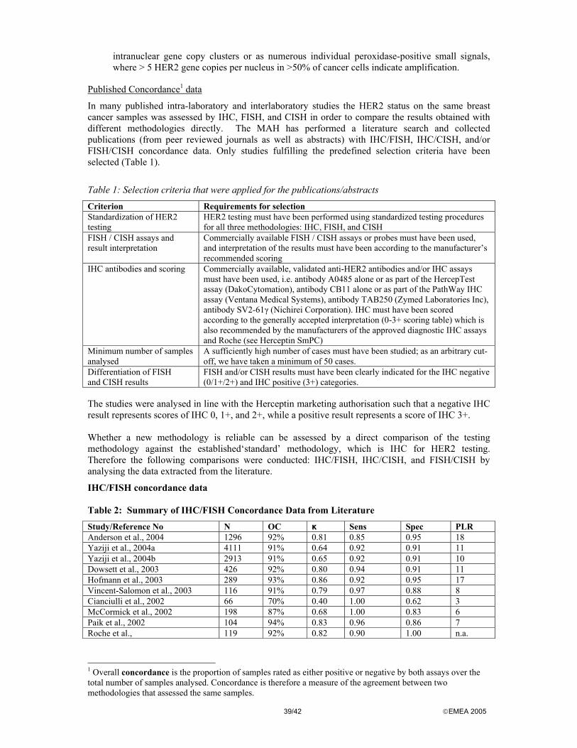
39/42 EMEA 2005
intranuclear gene copy clusters or as numerous individual peroxidase-positive small signals, where > 5 HER2 gene copies per nucleus in >50% of cancer cells indicate amplification.
Published Concordance1 data
In many published intra-laboratory and interlaboratory studies the HER2 status on the same breast cancer samples was assessed by IHC, FISH, and CISH in order to compare the results obtained with different methodologies directly. The MAH has performed a literature search and collected publications (from peer reviewed journals as well as abstracts) with IHC/FISH, IHC/CISH, and/or FISH/CISH concordance data. Only studies fulfilling the predefined selection criteria have been selected (Table 1).
Table 1: Selection criteria that were applied for the publications/abstracts
Criterion Requirements for selection Standardization of HER2 testing
HER2 testing must have been performed using standardized testing procedures for all three methodologies: IHC, FISH, and CISH
FISH / CISH assays and result interpretation
Commercially available FISH / CISH assays or probes must have been used, and interpretation of the results must have been according to the manufacturer’s recommended scoring
IHC antibodies and scoring Commercially available, validated anti-HER2 antibodies and/or IHC assays must have been used, i.e. antibody A0485 alone or as part of the HercepTest assay (DakoCytomation), antibody CB11 alone or as part of the PathWay IHC assay (Ventana Medical Systems), antibody TAB250 (Zymed Laboratories Inc), antibody SV2-61γ (Nichirei Corporation). IHC must have been scored according to the generally accepted interpretation (0-3+ scoring table) which is also recommended by the manufacturers of the approved diagnostic IHC assays and Roche (see Herceptin SmPC)
Minimum number of samples analysed
A sufficiently high number of cases must have been studied; as an arbitrary cut-off, we have taken a minimum of 50 cases.
Differentiation of FISH and CISH results
FISH and/or CISH results must have been clearly indicated for the IHC negative (0/1+/2+) and IHC positive (3+) categories.
The studies were analysed in line with the Herceptin marketing authorisation such that a negative IHC result represents scores of IHC 0, 1+, and 2+, while a positive result represents a score of IHC 3+. Whether a new methodology is reliable can be assessed by a direct comparison of the testing methodology against the established‘standard’ methodology, which is IHC for HER2 testing. Therefore the following comparisons were conducted: IHC/FISH, IHC/CISH, and FISH/CISH by analysing the data extracted from the literature.
IHC/FISH concordance data Table 2: Summary of IHC/FISH Concordance Data from Literature
Study/Reference No N OC κ Sens Spec PLR Anderson et al., 2004 1296 92% 0.81 0.85 0.95 18 Yaziji et al., 2004a 4111 91% 0.64 0.92 0.91 11 Yaziji et al., 2004b 2913 91% 0.65 0.92 0.91 10 Dowsett et al., 2003 426 92% 0.80 0.94 0.91 11 Hofmann et al., 2003 289 93% 0.86 0.92 0.95 17 Vincent-Salomon et al., 2003 116 91% 0.79 0.97 0.88 8 Cianciulli et al., 2002 66 70% 0.40 1.00 0.62 3 McCormick et al., 2002 198 87% 0.68 1.00 0.83 6 Paik et al., 2002 104 94% 0.83 0.96 0.86 7 Roche et al., 119 92% 0.82 0.90 1.00 n.a.
1 Overall concordance is the proportion of samples rated as either positive or negative by both assays over the total number of samples analysed. Concordance is therefore a measure of the agreement between two methodologies that assessed the same samples.
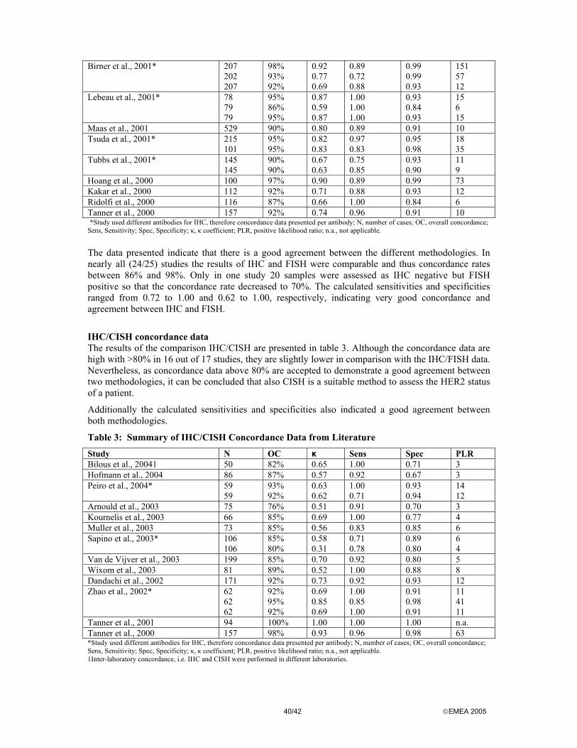
40/42 EMEA 2005
Birner et al., 2001* 207
202 207
98% 93% 92%
0.92 0.77 0.69
0.89 0.72 0.88
0.99 0.99 0.93
151 57 12
Lebeau et al., 2001* 78 79 79
95% 86% 95%
0.87 0.59 0.87
1.00 1.00 1.00
0.93 0.84 0.93
15 6 15
Maas et al., 2001 529 90% 0.80 0.89 0.91 10 Tsuda et al., 2001* 215
101 95% 95%
0.82 0.83
0.97 0.83
0.95 0.98
18 35
Tubbs et al., 2001* 145 145
90% 90%
0.67 0.63
0.75 0.85
0.93 0.90
11 9
Hoang et al., 2000 100 97% 0.90 0.89 0.99 73 Kakar et al., 2000 112 92% 0.71 0.88 0.93 12 Ridolfi et al., 2000 116 87% 0.66 1.00 0.84 6 Tanner et al., 2000 157 92% 0.74 0.96 0.91 10 *Study used different antibodies for IHC, therefore concordance data presented per antibody; N, number of cases; OC, overall concordance; Sens, Sensitivity; Spec, Specificity; κ, κ coefficient; PLR, positive likelihood ratio; n.a., not applicable. The data presented indicate that there is a good agreement between the different methodologies. In nearly all (24/25) studies the results of IHC and FISH were comparable and thus concordance rates between 86% and 98%. Only in one study 20 samples were assessed as IHC negative but FISH positive so that the concordance rate decreased to 70%. The calculated sensitivities and specificities ranged from 0.72 to 1.00 and 0.62 to 1.00, respectively, indicating very good concordance and agreement between IHC and FISH.
IHC/CISH concordance data The results of the comparison IHC/CISH are presented in table 3. Although the concordance data are high with >80% in 16 out of 17 studies, they are slightly lower in comparison with the IHC/FISH data. Nevertheless, as concordance data above 80% are accepted to demonstrate a good agreement between two methodologies, it can be concluded that also CISH is a suitable method to assess the HER2 status of a patient.
Additionally the calculated sensitivities and specificities also indicated a good agreement between both methodologies.
Table 3: Summary of IHC/CISH Concordance Data from Literature
Study N OC κ Sens Spec PLR Bilous et al., 20041 50 82% 0.65 1.00 0.71 3 Hofmann et al., 2004 86 87% 0.57 0.92 0.67 3 Peiro et al., 2004* 59
59 93% 92%
0.63 0.62
1.00 0.71
0.93 0.94
14 12
Arnould et al., 2003 75 76% 0.51 0.91 0.70 3 Kournelis et al., 2003 66 85% 0.69 1.00 0.77 4 Muller et al., 2003 73 85% 0.56 0.83 0.85 6 Sapino et al., 2003* 106
106 85% 80%
0.58 0.31
0.71 0.78
0.89 0.80
6 4
Van de Vijver et al., 2003 199 85% 0.70 0.92 0.80 5 Wixom et al., 2003 81 89% 0.52 1.00 0.88 8 Dandachi et al., 2002 171 92% 0.73 0.92 0.93 12 Zhao et al., 2002*
62 62 62
92% 95% 92%
0.69 0.85 0.69
1.00 0.85 1.00
0.91 0.98 0.91
11 41 11
Tanner et al., 2001 94 100% 1.00 1.00 1.00 n.a. Tanner et al., 2000 157 98% 0.93 0.96 0.98 63 *Study used different antibodies for IHC, therefore concordance data presented per antibody; N, number of cases; OC, overall concordance; Sens, Sensitivity; Spec, Specificity; κ, κ coefficient; PLR, positive likelihood ratio; n.a., not applicable. 1Inter-laboratory concordance, i.e. IHC and CISH were performed in different laboratories.
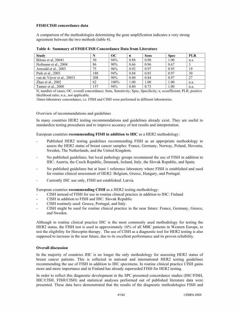
41/42 EMEA 2005
FISH/CISH concordance data A comparison of the methodologies determining the gene amplification indicates a very strong agreement between the two methods (table 4). Table 4: Summary of FISH/CISH Concordance Data from Literature
Study N OC κ Sens Spec PLR Bilous et al, 20041 50 94% 0.88 0.90 1.00 n.a Hofmann et al., 2004 86 90% 0.66 0.96 0.67 3 Arnould et al., 2003 75 96% 0.92 0.97 0.95 19 Park et al., 2003 188 94% 0.84 0.85 0.97 30 van de Vijver et al., 20031 208 90% 0.80 0.84 0.97 27 Zhao et al., 2002 62 100% 1.00 1.00 1.00 n.a. Tanner et al., 2000 157 94% 0.80 0.73 1.00 n.a. N, number of cases; OC, overall concordance; Sens, Sensitivity; Spec, Specificity; κ, κcoefficient; PLR, positive likelihood ratio; n.a., not applicable. 1Inter-laboratory concordance, i.e. FISH and CISH were performed in different laboratories.
Overview of recommendations and guidelines
In many countries HER2 testing recommendations and guidelines already exist. They are useful to standardize testing procedures and to improve accuracy of test results and interpretation. European countries recommending FISH in addition to IHC as a HER2 methodology:
- Published HER2 testing guidelines recommending FISH as an appropriate methodology to assess the HER2 status of breast cancer samples: France, Germany, Norway, Poland, Slovenia, Sweden, The Netherlands, and the United Kingdom.
- No published guidelines, but local pathology groups recommend the use of FISH in addition to IHC: Austria, the Czech Republic, Denmark, Ireland, Italy, the Slovak Republic, and Spain.
- No published guidelines but at least 1 reference laboratory where FISH is established and used for routine clinical assessment of HER2: Belgium, Greece, Hungary, and Portugal.
- Currently IHC use only, FISH not established: Latvia. European countries recommending CISH as a HER2 testing methodology: - CISH instead of FISH for use in routine clinical practice in addition to IHC: Finland - CISH in addition to FISH and IHC: Slovak Republic - CISH routinely used: Greece, Portugal, and Italy. - CISH might be used for routine clinical practice in the near future: France, Germany, Greece,
and Sweden. Although in routine clinical practice IHC is the most commonly used methodology for testing the HER2 status, the FISH test is used in approximately 16% of all MBC patients in Western Europe, to test the eligibility for Herceptin therapy. The use of CISH as a diagnostic tool for HER2 testing is also supposed to increase in the near future, due to its excellent performance and its proven reliability.
Overall discussion
In the majority of countries IHC is no longer the only methodology for assessing HER2 status of breast cancer patients. This is reflected in national and international HER2 testing guidelines recommending the use of FISH in addition to IHC specimens. In routine clinical practice CISH gains more and more importance and in Finland has already superseded FISH for HER2 testing.
In order to reflect this diagnostic development in the SPC presented concordance studies (IHC/FISH, IHC/CISH, FISH/CISH) and statistical analyses performed out of published literature data were presented. These data have demonstrated that the results of the diagnostic methodologies FISH and
42/42 EMEA 2005
CISH are accurate, reliable and robust. There is a strong association between HER2 overexpression and HER2 gene amplification demonstrating that direct determination of the HER2 gene copy number offers a valid alternative for assessing HER2 positivity. Thus FISH or CISH can be used for either primary HER2 testing or for re-testing cases for which the initial test result is difficult to interpret.
Specific characteristics of the different diagnostic methodologies including the interpretation of the test results are adequately addressed. In clinical practise, the quality of HER2 testing will depend on a good validation of the test methodology in the laboratories and an intra- and interlaboratory quality control and quality assurance.
There is ongoing scientific discussion about C/FISH methods and their concordance with (1) immunohistochemistry (IHC; can be negative despite positive IHC and vice versa) and (2) prediction of clinical response. Discussion on this concordance is controversial. The initial marketing authorization for Herceptin included data from a rather small study that showed a clinical benefit only in patients with strong overexpression of HER2 (3+). The efficacy database has not changed up to now, and thus, this target population should be kept. However, the situation “in the real world” might be different with various tests available. It might be considered if (1) the common approach should be undertaken to include any new method that emerges over time by an own type II variation, or (2) if in these sections a more general guidance could be given that gives a certain framework new methods have to stick to but would be allowed to be performed.
Amendments to the SPC.
This discussion is reflected in the SPC as follows: The indication section of the SPC includes a statement: Herceptin should only be used in patients whose tumours have either HER2 overexpression or HER2 gene amplification as determined by an accurate and validated assay and refers to 4.4 and 5.1 where general guidance is given. The guidance is strict enough to preclude use of this drug in patients with insufficient HER2 expression, since the risk-benefit ratio for these patients is critical due to considerable possible side effects of the drug.