scientific proceedings sixth symposium of the section of
TRANSCRIPT
Scientific Proceedings Sixth Symposium of the Section of Experimental Cancer Research (SEK) of the German Cancer Society
Sektion Experimentelle Krebsforschung (SEK) der Deutschen Krebsgesellschaft e.V.
Vorsitzender/Chairman: F. Oesch (Mainz) Stellv. Vorsitzender/Vice-Chairman: G. Eisenbrand (Kaiserslautern)
Wissenschaftl. Beirat/Scientific Council: H. Grunicke (Innsbruck), K. Havemann (Marburg), W. K. Lutz (ZSrich), K. MSIling (Berlin), H. Rabes (Menchen), M. F. Rajewsky (Essen), V. v. Schirrmacher (Heidelberg), M. Schwab (Heidelberg)
Sixth SEK-Symposium Heidelberg, 10-12 April, 1991
432 J Cancer Res Chn Oncol ISSN 0171-5216 JCROD7 117 (Suppl) $1 -$60 (1991) 017152169100034H
B1 EVIDENCE FOR ACTIVATION OF PHOSPHOLIPASE D INDUCED IN INTACT CELLS BY EPIDERMAL GROWTH FACTOR
Marietta ~ . ~ , n , Vo]ker Kmz¢l Department of Pathochmmsu'y, German Cancer Resea~h Center, Im Neuenhcnner Feld 280, D-6900 Heidelberg
S,gnal transducuon reduced by epidermal growth factor (EGF) vm ,ts receptor revolves two second messengers, chacylglycerol (DG) and momtol msphosphate, both products of phosphandylmo~to|-specdic
phosphohpase C wluch seem to pamctpate m the regulauon of cellular prohferauon by EGF We observed that A431 cells and HeL~ cells m response to EGF accumulate substanual amounts of phosphauchc actd (PA), the potenual product of DG kmase However, PA appears earher and stays longer elevated than expected from DG avadabxhty thus rinsing the poss~bd~ty of an EGF-mduced acuvatxon of a phosphohpase D (PLD) In the presence of prm=ry alcohols PLD effects the phosphandyl transfer reacuon producmg PA-alcohol, a reacuon known to be excluszvely ~ by PLD m mtact cells A431 cells and cells prelabeled w~th [14C]-oleate produce m the presence of ethanol (0 5%) or l-butanol (0 2%) substanual amounts of labeled PA-alcohol m response to EGF These results mchcate that acuvauon of PLD had occurred Acuvauon of PLD may x~present a novel mgnal transducing pathway wluch may pamctpate m the regulauon of cell prohferauon by EGF - a hypothes~s presently mvesugated m thts laboratory Supported by the Deutsche Forschungsgememschaft.
$1
B3 CASEIN KINASE H ANTISENSE OUGODEOXYNUCLEOTIDES INHIBIT MITOGENIC SIGNALING IN HUMAN FIBROBLASTS
R. Peppcrkok*, R. Jakob,, P Lorenz, W Ansorg¢* and W Pyerm Insumtc of Expcrm~ntal Pathology, DKFZ, and *European Molecular
Biology Laboralx~y, D-6900 Heidelberg, F R G
Mttogens such as the ¢p,dcrmal growth factor (EOF) or serum s.m,,1~c, when apphcd to cell cultures, n'anmendy CK II ac~wty of cy~osol suggcsung a role of CK u m nntogemc mgnahng The human CK II is a te~amenc enzyme (I) composed of two lanase-ac~ve subumts (a and a') and two regulatory subumts (B). Subumts ct and 13 w~'e sequenced recently (2,3), thus allowing to mvesugatc the role of CK II m nntogemc processus by anusense ohgcdeoxynucleoude methodology We demonsu~e that syntheuc ohgodeoxynucleoudes complementary to mRNA of ct and B subumts strongly mh,hlt the serum and the EGF prohferanon smnulanon of qmcscent pmmry human fibroblasts. When apphcd 2 h prior EGF, anUscnsc-ct and anusense-B mlubzted growth sumulauon by 51% and 64%, respccuvely, and when apphed prior serum, by 31% and 44%, ~ v c l y . Introdacuon of mutaUons into the ohgodeoxynucleoudes reduced thelr anuprohferauve effect, and apphcauon of sense-ohgodeoxynucleoudes neuwahzed the effect demonsu'almg the iugh specificity of the anusense acuon. The effect was revcrsthle, L¢., cells re-antered cell cycle after ohgodeoxynucleoud¢ removal. Thus the conunuous avadabxhty of CK H subumts ~s essential for the m~togcmc m~m.hng by senun and EGP to occur C~mges of CK H subumls at pro~n level arc cm'nmfly und~ mvesugauon. References ~ c~ aL (1987) B~oL Chem. Hogpe-Seyl= 368, 215- 227, (2) Mcu.e~ c~ al. (1989) Bmcbem~7 28, 4072-4076, (3) Sakol~, Voss. Pyenn (1989) Eur J. Btocbem. 183, 22%233
B2
UFE~ OF ~-~ ON BONBESIN-INDUCED 0PB~G OF K ÷- CHm~LS
Karl l~ly, Irina Bidder*,. F10rian I~g* and lbns H.Gru~.cke
Institute of Medical Chemistry and Bi.oche[i.'stry and the *Institute of Physlolocs, U~verslty o~
Innsbruck, Innsbruck, Austria
Addition of bombesin to quiescent NIH 3T3
flbroblasts transfected with a glucocort£oold-
inducible Ha-ras construct, elicits persistent
oscillating hyperpolarlsstlons of the plasma
membrane attributable to the activation of Ca 2+-
dependent K + channels. In the absence of the
inducer, bombesin evokes only single, transient
hyperpolarisations. In contrast to the activation
of K + channels, expression of Ha-ras attenuates
bombesln-induced inositol trisphosphete formation
and causes a reduction of the bombesin-medlated
release of intracellular Ca 2+ . The bombesin induced
hyperpolarlsatlon is insensitive to pertussls-
toxin, whereas the activation of phospholnosltide
hydrolysis by bombesln is inhibltable by the toxin.
As the mitogenlc activity of bombesin is enhanced
in cells expressing the Ha-ras oncogene, the
stimulation of the Ca 2+ dependent K+-channels seems
to be related to the mitogenic signal transduction
of bombesin.
B4 TUMOR PROMOTION AND PROTEIN KINASE C
P Borowslo, M Harbers, A Ludw,g, T Kischel, H Hdz Inshtut fOr Physmlogtsche Chem,e, Umvers,t~t Hamburg
TPA-,nduced promotion of ,ndmted ep,dermal cells m mouse skin can be mh,bltod by banzamlde (Bam) and analogs Barn effecbve- ly suppressed mflammatmn and accumulat,on of whrte blood cells ,n TPA-treated slon wrthout affect,on of hyperprol,feratton, sugge- sting that Its a~promotor effect came about by the ehm,n~on of granulocyte-danved reactwe oxygen species However, Barn and analogs also suppressed TPA-~nduced promot,on of "inrbatod" ept- dermal JB6 cells to the tumor phenotype (anchorage-,ndepsndent growth tumor promobon m nude mce) ,n the absence of granulo- cytes mdtat, ng that mechanisms not mvolwng reactive oxygen were respons,ble for promobon S,nce promotmn tn v~vo and ,n wt- ro requ,res prolonged exposure to TPA, we conmdered roducod rather than enhanced actrvdy of protein Ionase C (PKC) as a s, gnal for the ,nductmn of the tumor phenotype Ev,denca for such a me- chan,sm was provided by a Western blot analyses of TPA-mduced PKC deplebon, by the promobng act,wty of certain PKC ,nh0b~ors m the absence of TPA and by the synerglst,c effect wdh TPA of PKC anbsense ohgonucleot,des Promobon can also be effected by EGF and by TGF-~ Co-admmistrat,on of Peltusms toxin ehm,- hated ,nductton of anchorage-mdependant growth by all these pro- mot,ng substances A G,-I,ke protein, therefore, appears to be ,n- volvod m the transmlssmn of the s,gnal leading to the tumor phe- notype Part,al am,no acld sequence of G, ADP-nbosylated m wv( as well as ,mmunologtal data radiate that the PTX-sens~hve acceptor is a o,-2 contmnmg G, subspecms. A pcesable I,nk bet- ween PKC, active G, protein, and tumor promotmn wdl be disoas- sod
$2
B 5 MONO(XX)NAL AND POLYCLONAL ANTIBODIES AGAINST
CASEIN KINASE H SUBUN1TS
P ~ n z , A Wedell, RJakobx, W.Pycam
Insutate of Experimental Pathology, DKFZ, D-6900 Hezdelberg, F.R.G
Anuboches represent sensmve and effecave tools to mvesUgate slructure and functton of In'grams The Casem kmase ]I (CKII) ts composed of two catalync (a and (z') and two p~sumably regulalm~ subumts (~) These polypepudes show markedly conserved an~no acul sequences explaining the fadure to generate anuboches w~th the purified enzyme by routine strate~es Our approach therefore was to express the CKII subumts as fu~on proteins m E. coh usmg cDNA-cloues coding for b;~m~q CKILa and -~ subumts, respecuvely, developed m our lahorat:¢y, and a pEX31-denved expresston-veetor (Strebel et aL (1986) JVlrol $7 , 983@91) Three dLRerent fu.~on genius , hnkmg the polypcpudes to the N-terminal 10 5 kD-fragment of the MS2 RNA-polymerese J~-cham, were expressed under control of the temperature reducible ZPL pmmo- to~, (xFus54 contmmng the whole CKHa-cham, oFus26 containing amino acui sequence 1-128 of a-subumt, and [~Fus32 containing mmno aczd sequence 20-200 of ~subumt. Purtficuuon of fumon proteins was facthtated by thetr msolubdxty m 1M urea m contrast to the bulk of bac- terial proteins Crude sera obunned after one prm~mg and three booster mjeeuons mto xabbx~ thsplayed lngh rea~wty wzth human CKH holonnzyme (ELISA) and the resl~Uve subumts (Western blotting) For monoclonal anubody producuon, hybndomas were generated by fusing spleen cells of a mouse hyper-zmmumzed w~th [~Fus32 and mouse Ag8 653 myeloma cells Four clones secrelmg specific monoclonal aria- bodies reacting w~th the CKII holoenzyme were u:lentxfied by ELISA. They were grown m scram-free mechum and the anfihoches purified by chromatography on protein A- or protein G-sepharose Three of them were able to detect ~ protein on Western blots, even m crude cell exwacts Two of them were classified as IgG1, one as IgG2b and the fourth as IgG3, all of them possessing t¢ hght chains
B 7 GLUCOCORTICOID RECEPTOR DETERMINATION IN
ACUTE CHILDHOOD LEUKEMIA WITH REGARD TO THEIR CLINICAL RESPONSE TO STEROID THERAPY
V Mauch, S Wehner. B Komhuber and U Ebener Chm c of Pedmmcs~Dept. of Hematolo~v and Oncolo~f, J W Goethe
umv , Thendur-~tem-Km 7, D-60(0FFrankft~/M 70, Germany
The determma~on of the GCR-level m acute lenkexmas stall play an tm- porlant role m evaluating the chmcal response of leukemxas to steroul therapy. In adthuon to other factors ndluencmg the response to therapy, zt seems to offer a clue to the prognos~s of the chfferent types of leukenn- as In our study we concentrated on the mvesUga~on of these btochemtcal markers m leuken~ blasts m chddren (n=16) after phenotypmg vath an ~rnmKno~lzyl~1~C stamulg (APAAP-complex-techmque) The mononu- clear coIls (MNC) of tso!~_!_~_ bone marrow aspwa~s (BM) were analysed using a large panel of monoclonal anubed~s (MoAbs) m order to deter- nnne the lineage of the leukem¢ blasts as well as the~ stage of dd~e~n- ua~on. Accordmg to the results a classLficunon was rained out (n=16, 8 cALL, 4 AML, 3 T-ALL, I AUL) The GCR-level was meamn'ed by a whole eell-bmdmg-assay using [3H'J-Dexamethasone (DX) In each ex- periment the lenkennc cells were incubated ezther vnth racholabelled DX alone or in the present of a greater than I00 fold excess of unlabolled DX m order to determine the spee~c binding of the tracer In addmon to f~ddy from bone man-ow prepared leukermc ceils we also mvesugated the lealm~c cell line Molt 4 for th~tr receptor level as well as peripheral blood lymphocytes from healthy donors The number of receptor s~tes was esuma~ed mandy by sea¢haxd plot analysts In some experiments, due to the hn~ted cell numbers, comparusons were made by determining receptor binding at a single concenlrauon (near mammum) of [3H]-DX The results to-date point mto the dLreCUon that among the group of ALL the ~eceptor level ranged from 2 800 - 11.300 s~tes/cell (spec~c bmdmg between 21% and 67%) The GCR-level of the AML probes showed a low specdic binding rangmg from 2 100 to 8 400 snes/eell (percentage of spectfic binding between 15% and 60%) The receptor level of the cell line Molt 4 averages around 50 3%, which amounts to a receptor level of 8.600 sxtedcelL All results gathered flora our expenmunts so far are m favour of a corre- lallon between the GCR-level and the response to G(~-therapy although among the cases of ALL 2 cases showed devumng results
B 6 RELATIONSHIP BETWEEN 43 KDs EPIDERMAL GROWTH FACTOR-RELATED CLONOGENIC ACTIVITY AND CLINICAL PARAMETERS FOR BREAST-CANCER
K. E e k e r t , A. Granetzny , 3 . F i s c h e r , and R. Orosse, C e n t r a l I n s t i t u t e o f M o l e c u l a r B i o l o g y , ( K . E . , R . G . ) , C e n t r a I I n s t i t u t e o f Cancer Res. ( A . G . , 3 . F . ) , Ber lJn -Buch
The c lonogenJc a c t i v i t y o f e p r e v i o u s l y p u r i f i e d 43 kOs E G F - r e l s t e d p r o t e i n was e s t i m a t e d In e B i g - G e l P -30 f r a c t i o n o b t a i n e d from the ur ine o f b r e a s t cancer p a t i e n t s . I n c r e a s e o f a c t i v i t y was s t a t i s t i c a l l y s i g n i f i c a n t In lymph n o d e - p o s i t i v e p a t i e n t s , in a group o f p a t z e n t s w i t h l a r g e r c a r - cinomas, w i t h a c c e l e r a t e d tumor growth, in p r e - menopausal p a t i e n t s end younger age and in e s t r o - gen r e c e p t o r n e g a t i v e p a t i e n t s . In 31 p a t i e n t s the a c t i v i t y was e s t i m a t e d d u r i n g p o l y c h e m o t h e r s - py b e f o r e s u r g e r y . D i f f e r e n c e s between the v a l u e s a t the begin end the end o f t h e r a p y were compared between a l l groups be the K r u s k e l - W a l l i s t e s t ( p = 0 . 0 2 ) . P a t i e n t s with progress i ve d i s e a s e showed i n c r e a s i n g a c t x v l t i e s (mean v a l u e s from 31S to 811) w h i l e In those w i t h comple te remis- s ion a c t i v i t y decreased ( f rom 449 to 2 1 3 ) . P a r - t l a l - and non responders showed no change. I n a l o n g - t e r m f o l l o w - u p study wi th 25 p a t i e n t s the p r e - and p o s t o p e r a t i v e a c t i v i t i e s were e s t i m a t e d . In 17 o f 18 p a t i e n t s who had no l o c a l r e c u r r e n c e or m e t a s t a s i s the a c t x v t t y d e c l i n e d a f t e r s u r g e r y , whereas tn 6 o f 7 p a t i e n t s who d i e d , a c t i v i t y I n - creased 2-S f o l d p r i o r dea th . A l i f e t a b l e a n a l y - s~s w i t h a t o t a l o f 101 p a t i e n t s r e v e a l e d a t r e n d to s h o r t e r s u r v i v a l In the group w i t h h i g h e r a c t l - v z t y ( p = 0 . 0 4 2 ) . These o b s e r v a t i o n s suggest a r o l e f o r the EGF-11ke growth f a c t o r a c t i v i t y in b r e a s t cancer d i a g n o s i s and p r o g n o s i s .
B 8 DEVELOPMENT OF AN IN VITRO IRRITANCY AS AN ALTERNAT/VE TO THE ANIMAL DP.AIZE TEST
IC M61kr.Decker, G. F~mberser, L Vogt mM F Marks lasmnte of B~J~m~lry, German Cancor ~__p~c_ h Cm~r, Im Nemobcanor Fold 280, PRG-6900 HeKiePoe~
k mm¢ mmcny scamnng the rabbn eye umauon lest muodm:ed by DraBs ~s ~e me~l~__ cmreafly used to evelua~ lha mnlam ~ of chmucals de~gned for mdumrml phannacemcal a~d oosmeac me 3)m to ~ lack of vahdacd almmnves, flas te~ m s~U m me desp~ lhe ~ md educM cnnc~ms and tha lush costs Rccemly, vat, gum methods have been developed w~h the goal w repboe pamal|y or wholly the ~ I use m ~ ~ In ~ study a mecbamsnc approach zs cummdy ~ram~d to develop ~n a~amuve m vcro mecha& T~ sua~egy Is hased cn the steadily ucreasmg body of evldem,¢ mdcaang that chemcal and physcsl skin mmm~, iz~mq~mma~my me~haaxs and woaad hormo~s aldmugh followmg dd~e~m mechsms~ routes m epidermal tells reduce Ih¢ ,-eldest of amcludemc ac~d (AA) a~l ~ menboham into h,Bldy asUve b~mokcules along the cyclooxygemse and ~ p~hway Th~s, aracludomc acKI rekase and proslanold fonnanoo m I~a/mocy~ cells appear to be pmm~ng ca~hda~ for ,n ~uo paramut~ of n~mcy To ~ d a smlable ~es~ culm¢, skin d~n~l Immary and secondary haman ~ winch are as..quned to be d~e pnn~zy I~'g~ for n~tants m eWderm~s were compmed vnth x'espe~ to ~ catm~v/ to pmduc~ e~cosano~ a ~ chdkngmg wuh m-m~s or ixmnflammmmy mechators.
Imual sladcs revealed dmt the lxofik of e¢osanmds released upon ~aln~¢nt was mmlO¢ for inch can vjpe __~_a_: In Ml cea type~ mad, U~e relea~ of arachdon¢ as~d was smaukw, d by such as d~e nrnam Ca++~caop~ A2318"/, tha PKC acuvamr TPA or tha p¢ix~k ~ ~ a few memne¢ Whh respect to pros~mozd syn~e~s quid]laUra dlffeZ~lteS between tha tell types w~'¢ noted. Whd¢ m Izrm~a~ NHEK c~I]S and m Iha HPKII lme a izmdlel mc~mse OF bod~ AA and PGE2 was measurvd when Ih~ cells wore Irtalcd wzth bradylonm or A23187 m the HACAT cell ime a 2-4 hour delay of I~E 2 synthe~s as compared w~h AA.~Aease redressing zmmediately aft~ ~ of ~mnent was observed. Mm'eovm', m HACAT cells TPA-mduced pmslano~ release was foond to be mh~led by astmomy~m D and cyclolP.~,,mude md~m~g Irav.~zpumml mi wm~lanonal co~m~ of PGH-synthase These ~uha suBge~ dmt there ,,:" a couelauou between exposure to m'Imms and ¢~cosanod release m Immm~ EFlPKH aod NI.IEK bmmm:¢yu:s. 'These calls may prove ~refc~.., m be su~able as anunal fr~ ~.~ system foe dermal u n i t s
B9 LASER PHOTODYNAMIC TUI~ItAPY (PDT) AND PHOSPHOLIPID METABOLISM IN HUMAN TUMOR CELLS - 202 MHz mp-NMR
INVESTIGATIONS G Graechew, A Kilsel, W Hull, W. Lorenz, H. W Thielmann,
German Cancer Research Center, D-8900 Heidelberg, FRG
Aim To increase our basic understanding of sSP-NMR spectra o-~-talned at low field from tumors in patients and to lnterprete the changes occurring during tumor growth and therapy we investigated (saP-NMR spectra) human tumor xenografts In nude mice at high field and determined quantitatively several phosphollpid metabolttes" nucleosidedi- and -tHphosphates (NDP and NTP), phosphocreatine (PCr) and cytidinedlphosphecholine (CDPC)
~ : Cells of various human carcinomas (CX1 colon, MX1 m~mma) were cultured in basement membrane
gel For NMR measurements 2x10 v living cells embedded in gel vere placed in a home-built sample ehember and perfused at 1-2 ml/min with HAM's F-12 me(bum containing in addition 10~ fetal calf serum and 20 mM HEPES (pH 7 4) After 2 h incubation with PEOM-photesensitizer (S I~g/mi) samples were irradiated with laser light (wavelength. 880 nm, dose rate 80 mW/cm ~. dose 20 J / ca z)
~ e The metabohc response of tumor cells to PDT was d After 4-8 h, the intracellular pH decreased from
7 2 to 6 8 The total cellular phosphate detected decreased by 25-30% (shedding of damaged cells from the gel threads) NTP and NDP decreased from 2S-80% to 10-1S%, PCr levels from about S-8% to I-2% of total phosphate A signal from the ~-phosphate of CDPC at -8.98 ppm was detected CDPC level was S-6% of total phosphate 30 h al~er PDT, the lntracellular pH was identical with the external pH and the total cellular phosphate detected had decreased by 60-70% Only a small number of vital cells (8-556) was observed after PDT in the gel threads
C.oncinsions 1 Using s~P-NMR, It is possible to follow the efficiency of laser photodynamic therapy (PDT) of human tumore in a non-invasive way
After PDT, an increasing CDPC signal was observed and the total cellular phosphate detected decreased indicating a high shedding of damaged cells from the gel threads 3 (~uantitation and determination of the kinetics of CDPC accumulation in the ~P-NMR spectra of human tumors can be used as criteria for the efficiency of the PDT
B 10 COVALENT BINDING TO PROTEINS OF ARACHIDONIC ACID HETABOLITES FORNED BY PROSTAGLANDIN H SYNTNASE
G~sela H Degen and A l e x l u s F r e y b e r g e r I n s t . f~r T o x l k o l o g l e de r U n l v e r s ~ t a t Wuerzburg
S o n d e r f o r s c h u n g s b e r e 3 c h 172
P r o s t a g l a n d l n H s y n t h a s e (PHS) c o n v e r t s a r a c h l - d o n l c a c i d (ARA) t o p r o s t a n o 3 d s which have been ~mpl~cated as i m p o r t a n t m e d i a t o r s ~n tumor p r o - mo t ion and p r o g r e s s l o n . I n t e r e s t l n g l y , when m~crosomal PHS ~s ~ncubated w~th r a d ~ o l a b e l e d ARA, a n o n - e x t r a c t a b l e f r a c t i o n o f t h e r ad~o - a c t i v i t y p r e c i p i t a t e s w i t h p r o t e i n s ~nd~cat~ng f o r m a t i o n o f r e e c t ~ v e f a t t y a c i d ~ntermed~a- t e s / m e t a b o l ~ t e s . P ro te~n b~nd~ng was f u r t h e r s tud~ed ~n ~ncubat~ons w~th l e b e l e d ARA and p u r ~ e d PHS-holoenzyme and a n a l y z e d by SDS- p o l y a c r y l a m ~ d e ge l e l e c t r o p h o r e s ~ s , r a d i o a c t i - v i t y de te rm ined ~n t he s e p a r a t e d p r o t e ~ n bands ~nd l ca ted c o v a l e n t b l n d l n g o f r e a c t i v e ARA meta- b o l ~ t e ( s ) . I n c u b a t i o n s c a r r i e d o u t ~n t he p r e - sence o f album~n (BSA) r e s u l t e d ~n b~nd~ng no t o n l y t o PHS bu t a l s o t o BSA; 1~kew~ee o t h e r p r o t e ~ n s (g lobu1~n , ovalbum~n, a c t ~ n ) c o u l d be c o v a l e n t l y m o d i f i e d by ARA-metabo l~ tes rn v T t r o . The u n s t a b l e p r o s t a g l a n d ~ n (PG) ~n te rmed~ates PGG2 and PGH~ a r e bo th c o n s i d e r e d as c a n d i d a t e s f o r p r o t e ~ n a l k y l a t ~ o n a l t h o u g h t h ~ s a w a i t s f ~ n a l con f~ rmat~on . Our r e s u l t s show t h a t PHS-dependent ox~da t~on o f ARA ~s accompanied by t he f o r m a t i o n o f f a t t y a c i d m e t a b o l l t e s which b3nd c o v a l e n t l y t o p r o - t e~ns . F u r t h e r ~nves t~ga t3on 7n v r v o regard3ng t he p a t h o - / p h y s ~ o l o g 3 c a l s i g n i f i c a n c e o f t h ~ s p r o t e ~ n m o d ~ f l c a t l o n i s r e q u i r e d .
T h l s work has been s u p p o r t e d by a g r a n t f r o m t h e Deutsche For$chungsgemeTnschafC.
$3
B 11 ADDUCTS WITH HUMAN BLOOD MACROMOLECULES AS BIO- MONITOR FOR EXPOSURE TO SUSPECTED CARCINOGENS e.g 1,2-EPOXYBUTENE-3, THE REACTIVE INTERMEDIATE OF 1,3-BUTADIP/~ A. Muller. M Lmscheld*, Ulnke Hmdermcler, Ute Jorrltsma, K Golka, W FSIlmann, H. Peter, H.M Bolt
Exposure to suspected car~uogens is usually momtored by measunng the concentration m ambsent atr or by quanuficauon of the substance or ~ts metabohtes m blood or unne The genotoxtc effect can however not be momtored by these methods, since mtennd~wdual d~fferences m metabo- hsm and stereoehem~cal effects influence the b~olog~cal effectwe dose Binding products of alkylatmg agents with DNA m lymphocyte.s, or as surrogate wath blood proteins, reflect the mdtvldual health risk much better than measunng the parameters menttoned above In pharmacology, albumin "binding" of pharmaceut~cs ss well mvestagated, an rmplementsuon for exposure momtonng m toxicology is however not yet estsbhshed Thts study presents mmal experimental results for the use of adducts of 1,2- cpoxybutenc-3 with human blood macromolecules as b]omomtors which include the effects of mdwldusl variations of the exposure to 1,3-buta&ene
The reacave 1,2-epoxybutene-3 was added to a 5 ml aqueous solutton of 10 mg/ml human hemoglobin or human serum albumin and incubated for four hours m 22 ml head space wals under constant rolling at 370C, then shock frozen m hqmd mtrogen and lyophdlzed to remove umeacted epoxa- de. The proteins were redlSSOlved in 5 ml buffer (10rnmol Tns, 100 mmol sedsum acetate, 0.1 mmol C.aCt,, pH 7 4) and 0.5 ml ahquots were drawn for heat denaturation of proteins (950C for 30 mm and subsequent cooling m ice water prior to trypt~c or protemase K d~gestton with subsequent pept~de mapping through hplc analysks on a RP 18 column)
The alkylated amino acids within the peptsdes were then characterized by an hplc-mass spectrometry analysts Hplc was performed on a RP 18 m~crobore column vath methanol/R,0/l% mfluoroacetlc acd and glycerine for elutmn. Fast Atom Bombardm6nt (FAB) mass spectrometry analys~s was done on a MAT 90 spectrometer (Fmmgan, Bremen) Alkylatton products m human blood proteins were determined by both methods pepude mapping and mass spectrometry following hplc These methods are there- fore stated for further development as m wvo monetors for eccupatmnai exposure to 1,3-butadlene, of wbch 1,2-epoxybutene-3 ]s the mare bmlogl- c.ally reactive and ganotox¢ metabohte
Institut f~r Arbeasphyszologle, Ardeystr 67, W-4600 Dortmund 1 *Instttut filr Spektrochem~e, Bunsen-IGrchhoff-Str 2, W-4600 Dortmund 1
B 12 Adduot p a t t e r n s d e t e c t e d by 3 2 p - p o s t l a b e l l £ n g o f 1 - h y d r o x y n e t h y l p y r e n e s u l f a t e and 1 - c h l o r o - m e t h y l p y r e n e Ln Salmonel la Cyphimuxium and w i t h f r e e sa lmon sperm DNA
S Iosserjabs, ! I Phillips lj , t levertl, A Seidel, ! i . glair Institute of Toxicology, University,So6500 Raise, rig and z~ Seddow Laboratorleu,helsoat, Surrey Sl2 5IG, | [
1-ChlorosetbylHrese (tel), furled fret l-hydrozysethylpyreue sulfate {SIP} In the presence of C1- in phye/ological ueecentrsttsts, iu snub sore uotsgente it Seilolella t~pbt|nr/ua YA98 than 1S SlY (1) AeUSul|g that Plh adduces are the cntee of these sciatic|u, dlfferes©es either is the access to the Slb or in intrinsic reactivity towards 011 could sceOllt for the difference Ln letegeele |ctlvlty Yo dlltttgeish hetgees these tee possibilities, ve bare toe incubated CaP and SIP eltb bacteria nil free Billet sperm PSi in the presence and abselce of C1- lose, and atalyzed the guantity s|d the patters uf I |1 adduces Sider 811 theme conditions sddset| vere formed, the cbrosstogrephic pstterse costal|leg the usse four 1slop and several ninny adduce 8pot8 Ile |h|ttele vttb C|Y lad SlY, in the presence emd ahsetee of ¢1-, resulted is virtually identical adduce patterns, but variable adduce levels. In hsetetis, the adduce levels, like the sntesenie activity, folloeed the order CEY/Cl- > CaP/S:0 ~ $1Y/~I- -- SIP/S20 Ie reactloss vitb free e l | , the relative tsteneity of the haler spots aid the tuber of liner spoil itffered free the results obtained in bacteria, possibly dee to the d/armrest l/g ratios of basterial and ~ertebrete I l l SlY and CÁP forsed very stellar rid|act patterns, the quantitative difference belts limb smaller then i l the ezperisente in bacteria, loreuver, the presence ef Cl- bad oily e stab influence on the binding of SIP Hue resales show that SIP and CEY have qualitatively sad guaititetlvely sassier intrinsic reactivitieo tosardu hi& hnce, the different biological activity in bacteria is attributable tu differential access of the cospoeedu to the target hI1 lhereau tip is highly llpophillc, SiP is ionized st physiological p! valses and Cheretoru say here difficulties in penetrating cell teabranea
1} l . t Glair, i . Bessebler, | B Phillips, J B Blake, P Steinberg, h, Beidel, Y, 0eecb, he/roe lealtb Perepect., 88 {19g|) 43 - 48
$4
B 13 GLUCURONIDATION OF N-NrrROSO-
METHYLPENTYLAMINE IN RATS
K Wiench, E Fre=, P Schroth, M Wiessler
Instdut for Toxd(olege und Chemotherap=e
Deutsches Krebsforschungszentrum, He¢lelberg
Bes¢les an actwatDn to alkylat,ng spaoes, there ~s also a detoxdcat,on
pathway for N-n,trosodmlkylam,nes by glucuron¢lat,on The balance belwsen
UDP-glucuronosyltransferase (UDPG) and 8-glucuron~dase may determine
the amount of glucuron,,de found ,n a sedatn organ
In vwo and ,n vitro expenments were camed out 'ruth 14C-labelled N-
n~rosomethylpantyamme (NMPentA) and both possible 1-glucuron,des were
synthemzed as marker compounds
In hepatocytes from untreated rats 1 2% of 1SuM NMPentA were conjugated
to the glucuromde m the methyl-group Treatment of rats wdh phenobarbital
mh~ded 1-C-hydroxylatx)n of NMPentA. nevertheless 1 6e of 1SuM
NMPentA were conjugated to glucumn~e by these hepatocytss.
phenobarbital therefore induces a UDPG In hepatocytes of 3-
methylcholanthrene and aroch~or pretreated rats the 1-glucuromde m the
pentyl-cham was also formed 3-Mathylcholanthrene pretreatmant lead to
0 042o/o pentyl- and 0 85% methyl-glucurorade, whde arochlor lead to 0 08%
penlyl- and 1 1% methyl-glucuron¢le, ~f the respeofwe hepatocytes were
incubated with 15uM NMPentA. No glucummdes were formed m mcubaDons
of k¢lney cells wdh NMPentA In wvo 2 86% of 1 56 mg / kg rat NMPentA
were found m unne as glucuronK:le ,n the methyl-group The glucummdatmn of
NMPentA ,s therefore reg~oselectwe
B 15 Collagen Neosyntheszs In O.0.tured I-krmn Pet~tumerous Tissue Is I~3anced By Viscum Album Extract
D Pvot't:, P. Bartholmes
Inst for Biochemle der Unive~sit~t Wztten/Herdecke
Penetration of basement me~rene and Lnfllt~at!on of interstitial str~ma by invaslve tu~o~s requires lysis of cons~tuents of ext~acellulsr matrlx e g coll~n (i). Cllnlcal evldence exists, that e_nc~psulatzcn of Ixanor and reduced ~etastasis may be due to the actlon of Vlscum album exZr~cts in per/tumorous tissue With respect to othem tumor agents tb~s effect has been demon- strated using h~stolog~cal methods (2) The aim of th~s s%lld~ w~s to qusntlfy the r~te of neos~nthesls of collagen by flbr~blasts in perlDJmor~us connective tissue as a msmket- top encapsulatlon Further~D~e the act~vlty of tumor related collsEenases - whlch play a cr~tlcal role in metastatic processes was investigated T~ssue cul~ure was chosen as in vitro system with most ph~sio!oglcsl relevance. Extz-actlon and Qt~ntlflcatlon of collagen in small tissue cultures was developed A therapeutlcal concentratzon (0 0 ~ (v/v)) of aqueous extract of V~scum album L.,ssp. mall (Helixor ® ) enhanced the rate of" col- lagen ne?s~ntheszs by about ~3~ as measured v~a incorpora- tion of ~-~C-p~oLune ~nto ne~ly s~ntheszzed collagen during t3-~sue culture. On the other hand a 30% reduetzon of tumor related co]~momases activity was measured S~- ficent differences have been found FoP humsn nmm~a- and colon-carcinoma, respectlvely, taken fr~xn 14 patlents. Blochemlcal data were correlated wlth blstoloElcal charac- teristics.
(I) L.A Llotta, C N. Rao, S.H Ba~sk-y, Lab. Invest 49 636, 1983
(2) T. Kimoto, Cancer Detectlon and Prevention ~ 301, 1982
B 14 TOXICOKINETICS AND TOXICODYNAMICS OF 15,16-DIHYDROSIMPLEXIN AS A REPRESENTATIVE D1TERPENE ESTER OF THE DAPHNANE TYPE TUMOR PROMOTORS
T Schifer, M.Hergenhahn and E Hecker Inst. ffir Blochenue DKFZ HeMelberg
To study the tox¢okmeacs and tOXlcod~mam~s " m vxvo, a new radloacUve labeled denvaUve of simplexin, the prototype ox me daptmane type orthoesters, 15,16-d~hydroumplexm (DHS) synthes~ed by parttal hydroge~aUon of Excoeca~... factor 0P 2 7~0 mUum gas lh after etncataneous expo~m., o t A~A~a~nuce m 10 nmoles of ~ e DHS the acUwty m me sunace nines was ~y ~, after 611 decreasing to 7,5 ~ and then remaining constant up to 72h Dunng the first 24h the mdtoacmnty rathe eptoermat and the d e n ~ fracUon was 8 % and 7 ~ respecttvely of the album decreasing to 1 ~ up to 72h; m the subcuus actm~ was below O, of dose These fasts show that only a small quantity of radtoactwxty was transported to blood via skirt After a lag time.of l hrad, o a ~ t y appeared m the stomach, presumably because nuce ,cg mmr oacK stun and incorporate the dlm' l~e ester The intestine passage was very qmck - after 12h 60 % of the acfiwty was detected m the feces Aft~ 72h, apprommateJy 90 ~ of the apphed d.o~ weze detected m feces and urine Up to 6h the level m hver remamm constant (~-3 ~ , oecreasmg to 0,2 % after 72h Metabohtes were ldem,fied by HPLC and enzymauc reactmn$. In skm, DHS underwent no metabohc a l ~ The major metabohc l~hway started m hver tlu'ough opemng the oxn'ane rm.g by an epoxt ride hydrolase, yielding the 6,7-41ol The orthoester ano tts 6,7-dtol were glucuronated to gsve a rmxtme of varpas 5-, 20-, 6- a~2d 7: glucuromdes, wluch were found m hver and mtostme, ~ s e zt~c'yt- compounds we~ detected m mtesUne, but. not m excrements. In feces DH$ and glucuromdes and m m-me only the glucuromdes were found In general, the orthoest~ funcUon was not attacked Thus, the dtterp~e ester type promoters of the daplmane .umes are not activated by bmtransforrrrrrrrrnmtlon None of the metabohtes bmd to ~ e mouse bran membrane fra~on, comam~g ~ Kma~ C, suggesung that daphnane type esters themselves age the aoflv.e prmctpl~. ('ulumate promoters'), responmble for the plmotroplc mocnenucat reactmns observed
B 16 MODIFYING EFFECTS OF DITHIOCARBAMATES ON THE
INHIBITORY ACTIVITY OF PROTEIN SYNTHESIS BY N- MEFHYL-N'-N1TRO-N-N/TROGUANIDINE (MNNG)
ItS Bagewaddmr, B.Berwam, N.Frank and M Wiessler Insnmte of Toxtcology and Chemotherapy, German Cancer Research
Cent=, Fmdelberg, Germany
The mfluenco of dttlmocarbamates on the N-methyl-N'-mtro-N- mtmsogasmdme (MNNG) mduced mlnlnuon of cell free protem synthems was invesugate& Prolmeduluocarbamate (PDTC) and ðykhdnccarbam~ (DDTC) wc~ added to a protem synthesizing sys'~m using posumtochondnal supematant (S-30 fracuon) or a defined system conmsung of purified polysemes and 'pHS' enzyn~ fmcuon to observe thur effect on MNNG reduced mlubmon of . -~-o acid mcoqx~uon into polypepudes. PDTC showed a concenuauon dependent increase of MNNG reduced mlnbltmn of Amino acld mcorpofaUon, whale DDTC de~.ascs tlns mh~uon m S-30 fiacuon. However, the moddymg effect of PDTC aud DDTC showed a dtfferent Incmre m a protein synthesizing system consmmg of purified polysomes Here PDTC at increasing concentrations ¢hd not al~r the MNNG mduced mlubmon of amino amd mcoqxrmc~ while DDTC showed a pamal geductton m the mlnbitoty acuvny of MNNG When cytosol (S1O0) fracuon was ~td,d m the system the MNNG induced mhlbttton was reversed by both of these chthiocarbamates. Thus PDTC and DDTC show pamal modulation against mlnbltory asuvny of cell free protein synthes~ by MNNG.
B 17 A CALCIUM-UNRESPONSIVE PROTEIN KINASE C OF THE 5TYPE
Hanno Leiberaperger, Michael Gschwendt and Friedrich Marks Instimt fiir Biocbemie, Deutsches Krebsforschungsz~ntrum I--Icidelberg
A calcium-umespousive protein kinase (PKC3 was purified to homogeneity from porcine spleen. A characmrization of this enzyme will be presented- The novel kinase is recognized by an antiserum raised in rabbits against a pepfide of the COOH-t~rminus of 5PKC. It is neither reco~iTed by an ~J~,TPKC-specific nor bei an ePKC-specific antiserum. The 6PKC-specific antiserum reacts also with respective kinases of other species. The apparent molecular weights of these kinases differ slightly. An 82 kDa kinase (p82-kinase) is found in several murine tissues, predominantly in brain, lung, kidney, uterus, placenta, and epidermis. Whereas ~J3,TPKC is about equally distributed between the particulate and the soluble fraction of each tissue, the ~PKC-like p82-kinase is almost exclusively located in the particulate fraction. Murine epidermis contains much more p82-kinasc than 0gJS,yPKC. This is of special interest with respect to the phorbol ester- induced tumor promotion in mouse skin. Topical application of the phorbol ester TPA to mouse skin causes down-regulation of the epidermal 8PKC-like p82-kinase, as determined by immunohlotting. 18 hours after TPA more than 80% of the enzyme is degraded and the recovery is very slow.
B 18 PHOSPHORYLATION OF GLUTATHIONE SoTRANSFERASE BY PROTEINKINASE C
S.Fasco I, E.Frei 1, W.Pyerin 2 1 Institut fiir Toxikologie und Chemotherapie 2 Institot flit Experimentelle Pathologic Deutsches Krebsforschungszemrum, Heidelberg
Glutathione S-Trausferases (GST) play an important role in the metabolism of xenobiotics. They have three different functions: Conjugation with glutathione and peroxidase activity, reversible binding of hydrophobic compounds and covalent binding to reactive electrophiles. Taniguchi and Pyerin (1989, Bioch.Bioph.Res.Com 162. 903) succeeded in pbosphorylafing three GST isoenzymes by proteinkinase C (PKC) and found a decreased affinity of transferase 1-1 for bilimbin. Now the effects of phosphorylation of GST by PKC on the transfea'ase- and peroxidase-activity were examined. PKC was partially purified from rat brain. GST from rat liver (purchased from Sigma) moved as three bands on SDS/polyacryiamid-gdelectrophoresis all of which were phosphorylated. About 15% of the GST molecules incubated with PKC were phoshorylated within 45 minutes . The transferase activity (expressed as nmol/min/gg) with 1-C~loro-2,4-dinitmbenzene (CDNB) was 26.4 + 1 and after phosphorylation it was 26.3 + 0.1. Dephosphorylation of GST with alkafine phosphatase did not change the activity with CDNB either. The peroxidase activity with cumenhydroperoxide was 6.3 + 0.2 and after phosphorylalion 5.9 + 0.1. The data indicate that in vitro both catalytic activities of GST are not regulated by phosphorylation through PKC.
$ 5
B 19 I-IELA CELLS TREATED WITH ~ IONOPHORE A23187 ACCUMULATE ESTER- AND ETHER-LINKED PHOSPHATI- DATES THROUGH ACTIVATION OF PHOSPHOLIPASE D
Peter Plcin, Marietta Kaszldn, Volker Kinzel Department of Pathochemiswy, German Cancer Research Center, Im Neuenheimer Feld 280, D-6900 Heidelberg
Metabolism of phospholipids is crucial to cellular activation. Agonist-
induced accumulation of phosphatidic acid (PA) species gains increasing attention since PA does not simply iepgcsent a source of diacylglycerol,
the activator of protein kinase C, but an effector of its own in processes such as control of cellnlar proliferation, ceil cycle, secretion, etc. We tested the possibility of an involvement of phospholipase D (PLD) in the response of HeLa cells to calcium innophore A23187. HeLa cells prelabeled with [14C]arachidonic acid respond to ionophore A23187
with a rapid accumulation of comparable amounts of labeled 1,2 acyl-
phosphatidic acid (acyl-PA) and 1-0-alkyl-2 acyl PA (alk'yl-PA) with a first peak at 5 win and a second increase starting at 20-30 min. In ceils
prnlabeled with [14C]oleic acid the ionophore mobilizes relatively more of labeled acyl-PA. The generation of labeled acyl-PA in cells prelabeled with [laC]palmitoyl-lysophosphatidylcholine indicates the derivation of PA from phoaphatidylcholine. The transphosphatidylation of labeled acyi- and alkyl-PA to 1-butanol in all cases shows that activation of phospholipase D had occure~L The reaction became
detectable at 10 .6 M ionophore and was fully expressed at 10 -5 M. PA- butanol generated during 1 h treatment with ionophore amounted to approx. 0.5 nmol per 106 ceils (i.e. 10 -4 M cone. within ceils) as shown
by the use of [14C]-n butanol. The data demonstrate that HeLa cells accumnlate amounts of PA sufficient to alter membrane properties.
Supported by the DFG.
B 20 DL~e.RENT m.~IzCTS OF EXOGENOUS PI- AND PC-SPECIFIC PHOSPHOLIPASES C ON PHOSPHOLIPID METABOLISM OF INTACT H E I ~ f'~ J-q
Karin Schiess, Marietta Kaszkin, Lothar Scidler and Volkcr Kinzel Department of Pathochemistry, German Cancer R ~ h Center, Im Nenenheimer Feld 280, D-6900 I-Ieidelbeag
Agunists such as growth factors induce a cascade of changes in oelhilar phospholipid metabolism to yield inter- and intraceilnlar messengers for cellular stimulation. Diacylglycerol (DG) mostly derived from phoapha- fidylinusitol (Pl) or phosphatidylcholine (PC) through the action of specific phospholipases C (PLC) plays a key role in the activation of protein kinase C (PKC). In order to focus attention to consequences of activation of a particular PLC, intact HeLa cells were treated with the exogenous PC-PLC (from CI. perfringens) or PI-PLC (from B. cereus). Both enzymes rapidiy mobilize DG which is known to undergo a rapid trans-bllayer flip. Only PC-PLC led to morphological changes which were reversible on enzyme removal. In ceils prelabeld with radioactive arachidonic acid (AA) labeled IX} became mobilized. Only PC-PLC induced the release of AA with a lag of >10 rain. AA release requires a threshold dose of PC-PLC and a minimum time of treatment beyond which a AA release appears to be independent of the further presence of PC-PLC. AA release occm'red also after downregulation of PKC by TPA. Fluocinolone acetonid, an inhibitor of TPA-induced AA release, did not influence PC-PLC induced AA release thus indicating that pbospholipase A2 waS probably not responsible. The level of monogiycerids appeared to be elevated, a fact which may point to activation of a IX} lipase. It may be possible that diffelent DG species derived from PI and PC may contribute to the observed differences in the biological activity of both PLC. Supported by the Deutsche Forschungsgemeinschaft.
$ 6
B 21 CHARACTERIZATION OF g-LIPOXYGENASE ACTIVITY INDUCED BY TPA IN MOUSE SKIN IN VIVO
T. Jaenbi, G. Ffirstenberger, E. Besemfelder, M. Stephan*, W. D. Lehmann* and F. Marks, Department of Biochemistry and *Central Spec~oscopy Unit , German Cancer Research Center, D-6900 Heidelberg, F.R.G.
Dioxygenase-eatulyzed peroxidation of lipids is a phenomenon that leads to highly tissue-specific distribution patterns of biologically active hydroperoxides and hydroxides. The epithelial compartment of mnrine skin has been shown to contain a 8-1ipoxygenase activity only after treatment with the tumor promoter TPA (tetradecanoylphorbol acetate). This activity is the result of a de-novo synthesis which can be prevented by cycloheximide. The induction of 8-1ipoxygenase during the phorbol ester-triggered hyperplastic transformation of the epidermis is interpretable as a gene activation that is mediated by protein kinase C. Inhibition studies in cell-free systems have shown this enzyme to be a member of the lipoxygenase family. The primary product formed from arachidonic acid is 8-HPETE (hydroperoxyeicosatetraenoic acid) and the enzyme appears not to possess a significant peroxidase activity since 8-HPETE can be isolated from the reaction mixture. Clg-fatty acids like linoleic and linolenic acid are also accepted as subsfr~tes, but the turnover is lower than with arachidonic acid. The role of phospholipids as putative substrates is under investigation. 8-HPETE/8-I-IETE levels in vivo are dramatically increased after phorbol ester treatment - in agreement with the induction of 8- lipoxygenase observed, which raises the question of the immediate biochemical consequences. Preliminary results suggest a clastogenic effect of such oxidized eicosanoids. In spite of being a cytosolic enzyme 8-1ipoxygenase appea~ to possess a lipid binding site and is activated by lecithin. The enzyme does not require caleinm ions or ATP and shows a pH optimum at 7.5. The enzyme has been purified to near homogeneity and a peptide-derived antibody has been obtained. Oligonueleotides representing conserved regions in DNA sequences of cloned lipoxygenases are used to screen cDNA libraries obtained from phorbol ester-treated mouse epidermis with the intention to isolate the gene of 8-1ipoxygenase.
B 23 STIMULATION OF K§ SYSTDfS BY HA-RAS
Karl Maly, Markus Meyer and Hans Grunicke Institute of Medical Ch4aistry and Biocheaistry, University of Innsbruck, A-6OZO Innsbruck/Austria
The expression of Ha-ras in quiescent NIH3T3 cells carrying a glucocortlcoid-induclble htLman Ha-ras
gene (val-gly mutation at codon 12) stimulates total 86Rb+ influx. This effect is predominantly due to an elevated 86Rb+-uptake through an ouabaln- resistant, furosemlde-senslt• system. The
ouabaln-sensitive Na+/K+-ATPase is less affected. The transport which is resistant to both inhlbltors
is not altered by Ha-ras. 0verexpression of the Ha-
ras proto-oncogene causes only a marginal increase in total 86Rb+-uptake. The stimulation of the furosemide-sensitive influx by Ha-ras is paralleled
by an increase in mean cell volume which can be inhibited by furosemlde. A rapid stimulation of the furosemide-sensitive Rb+-influx is also observed after addition of bombesln to growth arrested
cells. Furosemide inhibits the mitogenic response after expression of Ha-ras or addition of bombesin. Both, the Ha-ras as well as the bombesin-induced stimulation of the furosemlde-sensltive Rb +- t r a n s p o r t Can be blocked by protein kinase C-
depletion or the protein kinase C inhibitor staurosporine. In contrast to boa~)esln-induced phosphatidylinositol-4,5-bisphosphate hydrolysis
which is down- modulated by He-ras, the stimulation of the furosemlde sensitive Rb+-influx by bombesln is elevated in Hs-ras expressing ceils. This is in accordance with the increased mitogenic activity of bombesin in Ha-ras expressing cells.
B 22 HEI ~.ROGENEITY OF TNF/LT RECEPTORS ON ACTIVATED
TCELLS M. Grail, B. Thoma, P.Scheurich.
I~inische Arbeitsgruppe dar MPG, G6ttincjen
Both tumor necrosis factor (TNF) and lymphotoxin (LT) act as costimulators for activated normal human T lymphocytas. Recent studies have shown that two distinct TNF/LT receptors molecules (1360 and 1:)80) are coexprsssed on neoplastic cell lines. Cross-competition studies on activated human T cells with TNF and LT indicate that both cytokines bind to one receptor type. In addition, Scatchard analyses of saturation binding experiments wi~ 1251-TNF and 1251-LT revealed only one class of high affinity binding sites. When the radiolabeled cytokines were croaslinked to the binding protein, in both cases a major 100 kDa complex was found, representing the pS0 receptor attached to monomerio TNF or LT. However, by the use of a p60 specific antibody (H398) we could show for the first time that both receptors ere coexprassed on T lymphocytes in an activation dependent manner. Though p60 receptors accounted for only 6% of total high affinity TNF/LT binding sites, blocking of ligand binding to 1060 by H398 strongly antagonized typical TNF and LT responses on activated T cells. Furlhermore, whereas both cytokines bound with high affinity to 1:)80, we show hare that the lower specific bioactiviy of LT, compared to TNF, correlates with its lower affinity to p60 receptors, indicating a potential causal relationship. Together, these results demonstrate that ligand binding to p60 and not to 1:)80 is response limiting in T cells and underline the crucial role of 1060 in TNF and LT signaling.
B 24 REGULATION OF THE PRODUCTION OF INSULIN-LIKE GROWTH FACTOR BINDING PROTEINS IN LUNG CANCER CELL LINES
Jaques, G., Wegmann, B., Havemann, K. ZIM, Abt. H~matologie/Onkologie der Univer- sit~t Marburg
The inslulin-like growth factors (IGFs) are shown to be a group of growth factors in lung cancer, which fulfill all criteria of the autocrine/paracrine model. We and others have shown that besides the IGFs and their recep- tors for IGF-I and IGF-II, at least three different IGF-binding proteins (IGF-BPs) are produced by small cell lung cancer cell lines (SCLC) and non-small cell lung cancer cell lines (MSCLC). These IGF-BPs can enhance or inhibit the growth stimulating action of the IGFSo Here we investigated the influence of various hormones (polypeptide hormones, steroid hormones) and growth factors on the biosynthesis of these IGF-BPs by western ligand blotting and Northern blot analysis. Under serumfree non-stimulating conditions different protein expression patterns were found in SCLC and NSCLC cell lines. These patterns were changed by the incubation with androgens and IGFs in these cell lines. These findings lead to the hypothesis that the IGF- activity at the cellular level can be regula- ted by hormones or growth factors which in- fluence the biosynthesis of the IGF-BPs.
B 25 THE FUNCTION OF DNA TOPOISOMERASES AND DNA POLYMERA,~ES IN THE REPAIR OF UV-INDUCED DNA DAMAGE IN HUMAN FIBROBLASIS
Odilia Popanda and Heinz Walter Thielmaun Institute of Biochemistry, German Cancer Research Center, Im Neuen- helmet Fold 280, D-6900 Heidelberg, Federal Republic of Germany
In order to investigate which role DNA topoisomernses I and H play in the repair of UV-irradiated genomic DNA, we determined (i) endo- nucleolytic cleavage (using the alkaline elution technique) in normal flbroblasts and (ii) DNA repair synthesis (monitoring [a-32p]dTMP incorporation) in sapouin-permeabilized fibroblasts in the presence of specific inhibitors of DNA topoisomerases. To dissect the contributions of DNA polymerases a, B, 6, and r reduction of UV-induced DNA repair synthesis was measured under the influence of specific inhibitors of DNA polymerasns. Dose-response experiments were performed throughout. The inhibitor of DNA tupeisomerase I, camptothecin, did not affect reparative DNA synthesis. However, several inhibitors of topoiso- merase II ~ h as novobiocin, coumermycin, m-AMSA, and quinacrine reduced [a-okP]dTMP incorporation by up to 80 %. By contrast, other topoisomerase II inhibitors such as nalidixic acid, oxolinic acid, and etoposide did not exert effects. A similar inhibition pattern was found when DNA-incising capacity was monitored. It cannot be excluded that the first group of inhibitors indead interacted with topoisomerase II but also impeded later steps of the excision repair cascade. The second group of inhibitors, however, might have inactivated topoisomerase u, but this effect could have been masked by topoisomerase I which substi- tuted for topoisomerase IL Therefore, combinations of inhibitora effec- tive for both enzymes were used; camptothecin plus one of the fol- lowing: nalidixic acid, oxolinic acid or etoposide. With these combina- tions, reduction of repair synthesis by 30 % was observed. These results suggest that topoisomerases (I or n ) are involved in DNA repair steps preceding incision, but the action of the enzymes is not rate-limiting. DNA repair synthesis was reduced by aphidicolin, butylphenyl-dGTP, butylanilino-dATP, and dd'ITP. Comparison of inhibition constants with /n vitro K.. values revealed that in permeabilized human fibroblasts reparative ]~NA synthesis is catalyzed by DNA polyroerase r
B 26 HUMAN CASEIN KINASE II: THE STRUCTURE OF THE GENE
ENCODING SUBUN1T BETA.
I-,l. Voss 2, U. Wirkner 1 , R. JLkobi 1 , W. Ansorge 2 and W. Pyerin 1 1 Institute of Experimental Pathology, DKFZ, and 2European Molecular
Biology Laboratozy, D-6900 Heldelbcxg, F.R.G.
The human casein kinase II (CKH) is an ubiquitous serine/dueonine pro- rein kinase with numerous functions in cell metabofism and growth. Special attention is being paid to CK ]]'s ability to phosphorylate and regulate oncogene products such as Myc, Myb, Fos, adenovirus Ela, SV40 large T antigen, HPV E7 as well as minor suppressor protein p53 and transcription factor SRF. CKII has a tetmnm'ic suucture; two cata- lyric subunits (a and a') form the holoenzyme together with two presumably regulatory subunits (B). We have isolated the gone encoding CKII subunit 13 from a human genomic DNA fibrin 7 and analyzed for its primary structure using exclusively non-radioactive procedures. The gone was found to span 4.2 kb and to be composed of seven r with all the exon-intron junctional sequences conforming to the canonical GT-AG rule. Primer extension analysis determined three transcription initiation sites. The wanslmion start is located early in the second exon; exon 1 is untranslated. The 3'-cleavage/polyadenyladon signal sequence is in the last exon at position 4173 bp relative to the first transcription initiation site. The coding sequence for ~ comprises 648 nucleotides identical in sequence to the pobfished CKHB-cDNA sequence (Jakobi, Voss, ])yet'in (1989) Eur. J. Biochem 183, 227-233). The upstream promoter region of the CKII8 gone contains multiple potential gone regulatory sequence elements but lacks a TATA-box and a CCAAT box at standard position, which, together with other features, render the ~ subunit gone a housekeeping gone. The ~ gone promotor structure shares comnz3n fearmes with that of other rnarnn~dian pr kinases.
$7
B 27 E~qHANCED EXPRES~ON OF MITOClIONDRIAL AND CELLULAR GENES IN X]E, RODERMA ]PIGMENTOSl/M FIBROBLAST STRAINS FROM VARIOUS COMPIJgMENTATION GROUPS
Heinz Walter Thlelmann , Manfred Rothe, and D~eter Wemer #
*Institute of Biochemistry and #Institute of Cell and Tumor Biology, German Cancer Research Center, D-6900 Heidelberg, Federal Republic of Germany, Im Neuenheimer Feld 280
eDNA libraries were constructed from RNA of normal and xeroderma pigmentosum (XP) fibroblast strains (complementation groups A, C, D, and XP variant) in both the X gtl0 vector and in the/n vitro transcrip- tion vector pBluescript. Approximately 16 000 plaques of each XP eDNA library in the Z gtl0 vector were differentially screened with in v i ~ transcripts of cDNA in the pBluescript vector from both the same XP strain (under investigation) and the normal flbroblast strain. Ten differential clones were detected, and verified in a second hybridization round, to cause stronger signals with transcripts from XP strains than with those from the normal strain. The eDNA clones were analyzed on the sequence level; 7 of them coded for 4 mitochondrial genes: subunit I os cytochrome c oxidase (subunit of complex IV), apocytochrome b (subuuit of complex HI), and 16 S rRNA. One clone coded for a nuclear gone: the carcinoembryonic antigen. Two clones representing i) subuuit I of cytochrome c oxidase and ii) 16 S rRNA diverged from the se- quence of the human mitochondrial ganome as present in the data base libraries. Clone i) exhibited a transition mutation (T > C') in position 7080 of the human mitochundrial DNA sequence corresponding to the exchange of phenylalanine by leucine. Clone ii) reflected a transcript of a mitochondrial genome rearranged in the 16 S rRNA gone.
The enhanced expression of mitochondrial genes in XP cells, together with the changes in DNA sequence, indicates that functions of the ATP- generating system were impaired. Whether this defect existed ab/n/do in XP individuals or originated from cumulative mutations due to lack of repair, cannot be decided. Nevertheless, human encephalomyopathies are known which result from mutant mitochondrial DNA. These disor- ders are characterized by several neurologic symptoms (among others) which are also common to XP: reduced intelligence, sensorineural deaf- ness, ataxia etc. Thus, abnormal functions of mitochondria might be one explanation for the neurologic symptoms of XP.
B 28 Expression of the Drosophila lethal (2) giant larvae Tumor Suppressor Gene Product p127 in Insect Cells Using the Baculovirus System
D. Strand, A. Kalmes, H-P Walther, R. OIio *,B. Mechler Inst. f. Biochemie, DKFZ, D-6900 Heidelberg *Inst. Pasteur, F-75724 Paris
We have used the baculovirus expression system to investigate the biochemical properties and biological function of the lethal (2) giant larvae (l(2)g/) gone product. Upon integration of the cloned 1(2)gl eDNA sequence downstream of the viral polyhedrin promoter, full length 1(2)gl protein of apparent molecular weight 127,000 was expressed. Here we report this protein is targeted to the membranes of infected cells, is found associated with the inner surface of the plasma membrane, and is immunologically indistinguishable from the p127 of Drosophila cells. Following cell disruption and partial membrane purification, this protein is retained in the membrane fraction after treatment with 1M NaCl, pH 11.5 and 6M Urea suggesting that the membrane attachment is by a similar mechanism as in Drosophila cells. These results indicate that the p127 protein expressed in baculovirus infected cells is biologically active and the availability of a large quanitity of presumably active 1(2)gl gone product is a significant advance to understand the function of 1(2)gl in tumor suppression.
$ 8
B 29 CLONING OF A NOVEL CATALYTIC SUBUN1T OF cAMP-
DEPENDENT PROTEIN KINASE
S. Wiemann, V. Kinznl and W. Pyerin Institute for Experimental Pathology, D K I ~ Heidelberg
The catalytic (C) subunit is the phosphcaylating component of the cAMP-depondent protein kinase, a key element in a multitude of hormonaily controlled cellular functions. The C-subunR, is known to be a group of isofm'ms comprising as yet Ca, C~, and Cy. We iso!~__~_ a full- length cDNA clone coding for a hitherto undiscovered isoform of the bovine C-subunit. The 5' end of the coding region and the 5' non-coding region of this 3365 bp clone ate unique whereas the rest of the coding region and the 3' non-coding region am identical with isoferm CI3. The clone has dierefore been named C[~2. The deduced amino acid sequance of C~2 has a length of 39? amino acid residues and a calculated molecular mass of 46.1 kDa thus being some 6 kDa higher than that of any known C-subunit. The unique aminoterminal sequence of C ~ lacks the usual myristoylation site of C-subunits. It contains a suetch of hydrophobic residues and a stretch which may fold into an amphiphilic a-helix conceivably serving targeting functions. The existence of isoform C[32 is confirmed (i) by the isolation of a second independent C-~2 clone, (ii) by the development of products of expected size and sequence upon amplification from total RNA of various bovine tissues with the polymerase chain reaction (PCR) using C[32-specific primers, and (iii) by
�9 Northern blots probed with a cDNA fragment containing exclusively C~2 sequence. C~2-mRNA has a size of 4.4 kb and is exp~ssed in various bovine tissues, mainly in heart and brain. Both the size and tissue distribution ave indistinguishable from that of C~-mRNA, thus explaining the failure of previous investigation to distinguish it from C~2. The deviation site is equivalent to the exon 1/exon 2 splice site of the mouse C-subunit. Since splice sites are highly conserved and since not a single mutation is found downstram of the deviation site, it is tempting to suppose that C~2 and C[3 are coded by one gene which possesses two alternatively spliced exons 1.
I 1 A PEPTIDE VACCINE AGAINST SIMIAN VIRUS 40 TUMORS. R. Hess, H.-G. Hanagarth, C. H&ssler, G. Brandner Abt. Virologie, Institut fLir Medizinische Mikrobiologie & Hygiene der Universit~t, Freiburg, Posffach 820, D-7800 Freiburg (Germany)
The cell-membrane-located fraction of simian virus 40 (SV40) large tumor (T) antigen can act as an inducer as well as a target of cellular cytotoxicity against SV40-transformed cells. In place of the complete T protein, the synthetic carboxyterminal undecapeptide lys(698)-thr(708) ["KT"] was found to adequately exert both these functions [J. Cancer Res. Clin. Oncol. 115: S 44 (1989)]. In addition, KT is a B cell epitope recognized by a monoclonal antibody, PAb 1605 [EMBO J. 3:1485-1492 (1984); Virology 166: 245-247 (1988)]. We show that KT can serve as a vaccine protecting mice against SV40 tumors. For this purpose KT is covalently ligated onto the cell membrane of living syngeneic (Balb/c) mouse spleen cells. Ligation is performed by 3-(2-pyridyldithio)propionic acid N- hydroxysuccinimide ester (SPDP) generating the (E-NH2)PDP amide of lys (698) which is assumed to ligate KT with SH-groups of the cell surface. The linkage is confirmed by surface immunofluorescence or by PAb1605-mediated adherence of Dynabeads. When immunized intraperitoneally with these KT- decorated "pseudo-tumor" cells, mice become immune against a subcutaneous challenge with a tumorigenic dose of genuine SV40- transformed tumor cells (VLM), just like mice vaccinated with 60- Co-irradiated VLM cells. Tumor immunity was not obtained by injection of lymphocytes preincubated with free KT, nor by free KT plus incomplete Freund adjuvant nor by KT ligated to bovine serum albumin. Spleen cells from mice vaccinated with genuine or "pseudo"-tumor cells were cytotoxic in vitro both for genuine and "pseudo"-tumor cells. Thus, covalent ligation with the cell membrane can convert a short synthetic epitope into a tumor-protecting vaccine. (Supported by DFG [Br 281/11.2], BMFT [01GA8709] and Stiftung Mi.iller-Fahnenberg)
1 2 A U G M E N T A T I O N OF T U M O R I M M U N O G E N I C I T Y BY N E W C A S T L E DISEASE VIRUS DOES NOT REQUIRE V IRUS INFECTIVITY
Christian Ertel and Volker Schirrmacher Institut fiir Immanologie and Genetik, DKFZ, 6900 Heidelberg
Newcastle disease virus (NDV) has been used succesfully to infect tumor cells to increase their immonogenicity. Clinical and animal studies with NDV-modified tumor vaccines applied postoporatively revealed antimetastatic immunotherapeufic effects. Furthermore, we have shown with metastatic mouse ESb lymphoma cells that NDV infected ESb cells elicit a better in vitro cytotoxic T cell (CTL) response than noninfected tumor ceils. In ESb cells the investigated virus strain (NDV-Ulster) goes through only one complete replication cycle: adhesion to the host cell (ESb), fusion of virus with the host cell membrane, t~anscription, translation and replication of viral RNA, assembly of viral proteins at the host cell membrane and b~di~g of the newly synthesised virions. The aim of these studies was to find out which of these events is essential for the observed augmentation of anti-ESb immunity? Our approach was to treat ESb cells with noninfectious NDV preparations, that lacked infectivity for different reasons: a) UV irrad~tion (NDV-UV), which destroys the viral genome and thus blocks the replication cycle at the level of RNA transcription/translation, b) NDV- F0, harvested from the culture super- natant of NDV-infected ESb cells. In contrast to egg-grown NDV, such virions lack a functional fusion protein (F). The F-protein requires a certain processing event for functionality that does not occur in mouse cells as the host. Thus, NDV-F0 is only capable to attach to the host cell. Its replication is blocked earlier than NDV-UV, at the stage of virus fusion with the host cell membrane. We found that both, NDV-UV and NDV-F0, upon adsorption to ESb stimulator cells, are capable to augment the in vitro anti-ESb CrL response. The required dose for this "NDV-effect" was the same as with infectious NDV preparations. We conclude, that even memlrane-attached virions (NDV-Fo~ hnprove the recognition of the minor cells by ~ NDV could either h~ip~o,,e lymphecyte-unnor cell contact via the viral adhesion molecule hemagglutinin (HN) or by altering the tumorcell surface via its neuraminidase activity, or both. It could also lead to the production of interferon(s) by responding cells in the culture.
I 3 ANTI-CD2 ANTIBODIES PREVENT CELL-MEDIATED IMMUNE RESPONSES IN VIVO
B. Gfickel, V. Schirrmacher and B. A. Kyewski Institute for Immunology and Genetics, German Cancer Research Center, D-6900 Heidelberg
The CD2 receptor functions as an adhesion and signal molecule in T cell recognition. Mnldmeric binding of CD2 on T cells to its physiologic ligant LFA-3 on cognate partner cells in vitro efficiendy augments the antigen- specific T cell signal delivered by the T cell receptur/CD3 complex. We analysed the role of CD2 in the murine immune response using a non- depleting anti-CD2 antibody which induces a marked, reversible modula- tion of CD2 exp~s ion on routine T and B cells in situ. This modulation is dose- and time-dependent, specific for CD2 and does not require the Fc- portion of the antibody. Anti-CD2 antibodies (rat IgG 1 or F(ab~)2) signifi-
cantly suppress the CD4 T cell-mediated response to hen egg lysozcyme and the CD8 T cell r response to a syngencic rumor cell line. In both cases anti-CD2 antibodies are only effective, when administered bofore or within 24 h after antigen priming. The reduction of the anti-tumor response results in a 5-10 fold reduction of specific CTL precursor cells and the ab- rogation of protective anti-tumor immunity. Beyond the effects of anti-CD2 antibodies on antigan-specific immune responses, a single antibody injection results in susstained un- responsiveness ("anergy") of T cells irrespective of antigen priming and CD2 modulation. These results document (1) that CD2 participates in the early efferent phase of T cell-dependent immone responses in vivo and (2) that CD2-mediated signals influence the state of T cell responsiveness in vivo.
14 Immunosuppress ive e f f ec t s of TGP-B in malignant 81iomes may be r e v e r s e d by TGP-B-Phosphoroth ioate -ant i sen , �9 o l i s o - nuc |eo t ides in v i tro .
Bogdahn. U. Jachimczak P. Schneider ]. Brysoh W . Schlingen- siepen W ,Drenkard D. Behl C, Winklar J. Apfel R. Meixsns- berger j a _ Dptms of Neurology and Neurosurgarya. Univ. of W[trzburg. MPI Biophysics. Univ. GSttingen ~ . FRG
TGF-B 2 is suppressing anti-tumor cytotoxic T-lymphocyte activation within malignant CNS-tumors i n r i v e and cytotoxic attack of CD8" - lymphocytes reap. proliferat/on upon stimuda- tion with autologous tumor targets in viLro. In 2 glioblestoma early passage cell cultures (HTZ-153. HTZ-209) autologous cyto- toxic - lymphocytes were enriched by in vitro mixed lympho- cyte tumor cultures and expanded with IL-2, A polyclonal neutralizing anti-TGF-8 antibody did not enhance lymphocyte proliferation upon stimulation with lethally irradiated tumor targets (3H-thymidine inoorporalion) and slightly stimulated lymphocyte oytotoxicity against autologous targets (as deter- mined by a colorimetric assay). Preincubstion of target cells for 24 hrs with TGF-B-phosphorothioate-antisense-oligonuoleo- t ides ( d e r i v e d f rom a TGF-~ 2- sequence, f.c. l u M , 14-mar) h o w - e v e r enhanced lymphocyte proliferation (up to 3-fold) and autologous tumor cytotoxicity (preliminary experiments, up to S0"A). These observations may have implications for i n wi f zo - expansion and in r i v e activation of specific autologous CD8* - 1ymphooytes within malignant gliomas.
15 LOCAL CELLULAR IMMUN REACTIVITY PATTERNS IN GASTRIC CANCER
PATIENTS, A COMPARETETIVE STUDY
K. Stulle, P. Marquardt, H. P. Vollmers, J. M(iller and H.-K. M(Jller-Hermel ink.
Pathologisches Institut der Universit~it WEirzburg
The reaction of peripheral blood (PBL) and lymphocytes out of regional metastatic (RLNL+) or non metastatic (RLNL-) lymph node and tumor inf i l t rat ing lymphocytes (TIL) were evaluated in gastric cancer. We determined the precursor frequency and the reactivity of T-cells to autologous tumor cells. Peripheral blood lymphocytes showed the highest natural k i l le r (NK) act iv i ty thus indicating the presence of many NK cells in this compartment. The precursor frequency was low (only 3%) and whole blood stimulation tests revealed that PBL of cancer patients had a low ab i l i ty to proliferate even after stimulation with mito- gens. RLNL- showed the highest proliferative frequency of al l four groups. As compared to PBL, RLNL+ had equal prol i- feration capacity. The precursor frequency of NK cells and their capacity to lyse NK sensitive target cells (K562) in RLNL+ and RLNL- were lower then in PBL. The proliferation frequency of tumor inf i l t rat ing T-lymphocytes (TIL) was relatively low to normal PBL and a lack of NK activi ty was detected. With our cloning conditions [low dose interleukin 2 (100 IU/ml)] no specifically ki l l ing T-cells can be found whithin al l four groups. By using autologous Mixed Lymphocytes-Tumor-Culture (MLTC) in TIL and PBL no specific cytotoxic T-cells were detected. Only in lymph node cell population using the same conditions the proliferation of specific cytotoxic T-cells can be observed.
I 6 Construction and expression of an antibody/en- zyme fusion molecule for antibody dependent enzyme prodrug therapy (ADEPT).
P. Lorenz +* , M. Schuermann + and G. Seemann* * Behring Research Laboratories, 3550 Marburg + Institute for Molecular Biology and Tumor
Research, 3550 Marburg, FRG
ADEPT has been developed with the aim to target the toxic effects of chemotherapy to the tumor site. To achieve this principle monoclonal an- tibodies (MAbs) are used as vehicles to deliver enzymes specifically to the tumor. The specific approach of our group was to link human $-glu- curonidase (Oshima, A. et al. 1987) to the humanized form of the murine MAb BW 431/26 (Seemann et al. in preparation) using recombi- nant DNA technology (Bosslet et al. 1991, this issue). The murine MAb BW 431/26 is highly specific for membrane associated CEA and is successfully applied in immunosointigraphy for detection of CEA-positive tumors. A part of the immunoglobulin heavy chain gene was fused to a modified cDNA coding for the mature form of human #-glucuronidase. The con- struct was cloned in an expression vector for eucaryotic cells and transfected in BHK-cells together with the humanized BW 431/26 light chain gene in BHK-cells using calcium phosphate precipitation. Transfectoma supernatants were tested for the presence of immunoglobulin frag- ments and transfectoma clones were identified which secrete the MAk/enzyme fusion molecules (Bosslet et al. 1991, this issue).
The strategy for the construction of the MAk/ enzyme hybrid gene will be presented.
Oshima et al., Proc. Natl. Acad. Sci. USA, (1987) 84:685-689
17 MONOCLONAL ANTIBODY UM-1H12 SHOWS DIFFERENT
IMMUNOBINDING TO NUCLEI OF NORMAL AND MAUGNANT CELLS
1Angelika Ptok, 1M. Ptok, 2T.E. Carey 1Univ.-HNO-KSnik T0bingen; 2Kresge Hearing Research institute, Univ.
of Michigan, Ann Arbor, MI 48109, USA
In the course of raising tumor specific monocicnal antibodies to the VX2 squamous cell carcinoma of rabbits, several monoclonal antibodies were Ixoduced. Hybrk~ma UM-1H12 was isolated after immunization with live VX2 and ELISA screening on cultured VX2 and rabbit fibroblasts as target sells. In ELISA the antitxxly showed a strong binding to the carcinoma colklino VX2 but also to a lesser degree to fibrobiasts. In Immunocytnohemical assays with UM-1H12 on fixed rabbit cell cultures (UM-VX2, rabbit tlorobiasts) or frozen tissue sections (rabbit VX2 cardnoma, normal tissue) a specific staining of nuclei of both normal and malignant cells could be observed. When tested against guinea pig and human sells (guinea pig fibroblasts, human squamous carcinoma calls, guinea pig skin, muscle, gut etc) the same degree of intense nuclear labeling was obtained. In mitotic nuclei the antibody stained condensed chromosomes but not other parts of the nucleus and in inteq)hase the staining was diffusely distributed throughout the nucleus. In normal cells the nuclear staining was uniform in all nuclei lout in the VX2. carcinoma and in human squamous cardnomas of the head and neck the staining varied gteatiy between cells. The different pattern el nuclear staining in the various phases of mitosis indicates that the UM-1H12 epitope is a chromatin-associated antigen. The variation in staining between sells in neoplastic lesions compared to the um~omdty in normal cells indicates that the antigen exhibits an aboorrnal expression pattern in malignant cells, In addition to DNA and hlstone proteins, possible nuclear antigens include nuclear protooncogene
such as c-myc, c-los and c-jun which are expressed only transiently during the cell cycle {although the uniform staining of Jntefj3hase nucieJ in normal tissues tends to role out ce, cycle phase specific antigens), other DNA binding proteins that regulate gene expression such as the p105 rotinoblastoma tumor suppressor gene pcoduct, or the W'dms' tumor suppressor gene product. It is likely that the UM-1H12 antigen is important in normal noclear functioning because it is conserved phyicgenotically and because its expression is altered, relative to normal cell types, in both human and rabbit squamous cell carcinomas.
$9
S I O
I 8 TNF-ALPHA GENE EXPRESSION IN CELLS OF SMALL CELL LUNG CARCINOMA LINES Steffen, M., Nitz, U.C., Everding, B., and HSlzel, F., Dept. of Internal Medicine and Dept. of Phys. Chemistry, University Hospital Eppendorf, Hamburg
Tumor Necrosis Factor alpha (TNF) is a multifunctional cytokine with a broad range of effects. It can act as a mediator in the regulation of inflammatory processes, of the immune response and of tumor cell growth. The expression of the TNF gene was analysed in 6 small cell lung cell carcinoma lines. Northern blot analysis revealed constitutive TNF ~RNA production in 2 of the lines. Only one of these lines produced TNF protein which was biologically active as shown in a bioassay using the TNF sensitive cell line WEHI 164. One of the cell lines without constitutive TNF mRNA expression could be induced to TNF mRNA production by cycloheximide. The results that there are cells of small cell lung carcinoma lines which produce TNF mRNA and the biologically active cytokine. In other cells of this differentiation only TNF mRNA is detectable, and obviously translation of the mRNA or secretion of TNF protein is blocked. Furthermore, there are cells of small cell lung carcinomas without TNF gene transcription. Obviously the TNF gene expression is differentially regulated in this cell type. The meaning of the TNF production by these tumor cells is not understood. Possibly, it plays a role in the regulation of tumor cell growth or differentiation.
1 10 MACROPHAGES IN TUMORS OF BRAIN ORIGIN DO NOT EXPRESS INTERLEUKIN-I OR TUMOR NECROSIS FACTOR-ALPHA. Immunohlstochemlcai Invee~gatlon
The brain is an immunologlcaUy privileged site. However, we know that ~n br~n tumors are numerous macropheges and lymphocytes. The goal of our experiments was to visualize the cellular immune reeponse In different brain tumors and to determ/ne the state of macrophage act/vatlon by Interleukln-i (II-1)and tumor necrosis factor-alpha (TNF} expresslon.
The ABC-peroxldase method was used for Lmmunohistochemlstry. Pour dlfferent tumor groups were investigated s menlngicn~as, gH0blastemas, low grade g~omas, and brain metastasis. For staining of astrocy%ee, macrophages, lymphocytes, ~nterieukin-l- and tumor necrosis factor-expression different monoclonal end polyclonai antibodles were tested. As control for I1-1- and TNF- expression activated and non-actlvated peripheral blood monocytss were used. Serial frozen sect/ons cf 5~m thickness were ~rformed.
Results~ We saw inflltrates of lymphocytes and maorophages I n g~oblastomas, metastases , and meninglomaS while in low grade astrocytomas were ordy a few. If-l- or TNF- expression was Inves~gated as slgn for macrophage actlvatlon. Only In metastases we detected some ce~s staining for Il- l which could he correlated to macroph&ges. In tumors of brain origin we did not find any staining of II-1 or TNF associated to macrophages.
19 IMPACT OF TNF-a ON THE MIGRATION BEHAVIOUR
OF LYMPHOCYIES G.Kantwerk-Funke, G.BoE, K.S.Z~,ke~r, Instl ' .Immunologle, Univ. Wltten/Herdecke, Stockamaers~.10, 5810 Wltten, FRG
Beside the well known cytotc~Ic effect cffTNF-a on t u m o u r cells in vitro there are some IndlcatloDs tha t th is cytoklne modula tes the effector function of immune cells. We are focuslr~ our interest on the m1~'atlon behavlour of h~m,me competent cells influenced b y endocrLne, neuro- endocrine and paracr lne react ing subs tances . Therefore we investigated the properties of TNF-a in respect to lymphocyte mlgraUon. We s tudied the two-dlmensional (2D) migrat ion in a th in glass c h a m b e r within a n establls- h ed hea t gradient and the three-dlmenslonal (3D) migrat ion into a collagen gel. ARer Incubat lon in med ium with TNF-a (50 r ~ / m l . 4-5 days) the m e a n velocity of hu ra~n peripheral blood lymphocytes [PBL) was highly Increa- sed in a 2D-approach. Native PBL exhibit ed a m e a n velocity of 5.4 ~ m / m l n a n d a posltlve thermotaxls. TNF-a t reated PBL increased slgngkmntly thel r m e a n migrat ion speed to 7.6 ~ m / m l n bu t dld not demonst ra te a t h e r m ~ dependent or lentat lo~ In order to exclude the possibility of failure b y undefinable prest lmulat lon cgPBL in vivo we used spleen lymphocytes of SPF-Balb/c mice for the 3D-exI~rlments. TNF-a incubated spleen lym- phocytes on top of the gel revealed a deeper ~ r into the gel compared to natlve spleen ceils within the first 12 hours . Extending the incubat ion t ime to 72 hours we observed an ~ m I s r a t l o n depth graduaBy decreasing to abou t 6096 of the unt rea ted control |100%). Using the same experimental design, unt rea ted lymphocytes migrated within the first 24 hours into collagen gels, ~ p e c t l v e l y of a TNF-a presence in the gel. Extending the migrat ion t ime the Imm1~atlon depth from top of the layer into the TNF-a c o n t a l r ~ gel was again reduced (60%) compared to TNF- u-free gel (I00%). No dose dependence could be observed ( I - I00 rig TNF- a/ml). Taking together the resul ts of the 2D a n d 3D ~ e n t s suggest . t ha t TNF-a on o re h a n d enhances the migration velodty of lymphocytes. b u t on the o ther h~nd TNF-a exhibits a Irapplng-llke effect on migrat ing lymphocytes. This obviously contradlctox 5, behavlour of lymphocytes in the presence of TNF-a becomes plausible in regard to the paracr lne role of th i s cytoklne. Therefore TNF-a increases the probabfllty of antlgen contact of t issue Inflllrating lymphocytes by accelerating the motlllty;, concomi- tan t ly Inhibiting the evaslon of lymphocytes out of the affected Ussue bu t b y t rapping them locany.
I 11 Onoogene Expression in Human Lung Tumor Cell
Lines of Various Differentiation
B.Everding*, M.Steffen*, F.HOlzel** Departments of Internal Medicine*, Physiological Chemistry and Obstetrics and Gynecology**, University Hospital Eppendorf, Hamburg, FRG
Cell lines derived from large cell carcinoma (SK-LC-3), squamous cell carcinoma (SK-LC-14), adenocarcinoma(SK-LC-15) and small cell carcinoma (NCI-H69) of the lung have very different patterns and levels of expression of cellular oncogenes. While K-ras-specific mRNA was expressed in pro- liferating cells of all cell lines examined, c-myc mRNA was found in SK-LC-3, SK-LC-14 and SK-LC-15 cells. C-fos ex- pression was determined in SK-LC-3- and SK-LC-15 cells. C-fms mRNA coding for CSF-1 receptor characteristic for hematopoietic cells, was expressed only in SK-LC-14 squa- incus carcinoma cells. In kinetics experiments, the proliferation of SK-LC-14 cells was inhibited by IFN ~ (5000 U/ml). At the onset of the antiproliferative effect of IFN X, an increase of c-myc end c-fins mRNA was demonstrated unexpectedly. Thus the level of c-myc m~RNA expression, largely considered as in- dicator of cell proliferation, appeared to be negatively correlated with SK-LC-14 cell growth. The exposure of SK-LC-15 adenocarcinoma cells to 7FN ~ led to inhibition of cell proliferation and down-regulation of c-fos ex- pression. Apparently, lung tu~or cells of various dlfferen- tiation may show various expression patterns in response to treatment with IFN ~.
1 12 Expres s ion of t h e CD56 (NKH-1) a n d the CD57 (HNK-1) Ant igen on Lung C a n c e r Cell Lines J. Heymanns, C. Hennig, M. Rotach, K. Havemann. Dept. I n t e r n a l Medicine, Div. Hematology/Oncology, B a l d i n g e r - s t r a s s e , D-3550 Marburg , Germany
Small cel l l u n g c a n c e r ce l l l ines (SCLC) e x p r e s s a number of n e u r e e n d o c r i n e f e a t u r e s , which non small ce l l l u n g c a n c e r ce l l l ines (NSCLC) g e n e r a l l y lack . The neu ra l cell a d h e s i o n molecules (NCAM, t h r e e i so forms wi th 180, 140, 120 kDa) were f i r s t found in n e r v o u s t i s s u e s and a r e p r e s e n t in n e u r o e n d o c r i n e t u m o r s t o ~ The p r e s e n c e of (NCAM) has been p r e v i o u s l y demon- s t r a t e d in SCLC (A/e t see -Uf rech t e t al. 1990, FEBS 267, 295). Lan ie r e t al. (1989, J. Exp. Med. 169, 2233) showed, t h a t the 140 kDa isoform of NCAM i s i d e n t i c a l to t he CD56 a n t i g e n , which i s p r e s e n t on NK-celis. SCLC a n d NSCLC cel l l i ne s were a n a l y s e d by flow c y t o m e t r y and immunoblot fo r t he e x p r e s s i o n of CD56 a n d CD57, wh ich is a c a r b o h y d r a t e ep i tope on t he NCAM a n t i g e n . All SCLC cel l l i ne s b u t one wi th a d h e s i v e g r o w t h p r o p e r t i e s were s t a i n e d by CD56, t h e coexp re s s ion of CD57 v a r i e d c o n s i d e r a b l y wi th two SCLC-V b e i n g nega t i ve . CD56 de t ec t ed a 120-125 kDa and a 180-185 kDa p r o t e i n in t he immunoblot , b u t no t t he p r e v i o u s l y d e s c r i b e d 140 kDa isoform of NCAM. CD57 d e t e c t e d a 120-126 kDa p r o t e i n in t he immunoblot p r o b a b l y t he low molecular form of NCAM. NSCLC were CD56 n e g a t i v e w i t h one except ion. A LCLC cel l l ine was found CD56 pos i t ive . The impor tance of NK-markers on tumors ce l l s f o r t he immunological defenoe is unkown and shou ld c o n c e r n f u r t h e r s t u d i e s .
1 14 MONOCYTE KILLING OF MALIGNANT BRAIN TUMOR CELLS - FIRST RESULTS.
~, ffl~l~ A t, ~"/~, H~ R', ~ I~, Blffl~ ^t ~ ~I~Q~T G L
In vitro and in vivo experiments with activated human peripheral blood monocytes showed enhanced tumoricidal activity against extracerebral tumor cells whereas normal cells stay unharmed. Monocytes were activated with biological response modifierS (BRM).
Peripheral blood monocytes from healthy donors were isolated by density separation and adherence. Following an incubation period of 24h in endotoxin-free medium and in the presence of BRMs (gamma- and beta- interferon, tumor necrosis factor-alpha, muramyl dipeptide) activated monocytes were tested against aH- thymidine labelled target cells to provide a target:effector cell ratio of 1:20,
Activated monocytas were tested against 5 different malignant gliome cell lines. The activation with lipopolysaccharide served as control (cytotoxicty rate about 53%).
We found high cytotoxicity rates after activation with beta-interferon (45% +5) and especially tumor necrosis faktor-alpha (72% • The combined effect of tumor necrosis factor-alpha plus beta-interferon decreased monocyte cytotoxicity compared . to single agents. Combination of beta-interferon or tumor necrosis factor-alpha with gamma-interferon or muramyl dipeptide enhanced monocyte tumoricidal activity compared to single agents.
S l l
1 13 Jfirgen R. Fischer, Sabine Stehr, Harald Lahml, Peter Drings and Peter H. Krammer2
Thoracic Clinic, Department of Medical Oncology, AmalienstraBe 5, 6900 Heidelberg-Rohrbach, FRG, IISREC, Ch. des Boveresses 155, 1066 Epalinges, CH and 2German Cancer Research Center, Division of Immunogenetics, 6900 Heidelberg, FRG
Small cell lung cancer (SCLC) lines release soluble mediators with autocrine and immuno- suppressive activity
To understand the biology of malignant growth the phenomena of autocrine growth stimulation have to be further elucidated. In addition, the interactions between tumor cells and the immune system on a molecular level have to be understood. Therefore, we investigated the effects of soluble mediators secreted by SCLC lines on growth of the producer cell and on modulation of cytokine-induced growth of lym- phocytes. We found that proliferation of SCLC is induced by a mediator(s) in the conditioned medium of SCLC lines. This autocrine activity is different from bombesin and IGF-I. In addi- tion, T cell growth is suppressed by a factor(s) in SCLC-conditioned medium. Initial biochemi- cal characterization of the immunosuppressive activity shows that this factor is dialysable, partially sensitive to temperature, pH 2 and I0, and is partially destroyed by proteinases.
1 15 REGULATORY FUNCTION OF PROTEIN KINASE C IN THYMOCYTE APOPTOSIS
S. Fr iedenauer , G. l~ r s t enbe rge r , D. L indner a n d F. M a r k s Instimt flir Biochemic, Deutscbes Krebsforschungszenu'mn Heidelberg
Apoptosis in thymocytcs can be induced by a variety of exogenous stimuli such as glucocordcoids, Ca ionophores, cAMP-elevating agents and phorbol esters .Thus. multiple mechanisms may be involved in this process. The apoptosis-inducing activity of phorbol esters indicates a critical role of protein kina~c(s) C in apopwsis. On a molar base the phorbol ester TPA tamed out to be as effective in inducing apoptosis as the glucocorticoid FA (EDS0=0.5 riM). The main .diffe~nce between both agents was an 8 hr lag phase of the apoptotic e•ect ooservea upon TPA but not upon FA U ~ m e n t . This lag phase coincided with the kinetics of PKC down modulation in TPA-u-eated thymocytes, as m c a s u r ~ by Western blotting and assaying enzyme activity. Both apop~osis and PKC down modulation could be inhibited by cycloheximide. Since TPA-dependent ~ i v a t i o n of PKC resulted in an inhibition of F A- induced apoptosis, it is concluded that thymocytcs are protected from apoptosis by active PKC and that PKC down modulation is a p~e, eXlulsite for phorbol ester-induced apoptosis. Whether the in- activation and down modulation of PKC observed also upon FA treat- ment is causally related to glucoconicoid-induced apoptosis remains to be shown.
S 12
K 1 STEREOCHEMICAL INFLUENCES IN THE REACTION OF CHIRAL EPOXIDES WITH NUCLEIC ACIDS INDICATING Dte~nKENT GENOTOXIC POTENTIAL OF THE ENANTIOMERS
H. Peter. B. Maz~,ymki. H. Karels, I-LW. ~
C~r~ c ~ a~ gazrmed t- ~e o ~ of m~ and a~ma~ ~em ~e mm w~ used plastic monomers pfopyknr vinyl chloride, acrylonitrik, l~3-butadiene and styrene. Tca~ ~ac~,c qx~c~s a~ 9%~/~__~_ to i, aa.t~ d~ mmsforms~o, pmo~ss in ~ ~ to turnout cells by binding ~o DNA. In the ~ o n of med~ni~ms of g ~ ' , the influence of st=eochemical diff=cnces in the ~ of r (in tem3s of configum- tioo) cpoxidcs wiin r (in t~ms of oonfocma~on) DNA has h i th~ nut barn ~ into consid=at~n. In om~aas~ num~ous cxampics of ~m~mr nmcdviW b~w~n cl~ml molccuk~ haw b~n ~ in phann~ok~" and b~cb~misWy of natm~ r
Both enantiomers of styrene 7,8-oxide and pmpylene oxide were inoabated with synthetic bomobasic polynuc.lco~id~ oligonucleotides and calf thymus DNA. With the methods of equilibrium dialysis, determination of the melting temperature of double slranded nucleic acids, circular dichroism spect, mcopy and hpic separation of specific alkylation products with nucle~ides, such enanticselective reactions weee i~v~tisated. Hom(R)asic single stranded polynuckot~s were poei~tnaed with the pure ~ t i o m ~ s of both epoxides and ra~uently hybridized with their o0mplementa-y of non(xm~plr mentary unreaOed ol.igonuclootxks in the equilthrium dialysis ~,,~,~et~s. Both,-~=,st~- mers of each epoxide investigated infirm.need h y ~ bonding in the recombined double strands differently. Although both enantiome~ of propylene oxide and styl~ae oxide enhanced the melting temperature of preincubated calf thymus DNA, this shi~ was signific~tly higher fc~ each R-enanticmef in O~nl~t'hon to i~ ~ ~%~nu:ling S-emmtio- mer. Enantioseleciive rea~vity was also confim~ed thmush investigation of ixct~ated homobasic polynucleofides with circular dichroism specU'oscopy, either in the single stranded form of after hybridization to the double strands. Finally, when aliqtmts of the preinoabated polynucieofides were hydmlyzed a 3 z y m ~ y with suhseqt~nt hpk sepa- ration of the nucieos~les, an emntin%uec~c binding with uridine was c~3served in the cme of S-styrene oxide and with cytidme in the ~ of R-propyicne oxide.
From the r~axlts prefamted it can be ooo=L-r that enantinmers of chiral epo~aks show a different reactivity towards nucleic acids. The det=minatinn of enantioselecIivc alkyin- tion products with constituents of nucleic acids indicates a difference in the genotoxic potential of the of.antinm=s. This ~ i c a l ph~0meo~ may also play a role in the interaction of the intermediate ctfiral r from vinyl c~lorkic, acrylonitrik: and 1,3- butadiene. The combination of equilibrium dialysis, melting bchaviour and circular dichroism spcctre~copy with hpic mmlysis pmvi~ .,i in vitro test system welt suited fof the investigation of such chiral effects in ganotoxiQly.
Iestitnt fi3r Arbr Ardcystr. 67, 4600 Dortmund 1, FRG
K2 DNA BASE MODIFICATIONS INDUCED BY SINGLET OXY-
GEN ARE RECOGNIZED BY ~ PROTEIN B. Epe, E. Mfiller, D. Schfitze and IS. Boiteux Ins t i tu te of Toxicology, Un ive r s i ty of W/ i rzburg , FRG and 1Institute Gus tave Roussy, 94805 Villejuif, France
Hydroxyl radicals and singlet oxygen represent two different types of reactive oxygen species that can be generated in and by biological sy s t ems a nd are k n o w n to react wi th DNA. In contrast to the s i tuat ion for hydroxyl radicals, little is known about the chemical na tu re and the repair of s inglet oxygen- induced DNA modifications. We modi f ied supercoiled D N A from bacter iophage PM2 in a cell-free sys tem by exposure to (i) hydroxyl radicals, generated by xanth ine and xanthine oxidase in the presence of FelILEDTA and fii) s inglet oxygen, generated either by thermal decomposi- t ion of NDPO2, an aromatic endoperoxide , or by me thy lene blue in the presence of light, a photosensi t iz ing system. Subse- quently, we determined the n u m b e r of single s t rand breaks and of var ious repair endonudease-sens i t ive sites. The results indi- cate that the damage induced by singlet oxygen consists predo- minant ly of base modificat ions which are recognized by form- a m i d o p y r i m i d i n e - D N A glycosylase (FPG protein) , a repair e n d o n u d e a s e so far k n o w n to recognize only imidazole ring- opened pur ines . Only very few sites of base loss (AP-sites), s ingle s t r and breaks and si tes sensi t ive to endonuc lease HI (d ihydropyr imidine derivatives) are present in the singlet oxy- gen-modif ied DNA, in marked contrast to the DNA damaged by hydroxy l radicals. Us i ng HPLC and an electrochemical detector, 8 -hydroxyguan ine was found to be excised f rom the singlet oxygen-modif ied DNA by FPG protein, indicating for the first t ime that this p romutagenic lesion is subject to repair by a glycosylase.
K 3 Formation of deoxyguanosine adducts as under- lying mechanism for mutagenicity and carcino- genicity of ~-alkyl substituted acroleins
E. Eder, C. Deininger and C. Hoffman Institut for Toxikologie, Universit~t WOrzburg
The importance of acrolein and its congeners as environmental pollutants is well-known. In the last few years these substances have gained increased interest as genotoxic products of lipid peroxidation, lipid and arachidonic acid oxidation and of reactive oxygen species. Some of them were revealed to be carcinogenic. Un- fortunately, contradictory results have been reported on the mutagenicity of these com- pounds. The difficulties in Salmonella muta- genicity testing depend on the high toxicity of these substances towards bacteria. When using a threefold bacterial density in the Ames pre- incubation test we found unambiguous mutageni- cities for crotonaldehyde, pentenal, hexenal, heptenal and 2,4-hexadienal but no mutagenicity for the extremely toxic 2,6-nonadienal. We also investigated the formation of deoxyguanosine adducts of these compounds (except nonadienal) and could identify and characterize cyclic l,N2-deoxyguanosine adducts, cyclic N7, C8- guanine adducts, linear NT-guanine adducts and biscyclic I,N 2, 7,8 adducts as well as cyclic, linear I,N z, N7 bisadducts. Ther e was a good correlation between the formation of adducts and mutagenicity. Adduct formation is most probably the underlying primary mechanism for mutagenicity and carcinogenicity.
K4
FREQUENT ACTIVATION OF c-Ki-Ras BY POINT MUTATION IN PANCREAS CANCER OF SYRIAN HAMSTER BUT NOT IN ADENOMAS OR CARCINOMAS OF RAT PANCREAS E. Scherer l, E. Vermeulen 1, H.I van Kranen 2 i Bax 1, R.A. Wouterson 3 and C E van Kreijl 2
1 Division of Chemical Carcinogenesis , The Netherlands Cancer Institute (Antoni van Leenwenhcek Huis), Amsterdam, 2 Laboratory of Carcinogenesis and Mutagenesis, National Institute of Public Health and Environmental ~ o n , Bilthoven. 3 Department of Biological Toxicology, TNO-Toxicoiogy and Nutrition Institute, Zeist; The Nethedands.
We have investigated the activation of ras genes by point mutation at codons 12, 13 or 61 in experimentally induced pancreas tumors of rat and Syrian hamster. Mutations were detected by allel-specific oligomer hybridization of ras sequences amplified f rom tumor DNA by the potymerase chain reaction. In rat no mutat ions were observed in c-Ki-ras or c-Haoras of 9 adenomas and 15 carcinomas of acinar origin indicating that activation o f these genes is not a frequent event in azaserine- induced rat pancreas carcinogenesis. In 19 out of 20 adenocaroinomas of hams te r pancreas codon 12 (85%) or 13 (10%) of c-Ki-ras .was mutated. All mutations were G->A transitions of the second base in the codons involved, leading to substitutions for glycine by aspartic acid. The consistency of this mutation pattern with DNA methylation-induced mutagenesis suggests that point mutation of c-Ki-ras is induced by the methylating carcinogen N - nitresobis(2-oxopropyl)amine (BOP), and thus could represent the first event in hamster pancreatic carcinogenesis. Our results underline the relevance of the BOP-Syrian hamster sys tem as experimental model for h u m a n pancreatic cancer.
K 5 CORRELATION BETWEEN CONVERSION AND 8- LIPOXYGENASE-MEDIATED CLASTOGENESIS IN TPA- TREATED MOUSE SKIN
B. Schurich, G. Ffirstenberger, H.Hagedorn, E. Besemfelder and F. Marks Insfitut flit Biochemie, Deutsches Krebsforschungszenu'nm Heidelberg
Initiation-promotion provides a well-established experimental approach of two-stage carcinogenesis. In mouse skin, initiation is achieved by single ueatrnent with a chemical carcinogen such as 7,12-dimethyl- bcnz(a)anthracene (DMBA) while promotion is carried out by chronic application of irritant sldn mitogens such as the phorbol ester 12-0- tcwadecanoylphorbol-13-acetate (TPA). Moreover, tumor induction in initiated skin can be subdivided in at least two stages brought about by subsequent treatment with TPA (converting tumor promoter;, conversion stage) and mezerein (Sencar mice) or 12-O-rednoylphorbol- 13-acetate (RPA; NMRI and CD-1 mice) so called non-converting rumor promoters (promotion stage). A striking difference between the non-converting promoter RPA and the converting promoter TPA is that only the latter induces chromosomal aberrations in mouse epidermal ceils in culture. Based on this finding, chromosomal alterations or damage have been proposed to play a role in the conversion stage of skin carcinogenesis. This view is supported by the observation that the converting promoter TPA but not the non-convening promoter RPA induced chromosomal aberrations when applied to mouse skin in vivo, i.e. under the conditions of conversion. Here we present evidence that TPA-induced conversion and clastogenesis can be mediated by metabolites derived from 8-1ipoxygenase-catalyzed arachidonic acid oxygenation. TPA has been shown to induce an epidermal 8- lipoxygenase activity. Moreover, the products of this enzyme reaction 8-hydroperoxy- as wee as 8-hydroxyeicc~atewaenoic acid have been found to induce chromosomal aberrations in mouse kcratinocytes. Prevention of lipoxygenase-catalyzed arachidonic acid metabolism impairs both the clastogenlc as well as the converting activity of TPA. These findings and the fact that the time point of maximum inkibiton of conversion by lipoxygcnase inhibitors coincides with the maximum of TPA-induced 8-lipoxygenase activity point to a critical function of 8- lipoxygenase-derived arachidonlc acid metabolites as endogenous mediators of phorbol ester-induced conversion.
K 6 LONG-CHAIN N-NITROSO-N-METHYLALKYLAMINES
OCCURENCE AND ANALYSIS
E. Kamp, G. Eisenbrand Food Chemis t ry and Environmental Toxicology Univers i ty of Kaisers lautern
Unsymmet r i ca l ly subst i tu ted, long-cha in N-ni t roso-N- me thy l a lky l amines are con taminan t s in cosmetics, house- hold cleaning preparat ions and l ight du ty d i s h w a s h i n g l iquids [Hecht et al., 1982; Morrison & Hecht, 1983; Morrison et al., 1983]. Information on potent ia l contami- nat ion of products from the German marke t is not ava i - lable yet. An analyt ical procedure for sc reen ing of a broad spec t rum of commercial consumer products was developed. Examinat ion of 86 products from the German marke t did not indicate s ignif icant contaminat ion. In contrast, analysis of a limited number of samples [26) from the US marke t revealed the presence of me thy l - a lky ln i t rosamines in hair care and d i s h w a s h i n g prepara- t ions conta in ing ta t ty amine oxides. Fa t ty amines and the corresponding N-oxides act as nitrosamine precur- sors. Investigations on factors of relevance for nitros- amine formation and it's prevention are in progress.
$ 1 3
K 7 NITROSODIETHANOLAMINE IN CUTTING FLUIDS
Spiegelhalder B) Bolm-Audorff U. 2 Bienfait H.G. = & Preussmann R? flns~Vte of Toxicology & Chemo~lerapy, Gem~ Canoer Research Center, HeideDerg, FRG. ~Ministery of Social Welfare, Dept. of Occupa#onal Safety, Division of Oocup~Modicine and Div. of Epidemiology. W~sbaden, FRG.
Since more than 10 years it is known that cutting fluids can contain consi~'able amounts of carcinogenic N-nitrosodiethanolarn;ne (NDE1A). Recently investigated grinding and cutting fluids, however, still showed high NDEIA levels. The introduction of legislative measures had not lead to a reco~niy~ble improvement. In order to get representative data about the present exposure situation to NDELA a survey was conducted in cooperation with the Governmental Hygienist in Hessen.
A total of 378 samples were taken in 91 factories of the metal working industry. About 1537 cutting, drilling, grinding and sawin~ workplaces were investigated.
Only 13 samples had no detectable NDE1A 0ower limit of detection was 0.1 ppm), 68 samples contained levels from 0.1 to 0.9 ppm NDEIA, 148 were between 1 and 4.9 ppm NDEIA. Levels above 5 ppm NDEIA were foandin 149 samples, of which 13 samples exceeded thelimit of 500 ppm. The highest concenwation was found in a cooling liquid concenWate with 2.2 percent NDEIA.
Nitrite levels in most samples were below I00 ppm and originated probably from nilrate in water by bacterial reduction. Some samples, however, contained nitrite up to 29 pcrc~t as an anti-corrosive agent.
In a follow-up study also air measurements and biological monitoring were performed to investigate workers exposure. NDEIA concentration in air was between 0 and 2.8 I,[g/mS. In urine after an exposure free weekend no NDEIA was detectable, whereas up to 15 ppb NDElA was found after exposure by cutting fluids. Air measurements and biological monitoring showed no correlatior It is assumed that exposure is mainly due to direct skin contact with cutting and gvndjn~ fluids.
K 8 SAFE AMINE DERIVED VULCANIZATION
ACCELERATORS AS A MEANS TO REDUCE HIGH NITROSAMINE LEVELS IN THE RUBBER INDUSTRY
C.-D. Wacker, R. Preussrnann, H. Kehl & B. SpiegeihaJder Institute of Toxicology& Chemotherapy, German Cancer Reseafch Center, Heidelberg, FRG.
The occurrence of caminogenic nilrosnmincs in the rubber and tyrr in- duslry is still an unsolved proble~ Air ncasmcments in German rubber factories flmmgbout 1988-89 showed ~ t l y N-nitrosodimethylamine (NDMA~ N-niwmedilmtylamine (N-DBA) and N-nim3somorpboline (NMOR) in concenWations up to 41 pg/m s.
to these carcinogens is caused by the indn~rial application of cemin vu l c~ -~em accekraurs such as t e a a m e t h y t t h i ~ (TMTD) or ~ - d i b u t y l d i t h k r m t ~ - . ~ (ZDBC). These amino derivatives me readily nRmsated by veriom niuosating agents at e l r temperatures, i.e. under curing conditions. In a project started 3 years ago we looked for alternatives to these and other commercial accelerators. The substitotes should be good acting accelerato~rs but unable to form ~ o g e n l c by-products. Since then, about 140 novel vulcanization accelerators derived from safe amines have br s~the$~.s SLfr amine,,S ~ r in Our undc~'stafiding as forming only non-carcinogenic nitrusocomtxamds ifnitrosated. In addition, some safe amines are much less nitrosatable than 'normal' an~P.,s due to st~.c hindrance.
As a remit, we have developed a number c~ safe amine de,iced accelenm~ such as bis-(4-methyl-piperazino)thinrarr~lieulfide ("Vulcasafe MPT")and zinc-(4-methyl-pipera,~o)dithiocarbama~ (~'Vulcasafe ZMP"), which only slightly differ in their vulcanization behaviour from the technical standards in their classes. Ill nltzosatlon ~ ~ ~rmd~t~! Valc~niT~tien conditions no otber nitroso compounds than the 'safe' nitresamines corresponding to the amino components in the accelerator molecules could be found. Thus, with the new accelenm~ a substsncial reduction of ca~'~genic ninmamine exposure in the rubber industry is now accessible.
This work was fully supported by the C.mnan MinisuT of Research and Technology (Grant No. 01HK496/1).
S I 4
K 9 N-NITROSAMINES IN AMBIENT AIR OF INDUSTRIAL
AREAS IN GERMANY.
Z. Akkan, R. Pmussmann & B. Spiegelhalder. Institute of Toxicology& Chemotherapy, German Cancer Research Center, Heidelberg, FRG.
Studies of eccupational and environmental exposure to catcinogeaic N-nitroso compounds revealed high levels of airborne nitrosamines in working places of industrial users of amines, and considerable contaminations can still be found in the ambient air nearby. To have a reliable survey of the emission situation in the FRG, a measuring programme has been set up, which includes systematic screening for volatile nitrosamines in the ambient air adjacent to certain industries in the whole country.
From July 1990 to January 1991 a total of 310 air samples was collected in industrial areas mainly in the middle west and south west of the country. Plants of the following branches were investigated: waste recycling industry, chemical industry, metal working industry, founderies, rubber industry, paper industry, tanneries, fish processing industry, mining industry.
Nitrosaxrdms wm'e ~ by gas chromatography in combination with cbemoluminesceace detection using validated analytical pmcedmes. About 25% of the air samples were found to contain N-nitrosamines with levels between 0.01 and 0.09 I~g/nP of N-nitrosodimethylamine, N-nitrosodiethylamine, N-nitrosodipropylamine, N-nitrosedibutylarnine and N-nitrosomorpholine. In some cases the presence of nitrosamines typical for the investigated industrial group could be confirmed. The highest frequeacy of contaminations was found nearby chemical plants with 30 % of the examined factories containing most frequently N- nitrosodimethylamine and N-nitrosodiethylamine in ambient air. This work was supported by B u n d e s g ~ t s a m t , Berlin, FRG
K I 0 QUALITATIVE AND QUANTITATIVE DETERMINATION OF
NITRATED HYDROCARBONS IN AUTOMOBILE EXHAUST
J. Rins, R. Pmussmann & B. Spiegelhalder Institute of Toxicology& Chemotherapy, German Cancer Research Center, Heidelberg, FRG.
Nitrated hydrocarbons (NHC), in particular nitrated polycyclic aromatic hydrocarbons (NPAH), have been identified in recent years as potent mutagens, some of which are carcinogens in experimental animals. The purpose of this stody is to qualifitafively and quantiadvely determine NHC in automobile exhaust emissions.
Emission measurements were carried out under simulated driving conditions on a motor testing bench equipped with a dilution tube. During these measurements, three different engines (Diesel and Diesel with exhaust recycling) under several driving conditions and other parameters which influence the comlmstion processes were investigated. A new sampling method was established to coilect nimated phenols. Particle-associated NPAH were collected by high-volume sampling, volatile NHC were sampled on organic adsorption cartridges. The samples were analysed by capillary gas chromatography using a NO- specific chemoluminesceace detector (Thermal Energy Analyzer, TEA) and by combined gas chromatography/mass spectrometry using both electron impact (El) and positive ion chemical ionisation (PICI).
Nitropheaols, methyinilrophenols, dimethyim'trophenols, dinitro- phenols and NPAH have been identified and quantified. In most instances 2-nitrophenol is the predominant component observed in concentrations ranging from 0.12 - 95.2 I~g/nP undiluted exhaust.
K 11 DEVELOPMENT OF AN ANALYTICAL METHOD FOR
PROTEINE PYROLYSIS PRODUCTS BY GAS CHROMATOGRAPHY USING NITROGEN SPECIFIC
CHEMILUMINESCENCE DETECTION
M. Meger, R. Pmussmann & B. Spiegelhalder Institute of Toxicology & Chemotherapy, German Cancer Research Center, Heidelberg, FRG.
Protein pyrolysis products constitute an important class of nutritional contmnim~ fur in cooked and chamd food. Some of these beterocyclic ~ i n . ~ fccmed by Maillard-type reactions during high temperature food preparation arc very potent mutagens whea tested with Ames-Salmonella and multipotcmt carcinogens in long term anlma] e x i t s . In order to asses the risk to human health it is essential to quantif7 the amotmt to which man is chronically exposed.
A gas chromatograph-chel~nmlnescence detectioll method has been developed for the simultaneous measuremeat of seventeen relevant smlnoaTAaren~ at low ppb levels. Extracticcx and purification steps are performed as described in literature. Because of their exlremlow volatiLity the me th~ employs derivatization with perfluorinated acylating agents. Up to now we have to deride the known heterocyctics into two classes which require different acylating conditions. Single peaks are identified by gas chromatographyhnass spectrometry. For quantification the compounds are ~ d u c e d via capillary column gas chromatograph into a catalytic heater, where they undergo pyrolysis and oxidation, with release of nltrnsyl radicals. The radicals are oxidized with ozone in an evacuated reaction chamber to give elecuunically excited nitrogen dioxide, which decays back to its ground state with emission of a characteristic chemilnmin~seeJnce.
The sensitive and selective analysis of nltx~gea containing substrates eaables us to simplify the isolation scheme with high recovery and thus improve the accuracy of quant~ication.
K 12 ALKYLATING ACTIVITY IN DAIRY FOODS BEFORE AND AFTER INCUBATION WITH NITRITE
S.E. Shephard, D. Gunz and W.K. Lutz, Institute of Toxicology, ETH and University of Z0rich, CH-8603 Sehwerzenbach, Switzerland
The diet contains a large number of constituents which can be nitrosated in the gastrointestinal tract to potentially electrophilic and carcinogenic nitroso compounds. Groenen and Busink recently reported the alkylating potential of a number of food items after incubation with nitrite, using 4-(4-nitrobenzyl)- pyddine (NBP) as a chromogenic nucleophilic reagent. We have continued these investigations. Yoghurt, quark, and buttermilk were incubated with nitrous acid. Nitrate was used for controls. The reaction mixture was loaded on an Extrelut column and lipophilic compounds were eluted with diethyl ether. Ether was evaporated and the non-volatile residue was incubated at 100~ with NBP. At 1.4 mM nitnte, the level of NBP adducts increased 1.4-, 3.7-, and 3.4-fold tar the three food items, respectively. Surprisingly, the alkylating potential detected in the absence of added nitnte was substantial, in the order buttermilk - yoghurt > quark. At nitnte concentrations found in the normal stomach (5-20 I~M), this preformed alkylat- ing potential becomes the predominant nsk factor. Compared with the standard precursor carbamoyl alanine and its nitroso derivative, it is estimated that one portion of quark contains the alkylating equivalent of about one ~rool nitroso compound of the alkyl urea type. The data indicate that fermentation of milk can result in the formation of products which are stable in acid (in the product and probably also in the stomach) but electro- philic under neutral conditions (as found in the stomach lining calls). The situation is compatible with the nitrosation of alkyl ureas and certain amino acids. Structural analysis will have to show the exact chemical nature of the reactions taking place dudng this type of food processing.
K 13 Biomonitof ing of endogenous piperazine ni t rosat ion
A.R. Tricker I, R. Kumar 2 M. ~'ddiqi 3 and R. Preussmann t
~ lnstitute for Toxicology and Chemotherapy, German Cancer Research Center, Ira NeuanheJmer Feld 280, D-6900 Heidelberg
2Department of Phormaceu~el Sciences and 3 D ~ t of Biochemistry,
University of Kashmir, Srinagar 190006, India
Antihelminfic treatment with piperazine (l,4-diazacyciohe~umr for micto- filarie parasitism re,nits in the endogenous formation of piper&ine-dedved N-nitrosamines. The urinary excretion of piperazino-dcxived N-nitros- amines was determined by biological monitoring of 14 patim~ts roceiving 2 g piperazine citrate.
The mean urinary excretion of non-carcinogenic N-mononitrosopipexazine (MNPz) was 27.0:1:26.7 pg/day (range 0.6-96.0/~g/day). In addition to the expected excretion of MNPz, trace levels of 0.73 + 0.92/~g/day N,N'- dinitrosopiperazine (DNPz) (range ND-2.8 #g/day) were also found in 7 out of 14 urine sample,. N-Nitroso-3-hydroxypyrrolidine (NHPYR), a metabolite of both MN'Pz and DNPz was detected in 11 out of 14 urine sample, at a mean concentration of 1.74 + 1.72 gg/day (range ND- 5.7 pg/day) together with ~ of N-nitrosodiethanolamine (N-DELA) in two sample, at levels of 0.3 and < 0.I/~g/day.
The results show that biological monitoring of urinary NHPYR may be a good indicator of endogenous MNPz formation. In view of the first demomtratinn of endogenously formed carcinogenic DNPz, pipetazine cannot be recommended as a safe precursor for monitoring endogenous nitrosation.
S 1 5
K 15 DIETARY AMINES, VOLATILE N-NITROSAMINES AND THE
POTENTIAL FOR ENDOGENOUS NITROSATION
B. Pfundstein, B. Spiegelhalder, A.R. Tdcker and R. Pmussmann Institute of Toxicology& Chemotherapy, German Cancer Research Center, Heidelberg, FRG.
Primary and secondary amino compounds may react with niaite within the human body to foam direct alkylafing species and carcinogenic N-nitroso compounds, respectively. To assess the potential for such endogenous nitrosafion reactions from amine precursors in foods amines were determined by a new analytical method using gas chromatography with a thermal energy analyzer allowing specific' detection of amines at the 10 pg/kg level
The results for a dietary survey on nitrosatable amines in the FRG will be presented. The most frequent primary amines in foodstuffs were found to be methylamine, ethylamine, propylamine, iso-butylamine and iso-pentylamine, while the most common secondary amines were dimethylamine, pyrrolidine and piperidine. The presence of these secondary amines in the diet confirm the occasional occurrence of the corresponding preformed N-nitrosamines.
The mean daily intake of primary and secondary amines was calculated to be 37 mg/day/man and 8 mg/day/man, respectively.
Endogenous nitrosation will also be discussed in relation to the presented data on the presence of amines and nitrosafing agents (nitrite, nitrate) in saliva, gastric juice, serum, urine and feces.
K 1 4
IDENTIFICATION OF NITROSAMINES FROM THE NITROSATION OF THE ANTIMICROBIAL DRUG
HEXETIDINE
P. Mende, R. Preussmann and B. Spiegelhalder Institute of Toxicology& Chemotherapy, German Cancer Research Center, Heidelberg, FRG.
Hexeddine ( 1,3 -bis(2-ethylhexyl)-5-amino-5-methyl-hexahydro- pyrimidine) is an anfimicrobial drug widely used for therapy of infections in the oral cavity. It contains twO tertiary and one primary amino group which are potentially able to undergo reactions with nitrusating agents.
We tested the nitrosatability of hexefidine under the standardized conditions of the "nitrosation assay procedure" (NAP) (10 mM drug, 40 mM nitrite, pH 2-4). Ni~-oso compounds were isolated by preparative HPLC and identified by mass spectrometry. The main ni~osation product is a heterocyclic dinitrose compound formed after rearrangement of the parent molecule. Five other nitrosamines identified as minor products derive from an r decomposition l~dac t of bexetidine.
According to the NAP-test, hexefidine is highly reactive towards nim~safing agents with total uilrosamine yields on file top of a comparative scale on nitrosatability of drugs. Despite this high reactivity, no preformed nitrosamines were detected in commercial hexetidine drug formulations.
However, a nitrosamine formation may occur by endogenous nitrosation of hexetidine in the gastric track especially if the drug is swallowed during the application. Data on possible gcnotoxic properties of the nitrosation products are therefore required for a risk assessment.
K 16 PHARMACOKINETICS OF ANTITOXIC AND ANTICARCI- NOGENIC DITHIOCARBAMATES
A. Scholl, N. Frank Instimt F~ Toxikologie und Chemothempie, Deutsches Krebs- forschungszcntrum, 6900 Heidelberg, Germany
Dithiocarbamtes (DTC) are well known as inhibitors of nitrosodialkylamine induced toxicity and carcinogenicty. Several mechanisms of these effects are discussed: inhibition of metabolizing enzymes, scavenging of ultimate carcinogens or stimulation of detoxifying systems. Recently a protective, antitoxic effect was found when a DTC was administered in combination with different chemotherapeutic agents. Since the mechanisms explaining the anficarcinogenesis are not sufficient to elucidate the antltoxic effects during chemotherapy, more mechanistic investigations should be performed. Here we report the phannacokinetics of proline- DTC (PDTC), sarcosine-DTC (SDTC) and diethyl-DTC (DDTC) in the rat. The amino acid derived DTC are more hydrophilic than DDTC and show distinct differences in their metabolism. The stability of PDTC and SDTC is higher in viu'o and in rive than that of DDTC. The in rive metabolism was measured by monitoring CS 2 exhalation and was found to be lowest for PDTC and highest for DDTC. An autoradiographic study showed an enrichment of radioactivity in the kidney and the urether. Whereas DDTC was not excreated, PDTC and SDTC could be detected in urine. DDTC is reported to be neurotoxic and is described to pass the blood brain barrier. In our investigations, PDTC was found not to enter into the brain. This could explain a lower toxicity of PDTC compared to DDTC, and makes PDTC a promising anticarcinogenic and anfitoxic agent.
$ 1 6
K 17 ACTIVATION OF THE BLADDER CARCINOGEN N-NITROSO- BUTYL-3-CARBOXYPROPYLAMINE BY MITOCHONDRIAL AND MICROSOMAL ENZYMES.
C. Ja~zowski I, D. Jacgb I, P. Goelzer I, I . Henn 2, H. Zank~, G. Eisenbrand"
iFachrichtung Lebensmittelchemie, 2Fachrichtung Humanbiologie der Universit~t Kaiserslautern
N-Nitrosobutyl-3-carboxypropylamine (NBCPA) se- lectively induces urinary bladder tumors in va- rious animal species. Its mechanism of action is not known yet. u-C-Hydroxylation of NBCPA, detect- ed in microsomes from liver and urinary bladder, is a possible activation mechanism (Janzowski et al., 1989, Toxicology, 59, 195). Metabolic rates of this reaction, however, were extremely low. An attractive alternative for biotransformation is 8- oxidation. Incubation of NBCPA with rat liver mitochondrlal fraction (9000xg sediment) yielded N-Nitrosobutyl- 2-oxopr~pylam~ne (NBOPA) at a rate of 1 nMol x mg protein "~ x h "~. In the absence of ATP this 6- oxidation reaction is drastically reduced. If S-9 fraction is used instead of mitochondria, NBOPA is not detected. In contrast to NBCPA, NBOPA is substantially debu- tylated by rat liver microsomes. Butyraldehyde is also formed by pig urinary bladder microsomes. NBOPA (>15 mM) is genotoxic inducing DNA single strand breaks in Namalva cells on activation with rat liver microsomes. NBOPA (0.3 mM) also induces chromosomal aberrations, micronuclei and SCE in Namalva cells. These in-vitro data show for the first time that mitochondrial enzymes are relevant for activation of carboxylated nitrosamines to proximate carcino- gens.
K 18 IN-VITRO M E T A B O L I S M O F a - T E R T I A R Y B R A N C H E D
N - N I T R O B A M I N E S K.-H. Zimlich, R. Preussmann and B. Spiegelhalder
Institute of Toxicology & Chemotherapy, German Cancer Research Center, Heidelberg, FRG.
Nitrosamines containing an a-tertiary branching neighboured to the N-nitrosu group axe known to be non-caroinogenic. Reduced alkylating activity of the metabofic i n ~ s is discussed to be responsible for rids effecL We therefore studied the metabolic conversion of a-terda~ branched N-nin'osamines in comparison to carcinogenic N-nitrosamines.
a-Tertiary branched nila'osamines can be hydroxylated only in the u- position of the n-alkyl side chain. This reaction (I) yields an text. c.m~oonium ion. We found, that this cafoonium ion is not only capable to produce the conesponding alcohol through electmphilic reaction, but also generales the analog alkenes by proton elimination (n). ,) ~ ~ ~ ~
R ! .N / ~ > I~../~C"~N RI . ~ . ~ RI.~ C~ 4, RI--CH:=O ' " , ~' ] aUatr/de o.N (~ o~N . r
. w / \ - I ' ~ . ~ -tho II) CI, CH2 m , . , . , , . , / - H~lk ICH~
g~=c, 4, .,-,o h - c - o . i
c a s j u e cth a~oh~ c ~ A series of alkyl-tert.alkyl-nitrosamin~ (ten-butyl, tert-pentyl or tcn-
hexyl) was synthesized. 7,6 ILMol nitrosamines (C=lmM) were metabolized for 30 rain. with aroclor-induced rat liver microsomes. Volatile metabolites such as alkenes and alcohols could be identified using headspa~ gas d . . . . . mgrsphy. Aklehydes could be identified as 2,4-diniw0phenylhydrmames by HPLC. Rel~esentative for nitrosamines containing one a-tert, branching, we list the results for n-alkyl-tert, pentylniUDsamines metabolites (nmnles).
n-alk~l 2-me-l-butene 2-me-2.4butenet-pentanol methyl- 27 41 316 ethyl- 5.6 5.9 43 pmpyl- 2.3 2.5 18.2 butyl- 1.2 1.3 12.4
1.7 ~.4 22.7
K 19 E N Z Y M E P O L Y M O R P H I S M IN T H E M E T A B O L I S M OF METHYLENE CHLORIDE, ETHYLENE OXIDE AND METHYL BROMIDE AND ITS IMPLICATIONS FOR EPIDEMIOLOGY
Barbara Gansewendt, Ricarda Thier, K.R. Schroeder, E. Hallier, H. Peter
The evaluation ot the carcinogenic potency of xenobiotics involves the use of celinlar tests for mutagenicity as a primary screening; in the case of a positive result, the substance is applied to laboratory animals in loog-term caroinogenicity studies. Definite proof of caroinogenicity to humans can however only be obtained through epidemiologic studies.
Since such human data is very often insufficient due to the limited size of the exposed populations investigated, an evaluation of the carcinogenic potency of a compound is often performed through extrapolation of animal data to humans.
In contrast to the genetically homogenous inbred strains used in animal experimentation, the human population is genetically heterogenous. Over 1000 hereditary enzyme polymorphisms have been identified until today. Such interindividual differences in metabolism can lead to a difference in disposition towards genotoxic effects induced by reactive chemicals, in the worst case to a "high risk" population. This phenomenon cannot be simula- ted in animal experiments.
In the experiments presented here, several suspected carcinogens were investigated for interindividual differences in metabolism in human blood. uC-labelled methylene chloride, ethylene oxide and methyl bromide were incubated in separate experiments with human blood samples in a closed exposure system. At regular intervals aliquots were drawn from the samples and the blood was fractionated into its cellular and subcellular components. The distribution of radioactivity in the fractious was determined by liquid scintillation counting. Marked differences in the distribution of radioactivity showed the existence of two distinct subpopulations, which were identical for all three substances investigated. Additional biochemical experiments with methyl bromide in the same exposure system showed that an enzymatic factor was responsible for the interindividual differences in metabolism.
Future investigations on the carcinogenic effect of methylene chloride, ethylene oxide and methyl bromide, especially epidemiologic research, should take the observed enzyme polymorphism in human blood into account.
Institut fiir Arbeitsphysiologie, Ardeystr. 67, 4600 Dortmund 1, FRG
K 2 0
I N V E S T I G A T I O N O F N - N I T R O S A M I N E S T R U C T U R - A C T I V I T Y R E L A T I O N S H I P A P P L Y I N G M O L E C U L A R
M O D E L U N G A N D AB-INITIO C A L C U L A T I O N S .
G. Moeckel, B. Spiegelhalder, R. Preussmann & *W. Heiden. "M. Waldherr-Teschner. *J. Brickmann
Institute of Toxicology& Chemotherapy, German Cancer Research Center, Heidelberg, FRG. ~ fOr Physikalische Chemie, Technische Hochschule Darmstadt
The chemically stable N-nitrosamines as such are not carcinogenic, but in vivo they metabolize to ultimate carcinogens after an enzymatic activation by ct-hydroxilase cytochrome P450.
This e n z y m e - s u b s a ~ inteaacfion will be studied using the molecular modelling program MOLCAD. It is able to generate a threedimensional representation of molecules and molecular systems which can be manipu!ated interacfively. The solid surface can he used to visualize the electrostatic or the hydrophobic potential. For the calculation of this hydrophobic potential a new algorithm was developed. By using a colour code the local 3D-molecular hydrophobic potential profile is shown on the sofid surface, depending on the structural environment and the torsi h ~ . This e~ables as to find out the accessibifity to the reactive site of the proteine for different groups of nitrosamines.
Additionally ab-in/t/o calcniafions with greater basis sets (6-31g*) are done for estimating geometry and molecular properties like the polarizability of the nitrosamine metabolites. With these theoretical methodes we examine characteristic intermediates of the activation and desactivation pathways, in order to ascertain differences in the behavior of carcinogenic and non-carcinogeulc nitroso compounds.
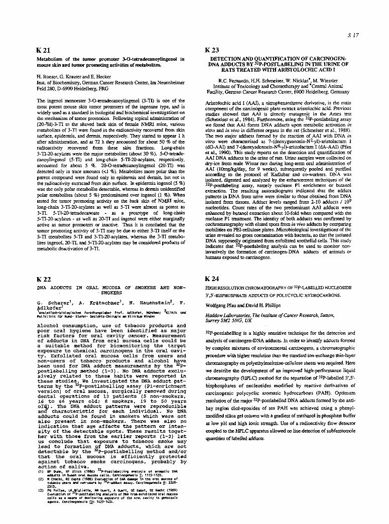
K 2 1 Metabolism of the tumor promoter 3-O-tetradecanoylingenol in mouse skin and tumor promoting activities of metabolites.
H. Roeser, G. Krauter and E. Hecker Inst. of Biochemistry, German Cancer Research Center, Im Neuenheimer Feld 280, D-6900 Heidelberg, FRG
The ingenol monoester 3-O-tetradecanoylingenol (3-TI) is one of the most potent mouse skin tumor promoters of the ingenane type, and is widely used as a standard in biological and biochemical investigations on the mechanism of tumor promotion. Following topical administration of [20-3H]-3-TI to the shaved back skin of female NMRI mice, various metabolites of 3-'17 were found in the radioactivity recovered from skin surface, epidermis, and dermis, respectively. They started to appear 1 h after administration, and at 72 h they accounted for about 50 % of the radioactivity recovered from these skin fractions. Long-chain 3-TI-20-acylates were the major metabolites (about 30 %). 5-O-tetrade- canoylingenol (5-TI) and long-chain 5-TI-20-acylates, respectively, accounted for about 5 %. 20-O-tetradecanoylingenol (20-TI) was detected only in trace amounts (<1%). Metabolites more polar than the parent compound were found only in epidermis and dermis, but not in the radioactivity extracted from skin surface. In epidermis ingenol (5 %) was the only polar metabolite detectable, whereas in dermis unidentified polar metabolites (about 5 %) predominated over ingenol (1%). When tested for tumor promoting activity on the back skin of NMRI mice, long-chain 3-TI-20-acylates as well as 5-TI were almost as potent as 3-TI. 5-TI-20-tetradecanoate - as a prototype of long-chain 5-TI-20-acylates - as well as 20-TI and ingenol were either marginally active as tumor promoters or inactive. Thus it is concluded that the tumor promoting activity of 3-TI may be due to either 3-'I7 itself or the 3-'17 metabolites 5-TI and 3-TI-20-acylates, whereas the 3-TI metabo- lites ingenol, 20-TI, and 5-TI-20-acylates may be considered products of metabolic deactivation of 3-TI.
K 22 DNA ADDUCTS IN ORAL MUCOSA OF SMOKERS AND NON-
SMOKERS
G. Scherer I, A. Kr~tschmer I, H. Hauenstein 2, F. ~dlkofer I
imatati$~-biot~isch~ Fors~ungat~aor Prof. ~tkofer, gOr~h~; ~tinik land ~tiktinik for gund- Kiefer- ~esichts*~irugie ~ Ktinik~Nind~l
Alcohol consumption, use of tobacco products and poor oral hygiene have been identified as major risk factors for oral cavity cancer. Measurement of adducts in DNA from oral mucosa cells could be a suitable method for biomonitoring the target exposure to chemical carcinogens in the oral cavi- ty. Exfoliated oral mucosa cells from users and non-users of tobacco products and alcohol have been used for DNA adduct measurements by the 32p_ postlabelling method (1-3). No DNA adducts exclu- sively related to these habits were reported in these studies. We investigated the DNA adduct pat- terns by the 32P-postlabelling assay (Pl-enrichment version) of oral mucosa, surgically removed during dental operations of 13 patients (5 non-smokers, 16 to 66 years old; 8 smokers, 19 to 50 years oi~. The DNA adduct patterns were reproducible and characteristic for each individual. No DNA adduots could be found in smokers which were not also present in non-smokers. There was also no indication that age affects the patte_--n or inten- sity of the detectable spots. These results toget- her with those from the earlier reports (1-3) let us conclude that exposure to tobacco smoke may lead to formation of DNA adducts, which are not detectable by the 32p-postlabelling method and/or that the oral mucosa is efficiently protected against tobacco smoke carcinogens, probably by action of saliva. (1) P Dunn, HF S t i ~ 0986) 52P-p~ttabetti~ ~ a t ~ i s of aromatic DNA
~ t s in human oratmuc~a carts. Carcinogenesis ~: 1115-1120. (2) N ~ k o , RC G~ta (1988) E v a t ~ i ~ of DgA damage in the ora[ nlucosa of
tobacco ~ers and non-~ers ~ P-edduct assay. Carcinogef~sis 9: ~ - ~15.
(3) PG F o i [ ~ , ~ i g [ i e t t a , ~ Q ~ r t , A q ~ r t # GC Kat~at, SS Hecht ( 1 9 ~ ) E v a t ~ t i ~ of ~ P - p o s t t a b e t i ~ nnat~is of DN* f r m e x f o t i a t ~ ora[ muc~a car ts ~ a m a ~ of t a ~ l i t o r i ~ exposure of the orat c a v i t y to g e t . t o x i c a g a r s . ~rc inogenes is ~ : 1 4 ~ - 1 4 ~ .
S 1 7
K 23 DETECTION AND QUANTIFICATION OF CARCINOGEN-
DNA ADDUCTS BY 32p-POSTLABELING IN THE URINE OF RATS TREATED WITH ARISTOLOCHIC ACID I
R.C. Femando, H.H. Schmeiser, W. Nicklas*, M. Wiessler Institute of Toxicology and Chemotherapy and *Central Animal
Facility, German Cancer Research Center, 6900 Heidelberg, Germany
Aristolochic acid I (AAI), a nitrophenanthrene derivative, is the main component of the carcinogenic plant extract aristolochic acid. Previous studies showed that AAI is directly mutagenic in the Ames test (Schmeiser et al., 1984). Furthermore, using the 32p-postlabeling assay we found that AAI forms DNA adduets upon metabolic activation in vitro and in vivo in different organs in the rat (Schmeiser et al., 1988). The two major adducts formed by the reaction of AAI with DNA in vitro were characterized as 7-(deoxyguanosin-N2-yl)-aristolactam I (dG-AAI) and 7-(deoxyadenosin-N6-yl)-aristolactam I (dA-AAI) (Pfau et al., 1990). This study reports on the detection and quantification of AAI DNA adducts in the urine of rats. Urine samples were collected on dry-ice from male Wistar rats during long-term oral administration of AAI (10mg/kg/day, for 9 weeks), subsequently pooled and purified according to the protocol of Kadlubar and co-workers. DNA was isolated, digested and analyzed by the enhancement techniques of the 32p-postlabeling assay, namely nuclease P1 enrichment or butanol extraction. The resulting autoradiograms indicated that the adduct patterns in DNA from urine were similar to those obtained from DNA isolated from tissues. Adduct levels ranged from 2-10 adducts / 109 nucleotides. Count rates of the two predominant AM adducts were enhanced by butanol extraction about 10-fold when compared with the nuclease P1 treatment. The identity of both adducts was confn'med by cochromatography with eluted spots from in vivo adducts by comparing mobilities on PEI-eellulose plates. Microbiological investigations of the urine revealed no gross contamination with bacteria, so that the isolated DNA supposedly originated from exfoliated urothelial cells. This study indicates that 32p-posflabeling analysis can be used to monitor non- invasively the formation of carcinogen-DNA adducts of animals or humans exposed to carcinogens.
K 24 HIGH RESOLUTION CHROMATOGRAPHY OF 32poLABELLED NUCI.~OSIDE
3',5'-BISPHOSPHATE ADDUCTS OF POLYCYCLIC HYDROCARBONS.
Wolfgang Pfau and David H. Phillips
Haddow Laboratories, The Institute of Cancer Research, Sutton, Surrey SM2 5NG, UK
32p-posflabelling is a highly sensitive technique for the detection and
analysis of carcinogen-DNA adducts. In order to identify adducts formed
by complex mixtures of environmental carcinogens, a chromatographic
procedure with higher resolution than the standard ion-exchange thin-layer
chromatography on polyethyleneimine~cellulose sheets was required. Here
we describe the development of an improved high-performance liquid
chromatography (HPLC) method for the separation of 32p-labelled 3',5'-
bisphosphates of nucleosides modified by reactive derivatives of
carcinogenic polycyclic aromatic hydrocarbons (PAH). Optimum
resolution of the major 32P-posflabelled DNA adducts formed by the anti-
bay region diol-epoxides of ten PAIl was achieved using a phenyl-
modified silica gel column with a gradient of methanol in phosphate buffer
at low pH and high ionic strength. Use of a radioactivity flow detector
coupled to the HPLC apparatus allowed on line detection of subfemtomole
quantifies of labelled adducts.
S 18
K 25 Development of higly sensitive detection of acrolein congener DNA adducts
E. Eder and S. Scheckenbach Insts of Toxicology, University of W~rzburg
~, G-unsaturated carbonyl compounds are ubiqui- tous environmental pollutants, occurring natu- rally, in food and as industrial products. They are formed during lipid peroxidation, in lipid and arachidonic acid oxidation and by reactive oxygen species. Acrolein and its congeners are therefore considered to play an important role in mutagenesis and carcinogenesis. The geno- toxic activities of these compounds probably depend on their ability to form linear and cyc- lic adducts with DNA components, in particular, with deoxyguanosine. Unfortunately the detecta- bility of most of these DNA-adducts is very low. In order to clarify the relationship be- tween the interactions of acrolein congeners and mutagenesis and carcinogenesis we are pre- sently developing highly sensitive methods for detection of such adducts, improving the DNA- hydrolysis and analysis methods and adapting the 3zP-post-labeling technique using HPLC for the nucleotide analysis. Difficulties arise from the incomplete enzymatic degradation of the modified DNA and, in particular, from the insufficient HPLC separation of the adducts from the unmodified nucleosides. We have achieved a successful separation using Sephadex LH20 gel filtration prior to the HPLC analysis. With this method tbe modified nucleosides could be analysed, isolated and identified much more readily with improved sensitivity, since the unmodified nucleoside peaks and those of the adducts no longer overlap.
K 27 COMPARISON OF THE GENOTOXIC POTENCY OF ETHYLENE OXIDE AND VINYL CHLORIDE ON THE BASIS OF DNA ADDUCTS
I-L Peter, H. Ottenw~ler, U. F~st
Ethylene oxide is an air pollutant emitted by various sources like exhaust fumes and cigarette smoke. In addition, it is metabolically formed in mammals after inhalation of ethylene, which is also produced in combustion processes and found as a phytohormon. The carcinogenic potency of ethylene oxide has clearly been proven in animal studies.
Vinyl chloride, a proven b,man carcinogen, is one of the most widely produced monomers for plastics. No natural sources of vinyl chloride are known. The substance is also produced from air pollutants like trichloroethyleue and perchloroethylene in soils by anaerobic bacteria leading to covtamination of drinking water.
Male Sprague-Dawley rats were exposed to either ethylene oxide or vinyl chloride (3 doses/exporiment; 3 animals/dose). Liver-DNA was isolated by phenol extraction and the amount of 7-(2-hydroxyethyl)- guanine in the case of ethylene oxide exposure and 7-(2-oxoethyl)- guanine in the case of vinyl chloride exposure were detected by gas chromatography/mass spectrometry. Chemical binding indices (CBI) according to Lutz were calculated from these data as well as from data published earlier by other groups. A comparison of the CBI's of ethylene oxide and vinyl chloride calculated from 7-alkylated guanines in liver DNA of pretreated rats showed an approximately 70-fold higher genotoxic risk for vinyl chloride than for ethylene oxide.
In additional experiments, the distribution pattern of ethylene oxide in the compartments of bnma. blood was investigated in vitro using 14C- labelled substance. It was found that an enzymatic turnover of ethylene oxide in bnman blood is probably subjected to an enzyme polymorphism, which may influence a binding of the substance to hemoglobin.
In order to evaluate the additional carcinogenic risk from endogenously produced ethylene oxide, lymphocyte DNA from non-exposed animals and b,ma,s was isolated and the amounts of 7-(2-hydroxycthyl)-guanine was determined as described above. Adducts specific for ethylene oxide can in part stem from endogenous production. The high level of adducts in non-exposed rats and humans cannot be explained by this production alone, so that exogenous sources such as an ubiquitous air pollution must be taken into consideration.
Institut f. Arbeitsphysiologie, Ardeystr. 67, D-4600 Dortmund 1
K 26 INVESTIGATION OF THE POTENTIAL FOR DNA BINDING OF STYRENE-7,8-OXIDE IN RAT AND MOUSE
S. Cantoreggi and W.K. Lutz, Institute of Toxicology, ETH and University of ZOrich, CH-8603 Schwerzenbach, Switzerland
Styrene oxide, the main intermediate metabolite of styrene, induces hyperkeratosis and tumors in the forestomach of rats and mice upon chronic administration in the diet. The aim of this study was to investigate whether DNA binding could be re- sponsible for the carcinogenic:effect observed. [7-3H]-Styrene - 7,8-oxide was administered by oral gavage in corn oil to male Sprague-Dawley rats at 2 dose levels (1.6 or 220 mg/kg). After 4 or 24 hours, forestomach, glandular stomach, and liver were excised and DNA was purified extensively. At the 4-hour time point, the DNA radioactivity was below the limit of detection in all samples. Expressed in the units of the Covalent Binding Index, CBI = (p.mol adduct per mol DNA nucleotide)l(mmol chemical administered per kg body wt.), the DNA-binding potency was below 3, 2, and 0.3, in the forestomach, glandular stomach, and liver, respectively. At the 24-hour time point, DNA isolated from both parts of the stomach (but not from the liver) was sl ightly radiolabel led. Metabol ic degradat ion of [7-3H]styrene oxide can result in the formation of tntiated water. It is therefore assumed that the DNA radioactivity in the gastro- intestinal tract represented biosynthetic incorporation of radio- label into newly synthesized DNA. In a second expenment, [7-3H]styrene-7,8-oxide was administered by intraperitoneal injection to male B6C3F1 mice. Uver DNA was analysed after 2 hours. No radioactivity was detectable at a limit of detection of CBI<0.6. It is concluded that the reactivity of styrene oxide with nucleic acids is not detectable in vivo at a low detection limit. This finding agrees with the relative stability of styrene oxide in the blood (half life - 20 min). Upon comparison with other caminogens, the maximum possible CBI values render a purely genotoxic mechanism of tumorigenic action unlikely.
K 28 MUTAGENIC ACTIVITY IN E. COLI BY
O6-N-BUTYLGUANINE AND O6-N-OCTYLGUANINE BUILT INTO A UNIQUE SITE IN A PLASMID
P.M. Banmgan, H.H. Schmeiser, H.-C. Kllem, S.Data, M. Wiessler Institut F~ Toxikologie und Chemotherapie, Deutsches K.rebsforschungszenmun, 6900 Heidelberg, Germany
The reaction of alkylating agents with DNA results in the formation of a divorse population of alkyl-DNA adducts, the processing of which is believed to lead to mutations and possibly cancer. One of the best investigated groups of alkylating agents are the N-niu'osamines. In contrast to other dialkylamines di-n-butylnitrosamine (NDBA) is not a typical liverearcinogen in the rat, but also induces tumors of the urinary bladder. NDBA was shown to occur in the annosphcre of factories of the robber industry (Spiegelhalder et al., 1983). Reactions with N- nin'oso-n-butylurea or NDBA lead to butylation of DNA. Hem we report on our studies with 06-n-butylguanine and 06-n-octylguaninc which we additionally included for comparison to a long-chain derivative. Using site directed mutagenesis we examined the mutagenic activity in E. coli of both modified guanines located at a unique site in a plasmid. The modified 10mer oligonuclcotides were prepared on a DNA synthesizer and characterized by HPLC, nucleoside composition analysis and 32p-posflabcling. These 10mers were ligated into pUC9 by "shot-gun" gene synthesis using another 10mer and 20mer with overlapping sequences. After cloning in E. coil BMI-I 71-18, mutants were identified by digestion with XhoI or PstI and sequenced by the dideoxy-method. O6-n-butyldG situated at a unique site in pUC9 showed mutagenic activity by targeted mutations, all of which being GC---~AT transitions. In contrast O6-n-octyldG leads to 70% GC--->AT transitions and to 30% GC --~ TA transversions. We assume that the mutagenic activity caused by 06-n-butyldG results from mispairing, whereas for 06-n-octyldG mispairing is partially inhibited sterically by the bulky n-octyl group. The 30% GC--#TA wansvcrsions could be the result of insertion of adenine opposite to a non-coding lesion, namely 06-n-octyldG.
K 29 EFFECT OF DNA ALKYLATION BY NITROSOUREAS ON SEMICONSERVATIVE DNA SYNTHESIS BY FROCARYOTIC DNA POLYMERASE C. Pfeiffer, A. Fuchs, G. Eisenbrand Universit&t Kaisersiautern, FR Lebensmlttel- chemie/Umwelttoxikologie, P.O.B. 3049, D-6750 Kaiserslautern, FRG
We have incubated M13 mp18 phage DNA with a series of homologue alkylnltrosoureas to study the effect of alkylation on the propensity of single stranded DNA to act as a template for DNA synthesis using E. coli polymerase Klenow fragment (Kf polymerase). Treatment with N- methyl-N-nitrosourea (MNU; 1-20 mM) resulted in a concentration dependent induction of DNA polymerase stops, 41/128 stop signals being clearly detectable. The sequencing gel revealed methylation predominantly one base 3'to template adenine residues (26/41), with some modification occuring also at sites 3'to template guanine (8/41) and cytidine (6/41). Treatment with longer chain nitrosoureas (ethyl- to n-butyl; 1-50mM) resulted in much weaker inhibitory effects on polymerase-mediated chain elongation with respect to frequency as well as intensity of stop signals. Moreover, no clear concentration dependency was detectable. Introduction of polymerase stops appeared to shift, however, with increasing chain length from one base 3'of adenine to one base 3'of guanine. In conclusion, the extent of induction of DNA polymerase stops mainly reflects the differential alkylating potency of homologuous nitrosoureas rather than the size of the alkylgroup transferred.
S I 9
K 31 SINGLE CELL ANALYSIS OF CARCINOGEN-INDUCED DNA SINGLE STRAND BREAKS BY IN SITU NICKTRANSLATION
U.M. Lieglbd, P. Schmez~, *B.L. Pool-Zobel Institute for Toxicology and Chemotherapy, DKFZ, Heidelberg. "Institute for Hygiene and Toxicology, Federal Research Center for Nutrition, Karlsruhe.
In order to assess toxicoklnerlc parameters and other mechanisms of genotoxic carcinogens it is useful to investigate compound-induced DNA single strand breaks in a variety of primary cells of the rat in viwo as well as in vivo. For this, we need methods that cart detect DNA damage in even a very small number of cells. One poteaatlal technique is the visualization of DNA-strand breaks ('nicks') by .= ~ - rock translation (NT). The slides with the fixed cells are incubated with the repair enzyme E. coli DNA Polymerase I and the nudcosldetriphosphates ATP, CTP, GTP and [3HTrTP for 10 minutes at 21~ Incorporation of [3H] into DNA is detected by autoradiography and quantified by counting the grains per nucleus (Anal H. et al., Cancer Letters 40, 33, 1988). To ~ the sensitivity of this new method we have performed some experiments after a one hour incubation of primary hepatocytes with the test substance and compared the results to those obtained by the wall established technique of alkal;n, filter elutlon (AE). As test compounds we used dlffereot N-nltros,amlnes most of which need metabolic activation for genotoxic activity. Results show that NT is able to detect single strand breaks in primary ceils. N-Nitrosodimethylamine induced DNA damage at approximately the same conematrations in both systems. N-Nitrosomethylbenzylamine induces detec- table single strand breaks in hepatocytes at 10 ~moles/ml in NT, whereas 1 ~mol is sufficient for AE. For the tobacco-specific nitrosamines NNN and NNK no effects can be measured with NT up to 25 ~mol/ml but NNK induces DNA- single swand breaks at 6.25/Lraoles measured with AE. Whereas the number of cells needed for one experiment is much lower for NT than for AE (1-2 x 10⁵/slide for NT; 1-5 x 10⁶/filter for AE), NT is somewhat less sensitive.
K 30 THE MICROGEL SINGLE CELL ASSAY: A RAPID AND SENSITIVE TECHNIQUE TO ANALYSE DNA DAMAGE IN PRIMARY RAT CELLS FROM VARIOUS ORGANS.
P. Schmezer 1, F. Kuchenmeister 1 and B.L. Pool-Zobel 2. 1: Institute for Toxicology and Chemotherapy, German Cancer Reseamh Center, INF 280, W-6900 Heidelberg, 2: Institute for Hygiene and Toxicology, Federal Research Center for Nutrition, Engesserstr. 20, W-7500 Karlsruhe, F.R.G.
The microgel single cell assay developed by Ostling and Johanson (1984, Biochem. Biophys. Res. Commun. 123:291) and modified by Singh et al. (1988, Exp. Cell. Res. 175:184) is an electrophoretic technique to detect DNA single-strand breaks and alkali labile sites in individual cells. We used this approach to analyse DNA-damaging compounds in primary cells deriVed from rat liver, stomach, blood and nasal mucosa in vitro and ex vivo. For this, the treated cells were embedded in agarose gels on micro- scope-slides, lysed and the remaining DNA was treated with alkali. Subsequently, a 20rain. electrophoresis (25V, 30OmA) was performed and the neutralized slides stained with ethidium bromide. As a quantitative measure of the induced DNA-damage, the migration patterns of the DNA in the agarose gels ware analyzed by fluorescence microscopy coupled with an image analyzer. So far e.g. the evaluation of 100 cells/slide/compound concentration or dose have revealed potent in vitro genotoxic effects of methyl- nitrosourea in tumor cells and nitrosodimethylamine (NDMA) in hepatocytes. Furthermore, methylnitrosoguanidine, NDMA, and a tobacco specific nitrosamine were genotoxic ex vivo in cells of various organs. In comparison to the well- established procedure of the alkaline filter elution, this technique showed a comparable sensitivity but was much less time-consuming. Moreover, it has the distinct advantage that very few or even single cells may be monitored.
K 32 A NICK TRANSLATION ASSAY AS A S~ECIFIC SCREENING METHOD FOR POTENTIAL INITIATING AGENTS OF SKIN
L.Steeb, H.Friesel, Th.Schneider and E.Hecker, Institute of Biochemistry, German Cancer Research Center, Im Neuenheimer Feld 280, D-6900 Heidelberg, F.R.G.
Polycyclic aromatic hydrocarbons (PAIl) such as 7,12-dimethylbenz- [a]anthracene (DMBA) are efficient initiators of mouse skin in vivo a ,d transforming agents in fibroblasts in vitro. These effects are irreversible and may depend on inidal covalent binding to DNA. One of the possible consequs~nces of such binding, the induction of DNA single strand (ss) breaks, was stodied with Balb c 3/T3 cells in an in vitro nick translation assay up to 24 hours after exposure. For this purpose the cells were maim'~ined and treated in 24-well culture plates for high sample throughput. Permeabilization was achieved by treatmeat with lysolecithin m~d monitored by trypan blue. Care was taken that the cells remained adherent throughout the nick translation reaction as ioitiated by addition of DNA polymerase I, 3H-dCTP and uniabelled dNTPs. DMBA (400nmole), the most potent initiator of skin in vivo, was compared to equimolar and equicytotoxic doses of either the PAH 9,10-dimethylanthracene (DMA) or the Utmor promoter 12-O-tetradecanoylphorbol-13-acetate (rPA). The incorporation of ~H-dCTP in nicked DNA (values for tested compounds corrected by subtraction of solvent control values) obtained with DMBA show that following a decrease during the first 4 hours the ss- breaks increase to a maximum at 7 hours, whereas DMA was inactive even at equicytotoxic doses. Also the tumor promoter TPA causes no detectable es-breaks. Therefore nick translation to measure u-breaks may be a specific in vitro assay for potential initiating acdvity, since it can efficiently discriminate between DMBA (a tumor initiator) and its inactive derivative DMA or TPA (a tumor promoter). This working hypothesis will be investigated using a large number of DNA-binding, initiating and non- initiating PAIl.
S 20
K 33 ACTIVATING MUTATIONS AT CODON 61 OF THE C-HA-
GENE IN TUMORS OF RATS AND MICE INDUCED BY ARISTOLOCHIC ACID
H.H. Schmeiser, H.R. Scherf and M. Wiessler
Institute of Toxicology and Chemotherapie, German Cancer Research Center, 6900 Heidelberg, Germany
Aristolochic acid I (AM), a nitrophenanthrene derivative, is the major component of the carcinogenic plant extract aristolochic acid, which has been used as a medicine since antiquity. Long term oral administration of AAI to male Wistar rats induces multiple tumors, mainly in the forestomach. We previously reported (Schmeiser et al., 1990, Cancer Res. 50, 5464-5469) on the detection of AT-->TA wansversion mutations in codon 61 of the c-Ha-ras gene (CAA to CTA) in 93% of the forestomach tumors analyzed. Here we applied PCR and mutation detection by selective oligonucleodde hybridization to 5 pm sections of formalin-fixed paraffin-embedded tissue. Paraffin blocks of forestomach squamous cell carcinomas from rats containing adjacent carcinoma and histologically normal tissue were cut and manually separated. DNA obtained from these histologically separated regions was amplified and analyzed. AT--4TA transversions in codon 61 of the c-Ha-ras gene were observed in the carcinoma portions, but not in the adjacent normal tissue. Moreover we screened 11 forestomach and 12 lung tumors of female NMRI mice induced by aristolochie acid (80% AAI;20% AAII) for c- Ha-ras gene mutations in codon 61 by the same procedure. Only 1 in 11 forestomach sections and 3 in 12 lung sections were successfully amplified. Identical AT->TA transversions were detected in the squamons cell carcinoma portion of the forestomach section as well as in one adenocarcinoma portion of one lung section. These results indicate that aristolochic acid activates ras genes in both rat and mouse tumors at deoxyadenosine residues.
K 34 MUTATIONS IN THE c-Ha-RAS GENE OF CARCINOGEN- AND TUMOR PROMOTER-INDUCED MOUSE LIVER TUMORS
A. Buchmann ~, R. Bauer-Hofmann 2, J. Mah# and M. Schwarz 2
qnsdtute of Toxicology, University of Tfibingen, D-7400 Tiibingen 2Institute of Experimental Pathology, German Cancer Research Center, D-6900 Heidelberg
It is well documented that different swains of rodents show characteristic differences in both, their spontaneous rate of liver tumor formation and their susceptibility towards hepatocarcinogenic and/or tumor promoting agents. Although the molecular basis for these differences in susceptibility to hopatocarcinogenesis is largely ,nknown, recent results fTom our laboratory bave indicated that mutational activation of the c-Ha-ras gene may be a major factor contributing to liver tumor development in sensitive but not in insensitive rodent strains. By analysis of liver tumors from various mouse strains which occurred either spoutanonsly or after carcinogen-treatment, we were able to detect mutations at codon 61 of the c-Ha-ras gene in 30-60% of spontaneous or carcinogen- induced liver tumors of different sensitive mouse swains (e.g. C3PdHe, B6C3FI and CF1 mice) but not in liver tumors of the comparatively insensitive C57BL/tl mouse. To further extend our findings, C3H mouse liver tumors induced by non-genotoxic tumor promoters such as phenobarbital or dieldrin were analyzed. Although treatment with these compounds more than doubled the number of liver minors pea- ~nimal as compared to u n m ~ d controb, the frequency of tumors with c-Ha-ras mutations was lowered from 60% in conU'ols to approximately 25% in promoter-treated groups. This finding suggests that c-Ha-ras mutations within promoter-induced liver tumors represent background muta~ons not related to treatment and, secondly, that tumor promote~ also confer a selective growth advantage on those initiated helmtocyt~ that do not contain an activated c4-Ia-ras gene.
K 35 SOLITARY CANCEROGENESIS BY 7,12-DIMETHYLBENZ[a]ARTHRA- CENE (DMBA) IN MOUSE EPIDERMIS: THE INFLUENCE OF DOSE,
DOSE-FREQUENCY AND OF THE TUMOR PROMOTER TPA
H. Frlesel, R. Schmtdt, F. Rlppmann, B. Stetnbauer, and E. Hecker. Insti tute of Biochemistry, German Cancer Research Center, Im Neuenhelmer Feld 280, D-tgOO
Heidelberg, F.R.G.
The so l i ta ry cancerogentc ef fect of DMBA was studied, varying the intervals between repeated eptcutaneous ad- ministrat ions to the back skin of female NMRI mice and, concomitantly, the parttal doses d In such a manner that they added up to equal total doses D over a given period of time. The tota l number of benign and that of malignant tumors was recorded weekly. The to ta l dose D = 800 nmol was highly cancerogenic when administered as d = 18 nmol twice weekly during 22 weeks and observ- ed up to 38 weeks; under these conditions, D = 800 nmol given as d - 54 nmol every 10 days was marginally ac t i - ve and d = 100 nmol every 4 weeks showed no e f fec t . 44 x d ~ 18 nmol proved to be even more ef fect ive than 5 x d - 1000 nmol. Similar ly performed l i fe long cont i - nuous applications led to drast ic increases in latency periods at longer intervals as well as decreases in the ra t io of carcinomas vs papillomas. These results sug- gest a partial revers ib i l i t y of cancerogenlc ef fects by DMBA, possibly due to a promoting ac t i v i t y requiring a constant stimulus. A l ternat ive ly , insu f f i c ien t DNA re- pair at short intervals may lead to cumulation of geno- tox ic lesions. Twtce weekly co-administrations of DNBA and TPA did not Increase the number of carcinomas but led to a htgh number of papillomas, slmtlar as with i n i t i a t i on by a single dose of OMBA followed by promo- t ion with TPA. This shows that TPA can a f fec t epider- mal cell populations d i f fe rent from those Involved tn so l i ta ry cancerogenesls.
K 36 Effects of Dietary Restriction on Mammary Tumorigenesis,
Sexual Hormones and Hormone Receptors
P. Zifu, B. Bunk, W. Hefter, K. Klinga and M.R. Berger
Institute of Toxicology and Chemotherapy German Cancer Research Center, Heidelberg, FRG
Female Sprague-Dawley rats were injected i.v. with 25rag N-methylni t rosouredkg on day 50 of llfe to induce mammary car- cinoma, and divided into 6 dietary groups: 50, or 35 kcal/day with 45, 35 and 25% (energy%) fat, respectively, were fed for 27 + 1 weeks. In accordance to a previous study from our laboratory, tumor burden was reduced in the low calorie groups, independently of the fat levels. Among the high calorie groups, tumor number and weight were significandy higher in the 35% than in the 25% fat group, but both levels showed no difference to the 45% fat group. By comparing all low calorie groups versus high calorie groups, both plasma estradiol (E2) and testosterone (T) levels at the stage of diestrns were lower in the low calorie group (E2:p<0.05 and T: p<0.01). Furthermore, an elevated T/E2 ratio in the low calorie group was found (p<0.0S). No relationship of either E2 or T levels to fat levels could be observed. Free estradiol and proge- strone receptor level in tumor cytosol showed no difference among the groups. Lower mean cytosolic androgen receptor level and higher dissociation constant were observed in tumor tissue of rats fed 35 kcal/day compared to the 50 kcal group (p<0.05), which could be a response to the elevated plasma T/E2 ratio found in the calorie restricted group. In conclusion, calorie restriction inhibits mammary tumor develop- ment, reduces plasma sexual hormone levels independently of dietary fat content, and elevates plasma T/E2 ratio; the latter might interfere with tumor burden.
K 37 DETERMINATION OF THE INITIATING AND PROMOTING ACTIVITY OF XENOBIOTICS IN RAT LIVER
M. Schwarzl, A. Buchmann 2, L.W. Robertson 3, G. Luebeck "4 and S. Moolgavkar 4
1projekt "Tumorpromotion in der Leber", Deutsches Krebsforschungs- zentrum, Heidelberg 2Institut ftir Toxikologie der Universit~t Tiibingen 3Grad. Center for Toxicology, University of Kentucky, Lexington, USA 4Division of Public Health Sciences, Fred Hutchinson Cancer Research Center, Seattle, USA.
The improvement of methods used to evaluate possible risks associated with exposure of humans to environmental compounds is one of the challenging tasks of toxicology. Recently developed biologically-based methods for cancer risk assessment seem to be very suitable in this respect both for inter-species and for low-dose extrapolation of experimental data. Compounds that increase the risk of cancer may exert their activity on the stages of initiation and/or promotion and/or progression. On the basis of theoretical considerations regarding the underlying mechanisms of action, characteristic differences can be expected to exist in the dose- response characteristics between initiating and promoting activities of such compounds. We have now determined the formation of enzyme-altered foci in livers of rats treated continuously with various hepatocarcinogens and in addition, in animals that were given various polyha.lo.g.en.ated biphenyls either alone or in combination with an mmaang dose of diethylnitrosamine. Differences in the effects of the various test compounds on the number and size distribution of foci were taken as indicators of differences in their initiating and promoting activity, respectively. In addition, loci data obtained with selected compounds were quantitatively analyzed within the framework of the model developed by Moolgavkar and colleagues. The outcome of these studies clearly indicates that this latter approach represents a very valuable tool for the quantitative analysis of such data and may significantly improve quantitative cancer risk assessment.
K 39 Expression of oncogenes in xiphophorine tumors of different etiology
Ch. Zechel, H. Peters, and F. Anders
Melanoma in Xiphophorus is mediated by a tumor gene complex (Tu-complex). Melanoma develops after deregulation of the Tu_ -~omplex a) in the germ line by mutation, b) in a pigment cell precursor by somatic mutation, and c) following the impairment of a tumor preventing block of cell differentiation by tumor promoters.
We studied the inheritance, structure and expression of several genes in f ishstrains sensitive for each of these kinds of tumor etiology.
Southern blot analysis and sequencing showed that a c-erbB-related gene (x-erbB*) is part of the Tu-complex. Expression and overexpression of x-erbB * was detected in spontaneous tumors as well as in in i t ia tor - and promoter-induced tumors. The highest expression was found in fast growing spontaneous and promoted melanomas. A second type of er__bbB-related gene (x-er__bbB) which probably encodes the common epidermal growth factor receptor (EGF-R) is expressed at a low level in all three kinds of tumors. Genes related to the human genes coding for the plateled derived growth factor (PDGF) and its receptor (PDGF-R) were identified in Xiphophorus by Southern Blot analysis and sequencing. Expression of x-pd~f was detected in approx, half of the spontaneous melanomas and fibrosarcomas. The x-pdgf~r is expressed in all tumors studied and is slightly overexpressed in spontaneous melanomas and fibrosarcomas. Expression of three types of identified x-erbA-genes that probably code for proteins of the thyroid hormone receptor family was undetectable in the majority of the tumors studied.
So far, only the x-erbB* appears to be involved in the in i t ia l steps leading to--melanoma and possibly to certain other tumors. The role of x-erbB, pdgf, pdgf-r, and x-er__bbA in tumor formation remains unclear. Supported by University of Giessen, DFG, and UBA.
S 21
K 3 8 Hormone~dependency, growth-rate and morphology of
mammary tumours in SD-rats treated with NMU
Torsten, U., Kowalczyk-Wagner, M., Weitzel/ H.
Dept. of Gynecology and Obstetrics
Klinikum Steglitz, Free University Berlin,
Hindenburgdamm 30, W - 1000 Berlin 45
FRG
SD-rats were deprived in estrogen by operation or
by application of anti-estrogens at different
stages in the development of breast cancer. One
group received ovariectomy and another group daily
application of the anti-estrogen TAMOXIFEN
simultaneously with the chemical induction by the
carcinogen NMU. In the ovariectomized group 62,5 %
of the animals showed no tumours compared to 80 %
of those which got TAMOXIFEN. In another series
one group was ovariectomized and another received
TAMOXIFEN after appearance of tumour nodules;
ovariectomy led to a 57 % complete remission,
whilst TAMOXIFEN at this stage of carcinogenesis
did never show a complete or partial remission.
K 40 ~ LIVER C~HCBR ZNRATB
S. Ambs, T.Kirohner and H.-G. Neumann Institut f~r Toxikologie und Institut fttr Pathologic, UniversitSt WUrzburg
2-Acetylaminofluorene (AAF) is a complete rat liver carcinogen (initiator and promotor). We studied the role of cytotoxicity for the promot- ing activity of AAF. Male Wistar rats were chro- nically fed 0.02% AAF in the diet for up to 16 weeks. The livers were analysed histologically. The following changes are seen in male rats: After 4 weeks the first GST-P positiv foci are detectable. Thereafter the area of these foci increases exponentially. Between the third and fifth week a new cell population appears in oxy- gen rich regions with a periportal accentuation. This cell population (gamma-GT positiv, ductular morphology similar to small bile ducts ) gener- ates septa encircling the centrilobular areas. The appearance reminds of the swine liver and is especially visualized by silver staining of reticulin fibers. Between week 8 and 12 livers become cirrhotic. The origin of these duct-like cells is unknown. One possible explanation is that AAF initiates transformation of hepatocytes to duct-like cells by a mito-inhibitory or hypoxic effect in the oxygen rich regions. Advanced stages exhibit multiple necrosis of hepatocytes and the deve- lopment of cirrhosis, in which the disturbance of microcirculation may produces selective pres- sure on hepatocyte proliferation. As a first step, we postulate the interaction of AAF meta- belites with mitochondria of hepatocytes. Mito- chondria in the periportal area are generally larger, have a higher oxygen turnover, and en- zyme activities are different from mitochondria of the centrilobular area. Investigations on isolated mitochondria are in progress.
S 22
K 41 G6PDH AND LDH AS MARKERS FOR FOCI INCIDENCE AFTER TREATMENT WITH DEN AND CLOPHEN A 50. Ic. E~nig, IE. E~genbrodt, 2D. Oesterle, 2E. Deml, ~G. Weisse, "U. Gerbracht
The rat liver foci bioassay is widespreadly used as an in vivo test system for the identification of chemically induced hepatocarcinogenesis. Following the two stage model of carcinogenesis which implicate the treatment with an initiator and a promotor, the rats received once i0 mg/kg b.wt of diethylnitrosamine (DEN) which is a strong liver-carcinogenic chemical. After 7 days the animals were treated twice a week with the promoter CiQphen A 50 (i0 mg/kg b.wt) for the following I0 weeks. After the experiment the rats were killed and the liver cryostat slices were stainded histochemically. The enzymes lacta~e-dehydrogenase (LDH) and glucoee-6- phosphate-dehydrogenase (G6PDH) of the carbohy- drate metabolism were used for the endpoint determination. The most of the loci were recognized in the animal group which had received the complete treatment schedule in contrast to the groups which were treated only with DEN or Clophen A 50. Also the mean area of the loci, the total area of the loci /cm 2 of the liver slice and the relative size distribution of the foci indicate that in the group which received the complete schedule the islands were significantly increased. 1 Vet. Biochemie Universit&t Gie~en Frankfurterstr. i00 6300 Gie~en 2 GSF Institut fQr Toxicol. Neuherberg 3 Merck, Institut f~r Toxikologie, Darmstadt 4 Pharmbiodyn Denzlingen
K 43 EFFECT OF THE HEPATCX)ARCINOGEN 2-ACEI~/I-Ad~IINOFLUORENE AND ITS NONCARCINOGENIC ISOMER 4-ACETYLAMINOFLUORENE ON THE LEVEL OF PLASMA MEMBRANE RECEPTORS AND ON THE ACTIVITY OF
CARBOHYDRATE-METABOLIZING ENZYMES IN RATS
W. Muster and P. Cikryt, Inst. for Toxikologie der Universit~t W0r-zburg
The aromatic amines 2-acetylaminofluorane (2AAF) and 4-acetyl- aminofluorene (4AAF) differ in their toxic and carcinogenic properties. 2AAF Is a potent hepatocarcinogen in the rat and has tumor-promot- ing properties. In contrast to 2AAF, the isomer 4AAF is regarded as a noncarcinogen or at least as a less potent carcinogen than 2AAF. It is established that the activity of carbohydrate-metabolizing enzymes changes during the carcinogenic process, but the relationship be- twean these changes and tumor promotion is not clear. For a number of diverse chemical tumor promotors, it is hypothesized that cellular receptors, e.g. plasma membrane receptors, may be involved. The objective of this study was to compare the effect of the complete car- cinogen 2AAF on carbohydrate-metabolizing enzymes and on plasma membrane receptors with the effect of the putative noncarcinogen 4AAF. Male Wistar rats were treated with 0.02 % 2AAF or 0.1% 4AAF in the diet for 2, 7 or 21 days. The level of epidermal growth factor re- ceptor (EGFR) of plasma membranes was down-regulated only by 2AAF after only 2 days of treatment and did not then recover. Microsomal EGFR content was reduced significantly by 2AAF and 4AAF. Cytosolic protein kinase C activity was significantly reduced by 2AAF after 21 days of treatment and by 4AAF after only 7 days of treatment. Changes in the activity of carbohydrate-metabolizing en- zymes were as follows: the gluconeogenetic enzyme phosphoenol- pyruvate carboxykinase was reduced to 28 % and 46 % of control values by 2AAF and 4AAF, respectively, after 21 days of treatment. Glucose-6-phosphatase activity was reduced by both aromatic amines after 7 days of treatment. Hexokinase activity remained un- changed. Only 2AAF treatment increased glucose-6-phosphate dehy- drogenase activity 5-fold after 21 days, The results of this study clearly demonstrate that some of the observed effects are only evoked by the complete carcinogen 2AAF. Other changes are also observed with the noncarcinogen 4AAF. The importance of these modifications for the process of tumor promotion remains to be elucidated.
K 42 Detection of glucose-6-phosphate dehydro- genase m-RNA in hepatocellular tumours and preneoplastic liver lesions of the rat
H. Stumpf,E. Filsinger und P. Bannasch
Institut f~r Exp. Pathologie, Deutsches Krebsforschungszentrum Heidelberg
The appearence of hepatocellular carcinomas, induced in rats with N- nitrosomorpholine, is preceded by a series of preneoplastic liver lesions, leading from glycogen-storing-loci (GSF) to mixed cell loci (MCF) and basophilic focl (BCF), which share a number of alterations in enzyme activity. One of the most prominent changes is the increase in the activity of the glucose-6-phospate dehydrogenase (G6PDH), the key enzyme of the pentose- phosphate-pathway. Using Northern-Blot-Analysis we were able to show increased levels of G6PDH- transcripts in 9 out of 10 hepatocellular carcinomas tested.To avoid dilution effects we used "in-situ"-hybridization to detect G6PDH-transcripts in focal lesions. GSF and the glycogen storing cells of MCF showed no detectable elevation in G6PDH m-RNA levels, though they were positive in enzyme activity. The basophilic cells of MCF and the BCF exhibited a strong increase in G6PDH-RNA. Three carcinomas that served as positive controls showed an even stronger elevation of the transcript.
K 44 EXPRESSION AND REGULATION OF PHENOL UDP-GLUCURONOSYL- 1RANSFERASE IN HEPATOCYTE CULTURES, HEPATONA (H4IIE) CELLS AND IN UNTRANSFORNED AND TRANSFORMED RAT LIVER EPITHELIAL CELLS
P. MQnzel, E. R~hrdanz and K.W. Bock Ins t i t u te of Toxikology, Univers i ty of T~bingen, W-7400 TQbingen, FRG
UDP-Glucuronosyltransferases (UGTs) are involved in de- tox icat ion and el iminat ion of endobiotics and of a vast array of xenobiotics including carcinogens. One isozyme of th is supergene fami ly, termed phenol UGT, is expressed at a low level in hepatocytes, but inducible by 3-methyl- cholanthrene (RC)-type inducers and by mult ip le other factors. The enzyme levels appears to be high in H4IIE hepatoma ce l ls and in ra t l i v e r ep i the l i a l ce l ls (RLEs). To study mechanisms responsible fo r const i tu t ive expres- sion of the isozyme, a phenol UGT-selective DNA probe was used. Two ol igonucleot ide primers, prepared according to i t s cDRA, and isolated ra t l i v e r DNAwere used to synthe- size a ~ : b ~ fragment (nucleotide 7,-350) by the poly- merase react ion. In the presence of 2 ,3 ,7 ,8- te t ra- chloro-dibenzo-p-dioxin (TCDD) phenol UGTBRNA level was increased lO-fo ld in primary hepatocytes. Pers istent ly increased expression (7- fo ld ) was observed in H4IIE ce l ls and expression was fu r the r inducible by TCDD. Regulation of phenol UGT was also studied in RLEs (McMahon et a l . 1986, Cancer Res. 46, 4665). Phenol UGT expression de- pended on ce l l density and passage number. Af la tox in B]- transformed RLEs and RLEs of higher passages showed even higher phenol UGT rd~RA levels than RLEs of low passage number. The resul ts suggest that ( in contrast to primary hepato- cytes) H4IIE ce l l s , RLEs and transformed RLEs show high persistent expression of phenol UGT, a behaviour which is also found at cancer prestages (hepatocyte foc i and hepa- tocyte nodules). The ce l l l ines may be useful to e luc i - date mechanisms responsible f o r high const i tu t ive expres- sion of the isozyme.
K 45 BIOLOGICAL ACTIVITIES OF NIXllJRES OF POLYCHLORINATED DIBENZO-P-DIOXINS (PCDDs) AND THEIR COILSTIllJENTS IN HEPG2 CELLS
H.-P. Lipp, T. WiesmQller*, H. Hagenmaier*, D. Schrenk and K.W. Bock Institute of Toxicology and *Institute of Organic Chemistry, University of TQbingen, W-7400 TQbingen, FRG
Polychlorinated dibenzo-p-dioxins (PCDDs) are environ- mental contaminants which cause a variety of toxic symptoms including body weight loss and thymic atrophy in rats. A good correlation has been reported between the toxic potency of various PCDDs in vivo and their potency to induce P450IAI activit ies in the H411E rat hepatoma cell line (Safe S., Ann. Rev. Pharmacol. ToxicoL 26, 371-399, 1986). In a previous study i t was found that inducing potencies of PCDDs in H4IIE cells and in rat hepatocytes are similar. However, l i t t l e is known about the inducing potency of PCDDs in human cells. Therefore, induction of P450IAl-dependent 7-ethoxyresorufin O-de- ethylase (EROD) was investigated in the human hepatoma cell line HepG2. The EC~-value for the most potent PCDD, 2378-CI,DD (TCDD), determined in HepG2 cells (706 pg/ plate) was much higher than those determined in rat H4IIE cells (8g pg/plate) and rat hepatocytes (37 pg/plate). The relative potency of other PCDDs was calculated from their respective EC~ values and is given as TCDD equivalents (TE). A similar rank order of TE for the 2378-substituted PCDDs was obtained in human and rat cells, though in HepG2 cells 12378-CI~D (TE 0.75) and 123478-CI,DD (TE ~75). were nearly as potent as 2378- CI,DD. Furthermore, CI~D did not lead to detectable EROD induction i n the human cell line (TE < 0.001). For a complex PCDD mixture containing 49 congeners the experi- mentally obtained EC~-value was in good agreement with that calculated from the sum of its 2378-substituted PCDDs, suggesting additive effects of the 6 most potent PCDDs. However, results obtained with mixtures containing > 50%CI~D suggest partial antagonistic effects of CI,DD.
K 47 A NOVEL CONTROL POINT IN CARCINOGENESIS
G. Bauer, P. H6fler, M. G6tschl, E. Viesel, J. JOrgensmeier, D. Schaefer, G. Picht Abteilung Virologie, Institut fQr Med. Mikro- biologie und Hygiene der Universit~t Freiburg
TGF-beta plays a dualistic role in the carcinogenesis of fibroblasts. It is both involved in the establishment of the trans- formed state and in its maintenance via an autocrinous loop. The transforming effect of TGF-beta is due to qualitative changes of the cells and not due to a positive selection process. This procarcinogenic effect of TGF-beta is directly opposed by a subsequent direct and very potent interaction against transformed cells. This inhibitory effect requires the presence of normal cells and is dependent on the dose of TGF-beta. It results in an effective elimination of the transformed cells due to a cytotoxic effect. Thus there exists a negative regulatory effect of TGF-beta in the process of carcinogenesis beyond initiation and promotion. There are several theoretical possibilities to overcome this inhibitory effect: i) interfering substances, which would contribute to carcinogenesis without necessarily beeing initiators or tumor promoters themselves; ii) resistance of transformed cells , and iii) inability of normal cells to perform TGF-beta-mediated inhibition. First experiments show the existence of interfering substances and demonstrate resistance of certain tumor cells. Further work on this control point might give us more information on cocarcinogenesis and tumor cell progression.
S 23
K 46 POLYCHLORINATED DIBENZO-p-DIOXINS (PCDDs) ETHINYLESTRADIOL AS GR(YdTH lq0OIJI_ATORS IN RAT HEPATOCYTE PRIMARY CULTURES
A. Karger, D. Schrenk and K.W. Bock Ins t i tu te of Toxicology, Univers i ty of TQbingen, W-7400 TQbingen, FRG
2,3,7,8-C1~D (TCDD) has been shown to act as a l i v e r tumor promotor in female rats (Pitot et a1., Cancer Res. 40, 3616-3620, 1980). To elucidate mechanisms responsible for this sex specific tumor promoting activity, the influence of 2,3,7,8-CI,DD, 1,2,3,4,6,7,8-CITDD(HCDD) and CI~D (OCDD, containing 0.5% HCDD) on EGF-stimulated DNA synthesis was studied in primary hepatocyte cultures of male and female rats in the presence of 5% fetal calf serum. Addition of TCDD (maximal effect at IO "n M), HCDD (m~ximal effect at 10 "11M), and OCDD (maximal effect at 10" M) increased DNA synthesis 30-50% in a s t r ic t ly EGF- dependent manner, the rank order of potency suggesting involvement of the Ah receptor. Induction of P450IAI- dependent 7-ethoxyresorufin O-deethylase act iv i ty occurred at PCDD concentrations which were higher than those leading to maximal stimulation of DNA synthesis. Addition of ethinylestradiol further increased TODD- mediated stimulation of DNA synthesis. This effect was variable. However, the hepatocyte preparations responding strongly to TCDD also responded strongest to ethinylestradiol. In the presence of 3 x 10 "12 M TCDD a 2.5-fold stimulation of DNA synthesis was obtained at 20 ~ ethinylestradiol. The results indicate that PCDDs enhance EGF-stimolated DNA synthesis in rat hepatocytes in the rank order of their binding aff in i ty to the Ah receptor. Furthermore, synergistic effects of ethinylestradiol suggest that estrogens faci l i tate tumor promoting actions of PCDDs.
K 48 ROLE OF SOLUBLE FACTORS IN THE TGF-BETA MEDIATED CYTOTOXIC EFFECT OF NORMAL CELLS AGAINST TRANSFORMED CELLS
J. JQrgensmeier, D. Schaefer, P. H6fler, J. Kiefer, G. Picht, G. Bauer. Abteilung Virologie, Inst. fQr Med. Mikro- biologie und Hygiene der Universit~t Freiburg
The TGF-beta mediated elimination of transformed cells by normal cells and interference with this process have been further characterized. The cytotoxic effect is caused by a soluble agent, which is produced by normal cells after treatment with TGF-beta. Cell-to-cell contact is not necessary for this process: When normal and transformed cells are separated by soft agar and treated with TGF-beta, elimination of trans- formed cells is still possible. Likewise, supernatant of TGF-Beta treated normal cells transfers a soluble factor, which is responsible for a direct cytotoxic effect on transformed cells. Interference with the here described cytotoxic effect can be obtained experimentally either with catechol or soluble factors, released from resistant tumor cells. The latter result indicates that tumor cells, which can escape TGF-beta mediated cytotoxic effects, release factors, which protect themselves and possibly mediate resistance to neighbouring transformed cells. Interference with the elimination step by exogenous chemicals or soluble factors from cells may be directly involved in the regulation of tumor development.
S 24
K 49 ELIMINATION OF TRANSFORMED CELLS BY A TGF-BETA- MEDIATED CYTOTOXIC EFFECT OF NORMAL CELLS
G. Bauer, P. H~fler, E. Viesel, J. J~rgensmeier, D. Schaefer, G. Picht Abteilung Virologie, Institut fur Medizinische Mikrobiologie und Hygiene, Universit~t Freiburg
Coculture of transformed fibroblasts with a surplus of normal cells in the presence of TGF- beta leads to a net loss of cells exhibiting the transformed phenotype. This effect is dependent on the dose of TGF-beta applied, the duration of coculture, and the density of the normal cells. Several possible alternative mechanims might equally explain the phenomeneon observed: reversible or irreversible inhibition of proliferation, reversion to the normal phenotype, or elimination of transformed cells. Using transformed cells with a neomycin resistance gene or a specific marker, detectable by immunofluorescence, the destiny of transformed cells in TGF-beta-controlled inhibition experiments was followed. Our analysis shows that a massive cytotoxic effect leads to the elimination of most of the transformed cells in coculture. The effect is specifically directed against transformed cells and therefore is discussed as a potent controlling step in carcinogenesis.
K 50 Expression and effects of TGF-BI during multistage carcinogenesis in mouse skin
P. Krieg*, R. Schnapke, S. Feil*, G. FUr- stenberger and F. Marks, Institute for virus Research* and Institute for Biochemistry, German Cancer Research Center, D-6900 Heidel- berg, Germany
The role of transforming growth factor B1 in multistage carcinogenesis in mouse skin was as- sessed by studying its growth inhibitory ef- fects on non-tumorigenic versus tumorigenic ke- ratinocytes and by examining its mRNA expres- sion in vitro and during epidermal hyperproli- feratien and multistage carcinogenesis. While primary basal keratinocytes have been found to be fully responsive to TGF-BI immortal non-ma- lignant cell lines and some tumorigenic cells had partially or completely lost this response to the growth-inhibitory activity of TGF-BI. Steady state levels of TGF-BI mRNA were shown to be high in all tumorigenic cell lines but low in primary basal cells and in non-tumorige- nic keratinocyte lines. Furthermore, Ca 2+- dependent modulation of TGF-BI expression seemed to be lost upon immortalization. Our in vivo studies showed that only tumor promoters, but not mitogenic or weak hyperplasiogenic agents were able to induce a transient expres- sion of TGF-BI mRNA in mouse epidermis. A con- stitutive overexpression of TGF-B1 mRNA was ob- served in malignant carcinomas but not in the benign premalignant lesions, indicating that overexpression might be associated with mali- gnant progression. Being interested in elucida- ting the function and mechanism of this tumor- specific overexpression we will desoribe the TGF-B induced modulation of cellular gene expression in malignant versus non-malignant mouse keratinocytes.
K 51 Studies on the inheritance, structure and expression of a xiphophorine gene related to the human gene coding for the PDGF-receptor
E.Wagner, U.Schleenbecker, A. Anders, Ch.Zechel
Two different types of the plateled derived growth factor receptor (PDGF-Rm and PDGF-R 8) are coded by two different human genes and are activated by binding of PDGF homodimers and/or heterodimers. I t is supposed that PDGF-Rw and 8 are involved in a signal transduction pathway important for the regulation of cel l -prol i ferat ion in normal cells and in tumor cells.
We investigated inheritance, structure, and expression of the putative pd~f-r gene of Xiphophorus. I t appeared that only one type of pdgf-r gene exists in Xiphophorus. We cloned genomic sequences of Xiphophorus that probably encode the juxtamembrane domain, half of the tyrosine kinase domain (TK I) and parts of the spacer of the putative xiphophorine PDGF-R. Computer mediated sequence analysis revealed that the most st r ik ing homology exists between the putative x-pdgf-r sequences and the human gene encoding the PDGF-R~.
Hybridization of a x-pdgf-r specif ic probe against mRNA from brain led to the ident i f ica t ion of a 5.9 kb mRNA. Besides the brain, eye, g i l l , heart, kidney, spleen, and testes express the 5.9 kb mRNA. Expression was not detectable in l i ve r and muscle. The 5.9 kb mRNA and a second mRNA of 4.1 kb is stored in the unfer t i l i zed egg. In embryogenesis, a f ter decreasing during the formation of blastula and gastrula, the amount of the 5.9 kb mRNA increaes s l i gh t l y and then stays almost constant during b i r th and in young f ish. In contrast, the amount of the 4.1 kb mRNA decreases completely during embryogenesis in the course of which i t becomes undetectable af ter organogenesis. A mRNA of 4.1 kb was also ident i f ied in a melanoma ce l l - l i ne , but was not detectable in any other cel l l ine and in any tissue of the adult f ish. Supported by the University of Giessen and DFG.
M1
Approaches to Molecular Clonlng of a Putative Tumor Suppressor Gene Controlllng HeLa Cell Tumorlgenlclty
M.M. Gross, S. Unger and E.J. Stanbridge* Inst. f. Biochemie, DKFZ, INF 280, D-6900 Heidelberg *University of California, Irvine, CA 927] 7 USA
The technique of somatic cell hybridization has established the p h e n o m e n o n on tumor suppression. The work of Stanbr idge et al. has p rov ided ev idence tha t a gene loca ted on normal human chromosome l I can suppress tumorigenici ty in hybrids der ived from a fusion be tween HeLa cells and normal human fibroblasts. The strategy used for cloning this gene took a d v a n t a g e of the fac t that Hela as well as the tumorigenic Hela x fibroblast hybrid express a 75 kDa cel l sur face an t i gen (= intest inal a lka l ine phosphatase IAP), which is neither present in the fibroblost pa ren t nor in the non- tumor igen ic hybrid. Retrovirol insertional mutagenesis has been e m p l o y e d to induce expression of IAP in the non-tumorigenic hybrid. From these experiments, 2 independent cell lines expressing lAP and the tumorigenic pheno type have been obta ined. Analysis of these c lones revea led a small number of inserted retroviruses, ranging from 3 to 4. We are in the process of isolating the flanking sequences at the sites of retroviral insertion to determine a common integrat ion site in the 2 di f ferent hybrid cel l lines that regulates expression of IAP and HeLa tumorigeniciW. This is be ing d o n e by conven t i ona l screening of g e n o m i c libraries of the cell lines using retroviral specific DNA probes. Ano ther a p p r o a c h is the use of the inverse PCR m e t h o d o l o g y to direct ly isolate g e n o m i c sequences flanking the refrovirol insertion sites.
M 2 Denendence of the oneo2enie notential of the c-Ha-ras oneo~ene o n t h e evto2enetie status of the recinlent ceU.
"Petra Boukamp, Ulrich Paseheberg, and Norbert E. Fusenig; DKFZ, Institut fi]r Biochemie, 6900 Heidelberg.
In the last years a number of models were proposed to explain the multistep process of human carcinogenesis. One step zs supposed to be correlated with the activation of proto-oncogenes leading to dominantly acting oncogenes. In order to study the role of the ras- oncogene, found to be activated in several human tumors, we transfected spontaneously immortalized human skin kerafinocytes (HaCaT) with the cellular Harvey-ras-oncogene. From 18 analyzed clones (HaCaT-ras) 12 were tumorigenic after subcutaneous injection into nude mice. According to the growth behavior and histology these clones were subdivided into two groups: 6 clones forming benign tumors (cysts) and 6 forming invasively growing carcinornas. Since the cells of both groups expressed similar levels of the oneogcne product (p21-vall2) malignant (versus benign) tumor growth obviously required additional changes. To study this hypothesis, chromosomes of different HaCaT-ras ciones which contained the integrated ras oneogene were isolated via mieronuelci- transfer and, after fusion with mouse cells, multiplied and analyzed. Identification of the human chromosomes by differeotial staining (G 11) and G-banding as well as loealiT~tion of the integration site of the ras-oncogene in the respective chromosomes by in situ hybridization clearly showed random integration, indicating that the integration site was not responsible for the respective pheootype. Transfer of these chromosomes into I-IaCaT cells of different passage levels further supported our hypothesis that (i) the ras-oncogene enabled the cells to grow in vivo (to give rise to benign tumors); (ii) however, the genetic status of the recipient cell (early versus later I-IaCaT passages) was causally related with the level of tumorigenicity. This argues against a dominant action of the oneogene and suggests synergistic or additive effect with other molecular events.
S 25
M 4 HUMAN NTS-1 TUMOR SUPPRESSOR: NUCLEOTIDE SEQUENCE AND FUNCTIONAL ANALYSIS BY SITE-DIRECTED MUTAGENESIS
R. Schiller, A. Hajnal, J. Balmer, R. Klemenz Division of Cancer Research, Institute of Pathology, University of Ziirich, CH-8091 Zfirich
Previously we have molecularly cloned the candidate human NTS-1 tumor suppressor on an 18 kb DNA restriction fragment originating from a revertant cell line. Transfer of NTS-1 sequences into H-ras transformed rat FE-8 cells results in suppression of anchorage independence and a reduction of tumorigenicity. Ras p21 expression is unimpaired in re'vertant FE-8 cells (1, 2). A geuomic subclone harboring 2.5 kb of human NTS-1 DNA is sufficient to induce reversion of FE-8 cells. We have determined the nucleotidr sequence of human NTS-1 DNA. Based on the genomic sequence, primers were sy~theslzed and used to isolate NTS-l-spccific cDNAs from mRNA of FE-8 r~vertaut cells by reverse transcription and PCR amplification. Three open reading frames encoding possible peptides of 13 to :56 amino acids were identified. The start codons of each of the predicted tmptides were modified by site- directed mutagenesis (ATG-t.TTG mutation). The functional activity of each DNA construct was assayed by transfcction into FE-8 cells. All mutated DNA scquonces were able to confer anchorage-dependence on H-ras transformed FE-8 cells. We conclude that none of the predicted tmptidcs is the product of the NTS-1 suppressor. Suppression of the neoplastic phenotype in FE-8 cells is probably caused by a different mechanism.
1 Sclg~ifer et al. (1988) Proc. Natl. Acad. Sci. USA 85:1590-94 2 Cavenee et al., Eds. (1989) Recessive Oncogcnes and Tumor Suppression, Cold Spring Harbor Laboratory.
M3 NEOPLASTIC TRANSFORMATION OF ESTABUSHED RAT 208F OR MOUSE 3T3 RBROBLASTS BY TRANSFECTION WITH AMPLIRC~TION PROMO~ING DNA SF,~UENCES. 1 t;I. Abken ' , U. H. Weidle , F. Grummt ~ K. Willecke . " lnst i tut for Genetik, Abt. Molel~,largenetik, Universit i t Bonn, R0merstr. 164, D-5300 Bonn 1; Boehr~nger Mannheim Gmbl-I, Nonnenwaldstr. 2, D-8122 Penzberg; ~ for Biochemie, Universit~t WQrzburg, R6ntgenring 11, D-8700 W0rzburg Cloned tauNTS1 DNA isolated from the nontranscribed spacer of murine rDNA, as well as a group of mouse DNA elements were shown to increase plasmid DNA amplification in mouse cells when linked to selectable marker ONA driven by a truncated promoter (1,2). Here, we have ligated these DNA elements (muNTS-1; element 1, 2, 4, 5, 8, 12) to phyg DNA coding for resistance to hygromycin B driven by a constitutive promoter. After transfection into established rat fibroblasts (208F), DNA of transfected mouse elements persists in low copy numbers (1 - 10 copies per cell) integrated into high molecular weight DNA. Furthermore, expression of a neoplastically transformed phenotype in 5% - 70% of hygromycin resistant colonies was found (dependent on mouse DNA sequence transfected). Transformed colonies induced tumors after injection into NMRI nu/mu mice. Mouse NIH 31"3 cells are also neoplastically transformed after transfection of these mouse DNA elements. In control experiments, transfection of phyg DNA did not induce neoplastic transformation of rat 208F cells or mouse 3T3 cells. The transforming activity of mouse DNA element 4 could be consecutively transferred in 208F cells using total cellular DNA from primary transfectants. After transfection into primary rat fibroblasts, mouse DNA element 4 does not exhibit neoplastic transformation, nor does this DNA element have the capability to immortalize pre-senescent rat fibroblasts. These mouse DNA elements were suggested (1, 2) to stimulate DNA replication directly by providing start sites for DNA replication or by acting as enhancers of replication. Recently, tauNTS1 DNA was shown to interact with HMG-I protein (2) that possibly participates in DNA replication or recombination, and with two other proteins not yet identified (3). We speculate that these DNA binding proteins may mediate the activity to induce neoplastic transformation of 208F cells by deregulation of DNA replication. 1. Hoist et al., Cell 52, 355 - 365 (1988) 2. Wegner et al., Nucl. Acid Res. 17, 9909 - 9932 (1989) 3. Wegner et al., J. Biol. Chem. 265, 13925 - 13932 (1990)
M 5 FIRST DEMONSTRATION OF P53 AND GsPROTEIN (GSP) GENE MUTATIONS IN HUMAN THYROID CELL-LINES (FTC 133/236/238)
P.E.Goretzki, H.Reishaus, M.Demeure, H.Haubruck, J.Lyons, H.D.R6her; Chir.univ.Klinik D~sseldorf
Only little is known about the genetic changes leading to thyroid cancer. Thus mutational activation of ras oncogenes has been demonstrated in benign and malignant thyroid tumours and a specific tyrosine kinase gene activation (PTC) has been found in papillary thyroid cancer. After establishing 3 permanent cell lines from a low differentiated thyroi d cancer of one patient we now investigated the underlying genetic changes in the primary tumour and the consecutive cell lines. From 1986 on the primary tumour demonstrated a p53 mutation at codon 273, which could not be found in tissue from the first operation of 1981. The cell-lines deriving from tissue, resected in 1987 & 1989, additionallly lacked a normal p53 allel. Only in
1986 some tumor tissue demonstrated N-Ras oncogene activation (codon 13) and the cells from 1989 were positive for mutational activation of Gs-protein (codon 201). The established 3 human thyrocyte cell lines will be of significant importance for further investigations on the interaction of p53, ras and GSP mutations in human thyrocytes.
S 26
M 6 The tyrosine phosphorylation of v-fms oncogene product. Sylvia Trouliaris*, Angelika Hadwiger- Fangmeier*, Elke Simon*, Heiner Niemann 2 and Teruko Tamura*.
I. Institut f~r Virologie, Justus-Liebig- U~iversit~t Giessen, Frankfurter Sir. 107, D- 6300 Giessen, 2. Institut f~r Mikrobiologie, Bundesforschungsanstalt ffir Viruskrankheiten der Tiere, D-7400 T~bingen
The v-fms oncogene of the McDonough strain of feline sarcoma virus (SM-FeSV) encodes a receptor for the colony stimulating factor 1 of macrophages (CSF-1) which exhibits tyrosine kinase activity. The v-fms-product is phosphorylated at tyrosine residues by autophosphorylation or cross-phosphorylation. The cellular counterpart, the c-fms product (receptor for CSF-1),is also phosphorylated at tyrosine residues upon stimulation by CSF-I. In this study, we have compared the tyrosine phosphorylation sites of v-fms and feline c-fms gene products: 1) The tyrosine phosphorylation sites of the v-
fms and the feline c-fms product are completely identical.
2) The fms-products are phosphorylated at seven different residues.
3) Four sites (Y-553, Y-696, Y-705 and Y-807) were mapped by tryptic peptide mapping and CNBr-fragmentation.
4) The .4 tyrosine residues were mutated into phenylalanine residues by site-directed mutagenesis to study their role on the transformation potency of the fms-gene product.
M 8 DISLOCATION OF CHROMATIN ELEMENTS IN PROPHASE INDUCED
BY 3,3'-DIETHYLSTILBESTROL: A NOVEL MECHANISM BY WHICH
IVl][C2;',ONUCLEI CAN ARISE
Helga Stopper, Dietmar Schiffmann and Umberto De Boni*, Inst. of Toxico-
logy, University of Wtirzburg, F.R.G., * Dept. of Physiology, University of
Toronto, Canada.
The mechanisms of estrogen-mediated carcinogenesis are currently under
much debate. The synthetic estrogen diethylstilbestrol (DES) transforms
Syrian hamster embryo (SHE) fibroblasts neoplastically in vitro without
detectable DNA damage. Instead, it is known that DES induces micronudei
(MN) and near-diploid aneuploidy in these cells. In order to clarify the ori-
gin of these MN and their possible role as an intermediate step in aneuploidy
induction, we have now studied the time course of their formation in living
SHE cells during mitosis. The analog 3,3'-DES was chosen to induce MN since
it proved to be more efficient than DES. The cells were labelled with DAPI
(DNA fluorescence) and monitored live using UV-light and phase contrast
microscopy. A S.LT. (silicon intensifier target) camera was used to allow low
UV-dosage in order to avoid cellular damage. Lagging chromosome elements
were detected as early as prophase, persisting throughout metaphase/ana-
phase. These structures formed MN after karyokinesis. Visualization of ki-
netochores in 3,3'-DES-induced MN by CREST-antibodies yielded a consider-
able number of CREST-reactive MN (> 60 %) most likely indicating the pre-
sence of whole chrornosomes/chromatids. Furtl~rmore, these results suggest a
causal relationship between the interference of estrogens with the function O f
the mitotic apparatus, the formation of MN and aneuploidy induction.
M7 MUTAGENICITY AND CYI'OGENETIC EFFECTS (SCE) OF CHLO- ROPRENE (2-CHLORO-1,3-BUTADIENE) G. Westphal, Elke Turner, H. Karels, M. Blaszkewicz, H. Peter
Chloroprene is used for the production of plastics (Neoprene TM) and glues. The substance has not been classified by the IARC (International Agency for Research on Cancer) in regard to its carcinogenic potential. Since the chemically closely related substance 1,3-butadiene is a proven carcinogen in animal experiments, and the previous data on chloroprene is insufficient and inconsistent, experiments were performed to investigate the mutagenicity of chloroprene.
Chloroprene was purchased from Matthey Co., FRG, as a 50% solution in toluene. Pure chloroprene is unstable. It was therefore freshly distillated for each experiment. Due to the volat i le nature of chloroprene, it was preincuhated with S. typh/mur/um TA100 in gas tight screw cap vials with or without $9 Mix or glutathione.
A differentiation between cytotoxicity and mutagenicity was investiga- ted in two parallel experiments: for the mutagenicity test the preincubated S. typh/mur/um were processed according to Ames . For the cytotoxicity test an aliquot of the stock culture was diluted l:106with PBS buffer, preincu- bated with chloroprene in the manner described above, and subsequently plated on nutrient agar. This parallel procedure minimized the error due to dilution in the cytotoxicity test.
Reproducible results in terms of mutated revertants could only be obtained when the time interval between distillation and testing was main- tained. The time dependent aging of chloroprene was controlled by gas chromatography. The mutagenicity of freshly prepared chloroprene was not enhanced after addition of $9 mix. Addition of giutathione (with or without $9 mix) reduced mutagenicity and cytogenici ty of the substance. The mutagenic effect observed was found to increase with the age of the distilled substance.
In additional experiments with human lymphocytes the rate of sister chromatid exchange (SCE) induced by incubation with chloroprene was determined according to the method described by Perry & Wolf. In contrast to literature, an increase of SCE was found. A strong lymphotoxicity was observed as well. The recently described immunosupressive effect of chlo- roprene could be connected with this toxic effect on lymphocytes.
The results show that aging of glues containing chloroprene could lead to an increase of health risk for the user.
Inst. f. Arbeitsphys., Ardeystr. 67, 4600 Dortmund 1, FRG
M9 INDUCTION OF MICRONUCLEI BY DIETHYLSTILBESTROL AND ESTRADIOL: COMPARISON OF VARIOUS MAMMALIAN PRIMARY CELLS IN CULTURE
M. Schuler, R. Schnitzler, H. Zankl*, M. Metzler Dept. of Food Chemistry and Environmental Toxi- cology, and *Dept. of Human Biology and Human Genetics, University of Kaiserslautern, P.O. Box 3049, D-6750 Kaiserslautern, Fed. Rep. Germany
The steroidal estrogen 17B-estradiol (E2) and the stilbene estrogen diethylstilbestrol (DES) are known to induce micronuclei (MN), aneuploidy and neoplastic cell transformation in Syrian hamster embryo (SHE} fibroblasts. It has been reported recently (Tsutsui et al., Toxicology in Vitro ~, 75-84, 1990} that the aneuploidogenic activities of E2 and DES are markedly lower in haman foreskin fibroblasts than in SHE cells.
We have compared three different human primary cell systems, i.e. amnion fluid cells, chorion villi cells and human peripheral lymphocytes, with SHE and sheep seminal vesicle (SSV) cells with regard to estrogen-induced MN. Significant formation of MN was observed after treatment with E2 or DES at various concentrations in all human cell systems. In SSV cells, the induction of MN was about the same as in SHE cells. All three human cell types were less sensitive to E2 and DES treatment than were SHE and SSV cells. These data are in accordance with the results reported by Tsutsui et al. Whether differences in the interactions with the mitotic spindle apparatus or different detoxifying capabilities are responsible for the different susceptibili- ties of the cells remains to be clarified.
M 10 COVALENT AND NON-COVALENT BINDING OF QUINONES TO MICROTUBULAR PROTEINS: STRUCTURE/ACTIVITY RELA- TIONS AND IMPLICATIONS FOR MICROTUBULE ASSEMBLY
E. Pfeiffer, R. Aulenbacher and M. Metzler Dept. of Food Chemistry and Environmental Toxi- cology, University of Kaiserslautern, P.O. Box 3049, D-6750 Kaiserslautern, Fed. Rep. Germany
For carcinogenic estrogens and benzene, the in- duction of near-diploid aneuploidy has been pro- posed as an early genetic event in neoplastic cell transformation. The biochemical mechanisms of aneuploidy induction appear to involve the interaction of quinone metabolites of these agents with the mitotic spindle apparatus. In order to elucidate the aneuploidogenic effects of other quinones and to clarify the structural requirements for this activity, we have studied the interactions with microtubular proteins (MTP) in vitro of several methyl substituted para- and ortho-benzoquinones and naphthoqui- nones as well as a series of quinones of poly- cyclic aromatic hydrocarbons, e.g. benzo(a)- pyrene and chrysene. We found that all quinones unsubstituted in the ortho-position and allowing Michael addition are able to bind covalently to MTP and to inhibit microtubule (MT) polymeriza- tion. Covalent binding reduced the number of free sulfhydryl groups (determined with Ellman's reagent) Compared with control MTP and so invol- ved the MTP cysteines. Moreover, binding to MTP and inhibition of MT assembly could be complete- ly prevented by the addition of cysteine. Inte- restingly, a marked inhibition of MT polymeriza- tion was observed with phenanthrene-9,10-quinone in the absence of covalent binding. Thus, cer- tain quinones of aromatic hydrocarbons are able to interact either covalently or non-covalently with MTP. They disturb MT assembly and may have aneuploidogenic and carcinogenic potential.
M 11 DIFFERENT CYTOGENETIC TOXICITY OF METHYL BROMIDE IN HUMAN "CONJUGATORS" AND "NON-CONJUGATORS"
T. Lan~,hof. K.R. Schr6der, H. Peter, E. Hallier
Methyl bromide, which is a direct alkylant, has been classified as a possible carcinogen by the "Deutsche Forschungagemeinschaft" and the IARC. In 1990, our group reported an enzymatic conjugation of methyl bromide to gluthatione in human erythrocytes. A distinct enzyme polymor- phism was observed for this metabolism; 2/3 of the investigated individual blood samples showed a marked enzyme activity (conjugaturs) whereas 1/3 did not (non-conjugators). The implications of this enzyme polymor- phism remained to be clarified.
In the study presented here, individual whole blood samples of 5 con- jugators and 5 non-conjugamm were incubated for one hour in 22 mi head space vials with 3.6 Izmole methyl bromide. Acute cytotoxic effects of the substance on lymphocytes were not observed at the chosen concentration in the samples of either subpopulation.
During the incubation, a rapid disappearance of methyl bromide fa'om the gas phase of the head space vials was observed in the case of the conjugators in contrast to the non-conjugators. Following the incubation, the lymphocy- tes were isolated fi'om the blood samples, cultivated and processed for SCE- determination according to Perry and Wolff.
The microscopic evaluation showed a substantial increase of SCE in the lymphocytes of each non-conjugator from 6.1"*'1.65 SCE/mitosis to 11.36,-2.45 SCE/mitosis in comparison to samples which had not been incubated with methyl bromide. In the case of the conjugators, no increase of the SCE was observed.
This result indicates a protective effect of the conjugation of methyl bromide to glutathione in erythrocytes which influences the cy~genetic toxicity of the substance to lymphocytes.
Institut ffir Arbeitsphysiologie an der Universiflit Dortmund, Abteilung Toxikologie und Arbeitamedizin, Ardeystr. 67, 4600 Dortmund 1, FRO
M 12 Cvto~enetic find~n~ ~n differen~ ~ype~
of ~astric carcinoma
K. Saal, H.P. Vollmers and H.K. Mfiller-Hermelink
Inst. f. Pathologie, Universit~t Wfirzburg, 8700 WOrzburg, Josef-Scbneider-Str. 2, FRG
Only a few solid tumors are cytogenetically well investigated, e.g. renal cell carcinoma or lung cancers. The stomach carcinoma is, in spite of its high incidence, cytogenetically only poorly investigated. One of the reasons is the difficulty in culturing the tumor cells. This study describes the chromosome analysis of 27 human gastric cancers from male and female patients in the age of 23-84 years. The tissue was received immediatly after surgery and the cells were mechanically or enzymatically prepared. Mitoses were prepared by direct preparation after 2-17 h or from primary cultures after I-I0 days. Lymphocytes from the same patients served as controls. Karyctyping was done by G- and/or R-banding methods. Significant chromosomal differences could be observed between signet-ring cell carcinomas and adenocarcinomas of the intestinal typ. The relation of tumorigenicity and tumorprogression to the foung aberrations has to be discussed.
$ 2 7
M 13 SCREENING OF SIMULTANEOUS MULTIPLE TUMORS IN A YOUNG WOMAN BY CYTOGENETIC AND MOLECULAR METHODS
Henn W., Seltz G., Lagoda P., Christmann A., Blin N., Welter C. Inst. f. Humangenetik, Univ. d. Saarlandes, Homburg/Saar
In a young fem~tle patient (22 y) who had survived an adrenal carcinoma at the age of 2, multiple tumors were detected; a metastatic invasive lobular mammary carcinoma (pT2 GII pNIb), a huge granulosa cell tumor of left ovary (6.8 kg), focal nodular hyperplaslas of the liver and uterine lelomyoma. The breast carcinoma and the uterine leiomyoma expressed estrogen receptors but no progesterone receptors. Cytogenetlc evaluation of tumors and normal cells (from peripheral blood, healthy liver and fibroblasts) all yielded a normal 46.XX karyotype. Moreover, DNA was isolated from the same sources and tested for DNA fingerprints by a (GTG)s ollgonucleotlde and the 33-15 minlsatellite. AluI and HaeIII patterns were identical in all cases compared. These results indicate that no gross structural rearrangements of the genome have taken place despite the genesis of (an uncommon) multiple neoplasla.
S 28
M 14 Involvement of the tumor modifier locus of Xiphophorus in transcriptional regulation of the melanoma oncogene Xmrk
D. Adam, D. FOmzler, C. Winlder, W. M~eler and M. Schartl MPI-Biochemistry/C~ne Center, D-8033 Martinsried
The melanoma inducing gene Xrark of Xiphophorus represents a novel receptor tyrosinc kinase of the EGF receptor type. We have shown that Xmrk can be encoded by three different loci. One locus representing the proto-oncogene is an essential part of the genome and not involved in tumorigenesis. The other two loci were generated by gene duplication/translocation of the proto-oncogene. Both have oncogenic potential after elimination of the tumor modifier locus R. Loss of this locus leads to overexpression in melanoma of both oncogenic loci but not of the proto-oncogene. Primer extension analysis revealed the utilization of different promoters in the oncogenic loci compared to the prOto-oncogane. A CAT-gene driven by the promoter from one of the oncogenic loci was active in a Xiphophorus melanoma cell line but showed only barely detectable activity in an embryonal cell line. In vivo studies in transgenic fish showed differential activitiy of the consu'uct at different embryonic stages. This indicates a Wanscriptional regulation of the oncogenic loci in fish carrying the tumor modifier locus whereas the proto-oncogene is obviously not regulated by the R locus. Analysis of transcriptional control elements acting on the oncogenic Xmrk promoters witl facilitate the identification and isolation of the tumor modifier locus. In a top-bottom approach methods of reverse genetics were applied to identify the tumor modifier locus. We have defined more prectsely the linkage between carboxylesterase ES] and the R locus. Polymorphic loci with high polymorphism information content have been isolated and tested for their linkage to the R locus in order to obtain molecular markexs located closer to the R locus than the distant isozymc locus.
M 16 Expression of N-myc in human germ cell tumors
ROther., U., Nunnensiek,C., MOller, H.A.G., Rupp, N., LOthgens, M., 3ipp, P.
Cellular oncogenes are frequently activated of
deregulated in human tumor cells. Expression of
N-myc was found in significant amounts in neuro-
blastoma, small cell bronchial carcinoma. Wilms
tumor and in human and murine teratoma cell lines.
The expression of N-myc in testicular biopsy
specimens from 20 patients with testicular cancer
seminoma (n=8), embryonal carcinoma (n=5),
embryonal carcinoma + teratome (n=l), teratoma
(n=4), Leydig cell tumor (n=l) were analyzed by
in situ hybridization. Specimens from 7 of 20
patients including all patients with embryonal
carcinoma and 1 of 4 patients with teratoma
showed intense hybridization for N-myc to
numerous but not all tumor cells, whereas the
oncogene could not be detected in seminoma of
Leydig cell tumor. Our study provides evidence for
the association of N-myc activation especially
with human embryonal carcinoma. However, the role
of N-myc oncogene in tumorigenesis of human
testicular cancer needs further investigation.
Present address: Katharinenhospital Stuttgart, Kriegsbergstr.60, 7000 Stuttgart l, FRG
M 15 EXPRESSION OF THE BREAST CANCER-ASSOCIATED PROTEIN PS2 IN ADENOSQUAMOUS TUMORS OF THE HUMAN GASTRO- INTESTINAL TRACT
Welter C., Theisinger B., Seltz G., Sch~der G., Blln N. Inst. f. Humangenetlk, Pathologie und Allgemenlne Chirurgle der Universit~It des Saarlandes, H0mburg/Saar
The human pS2 gene, isolated from the breast carcinoma cell line MCF7 and shown to be under estrogen transcriptional control in a subclass of breast cancer cells was reported to be expressed in normal stomach surface epithelial cells whereas additional gastrointestinal tissues like pancreas and colon do not produce pS2 at all. On RNA (Northern blots) and protein (Immunohlstochemistry) level we have demon- strated pS2 expression in various tumors of the human gastrointestinal tract like carcinoma of the stomach, colon, pancreas and blliary tract. The tumors investigated by us did not show any estrogen receptors neither by Immunohi- stochemlstry nor by biochemistry. Thus, the transcription of pS2 is estrogen independent in contrast to breast carcinomas. The mechanism of pS2 activation remains to be elucidated. The physiological role of the pS2 protein is still unknown. As in breast tumor cells, pS2 Immunostalnlng in stomach mucosa cells was predominantly cytoplasmic with an uneven distribution and often perinuclear localizatlon corresponding to the Golgl apparatus. This result indicates that the pS2 gene codes for a secretory protein. To investigate such function in more detail 14 adenosquamous carcinomas of the human gastrointestinal tract were screened for pS2 expres- sion. Only the adenomatous areas of the tumors were posi- tive whereas the squamous parts remained negative as Jud- ged by pS2 staining. Our data support the conclusion tha~ the pS2 protein is being secreted because of its exclusive localization in the adenocarcinomas. First structural evidence suggested that pS2 might be a growth factor or a degrading enzyme of the extracellular matrix.
M 17 ONC-GENE EXPRESSION IN HEMATOPOIETIC TISSUE DURING
CHEMICALLY INDUCED MUEINE T-CELL LEUKEMOGENESIS
I. Kinzler, M. Gulich, H.J. Seidel Inst. of Occupation~l a~d Social Medicine,
Umiversity of Ulm
In our laboratory we use a very effective model of T- cell leukemogenesis in adult C57BI/6 x DBA/2 hybrid mice - a single injection of 50mg/k$ of methylnitros- urea (MNU) is followed by a high incidence of thymomas (up to 90Z in se--e experiments) of a variety of T-cell
~ henotypes (KreJa et. el. Leuk. Res. 9, 321-327, 1985), eginning about 12 weeks after the injection. All thy-
momas at least showed trisomle 15 (for review see Seidel, Thieme Verlag 1986). Molecular changes at the level of oncogen mutation and oncogen expresslon have been described for the leukemic cells in this model by several groups. - We were interested to see whether the expression of oncogenes which are known to play a role in the proliferation and/or differentiation Of hemato- poietic tissues would be altered during the latency pe- riod (and also in leukemic cells), as a contribution to the target cell question. N-ras, K/-ras, myb, myc and sis were studied in their expression in the bone marrow and the thymus after isolation of m-RNA. - We found no detectable Ki-ras expression in untreated s-~,~is, but a significant amount as soon as 1 day of ~ applica- tion in the thymus and the bone marrow, and also at la- ter intervals during the latency period (and in 5 out of 7 thFmo~a~ Myc expression was absent at day 1 and 3 after MNU, but then reappeared. It was present in all thymomas with increased expression in 2 out of 7. Myb was as in controls during the latency period, An 3 out of 7 thymomas no expresslonwas seen. Sis remained un- changed after MNU application. The presence of mutated genes during the latency period will be studied now.
M 18 MUTATIONAL ACTIVATION OF RAS AND Gs-PROTEIN IN DIFFERENTIATED THYROID CANCER FROM GERMAN AND CALIFORNIAN PATIENTS
P.E.Goretzki, J.Lyons, M.Demeure, O.H.Clark, H.D.R6her, F.McCormick, H.R.Bourne; Chir.Univ.Klinik D~sseldorf
Mutational activation of Ras oncogenes prove significant importance for a variety of different human tumours. Recently also oncogene activation of genes coding for G-proteins have been demonstrated, with tissue specific stimulation of Gi or Gs. Thus mutational activation of GSP was shown in some pituitary tumours and a single thyroid nodule, while GIP was found in some adrenal tumours. To further investigate the importance of Ras and GSP oncogene activation in human thyroid cancer we assessed 27 German and I0 American tissues from patients with papillary (n=28) or follicular (n=8) thyroid cancer. Tissue sectioning and separate PCR amplification as well as hybridization revealed a heterogeneous distribution for Ras and GSP mutations. While Ras oncogene activation was found only on N-Ras (codon 12/13/61) in German and American tissues with similar frequency (11% vs. 16% of tissue fractions), GSP oncogenes were found more often in German 77/215 (36~) thanAmerican 4/51 (8%) tissue fractions (p<0.001 by Fisher's exact test). The primary underlying genetic changes in differentiated thyroid cancer from German (low iodide) and American (high iodide) patients is therefore different, which has to be considered, when both patient groups are compared.
M 20 FRACTIONATION OF TOPOISOMERASE II IN A MULTI-DRUG RE- SISTANT HL-60 CELL UNE REVEALES MULTIPLE ISO-ACTNITIES WITH DIFFERENT SENSITIWTY PATrlBRNS. Fritz Boe~, Frank Gieseier, Harold Biersad(, Mid/met Clark end Klous u Medizinische PoUklinik der Universit~t WLirzburg, Klinikstral3e 6-8, 8700 WiJrzburg
We have fractionated nuclear extracts from a multi-drug resistant subdone of the human promyelocytic subiine HL-60 by anion exchange and chromate-focussing and have isolated at least two structurally and functionally different subtypes of topoisemerase II (Tope II) which are not identical to the known a-and ~-iso-forms of the enzyme since beth forms are con- tsined in each subtype as monitored by immunoblot analysis. Both subtypes exhibit ATP-dependent pBR322 DNA relaxation and phage P4 DNA unknotting activity. The two subtypes are present in about equal proportion and differ remarkably with respect to pH-optimum and sensitivity to m-amsacrine and orthovonadate. Both types are highly insensitive to etoposide inhibition in-vitro, which is in agreement with the >lO0-fold resistance of the cells to this drug in viva. Functional and structural alterations of Tope II have been described in certain types of multi-drug resistance of malignant cells. Our findings suggest that both iso-forms of Tope II (r and (3) can be subject to epigenetic structural modifications in viva. This would be a candidate mechanism for the regulation of Tope U function, which allows malignant cells to swiftly acquire target protein resistance to Tope I1 inhibitors such as anthra- cycUnes, epipodophytlotoxines and m-amsacrine.
Supported by Wilhelm Sander-Stiftung, grant 90.038.1, and Deutsche Forschungs Gemeinschaft, SFB 172, C9.
S 29
M 19 EFFECT OF TOPO II INHIBITORS ON HUMAN MYELOIC CELL
DIFFERENTIATION F. Gieselsr, F. Boege, H. Biersack, B. Spohn, M. Clark, K. Wilms
Medizinlsche Poliklinik, University of Wuerzburg, D8700 Wuerzburg, FRG
Bockqround: in the course of differentiation, transcriptional re- gulation of genetic programms are necessary to switch from proliferation to differentiation. The conforrnational state of the DNA is an important factor for the transcription frequency and is regulated by a group of enzymes catted topoisomerases (topos). Several cytostatics such as anthracyclines, epi- podophyUotaxines, mitaxantrone, and mAMSA act via inhibition of these enzymes. Results: We have studied the effect of tope II inhibitors on the differentiation of HL-60 cells: mitoxantrane induces granulocytic differentiation, mAMSA inhibits DMSO induced granu[ocytic differentiation, undifferentiated HL-60 cells are highly sensitive to etoposide and resistant to mAMSA, differentiated HL-60 cells are highly resistant to etoposide and sensitive to mAMSA. We could distinguish different tope II iseactivities with specific pH-activity optima which results in a characteristic pH-activity profile of DNA relaxing enzymes in the nuclear extracts of the ceils. Obviously, the isoactivities can be inhibited by different substances shown in o etoposid resistant celUne. Conclusion: Modification of tope II, resulting in the topological alteration of different DNA areas, is a possible molecular mechanism of HL-60 cell differentiation. This can be influenced by the selective inhibition of tope n isouctivities.
supported by Bonder Stiftung, grant 90.0381 and Deutsche Forschungs Gemeinschaft (SFB 172, 03)
M 21
FUP2 MYC PROMOTER SHIFT IN BURKITT'S LYMPHOMA CgLI.q IS A POSITION EFFECT OF THE TRANSLOCATION CHROMOSOME Axel Polack, Lothax Strobl, Regina Feederle, Matthias Schweizer, Dirk Eick, and Georg W. Bomkamm. Institut fiir Klinische Molekular-biologie und Tumorgenetik der GSF, MaxchioninistL 25, DS000 M~uuchen 70, Germany. Preferenti~ usage of the c-myc promoter P1 is a common feature of Burkitt's lymphoma cells in which the expressed c-myc allele is characterized by mutations or rearrangements within and flanking the first exon. To define mechanisms leading to c-myc deregulation, we have studied the expression of a mutated c-myc allele with and without the influence of sequences from the previously described bvr-1 (Burkitt's variants rearranging region 1) and part of the human immunoglobulin x locus after stable transfer into Burkitt's lymphoma cells. We used for these experiments the mutated c-myc allele from BL60, a Burkitt's lymphoma cell line carrying a t(8;22) translocation with a particularly pronounced usage of the P1 promoter. Studying the expression of various constructs we could show that a mutated c- myc gene without enhancer is only weakly expressed and that a mutated r gene with or without a x intron enhancer shows a P2/P1 usage characteristic of a normal c-myc gene. A mutated c- myc gene alone or in conjunction with a reconstructed chromosomal breakpoint carrying part of bvr-1, the x intron enhancer and
constant region is thus not sufficient to cause the characteristic promoter shift in Burkitt's lymphoma cells.
S 30
M 2 2
AMPLIFICATION OF MOUSE MAJOR SATELLITE DNA IN LARGE MARKER CHROMOSOMES OF MULT1DRUC RESISTANT EHRLICH- LETrR]~ ASCITES CELLS AS SHOWN BY NON ISOTOPIC IN SITU HYBRIDIZATION
Kopun, M., "Scberthan, H., and Granzow, C.
Inst. ffir Zeil-und Tumorbiologie, DKIFZ Heidelberg and *Inst. ffir Humangenetik und Anthtopologie dec Univecsit~t Heidelberg
It is known that satellite DNA or other repeated sequences can be amplified during selection for drug resistant cells. Such cells usually display gene amplification implicating cytogenetie characteristics including expanded chromosomal or abnormal banding regions. However, a spontaneously arisen, multidrug resistant in vitro cell strain ( IC 50 of colchicin~- 3.6x10 "7 M) of Ehrlich-Lett~ aseites cells does not show striking changes of the karyotype compared to the parental chemosensitive cell line (IC 50 of colchicine=l.Sxl0 "8 M). The resistant cells were further induced to higher resistance levels by stepwise treatment with colchicine and became capable of growing permanently in the presence of 4x10"6M colchicine. The cytogenetic analysis of this stable, highly resistant cell line revealed few large marker chromosomes with tandemly repeated C- bands and several dicentric chromosomes. In situ hybridization with biotinylated oligomec DNA derived from the consensus sequence of the mouse major satellite DNA showed positive hybridization to all C- bands. The samples were further probed with CREST serum (anti- centromere antibodies) which labeled exclusively the distal C-bands (active centromeces). NOR-silvec staining of the expanded regions on marker chromosomes was negative. After in situ hybridization of the same probe to resistant mouse x man interspeeific hybrid ceils, only the heterochrematin of mouse chromosomes was labeled confirming the specifity of the oligoraere probe.
M 2 4
MOLECULAR EVENTS IN EXPERIMENTAL O S ~ E N E S I S
P. G. Strauss, K. MOiler*, and V. Erfle GSF-Abteilung for Molekulare Zellpathologie, *GSF-
Institut for Pathologie, D-8042 Neuherberg
Multiple steps are considered to be necessary in a cell for deploying a full tumor phenotype. Such events may include the activation of oncogenes, inactivation of suppressor genes, or the effect of mobile elements like endogenous retroviruses. We have analyzed alterations of the structure and expression of these genes in radiation-induced murine osteosarcomas. 1.) Endogenous ecotropic retroviruses, which are activated dose dependently upon irradiation early during the latency period, integrated de novo into the cellular genome. Newly acquired proviruses were found in 60% of the tumors and indicate a clonal growth of these cells. 2.) A variable karyotype was found, with excess of extrachromosomal double minutes containig amplified c-myc sequences. The c-myc gene was found amplified in 30% of the tumors. As a consequence of the c-myc amplif ication the level of c-myc RNA was increased. 3.) The growth suppresor gene p53 was altered in DNA structure or RNA expression in 65% of the tumors. Tumors that showed high expression of p53 RNA did not reach an advanced stage of osteogenic differentiation. The data indicate that p53, in addition to its role in DNA replication, may have a distinct function in the control of osteogenic differentiation.
M 2 3
Chromosomal Localization of Retrovlral Integration Sites In HeLa/Flbroblast Hybrids
P. Janiaud, D. RueB, S. Unger, B.M. Mechler and M.M. Gross Inst. f. Biochemie, DKP-Z, INF 280, D-6900 Heidelberg
In the human hybrid cell system, a fusion between HeLa cells and normal human flbroblasts, tumorigenici ty is suppressed. With pro longed culture condit ions, rare tumorigenic segregants anse. They reexpress a cell surface antigen, IAP (intestinal alkaline phosphafase), which is neither present in the fibroblast parent nor in the tumorigenic segregant. By insertional mutagenesis, we have induced reexpression of the tumor marker IAP in the non-tumorigenic hybrid. From these experiments, 2 independent cell lines expressing IAP and displaying the tumorigenic phenotype have been obtained. Southern analysis of these clones revealed a small number of inserted retroviruses, ranging from 3to4. In situ hybridization of these cell clones using retroviral probes indicate the chromosomal location of the retroviral integrations. Furthermore the genomic sequences, flanking the retroviral insertion sites, which have been derived by cloning and inverse PCR (see also abstract M.M. Gross et al.), can be localized by in situ hybridization and Southern analysis.
M 2 5
NUCLEAR ACCUMULATION OF P53: AN INDICATOR OF CHROMATIN INJURY?
M. Fritsche, C. Haessler, G. Brandner Abteilung Virologie, Institut fiir Medizinische Mikrobiologie & Hygiene der Universit~t, 7800 Freiburg (Germany)
AS a matter of fact, in the nuclei of numerous tumor cells the phosphoprotein p53 is found accumulated in a metabolically stabilized form. AS a hypothesis, the regulative function of intact p53 for the cell cycle is thought to be imbalanced if p53 is mutated. Here we report an as yet unknown additional feature of p53: We have observed in normal (mouse late embryonic ), immortalised (primate kidney , mouse NIH 3T3 ) and transformed cells (mouse L929) a drastic and long lasting accumulation of nuclear p53 effected by a number of agents known to act onto cellular DNA. From those agents very potent ones are cytostatic drugs used in tumor therapy, e.g. actinomycin D, cisplatinum, doxorubicin, mitomycin, etoposid, 5-fluoruracil, bleomycin, and gamma-irradiation. In the case of doxorubicin, etoposide, mitomycin and cisplatinum, the nuclear accumulation of p53 is accompanied by formation of multinuclear giant cells representing an increased cell mass up to the 30fold amount of untreated cells. Induction of p53 accumulation by these agents shows indepence from de novo transcription. On the other hand, inhibition of translation by cycloheximide prevents p53 accumulation suggesting de novo translation from a hypothetic RNA stock followed by metabolic protein stabilization by unknown mechanisms. The accumulation through stabilization is confirmed by p53 pulse-chase-labelling with S-met followed by immunoprecipitation demonstrating long half- life of p53. The common feature of the above p53 "inducers" is impairment of chromatin function. Therefore, accumulation of p53 at the site of DNA-injury may indicate an as yet unknown function of p53 in repair or destruction of chromatin. (Supported by DFG, Br 281/13.2).
M 26 C~q3MO~E I CRAPACI~RI2~TIC~ IN PRIMARY NE3~CELI%.~I~MAS
BY I~vI~a~4ASE CYrOC~NErICS
H. Christiansem, J. Schestag, N.M. Christie, F. Lampert Univ.-Kinderklinik, Feulgergstr.12, D-6300 GieSen, Germany
In order to eliminate neurc~lastomas with progressive potential by screening cytogenetic data with different prognostic meaning have to be ~ . In 52 primary we could establish the ~ t i c significance of ntmerical and structural aberraticr~s of chrr I. Metaphase cyto- genetics, hosever, has the disadvantage of being successful in only 20% of ~ , and unly few metaphase cells can be analyzed per tumor. We, therefore, turned to interphase cytogenetics which alloss not only the evaluation of an m~- restricted number of nuclei in the individual tumor (about 1000 nuclei are evaluated routinely), but increased the positive results on numerical behavior of ~ I to over 90% of al I tumlors studied. The c_hrum:s~e I cent,m.mric ~ Ic (pLE1.77) was used to determine numerical changes by in situ hybridization cn methanol/acetic acid fixed nuclei prepared from primary neuroblastoma ~ . The three predominant hybridization patterns, i.e. pUCI.77 signals per nucleus, were: 2 (= disomy of chromosome I), 3 (= triscmy of chromosome I l, 2 and 3 (-discmy and trisomy of ch~-,--~Te I in two differemt cell clones). 9 out of 14 stage I and II tumors (=64%) ~ 3 or 2 and 3 pUCI.77 signals per nucleus, both 2 stage IVs tumors examined had 3 pUC1.77 signals per nucleus, ~heress only I out of 12 stage IV tumors had 3 pOC1.77 signals per nucleus (=8%). This strong correlaticm of triscmy or di/triscmy of chromosome I with p~tically fagprable tumor stages thus would qualify inte~ cytocjex~tics as a useful method to characterize the neuroblastumas ~hich can be detected by screening.
The application of additicmal probes as Ipt (BG2.8) in double-hybridization protocols delineated also stlmlctural aberrations of c_hr~x~ie I as the neuroblastoma-specific Ip-deletion in interp~ nuclei. Its predominant occur- rence in advanced clinical tumor stages eve~ further empha- sizes the value of inte~ cytcge~etics. T h i s s t u d y was S u p p o r t t d b y t l t t D e u t s c h e F o r s c h u n ~ s ~ u m i n s c f l a f t L a 1 1 5 / 8 - ~
S 31
M 28 THE SPREADING OF DNA METHYLATION ACROSS INTE- GRATED FOREIGN (ADENOVIRUS TPYE 12) GENOMES IN MAMMALIAN CELLS
~. Orend 1, I. Kuhlmann 2, and W. Doerfler 1 _Institut ftlr Genetik, Universit~t zu KSln und 2Tierforschungsanlage der UniversitMt Konstanz
The establishment of de novo generated patterns of DNA methylation is characterized by the gradual spreading of DNA methylation. We have used integrated adenovirus type 12 (Adl2) genomes in hamster tumor cells as model system to study the mechanism of de novo DNA methylation. Ad12 induces tumors in neonate hamsters, and the viral DNA is integrated into the hamster genome. The integrated Ad12 DNA in the tumor cells is weakly methylated at the 5'- CCGG-3, sequences. Upon explantation of the tumor cells into culture, DNA methylation gradually spreads across the integrated viral genomes with increasing passages of cells in culture. Methylation is reproducibly initiated between 30 and 50 map units on the integrated viral genome and progresses from there in either direction on the genome. Eventually, the genome is strongly methylated, except for the terminal 2-5% on either end which remain hypomethylated. Similar observations have been made in tumor cell lines with different sites of Adl2 DNA integration. In contrast, the levels of DNA methylation do not change after tumor cell explantation in several segments of hamster cell DNA of the unique or repetitive type. In the integrated foreign DNA nucleotide sequences or structures or chromatin arrange- ments are presumed to exist that can be preferentially recognized by the system responsible for de novo DNA methylation in mammalian cells.
M 27 FORMATION OF METHYLATION PATYERNS IN TERATOCARCINOMA CELL I/NES Wolfgang A. Schulz, Andreas Hasse, and Helmut Sies Institut f. Physiologische Chemie I, Heinrich-Heine-Universit~t, D- 4000 D/isseldor~ Germany
The mechanisms that establish normal patterns of DNA- methylation specific for cell types and developmental stages and aberrant ones in tumor cells are not yet known. Because of their high de-novo methylation activity, embryonic cell lines are particular suitable to study these mechanisms. We have investigated the contribution of several parameters, i.e. gent activity, site and pattern of chromosomal integration, copy number, to the formation of de- novo-methylation patterns on p|asmids trausfected into the murine teratocarcinoma cell line F9.
In individual cell clones, methylation patterns of CAT-reporter gene plasmids bearing different promoters were found to be specific foreach constru, ct, suggesting that site of integration, copy number and pattern ot integrat,on (head-to head vs head-to-tail) were n o t
decisive factors. Relative methylation did not correlate with promoter strength or with transcriptional activity determined by CAT assays. Thus, a promoterless construct as well as constructs containing the moderately active SV40 early and the HSV tk promoters, respectively, were found essentially unmethylated, whereas the highly actwe CMVCAT promoter construct was moderately methylated. Plasmids containing the rat AFP promoter or the mouse metallothionein promoter, both transcriptionally inactive, obtained the methylation levels of the respective endogenous promoters, i.e becoming highly methyland and remaining unmethylated, respectively.
These data suggest that promoter sequence determines de- novo-methylation patterns of the complete transfected DNA, hut that promoter activity itself is not important for methylation pattern. In contrast to previous assumptions, de-novo-methylation in teratocarcinoma cells does not necessarily result from transcriptional inactivity.
M 29 PATTERNS OF DNA METHYLATION ARE INDISTIN- GUISHABLE IN DIFFERENT INDIVIDUALS OVER A WIDE RANGE OF HUMAN DNA SEQUENCES
A. Behn-Krappa, I. HSlker, U. Sandaradura de Silva, and W. Doerfler Institut ftlr Genetik, Universit~t zu KSln
Patterns of DNA methylation at 5'-CCGG-3' and 5'-GCGC-3' sequences were determined in about 500 kilobase pairs (kbp), corresponding to approximately 0.02 % of the human genome, by using HpaII and HhaI restriction endonucleases, respectively, and randomly selected cosmid clones of human DNA as hybridization probes. Many of these human DNA sequences were of the repetitive type. The DNAs from human lympho- cytes, from a mixture of all blood cells or from several established human cell lines (HeLa, KB, 293, or DEV) were included in these analyses. The patterns of DNA methylation were characterized by often completely or partly methylated 5'-CCGG-3' or by partly methylated 5'-GCGC-3' sequences. Even among individuals of different genetic origins (East-Asian or Caucasian), these patterns proved indistin- guishable by the method applied. Similar re- sults emerged when the oncogene c-myb or the lamin C gene was used as hybridization probe. The cytokine-dependent stimulation of human lymphocytes did not affect the stability of these patterns. In the same DNA sequences from several human cell lines, much lower levels of DNA methylation were observed. In human HeLa cells the investigated sequences were unmeth- ylated. The human genome apparently exhibits highly cell type-specific patterns of DNA methylation which were often indistinguishable among different individuals even of quite distinct genetic backgrounds.
S 32
M 30 THE REVISED CARBOXYL-TERNINAL SEQUENCE OF TEE v-fss 0NCOGENE PRODUCT: IMPLICATIONS FOR PMOSPHORYLATION BY CDC2 KINASE:
Ute Smola*, Dagmar Hennig*, Anqelika Hadwiger-Fangmeier*, Burkhard Schfitz *, Heiner Niemann =, and Teruko Tasura*.
I. Institut fQr Yirologie, Justus-Liebig-Universitat Giessen, Frankfurter Str. 107, D-6300 Giessen, 2. Institut ffir Mikrobiologie, B u n d e s f o r s c h u n g s a n s t a l t fQr V i r u s k r a n k h e i t e n de r T i e r e , D-7400 TQbiugen, F e d e r a l Repub l i c of Germany.
The v-fms oncogene product of the McDonough strain of feline sarcoma virus as deduced from the published
sequence (Hampe et al., 1984) and its cellular counterpart, c-fms, which encodes the CSF-1 receptor, differ in only 8 point mutations and 11 unrelated amino acid residues of v-fas replacing the 50 C-terminal c-fms specific residues. Resequencing of t h e v-fms gene indicated the presence of an additional A residue in position 2809 and further demonstrated that the sequence encoding the C-terminus os v-fms corresponded to a region present in the 3'-noncoding region of the c-fms qene. Thus, the newly found C-terminus, Q-R-T-P-P-V-A-R, differed from the published sequence, R-G-F-P-L. Antibody was raised against the synthetic peptide correspoding to this new sequence. This antibody precipitated the v-fms protein derived from the v-fms transformed NRg cells as well as SH-FeSV infected cat fibroblasts. In agreement with the presence of a cdc2 kinase consensus sequence in the newly found sequence, a synthetic peptide carrying the new sequence served as a substrate for cdc2-kinase in vitro. Phosphoamin0 acid analyses of the peptide indicated that the threonine residue was phosphorylated. Furthermore, tryptic peptide mapping of gp140 v-f-, derived from SM-FeSV transformed NRK cells revealed that the identical v-fms specific peptide was phosphorylated in vivo. The kinase activity of the v-fns product was enhanced after cdc2 kinase phosphoru in vitro.
P 2 EXCESS CHROKOSOKES #4, #6 and #7 REDUCE THE LATERCY PERIOD UNTIL PORXATION OF TR~ RAT H~PATOKA CL 52 Kerler R. and Rabes H.N., Institute of Pathology, Univer- sity of Xunlch. Thalklrohner Sir. 36, FRG-8000 Munich 2
A single clone in rat liver culture CL 52. initiated in vlvo by DEN and established in vitro at a late preneo- /early neoplastic stage, progreSSed to slow growth after 8 months and was finally ssparated into parallel lines to circumvent loss of this early neoplastlc culture (Kerler and Rabes. J.Cancer Res. Clln.Oncol.114,113-123,1988). 7 karyotypes of the 4 lines at early and progressed stages have already been shown (Ibid 115.$26. 1989). In summary: analysis at early stages revealed 42,XY, var(1), followed by p-losses on #3 and #ii and lateron chromosome Ealns in one of the lines (52b). At that time in vivc karyotypes were not yet available, due to a long latency period until tumor form~tlon. Cell lines inoculated into newborn rats at the var(1) stage produced tumors after 23-34 weeks and represented, compared to their appropriate cell lines at a similar time, the additional aberrations +4, t(del(6);?) sr +6 and +7. This partially trlsomlc "tumor" karyotype was reached by some o f the cell lines after an in vitro extended progression period of = 6 months. Retransplanta- tlon sf the tumor, as well as implantation of cell llne 52a2, which attained the characteristic trlsomy at first, gave rise to tumers within 5~-7 weeks only. The results imply, that excess chromosomes #4, #6 or #~ are advanta- geous for the growth of this hepatoma. An influence of the once typical der(1)-mrkers or the now often translecated 3q and llq cannot be ruled out. Comparison (in prep. )of sur results with 27 karyotypes analysed in 17 liver tumors described in the literature, reveal that actually no liver- or carclnogen-speclflc chromosome change Is to be found in these experimental tumors. Gene expression and localization in tumor karyotypes with snly 1 or 2 aberra- tions may finally give more information. However, a rat gene map hardly exlsts and most genes have only been attached to a specfflc chromosome, but localization to = detailled band or region remains unclear.
P1 Determination of Tissue Levels of Monohydroxylated
Fatty Acids (HODEs and HETEs) in Normal, Hyperplastic and Neoplastic Mouse Epidermis by Gas Chromatography-
Mass Spectrometry and Stable Isotope Dilution
M.Stephan*, W.D.Lehmann*, TJacobi #, G.FIlrstenberger # Central Spectroscopy DeparUnent* and Institute of Bicx:hemisn7 #
German Cancer Research CenteL D-69(]0 HEIDELBERG
ABSTRACT In vitro incubation of mouse epidermis homogenate with arachidonJc
acid shows the production of small amounts of 8-HETE and 12-HETE by 8- and 12-1ipoxygenase, r~tively. To investigate the tissue levels of monohyckoxylated products of linoleic acid (18:2) and arachidonic acid (20:4) in normal, hyperplastic and neoplastic mouse epidermis an assay was developed based on homogedisation at low temperature, solid- phase extraction, derivatisation and gas chromatography-mass spectrometry (GC-MS). This assay enables the determination of tissue levels of linoleic acid and arachidonic acid down to about 200 ppb and those of the corresponding lipoxygenase products (9- and 13- hydroxyoctadecadif.noic acid, HODEs, and 5-. 8-, 9-, I1, 12- and 15- hydroxyeicosateWaenoic acids. HETEs) down to about 3 ppb in samples of about 200 mg of tissue. Quantification is performed by stable isotope dilution relative to [carboxyl-1802]-labe,od analogues added as internal sL~ndard to the tissue homogonate.
Determinations were performed in epidermis of adult (7w) mice, as well as in DMBA/rPA papillomas and DMBA/TPA carcinomas. The Table summarizing these data shows markedly elevated tissue levels of 8- HETE and 12-HETE in the papillomas and carcinomas, 12-HETE being the most abundant species.
. . . . . . . . . . . . Tissue . . . . . . . . . . . . . . . . . . . . . . . . . . . . . . . . . Tissue Level . . . . . . . . . . . . . . . R-HETE (n~,l~,] 12-HETE (nell,)
control epi~'mis (7w) 71 + 34 -(n=3) 43 +_ 28 (n=-3) . DMBATrPA papilloma 672 + 133 (n=3) 1.898 + 588 (n=3) DMBA/TPAcarcinoma 134+ 85 (n=7) 1.775+545 (n=7)
The observed alterations in the tissue levels of fipoxygenase derived products indicate a possible mediator function of HETEs in tumor promotion by TPA and they show that 8- and 12-1ipoxygenase reactions provide an important contribution to arachidonic acid metabolism in the papillomas and carcinomas investigated.
P3 IN VITRO AMPLIFICATION OF BCR/ABL mRNA AS A DIA- GNOSTIC TOOL FOR Ph+ CHRONIC MYELOGENOUS LEUKEMIA (CML) AND FOR THE DETECTION OF RESIDUAL LEUKEMIC CELLS AFTER ALLOGENIC BONE MARROW TRANSPLANTATION
G. D61ken, A.A. Fauser, R. Herkert, U. Ragoczy, R. Fritzen, W. Lange, J. Finks, B. Nowicki, E. Schalipp, W. Siegert*, R. Mertelsmann Abt. H~matol. Onkol., Med. Univ. Klinik Freiburg und Univ. Klinikum Charlottenburg, Berlin(*)
The Philadelphia chromosome (Ph) is found in about 95~ of patients with chronic myelogenous leukemia (CML). The basic molecular event is a transloca- tion of a part of the ABL gene from chromosome 9 into the BCR gene located on chromosome 22. We have established a two-step polymerase chain re- action (PCR) to amplify BCR/ABL mRNA from total RNA prepared from peripheral blood lymphocytes or bone marrow cells from patients with CML. Until now, 57 patients with CML were analyzed by PCR: 45 patients (79~) expressed the K-28 type of BCR/ABL mRNA, I0 (18~) the L-6 type and 2 (3~) both mRNAs. 14 patients with other myeloproliferative syndro- mes (polycythemia vera, idiopathic myelofibrosis, thrombocythemia) and 20 healthy volunteers were negative for BCR/ABLmRNA by two-step PCR. Because of its high sensitivity this technique is very suitable to follow up patients who received an sllogoneic bone marrow transplant for CML. A total of 23 patients were studied up to seven times during intervals of 1-40 months after transplan- tation. 17 patients were transplanted in chronic phase: they all are alive and in complete clinical and cytogentic remission. 14 are negative by one step PCR, 7 also by two-step PCR. From these data it can be concluded that in most of these patients a clinical and cytogenetic remission is followed - after months or even years - by a disappearance of BCR/AEL mRNA positive cells, i.e. a molecular remission.
P 4
U. ~illi~, H.J. Sinn, W. Maier-Borst, E.A. Friedri~ D~tmi~ K~J~f~mt~m, h~t. f~ R~/al~jie m~ ~ physiolo~ie, Im New.n~imP, Feld 280, I)-6900 I~,t~l.berg, FRG
Pat serBmallmmis (RSA) ~ iod~ated in two ways: (I) *3~Ixxtine was d/rectly bound to a tyrosis residue of RSA ~ (2) *~*T,~.~ was b~md to a cum.lative marker ( t ~ o b i ~ (T(3) ~ t ~ ~eoxysorbitol (T0S)) and this com~le~ was than oo~alently ~o~ to the ~tain. T~ ~is~tS~ of t~ label~ P~ in ra ts ~ s m~/- toted ~ a ~a-c~aera (Ph~ V, ~earle/Sie~ms, FRG). Data ~ r e cumputed by a Gaede medwc~ker N2. Five minute flames were ta- ken at differ~t tim~. Regin0s of int~t (ROI) were p~itioaed over the whole b0~, liver, left hi~ leg (tumor r e ~ ) a~ right hind leg (m~e region). In previous experimants we ~ able to show that the uptake of plam~qzoteins in different t~.u~ of the rat (~mte~ intra~ into the left kind leg) is h i ~ than in normal tissue. If K~ in indiuated with a resi~ali~ market (T~ or ~I~) the uptake of m(tioa~wity in tumors (e.g. ovarian carcinoma of ~)IX rats) is ~ to f ive times higher oom~are~ to directly l a thed albumin (*~*I-RS~. The whole ~dy r~ioacti- vity of m~ i~d with ~ati~ la~eled ~ decreased slo~er than in rats injected with ~kirectly io~ti~at~ RSA. Plamaa curvas show that the biological half-li~ of *s*I-TD~- or *s*I-TCB~RSA is ~y slightly ~creased com~ to ~ RSA. Biopsies of the tu- .~rs and frac~ti~n of the tu.~r cells show an ~ of ra- 8/oactivity is the lysosunal- and golgi-fractin~ up to 72 hours
were injected with A ~ i r ~ P h t a l t y r i m i d - ~ e o ~ i t o l - ~ ~ r m ~ - ~ ) ~ /c~ emitta a ~re~n f luc~see~e (z4~n~ ~an e ~ t e d with blue light (380-420~). Kryosecticms prepared frcm tumors 24
~t in~ection sho~ a ]~ght flaoresc~e in the tumor ce/is D~t not in muscle cells. From these results ~ ccmcl~de that albu- min is tsl~s up ~ the tm~s ~ is .,~taholJ~ in the tu~ cells.
S 33
P6 A HOUSE MODEL FOR THE GVH-DISEASE (GVHO)-ASSO- CIATED GRAFT-VERSUS-LEUKEMIA (GVL) EFFECT
3. Mi l leck Z e n t r a l i n s t i t u t fo r Krebsforschung Berl in-Buch
GVHO is induced by intravenous injection of pa- rental spleen cells into sublethally irradia- ted F1 hybrids of the mouse strains XVlI/Sln (H-2K) and C57816/Jena (H-2b). Dependent on the number of injected spleen cells F1 hybrids de- velop a le tha l GVHO. To estimate the 8VL e f fec t the 9VH-diseased mice are challenged with I03 c e l l s of a v i r us - induced,aggressive myeloblast ic leukemia which had been establ ished in mice of the s t ra in XVII /BIn. Seven days l a te r spleens of mice are examined fo r v i t a l leukemia c e l l s by in~ect ion of spleen c e l l s in to naive mice of the s t ra i n XVI I /B ln. I f spleens contain v i t a l leukemia c e l l s recipien~ mice w i l l d ie during 4 weeks. Results of 5 experiments show tha t 27 % of the GVH-diseased mice had been protected from the growth of leukemia ce l l s . In t h i s case the GVL e f fec t had been achieved although leukemia cells and donor lymphocytes did not differ ge- netically.
P5 Quantitative analysis of monoclonal antibody accumulation in human colon carcinoma after in vivo and ex v/vo application
L6hde, E.; L0ck,M; Raude, H.; Schlicker,H.;'Barzen,G.; Kraas,E Dep, of Surgery and Nuclearmedicine, Hospital Moabit, Berlin *Dep. of Nuclearmedicineo University Rudolf-Virchow, Berlin
The crucial point of immunoscintigraphic diagnosis of colD-rec- tal cancer is the amount of rnonoclonal antibodies accumula- ting in the tumor. Aim was to define the mab quantity in the tumor after /n v/vo application in comparison to the scintigra- phic detectability pre-operatively and to investigate mab accu- mulation limits by perfusing the tumor post-operatively in an ex vivo perlusion system. Mdhods: The mab BW431126 was i.v. applicated 48h prior to the operation in 10 ptx. suffering from colon cancer. All ptx. gave informed consent.. Immunoscintigraphy was performed 24h prior to the op.. After tin-resection measurements of tissue activity and tm-volume as well as immunochemical work up were done. Additionally 5 of these tumors underwent a further mab challenge by perfusing the resected colon-segment in an oxigenized ex ~ o per/usion system (meth.:L0hde,E; Br.J. Cancer 62, suppl.X 1990) using the same mab in higher con- centrations. Resul~: The scintig-aphic tumor localization pre-op, was posi- tive in 6 cases, Ix indefinite and 3x neg,. However all tumors turned out to be marked by the mab. The range was 0.1-I .Bug Ig respectively 0.01-O.09% of the i.v. given mab. The/n Wvo detectability turned out to require a total uptake of 5-6ug mab in the tumor being the product of tin-volume and specific accu- mu-lation per g tissue. The ex ~fvo perfusion can increase the mab uptake but 3,5ug Ig seems to be kind of limit. Immuno- chemnistry shows heterotopic mab localization. O~dmien: The data quantify mab enrichment in human colon cancer and permit calculations for tumor targeting.
P7 Lymphocytic infiltration in,renal cell carcinomas and oncocytomas
R. Keymerl; S. StSrkell; T. Reichertl; F. Steinbach~; R. Lippold3~and W. Thoenes 1 InstitutfUr Pathologie ~, Urologische Klinik ~ und IMSD = der Universit~t Mainz
The analysis of tumour cell characteristics and lymphocytic infiltration enables to study the mechanisms of tumour defense. We investigated 60 epithelial renal cell tumours with respect to MHC- and integrin-expression and compared the results with quantitative and qualitative findings of . lymphocyte subsets (B-, T-, LFA-I--, CD4 ~- , CD8 ~- , CDl6+-cells). We further focussed on the topography of the lymphocytic infiltration and on the different tumour types. The infiltration density correlates with ICAM-1- and MHC-I- expression on the tumour cell membrane. ICAM-1 positive clear cell carcinomas and the majority of chromophilic carcinomas show a higher infiltration rate than ICAM-I negative oncocytomas, chromophobic carcinomas and some ICAM-1- negative chromophilic carcinomas. The infiltration density depends as well on the differentiation of the turnouts (cell type, grading) as on some lymphocyte subsets. T- helper cells and B-cells accumulate in the tumour surrounding tissue whereas cytotoxic T cells and NK cells are mainly situated in peripherial tumour areas. The results obtained can be the basis for further investigations to study the effect of immunologic response modifiers.
(*supported by DFG STO 197/1-2)
$ 3 4
P8 Histological factors influencing prognosis in oral squamous cell carcinomas: a multivariate analysis T. Rei~ert 1, S. St6rkel 1, R. Keymer 1, R. Lil~old2,,and W. Wagner ~ Dept. qf Pathology x, Medical StatisficsJ" and ]v,=~311ofaciat Surgery', Univer~ty of ~ , FRO
Staging and grading are the basic parameters to predict the c l i n i ~ outcome of patients suffering from carcinomas. We studied retrosla~vely 100 cases of surgically removed oral squaraons cell carcinomas (follow up > 5 years) and compared the prognostic value of the following i~ramete~: gradin.l~ according to Broders (1920), tumor front grading according to Auneroth (1987), degree of keratinization, nuclear atypia, the number of mitotic figures, pattern of invasion according to Jul0obsson (1973), and the number of tumoral und peritumoral tissue infiltrating To, B-lympbocytes and marcophages. The prognostic value of all parameters and diffemat scores was proven statistically with Cox multivariate survival regression analys~s. Results: The tumor front grading is a better prognostic indicator than Broders' grading (CHI-SQARE 17.1/12.5, P = 0.0000/0.0004). Keratinizatiun, nuclear atypia, and number of mitotic figures ind/pendently have a significant effect on the prognosis (CHI-SQARE 6.5/14.7/17.6, P = 0.0108/0.0001/0.0000). The pattern of invasion is an important prognostic indicator (CHI-SQARE 21.8, P=0.0000): The worse the borderline is defined, the worse is the prognosis of the patient. T-lymphonytes am the predominant infiltmling cell type m the tumor and in the peritumoral tissue, whereas ]3- l)rmphocytes are found nearly absent. Only the T-lymphocyt~ in the peritumoral tissue are of prognostic value (CHI-SQARE 13.6, P=0.0003). The rnarcophage infiltration exhibits no correlation to the prognosis. The statistical analyses favours the malignancy score with following parameters: Degree of keratinization, nuclear atypia, number of mitosis, pattern of the invasion front and degree of peritumoral T-lymphueytu infiltration (CHI-SQARE 26.1, p=0.0000). This score resulted in a superior prediction of the survival rate.
P9 PCNA and EGF-R characteristics of oral squamous ceiL] Carcinomas
S. St6rkel 1, T. Reiqhert 1. K.-A. Reiffen 2, and W.Wagner 3 Dept. of Pathology z an cL,of Maxillofacial Surgery ~ University of Mainz, FRG; Fa. Merck ~, Darmstadt
Growth factors are known to induce DlqA replication and cell division in a variety of human carcinomas. We investigated I00 squamous cell carcinomas of the oral cavity with respect to stologi g, admg, momb eous sta ng for the E F- receptor, nuclear PCNA (proliferating cell nuclear antigen) - expression and mitosis count. The immunohistological results were combined in a score (product of the number of positive stained cells and the staining intensity). Using a histological tumor front grading which is superior to the Broders'grading (Brync et al., 1989) there were 36 carcinomas of grade I, 32 carcinomas of grade 2 and 32 carcinomas of grade 3. The membrancous EGF-R expression was always accentuated at the tumor front with a mean staining score of 8.5 for grade 1-, 11.5 for grade 2- and 13.0 for grade 3 carcinomas (con-elation coeffizient of the linear regression r=0.642). The PCNA tneparations showed a qualitative wide mange of positive nucle/ with a medium staining score of 1.7 for grade 1-, 2.4 for grade 2- and 3.1 for grade 3 carcinomas. These PCNA-data were in good correlation to the real mitosis count (r=0.733). Our results point out: 1. The PCNA-smining score shows a good con'elation to.the real mitosis count, which proves the PCNA antibody as a valuable tool to detect proliferating celis in pm'al~ sections; 2. There is a dose relatio,ship between the tumor front grading on one hand and the expresston of the EGF-receptor and the proliferation rate on the other hand. Thus enhanced expression of the EGF receptor is closely related to tumor proliferation and finally to the prognosis in sqamous cell carcinomas.
P 10 ENDOGENOUS LECTINS: A MARKER CLASS IN NEUROPATHOLOGY ?
A. gardosi, D. Brkovic, H-J. Gabius I ns t . o f Pathology, Detmold, Mex-Planck Ins t , f o r exp. Medicine, G~ttingen.
The carbohydrate part of glycoconjugates (glycoproteins, glycolipids, proteoglycans) appears to f u l f i l l a prerequi- site to serve as carrier of biological informations in a biological recognitive system of protein-carbohydrate in- teractions by the inherent Potential for structural diver- sity. Evidence for regulation and structural diversity can be detected by sugar-specific probes. Using an assortment of labelled synthetic neoglycoproteins and naturally occu- ring glycoproteins with different sugar moieties, the 91y- cohistochemical patterns of endogenous sugar-binding prot- eins, called endogenous lectins, were analyzed in tumors of human peripheral (neurinomas and neurofibromas) and central nervous system (n~ningiomas of different subtypes, diffe- rentiated and anaplast tc oligodendrogliomas, ependymomas, gl ioblastomas, gangliocyto~as and astrocytOmas). The glyco- histochemical spectra o f endogenous sugar receptors were d i f f e r e n t in the d i s t i n c t types of tumors. Dif ferences of the s ta in ing pat terns could be observed wi th in the same tu- mor, showing se lec t ive sta in ing of Structures charac ter i - s t i c f o r e given tumor type (whorl formations or lobulated arrangement o f the tumor ce l l s in mentngotheliomatous and submalignant meningiomas). There were differentiation de- pendent changes In the pattern of endogenous lectins in tu- mor cells, suggesting the Possible involvement of glycocon- jugate-endogenous-receptor system in the cell transforma- tion and development. Exemplary biochemical analysis by af- f in i ty chromatography of 91ioblastomas revealed presence of various proteins with specific sugar-blndin9 capacity, suP- porting the glycohistochemical results. Our glycohistoche- mical and biochemical observations on endogenous lectins suggest the potential value of this class of proteins to aid in tumor classification and in understanding of mecha- nism of tumor differentiation and development.
P 11 Quantitative Determination of ~R 15-3, Cathepsin D. ~ammayGlu~amyltransferase, estrogen and Droqesteron receptor in Breast Cancer Tissue Brandt B., Jackisch C., Seltzer D., Hillebrand M., Inst. f. Klin. Chem. d. Universitfit M~nster
In tumour tissue of 96 patients suffering from primary breast cancer the tumour marker CA 15- 3, Cathepsin D, gamma-Glutamyltransferase, estrogen and progesteron receptor were studied. The preparation of the examinated compartments based on ultracentrifugation methods at 100,000xg, 4"C. The assay results were not dependent on age and menopausal status. A significant reciprocal relation was revealed between histological grading and CA 15-3 concentration in cytosol and membrane fraction. Multifactorial cog analysis and Kaplan-Meier lifetable analysis revealed a significantly increased risk of cancer recurrence w~th CA 15- 3 concentrations in cytosol and membrane fraction below 40 U/my ( p < 0.01 ) and cathepsin D values in cytosol above 35 pmol/mg (p < 0.05 ). Cytosolic values of our parameter panel from patients with hyperplasia (mastopathia I-IIl), l ymphangiosis carcinomatosa and perinodulous carcinosis of fat tissue have been compared with those of patients without this negative factor of prognosis. A relation to elevated specific activities Of gamma-glutamyltransferase in cytosol indicated with l yl~phangiosis carcinomatosis ( p < 0.05 ) and perinodulous carcinosis ( p < 0.05 ). The measurement of biochemical parameters in breast cancer tissue improved to be a valid complementation of morphologically qualitative diagnosis, especially considering prognostic criteria at the time of first therapy decision.
P 12 IMMUNOPHENOTYPING OF ROUTINELY PREPARED
PERIPHERAL BLOOD AND BONE MARROW SMEARS AFI'ER PROLONGED STORAGE WITH CLINICAL VALUE IN THE
DIAGNOSIS OF MALIGNANT DISEASE
�9 . S. Wehner, B. Kornhuber and U. Ebener Chine.of Pediaffics; Dept. of Hematolokrv and Oncolok~/, J.W. Goethe
omv., Theooor-~tern-Kai 7, D-6000T-.raukfm't/M. 70, Germany
Routine haematological blood smears are appropriate to identify cells with phenoWpic characteristics in an immunological assay in conjunc- tion with specific monoclonal antibodies (MoAbs). In the present study we describe an extensive investigation of long-team stability of antigenic reaction derived from normal lymphocy~s or from tumonr cells. The phenotyping of routinely prepared and unfixed air dried peripheral blood and bone marrow smears after prolonged storage is demonstrated. We preferred the APAA.P labeling reaction with mono- clonal complexes of alkaline phosphatase and monoclonal anti-.lk.llne phosphatase carried out in smears under different storage conditions. We evaluated more than 100 cases of smears swred either at room tempera- ture or at 4"C, using a large panel of MoAbs reacting with T- and B- cells, myeloid-monocyfic cells, activated and pruliferating lymphocytes and MoAbs binding at various stages of differentiation, The retrospecti- ve analyses were performed at smears of blood and bone marrow sam- ples from normal donors and from patients with haematological disor- ders including acute leukemia (n=30) and neuroblastoma (n=7). The re- suhs were compared with the results received from freshly prepared mo- nonuclear cells at time of diagnosis. Depending on conditions such as the quality and density of cells and the size of smears itself on the slide up m sxx separate reaction fields are available for the typing procedure, The flooding and mixture of applied antibodies between the differem reaction fields is excluded by the usage of self developed Silicon-r stem (Hanno-SCS). To facilitate the analysis of the cell morphology the cells were counterstained after FastzRed-Indieator-Reaction using Hema- laun. Analysing smears we could detect the typical m~ker profile in all cases of storage up to 19 month at 4"(]. Surface markers as well as intra- cytoplasmic or nuclear antigens could be detected in each case. A loss of antigens under the refrigerated conditions was not observed with the exception of smears stored at room temperatme for more than ,4 weeks. These findings strongly suggest that it is suitable to send unfixed blood and bone marrow smears to reference laboratories during a period of se- veral weeks.
P 13 ANALYSIS OF HUMAN TUMOR CELL PROLIFERATION BY COMBI- NATION OF Ki~Y ANTIGEN DECTECTION AND LAB~J.rNG WITH 3H-THYMIDINE AND ~BROMODEOXYURIDINE
F. A. Habermann and H. M. Rabes Pathologisches Ins~tut der Universit~I Miinchen
The cell cycle-dependent appeerance of the proliferation-associated nuclear Ki-67 antigen was analysed on non-synchronized exponen- tially growing human colon carcinoma cells in rico. Parallel" mono- layer cultures of HCT-8 cells were successively pulse-labelled with 3H-thymidine (3H-TdID and 5-bromodeoxyu~dine (BrdU) in variable intervals. Ki-67 antigen, SH-TdR and BrdU were detected simultane- ously by immunocytcchemical double staining and autoradiog~aphy. By this method it was possible to calculate the duration of the cell cycle and its phases, to determine the cell cycle position of individ- ual cells and to analyse their phase-specific Ki-67 antigen expressien microscopically avoiding artefacts due to cell synchronization. Cell cycle progression was characterized by low nucleoler Ki-67 antigen content in early S phase, accumulating deposits in the en- tire karyoplasm during late S and G2 phase and highest levels at mitotic chromesomes. After telophase Ki-67 antigen g, adually de- creased and was redistributed to nucleoli
To investigate cell proliferation in human colon carcinomas in v/vo surgically resected tumor-bearing colon segments were exposed to 3H-TdR by vascular perh.=ion under "simulated physiological condi- tions. Ki-b7 antigen and SH-TdR were ~ simultaneously on kryostat sectors by tmmunestatning and autoradiography. Growth fraction determined by Ki-67 and S phase compartment could be conelated dilectiy on identical slides. The quantitative difference of Ki-67 an~gen expre~on between Ca and G2M cells was used to identify prolifexa~ng tumor cells in GI. S and G2M phase and to study their di~ibution within the preserved ti~-ue architecture. The different carcinomas showed a relatively cor~ant di~ribution of the prulfferath~ tumor cells to the cell cycle c o - - a n t s G1. S and G'2M irrespective of the widely varying size of the growth fractio~n.
S 35
P 14 CHARACTERIZATION OF CHEMICALLY INDUCED TUMOR-ASSO- CIATED PROTEIN VARIANTS SYNTHESIZED IN RAT HEPA- TOMAS AND RAT HEPATOMA CELL LINES
Zeindl-Eberhart, Evelyn, and H.M. Rabes Institute of Pathology, University of Munich, Thalkirchner Str.36, D-8000 Munich 2, F.R.Germany
Tumor-associated proteins have been determined after separating soluble proteins from N-methyl-N- nitrosourea-induced rat hepatomas and transformed rat liver cell lines by high resolution 2-dimensi- onal gel electrophoresis (2DE). Corrections for variants due to cell proliferation and culture conditions were made by comparison with gels from regenerating liver and short-term cultivated liver cells. Among these tumor-associated protein variants one spot series was detected consisting of six diffe- rent variants.Three of these spots were localized at MW 35.000, three others at MW 35.500. All six variants had only minor pI differences ( 6.6, 6.7, 6.8, 7.0, 7.3 and 7.4 ). One variant was up-regu- lated only in hepatomas and transformed cells. An- other variant was significantly up-regulated in hepatomas and transformed cells but also slightly expressed in short-term cultivated cells. Both variants may represent tumor-associated proteins. As they were not detectable in controls and rege- nerating liver, they may be related to carcinoge- nesis. Four variants showed a reduced expression in tumors, transformed cell lines and short-term cultivated cells, thus being putative organspeci- fic proteins and changed by dedifferentiation processes. Because of the similar position in 2DE gels these different isoelectric variants may result from a single gene or from two genes which show an al- tered expression, or gene products which are the result of altered posttranslational modification during carcinogenesis.
P I 5 EFFECT OF AGING ON BUMBER AND SIZE DISTRIBUTION OF SPONTANEOUS AND DEN-INDUCED ENZY]~-ALTERED FOCI IN RAT LIVER. WITH AND WITHOUT PHENOBARBITAL PRONOTION C. Robl and H. M. Rabes , Institute of Pathology, Univer- sity of Munich, Thalklrchner Str. 36, FRG-8000 Munich 2
Age-dependent carcinogen sensitivity of the liver was measured by inducing ATPase-deflclent focl in male young adult (6-8 months), old (18-20 months) and senescent (28- 34 months> u AF/Han rats by N-nltrosodiethylamlne (DEN) (5 mg/kg/day) for 14 days in the drinking water following partial hepatectomy (PH). Untreated and merely hepatectomlzed rats served as controls. To evaluate the effects of promotion, other groups of rats were treated identlcally but additionally fed 0.05 % phenobarbital (PB) for ii weeks, beglnnln E at the 15th day after PH. After 3 months number/c~ and size of ATPase-deflclent focl were measured. In carclnogen-treated rats a non-siEnlflcant decrease of focl number is observed with increasing age, whereas control groups show a significant increase of spontaneous fool wlth age, The mean size of fool increases with age in all groups except the PH-DEN group, where it decreases slgnlflcan%ly in senescent rats after a mexlmem in 18-20 months old animals. When comparln E carcinogen- treated groups with controls, the age-related increase of the mean focl size of DEN-treated rats is apparently due to a shift of this mean size to a higher value by a high number of pre-existing focl in old and senescent rats (the mean focl size of which exceeds the mean focl size of DEN- treated animals), and less likely to increased cell proli- feration. PB treatment generally increases the number of DE~-induced as well as of spontaneous loci. The promotion effect (calculated as difference of the mean area percen- tage occupied by focl between PB- treated and their cor- responding groups) increases with age in DEN- treated rats and PH- controls. A~e-related differences show the same tendencies as in non-promoted groups and are more dis- tinct. It is concluded that DEN sensitivity is less ex- pressed in the liver of old rats, while occurrence and size of spontaneous focl increase with age. The response to promotion appears more pronounced in older animals.
S 36
P 16 INITIAL REPAIR OF 06-ETHYLDEOXYGUANOSINE (06-EtdGuo) IN DIFFERENT RAT LIVER CELL LIMES AFTER N-ETHYL-N-NITEOSOUREA (EMU) EXPOSURE V. RSttgen and H.M. Rabes Institute of Pathology, University of Munich, Thalkirchner Str. 36, D-8000 Munich 2, F.R.G. The capacity for removal of 06-EtdGuo was determined in two t umor igen ic r a t l i v e r c e l l l i n e s CL 38 and CL 49 and a non- tumor igen ic r a t l i v e r c e l l l i n e NT 27 which were e s t a b l i s - hed in our l a b o r a t o r y (Ker l e r and Rabes, 1988). A monoclo- nal antibody (gift from Dr. M. Rajewsky, gssen) was used to evaluate by an immuno slot blot method (Mehls et al., 1984) with slight modifications the molar concentration of 06- EtdGuo after EMU exposure. The initial concentration was 0.7, 0.9 and 1.2 fmol 06-EtdGuo/~g DNA for CI 38, CL 49 and NT 27, respectively, after the end of a 20 min ENU ex- posure (100 ~g/ml). After 60 min only 8% of the initial value was found in the non-tumorigenic line NT 27, whereas 15% and 20% of the initial value were observed in CL 38 and CL 49, respectively. As the low initial concentration of 06-EtdGuo in these cell lines is in contrast to the higher 06-EtdGuo concentration seen in DNA of rat liver 30 mln after an intravenous injectlon of ENU (50 ~g/g body weight} we determined the formation kinetics of 06-EtdGuo in DNA of the c e l l l i n e s du r ing the 20 min exposure to ENU. 5 min a f - t e r s t a r t of ENU exposure the f i r s t 06-EtdGuo is d e t e c t a b l e . I t s c o n c e n t r a t i o n r i s e s u n t i l the end of the ENU exposure at 20 min to its maximum. At this time, however, 25% of he- t e r o l y t i c decomposing ENU is s t i l l p r e s e n t in the medium a t pH 7 .2 . We excluded f u r t h e r the p o s s i b i l i t y of an i n f luence of the rate of proliferation on different loss of 06-EtdGuo in the c e l l l i n e s by measur ing the e l i m i n a t i o n k i n e t i c s of 3H in DNA after a 3H-TdR pulse for 25 min. The half-life of the specific activity of DNA yielded values of 48 h, 36 h and 55 h for CL 38, CL 49 and NT 27, respectively, indica- ting that the rate of proliferation of the cell lines is too slow to be responslble for the differences of 06-EtdGuo elimination kinetics. Experiments with other tumorigenic and non-tumorigenic liver cell lines will clarify whether the neoplastic cells possess as a rule a lower initial repair capacity than non-tumorigenic liver cell lines.
P 17 Effects of N-Methy1-N-Nitro-N-Nitrosoguani- dine and Diethylnitrosamine on fetal hamster tracheal explants
C. Nowak, H.'B. Richter-Reichhelm Max-von-Pettenkofer Institut, Bundesgesundheitsamt Berlin
Tracheas were excised from fetal Syrian gol- den hamsters on the 15th day of gestation. The tracheal explants were cultivated in vitro and exposed to different dose levels of N-Methyl-N-Nitro-N-Nitrosoguanidine (MNNG) or Diethylnitrosamine (DEN). After a 24h-treat- ment the nitrosamines were washed off and the organ explants were cultivated for another 28 days. After fixation tracheal explants were transversely cut with serial section techni- ques and scored for morphological changes of the epithelium by light microscopy. Most of the control tracheal explants showed normal morphology after 4 weeks of in vitro cultivation, except for some decrease in the number of ciliated cells and minimal squamous metaplasia. DEN concentrations of 0.i, i, I0 and i00 ug/ml and MNNG concentrations of 0.i, 0.5, I, 5 and 50 ug/ml led to various changes of the epithelium, such as squamous metaplasia or hyperplastic foci. But a stringent dose- dependent increase of metaplastic or hyper- plastic events could not be determined. Nevertheless this in vitro system seems to be suited to detect preneoplastic changes after nitrosamine treatment.
P 18 STUDIES ON THE PREVALENCE OF RAS GENE POINT MUTA- TIONS IN N-METHYL-N-NITROSOUREA(MNU)-INDUCED INTESTINAL TUMORS OF THE RAT
V. Waldmann and H.M. Rabes Institute of Pathology, University of Munich, Thalkirchner Str.36, D-8000 M~nchen 2, F.R.G.
Recent investigations revealed ras gene point mu- tations to be involved in particular in human in- testinal carcinogenesis, but also in a number of experimental carcinogenesis model systems using MNU as initiating carcinogen. However, both time and necessity of activation of ras genes for the carcinogenesis process remains unclear. Aiming at a sequential analysis of the role of ras gene mutations during intestinal carcinogene- sis we established an experimental rat tumor mo- del: Male Wistar AF/Han rats were partially hepa- tectomized, infused via tail vein from 14 to 24 hours after operation with hydroxyurea (HU) and received a single dose of MNU (25 or 50mg/kg) at the start of DNA synthesis after HU block. This treatment resulted in a high incidence of intes- tinal adenomas and carcinomas, besides liver tu- mors. DNA of the induced adenomas and carcinomas was extracted and amplified by PCR using oligonucleo- tide primers up- and downstream of codon 12 and 61 of H-, K- or N-ras. In dot blots amplified DNA was evaluated for the ~sesence of point mutations by hybridization with --P-labelled oligonucleoti- de probes specific for all possible point muta- tions at codon 12 or 61. Inspire of the ability to detect even low proportions of in vitro-syn- thesized test oligonucleotides with mutations at specific sites we did not detect any point muta- tion of ras genes in 39 tumors. We conclude that ras gene point mutations are not involved in MNU-induced intestinal carcinogenesis of Wistar rats.
P 19 THE ROLE OF K-RAS POINT MUTATIONS IN PRIMARY AND METASTATIC HUMAN COLON CARCINOMAS. A CORRELATION OF MOLECULAR RESULTS AND CLINICAL DATA.
B. Suchy, Ch. Zietz, and H.M. Rabes. Institute of Pathology, University of Munich Thalklrchnmrstr. 36, 8000 Munich 2, F.R.G.
I% is not known wether ras mutations are involved in early stages of colon tumor formation or correlate with tumor progression. We analysed material from paraffin sections of 110 colon tumor patients. PCR, dot blots and allele-speclflc analytical ollgonucleotldes were used to detect point mutations. Mutations in K-ras codon 12, position 2 were found in 21.8 ~ of these samples. This is a similar frequency as described in the literature for the analysis of fresh material from colorectal carcinomas. Direct comparison of primary tumors and metastases revealed a homologous pattern of either occurrence or absence of K-ras mutations in both groups. Metastases do not show a higher incidence of ras gene mutations than primary tumors. Mutation pattern of primary tumors and metastases are identical wlth respect to GAT transition and GTT transverslon mutations. A comparison of the clinical data from patients with and without K-ras codon12/2 point mutations r e v e a l e d n e a r l y i d e n t i c a l v a l u e s w i t h r e s p e c t t o age, sex or tumor type. Differences were observed in tumor localization wlth the prevalence of point mutations being slightly higher in coecum tumors than in slgmold tumors. Mutations were found exclusively in TS and T4 tumors. The grade of differentiation was equal in both groups. Wlth regard %o ploidy tumors with aneuploid pattern showed a higher prevalence of ras mutations as compared wlth euploid tumors. A clonal selection of cells wlth K-ras mutation during the metastatic p r o c e s s i s u n l i k e l y .
P 20 DEMONSTRATION OF TEMPORAL AND TOPICAL C-MYC EXPRESSION IN REGENERATING RAT LIVER AND IN NEOPLASTIC NODULES BY IN SITU HYBRIDIZATION M.Sarafoff and H.H.Rabes Institute of Pathology, University of Munich Thalkirchner Sir. $6, 8000 HOnehen 2, FRG
In situ hybridization of tissue sections was used to detect in rat liver the temporal and topical expression of c-myc mRNA known from biochemical studies to be elevated during regeneration and hepatocareinogenesis. The expression of e-myc was studied 1,3 and 6 hr after partial hepatectomy (PH) and in liver nodules produced by continuous administration of diethylnitrosamine (5 mg/kg/day for 42 days). Frozen liver sections, fixed in 4~ buffered paraformaldehyde, were hybridized to 3sS-labelled REA-probes of a rat c-mye, produced from a 240-base pair PCR amplified fragment of exon3, subcloned into a pBS vector. The specificity of the hybridization with t h e antisense RNA was controlled by using ~sS-labelled sense transcripts as a negative control. Expression was found to be correlated with cell proliferation: a significant transient increase of c-mye mRNA was observed between 1 and 3 hr after PH. Six hr after PH the level of c-myc mRNA decreased t o a nearly normal value.Despite the wellknown specific intralobular distribution of DNA synthesizing hepatocytes as a function of time after PH, only minor topical differences were found for e-myc expression. A quantitative evaluation is under way. High e-myc levels were observed in neoplastic liver nodules. Liver tissue aJacent to the nodules showed low c-mye expression. The results suggest that expression of e-myc is activated shortly after PH wlthout striking differences in the liver lobule, but reverses to normal values rapidly, in contrast to neoplastic nodules with a constitutively enhanced c-myc expression.
P 2 1 Mutational activation of the H-, K- and N-ras genes is generally not involved in initiation and progression of carclnogen-induced liver tumors of the rat Richard Osterma~rr and Hartmut M. Rabes Institute Of Pathology, University of Munich Thalkirchnerstrasse 36, 8000 Htt~ich 2 Functionally important sites of the H-, K- and N-ras genes were isolated from small areas of histologically characterized eryostat sections by the Polymerase Chain Reaction and analyzed by analytical oligonueleetide hybridization. We examined mutational activation of exon and intron positions in preneoplastie loci, neoplastic nodules and carcinomas of the rat liver induced by diethylnitrosamlne and G-A transitions in codon 12 of the H-ras gene in hepatoeellular carcinomas induced by N-methyl-N-nitroeourea~ No mutations were detected in the investigated hepatic lesions suggesting that neither mutations at codon 12, 13 and 61 of H-, K- and N-ras nor a mutation in the last intron of the H-ras 9ene are generally involved in initiation and progression of rat hepatocellular carcinomas. Besides the normal N-ras gene we amplified a variant gene which may be a pseudogene. In addition t o these sequences (designated N-rasA and N-rasB) recently a third N-ras variant (N-rasC) with a mutation at codon 18 and putative oncogenie potential was found in normal liver of Fischer rats (MeHahon et al. Proc Nail Acad Sci USA 87:1104-1108, 1990). N-rasC could not be detected i n Wistar rats used in our experiments.
$ 3 7
P 22 DISTRIBUTION AND PROGNOSTIC SIGNIFICANCE OF C- ERBB-2, C-KI-RAS, AND C-MYC PROTOONCOGENE (ONC) OVEREXPRESSION IN NON SMALL CELL LUNG CANCER (NSCLC) I ~ I J. Lorenz ~, T. ,Friedberg ~, W. Paulus ~, R. Ferlinz ~, F. Oesch ~ IIII. Medizinische Klinik-Pneumologie, 2Institut f~r Toxikologie, Universit~t Mainz
Activation of certain ones by overexpression is associated with rapid proliferation and altered phenotype in vitro in some tumors. We therefore investigated the expression of c-erbB-2, c-Ki- ras, and c-myc in tumor and lung tissue biopsies of 45 patients (pts) with NSCLC. Lung tissue was available in 27 cases. We applied a RNA hybridi- sation technique with the use of Sl-nuclease digestion of single stranded RNA. The clinical follow up of 36 pts undergoing surgical resec- tion with curative intend was observed up to 21 months. The propability of disease free survival was estimated and groups without and with one overexpression were compared using the logrank test. In only one lung sample a slight 2-fold overexpression of c-erbB-2 was seen. Tumor samples showed a slight (+,2 - 4-fold) over- expression of c-erbB-2 in 36% and a strong (++,>4-fold) overexpression in 29%. Overex- pression of c-Ki-ras was observed in 32% (+) and 17% (++) and that of c-myc in 16% (+) and 27% (++). Some preference of c-erbB-2 and c-Ki-ras overexpression in adenocarcinoma (82% and 58% respectively) was observed, while c-myc was preferentially (72%) overexpressed in undif- ferentiated large cell carcinoma. Only c-erbB-2 overexpression had a significantly adverse impact on the disease free survival (p= 0,0345).
T1 HYDROGEN ION-MEDIATED ACTIVATION OF CYTOTOXIC DRUGS: A STRATEGY TO IMPROVE THE THERAPEUTIC INDEX OF ANTI-CANCER AGENTS
E. Jahde, IC-H. Gltisenkamp, LF. Tietze*, and M.F. Rajewsky Institute of Cell Biology (Cancer Research), University of Essen Medical School and *Institute of Organic Chemistry, University of G6ttingen
Tumor-selective chemotherapy must be based on phenotypic differences that distinguish cancer cells from normals cells. It has been well established, both in animal and human tumors, that a metabolic hallmark of malignant cells, lactic acid, production, can be exploited to increase the concentration of H ~" ions 5 - 10-fold f t ~ in tumor tissues. We are investigating whether such a transient modification of the internal milieu of malignant tumors can be used to ~mprove the therapeutic index of anti-cancer agents by increasing the,r cytotoxicity ~ in malignant tissues n~ vTvo. Three mechanisms of H + ion-mediated drug activation axe being evaluated: (i) Increased reactivity (r of alkylalJng drugs at reduced pH. For example, at pH 6.2 - the mean pH in tumors following glucose-mediated stimulation of lactic acid production - the cytotoxicity of "activated" cyclophosphamide on cultured malignant cells was potentiated by a factor of 200 as compared to control cells treated at pH 7.4. In addition, this increase in cytotoxicity was sufficient to full~, compensate drug resistance in cyclophosphamide resistant cells. (ii) H ~ ion sensitive prodmgs of alkylatlng agents were designed which are non-toxic at pH 7.4. At low pH, however, these prodrugs decompose with liberation of the acuve alkylating species. For this approach ketalglycosides and carboxyamides were chosen as acid-labile (activating) moieties. At pH 6.2 the c/toxic effects of this class of agents, as measured by the survival rate of colony-forming cells, wasinczeased by several orders of magnitude as compared to physiological pH. ( i i i )Our third approach aims at activation of non-toxic prodrugs by pH-dependent enzymatic reactions in conjunction with enzyme targeting by monodonal antibodies. Prodmgs were synthesized which, in the presence of the corresponding enzymes, are not active at physiological pH. However, at "intratumoral" pH (6.2) the activiv/of the enzymes was increased resulting in dose-dependent r of the prodrugs.
S 38
T 2 Cisplatin-resistance associated karyotypic evolution from het- eroploidy to near-diploidy in a rat ovarian tumor cell line �9 asayed by flow cytomen-y and determination of chromosome number distribution G. Chert1), IL-J. Hutter2), J. BuUerdiek 3) and 'r162 J. Zeller 1) 1)Institute of Toxicology and Chemotherapy, 2)Institute of Radiology and Pathophysiology, Applied Immunology, Flow Cytometry, German Cancer Research Center, 6900 Heidelberg, FRG; 3)Center of Human Genetics and Genetic Counselling, Universky of Bremen, 2800 Bremen 33, FRG. In a rat ovarian tumor cell line a 33-fold resistance to DDP (O-342/DDP) was developed in vitro by continuous exposure of the parental cell line (0-342) to a stepwise increase of DDP concentration in the culture medium. Induction of resistance lasted about 6 months before sensitivity of both lines against cis- platin was determined by colony forming efficiency. Both cell lines had a similar growth rate wkh a doubling time of about 24 h in log phase growth in vitro. Development of resistance was accompanied by evolution of the karyotype from heteroploidy in chemosensitive 0-342 cells to near-diploidy in resistant O-342/DDP cells as shown by chromosome number distribu- tion. This finding was confirmed by measuring cellular DNA content using flow-cytometry analysis. In addition, flow karyo- typing showed significant differences in chromosomal DNA con- tents between both cell lines. Our results suggest that the paren- tal line 0 -342 consists of at least two subpopulations, a DDP-sensitive and a DDP-resistant one, corresponding to het- eroploidy and near-diploidy, respectively. Continuous DDP-ex- posure of O-342 selectively killed the sensitive fraction, resulting in the karyotypic evolution observed.
T 4 Evidence for Hormone Receptor Mediated Action of an
Androgen-linked Nitrosourea Derivative in MCF7 Cells
W. H e f t , P. Zhu, M. Gebelcin, B. Naser- Hijazi, G. Eiscnbrand, MR. Berger
Institute of Toxicology and Chemotherapy German Cancer Research Center, 6900 Heidelberg, F.R.G.
N-(2-hydroxyethyl)-N'-nitrosourea-succinato- dihydrotestos- terone-17-ester (I-IECNU-suc-DHT) is a very effective cytostatic agent in the methylnitrosourea-induced rat mammary carcinoma. In vitro investigations were made to elucidate a possibly hormone receptor mediated mode of action of this compound. After 6 hours of incubation with 100 ILM of the mixture of HECNU-suc + DHT the growth of MCF7 cells -determined at 4 days after treat- merit- was inhibked by only 27%, 50/~M had no inhibitory effect at all. HECNU-suc -DHT strongly inhibited cell growth by 90% and 79%, respectively, at the indicated concentrations. Coincubation with estradiol (E2) and dihydrotestosterone (DHT) for 4 hours antagonized the cell growth inhibition in a dose-de- pendend manner. 50/~M E 2 antagonized the cell growth inhibition of 20 ~ HECNU-suc -DHT distincly from 39% to 9% and that of 50/~M from 79% to 61%. DHT was less effective than E 2. 50 I~M DHT reduced the growth inhibition of 20 ~ H E C N U - s u c - DHT from 37% to 19% and that of 50 gM from 66% to 53%. The KB epidermoid carcinoma cell line showed a similarly strong inhibition of cell growth by HECNU-suc -DHT but the effect could not be competitively antagonized by either hormone. Therefore we conclude that part of the growth inhibitory effect of H E C N U - s u c - D H T in the receptor positive human breast cancer cell line MCF7 is mediated by steroid hormone receptors, mainly the estrogen receptor.
T3 THE STABLE PROSTACYCLIN ANALOGUE CICAPROST IN- HIBITS TUMOR METASTASES IN DIFFERENT TUMOR MODELS IN RODENTS M. Schirner and M.R. Schneider Research Laboratories of Schering AG Berlin
The concept of the metastatic tumor spread im- plies a potential therapeutic role for compounds which inhibit tumor cell-platelet aggregation. Compounds with antiaggregatory properties were shown to reduce tumor cell platelet aggregation - a crucial step in tumor dissemination. Therefore, we studied the effect of Cicaprost, a stable, orally active prostacyclin-analogue, on the num- ber of tumor metastases in the M5076 reticulum sarcoma of the mouse and the R3327 MAT Lu pro- state carcinoma of the rat. In the s.c.-implan- ted, spontaneously metastasizing M5076 reticulum sarcoma, the number of visible liver metastases was significantly inhibited by all three doses tested (0.1, 0.3 and 1.0 mg/kg) ~. In the 1.0 mg/kg dose, Cicaprost reduced the number of metastases by 90% compared to the control. In the second test, Cicaprost decreased the number of liver metastases of i .v. -injected M5076 reticulum sarcoma cells when administered 3 or 6 hours before tumor implantation. No extrahepatic metastases were found in either treatment group. In the s.c.-implanted spontaneously metastasizing R3327 MAT Lu prostate carcinoma of the Cop rat, Cicaprost was given in a dose of 1.0 mg/kg p.o. daily throughout the experiment without removing the primary tumor. At the end of the experiment the number of lung metastases was reduced by more than 80% compared to the control. These data indicate that the stable prostacyclin analogue Cicaprost has pronounced antimetastatic activity in spontaneously as well as i .v. -injected metastasizing tumor models and therefore is a promising candidate for specific antimetastatic therapy.
T5 MURINE MONOCLONAL ANTIBODIES AND A RECOMBINANT SINGLE CHAIN Fv FRAGMENT SPECIFIC FOR THE EXTHACELLULAR DOMAIN OF THE HUMAN c-erbB-2 PROTEIN: NOVEL APPROACHES TO TARGETED DRUG THERAPY. Winfried Wels, Ina-Marie Harwerth, Bernd Groner and Nancy E. Hynes Friedrich Miescher Institute, P.O. Box 2543 4002 Basel, Switzerland
The c-erbB-2 proto-oncogene encodes a transmembrane glycoproteln of 185 kd. This protein has structural homology with the growth factor receptor-tyrosine kinases and is most likely important for the growth or differentiation of specific epithelial cells. Enhanced expression of the gp185 c-erbB-2 protein is found in about 30% of human primary breast and ovarian cancers, making this protein an interesting target for directing cytotoxic reagents to these tumors. We have prepared a panel of monoclonal antibodies (MAbs) which specifically react with the extracellular domain of the c-erbB-2 protein. The MAbs were tested for their effects upon c-erbB-2 receptor internalization and upon anchorage dependent and independent growth of cells expressing elevated c-erbB-2 levels. The variable domains of one of the MAbs, FRP5, were cloned by reverse transcription of poly-ARNA frdmhybridoma cells and amplification of cDNA by the polymerase chain reaction (PCR). A fusion gene coding for a single chain Fv fragment was constructed and used for the expression of a 26 kd Fv in E.coli. The recombinant single chain Fv, like the original MAb, shows specific binding to the c-erbB-2 protein and to cells expressing high c-erbB-2 levels. Strategies for the construction of chimeric Fv constructs, encoding linked enzymes, have been evaluated.
T 6 Increased ADP-r ibose transferase (ADPRT) activity in a DDP-res is tan t ovarian tumor cell line and its relation to DNA repair. G. Chen and W.J. Zeller Institute of Toxicology and Chemotherapy, German Cancer Research Center, 6900 Heidelberg, FRG Serial determination of ADPRT activity following cisplatin (DDP) exposure in a DDP-resistant rat ovarian tumor cell line (O-342/DDP) and its j~arental counterpart (0-342) was carried out by measuring [ aH] -NAD (a substrate for ADPRT) incorporation. ADPRT activity in O-342 /DDP cells was found to be 2.85 times as high as that in 0 - 3 4 2 cells (839 vs. 294 dpm/106 cells) in 4 separate expeximents. Following DDP exposure (20 t~l, 2 h), ADPRT activity in 0 - 3 4 2 cells was at first slighdy inhibited, then stimulated with a maximum at 24 h (50 % increase vs. control), while in O-342 /DDP cells the activity of this enzyme was continuously decreased at all time points following DDP exposure, although at 12 h it almost recovered to the control level (799 vs. 839 dpm/106 cells). Correspondingly, DNA single strand breaks (DNA-SSB), which are known to stimulate ADPRT activity, mostly occurred at 24 h in 0 - 3 4 2 cells and at 12 h in O-342 /DDP cells after DDP treatment as assayed by alkaline elution. Accordingly, 3-aminobenzamide 0AB), a wel l -known inhibitor of ADPRT, maximally inhibited ADPRT activity at 24 h and 12 h post DDP treatment in 0 - 3 4 2 and O-342 /DDP cells, respectively. Furthermore, 3AB was shown to significantly decrease DNA interstrand crosslinks (ISCL) at 24 h in 0 - 3 4 2 cells and at 12 h in O-342 /DDP cells following DDP treatment. Altogether, our results suggest that increased ADPRT activity and a change of DNA repair as shown by alkaline elution may be involved in development of DDP resistance.
T7 In vivo Effect of Hexadecylphosphocholine On Methylnitrosourea (MNU)-induced transplanted m~mmary carcinoma in Rats
M. Reinhardt, P. Heyl, M.R. Berger and D. Sehm~ihlT German Cancer Research Center, Institute for Toxicology and Chemotherapy, Im Neuenbeimer Feld 280, 6900 Heidelberg (FRG) The new antineoplastic agent bexadecylphosphocholine (HPC) shows high antineoplastic activity in MNU-induced mammary carcinoma. In other tumor models, however, HPC has only a small effect or is inactive. The mechanism of action of HPC is probably membrane directed. To elucidate this mechanism we transplanted MNU-induced mammary carcinomas serially to obtain sensitive and resistant tumors and characterized a possible change in sensitivity by cytoskeleton examination in all passages. Virnentin could clearly be detected in all passages whereas cytokeratin was marginally expressed at early passages, and appeared clearly only after repeated transplantations. In passages 5 and 10 the animals were treated with pulse doses of HPC once weekly for five weeks to determine the sensitivity to this agent. During therapy the body weight loss was used as indicator of toxicity and tumor volume served as parameter of therapy success. In the 5 th passage HPC (112,Smg/kg body weight) effected a significant reduction of the tumor volume by about 50*, whereas in the 10 th passage treatment with the same dose had no significant effect on tumor volume. This change in chemosensitivity was parallded by a continuous acceleration of tumor growth, as characterized by the time of tumor manifestation following implantation of the tumor cell suspension: This time decreased from 15 weeks (passage 1) to 1 week (passage 10). The selection pressure of passaging probably was responsible for the change in chemosensitivity of the transplanted tumor.
S 39
T 8 THROMBIN INDUCES ACTIVATION OF pp60 �9 PROTEIN TYROSINE KINASE IN HUMAN PLATELETS
P. Presek, U. Liebenhoff and D. Findik
Stimulation of human platelets by thrombin, the phorbolester TPA, collagen and other agonists causes a transcient increase in tyrosine phos- phorylation of a variety of proteins. Changes in tyrosine phosphorylation occurred in three tem- poral waves. Proteins of 48-50, 55, 64-70, 95 and 115-130 kDa were affected. Looking for the kinase responsible for the enhanced tyrosine phosphorylation we studied pp60 c-"rc , the most abundant protein tyrosine kinase in platelets. pp60 �9169 represents the cellular homologue of the transforming protein of Rous sarcoma virus. pp60 �9169 is maximally stimulated 2-3 fold by thrombin in a time- and concentration-dependent manner. TPA and collagen were also effective. Analysis of immunoprecipitated pp60 r revealed an enhanced phosphory-lation of the protein due to stimulation. Distinct changes in pp60 t-'re phosphorylation after thrombin treatment are discussed in relationship to al- ready studied pathways of pp60 e-sre stimulation. The present study focuses on a mechanism in- volved in platelet activation and demonstrates pp60 t-,re function in mature cells additionally to its apparent role in differentiation processes.
Rudolf-Buchheim-Institut f~r Pharmakologie, Frankfurter Str. 107, 6300 Gie~en, FRG
T9
STUDY OF AN IN VITRO ASSAY TO PREDICT HUMAN BRAIN TUMOUR RADIOSENSITIV1TY: CORRELATION BETWEEN AN IN VITRO RADIOSENS1TIV1TY ASSAY AND TREATMENT FOLLOW-
UP
G. H. Hartmunn 1, H. Fischer 1, C. Kliesch 1, (3. Schackert 2, F. Albert 2, s . i / ~ / ~ . Wan~enmache?
1 Deutsches I~ebsforschungszentrum, Heidelberg. 2 Neurochirurgisches Zentrum und ~ Klinische Radiologie, Un/versit~tskllnik Heidelberg
Recently, a great interest in laboratory assays which might predict tumor treatment response has grown. The question tackled in this work was: how important is intrinsic cellular radiosensitivity as an independent determinate of brain tumour response to radiation.
At 54 patients the radiosensitivity of brain tumours was determined by an in vitro assay using tissue specimens that are taken during surgery (1). This was done parallel to a postoperative radiotherapy. For each patient the results of both the in vitro assay and the clinical therapy are correlated by evaluation of his medical records.
(31ial turnouts, meningiomas and brain metastases of extracranial tumours were subject of our investigations. According to their in vitro reaction to x- ray doses from 30 to 90 Gy they were classified into a) sensitive, b) moderately sensitive and e) resistant tumours. The highest and mainly homogenous radioresistance was found in the group of the meningiomas. For comparison various specimens of the same merfingioma were esposed to a 60 Gy single dose and to a fractionated scheme (5x12 Gy/day), r ~ e l y . A substantial difference could not be detected. However, a remarkable individual spread of radiosensitivity was found for glioblastomas. The same applies for grade II and IIIogliomas.
Comparing eaperimental and clinical data, it appears that there is indeed a dose correlation. For example, some patients showed recurrence of tumour only a few months after surgery and radiotherapy. All these turnouts had already been conspicuous in the cell culture assay by an especially high radioresistance and rapid growth.
(1) Fischer et aL, Acta Nenrochir. (1987) 85:46-49
S 40
T 10 FIRST EXPERIENCES WITH INTRAOPERATIVE RADIATION THERAPY (IORT) FOR GASTRIC CANCER
J. Bcese-Landgraf, E. Lorenz, D. Albrecht and M. Dulce; Klinikum Steglitz, Berlin.
In an effort to treat the advanced gastric cancer more successfully we combined standard surgical procedure with IORT (15Gy) of the compartment II plus post- operative external radiation therapy to a total dose of 60Gy. Nineteen of 21 patients had histologically an advanced cancer: 9 diffuse, 9 intestinal- and 1 inter- mediate type gastric carcinoma. In a follow-up period of 17 months 6 patients with a diffuse type received metastat ic recurrence, none of the others. As intestinal-type gastric carcinoma tend to recur only locally in 30%, this type of carcinoma may profit from adjuvant radiation regimen.
T 12 Isolated Pelvic/ 'effusion (IPP) For Nonresectable Pelvic Tumors
H. Mfiller, K.R. Aigner Dept. of Surgery, K K H Trustberg, FRG (Head: K.R. Aigner)
In an attempt to incrase locoregional drug exposure in pelvic tumors the aorta and vena cava were cannulated just above the bifurkation and perfnsed at a low flow rate (100ml/min.) for 60 rain. using a heart lung machine (HLM). In order to keep the perfused volume minimal Esmarch bandages were placed around both roots of the limbs.
Material: 36 isolated pelvic perfusious were carried out in 33 patients (3 second perfuusious) with nonresectable pelvic tumors. (19 rectum cancers, 4 melanomas, 6 gynecologic tumors, 4 sarcomas). A three drug combination was used in rectum cancer patients (MMC, 5-FU, ACNU), in melanomas (MMC, CDDP, ACNU) and in sarcomas (MMC, CDDP, L- Pare), whereas gynecologic tumors were treated with a two drug combination (MMC, CDDP). In 6 cases with recurrent rectum cancer we used Mitoxantron as a single agent.
Results: Mitoxantron and ACNU pharmacoklnetics showed a steep increase of regional drug exposure compared to systemic plasmaleveis (20:.I). Medium plasma half life was I 1.8 rain. and I 1.4 rain. respectively.
According to CT-scan and tumormarker levels there were 2/33 CR (I rectal-, I anal carcinoma) 24/33 PR (19 rectal carcinomas, 4 melanomas, 3 gynecologic tumors, 2 sarcomas), 1/33 MR (l rectal carcinoma, 1/33 SD (I sarcoma) and 5/33 NR. Resectability could be achieved in 12 cases out of 29 patients.
Complications: After the operation three patients had bleeding complications and one had ileofemoral venous thrombosis. Transient myelosuppression was observed in 6 ~ s .
T 11 Intraarterial Infusion of MMC, 5-FU for Recurrent Breast Cancer - A Phase II Study in 94 Patients K.R. Aigner, N. Thiem, H. Mfdler, M. Leonardi Dept. of Surgery, K K H Trnstberg, FRG (Head: K.R. Aigner)
In 94 patients with recurrent breast cancer subclavian artery infusion was performed via an implantable Jet Port catheter (PfM, Cologne, FRG). Chemotherapy consisting of MMC (14 rag) on day l and 5- Fluorouraoil, (1000 rag) on day 2 - 5, infused over 60 rain. each is given in four cycles in four weeks intervals.
Results: The overall response rate was 88 % (CR 300/0, PR 45%, MR 17%). In the non pretreated group (n-21) there were 47.6 % CR and 33.3 % PR, in the gorup with prior chemotherapy and radiation the CR rate was only 13.9 % and 52.8 % respectively.
Pretreatment CR PR MR NR
None (n-21) 47.6 % 33.3 % 9.5 % 9.5 % Chemotherapy 41.2 % 29.4 % 17.6 % 11.8 % (n-17) Radiotherapy 30.0 % 55.0 % 5.0 % 10.0 % (n-20) Radio- and 13.9 % 52.8 % 27.8 % 5.6 % Chemotherapy (n-36)
Subclavian artery infusion for locally recurrent breast cancer - response related to pretreatment (n - 94)
Side effects and comolications: Quality of life is not affected and pain relief is usually achieved after the first cycle. Three patients had spot like skin and soft tissue burn from drug streaming.
T 13 A 3 D - M o d e l to Study I n t e r a c t i o n s ( D r u g - D r u g , D r u g - R a d i a t i o n ) for S e q u e n t i a l M o d a l i t y Combinat ion - Therapy in v i t ro U. B o g d a h n , A. Jus th , D. D r e n k a r d , M. L u t z , R. Apfe l . C. Behl - D p t m s of N e u r o l o g y , U n i v . of W Q r z b u r g . F.R.G. a n d I n t e r n a l M e d i c i n e . Un iv . T ~ b i n g e n . F.R.G.
C o m b i n a t i o n c h e m o t h e r a p y is w i d e l y e m p l o y e d in c l i n i c a l o n c o l o g y , h o w e v e r , t h e r e is n o g e n e r a l l y a c c e p t e d m o d e l to e v a l u a t e i n d i v i d u a l t u r n o u t s u s c e p t i b i l i t y to a g i v e n d r u g c o m b i n a t i o n r e g i m e r a sp . to a c o m b i n a t i o n of r a d i o - a n d c h e m o t h e r a p y . We t h e r e f o r e i n v e s t i g a t e d d r u g i n t e r a c t i o n of IFOSFAMID (4-Hydroxy-Paroxy-ls163 a n d ACNU a n d IFOSFAMIDand r a d i o t h e r a p y i n a r e c e n t l y d e v e l o p e d in v i t r o m o d e l of p a i r e d s e q u e n t i a l c o m b i n a t i o n t h e r a p y . A l o n g t e r m s t a n d a r d ( 6 , 3 - 3 H ~ - T h y m i d i n e - i n c o r p o r a t i o n a s s a y e m p l o y i n g a l i q u i d s c i n t i l l a t i o n c o u n t i n g p r o t o c o l w a s s e l e c t e d to assess h u m a n t u r n o u t s e n s i t i v i t y . In v i t r o d r u g e x p o s u r e s w e r e d e r i v e d f r o m c o r r e l a t i n g i n v i v o - ( s y s t e m i c a n d CNS-) a n d in v i t r o - p h a r m a c o k i n e t i c d r u g p a r a m e t e r s . In c o m b i n a t i o n e x p e r i - m e n t s t u r n o u t ce l l s w e r e t r e a t e d s e q u e n t i a l l y b y t h e 2 d r u g s in b o t h s e q u e n c e s : d r u g e x p o s u r e s w o r e c a l c u l a t e d for 2 hrs . in b e t w e e n t r e a t m e n t s a I h o u r i n t e r v a l w i t h o u t d r u g p r e s e n t w a s i n s t a l l ed . "Cu t -o f f " c o n c e n t r a t i o n s ( m a x i m u m i n v i t r o e x p o s u r e doses) w e r e 1.74 ttM ( p r i m a r y C N S - t u m o u r s 0.58 |tM) for IFOS- FAMID a n d 5.4 Idv[ (CNS: 1.33 ;tM) for ACNU. Dose r e s p o n s e r e l a t i o n s w e r e d e r i v e d f r o m i so tope i n c o r p o r a t i o n r a t e s a f t e r ce i l s h a d g r o w n for a p p r o x . 5 p o p u l a t i o n d o u b l i n g t imes. S e q u e n t i a l r a d i o - / c h e m o t h e r a p y w a s p e r f o r m e d a c c o r d i n g l y : r a d i a t i o n doses i n c l u d e d e x p o n e n t i a l d o s e - i n t e r v a l s f r o m 0.5 to 3.25 G y . C o m b i n a t i o n t h e r a p y r e s p o n s e s u r f a c e s w e r e d e r i v e d f r o m t h e s e in v i t r o d a t a . a l l o w i n g i sobo lo a n a l y s i s of p u t a t i v e s u b - . s u p r a - a n d a d d i t i v e c o m b i n a t i o n t h e r a p y ef fec ts w i t h a 3D m o d e l I sobolas m a y b e c o n s t r u c t e d a t a n y e f f ec t l e v e l a n d i n i n d i v i d u a l c o m b i n a t i o n d i r e c t i o n s . A n a l y s i n g t h e resul t s of 3 glioblasfomas.3 bronchial carcinomas (brain metastases), 3 malignant melanomas. 2 hypernephroid carcinomas for d r u g c o m b i n a t i o n s w e c o n c l u d e , t h a t d r u g c o m b i n a t i o n c h e m o - t h e r a p y ef fec ts a t t h e c e l l u l a r l e v e l m a y b e e x t r e m e l y he t e ro - g e n e o u s : It is h y p o t h e s i z e d t h a t c l i n i c a l ef fects of d r u g c o m b i - n a t i o n s m a y b e o p t i m i z e d b y d r u g m o d e l l i n g d e p e n d i n g o n t u r n o u t h i s t o l o g y , s p e c i f i c i n d i v i d u a l l y a c t i v e s u b s t a n c e s a n d i n d i v i d u a l l y s e l e c t e d d r u g s e q u e n c e s . F i n a l l y . it is f ea s ib l e to a n a l y s e for d r u g - r a d i o t h e r a p y i n t e r a c t i o n w i t h t h e p r o p o s e d m o d e l .
T 14 COMPUTER AIDED DRUG DESIGN (CADD): AIDS TO GET A BETTER UNDERSTANDING OF A NEW PORPHYRINE DERIVATIVE P4P-mD FOR TUMOR DIAGNOSIS AND PHOTODYNAMIC THERAPY
E. Lang, C.W.v.d. Lieth, G. Graschew 1, H. Sinn 2
DeuUc~s ~ . - ~ - - . , ~ m I~.~n,ug, Zem~e Spektrosd~pie
Porphyrins can be used as photosenskizers cumulating in tumors and making them accessible to laser llght-induced fluorescence diag/*osis and photodynamic therapy. The aim of recent investigations was to find substances which possess high local spedfity, a low side effect rate and a good photosensitizing effect. The Cambridge Crystallographic Data Files (CCDF) are worldwide the largest collection of 3-dimensional coordinates and certain properties of substances which have been investigated by X-ray diffraction. Over thirty tetraphenyl porphyrin derivatives have been extracted from the CCDF by means of a powerful substructure search routine developed at the department of spectroscopy of the DKFZ which allows to retrieve substances with similar structures. The applicability of the tetraphenyl porphyrins bound to the carrier system PEOM could be estimated according to their structural properties. Geometric analyses and molecular dynamics have been performed with the set of retrieved structures. It could be shown that the porphyrin skeleton is not conjugated with the phenyl substituents and that it is very stable. The phenyl rings show steric hindrance and are not rotatable. CADD methods could give aids to design a porphyrin derivative which is stable (low bleaching), can be covatendy bound to the carrier system PEOM and accumulates at a higher level in the malignant tissue than most photosensitizers do.
T 15 Technique of sample extraction and
High-Pressure-Liquid-Chromatography (HPLC) for comparative pharmacokinetic studies with
estradiol linked nitrosoureas.
B.R. Betsch, B: Spiegelhalder, M.R. Berger, D. Schn~hl
lnstituZe of Toxicology and Chemotherapy, German Cancer Research Center, 6900 Heidelberg
Estradiol-linked nitrosoureas are offering new perspectives in the treatment ofestradiol receptor positive tumor tissues. For pharmacokinr studies a new method was developed for the simultaneous analysis of the esWadiol-linked rdtrosoureas N'-(2-chloroethy1)-N'-nin'oso-carbamoyl- L-alanine-estradiob17-amide (CNC-ala-ErA) and N'-(2-chlorcethyl)- N-nitroso-carbamoyl-L-alanine-esWadiol-17-ester (CNC-ala-Ez-E) with their respective metabolim N'-(2-chlorecthyl)-N-nin'oso-carbamoyl-L- alanine (CNC-ala).
All three compounds were ex~acted from plasma with C n silicagel reversed phase carniges (Baker, GroB-Gerau, F.R.G). The clean up technique delivered clear samples only slightly contaminated with the biological man'ix. The recovery from plasma was 70/:5% for CNC-ala and 75 :[:5% for the eswadiol-linked compounds.
The analysis was carried out by means of HPLC (HP 1090 Hewlett Packard, Waldbronn, F.R.G.). To allow a simultaneous detection of CNC-
ala and CNC-ala-E2-A or CNC-ala-E2-E , respectively, the unlinked single agent CNC-ala was derivated with diazomcthane prior to analysis. The compounds were detected by an UV detector at 230 nm after separation over a spherisorb-C1s column, 25 x 0.4 cm/5 tt (Latek, Heidelberg, F.R.G.) with a solvent gradient (1 ml/min) of methanol=m, acetonitril=a, and water=w (start: m/a/w/=10/20/'/O; end: m/a/w/=80/0/20). The limit of detection was 1 nmol/ml. The described technique provides a selective, highly sensitive and well reproducible procedure for the analysis of estradiol-linked nitrosoureas.
S 41
T 16 DNA STRAND BREAKS CAUSED BY CHEMOTHERAPY WITH ALKYLATING AGENTS IN HUMANS
Jan Georg Hengstler, J~rgen Fuchs, Franz 0esch; Institut f~r Toxikologie, Universit~t Mainz, 0bere Zahlbacherstr. 67, D-6500 Mainz
The therapy with alkylating agents caused DNA strand breaks in mononuclear blood cells of cancer patients which was measured as an accelerated elution rate with the alkaline filter elution method. An investigation of 18 patients suffering from different types of cancer showed an increased elution rate of 128% on the average in those patients receiving alkylating agents compared to the control group. The difference was highly significant with p < 0.005 (U-test). In the group of patients who received non alkylating antitumor agents no increase in DNA strand breaks could be detected. In a successive study DNA strand breaks and DNA cross links were investigated prior to and after a therapy cycle in 15 ovarian carcinoma patients who received therapy with alkylating agents (CP-scheme: cyclo- phosphamide and carboplatin). The patients showed an increased elution rate of 37% compared to controls already before the current chemotherapy cycle. At the end of the actual cycle of therapy an acceleration of the elution rate of 157% on the average compared to controls was found. The amount of DNA cross links was also increased at the end of the therapy cycle. The patients showed different responses after drug intake. While some patients showed nearly no acceleration of the elution rate, others showed an acceleration up to 400%. Monitoring the course of the disease in 6 of these patients indicated that a strong acceleration of the elution rate during the therapy is possibly linked to the success of this cancer treatment measured as a decrease in the tumor marker CA12-5 to the normal level.
T 1 7 INFLUENCE OF A DNA-CROSSLINKING AGENT WITH AF- FINITY TO THE ANDROGEN RECEPTOR ON THE MOUSE MAMMARY TUMOR VIRUS HORMONE RESPONSE ELEMENT IN MAMMARY CARCINOMA CELLS ~.J. Buschq~ A.B.C. Cato 2, G. Eisenbrand 1 - Univ. Kaisersl., FR Le~.chem/Umw.tox. P.O.B. 3049, D-6750 K'lautern, "Kernforsch., Inst. f. Gen. u. Tox. v. Spaltst., P.O.B. 3640, D-7500 Karlsr., FRG.
1-(2-chloroethyl)-l-nitrosocarbamoyl-L-alanine- dihydrotestosterone-17-ester (CNC-ala-DHT) is an antitumor drug with affinity to the androgen receptor highly effective in experimental mammary carcinomas. Interaction with the steroid hormone response element (HRE) was studied in T 47-D cells stably cotransfected with mouse mammary tumor virus long terminal repeat sequences (M~ITV- LTR) linked to the chloramphenicol acetyltransferase (CAT) gene and a chimeric construct containing the SV 40 promotor linked to the neomycin resistance gene. Sequences within the LTR region of MMTV confer androgen in- ducibility to the M~4TV-LTR promotor. CAT activity and transcription at the I~MTV-Ltr region were measured. CNC-ala-DHT (10"4M; 15 min) induced a strongly decreased expression at the MMTV-LTR pron~Dtor in the CAT-assay compared to DHT. Simultaneous treatment with an equimolar dose of DHT resulted in a slight increase in CAT- activity. Likewise, transcript$on at the MMTV-LTR promotor was inhibited by 10-M ~ CNC-ala-DHT and there was no substantial i~crease after simul- taneous incubation with i0" %M DHT. In contrast, ~ncubation with a mixture of CNC-ala and DHT (i0" M; 15 min) led to marked overind~ction of MMTV transcription in comparison to 10"~M DHT alone. The results suggest a specific interaction bet- ween CNC-ala-DHT and the LTR region or with the hormone receptor.
S 42
T 18 ATTEMPTS TO EXPLOIT PLASMIN ACTIVATOR ACTIVITY OF THMOHR CELLS FOR A BETTER TARGETTING OF DNA CROSSLINKING AGENTS
G. Schied, W. Tang, G. Eisenbrand Dept. of Food Chemistry and Environm. Toxicology, University of Kalseralautern, D-6750 Kaiserslautern, FRG
A series of malignant tumor cell lines as well as human and animal solid tumors secrete substantially increased amounts of plasminogen-activators compared to normal tissues. As a consequence, an enhanced plasmin activity is formed from plasminogen in the vicinity of tumour cells. We try to achieve a more selective antitumour activity of alkylating agents by linking them to plasmin substrates. New aminoalkyl-chloroe- thylnitrosoureas (aminoalkyl-CNUs) and their conjugates with appropriate plasmin substrates such as D-val-leu-lys, pyr- phe-lys, Z-ala-ala-lys, and D-val-phe-lys were synthesized. DNA crosslinking activity of 2-aminoethyl-CNU and its conju- gate D-alanyl-phenylalanyl-lysyl-aminoethyl-C NU has been tested in Namalva cells, a human lymphoblastoid cell line, with or without added plasmin. The free aminoethyl-CNU (IO-4M) effected a crosslinking activity (rad equivalent) of 41 whereas the conjugate showed only an activity of 15 (10-r Addition of ptasmin (OA Ll/ml) elevated the crosslinking activity for both, the free aminoethyl-CNU and its conjugate to 95 and 91 (10-r respectively. Since incubation with plas- min lead also to an increased uptake of trypan blue, we conclude that proteolytic enhancement of membrane permea- bility also contributes to the observed effects. Further inve- stigations using cells with high plasminogen activator activity are under way.
T 19 GROWTH FACTOR EXPRESSION IN HUMAN MALIGNANT GLIOMAS AFTER IN VITRO BCNU CHEMOTHERAPY"
BOGDAHN. U. Richter B.. Behl C.. Apfel P,.. Drenkard D, Dept. of NeurologT, WCuzburg Univ.. D-8700 WOrzburg. F/ZG.
Three human gliobMstoma early passage cultures HTZ-I7, HTZ-I,4~b, HTZ-194A. were t r e a t e d for three t reatment b locks w/th a n a v e r a g e m a x i m a / c l i n i c a l e x p o s u r e d o s e of BCNU (81tam for 2hrs]. The chemosens i t iv i ty of these ce l l s for BCNU w a s de t e rmined for e a c h tumor before, after the rapy a n d for un t rea ted controls, mIT.NA e x p r e s s i o n leve l s for PDGF-A-chain, -B-chain. TGF-or bFGF, TGF-~ 1. TGF-132. PDGF-receptor A - type a n d B-type . a n d EGF-receptor in r e s p e c t i v e ce l l popu la t i ons w e r e de t e rmined b y norhhem hybridiT~tions. #/though chemcr remainea exacuy unchanged in all 3 cultures, HI'Z-IT d i s p l a y e a a n increa._~e. in TGF-I~ I mRNA, H'rz-146 a n i n c r e a s e in PDGF-A-chain mRNA, a n d HTZ-104A in bFG-mRNA-levels in t rea ted vemus un t rea ted cells. This i nd i ca t e s a pu ta t ive role of TGFT,~ 1 , bFGF a n d PDGF in g l ioma ce l l s su rv iv ing BCNU--cnemome- rapy. We c o n c l u d e that ind iv idua l malignan.., t b ra in . . t~ .o rs may reac t to n i t rosomea c h e m o t h e r a p y with air. ind iv idual pa te rn of a d a p t e d growth factor mRNA-expresmon.
T 20 ~x~ma- OF BBV~N TO~4OR ~ FACTOR-or AM) A H I ~ ED, LTION OF ~.Alff--l~ZV~ ~ 0~ ~ AND t"~nn.tR SITIOg OF A RAT A.~u.'n~ TIRQR
C. Schaefer, D. K. Kelleher and P. Vaupel Institute of Physiology and Pathophysiology, University of Mainz, D-6500 Mainz, Germany
The impactof recombinant human tumor necrosis factor-iX (rhuTNF-o0 and a high-molecular fraction of the combined plant- derived extracts (HMFPE) from Haptisia tinctoria, Echinacea purpurea, and Thuja occidentalis on growth of a rat ascitas tumor model ~8-carcinosarcoma) was studied. Tumors were implanted by i.p. injection of 2 x 10⁸ tumor cells and TNF-0t(I mg kg -I) and HPFPE (i0 m E kg-l) were administered every 12 h i.p., starting 8 h after tumor implantation. Injections of the appropriate vehicle were given to control animals. After 7 days the animals were sacrificed and ascites volume, cytocrit, and cell count per unit ascites volume determined. Cell smears were prepared (Rome- nowsky-Giemsa stain) and differentiated into intact tumor cells, tumor cell ghosts, monocytes, lymphocytes and polymorphonuclear leukocytas. Asc_ites weight was significantly reduced following TNF-~ or }~FPE treatment. Total cell count was found to be significantly reduced in TNF-~-treated animals whereas upon HMFPE treatment no significant change was seerL Growth changes were accompanied by changes in the proportion of different cell types in the ascites. The proportion of viable tumor cells was significantly reduced by 30~ upon TNF-~t or by 57~ with HMFP~ However, different effects on the leukocyte proportion were seen with these agents. TNF-(% produced significant increases in the proportion of monocytes, lymphocytes and polymorphonuclear leukocytes. HMFPE produced a significant increase only in the proportion of polymorphonuclear leukocytes.
TNF- ~was provided by Knoll AG (iadwigshafen), HMFPE was donated by Schaper & Brfimmer (Salzgitter).
T 21 EFFECT OF RECOMBINANT HUMAN ERYTHROPOIETIN (rhEpo) ON THE GROWTH OF HUMAN TUMOR CELL LINES IN VITRO
D. Mundt, M.R. Berger, Institut f'fir Toxik010gie and Chemotherapie, DKFZ, Heidelberg
Several growth factors are well-known for their stimulation of human tumor cell lines in vitro. We tested the effect of rhEpo on the proliferation of human skin, kidney, mamma, liver, colon, and lung tumors with the microculture tetrazolium assay (MTT-Assay). A range of 1, 10, 100, 1000, and 5000 units of rhEpo (Boehringer Mannheim GmbH) per ml containing 10 x 104 cells were incubated for three to six days including generally two cell doubling times. None of the rhEpo dosages had any effect on the growth of any of the tested human tumor cells, neither in a stimulatory nor an inhibitory way. Those of our cell lines which form colonies in the Hamburger- Saimon-Assay were also incubated with different dosages (1-5000 U/assay). Colonies were counted after one, two, and three weeks of incubation. There was no colony-stimulating effect on the cells by any dosage of rhEpo. We saw a slight colony reduction after three weeks of incubation, probably due to insufficient nutrition after that period of time. The results are of clinical relevance in that rhEpo may be an untoxic adjuvant therapy of tumor- and chemotherapy-associated anemia.
T 22 Tolembllity and efficacy of aequentlsl therapy with rec. human IL-3 and rec. GM-CSF after Intensive chemotherapy In patients with ovarian and breast cancer
H. H. Barisch', H. Meden", M. Meyer*, K. Vehnaeyolr', R. MullS, anti W. Kuhn" �9 Abt. I-~mafoicgle/Ordtologle Univ. G0ttlngen, " Univ. Frauenkllnlk GOltlngen, ,n Sandoz Pharma Ltd., Basel, Schwelz
Hemopolstic growth factors such as IL-3 (muitl-CSF), GM-CSF and G-CSF warn Investlgnted In clinical phase-I and phaas-II trials for their efficacy to reduce chemotherapy Induced hematologic toxi- city In patients with mallgnlmt d __ls*ys~___~ Up fo now them Is little Information about the conablned effect of CSIms whiCh act on early (IL-3) and determlnstnd (GM-CSF) progenitor cells. We started �9 clinical phase-I trial to investlgste the tolorabllity and efficacy of rhlL-3 alone, given as 24 houm cont. Infusion for 5 days or the se- quentlsl application of rhlL-3 (same dose + schedule) and rhGM- CSF as s.c. Injection for 7 days. Pts. with a d v a n ~ OVarian and breas~cancer receive cycicphospJ~ld 500 mg/m', eplmbiCln 50 rag/m" and carboplntln 300 nag/re'. Outing the second treatment cycle pts. receive additicnal rhlL-3 alone (group I + 3) or asquentlal rhlL-3 and rhGM-CSF (group 2 + 4). rhlL-3 doses am 0,5 mcg/kg and 2,0 nacg/kg while rhGM-CSF dose Is 3,0 mcg/kg. Seven pts. am Included Into each treatment group Irom whom one pat. receives placebo Infusion. Up to now 18 pts. are enrolled In this trial and 14 pts. have completed the study. Them were no cllnk-.al side effects according to WHO.crlterls observed during rhlL-3 Infusion In the low dose group either alone or during asquentlal therapy with rhGM-CSF. We found a clear Inc _m~___~3~_ In the number of eoslnophll gmnulocytas and an Incmaasd recovely of platelsts In group 1. After sequential applicallon of rhlL-3 and rhGM-CSF there was also a significant Increase In retlculocytea documented. We also _n~___Jmd a remarkable Inc__ms~__ of circulating peripheral stem calls wfth a max. no. of 600 colonlss/ml on day 14. Inamunphenotyplng of pertpheral lymphocytas revealed evidence of IL.3 Induced expression of IL-2 receptors.
$43
T 24
P H A R M A C O K I N E T I C S O F A N T I C A N C E R O U S O X Y S T E R O L S
Ch. Moog, B. Bertram, N. Frank, B. Luu*, D. S c h r n ~ l
Insfitut f'tir Toxikologie und Chemotherapie, Deutsches
Krebsforschungszentrum, Heidelberg, * CNRS, URA 31, Strasbourg
Oxysterols, a family of naturally occurring substances have been shown
to possess several biological activities. In particular some of them are
toxic towards tumor cells in vitro. In vivo studies were difficult to carry
out because of solubility problems. Therefore new hydrosolublc
derivatives of oxystcrols (phosphodiestcrs of oxysterols linked to
nuclcosides) have bccn synthesized. These prodrugs were active in
vitro and in vivo.
The pharmacokinetics of one of these compounds, i. c. tbe sodium salt
of 3-(713-hydroxy-cholcstcryl)-5'-(2'-dcoxyuridylyl)-monophosphate
(JB 69) has bccn studied. Male Sprague-Dawley rats weighing 100 g
were used. YB 69 was diluted in phosphate buffer pH 7.2 and applicatcd
i.p. (50 mg/kg). After different time points (5', lh, 4h, 24h) blood was
taken. After killing the animals, liver and kidneys wcrc removed,
prepared and extracted for hplc examination.
JB 69 is stable in the organism for at least 1 h as it could bc isolated
from the liver in 10 % yield. JB 69 is supposed to be hydrolysed in vivo
to release 7B-hydroxycholestcrol. Preliminary results showed fl~at this
product of hydrolysis is indeed formed in vitro, but until now we did
not succeed to prove such a metabolic pathway in vivo. Further
experiments are underway.
T 23 STEROID-LINKED ANTHRACYCLINES: RECEPTOR BINDING
AFFINITY AND ANTITUMOR ACTIVITY
S.Hoffrnarm, W.Tang , D. Berger*, H. F iebig ' , G. E isenbrand Depart . o f Food Chemis t ry and Environm. Toxicology, Univer- s i ty o f Kaise rs lau te rn , D-6750 Kaisers lau te rn , FRG m Depart . o f In te rna l Medicine, Universi ty o f Fretburg, D-7800 Freiburg, FRG
Stero id- l inked adr iamycin , daunomyc in and epirubic in have been syn thes ized in an a t t e m p t to achieve a b e t t e r t a r g e t t e d d rug delivery t o h o r m o n e - r e c e p t o r con ta in ing t u m o r t i s sues . Es t rad io l (E2) a n d d i h y d r o t e s t o s t e r o n e (DHT) have been se lec - t ed as ca r r ie r molecu les and N-hemisucc ina to -o l igopep t ides wi th a l t e rna t ing leucine and alanine sequences as s p a c e r s . The C- t e rmina l amino acid was connec t ed to the 3"-NH2 g r o u p o f an th racyc l ines a n d the N- te rminus t o the 17-OH g r o u p o f s t e - ro ids by a s u c c i n a t o e s t e r bond . Relative b ind ing aff ini t ies (RBA) o f the E2-conjuga tes t o e s t r o g e n r ecep to r f r o m c a l f u t e ru s cy toso l were very low (O.1 t o O.2). The DHT-con j nga - tes , however had RBA-values t o a n d r o g e n r e c e p t o r f r o m O.S to 2.0, depending o n n u m b e r and na tu re o f amino acids . In e igh t human x e n o g r a f t cel l l ines and P 388 mouse leukemia cells adriarnycin-leu-ala-succ-DHT was only active at concen- tration as high as 2~g/ml, whereas tmlinked adriamycin was active already at O.021~g/mL Similarly poor activity was found in human xenograft cell lines and P 388 mouse leukemia cells with 3 further adriamycin conjugates having various spacers. The poor antineoplastic effectiveness of the conjugates appe- ars to result from their high stability.
T 25 Experimental antitumor activity of new metal complex compounds.
S. Fr%ihauf, B. K. Keppler and W. J. Zeller
Institute of Toxicology and Chemotherapy, German Cancer Research Center, 6900 Heidelberg, Federal Republic of Germany.
Eight new metal complex compounds (3 platinum
(Pt)-, 1 titanium (Ti)-, 4 ruthenium (Ru)-
derivatives) were tested in a cisplatin (DDP)-sensitive (0-342) and a DDP-resistant (O-342/DDP) ovarian tumor line using the clonogenic assay. In 0-342, CTDP was the most active new Pt-compound resulting in a %
control plating efficiency (CPE) (• SE) of I~i, 12~8 and 40~21 following continuous
exposure (CE) to i0, 1 and 0,1 ~M, respectively; it was comparable to the
activity of DDP at equimolar concentration. In O-342/DDP, CTDP (I0 ~M) effected a % CPE of 18~8 (equimolar concentration of DDP: 26• The Ti-compound budotitane was
promising at I00 ~M (short-term exposure) in both lines; at this concentration, activity
in the resistant line was higher than in sensitive line (7~7 vs. 34~17 % CPE at i00
~M). All Ru-complexes were higher active in O-342/DDP than in 0-342; ICR was the most
active candidate. Following CE of O-342/DDP
cells to i0 ~M ICR, % CPE was reduced to 18_+4. Altogether, the results obtained with CTDP, budotitane and ICR warrant further
investigation of these compounds.
S 44
T 26 Efficacy of two new pla t inum-l inked phosphonates in the t r ~ t m e a t of colorect~ cancer of the rat and in vitro in two
human colon carcinoma cell lines.
A. Galeano, M.R. Berger, B.K. Keppler Institute of Toxikology and Chemotherapy, German Cancer
Research Center, 6900 Heidelberg, FRG
Two new platinum-containing phosphonate compounds, cis- diammine[nitrilotris(methylphosphonatoX2 - ) - O 1,N1]platinum 1I, (AMDP) and cis-cyclohexane-l,2-diamine[nitrilotris(methylpho-
sphonato)(2-)-O1,N 1] (DADP) were investigated in acetoxyme- thylmethylnitrosamine induced colorectal rat adenocarcinomas as
well as in two human colon cancer cell lines (SW707 and $W948) in vitro. In the in vivo model the two compounds, AMDP and
DADP, were administered at doses of 8 and -13 mg/kg i.v. as well as at 16 and 26 mg/kg p.o., twice/weekly, for 10 weeks, respectively. AMDP caused with both dosages a higher antkumor
effect but also showed a more intensive toxicky; DADP on the other hand showed also significant tumor volume inhibition with
both modes of application and low toxicity, The in vitro assays were performed with two cell lines derived from human colorectal adenocarcinomas. Utilizing both microculture tetrazolium test
(MTT) and the Coulter Counter method, AMDP (IC50-15 and 30
~g/ml in SW707 and SW948) was more effective than DADP
(IC50-250 and 400 tzg/ml in $W707 and $W948) in inhibiting the cell growth at doses which might be realistic in ,/,ivo. These resuks correspond to those obtained in vivo and warrant further investigations wkh compounds of this class to elucidate their role in
the treatment of colorectal cancer.
T 27 SENSITIVITY PAt I~RN OF DIFFERENT OSTEOSARCOMA LUNG METASTASIS CELL LINES EXPOSED TO NEW PLATINUM CONTAINING PHOSPHONIC COMPLEXES IN- VITRO.
P. Valenzuela-Paz 1, T. Klenner 1, B. K. Kepplex 2 1 Institute of Toxicology and Chemotherapy, German Cancer Research Center; 2 Institute of Inorganic Chemistry, University of Heidelberg, 6900 Heidelberg, FRG
Two tumorigenic cell lines (C 42, C 39) and a non-tumorigenic one (C 10) of a lung metastasis from an osteosarcoma bearing rat were isolated. Their doubling times in vitro during log-phase were 18 h (C 42), 37h (C 39) and 22h (C 10). The exposure to different platinum- containing phosphonic complexes (AMDP: cis-Diammine[nitrllo- tris(methylphosphonato)(2-)-o'l,N1]platinum ([I), IMD: cis-Diammine- [iminobis(methylphosphonato)(2-)-O1,N1]platinum (II)) and their diaminocyclohexane analogs (DADP: cis-Cyclohexane-l,2-diamine- [nitrilotris(methylphosphonato)(2-)-O1,N1]platinum riD, DIMD: cis- Cydohexane-1,2-diamine-[iminobis(methylphosphonato) (2-)-O 1 ,N 1 ]- platinum riD) lasted either 4h or 24h using concentrations of 2.5 to 10 x 10 -5 M. The compounds AMDP and IMD were effective in all three cell lines; in a time- and concentration dependent manner In C 39 and C 10 with a maximum inhibition of 80% ((:39, AMDP) and 89% (C10, AMDP) at day 5. The inhibitory effect of AMDP and IMD on cell proliferation of C 42 was to a large extent independent from time and concentration of exposure with a maximum of 98% (AMDP) In contrast, the diaminocyclohexane analogs were effective only in C 42. Viability was significantly reduced in C 42 by AMDP and IMD and in C 10 only by AMDP. In conclusion: These results elucidate the high potency of the new platinum containing phosphonic complexes in inhibiting proliferation of ostensarcoma lung metastases cell lines and the structure-activity relationship in respect to different growth pattern.
T 2 8 c~,o)-Bisacylphosphonates: New Drugs interacting wi th Hydro- xyapatite and inh ib i t ing tumor- induced Osteolysis.
H. Stadler 1, T. Klenner 1, G. Golomb 2, E. Breuer 3
1 Institute of Toxicology and C h e m o t h ~ a p y , German Cancer Researc~ Center, 6900 Heidelberg, FRG; Department of Phar- macy, o Department of Pharmaceutical Chemistry, School of Pharmacy, Hebrew University, Jerusalem 91120, Israel
Bisphosphonates are used dinically for a number of important b o n e / c a l d u m metabolism related diseases. Although the gemi- hal bisphosphonates are potent drugs for these indications their use is hindered by lack of selectivity and side-e.ffects, e.g. when treating patients orally. Bisacylphosphonates, such as AdBP (disodium dihydrogen adipoylphosphonate) and SuBP (disodium dihydrogen suberoylphosphonate), have been syn- thesized and found to affect hydroxyapati te formation and dis- solution in-vitro and to inhibit tumor- induced osteolysis in the Walkercardnosarcoma rat model similar to the geminal bisphosphonates, e.g. ABP (disodium hydroxypropyl- bisphosphonate). In contrast, the dideoxo bisphosphonate HBP (lacking the functional oxo-groups), as negative control, showed no activity. In detail, the efficacy was assessed by x-ray exami- nation at day 10 after the transplantation (p. tr.) us ing an arbi- trary scale. Compounds were administrated on day 3 till day 5 p.tr. in single doses: 0,27 m m o l / k g (A), 0,41 m m o l / k g (B), 0,61 m m o l / k g (C). When taking the mean osteolysis of controls as 100%, the ~reatment effect can be calculated as follows:
AdBP: A - 108%, B - 84%, C - 92%; SuBP: A - 86%, B - 36%, C - 59%; HBP: C - 140%; ABP (0,05 mmol /kg ) - 45%
The bisacylphosphonates possess lower toxidty and their cal- d u m complexes/sal ts have improved solubility properties. In conclusion: These are the first instances of non-geminal [P-(C) n- P, n~_2] bisphosphonates that have been reported to be highly active in inhibiting tumor-induced osteolysis.
T 29 COMPARISON BETWEEN INTRA ARTERIAL (i.a.) AND INTRAVENOUSLY (iv.) $'DEOXY-$.FLUOROURIDINE ($'dFUR) ADMINISTRATION IN THE TREATMENT OF DS- SARCOMA AFTER I.M. TRANSPLANTATION IN RATS.
B. Naser-Hijazi, M. Gebeleia, W. Hefter and M.R. eerger Insfitut f'tir ToxLkologie und Chemotherapie
Deutsches Krebsforschongszentrum, INF 280, 6900 Heidelberg
5'dFUR is one of the 5-fluorouracil metabolites and has been used in the therapy of liver metastases from colorectal carcinoma. The influence of infusion time and dose on the anficancer efficacy of 5'dFUR was investigated after variation of the administration mode (i.a. and i.v. therapy). 99 SD-rats bearing the wansplanted DS-Sarcoma i.m. in the right thigh were randomly distributed ia 25 groups. 5'dFUR was adrainistered via catheter into the ~ artery (loco-regional; 54 rats) or the iliolumbal vein (systemic; 45 rats). Treatment began 6 days after tumor implantation and lasted five days. The duration of administration (bolus, l-h, 5-h and 24-h) and the dose (47.3, 94.6 and 189.2 mg/kg) were varied. The change in median tumor volume following 5'dFUR treatment served as indicator of therapy response. After end of therapy we observed either tumor remission or progression. Following i.a. therapy there were more cases of complete remission (40%) than following i.v. therapy (27%). The long adminiswation times (5-h and 24-h infusions) were more effective than bolus injection and l-h infusion time. The data concerning median tumor volume and survival time suggest that a therapeutic improvement can be obtained after loco- regional chemotherapy.
T 3 0
TUMOR INHIBITING EFFECT OF A NEW CYCLOBUTANE- PLATINUM COMPLEX (D-19466) IN THE TRANSPLANT- ABLE OSTEOSARCOMA OF THE RAT.
T. Klenner 1, R. Voegeli 2 P. Hilgard 2
1 Institute of Toxicology and Chemotherapy, German Cancer Research Center, 6900 Heidelberg, FRG; 2 ASTA Pharma AG, POB 100105, 6000 Frankfurt, FRG
D-19466, a new cydobutane-platinum complex has shown high antineoplastic activity in different in-vitro cell lines and a num- ber of rodent tumors and human xenografts in-vivo. These re- sults and the lack of nephr0toxidty, which is dose-limiting in clinical use of c i s p l a ~ (CDDP), has prompted further investiga- tion in solid tumors, as the transplantable osteosarcoma of the rat. This tumor is characterized by bone-matrix formation, regu- lar dissemination to the lungs and slow growth pattern. I~19466 was given i.v. twice a week for three weeks with a maximum total dose of 23.84 m g / k g (A), 11.94 m g / k g (B) and 5.94 m g / k g (C) versus CDDP 9 m g / k g as positive control. The tumor inhibi- ting effect was assessed by T/C% values at the end of the treat- ment period~ D-19466 A: 33%, B: 42%, C: 50%, CDDP: 33%. Mortality after the treatment was found only in the CDDP treat- ed group (25%) and the controls (12.5%). The body weight of the treated animals developed similar to that of the controls. In conclusion: 1)-19466 is as potent as CDDP in inhibiting the growth of the solid osteosarcoma without being nephrotoxic. Even better results should be obtainable with D-19466 as the maximally tolerable dose was not attained in this study.
T 32
d ,#~ - -re , i , t an~ by nice~-o,-ide in a r,t ova- r i ~ minor c~i liar h~ ~ z o us well zs in vivo. G. Chen and W.J. Zeller In.~tute of Toxicology and Chemotherapy, German Cancer 11 _,r~rch Ceater, 6900 Heiddber~ FRG Nicodnamide (NA) significantly potentiated cytotoxicity of cis- platin ('DDP) in a DDP resistant ovarian tumor cell line (O-342/DDP) in vitro, while k had no enhancing effect on DDP ~dvky against the san~five l~remal cell line (0-342), as assayed with the MTT-test. Furthermore, in vlvo NA increased the mean sur~val thne (MST) of O-342/DDP-be~/ng NMR/ nude mice from 21.1 days in the DDP treated group to 27.1 days in the NA plus DDP treated group (p < 0.01), although DDP alone showed no antitumor activity in this in vivo model (MST: 20.7 days in controls vs. 21.1 in the DDP group). Activity of ADP-ribose trans~ase (ADPRT) was found to be almost three fold higher in O-342/DDP cells than in 0 - 3 4 2 cells ([3H]-NAD incorporation: 839 vs. 294 dpm/106 cells); NA (2.5 raM) inhibited ADPRT activity in O-342/DDP cells (control value: 869 dpm/106 cells) to a similar level (234 dpm/106 cells) as observed in 0 -342 cells without any treatment (294 dpm/106 ceUs). A~k~line dution showed that NA had no ef{ect on induction of DNA single strand breaks (SSB) by DDP but could significantly decr~se the production of DNA interstrand crosslinks (ISCL) following DDP exposure. The latter observadon parallels results obtained with 3-aminobenzamide, another inhibitor of ADPRT, which underlines that reversal of DDP-resistance must not be mediated by an increase in ISCL.
S 45
T 3 1 FURTHE~ CHARACTERIZATION OF [3-19466, A PLATINUM
COMPLEX WITH ANTICANCE~ EFFICACY
R. Voegeli*, H.R. Nowrousian 2, P. Hilgard*, P. Aulenbacher i , H. Fiebig 3 , B. Winterhalter'
Lactato-O, O ~ -I, 2-bis (aminomethy i ) cyc 1 obuta ne- platinum(II) (D-19466) has been identified as a platint~, complex with high anticancer efficacy. It is lacking the nephrotoxic potential of cisplatin, its main adverse side effects being its hematological toxicity comparable to that of carboplatin. D-19466 showed excellent activity superior to that of cisplatin and carboplatin in the murine P388 and Ll210-1eukemia models after single or multiple intraperitoneal treatment. Similarly positive results could be obtained in the treatment of the Ehrlich Ascites carcinoma model. D-19466 was also evaluated for its activity against a cisplatin-resistant P388 subline. It showed substantial activity in this model in vitro as well as in vivo. A panel of human tumor xenografts in nude mice was treated with D-19466 or cisplatin. The effect of D-19466 was in all cases superior to that of cisplatin. D-19466 was selected for clinical development since its pharmacological profile differs favourably from that of other platinu~n ccmlolexes. Phase I studies are under wa~ and will be completed later this year.
t ASTA Pharma AG, D-6000 Frankfurt am Main 1 2 Westdeutsches 1~,orzentr~n, D-4300 Essen 1 3 Medizinische Universit~tsklir%ik, D-7800 Freiburg
T 33 PHOTOACTIVATED NAPAVIN SUPPRESSES TUMORIGE2~ICITY IN WILD TYPE AND MUTANT, MULTIDRUG-RESISTANT EHRLICH-
MOUSE AscrfEs TLrMOUR CELLS
C. Granzow, M. StShr L and H. Ponstingl 2 Institut f/ir Zell- und Tumorbiologie, lInstitut fiir RacUologie und Pathophysiologie , 2 Projekt Molekulare Biologie der Mitose, Deutsches Krebsforschungszentrum, Heidelberg
The effect on tumorigenicity of Napavin, a new photoreactive vinblastine derivative (Tetrahedron Lett. 30, 5881, 1989), was investigated in the drug sensitive, wild type HD34K and in the vinblastine resistant, mutant HD33 sublines of the Ehrlich-Lettr4 ascites turnout. Explanted ceils were incubated for 30 rain with Napavin before irradiation with an argon laser (458 nm, 150 m W / c m 2, 4 m/n), or were left unirradiated. The Napavin concentrations used were 0.25, 0.5 and 1 pA4 for HD34K and 1.5, 3, and 6 pM for HD33 tumour cells. Controls were incubated with MEM, vinblastine or NAPA (the photoreactive moiety of Napavin). With the exception of the photoactivation step, all procedures involving exposure to Napavin or control substances were carried out in the dark or under light of above 650 nm wavelength. 10 s irradiated or unirradiated test or control cells per animal were injected i.p. to groups of 5 NMRI mice each. Two weeks later, 30/30 and 28/30 of the respective HD34K and HD33 control animals had devdoped large ascites turnouts (>9 ml) and were sacrificed. The corresponding figures in the Napavin test groups without photoactivation were 15/15 and 14/15. However, two months later, no tumours had developed in the 15 HD33 test animals with photoactivation, whereas 2, 3, and 5 mice survived tumour-free in the HD34K test groups injected with cells incubated respsctively with 0.25, 0.5, and 1 }aM Napavin before irradiation. Napavin was previously found to inhibit growth of the asdtes ceils in vitro transiently only (Cancer Res. 50, 403,1990). The lasting sup- pression of tumorigenicity observed in vivo might be mediated immunologically after an initial growth arrest in the drug sensitive and multidrug resistant tumour cells due to activated Napavin.
S 46
T 34 SENSITIZATION OF MULTIDRUG-RESISTANT KB CELLS BY PHOTOACTIVA'I~D NAPAVIN
C. Gtanzow and P. Dtings 1 Institut fiir Zell- und Tumorbiologie, Deutsc.hes Krebsforschungszentnm~, Heidelberg, und IThoraxldinik Heidelberg-Rohrbach
Napavin, a new photoreactive vinblastine derivative (Tetrahedron Left. 30, 5881, 1989), ser~tizes mttltidrug-resistant (mdr) rodent cells in vitro (Cancer Res. 50, 403, 1990), and suppresses the in vivo tumongunicRy Of hlgldy malignant drug sensitive and mdr mouse ascites ceils (Granzow, StShr and Ponstingl, this meeting). To investigate the action of Napavin on mdr human carcinoma cells, KB cells were selected for multidrug resistance. The sublines KBC-6 and KBC-5 could be established which grow permanently with 10~M and 10-SM colchicine, respectively. KBC-6 and KBC-5 cells are mdr; the respective relative resistance levels of KB, KBC-6 and KBC-5 cells are 1, 200, and 1600 for colchicine, and 1, 30 and 300 for doxorubicin. The effect of Napavin was studied ~sing freshly harvested KB, KBC- 6, and IO3C-5 cells. Incubation for 30 rain with Napavin was followed by resuspension in fresh medium, irradiation with an argon laser (458 rim, 0 to 15 mWlcm2) for up to 5 rain, and incubation under standard conditions for a further 72 hours. ConWols were exposed to culture medium, vinblastine or NAPA, the photoreactive moiety of Napavin. All steps except the photoactivation were performed in the dark or under light o[ above 650 nm wavelength. The relative resistance of the cell swains to unirradiated Napavin corresponded to that determined with colchicine and doxorubicine. However, depending on resistance levels and incubation/irradiation conditions, the photoactivation after Napavin incubation led to an increase in drug sensitivity of one to two orders of magnitude which was most prominent in KBC-5 cells and was followed by a prolonged growth arrest. These observations indicate that Napavin is a potent chemosensitizer also of mdr human carcinoma cells under conditions in viWo. Supported by the Deutsche Krebshilfe (W 33/89/Dr I). The authors are indebted to H. Ponstingl for the gift of Napevin and NAPA.
T 35 Reversing of multidrug resistance and persistence in murine
advanced tumors by pretreatmeat with amphiphilic cationic drugs
H. Osswald Institute of Toxicology and Chemotherapy, German Cancer Rcscarch Center, 6900 Heidelberg, Germany
Mullidrug resistance and tumor cell persistence in advanced solid tumors rcp~scnt the main problems of chemotherapy. Both phenomenons seem to be related to diffr almretions of the celi n~mbrane. The mechanism of multidrag resistance is studied widely, whelreas tumor cell per~stence occurs predominantly in solid tumors after achieving a critical volume even ff the necrobiotic part of the primary tumor and mcmsmscs increase considerably in relation to the dividing tumor cell population. The assumption of a diffusion of the advanced aunor by drugs is only one insuffi~nt explanation for the missing chemosensifivity of the tmnor cells in the better pcrfused areas of the neoplasm. The destructive growth of the solid tumor caused a decrease of functioning lymph vessels and consequently an increase of interstitial prcssme linked with a of the local pH reducing not only the interstitial transport but also changing the membrane potentials in the ~gions as well leading to tumor cell persismnce.
Within the group of cationic amphiphilic drugs quinidinc seems to he one of the interesting candidates xeversing not only muhidrug resistance in routine tumors (L1210, S180 Lewis Lung ~ ) against ~nipnsidc or mitoxunn'one but also l~tsistence in far advanc~ solid tumors ($180, Lewis Lung Carcinoma, M5076 Sarcoma) which are exerting a slow progression during repeated high dosed c h e ~ with cyclophosphamidc, tcniposidc, mitoxantronr or teniposidc mitoxantmam combination but can he brought to a tumor regression of more than 80 pm~nt or to complete regression if quinidinc is applied in a dose of 12-16 mg/kg sc. 1 hour hefom the untitumor drug(s) allowing to go beyond the previous bounds set to chemotherapy.
T 36 ANTrrUMOR PROPF.~TIES OF OCTADECYLPHOSPHO- CHOLINE (OPC); a~d -TRANS-OLEYLPHOSPHOCHOLINE (e-OIPC) AND dg-~-CIS-OLEYLPHOSPHOCHOLINE (t-OIPC) IN VITRO. S.B. SoboRka. M.IL Be~er. In~mte of Toxicology and Chemo- therapy, German Cancer Research Center, Heidelberg, FRG.
Alkylpbo~lines (APC) am a new class of antincoplastic agents. To investigare sumctural r~mm(mts in the cytotoxic action of APC we examined the antiproliferative activity of three new compounds (OPC, t-OIPC and c-OIPC) in a tumor cell panel by cell count and MTr-a.~say. The cell lines I/C2 and I/C32 (cell clones from a methylrdtmsourea- induced mammary carcinoma of a female BDVI rat), MDA-MB231 and MCF-7 (human mammary carcinoma cell lines), HT-29 (human colon carcinoma) and KB (human epidermoid carcinoma) were incubated with increasing concentrations (0.4 - 250 lanoIA) of the APC derivatives up to 72 hours. The studies revealed conccnu-ations inhibiting cell growth by 50% (ICt!.0) ranging., from 08.. to 110 gmoUl after 72 hrs. The individual senmuwt3es of the cell lines were ex~'emely different. The following table shows the ICs0 values resulting from cell count and MTT-assay in dctalh
OPC t-OIPC c-OIPC CC/M'rr CC/M'rT CC/MTr
1/C'2 21.3122.9 p.mol/l 21.8/29.3 gmol/l 41.5/51.0 gmol/l 1/C32 22.8/22.9 [.Lmol/l 24.2/33,9 gmol/l 38.6/ 54.9 p, mol/l MDA-MB231 8.8/9.6 }~nol/l 11,7/22.4 lunol/l 25.0/ 36.7 pmol/l MCF-7 42.4143.9 JamoVl 61.8/70.4 p.mol/l 83.7/110.9 ~moFl HT-29 4.4/ 5.9 p.mol/1 5.8/7.1 p.mol/l 17.8/22.1 [amol/l KB 0.8/ 0.9 pmop1 1.6/ 1.8 Ixmol/1 3.0/ 3.2 ItmoFl
These data indicate that a double bond between C 9 and C10 within the alkyl-chain is linked with a considerable decrease of the anti- pmfiferafive activity with the cis-isomer being less effective than the n'ans-isomer. Following treaUnem with c-OIPC and t-O1PC the ICs0 values of the M'rr-assay were on average 30% higher than those from cell counting, indicating that the MTT-assay tended to undere, st/mate the antimmor efficacy.
T 37 ANTITUMORAL ACTIVITY OF TNF, XANTHATE AND MONO-
CARBONIC ACID: I. EFFECT ON HUMAN CARCINOMA XENOGRAFTS
E. Amtmann and G. Sauer Institute for Virus Research, German Cancer Research Center, 6900 Heidelberg
The clinical use of TNF as an antitumoral agent is limited by its side effects. Here, we describe conditions, that increase the therapeutic index to such an extent that extensive necrosis can be generated in various human carcinoma xenotrans- plants in athymlc mice after only one intravene- ous infusion (Amtmann and Sauer, Int. J. Cancer 45, 1113 (1990)); Sauer et al., Cancer Letters 53, 97 (1990)). These antitumoral effects are ob- ~-alned in the presence of laurlc acid and the xanthate compound tricyclodecan-9-yl-xanthogenate (D609) which is an inhibitor of phosphollpase C (M(lller-Decker, Biophys. Biochem. Res. Comm. 162, 198 (1989)) and, thus, also of protein kinase--C- activation (MQller-Decker et al., Exp. Cell Res. 177, 295 (1988)). The principle by which the cy- ]~6-cidal effect is being exerted appears to be based upon the depletion of the energy supply of tumor cells. D609, by inhibiting the activation of protein kinase C is capable of increasing the cytocidal effect of TNF, while lauric acid stimu- lates the synthesis of triglycerides and becomes itself incorporated. This enhanced synthesis is an energy-demanding process which in conjunction with the enhancement of the cytocidal activity of TNF at low glucose concentrations in the central parts of tumors causes cell death. Further de- tails about the antltumoral mechanism are given in the two accompanying abstracts by Hornung et al., and Vollend et al.
T 38 ANTITUMORAL ACTIVITY OF TNF, XANTHATE AND MONO-
CARBONIC ACID: II. STIMULATION OF TRIACYLGLYCEROL SYNTHESIS BY
MEDIUM CHAIN LENGTH MONOCARBONIC ACIDS
B. Hornung, E. Amtmann and G. Sauer Institute for Virus Research, German Cancer Research Center, 6900 Heidelberg
The antitumoral activity of TNF is enhanced in the presence of D609 and undecanoic acid (Cll) or lauric acid (C12). The synthesis of triacylglyce- rols was stimulated by Cll and C12 up to 10-fold in normal and tumor cell lines. Shorter or longer chain length monocarbonic acids had either no ef- fect or were less effective. The triacylglycerol synthesis from all monocarbonic acids independent of their chain length, was found to be enhanced by Cll and C12. This leads to the speculation that Cll and C12 may possibly play a regulatory role in the synthesis of triacylglycerols. The C12-induced de novo synthesis of trlacylglycerol led to a 7-fold increase of the total amount of triacylglycerols per cell during the first 10 hours of incubation. The hydrolysis of triacyl- glycerols was prevented by D609. Such considera- ble stimulation of triacylglycerol synthesis by Cll or C12 without subsequent decomposition may lead to a deflcitary energy and glucose balance in the cell. Furthermore, the increased amount of C12-containlng triacylglycerols, when incorpora- ted into the cellular membrane, may alter its permeability and fluidity, thus, enhancing the cytotoxlc activity of TNF.
S 47
V1 TRANSFORMATION OF MURINE FIBROBLASTS BY HERPES SIMPLEX VIRUS: LACK OF REQUIREMENT OF VIRAL DNA REPLICATION FOR TRANSFORMATION AND OF VIRAL GENE FUNCTION FOR THE STABLE MAINTENANCE OF THE TRANSFORMED STATE
G. Bauer, S. Kahl, P. Hbfler, R. Gerspach, B. Matz Abteilung Virologie, Institut fur Med. Mikro- biologie und Hygiene der Universit~t Freiburg
The role of HSV in carcinogenesis is discussed controversely since many years. We have used a new approach to study the role of HSV in the transformation of murine fibroblasts: i) We use an optimized transformation system which allows measurement of transformation without interference by neighbouring cells; ii) the use of defined temperature-sensitive mutants allowed restriction to immediate early and early gene expression, thus avoiding viral DNA synthesis and virus replication. Our data show that transformation by HSV requires viral gene expression but is independent of viral DNA replication. Cells transformed by HSV maintain their state stably, i. e they show criss-cross morphology and are anchorage-independent. Daughter cells remain transformed and are free of viral DNA. These data indicate that HSV induces stable changes in the originally transformed cell which are sufficient for the maintenance and inheritance of the transformed state without further presence of viral DNA or viral gene expression.
T 39 ANTITUMORAL ACTIVITY OF TNF, XANTHATE AND MONO-
CARBONIC ACID: III. THE ANTITUMORAL ACTIVITY OF TNF IS ENHANCED
IN THE ABSENCE OF GLUCOSE
S. volland, E. Amtmann and G. Sauer Institute for Virus Research, German Cancer Research Center, 6900 Heidelberg
TNF-treated tumors in mice display extensive ne- crosis in their central parts while the periphe- ral sections are affected to a lesser extent (Sauer et al., Cancer Letters 53, 97 (1990)). In vitro most tumor cells resist ~-Ee cytocidal acti- vity of TNF. Cancer cells tend to generate their energy supply via the inefficient lactate fermen- tation and produce less than 1/18th of the energy that can be obtained via the aerobic pathway in normal cells. As a consequence, the glucose con- centration within the tumors is reduced to about 1/100th of that in the peripheral tissue (Yon Ardenne, Krebs-Mehrschritt-Therapie, Tell II (1970)). Therefore, we have examined the rela- tionship between the glucose concentration in tissue culture medium and the cytocidal effect of TNF on the cells. Various malignant cell lines were treated with TNF in medium either with or without glucose. Increasing concentrations of (rh) TNF led to cell death in glucose-free me- dium, while the cell growth in glucose-containing medium remained unaffected or, was even stimula- ted. We found that normal flbroblasts were resi- stant against TNF both in the absence or presence of glucose. These results demonstrate, in com- pliance with the histological observations men- tioned above that the antitumoral activity of TNF is enhanced not only in the central parts of tu- mors but also in cultures of transformed cells under conditions of limited glucose supply.
V2
RETROVIRUS-ONCOGENE COOPERATION ENHANCES
CARCINOGENESIS IN TRANSGENIC MICE
L Scbmidt, M. Lipp, G. Btehm, A. Luz, U. Rflthe=, S. Weadel, P.G. Strau~ V. F_xfle GSF-Ab~ilung f~ Mo)~ha,=e Z e ~ mid Insdvat ~ Pa~ologic, N~herbczg, EMBL I . t ~ h ( ~ m~l Ludwig- Ma,~milians-Univ~, l~lncl~m
The introducfon of oncogenes into the germ llne of mice allows the
elucidation Of their ~ pathogenic eff~:ts and the idantification of the
target tissue(s). The expression of a given oncogcne in the transgenic
mouse, howard, is eith~ insufficient for tumor induction or tum~ inci-
dence is generally low. This implies that oncogcne expression
predisposes mice to cellular alterations, and additional factors and/or
consecutive events are neccessary for the generation of a progressively
proliferating fully malignant cell. Endogenous rea-oviruses, present in
numerous copies in the mouse genome and activated spontaneously or by
carcinogenic agents in different mouse sa-ains are likely candidates to act
as cooperative agents in the tumorigenic process.
We have infected c-myc and c-los transgenic mice with Akv, a replication
competent prototype endogenous rea'ovirus. Infected mice of the two
transgenic strains showed increased tumor incidence and a shortened
latency period. The tumors of Akv-infected c-fos-lransgenic mice differ
from those which develop either in Akv-infectexl control mice or in c-los
tz~asgenic mice. They contain bonetogethor with fibrous tissue and lack
invasive growth. These data indicate that endogenous retroviruses
significantly enhance tumor formation in transgenie mice thereby
representing a powerful model for the elucidation of their role of in
carcinogenesis.
S 48
V 3 DNA-BINDING AND TRANSACTIVATION PROPERTIES OF HU-C-MYB AND V-MYB PROTEINS. S. Guehmann, A. Zobel, F. Kalkbrenner, G. Vorbrfiggen and K. Moelling. Max-Planck-Institut fuer Molekulare Genetik, Abt. Schuster, lhnestr. 73, D-1000 Berlin 33, FRG.
The human 0au-)c-Myb proto-oncogene is the cellular progenitor of the viral v-Myb oncogene and codes for a 75 kD protein involved in growth regulation and differentiation in a number of cells.
The aminoterminal DNA-binding domain of c-Myb proteins exhibits a tripartide structure with three imperfect repeats. In comparison to the proto-oncogene c-Myb the first 70 amino acid residues which contain the first half of the first repeat are deleted in v-Myb expressed in AMV. We raised monoclonal antibodies (Mab) against the c-Myb protein, one (3/7) crossreacts with v-Myb and interferes with DNA binding of c-Myb in gel retardation assays, the other one (6/22) binds to the N-terminus of c-Myb (amino acids 38-70) and increases retardation of complexes in the shift assay. This result supports the notion that the first repeat is not essential for DNA binding.
To study the function of the Myb protein as transactivator, fusion proteins were constructed in which human c-myb sequences are linked to the DNA-binding domain of the yeast transaetivator GAL4. They activate transcription in transient transfection assays from a reporter gene which carries the chloramphenico} acetyl transferuse (CAT) gene linked in cis to a repeat of the GAI_A binding site. Deletions of amino- and carboxyterminal sequences alloWed identification of the domain responsible for transcriptional a0flvation, which is located between amino acid residues 275 to 327. The intact GALA-v-Myb protein can also activate transcription whereas no transactivation by the intact GAIA-c-Myb is observed, indicating that a carboxyterminal domain of c-Myb which is absent from v-Myb apparently negatively regulates transcriptional activation.
Furthermore, we noted Myb-DNA binding sites (MBS) on the upstream regulatory region of the human c-myc gene which suggested that this gene might be regulated by Myb. Footprint analyses of several regions with MBS exhibit a hierarchic of binding affinities. Cotransfection studies are underway to investigate a la'ansactivation effect of Myb on c-myc expression at the cellular level.
V 5 The Genesis of Harvey Sarcoma Virus: Necessary Events
To Confer Transforming Function Upon Proto-ras
Klaus Cichutek, Iris Treinies and Matthias Lang, Paul-Ehrlich- Institute, Pani-Ehrlich-Stzeet 51- 59, D-6070 Langen, Federal Republic of Germany
The genesis of Harvey sarcoma virus fflaSV) includes insertion of a mtrovira] LTR into the ras locus, addition of a 3"LTR probably by recombination with helper virus, and viral replication of a prototype virus with proto-ras sequences in presence of helper virus. Further structmal changes in the course of the generation of HaSV from such a prototype vires were defined us (1) deletion of the cellular ras promotor and poly(a)-signal, (2) deletion of all or parts of non-coding exons, one of them exon 1 (Cichutek et aL, PNAS 83, 2340, 1986; Cicbutek et al., J.ViroL 63, 1377, 1990), (3) introduction of virus-specific codons 12 and 59 by point mutation. To test the oncogenic potential of ras genes that include some of the described structural changes, we constructed various normal Harvey rats genes with LTRs inserted in the vicinity including a prototype viral ras gene. Transfection into NIH 3T3 cells demonstrated that 5"but not 3 %TR-iasertion is sufficient to activate the wansforming potential of proto-ras in cell lines. LTR- insertions into normal protc~ras have been found in a chicken minor and a tumorigenic Ixme marrow cell line and can thus be correlated with the subsequent malignant transformation.
LTR-ras genes were unable to elicit wansformadon of primary rat embryo fibroblusts (REF) after transfeetion, whereas replicating prototype viruses with proto-ras codons and cellular prornotor elements efficiently wansfonned REF. In addition, we were able to demonstrate a strong upregniation of proto-jun mRNA expression in such ceils. The contribution of this effect to wansformation is currently under investigation. To test the hypothesis that viral replication is necesary for primary cell but not cell line transformation, we generated prototype Harvey sarcoma virus variants in helperfiee cells the wansformation potentials of which are currently tested. In addition, a new homology region 5"of exonl of proto-ras that is also found in the viral ras sequence is functionally tested to further investigate so far unknown elements of normal proto-ras.
V 4 DISSECTION OF TRANSFORMING AND TRANS-ACTIVATING
FUNCTIONS IN THE 3'-TRANSFORMING REGION OF HUMAN
PAPILLOMA VIRUS 8
T. lfiner, A.H. Mailer, P.G. Fuchs and [-1. Pfister. lnstitut far Klinische und Molekulare Virologic, Loschgestr, 7,
Universitat Erlangen-Narnberg, 8520 Erlangen
Human Papillomavirus (HPV) 8, which belongs to the group of HPVs associated with skin lesions, induces squamoas cell carcinomas in patients with epidermodysplasia verraciformis. In a previous study we localized an in vitro transforming activity within ORF E6 but not in E7, in contrast to the genital pupillomavirases. A second transforming region could be identified in the 2.4 kb Bcll-BamHl fragment covering the 3'-early domain with the trans-activating E 2 0 R F and ORF E4 as well as the 5"-part o f ORF L2. C127 mouse fibroblast lines, containing a retroviral expression vector (pZip-Neo-SV(X)I ) for the Bcll-BamHl fragment, revealed a reduced serum requirement and formed colonies in soft agar but did not show any morphological alterations. To examine a possible role o f the trans-activating E2 protein in the transformation process, we constructed linker insertion mutants within the Is All cell lines established with the mutant expression vectors were severly reduced in their ability to trans-activate a HPV8 enhancer-cat-construct and could not grow in medium with low serum content. In contrast to this, a cell line containing a mutant within the carboxy terminus o f E2 revealed almost wild type activity when tested for growth in soft agar. The cloning efficiencies o f two other cell lines with mutations in the aminoterminol and middle part orE2 were much more reduced, but still above the level o f the negative control. We are presently testing a possible participation of the E 4 0 R F in the anchorage independent growth, by investigating E4-specific termination mutants.
V 6 AN SV40-T-ANTIGEN-SPECIFIC ANTIBODY, PAB 1605, REACTIVE WITH A HUMAN LYMPHOMA PROTEIN
C. Braun, C. Haessler, M. Fritsche, and G. Brandner, Abt. Virologic, Inst. f. Medizinische Mikrobiologie & Hygiene der Univ., 78 Freiburg.
PAb 1605, a monoclonal antibody directed against Simian Virus 40 (SV40) large tumor0") antigen is specific for the sequence thr(701)-pro(706) within the C-terminal undecapeptide, lys(698)-thr(708), of this protein [EMBO J. 3:1485-1491 (1984); Virology 166:245-247 (1988)]. The same sequence is an epitope also for cytotoxic lymphocytes directed against SV40-transformed mouse cells [J. Cancer Res. Clin. Oncol. 115:$44 (1989)]. However, PAb 1605 is reactive also with human SV40- negative lymphoid cells: We have found an antigen expressed in B- and T-lymphoma cells, but not in untransformed peripheral blood lymphocytes. Upon cell extraction, separation in SDS-polyacrylamide gels and immunoblotting, a protein becomes visible with PAb 1605 exhibiting an apparent molecular weight of approx. 30 kD. The sequence homology of this unknown human lymphoma protein with SV40 T-antigen is probably low since the monoclonal antibody KT3, specific for the adjacent sequence pro(703)-thr(708) within the C-terminal undecapeptide of SV40 T-antigen [H. MacArthur, G. Walter, J. Virol. 61:670-681 (1984)] gave no positive im- munofluorescence with several human B-lymphoma cells. In addition, a protein sequence data search revealed no homology of lys(698)-thr(708) or other SV40 T-antigen sequence with known human lymphoid proteins. Thus, the molecular basis of this mimicry remains unknown as yet. (Supported by BMFT, 01GA8709, Stiftung MLiller-Fahnenberg, and DFG, Br 281/11.2).
V 7 A SYNTHETIC PEPTIDE DERIVED FROM THE ENVELOPE PROTEIN gl~.l of HIV-1 SUPPRESSES LYMPHOCYTE PROLIFERATION
.Donner, J., Norley, S., and Kurth, R. Paul -Ehr l icb- lns t i tu t , Langen
Although the gcnome of HIV-I is well studied, the sequence(s) responsible for its immunopathogenicity is still unknown. We provide evidence that a 17-mar peptide, derived from the trans- membrane glycoprotein gp41 of HIV-I (aa583-599) inhibits Con A-induced stimulation of human lymphocytes. This pepride is related to the evolut ionary highly conserved immuno- suppressive peptides of type C and D retroviruscs. Like these it is interspecies-activc, e.g. it inhibits IL-2 dependent proli- feration of routine T-cells. In order to suppress the in vitro immune reactions the pcptide has to be conjugated to a carrier protein, so simulating an intact gp41 molecule. Unconjugated peptide and control pcptide conjugates have no inhibitory effect. The N-terminal octamer (aa583-590) represents the minimal immunosuppresalve domain and there is an immuno- genie epitopr located C-terminal to this region. The humoral immune response of responding AIDS patients decreases with progression of the disease. Although the immunosuppressive activity of this peptide is clearly shown, its mechanism of action and its possible role in AIDS is still unclear. However it has been shown that during progression of AIDS the titer of HIV-I and therefore the concentrat ion of the immuno- suppress ive pept ide increases . Using immunof lu rescence methods and biotinylated pcptide we are now characterising the putative receptor on the cell membrane of lympbocytes. Comparison of the immunosuppressive peptide of HIV-I with corresponding sequences of less pathogenic human (HIV-2) and non-pa thogen ic s imian i m m u n o d e f i c i e n c y v i r u s e s (SIVagm) revealed considerable differences in their amino acid sequences and their immunosuppressive activities. In addition, the immunosuppressivr pcptidc of HIV-I inhibits the cytopathic effect of HIV-I on MT4 cells, suggesting that it may interfere with virus infection. Again, only conjugated HIV583-599 has this property, whereas unconjugatcd pcptide and control peptide conjugates are ineffective.
Z 1 ONCOGENE COMPLEMENTATION IN FETAL BRAIN TRANSPLANTS
O.D. Wiastler, A. Aguzzi, A. von Deimllng, M. Schneemann, R. Elbl, P. KJeihues and H. Land Abtailung Neuropathologia, Universlt~tsspital Z[Jdch und Imperial Cancer Research Fund, London
Using a neural transplantation model and retrovirus-mediatad gene transfer, we have introduced activated oncogenes into the developing rat brain. With mock-infected donor cells, the transplants gave dse to highly differentiated neuroectodermal tissue containing all major CNS cell types. This neural transplantation system was used to study potential effects of the oncogenes v-Ha-ras and v-myc on the developing central nervous system in vivo. Upon insertion of a construct encoding v-Ha-ras and the E. coil 8-galactosidase marker gene, the retrovIral vector was found to be expressed in neurons, astrocytes and endothelial cells of the graft. After latency pedods of several months, immature glial neoplasms were observed in 50 % of the transplants exposed to this retroviral vector. By X- gel histochemistry, the retroviral construct was shown to be highly expressed in the tumors as well as in morphologically intact donor cells indicating that an activated Ha-ras oncogene has the potential to initiate neoplastic transformation of glial cells. Introduction of the v-n'~c oncogene into 15 grafts resulted in only a single primitive neuroectodermal tumor. Preliminary evidence suggests that this neoplam resulted from insertional mutagenesis of a cellular gene. A replication-defective retroviral vector which encodes both oncogenes was used to study a complementary effect of these transforming genes on the brain. Simultaneous expression of the v-Ha-res and v-myc genes yielded highly malignant, polyclonsi neoplasms in all recipient animals with vital grafts. Large tumors were observed as early as 13 days after transplantation. In addition, neoplastic transformation was also observed in vitro following introduction of the ras&rnyc construct into embryonic CNS cultures. These data indicate that ms and myc oncogenes exert a powerful complementary transformation potential on neural progenitor ceils both, in vitro and in vivo.
S 49
V 8 INHIBITION OF TRANSCRIPTION OF THE T-CELL
LYHPHOMA INDUCING MOLONEY-HURINE-LEUKEMIA VIRUS BY A XANTHATE COMPOUND
L. MUSIC and G. Sauer Institute for Virus Research, German Cancer Research Canter, 6900 Heidelberg
We are interested in the mechanism controlling the expression of proviral moloney-murine-leuke- mle virus (Mo-MLV) genomes. In particular, the regulation of viral transcription was investiga- ted by using stably transformed Mo-MLV NIH-mouse fibroblasts. After treatment of these producer cells for 24 hwith the antivlral xanthate D609 the production of infectious virus was inhibited up to 90 %. This effect was accompanied by the selective inhibition of viral transcripts as shown in Northern blots. Additionally, immunoblot assays demonstrated the lack of viral proteins after treatment of producer cells with D609. The compound inhibits the activation of some phospho- llpase C isoenzymes and blocks as a consequence the protein kinase C mediated phosphorylation of different target proteins amongst which transac- ting factors could be identified. The functional significance of such modification of transacting factors that control the viral transcription is examined.
Z 2 Induction of Differentiation in Choriocarcinoma Cells by Extracellular Matrix: On the Role of Physical Properties of the Matrix.
H.-P. Hohn", M. H66k*, H.-W. Denker'; ") Inst. f. Anatomie, University Hospital, Essen, FRG; ") Dept. of Biochemistry, University of Alabama at Birmingham, USA
Human choriocarcinoma cells provide a useful ex- perimental model for the study of differentiation potent ia ls of t u m o r cel ls and their role in mal ignancy. We have invest iga ted the abi l i ty of extracel lu lar matr ix to induce differentiat ion in BeWo chor iocarc inoma cells in vitro. The response of all dif ferentiat ion markers moni tored (steroid and pept ide hormone product ion, prol i feration, morpholo- gy, alkal ine phosphatase) was highly pronounced on three- d imensional f lexible matrices (gels: col lagen I, Matr igel TM, placental matrix) as opposed to rigid substrata ( including addi t ional ly: f ibronectin, laminin, col lagen IV). The results indicate that not only the type of effector molecules but also the physical propert ies of the matrix contdbute important information for the induction of differentiation in t rophoblast tumor cells. This is supported by exper iments with artif icial f lex ib le subst ra ta (gels of der ivat ized agarose with or without matdx molecules coupled to it) or on plast ic coated with po ly -HEMA (different concentrations: modulat ion of cell shape) . Cel ls a lways showed increased express ion o f differentiation markers when having a rounded as opposed to f lat tened morphology. It is concluded that modif icat ion of cel l shape can prov ide a d i f ferent iat ion s t imulus for chodocarc inoma cells that does not necessari ly depend on the recognit ion of specif ic adhesion molecules.
Supported by Dr. Mildred Scheel Stittung fOr Krebsforschung
S 50
Z 3 Rapid aggregation and tight junction formation in single cell suspensions of tumor calls after very low dose trypsin treatment
W.Eemmner and K.Zaar, Inst. f. Biochemie II and Inst. f. Anatomie und Zellbiologie II, Uni- versitfit Heidelberg.
The tight junction (zonula occludens), a belt- like region of contact on the apical pole of epithelial cells, serves as a barrier control- ling the paracellular pathway of large molecules. A variety of experimental conditions have been reported to induce tight junctions in cultured cells or tissues. The human adenocarcinoma cell HT29 in culture virtually grows without tight junctions. After short trypsin treatment of adherent ccnfluent cells grown on petri-dishes for 4 to 5 days the formation of tight junctions on 50-60% of plasma membranes counted on freeze-fracture replicas has been found. In order to further study the relationship between tight junction formation and intercellular adhesion of tumor cells we have examined the trypsin-mediated processes in suspensions of single tumor cells after their detachment from culture dishes. In such a system both aggregation and tight junction formation can be followed. Very low dose trypsin treatment (0.15 ~g/ml,30 min, 37"C) induced rapid intercellular adhesion of suspended HT29 colon carcinoma cells. Intercellular adhesion was independent of the presence of divalent cations. Electron microscopy of freeze-fractured membrane fragments of trypsin-treatedHT29 cells revealed a dramatic increase of typical tight junction structures during aggregation.
Z 4 Influence of MvoDl and 5-aza-2'deoxvevtidine treatment on the stabilRv of differentiation of the human skin kertinecvtes line HaqaT Petra Boukamp*, Peter A. Jones**. and Norbert E. Fusenig*; * DKFZ, Heidelberg, FRG; ** USC, Los Angeles, USA Epidermal differentiation is a complex process strictly linked to the structural organization of the stratified squamous epithelium. This process involves an ordered sequence of defined morphological changes, accompanied by the sequential expression and modification of specific differentiation products. While many of these products are well characterized little is known about the mechanism or genes involved in the regulation of the differentiation process. For myogenesis, on the other hand, regulatory genes are described with the propensity to induce myogenic differentiation also in non-muscle cells. One of these genes MyoD1 (myogenic de.tennination gene 1) was transfected into the spontaneously immortalized human keratinocyte line HaCaT and the consequence of the forced expression of MyoDl in a cell of ectodermal linage analyzed, as to whether the myogenic expression would affect growth and differentiation properties o f the well characterized epidermal phenotype. MyoD1 expression in the HaCaT cells resulted in the reduction of myogenic differentiation products (myosin, desmin, vimentin) without disturbing epidermal diffenmtiatiun in vitro (keratin profile) and in vivo (formation of a well stratified and differentiated epidermis in transplants). Additional treatment with the hypomethylating drug 5-aza-CdR led to loss of tissue morphogenesis without affecting the expression of cyto- differentiation makers, indicating two independent modes of regulation. From these cells a subfraction could be isolated which no longer expressed any of the epidermal differentiation markers neither in vitro nor in vivo but instead morphological and biochemical traits of mesenchymal/myogenic cells. Thus, by introducing a muscle determination gene into human epidermal cells and by decreasing the meth~lation status (treatment with 5-aza-CdR) we could induce a shift from epidermal to mesenchymal/myogenic phenotype, indicating that fully determined epidermal cells are able to "trans-differentiate" under certain conditions. On the other hand, the data also stress the tenacity with which the epidermal phenotype is maintained despite major manipulations.
Z 5 DEVELOPMENT OF INVASIVE AND GROWTH FACTOR-
INDEPENDENT PRIMARY HUMAN MELANOMAS
I t Kath und M. Herlyn*
Univerm'~ttsklinikum Essen, Innere Klinik und Poliklinik
(Tmnofforschung), *Wistar Institute of Anatomy and Biology,
Philadelphia, PA
Growth autonomy and high levels of invasivoness are
characteristics of human melanoma celt lines that are metastatic in
viva. By consecutive passage through a rec~structed basement
membrane, we have selected from 5 out of 6 primary melanoma
cell lines variants which show an up to 10-fold increase in
invasiveness. The invasive variants grew more rapidly than the
parental, non-invasive cells in serum- and growth factor-free
medium and one of the three variant cell lines with the highest
invas~ve capacity in vitro metastasized to the lungs when injected
subcutaneously into nude mice. In a second approach, variants of
six primary melanoma cell lines were clunally selected in medium
without exogenous growth factors (protein-frce medium). These
selected cells showed higher invasive properties in vitro and in
vivo than the parental cells, Clones of invasive and growth factor-
independent cell variants were heterogenous and changed over
time in the absence of selected pressure to a phenotype similar to
that of parental non-selected cells. These results indicate that
primary melanoma cells contain subpopulations of cells that have
the phenotype of an advanced (metastatic) stage of tumor
progression, but this phenotype is not stable without selective
press~-'r
Z 6 I N D I C A T I O N OF NONGENOTO:~C CARCINOGENS IN HF.PATOCYTE
CULTURES BY FLOW CYTOMEFRY
P.Maier, H.P.Schawalder, J.Elsuer. Inst. fur Toxikologie,
Universitit und ETH Zilrich
The response of the nongenotoxic rodent carcinogen
thioacetamide (TA) was characterized in rat hepatocytes cultured at 4% 02 or 13% O2. Cells were exposed to TA from 24 -72 hours
after isolation at exposure levels between 0.01 and 0.33raM.
Hcpatocytes and isolated nuclei of them were analyzed by DNA-
protein flow cytometry. The proportion of S-phase cells and of
diploid, tetraploid or octoploid as well as of hinucteated
hepatocytes were measured or calculated. The proportion of S-
phase cells in diploid hepatocytes increased with increasing
concentration of TA up to .3.9 fold. The corresponding increase in
S-phase mononucleatcd tetraploid cells was only 1.8 fold and S-
phase binucleated tetraploid cells showed no increase. The mitogenic stimuli was stronger in cultures maintained at 4% 02.
The relative contribution of binuclear cells was increased 1.2-
fold in the tetraploid and 1.5-fold in the octoploid hepatocytes.
Therefore TA stimulates preferentially diploid cells to undergo
complete mitosis and/or post mitotic diploid binucleated cells to
undergo cytokinesis. The mitogenic activity of the chemical was
not associated with an increase in cellular protein content. The
results obtained correspond well with data from in viva studies.
S 51
Z7 ACTION OF RAT LIVER TUMOR PROMOTERS ON DNA SYNTHE- SIS IN CULTURED HUMAN HEPATOCYTES.
W. Parzefall, E. Erber, R. Sedivy, R. Schulte-Hermann Inst. f~r Tumorbiologie-Krebsforschung, Borschkegasse 8a, A-1090 Wien, Austria
A large number of compounds in divergent human use are known to accelerate and enhance liver tumor formation in rodents after the animals have been dosed by an initiating carcinogen. This applies e.g. to the following model compounds: cyproterone acetate (CPA), ~-hexachlorocyclohexane, (HCH) ; Pregnenolon-16~-carbonitril, (PCN) ; Nafenopin, (NAF), and Phenobarbital, (PB). These so-called tumor-promoters are also inducers of drug metabo- lizing enzymes and of liver growth in intact rats. It is not known whether or not they pose a risk for human cancer formation. The growth response is thought to be a common property of liver tumor pro- moters. Therefore we have developed a culture sys- tem with primary rat heDatocvtes to test for the induction of DNA synthesis by measuring thymidine incorporation either biochemically or by autoradio- graphic techniques as an indicator of possible tumor promoting activity. The growth factor EGF served as reference stimulus. It was found that HCH, PCN and NAF stimulated DNA synthesis in a dose dependent manner after a lag phase of approximately 24 hours. PB was the least potent of all compounds. EGF was consistently stimulatory in rat cells. The very culture conditions were then used with seven preparations of human heDatocvtes. Again, EGF induced DNA synthesis, although the number of labeled cells was approximately by an order of mag- nitude lower than with rat hepatocytes. In contrast to rat hepatocytes no significant increases in DNA synthesis could be found after treatment with CPA, HCH, NAF, PB or with rifampicin. These findings suggest that human hepatocytes are much less sensitive to growth stimulation by rat liver tumor promoters and possibly also to their tumorigenic effects. A larger number of preparations will be needed to prove the universality of these findings.
Z8 DNA PLOIDY AND METASTASIS IN ORAL CARCINOMA
J. Hemmer
Dept. of Maxillofacial Surgery, Laboratory for
Experimental Cytology, University of Ulm, FRG.
The ploidy status of 183 primary squamous cell
carcinomas of theoral cavity was analysed by
DNA flow cytometry. In 73% of the cases, cell
lines with abnormal DNA content were detected.
Aneuploidy rate increased with size from 21%
in T1 to 76% in T2 and 91% in T3. Obviously,
early tumor stages consist of diploid tumor
cells only, the emergence of aneuploid cell
lines represents a characteristic property of
late tumor progression. Furthermore, 88% of
the patients with cervical lymph node involve-
ment had aneuploid primary tumors. Only a
minority of diploid cases had metastases indi-
cating a largely reduced ability of cell
dissemination of diploid clones. Cell lines of
corresponding metastases and primary tumors
showed identical DNA contents indicating that
the aneuploid genotype, in fact, represents a
highly metastatic phenotype.
Z9 Reserved differentiation capacity of SV40. RpV a~d s~ntaneous immortalized human keratlnocTt~s. P.Tomakldi, P.Boukamp, D.Sreitkreutz, N.E.Fusenig DKFZ-Institut f~r Biochemie, Heidelberg.
From earlier studies it had been postulated that block in differentiation is an early and obligatory prerequi- site for immortalization of human skin keratinocytes. To test this hypothesis we have investigated the diffe- rentlation capacity in vitro and in vivo of six human keratinocyte cell lines which were either obtained after transfection with SV40 (HaSV, SVKI4) or HPV16 (HPKI and II), or arose spontaneously (HaCaT, NM1). Under in vitro conditions all lines expressed the kera- tin typical for epidermal basal cells (K14), the "hyperproliferative" keratins K6 and K16 as well as the "simple type" keratins K7, 8, 18 and 19. The differen- tiation specific keratins K1 and I0 were expressed by the spontaneously immortalized as well as HPVI6 trans- fected cell lines but absent in the SV-40 immortialized cell lines. These data obtained by 2D gelelectrophore- sis were confirmed by immunofluorescence using specific antibodies for the respective keratins. In vivo diffe- rentiation was tested after transplantation of the cells as organotypic cultures (on collagen gels) onto nude mice. All cell lines developed stratified epi- dermis-like epithelia except NMI and SVKI4. Moreover, the simple type keratins were largely down-regulated or completely suppressed in the transplants and the epi- dermal keratins became predominant, indicating that these cell lines had maintained the propensity for epidermal differentiation upon appropriate stimulation. On the other hand SVK14 formed undifferentiated epithe- lia with no indication of differentiation specific keratins, while NM1 did not stratify well but synthe- sised KI and I0. This clearly demonstrates that, ~ with one exception, (i) all cell lines are still capable to express epidermal differentiation markers although to a variable degree and (ii) the degree of differentiation did not correlate with the respective mode of immorta- lization.
Z l O P i r f u s i o n a n d m e t a b o l i c ~ 4 c r o m i l i e u o f t t ~ o r s d @ - ri~fromras transfozmedfibroblasts
F. Kallinowski, W. Strauss I, A.L. Brownell 2
Chirurg. Univ.-Klinik Heidelberg, iDept, of Nu- clear Medicine, and 2physics Research Laboratory,
Massachusetts General Hospital, Boston, USA
Mutated ras genes are frequently found in human
tumors. Ras transformed fibroblasts in nude mice were studied in order to assess therapeutically relevant changes of the tumor micromilieu. Tissue
perfusion was obtained with a radioactive tracer technique using thallous chloride. Tumor tissue
oxygenation was evaluated with oxygen sensitive
electrodes. Glucose utilization was imaged with high resolutionpositron emission tomography af- ter the injection of 18F-2-deoxy-D-glucose.
Resnlts: Compared with parental Ratl cells, ras
transformed tumors exhibited accelerated growth
rates. Perfusion rates of ras transformed tumor lines were reduced (mean • SE: 0.68 • 0.12 vs.
0.44 • 0.08 ml/g/min). This was concomitant with
a reduction in median oxygenation values from 14
to 8mmHg. In contrast, similar glucose metabolic rates were found in all tumor lines (11.0 • 2.1
vs. 11.6 • 1.7 ~mol/100cc/min).
It is concluded that ras transformation accelera-
tes growth leading to a reduction of tumor perfu-
sion and oxygenation. An increased glucose ex-
traction compensates the reduced supply leading to comparable metabolic rates.
S 52
Z 11 PRESERVATION AND REUTILIZATION OF THE VASCULAR TUMOR BED DURING REPOPULATION OF THE MOUSE ADENOCARCINOMA EO771
AFTER CYCLOPHOSPHAMiDE
I.D. Bassukss, G. Vester and B. Maurer-Schultze Inst. f. Med. Strehlenkuncle d. Univerelt~t WQrzburg
Tumor growth depends on anglogenesis, however, how far this Is also true for tumor repopulatlon after treatment Is not known. K]natles of tumor cell repopulatlon and slmuRaneoudy of Intratumoral enclothallal cell proliferation of the transplantable murlne adenocaminoma EO771 have been studied after a single Injection of cyclophosphamkie (CY; 300 mg/kg). Tumor size, call number/tumor, tumor cell size, DNA histogran~ and =H-thymldlne 0"dR) labeling indices of tumor ceils 0Jtu) and of eodothallal ce~ls (Llend) have been determined as a function of time after tumor Inocula~on and treatment usJng morphometry, DNA flow cytometty and =H-TdR autoradlography. The untreated tumor grows according to the Gompertz mode of growth with LI of tumor and endothelisl ceils decreasing as a function of tumor age. Although almost ell tumor cells are killed by one single Injection of CY (log kill-6), there is no corresponding reduction In tumor size, since some sublethelly damaged tumor cells (-15%) develop via multiple endomitoses into giant reproductive sterile cells which fill the place of the tumor cells that are Immediately killed and removed (~85%). About one week after treatment tumor repopulaUon starts with aggregates of typical tumor cells in the dose vicinity of preserved, slnusoid-Ilke enlarged Intratumorel capillaries. Two weeks later these cells have spread throughout the enUre tumor by Irtflltratlng and destructing the giant tumor cells. This process IS accompagnled by a sharp Increase of the Lltu up to values higher than that at the time of treatment. In contrast to the Lltu the l-land does not Increase dudng tumor c~l repepulatlon. These findings suggest that the repepulatlon of the tumor after treatment does not depend on anglogenesis, however, the regrowlng tumor reotnlzss the preserved vascular bed of the tumor whlch is protected from collap~ng by the giant tumor cells providing the rapid tumor relapse after suhouratlve treatment.
Z 13 CATHEPSIN B: ACTIVITY AND CYTOCHEMISTK'Y IN HUMAN LUNG TUMOR CELL LINES
EBEI~-IARD SPIESS I, GONTHER TREFZ2, and WERNER EBERT2 lInstitut for Zell und Tumorbiologie, DKFZ, D 6900 Heidelberg, FRG und 2Thoraxldh~Ik der LVA Baden. D 6900 Heidelberg, FRG.
Proteolytlc activity is considered to play an essential role in the invasive process which is necessarily envolved in the metastatic cascade. The cysteln proteinase cathepsin B [CB) has attrack'ted particular interest as CB-like activities appeared abnormally also outside the lysosomal compartment M tumors or tumor ceils. We have investigated human lung tumor derived cell lines {HS24 isolated from a SCluamous cell carcinoma, SB3 isolated from a metastasis of an adenocarcinoma) for CB activities by biochemical and cytochemical methods using synthetic substrates (Z-Arg-Arg-AMC. Z- Arg-Arg-4M~NA. Z-Val-Lys-Lys-Arg- 4M ~NA). Both cell lines are potent CB producers. Under norraal culture conditions neither active nor latent CB is secreted into the culture medium. CB release could be stimulated by the addition of EGF and NaCI at concentrations of i0 ng /ml and 150 raM, respectively. In contrast to purified h u man CB (pH optimum at 6.2) the pH optimum of CB activity shifted in extracts of tumor cells to 6.8 - 7.2; however, a similar result was found for the same type of extract from normal h u man flbroblasts (Wi38). On the cellular level in living or formaldehyde fixed cells, the CB activity peaked at pH 6.2. was almost unreduced at pH 6.8 and was still considerably high at pH 7.2 in all mentioned cells. Cytochemlcal localization at the light microscopical level revealed intraceilular but also plasmamembrsne associated acti~ty. These results were confirmed at the electron microscopical level: besides lysosomes, the ER showed high activity: but alss the nuclear membrane and the pla.~aamembrane showed centers of activity. These experiments reveal the first indication for a possible involvement of EGF in the release of CB. show CB activity at non-ly~somal pH values under in vivo conditions and directly prove the non-lysssomal localization of CB in tumor cells confirming biochemical data from the literature, The results thus open the possibility of art involvement of released or plasma.membrane bound CB in the degradation (direct or indirect) of extraceilular matrix material and a potential role of this enzyme in the course of the mvaslve and metastatic process.
Z 12 CHEMICALLY-INDUCED MURINE R H A B D O M Y O S A R C O M A (RMS| - TUMORBIOLOGICAL ASPECTS OF SARCOMAS, CELL CULTURES AND NUDE MOUSE ALLOTRANSPLANTS.
1,. l , . ~ a ~ l n , IL Koemehl. D. Kat*.=k=,,lp.
Insti tute of Cell- and Tumorbiology, DKFZ, Heidelberg; Institute of Pathology, Frledrlch Schiller University, J ena
H u m a n and experlmental RMSs comprise a mixture of primitive m e s e n c h y m a l ceils a n d eel ls showing v a r i o u s s t a g e s of r h a b d o m y o m a t o u s different iat ion (RHD]. The qual i ta t ive and quantitative degree of RHD can be evaluated by morphology (light- and electron microscopy) and expression of defined s t ruc tu ra l and functional proteins (virnentin. desmin, cytokeratins No. S, 18, 19; actln, myoglobin, fibronectin, laminin) which were considered to give informations on tumor diagnosis as well as prognosis and biological behavior of RMS. We investigated aspects which may influence RHD and cellular matura t ion in Methylcholanthrene-induced mur ine RMS in vivo, including after surgical tumor diminution, in differentiated cell c u l t u r e s (CC) with va ry ing g rowth cond i t ions a nd a f t e r (re-)transplantation (tumor pieces, cultured cells) into athymic nude mice. When we compare solid tumors wlth adherent CC of decreasing ceil densities -up to single cellS- with suspension CC. a gradual decline of RHD ("dedifferentiation") was seen paralleled by a decrease or lost of markers (e.g. desmin, myoglobin; cytokeratins only in tumor tissue) and clearly correlatable to a lowered extent of cell-cell /cell-substrate contacts and changed extraceilular matrix formations. In suspension CC without any cellular contacts, no RHD was seen, The process of dedffferentiation was reversible after restorat ion of cell contacts ( ad he ren t growth with inc reas ing cell densi t ies) or a f te r transplantation, Therefore we suggest that the extent of celi-substrate contac t s and n u m b e r of cell-ten interactions, both media ted by extraceilular matrix components, are responsible, in part at least, for the capability of a mal ignant mesenchymal cell, which is able to undergo RHD, to achieve various stages of maturation. Transplantation of RMS (ceils from cultures or tissue pieces) results in differentiated or dedifferentiated solid tumor allografts independent on the RHD of original tumors. Metastases in original tumors were seen very rare, even after surgical diminution of RMS. Only after t ransp lan ta t ion of dedifferentiated cultured RMS (appr. 80. Pass) followed by tumor surgery metastatic rate was significantly enhanced suggesting dedifferentiation a s a possible prerequisite of metastazation in RMS.
Z 14 t"~! J~.SURFACE PHOSVITIN/CASEIN KINASE:
ASPECTS OF SUBSTRATE/ENZYME ORIENTATION ON THE
MEMBRANE
Peter Jordan and Dieter Ktlbler Department of Pathocheraisiry, German Cancer Research Ccmer, Heidelberg
A number of celt types carry two kinds of protein kinasc (PK) activity on their
extracellular surface (ectoPK) with characteristics of phosvitin/eascin type II-
PK (TBC 257, 322) and cAMP-dependent PK OBC 264, 14599). The casein
kinase catalyses pbosphorylation of a pattern of plasmamcmbrane proteius
characteristic for each cell type as well as exogenous substrate proteins, Two
highly phosphotylated surface proteins, designated lap 120 and pp 100, arc wide-spread among ceils and have been characterized previously as integral
membrane proteins. In this study we have attempted to clarify the spatial
relationship of these two proteins by investigating their accessibility to
externally applied modifiers. Membrane-impermeable cross-linking agents of
the succinimidyl-type (DSP and SAND) affected some of the surface
phosphoproteins, but pp 120 and pp 100 were cross-linked neither with each
other nor to other membrane proteins. The pp 120-phosphorylation alone was
abolished in the presence of 2 mM Ca 2+. This led us to look at several other
divalent cations. The results, showing inhibition by different cations of pp 120-
phosphorylation, suggest a conformatiooal change of the protein in the
membrane. Exogenous substrate proteins can release ectoPK from the i~tact cell into the
incubation fluid (PNAS 80, 4021) by a yet unknown mechanism. Thus,
membrane protein phosphorylation might be Controlled by the available ectoPK
activity. In view of the fact that several other ectcenzymes are membrane-
bound via a glycosyl-phosphatidylinositol anchor we looked for such linkage of
ectoPK. Therefore, HeLa cells wen: incubated with PI-PLC: however, no
ectoPK activity was liberated by this treatment.
Z 15 EPIDERMAL GROWTH FACTOR RECEPTORS EXPRESSED ON METASTATIC 13762NF RAT MAMMARY ADENOCARCINOMA CELLS EXHIBIT LIGAND INDUCED ACTIVATION WITHOUT SIGNIFICANT RECEPTOR P~OSPHORYLATION 1 Rosemarie B. ~ichtner , Mari~n Wiedemuth , An- nett~ Kittmann x, Axel Ullric~ , volker Schirrma- ~her ~ ad Khashayarsha Khazaie ~ Department of Immunology and Genetics, German
Cancer Research Center, 6900 Heidelberg and "Max Planck Institute for Biochemistry, 8033 Martins- ried, Federal Republic of Germany.
Expression and function of epidermal growth fac- tor receptor (EGFR) was investigated in a meta- static cell clone (MTLn3) derived from the 13762NF rat mammary adenocarcinoma. MTLn3 cells showed growth in response to EGF and expressed EGFR specifi~2~ranscripts. Cross-l~king experi- ments with ~ ~I-EGF, as well as ~ labelling followed by immunoprecipitation with receptor specific antibodies readily detected in MTLn3 cells the expected 170 kd EGFR protein. Ho- wever, no receptor phosphorylation could be iden- tified in intact cells or in membrane prepara- tions, while EGF-dependent phosphorylation of substrates occured in intact cells. Furthermore, EGFRs bound in immunocomplexes or associated with the cytoskeleton of detergent treated cells were able to undergo basal and EGF-induced phos- phorylation in vitro. In addition, two proteins ~h M of 420-480 and 95 kd specifically bound "~I-E~F on intact MTLn3 and sparse cultures of
A431 cells. Only the 420-480 but not the 95 kd protein was phosphorylated. The functional acti- vity of EGFR without detectable receptor au- tophosphorylation and the presence of alternative forms of EGFR in the highly metastatic cell clone MTLn3 were unexpected. These novel properties of MTLn3 may possibly relate to the aberrant growth and dissemination of these cells in syngeneic animals.
Z 16 ON THE ROLE OF THE NEURAL CELL ADHESION MOLECULE L1 IN METASTASIS OF THE MURINE LYMPHOMA ADHESION VARIANT ESB-MP.
Aiga KowitT. Guni Kadmon, Volker Schirrmacher, and Peter Altevogt Institut fiir Immunologie and Genetik, Deutscbes Krebsforschungszentrum, 6900 Heidelberg, F.R.G.
Cell adhesion molecules may be involved in a variety of cellular functions and may play a role in certain stages of the metastatic process of tumor cells. We have studied an adhesive tumor cell variant (ESb-MP) of the highly metastatic murine lymphoma ESb. In conuast to the parental cells the adhesive variant ESb-MP is decreased in malignancy and had aqulred an increased expression of several adhesion molecules including the neural adhesion molecule L1 and the cell-cell interaction molecules CD2 and LFA-1. In further experiments we have investigated the influence of L1 expression on malignant behaviour of ESb-MP lymphoid cells. First, Ll-nega~ve variants of these cells were established using fluorescence-aedvated cell sorting. Second, Ll-positive revertants of the Ll-negative variants were selected using the same proceedure. Testing the malignancy of these different L1 expressing clones in syngenic mice revealed that Ll-negative clones lead to enhanced metastasis and earlier death of the animals. Since L1 mediates Ca2+-dependent cell-cell interaction the homotypic aggregation of these ESb-MP clones was studied in Ca2§ medium. It could be shown that the Ll-dependent aggregation was their only Ca2+-independent mechanism. The Ll-dependent homot~pic aggregation of these different L1- expressing clones in v~tro was found to be correlated with their malignancy in vivo. This suggests that L1 mediated cell-cell adhesion at the site of the primary tumor and/or metastases would lead to reduced release of tumor cells.
S 53
Z 17 E-CADHERIN-MEDIATED CELL-CELL ADHESION PREVENTS INVASIVENESS OF HUMAN CARCINOMA CELLS
U.H. Frixen, J. Behrens, J. Schipper, M. Sachs, and W. Birchmeier Institut fhr Zellbiologie (Tumorforschung), Essen Medical School, Virchowstr. 173, 4300 Essen I, FRG
The ability of carcinomas to invade and to metastasize largely depends on the degree of epithelial differentiation within the tumors, i.e. poorly differentiated being more invasive than well differentiated carcinomas. Here we confirmed this correlation by examining various human cell lines derived from bladder, breast, lung, and pancreas carcinomas. We found that carcinoma cell lines with an epithelioid phenotype were noninvasive and expressed the epithelium-specific cell-cell adhesion molecule E-cadherin (also known as Arc-l, uvomorulin, and cell-CAM 120/80), as visualized by immunofluorescence microscopy and by Western and Northern blotting, whereas carcinoma cell lines with a fibroblastoid phenotype were invasive and had lost E-cadherin expression. Invasiveness of these latter cell could be prevented by transfection with E-cadherin cDNA and was again induced by treatment of the transfected cells with anti-E-cadherin monoclonal antibodies. The analysis of normal human squamous epithelium and 18 squamous cell carcinomas of the head and neck revealed that E-cadherin is expressed in normal tissue and well differentiated tumors (6), weakly expressed in moderately (7) and not present in poorly differentiated tumors (5). These findings indicate that the selective loss of E-cadherin expression can generate dedif ferentiation and invasiveness of human carcinoma cells, and they suggest further that E-cadherin acts as an invasion suppressor.
Z 18 AUTOREGULATION OF ANDROGEN RECEPTOR LEVEL IN THE NEW HUMAN B~EAST CANCER CELL LINE MFM-223
R. H a c k e n b e r g , T h . " H a w i g h o r s t , J . Hofmann , M. Beato s and K.-D: Schulz Frauenklinik und Institut ffir Molekularbiologie und Tumorforschung * der Philipps-Universit~t Marburg
The recently established cell line MFM-223 contains high levels of androgen receptors (90.000 binding sites per cell, ~ = 0.12 nM), but lacks significant amounts of estrogen and progesterone receptors. Proliferation is effectively inhibited by 10 nM DHT. Inhibition by 10 nM DHT can be partially antagonized by antiandrogens underlining the involvement of the androgen receptor.
AR protein level was assayed by a whole cell binding assay using SH-R1881. After preincubation with androgen, cells were rinsed severall times prior to the receptor assay. Complete removal of the unlabeld androgen was confirmed by constant Ka-values. AR level was down regulated to 60% after treatment with 10 nM DHT for 24h. Incubation for 14 days yielded a down regulation by more than 50%. After withdrawal of long term androgen incubation AR increased again (75% of the control level). Cell population doubling time after long term incubation with androgen was considerably longer than of wild type cells, suggesting the selection of androgen receptor- poor cells during prolonged incubation with androgen.
AR mRNA was demostrated by Northern Blot hybridization. The reduction of the mRNA level was observed after treating the cells with 10 nM DHT for 6h and was more pronounced after prolonged incubation.
The data show down-regulation of the androgen receptor by its physiological llgand DRT.
S 54
Z 19 Characterization of Insulin-like growth factor I and II receptors in human lung cancer cell lines M. Rotsch, C. Hennig, C. Erbil, M. Maasberg, K. Havemann, Dept. Int. Medicine, Div. Hematology/Oncology, Baldingerstrasse, D-3550 Marburg, Germany
The Insulin-like growth factor I and II have been identified as mitogenic for a variety of cell types including lung tumor cell lines. Small cell lung cancer (SCLC) and Non-Small cell lung cancer (NSCLC) cell lines were investigated for the IGF I and the IGF II receptor expression. The IGF I and the IGF II receptor were characterized by radioreceptor assays, chemical cross-linking and northern blot hybridization. All tested SCLC and NSCLC cell lines showed specific ~sI-IGF I binding with a high affinity (0.6-3 nM). tzsI-IGF II binds simultaneously to the IGF I and the IGF II receptors. The Scatohard analysis was linear although the binding had occurred at both receptors. Radiore- ceptorassays performed on membrane preparations of the cells gave evidence that u-sI-IGF II binds to a high (30-80 pM) and low (1-4nM) affinity site, which are differently distributed in subcellular fractions. The high affinity binding site was identified as IGF I receptor, the low affinity sites as IGF II receptor. Northern blot hybridization indicated that IGF I and IGF II mRNA was expressed by all tested cell lines. The IGF I binding was inhibited by an IGF I receptor antibody (a-IR3). The IGF II binding to its receptor was likewise inhibited by an IGF II receptor antiserum, The binding capacity and the affinity of ~sI-IGF II for the IGF II receptor was enhanced in the presence of mannose-6- phosphate.
Z 20 MALIGNANT TI~NSFOPJ4KTZON OF SEeoNDKRY ~ E~SRYO F I B R O -
BLASTS BY T24HA-RAS IS ~SOClKTED WITH AN RLTERATIOM IN OXYG~ CONS~TION
L.A. Kunz, A. Sitars*, O. Adam*, and W. Mueller-Klieser; Inst. of Physiol. andPathophysiol., Univ. of Mainz; *Faculty of Biology, Univ. of Konstanz.
The effect of T24Ha-ras transfection on cellular growth, volume and oxygen consumption (QO2) was inves- tigated using monolayer cultures of nontumorigenic, spontaneously inuuortalized (Ratl) and c-myc transfected (MI) rat embryo fibroblasts and an additionally T24Ha- ras transfected, tumorigenic clone of each (Ratl-Tl and MR1, respectively). Cellular growth and volume ware quantified by standard methods. The respiratory activi- ty of the cells was assessed by a photometric tech- nique. T24Ha-ras-transfaction of both Ratl and M1 calls caused morphological alterations such as a statistical- ly significant 30 - 50 % decrease (p < 0.001) in cell volume of both exponentially growing and plateau phase cells. Among other factors this effect might be respon- sible for the 8 and 5 times higher cell density of Ratl-T1 and MR1 calls in the plateau phase. Further- more, an expected stimulation of the cellular growth after T24Ha-ras transfection was documented. In con- trast, oxygen consu4nption rates Of Ratl-Tl and MR1 were significantly lower (p < 0,02) than those of Rat1 and M1 cells with different kinetics during monolayer growth. Whereas the ~2 of the tumorigenic cells de- creased or stagnated throughout the entire period of plateau phase, a decrease in oxygen uptake was observed in Ratl and M1 cells until day 4 - 5 only, followed by an increase with values almost returning to those of the early exponential phase. In conclusion, the trans- fection of Rail and M1 cells with T24Ha-ras oncogene alters not only cellular m~rphology and growth but also the oxygen metabolism. Supported by the DFG (Mu576/2-4)
Z 21 RAT MAMMARY ADENOCARCINOMA CELL CLONES OF DIFFERENT METASTATIC POTENTIAL REACT DIFFERENTLY TO TRANSFORMING GROWTH FACTOR BETA 1 (TGF-81)
And~as M. Kaufmann, Michael Stoeck, Volker Schirrmacher and Rosemarla B. Lichtner; Institut for Immunologie und Genetik, DKFZ, 69-Heidelberg.
The rat mammary adenocarcinoma system 13762NF consists of several clones of common origin but with differing metastatic potentials. The TGF-8's are involved as autocrine or paracrine factors in the control of growth and differentiation of many cells. In particular the production of extracellular matrix (ECM), proteases and their inhibitors are regulated by this factor, suggesting that TGF-8 might play a role in metastasis. We therefore investigated the effects of porcine TGF-81 on cell clones with defined metastatic potentials. TGF-81 inhibited growth of nonmetastatic clone MTC (IC50 - 30 pg/ml) but not of clone MTLn2 of intermediate and clone MTLn3 of high metastatic potential.
Low concentrations of TGF-81 (lpg/ml) increased 3H-thymidine incorporation in MTLn3 cells. Upon TGF-8 treatment all three clones exhibited morphological changes such as flattening of cells and reorganisation of actin fibers indicating altered adhesive properties. In MTLn2 cells TGF-81 induced a reduction of cytokeratin content (no. 8, 14, 19). The same effects were achieved when untreated MTLn2 cells were plated on ECM produced by MTLn3 or TGF-B1 stimulated MTLn2 cells. These changes were reversible upon replating of cells. Our data suggest that metastatic tumor ceils may modulate biological properties of adjacent cells by virtue of their ECM composition and/or local diffusible factors.
Z 22 Altered m-owth behaviour of human keriltinocvtes after ras-
Hfilsen A., Boukamp P., Game S.*, Donnelly M.*, Fusenig N. E. Abt. Carcinogenese und Differenzierung in vitro, DKFZ, 6900 Heidelberg, FRG; * Dept. of Oral Medicine, Dental Hospital, Bristol BS1 2LY, UK
Cells of a spontaneously immortalized human keradnocytc ceU line (HaCaT) were transfected with the cellular Harvey ras-oncogcne. Only clones, which express the mutated ras-protein p21, were tumorigenic in nude mice, forming either benign tumors or malignant, invasive carcinomas. These tumorigenic clones had a higher plating-efficiency, viability and growth-rate than nontumongenic clones under serum- and growth factor-free conditions in vitro. Scram withdrawal in addition to low cell densities caused a strong decrease of colony-forming-efficivncy (<5%) of nontumori~.enic cells, whereas tumorigenic cells, seeded at clonal densities, still formed colonies on an average efficiency of 16%. This increased growth autonomy of the malignant HaCaT-ras- clones might be due to higher expression of EGF-likc factors (possibly TGF-a), as detected in confluent cultures under serumfree conditiones. The classical sofl-agar-test did not show any con~lation to the transformed phcnotype of these cells. On the contrary, while the parent I-IaCaT-line formed colonies in this semisolid medium, EI- ras-transfected clones, even the malignant ones, had generally lost the ability of anchorage-independent growth. So far, only one in vitro growth parameter correlated with in vivo malignancy: while TGF-B reduced growth-rates of nontumorigenic and benign HaCaT~:lones in serumfree medium, the carcinoma~ forming HaCaT-clones were either refractory or even stimulated by TGF-~. Investigations on representative clones of the nontumorigenic, benign and malignant type showed that their TGF-~ synthesis was not correlated to their TGF-g response, but that the refratory or stimulated clones expressed less TGF-B receptors than the inhibition- sensitive clones.
Z 23 Epidermal differentiation of Ha-ras transfected human HaCaT cells. D. Breitkreutz, P. Boukamp, C. Ryle*, H.-J. Stark, N.E. Fusenig, Iqstitut for Biochemie, DKFZ, Heidelberg; --Neuroscience Group, Molecular Medicine, University of Oxford, UK
Tumorigenic clones evolving from the human epidermal cell line HaCaT after transfection with c-Ha-ras oncogene (val-12) behaved differently when injected s.c. in nude mice. Malignant clones developed carcinomas and grew invasively in surface transplants [Boukamp et al., Cancer Res 50:2840-2847, 1990]. In culture, epidermal markers, such as keratins K1 and i0, were expressed at high cell density or upon depletion of retinoic acid (RA). Accordingly, all clones formed stratified epithelia in transplants with significant amounts of K1 and i0. Simple epithelial type keratins and KI3 (common in internal stratified epithelia) were largely suppressed in vivo like upon RA depletion. In transplants of HaCaT cells all layers were stained by Kl-antibodies (inununofluorescence), in contrast to the regular suprabasal location of K1 in normal transplants, while KI0 appeared regularly in mainly suprabasal position. Tumorigenic clones showed, in addition to Kl-/10-dissociation, a general shift of these keratins towards upper layers. Highly invasive cell masses reacted similarly, also comparable to corresponding carcinomas. Thus, differentiation potential was not inversely related to malignant properties, correlating to previous data on a series of mouse keratinocytes. [Breitkreutz et al., Eur J Cell Biol 42:255-267, 1986]
Z 24 Platelel Derived Growth Factor (PDGF) as Autocfine Growth Factor for a Human Melanoma Cell Line
Behl C., Winkler J.. Apfel R., *Schlingensiepen K.-H.,* Brysch W., Bogdahn U. Dept. of Neurology, Wtlrzburg Univ., D--8700 Wt]rzburg. FR.G. * MPI for Biophysics. D-3400 G~ttingen, FR.G.
The human melanoma cell line HTZ-19 was screened for the expression of different growth factors by northern hybridizations. The secretion of factors was tested by weslem blotting of the conditioned culture supematants. A putative amplification of some growth factor genes was investigated by southern blotting. HTZ-19 was positive for mRNAs of PDGF-A and -B--chair~, TGF-[51, IGF-I and bFGF and negativ for TGF-~,.TGF-1~2, TGF- [53, insulin and IGF-ll. PDGF-AA-, TGF-IB-- ana Di'-~i--pep- tides could be detected. Southern Blots showed no amplifi- cation of the PDGF-A--chain-, -B-chain- and bFGF-gene. Although H'FZ-IQ--cells growing under serum-f~ee conditions only relatively minor amounts of PDGF-A-mRNA could be detected, expression of the PDGF-A-chain-gene was remarkably enhanced after incubation with purified PDGF. The highest expression level was seen after a 4h-PDGF- stimulation. This indicates a PDGF-induced autocrine stimulation involving PDGF-AA which could amplify the response to PDGF. After addition of purified PDGF to the cells the~ growth was s~mulated as shown by 3H-Thymidin. inconporation (20% growth sfim~ation at 10ng/nil s PDGFI
A 48h ir~ubation of Hq'Z-IQ with phosphoroh~doate~Hgc~ nucleotides as specific an0sense against PDGF-A~hain- mRNA leads to a growth inh}bition (approx. 60% at 02f~M f.c. antisense). These data suggest that PDGF may play an important role in growth stimulation of HTZ-19 melanoma cells and that PDGF may be a part of an autocrine growth regulatory loop.
S 55
Z 25 GENETIC, DEVELOPMENTAL AND MOLECULAR ANALYSIS OF THE TUMORSUPPRESSOR GENE LETHAL(2)TUMOROUS IMAGI- NAL DISCS l(2)tid OF DROSOPHILA MELANOGASTER
Kurzik-Dumke, U., Phannavong, B., Gundacker, D., Gateff, E.
Institute of Genetics, Johannes Gutenberg-Univer- sity, 6500 Mainz, F. R. G.
A great number of lethal mutations in Drosophila interfere with imaginal disc development. Three tumorsuppressor Genes are instrumental in imaginal disc differentiation (Gateff,E. Science 200, 1448, 1978). One of these three genes called lethal(2) tumorous imaginal discs l(2)tid) was analysed (i) genetically, (ii) cytogenetically, (iii) anatomi- cally, (iv) histologically and (v) developmentally. The gene is located on the giant chromosome If in the band 59F4-6. The homozygous mutant animals ex- hibit tumorous imaginal discs and die at the end of larval life.The differentiation capacity of the imaginal discs is limited to only cuticle and few hairs. Transplanted into the abdomen of wild-type flies the mutant pieces grow in a similar lethal, tumorous fashion as in situ. In order to determine the time of l(2)tid gene activity, we made double mutants between l(2)tid and three other imaginal disc mutants such as l _ ~ ~ R t larvae (l(2)gl), lethal(2)fat = (l(2)ft ~) and lethal(2) 37Cf (i(2)37Cf). The analysis of the morphology, histology and developmental capacities of the imaginal discs of these double mutants allowed us to determine the following order of gene expres- sion:
I(2)37Cf l(2)gl i(2937Cf l(2)tid l(2)ft gd
maternal zygotic e ~ p r e s s i o n
Z 26 DO METASTATIC VARIANT CELLS EXHIBIT DIFFERENCES IN THEIR CELL SU~ACE LECTIN EXPRESSION?
S. Gabius 1, V. Schirrmacher 2, S.S. Joshi 3, H. Franz I and H.-J. Gabius 4
IMedizinische Universit~tsklinik G6ttingen, 2Deutsches Krebsforschungszentrum, Heidelberg, 3University of Ne- braska Medical Center, Omaha and 4Max-Planck-lnstitut f~r experimentelle Medizin, G6ttingen.
The expression of cell surface receptors is crucial to the biological behavior of tumor cells. Among diverse types of molecular recognition protein-carbohydrate interaction can be involved in mediation of cellular contacts. TO quantify cell surface expression of sugar receptors (lectins) of native cells, chemical glycosyla- tion of E. coli ~-galactosidase is employed to generate a versatile glycocytological tool for this purpose (Gabius et al. (1989) Anal. Biochem. 182, 447-451). Model systems of strongly versus weakly metastatic cells including the lymphoma Eb/ESb, the lymphosarcoma RAW117 P/HI0 and the melanoma BI6 FI/FI0 were quantitatively assessed for cell surface binding using a panel of neoglycoenzyr0es, carrying components of the carbohydrate part of cellular glycoconjugates. Significant diffe- rences in receptor quantity were revealed, e.g. for galNAc-exposing probes. Differential binding of tumor cells to plastic-iK~nobilized neoglycoproteins substan- tiated that such alterations can translate into notable changes of lectin-mediated adhesion. Moreover, modula- tion of lectin expression was seen after chemically induced differentiation. The measured differences may have a bearing on adhesive contacts during the metastatic process.
S 56
Z 2 7 ATTACHMENT OF HUMAN CHORIOCARCINOMA CELL SPHEROIDS TO UTERINE EPITHELIAL MONOLAYERS: QUANTITATIVE MEASUREMENTS OF ADHESION
N.J. John, R. G~mmer, and H.W. Denker Inst. fOr Anatomie der UniversitAt Essen
Understanding of cell-to-cell interactions responsible for the invasive property of cancer cells is still rudimentary. In particular, the parameters involved in intercellular adhesion need to be defined. As a model for the study of adhesion and invasion, human choriocarcinoma cell lines (BeWo and JAr) grown as multicellular spheroids and human epithelial endometrial cell lines (RL95-2, HEC-1-A, KLE, AN3-CA) grown as monolayers were used in an in vit~ model system. As determined by MAIA or RIA, BeWo spheroids released the placental hormones human chodonic gonadotropin (hCG), progesterone (P), and 17-8-estradiol (E2) into the culture medium. JAr spheroids produced hCG and P. Indirect immunofluorescence of.the endometdal cell lines revealed distinct pattems of structural determinants charactedstic of epithelial cells (RL95-2: + c-keratin; HEC-1-A: + u-keratin and uvomorulin; KLE: + r and vimentin; and AN3-CA: + vimentin). Attachment of BeWo or JAr spheroids to monolayer cultures of the endometrial ceil lines was quantified using a centrifugal force-based adhesion assay. Results showed similar patterns of attachment for both BeWo and JAr spheroids to the different endomstdal cell monolayers (a) after 1 hr incubation: adhesion (measured following exposure to 12xg RCF) was greatest with cell line RL95-2 (>70=/0 of the spheroids were attached compared to 50% for poly-d-lysine control), minimal with HEC-1-A (<10=/, attached), and null with KLE and AN3-CA and (b) after 24 hrs incubation: attachment increased over time (with the exception of JAr x AN3-CA where 0 attachment was maintained). Significant differences between BeWo and JAr were most notably measured when comparing adhesion (a) with HEC-1-A at 5 and 24 hrs (JAr > BeWo); and (b) with AN3-CA at 24 hrs (BeWo > JAr). This assay system has identified specific endometdal cell lines which are adhesive vs. non-adhesive for chodocarcinoma cells. Further experimentation to identify and study regulation of the specific intercellular adhesive molecules involved is now possible.
Z 28 O X Y G E N A T I O N S T A T U S OF TUMOR S P H E R O I D S A S S E S S E D B Y
SIMULTANEOUS PO 2 M I C R O E L E C T R O D E M E A S ~ S AND M I S O -
NIDA~OLE LABELING
M. W. Gross, U. Karbach and W. Mueller-Klieser
Inst. fur Physiologie& Pathophysiologie der Universi-
t~tMainz
TO quantify the oxygen dependence of MISO binding,
oxygen tension (PO2) profiles were recorded in EMT6/Ro
tumor spheroids (mean diameter ~ SD, 830 • 30 ~m) using
O2-sensitive microelectrodee immediately before and
after a 1 hr incubation with 50 gmol/l 3H-MISO. The
distribution pattern of the misonidazoleadducts was
then assessed by conventional autoradiography. The data
showed a statistically significant (Mann-Whitney-U-
teat, P < 0.005) accumulation in spheroid regions with
PO 2 values < 36 mmHg compared to the peripheral well
oxygenated regions. Owing to an alteration of cellular
respiration, the central oxygen tension values were
elevated by 15 ~ 8 mmHg (mean ~ SD) during incubation
with 3H-MISO. When corresponding experiments were
performed with spheroids (diameter 870 ~ 16 gm, mean
SD) in lowered oxygen tensions (40mmHg) and increased
glucose (4.5 g/l), the average central PO 2 was elevated
by 4 ! 2 mmHg (mean ~ SD). A significant increase in
labeling in the inner parts compared to the peripheral
spheroid regions could be observed at 02 tensions below
8 mznHg (P < 0.005). The data suggest that MISO labeling
may yield qualitative information about the presence of
areas with relatively low oxygen tensions in malignant
tissue but may not exclusively detect radiobiologically
hypoxic cells with PO 2 values < 2 mmHg.
The results presented here are part of the doctoral
thesis of M. W. Gross.
Supported by the DFG: Mu 576/2-4