“screening” for autoimmune disease - home page · pdf file“screening”...
TRANSCRIPT

8/5/2015
1
“Screening” for Autoimmune Disease
JENNIFER A. BRACKNEY, DO, FACOI, FACRRHEUMATOLOGY
VA PITTSBURGH HEALTHCARE SYSTEM
LECOM Primary Care ConferenceAugust 16, 2015
Objectives
• Identify when to “screen” for autoimmune disease• Explain ANA testing• Review common symptoms, physical findings and
laboratory abnormalities associated with autoimmune diseases

8/5/2015
2
Case #1• 45 year-old male patient presents to your
office with c/o generalized fatigue, diffuse arthralgias and diffuse myalgias
• PMHx/PSHx: HTN, PTSD, appendectomy• FHx: Mother age 65 with “some kind of
arthritis” affecting her hands and knees• SocHx: Recently divorced. No children.
Construction worker – recently unemployed. Smokes 1-2 ppd for 25 years. Drinks 2 beers each night, more on weekends. No IVDA.
Case #2• 32 year-female patient presents with
complaints of a new rash on her face, fatigue and hand pain. She states she hasn’t been able to get her rings on and off easily. Her knuckles feel swollen & stiff.
• PMHx/PSHx: G2P2. C-section x2• FHx: Sister with RA. • SocHx: Married. SAHM. 2 children ages 3
and 6. Smokes on the weekends socially, but not daily. ETOH on weekends: 2-4 beers. No IVDA.

8/5/2015
3
Case #3• 55 year-female patient presents with complaints of
her fingers turning white and blue with cold exposure.
• PMHx/PSHx: GERD• FHx: Unremarkable.• SocHx: Divorced. Lab tech. No children. Smokes 1-
2ppd for 20+ years. Denies routine ETOH. No IVDA.
Overview• Complaints of chronically low energy, arthralgias and
myalgias are common• Fact:
o Few of these pts will have lupus or other CTDo Many will be diagnosed with Fibromyalgia (FMS)
• Autoantibody testing is best reserved for pts whose pretest odds of an autoimmune disease are high
• All rheum lab tests must be interpreted in the context of the history and physical exam

8/5/2015
4
Demographics• Lupus is not a common disease
US prevalence:white women 10-50/100,000black women 4-5 x’s higher
• FMS is common o US prevalence: 1% in women 18-29 years old; 7% in women over age 59At least 20 x’s more prevalent than lupus in white women
ANA• Short for “anti-nuclear antibody”• Positive ANAs are commonly found in the normal
population• False positive ANAs (ie, ANAs in the absence of
autoimmune disease or known antigenic stimuli) are more commonly seen in women and in elderly patients. The majority of these are present in low titer.

8/5/2015
5
How common are they?• DeVlam et al looked at healthy blood donors
o 20% of women & 7% of men studied had a positive ANAo Women >40 years old - 31% ANA+
• Tan et al studied healthy adults ages 20-60o 32% 1:40, 13% 1:80, 3% 1:160o 39% of pts with “soft tissue rheumatism” 1:40, 23% 1:80
• Slater et al reviewed 1010 ANA resultso False positive rate was 72% in pts <65; 90% in >65 groupo Even ANAs 1:320 or greater were more likely to be falsely positive (55%)
than indicative of rheumatic disease (45%)
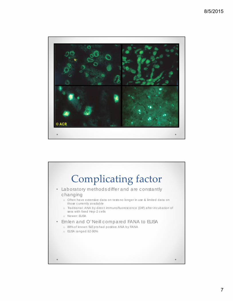
Methods of Detection• FANA – standard method
o Sera incubated with substrate cells that have been fixed with acetone
o Bound antibodies are detected by fluorecein-conjugated anti-human IgG
o Viewed through fluorescence microscope, antibodies bound to nuclear antigens produce a nuclear pattern
o Dilution at which nuclear fluorescence disappears = titero Results: pattern and titer
• Others:o Immunodiffusiono Counterummunoelectrophoresis (CIE)o Immunoprecipitationo Immunobloto Enzyme Immunossay (ELISA)

8/5/2015
6
Principle of indirect
immunofluorescence (diagram)
Principles of enzyme‐linked immunosorbent assay (diagram)
8/5/2015
7
Antinuclear antibodies (photomicrographs)
Complicating factor• Laboratory methods differ and are constantly
changingo Often have extensive data on tests no longer in use & limited data on
those currently availableo Traditional: ANA by direct immunofluorescence (DIF) after incubation of
sera with fixed Hep-2 cellso Newer: ELISA
• Emlen and O’Neill compared FANA to ELISAo 88% of known SLE pts had positive ANA by FANAo ELISA ranged 62-90%

8/5/2015
8
H&P is the key• Advances in diagnostic testing have not
supplanted a carefully performed H&P• History & ROS should seek clues of autoimmune
disease and also evidence of FMS• When a lab test (eg, ANA) is not very specific, it is
essential to determine the pre-test likelihood of the disease
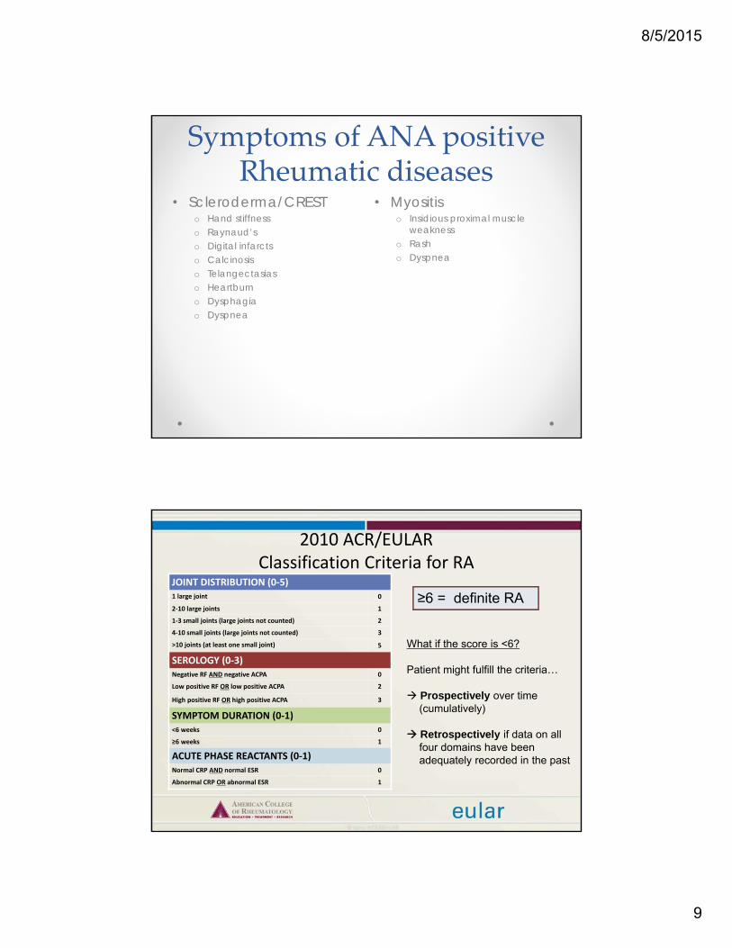
Symptoms of ANA positive Rheumatic diseases
• Lupuso Alopeciao Oral or nasal ulcerso Malar rasho Photosensitivityo Raynaud’so Pleuritic chest paino Joint pain and stiffnesso Unexplained fevero Unexplained weight losso Unexplained LAD
• Sjogren’so Dry eyeso Dry moutho Vaginal drynesso Parotid swellingo Accelerated dential caries or
gingivitis
8/5/2015
9
Symptoms of ANA positive Rheumatic diseases
• Myositiso Insidious proximal muscle
weaknesso Rasho Dyspnea
• Scleroderma/CRESTo Hand stiffnesso Raynaud’so Digital infarctso Calcinosiso Telangectasiaso Heartburno Dysphagiao Dyspnea
2010 ACR/EULARClassification Criteria for RA
JOINT DISTRIBUTION (0‐5)1 large joint 0
2‐10 large joints 1
1‐3 small joints (large joints not counted) 2
4‐10 small joints (large joints not counted) 3
>10 joints (at least one small joint) 5
SEROLOGY (0‐3)Negative RF AND negative ACPA 0
Low positive RF OR low positive ACPA 2
High positive RF OR high positive ACPA 3
SYMPTOM DURATION (0‐1)<6 weeks 0
≥6 weeks 1
ACUTE PHASE REACTANTS (0‐1)Normal CRP AND normal ESR 0
Abnormal CRP OR abnormal ESR 1
≥6 = definite RA
What if the score is <6?
Patient might fulfill the criteria…
Prospectively over time (cumulatively)
Retrospectively if data on all four domains have been adequately recorded in the past
8/5/2015
10
Algorithm to Classification of RA Including Radiographs
Longstanding Longstanding inactive diseasesuspected?
≥6/10 on the scoring system?
Not RANot RA
RA
No
already availableRadiographs
already available
Perform radiographic Perform radiographic assessment
Yes Erosions typical forErosions typical forRA present?
Yes≥1 swollen joint,
which is best explained by ≥1 swollen joint,
which is not best explained by another disease?
No
No
No
Yes
Document result of the scoring system
Yes
Yes
No
SLE: Diagnostic criteria
D: discoid rash. Erythematous raised lesions.O: oral and nasal ulcers.P: photosensitivityA: arthritis. (nonerosive, 2+ jts, symm)M: malar rash. Fixed erythema over malar eminences.I: immunologic. Anti-dsDNA, anti-Sm, or APL abs.N: neuropsych (sz or psychosis)R: renal. Proteinuria or cellular casts.A: +ANAS: serositis. Pleurisy or pericarditisH: hematologic. (hemolytic anemia, WBCs, plts)
(Any 4 or more of the 11 criteria present, serially or simultaneously, during any interval of observation)

8/5/2015
11
SLE: rash, face and neck
SLE: butterfly rash, discoid type

8/5/2015
12
Alopecia, scalp
Copyright © 1972-2004 American College of Rheumatology Slide Collection. All rights reserved.
SLE: bullous lesions, palate
Copyright © 1972-2004 American College of Rheumatology Slide Collection. All rights reserved.

8/5/2015
13
Raynaud’s phenomenon, blanching of hands
Copyright © 1972-2004 American College of Rheumatology Slide Collection. All rights reserved.
Raynaud’s phenomenon: hands
Copyright © 1972-2004 American College of Rheumatology Slide Collection. All rights reserved.

8/5/2015
14
Raynaud’s phenomenon: cyanosis of the hands
Copyright © 1972-2004 American College of Rheumatology Slide Collection. All rights reserved.
Fusiform swelling, hand

8/5/2015
15
Jaccoud’s arthropathy (clinical and radiograph)
Copyright © 1972-2004 American College of Rheumatology Slide Collection. All rights reserved.
Photosensitivity, face and neck

8/5/2015
16
Laboratory abnormalities in lupus
• CBCo Leukopenia: usually
lymphopenia, occ neutropeniao Anemia: chronic disease,
hemolytico Thrombocytopenia
• Serum Chemistryo Elevated Cro Low albumino Polyclonal hyperglobulinemiao Elevated CPK
• UAo Proteinuriao Microscopic hematuriao RBC or hyaline casts
History & Physical Exam
• Differentiating lupus from FMS by history alone can sometimes be difficult
o Fatigue, arthralgia, morning stiffness, cold intolerance, chest wall pain and subjective deficits in memory and concentration
o The likelihood of lupus increases if the pt gives a convincing history of lupus that would not ordinarily occer in FMS
• While excluding lupus, look for fibromyalgia

8/5/2015
17
Fibromyalgia• Not merely a diagnosis of exclusion• Often occurs in a setting of stress, depression,
anxiety, lack of sleep, lack of exercise, and traumatic life experiences
• Related symptoms:o Chronic headaches, memory loss, loss of concentration, parasthesias of
the extremities, irritable bowel or bladder
• Normal lab values: CBC, CMP, UA• When the history, PE, and routine lab testing
supports a diagnosis of FMS, autoantibody testing is not necessary
Fibromyalgia Tender Points

8/5/2015
18
Case #1• 45 year-old male patient presents to your
office with complaints of generalized fatigue, diffuse arthralgias and diffuse myalgias
• PMHx is significant for HTN, PTSD• PSHx: vasectomy• FHx: Mother age 65 with “some kind of
arthritis” affecting her hands and knees• SocHx: Recently divorced. No children.
Construction worker – recently unemployed. Smokes 1-2 ppd for 25 years. Drinks 2 beers each night, more on weekends. No IVDA.

8/5/2015
19
Case #2• 32 year-female patient presents with
complaints of a new rash on her face, fatigue and hand pain. She states she hasn’t been able to get her rings on and off easily. Her knuckles feel swollen & stiff.
• PMHx: Unremarkable. G2P2.• PSHx: C-section x2.• FHx: Sister with RA. • SocHx: Married. SAHM. 2 children ages 3
and 6. Smokes on the weekends socially, but not daily. ETOH on weekends: 2-4 beers/night. No IVDA.
Case #3• 55 year-female patient presents with complaints of
her fingers turning white and blue with cold exposure.
• PMHx/PSHx: GERD• FHx: Unremarkable.• SocHx: Divorced. Lab tech. No children. Smokes 1-
2ppd for 20+ years. Denies routine ETOH. No IVDA.

8/5/2015
20
QUESTIONS?
Bibliography• Blumenthal DE. Tired, aching, ANA-positive: Does your patient
have lupus or fibromyalgia? Clev Clin J Med 2002; 69(2):143-152.
• De Vlam K, et al. Detection and identification of antinuclear antibodies in the serum of normal blood donors. Clin Exp Rheumatol 1993; 11:393-397.
• Emlen W, O’Neill L. Clinical significance of antinuclear antibodies. Arthritis Rheum 1997; 40:1612-1618.
• Slater CA, et al. Antinuclear antibody testing. A study of clinical utility. Arch Intern Med 1996; 156:1421-1425.
• Tan EM, et al. Range of antinuclear antibodies in “healthy” individuals. Arthritis Rheum 1997; 40:1601-1611.