screening for pre-eclampsia: a systematic review of tests ... · ana catarina pinho freitas de...
TRANSCRIPT

Ana Catarina Pinho Freitas de Carvalho Pedrosa
Screening for pre-eclampsia: a systematic review
of tests combining uterine artery Doppler with other markers
2010/2011
Abril, 2011
Ana Catarina Pinho Freitas de Carvalho Pedrosa
Screening for pre-eclampsia: a systematic review
of tests combining uterine artery Doppler with other markers
Mestrado Integrado em Medicina
Área: Ginecologia e Obstetrícia
Trabalho efectuado sob a Orientação de:
Professora Doutora Alexandra Matias
Pereira da Cunha Coelho de Macedo
Revista: Journal of Perinatal Medicine
Abril, 2011


Projecto de Opção do 6º ano - DECLARAÇÃO DE INTEGRIDADE
Unidade Curricular “Dissertação/Monografia/Relatório de Estágio Profissionalizante”
Eu, Ana Catarina Pinho Freitas de Carvalho Pedrosa, abaixo assinado, nº mecanográfico 050801116,
estudante do 6º ano do Mestrado Integrado em Medicina, na Faculdade de Medicina da Universidade
do Porto, declaro ter actuado com absoluta integridade na elaboração deste projecto de opção.
Neste sentido, confirmo que NÃO incorri em plágio (acto pelo qual um indivíduo, mesmo por omissão,
assume a autoria de um determinado trabalho intelectual, ou partes dele). Mais declaro que todas as
frases que retirei de trabalhos anteriores pertencentes a outros autores, foram referenciadas, ou
redigidas com novas palavras, tendo colocado, neste caso, a citação da fonte bibliográfica.
Faculdade de Medicina da Universidade do Porto, 01/03/2011
Assinatura:

Faculdade de Medicina da Universidade do Porto
2010/2011
Unidade Curricular “Dissertação/Monografia/Relatório de Estágio Profissionalizante”
Projecto de Opção do 6º ano – DECLARAÇÃO DE REPRODUÇÃ O
Nome: Ana Catarina Pinho Freitas de Carvalho Pedrosa
Endereço electrónico: [email protected] Telefone ou Telemóvel:
Número do Bilhete de Identidade:
Título da Dissertação/Monografia/Relatório de Estágio Profissionalizante (cortar o que não
interessa):
Screening for pre-eclampsia: a systematic review of tests combining uterine artery Doppler with other
markers
Orientador:
Professora Doutora Alexandra Matias Pereira da Cunha Coelho de Macedo
Ano de conclusão: 2011
Designação da área do projecto:
Ginecologia e Obstetrícia
É autorizada a reprodução integral desta Dissertação/Monografia/Relatório de Estágio
Profissionalizante (cortar o que não interessar) para efeitos de investigação e de divulgação
pedagógica, em programas e projectos coordenados pela FMUP.
Faculdade de Medicina da Universidade do Porto, 01/03/2011
Assinatura:

ACKNOWLEDGMENTS
I would like to express my most sincere thanks to Professor Alexandra Matias for her
excellent guidance throughout this project. Her insight, ideas and suggestions were
invaluable. Her dedication, enthusiasm and encouragement were inspiring.
I also wish to thank my parents for everything they taught me, for their guidance and for
their unconditional love and support.

TABLE OF CONTENTS
1. TITLE PAGE. .................................................................................................................... 1
2. ABSTRACT. ..................................................................................................................... 2
3. KEYWORDS. ................................................................................................................... 3
4. INTRODUCTION. .............................................................................................................. 4
5. METHODS. ...................................................................................................................... 6
6. RESULTS. ........................................................................................................................ 8
7. DISCUSSION. ................................................................................................................. 10
8. REFERENCE LIST. ......................................................................................................... 16

1
COMPLETE TITLE
Screening for pre-eclampsia: a systematic review of tests combining uterine artery Doppler
with other markers
SHORT TITLE
Screening for pre-eclampsia: a systematic review

2
ABSTRACT
Aims
To perform a systematic review of screening for pre-eclampsia (PE) with the
combination of uterine artery Doppler (UAD), maternal history, mean arterial pressure and/or
maternal serum markers.
Methods
We identified eligible studies through a search of Medline, and, for each included study,
we assessed the risk of bias and extracted relevant data. We reported the performance of
screening tests according to the target population (low or high-risk), the trimester of screening
(first and/or second) and the subset of PE screened for (early and late).
Results
Several tests provided moderate or convincing prediction of early PE, but screening for
late PE was poor. Although UAD is more accurate in the second-trimester, we found
encouraging results for first-trimester screening when it was combined with other markers.
Performance of screening was consistently lower in populations with risk factors for PE in the
maternal history.
Conclusions
We present encouraging results for the prediction of early PE, even in the first-trimester
of pregnancy. The different performance of tests in screening for early versus late PE, and of
low versus high-risk populations, supports the concept that PE is a heterogeneous disease.

3
KEYWORDS
Biochemical serum markers
Blood pressure
Doppler
First trimester
Maternal history
Pre-eclampsia
Screening
Second trimester
Systematic review

4
INTRODUCTION
Pre-eclampsia (PE) affects 2-8% of pregnancies and is a major cause of maternal and
perinatal morbidity and mortality [45]. In mothers, PE may lead to disseminated
coagulopathy, pulmonary edema, renal or liver failure, eclampsia, stroke and placental
abruption [38]. In fetuses, it may cause intrauterine growth restriction (IUGR), hypoxia-
neurologic injury and preterm delivery [38]. Ultimately, PE may lead to death of the mother
and/or the fetus [38].
Considering the impact of PE in obstetrics, the development of an accurate screening
method would be of great value. Although, at present, there is no effective preventive
intervention for PE [38,45], screening would allow us to select a group of pregnant women
who would receive increased maternal and fetal monitoring [12]. From a research point of
view, it would be essential for future development of effective prophylactic measures, as it
would enable the recruitment of high-risk women in which the effect of those measures could
be evaluated [12].
Classically, PE has been associated with inadequate trophoblast invasion of the spiral
arteries and consequent failure of development of a low-resistance uteroplacental circulation
that characterizes normal pregnancies [10,13]. Therefore, uterine artery Doppler (UAD) has
been extensively studied as a screening test for PE. A recent meta-analysis [11] reported that a
high second-trimester pulsatility index (PI) detects 42% of PE cases with a specificity of 91%.
In the first-trimester, the accuracy is lower, with a sensitivity of 25% and a specificity of 95%.
The National Institute for Health and Clinical Excellence [24] currently recommends
the assessment of each woman’s risk for PE on the basis of maternal history. Age ≥ 40 years,
body mass index ≥ 30 Kg/m2, pre-existing vascular or renal disease, nulliparity or pregnancy
interval of > 10 years, prior or family history of PE and multiple pregnancy increase the
probability of developing PE. However, accuracy of screening with maternal history alone is
low [32].
In addition to UAD and maternal history, a large number of maternal serum markers
have been investigated for the prediction of PE, but their use as single screening tests has also
been disappointing [6]. Finally, early measurement of mean arterial pressure (MAP) is
another screening test that should not be forgotten because it is simple and inexpensive and
appears to be an important predictor of subsequent PE [6].

5
Despite great research efforts, in 2004, the World Health Organization [12] concluded
that no single test was yet available to provide accurate screening for PE. Since then, there has
been growing interest in the combination of markers for PE screening. Recently, this was
reviewed by Giguère et al. [17], who concluded that the combination of biochemical and
ultrasonographic markers improves prediction of PE. However, the authors did not
systematically evaluate the contribution of maternal history and MAP to combined screening.
In this context, we performed the current systematic review to evaluate first and second-
trimester screening for PE with tests that combine UAD with maternal history, MAP and/or
maternal serum markers.

6
METHODS
A search was conducted in the Medline database using the following MeSH terms or
keywords, with no limits:
preeclampsia, pre-eclampsia, diagnosis, screening, prediction, uterine artery, Doppler,
clinical, maternal, characteristics, factors, history, blood pressure, pregnancy-associated
plasma protein-A, PAPP-A, chorionic gonadotropin, hCG, alpha-fetoprotein, inhibin A,
activin A, placental protein 13, placental growth factor, soluble fms-like tyrosine kinase 1,
soluble vascular endothelial growth factor receptor-1.
Additional keywords were tested but were not included in the final query because they
did not improve the sensitivity of the search.
Table 1 presents the selection criteria used to determine the eligibility of the studies
identified by the search, and, when appropriate, the rationale for using those criteria. They
were applied in two stages: first, to the titles and abstracts of the articles yielded by the query;
second, to the full texts of the articles selected in the first stage.
The reference list of the selected articles was also searched to identify additional
potential articles of interest, which were then retrieved and submitted to the selection criteria.
The search was updated periodically and was last run on 10 December 2010.
Relevant data were extracted from each article using a standardized form. Risk of bias
was assessed according to criteria that had been previously used [12] and that we adapted to
our own review (Table 2). In nested case-control studies, we considered selection adequate
when cases included “all (or a representative sample of) individuals with the outcome of
interest occurring in the defined cohort” and controls were “a random sample of the
individuals remaining in the cohort” [47]. This way, investigators ensured that cases were
representative of individuals with the outcome in the population studied and that controls
were representative of individuals without the outcome in the same population.
When available, we reported the following measures of accuracy: area under the ROC
curve (AUC), specificity (Sp), sensitivity (Sn), and likelihood ratios of the positive (LR+) and
negative (LR-) results. The LR+ is defined as Sn/(1-Sp) and the LR- as (1-Sn)/Sp [12].
Convincing prediction is provided by tests with LR+ > 10 and LR- < 0.1 [11,12]. On the other

7
hand, tests with LR+ < 5 or LR- > 0.2 achieve only minimal prediction and the other tests
achieve moderate prediction [12].
We followed the PRISMA guidelines [22] in order to maximize the quality of the report
of our systematic review.

8
RESULTS
Figure 1 depicts the results of each stage of the selection process. According to the
selection criteria previously described, 35 articles were eligible and, of those, three [34,36,37]
reported results of the same cohort. Therefore, 33 studies were reviewed, which are described
in Table 3.
Twenty-one cohort and 12 nested case-control studies were included. Seven were
conducted in high-risk populations, defined by the presence of risk factors for PE in the
maternal history, an abnormal second-trimester UAD or high second-trimester levels of
human chorionic gonadotropin (hCG) and alpha-fetoprotein (AFP).
Studies of low-risk populations reported a prevalence of PE that varied between 1.2%
and 10.5%, although it was ≤3.0% in the majority. Those which evaluated screening for early
PE reported a prevalence of this outcome of 0.3%-0.8%, with the exception of one study
which reported a prevalence of 2.3%.
The conclusions of some studies are limited by the size of their samples, which is partly
related to the low frequency of PE and especially early PE in the general population. For
example, six studies included only ≤10 participants with early PE.
Screening tests were performed in the first-trimester in 12 studies, in the second-
trimester in 15 and in both trimesters in six.
Maternal history was evaluated in 16 studies, pregnancy-associated plasma protein-A
(PAPP-A) in 11, inhibin A in nine, placental growth factor (PlGF) in eight, hCG, activin A
and placental protein 13 (PP13) in six each, soluble fms-like tyrosine kinase 1 (sFlt1) in four,
MAP and AFP in three each.
It should be noted that some studies are related. Seven of them [1-3,30,33-35] were
conducted as part of the same research program and had overlapping study groups. This also
happened in two additional studies [27,49]. To avoid data duplication, whenever a screening
test was evaluated in more than one of these studies, we only reported its performance in the
study with the largest sample. Other studies [40 and 44, 42 and 43] also had common
participants but we considered them separately because they evaluated different screening
tests.

9
Figure 2 presents the results of the assessment of the risk of bias. Selection of the study
participants was generally adequate but occasionally inadequate or unreported. The study
population was adequately described in nearly all studies whereas description of the screening
tests was frequently inadequate because the selected cut-off points were not specified.
Blinding of the readers of the screening tests was usually adequate, but sometimes unreported.
On the other hand, complete blinding of the readers of the reference standard was
accomplished in only two studies and was inadequate or unreported in the remaining. Follow-
up and verification was adequate in most studies but in several it was unreported.
Tables 4, 5, 6 and 7 summarize the results of each study. The performance of screening
tests is reported quantitatively, through AUC, Sp, Sn, LR+ and LR-, and qualitatively,
through a citation of the article.
Figures 3 and 4 show the screening tests that provided moderate or convincing
prediction, according to the LR values. None of the screening tests for late PE qualified. A
few screening tests for total PE were moderately predictive, all of which involved second-
trimester testing. On the other hand, screening for early PE was accomplished with moderate
accuracy by several first and second-trimester tests. Additionally, four tests were on the verge
of providing convincing prediction, and one first-trimester test was highly predictive.

10
DISCUSSION
Best first-trimester screening tests for early PE
Our results suggest that, in the first-trimester, accurate screening for early PE probably
requires the combination of several markers. Two tests that combined UAD with four other
markers had a very good performance.
The combination of mean-PI (M-PI), maternal history, MAP, PAPP-A and PlGF was
highly predictive of early PE, in a nested case-control study with almost 30 cases of early PE
[33]. Of the 33 studies reviewed, one included the same number of early PE cases and only
six included a higher number. Unfortunately, there were additional cases of early PE in the
original cohort who did not have available serum for the measurement of PlGF and thus were
not included in the case-control study. Although selection of cases was simply based on
serum availability, we cannot be certain that the sample is representative of the original
population. We conclude that these results are very encouraging but require confirmation by
future studies.
The other test combined lowest-PI (L-PI), maternal history, PAPP-A, inhibin A and
PlGF and was applied to a cohort of nulliparous women [8]. However, it should be considered
with caution because the cohort included only four participants who subsequently developed
early PE. Furthermore, the authors reported that L-PI was not significantly different between
the PE and normal outcome groups and that it did not improve screening by maternal history
combined with the three serum markers.
Several other tests had a good performance in the first-trimester, virtually all of them
with a LR+ > 8.6 and a LR- ≤ 0.18. The majority took into account the maternal history.
Other markers frequently considered were MAP, PAPP-A, inhibin A and PlGF. In particular,
we’d like to highlight that screening with L-PI, maternal history, MAP and PAPP-A achieved
a LR+ of 9.46 and a LR- of 0.06, in a cohort of over 8300 women, including 37 with early PE
[34,36,37].
One study [25] also achieved promising results with the combination of M-PI and PP13,
albeit in a sample that included only ten early PE cases. These results are supported by other
studies, which demonstrated that first-trimester PP13 is a significant predictor of early PE [2]
and that it improves screening with first [21] and second-trimester UAD [41]. However, one
study did not confirm the predictive accuracy of PP13 [8].

11
Best second-trimester screening tests for early PE
Screening for early PE in the second-trimester probably requires the combination of
fewer markers, as UAD alone is substantially more accurate when performed in this trimester
[11].
The association of M-PI, maternal history and MAP almost provided convincing
prediction in a cohort of over 3000 women, including 23 with early PE [26]. The same
promising results were obtained when maternal history and first-trimester UAD were
combined with the assessment of the ratio between M-PI in the second and first trimesters, in
a cohort of similar dimensions [31]. Therefore, measurement of maternal serum markers may
not be necessary to provide accurate screening for PE in the second-trimester, because the
combination of UAD with the simple and inexpensive evaluation of maternal history and
MAP may be sufficient.
In women with abnormal second-trimester UAD, sFlt1 appeared to be useful in early PE
screening. In one study [14], sFlt1 provided moderate prediction and the sFlt1/PlGF ratio
almost provided convincing prediction. A similar study [46] demonstrated a good
performance of the two serum markers when they were concurrently measured but not
combined in a ratio. However, both studies were limited by the size of their samples, which
included only 8 and 9 women with early PE, respectively. Moreover, their results were
challenged by a larger study, which found that sFlt1 was not able to predict early PE in a
group of women with abnormal UAD [15].
In one second-trimester study [40], combination of UAD and PP13, with or without
other serum markers, achieved moderate prediction of early PE (LR+=5, LR-=0). However,
this was a case-control study which included only five cases of early PE that were selected
from a cohort on the basis of serum availability. Furthermore, the authors reported that the
addition of PP13 to UAD did not improve screening.
Best screening tests for late and total PE
In contrast with early PE, none of the tests were even moderately predictive of late PE.
On the other hand, prediction of total PE was accomplished with moderate accuracy by some
tests, all of which involved second-trimester screening and combined UAD with inhibin A,
activin A, PlGF and/or sFlt1.

12
What to screen? Early versus late PE
The performance of screening tests was consistently and substantially better in the
prediction of early PE, comparing to late PE. Our results support the concept that these are
distinct disease entities [20,48], with impaired placentation and defective angiogenesis being
related especially to early PE [13,14,45], and cardiovascular and metabolic risk factors
probably leading to late PE [45]. Although much less frequent than the late form of the
condition [20], early PE is the main contributor to the maternal and perinatal morbidity and
mortality seen in PE [1,31], as it is associated with premature delivery, a higher risk of IUGR,
more severe maternal disease and a higher rate of pregnancy-related maternal death [20,48].
Considering its impact, the low incidence of early PE should not prevent routine screening.
Moreover, trisomy 21, for which screening is currently performed, is even less frequent,
affecting only 0.14% of newborns in the absence of any intervention [24].
When to screen? First versus second-trimester screening
Even though UAD is more accurate in the second-trimester [11], we found encouraging
results for first-trimester screening when it was combined with other markers. We believe that
screening for PE is most relevant in the first-trimester because, as suggested by a recent meta-
analysis [10], preventive interventions are more likely to be effective if initiated early in
pregnancy, when pathogenic mechanisms can still be modified.
Who to screen? Low versus high-risk populations
Three studies evaluated the performance of screening tests in high-risk populations
characterized by the presence of certain risk factors in the maternal history.
Screening a preselected high-risk population is of particular interest when the condition
screened is infrequent, as is the case of early PE. Assuming that the sensitivity and specificity
of the screening test remain constant, the positive predictive value increases when it is applied
to a population with higher disease prevalence.
Interestingly, however, the accuracy of screening tests was consistently lower in the
previously mentioned studies than in studies of low-risk populations. In nulliparous women
[8], the performance of screening with L-PI and maternal history was poorer than in
unselected pregnancies [34,36,37]. In women with risk factors such as chronic hypertension,
pregestational diabetes mellitus and obesity, screening with M-PI and maternal history [19] or
with M-PI and PP13 [21] was also less predictive than in low-risk populations [31,25].

13
In the meta-analysis performed by Cnossen et al. [11], screening with the pulsatility
index was less accurate in high-risk populations. Thus, our results may be explained by the
lower performance of UAD in women with historical risk factors. They further support the
concept of PE as a heterogeneous disease [13,38,48] and suggest that, in these women,
impaired placentation may play a less important role in the development of PE [19].
Limitations of the reviewed studies
Several studies that we reviewed were limited by the size of their samples and by the
risk of bias in certain methodological areas.
Selection of study participants was occasionally inadequate. Some nested case-control
studies included only a subset of PE cases of the original cohort because only those had
available blood samples for the measurement of specific markers. Additionally, some cohort
studies did not apply the screening tests to all eligible participants.
Outcome assessors were not completely blinded to the results of the screening tests in
several studies. In some cases, blinding was inadequate because the results of UAD
influenced the subsequent management of pregnancies. In other cases, investigators evaluated
screening for PE with first-trimester PAPP-A or hCG, or with second-trimester hCG or AFP,
and had to communicate the results of the screening tests to pregnant women and their
managing clinicians because they were necessary for routine assessment of trisomy 21 risk.
This shows that, at times, although the methodology of the study may lead to bias, it is the
best that researchers are able to do, and for that reason the PRISMA statement [22]
recommends that the term “quality” be replaced by “risk of bias”.
The comparison of individual studies is limited by differences observed among them,
concerning, for example, the definitions of PE and early PE and the UAD technique.
Although, in the studies reviewed, the definitions of PE always included the concurrent
presence of hypertension and proteinuria, we identified among them several differences.
Concerning hypertension, some studies considered it to be present when either the systolic
(SBP) or diastolic (DBP) blood pressure (BP) was elevated, while others did so only in the
presence of high DBP. Generally, the diagnosis of hypertension required at least two
recordings of elevated BP with a minimal 4-6 hour interval, and cutoffs of 140 mmHg and 90
mmHg were considered for high SBP and DBP, respectively. Occasionally, however, studies
reported the presence of hypertension when DBP was ≥ 110 mmHg on any occasion or ≥ 90
mmHg on at least two occasions. Proteinuria was usually defined as protein excretion of

14
≥ 300 mg in a 24-hour urine collection, but two dipstick readings of ≥ 2+, or occasionally
≥ 1+, were also frequently considered diagnostic if no 24-hour collection was available.
The definition of early PE was also not uniform and almost one third of the studies did
not report screening for this outcome.
The technical performance of UAD was usually but not always clearly reported.
Investigators used color Doppler to identify the uterine arteries (UA) and pulsed wave
Doppler to obtain the flow velocity waveforms. In the first-trimester, UAD was generally
performed using a transabdominal approach. In the second-trimester, transabdominal UAD
was also used, but the transvaginal approach was more frequent, often because investigators
concurrently measured the cervical length for the assessment of the risk of premature
delivery. In the majority of studies, waveforms were obtained from the UA at the level of the
internal cervical os but in several others they were obtained at or one centimeter distal to the
crossover point with the external iliac artery. Investigators variably required an angle of
insonation below 30º, 50º or 60º, and several did not describe it. Although PI was most
frequently reported, some studies evaluated other Doppler parameters, such as the resistance
index or the presence of early diastolic notches.
Limitations and strengths of our review
The strength of our conclusions is limited by the multiplicity of combinations evaluated,
the variability of the gestational age at which tests were performed (even within the same
trimester) and the diversity of populations studied. Additionally, as in any review, it is limited
by the shortcomings of the original studies, which we have previously discussed.
On the other hand, our review has several strengths: we used explicit and reproducible
methodology; we minimized the risk of bias using rigorously predefined selection criteria and
a standardized data extraction form; we assessed the risk of bias of the included studies with
objective criteria; we reported the findings of each study and summarized those findings in a
systematic way.
Our review does not provide definitive conclusions but rather highlights important
advances that have been made in PE screening and offers guidance and optimism for future
research. To our knowledge, it is the first review that systematically evaluates the
combination of UAD, maternal history, MAP and serum markers in the prediction of PE.
While screening for total and especially late PE remains disappointing, we have
demonstrated encouraging results in the prediction of early PE. In addition to the eight serum

15
markers we reviewed, others, such as soluble endoglin and homocysteine, might be useful
when combined with UAD.
We believe that future research should focus on first-trimester screening for early PE,
with a combination of UAD, maternal history, MAP, and serum markers such as PAPP-A,
inhibin A and PlGF. Large cohort studies are needed in order to accurately study this
relatively infrequent outcome of pregnancy.

16
REFERENCE LIST
1. Akolekar R, Minekawa R, Veduta A, Romero XC, Nicolaides KH. Maternal plasma
inhibin A at 11-13 weeks of gestation in hypertensive disorders of pregnancy. Prenat
Diagn. 2009;29:753-60.
2. Akolekar R, Syngelaki A, Beta J, Kocylowski R, Nicolaides KH. Maternal serum
placental protein 13 at 11-13 weeks of gestation in preeclampsia. Prenat Diagn.
2009;29:1103-8.
3. Akolekar R, Zaragoza E, Poon LC, Pepes S, Nicolaides KH. Maternal serum placental
growth factor at 11 + 0 to 13 + 6 weeks of gestation in the prediction of pre-eclampsia.
Ultrasound Obstet Gynecol. 2008;32:732-9.
4. Alkazaleh F, Chaddha V, Viero S, Malik A, Anastasiades C, Sroka H, et al. Second-
trimester prediction of severe placental complications in women with combined
elevations in alpha-fetoprotein and human chorionic gonadotrophin. Am J Obstet
Gynecol. 2006;194:821-7.
5. Aquilina J, Thompson O, Thilaganathan B, Harrington K. Improved early prediction of
pre-eclampsia by combining second-trimester maternal serum inhibin-A and uterine
artery Doppler. Ultrasound Obstet Gynecol. 2001;17:477-84.
6. Audibert F. Maternal serum screening for preeclampsia: is performance enough? Clin
Biochem. 2010;43:707-8.
7. Audibert F, Benchimol Y, Benattar C, Champagne C, Frydman R. Prediction of
preeclampsia or intrauterine growth restriction by second trimester serum screening and
uterine Doppler velocimetry. Fetal Diagn Ther. 2005;20:48-53.
8. Audibert F, Boucoiran I, An N, Aleksandrov N, Delvin E, Bujold E, et al. Screening for
preeclampsia using first-trimester serum markers and uterine artery Doppler in
nulliparous women. Am J Obstet Gynecol. 2010;203:383.e1-8.
9. Ay E, Kavak ZN, Elter K, Gokaslan H, Pekin T. Screening for pre-eclampsia by using
maternal serum inhibin A, activin A, human chorionic gonadotropin, unconjugated
estriol, and alpha-fetoprotein levels and uterine artery Doppler in the second trimester of
pregnancy. Aust N Z J Obstet Gynaecol. 2005;45:283-8.

17
10. Bujold E, Morency AM, Roberge S, Lacasse Y, Forest JC, Giguère Y. Acetylsalicylic
acid for the prevention of preeclampsia and intra-uterine growth restriction in women
with abnormal uterine artery Doppler: a systematic review and meta-analysis. J Obstet
Gynaecol Can. 2009;31:818-26.
11. Cnossen JS, Morris RK, ter Riet G, Mol BW, van der Post JA, Coomarasamy A, et al.
Use of uterine artery Doppler ultrasonography to predict pre-eclampsia and intrauterine
growth restriction: a systematic review and bivariable meta-analysis. CMAJ.
2008;178:701-11.
12. Conde-Agudelo A, Villar J, Lindheimer M. World Health Organization systematic
review of screening tests for preeclampsia. Obstet Gynecol. 2004;104:1367-91.
13. Crispi F, Domínguez C, Llurba E, Martín-Gallán P, Cabero L, Gratacós E. Placental
angiogenic growth factors and uterine artery Doppler findings for characterization of
different subsets in preeclampsia and in isolated intrauterine growth restriction. Am J
Obstet Gynecol. 2006;195:201-7.
14. Diab AE, El-Behery MM, Ebrahiem MA, Shehata AE. Angiogenic factors for the
prediction of pre-eclampsia in women with abnormal midtrimester uterine artery
Doppler velocimetry. Int J Gynaecol Obstet. 2008;102:146-51.
15. Espinoza J, Romero R, Nien JK, Gomez R, Kusanovic JP, Gonçalves LF, et al.
Identification of patients at risk for early onset and/or severe preeclampsia with the use
of uterine artery Doppler velocimetry and placental growth factor. Am J Obstet
Gynecol. 2007;196:326.e1-13.
16. Florio P, Reis FM, Pezzani I, Luisi S, Severi FM, Petraglia F. The addition of activin A
and inhibin A measurement to uterine artery Doppler velocimetry to improve the early
prediction of pre-eclampsia. Ultrasound Obstet Gynecol. 2003;21:165-9.
17. Giguère Y, Charland M, Bujold E, Bernard N, Grenier S, Rousseau F, et al. Combining
biochemical and ultrasonographic markers in predicting preeclampsia: a systematic
review. Clin Chem. 2010;56:361-75.
18. Gilbert WM. The cost of preterm birth: the low cost versus high value of tocolysis.
BJOG. 2006;113 Suppl 3:4-9.

18
19. Herraiz I, Arbués J, Camaño I, Gómez-Montes E, Grañeras A, Galindo A. Application
of a first-trimester prediction model for pre-eclampsia based on uterine arteries and
maternal history in high-risk pregnancies. Prenat Diagn. 2009;29:1123-9.
20. Huppertz B. Placental origins of preeclampsia: challenging the current hypothesis.
Hypertension. 2008;51:970-5.
21. Khalil A, Cowans NJ, Spencer K, Goichman S, Meiri H, Harrington K. First-trimester
markers for the prediction of pre-eclampsia in women with a-priori high risk.
Ultrasound Obstet Gynecol. 2010;35:671-9.
22. Liberati A, Altman DG, Tetzlaff J, Mulrow C, Gøtzsche PC, Ioannidis JP, et al. The
PRISMA statement for reporting systematic reviews and meta-analyses of studies that
evaluate healthcare interventions: explanation and elaboration. BMJ. 2009;339:b2700.
23. Llurba E, Carreras E, Gratacós E, Juan M, Astor J, Vives A, et al. Maternal history and
uterine artery Doppler in the assessment of risk for development of early- and late-onset
preeclampsia and intrauterine growth restriction. Obstet Gynecol Int.
2009;2009:275613.
24. National Collaborating Centre for Women’s and Children’s Health, commissioned by
the National Institute for Health and Clinical Excellence. Antenatal care: routine care
for the healthy pregnant woman. London: RCOG Press; 2008.
25. Nicolaides KH, Bindra R, Turan OM, Chefetz I, Sammar M, Meiri H, et al. A novel
approach to first-trimester screening for early pre-eclampsia combining serum PP-13
and Doppler ultrasound. Ultrasound Obstet Gynecol. 2006;27:13-7.
26. Onwudiwe N, Yu CK, Poon LC, Spiliopoulos I, Nicolaides KH. Prediction of pre-
eclampsia by a combination of maternal history, uterine artery Doppler and mean
arterial pressure. Ultrasound Obstet Gynecol. 2008;32:877-83.
27. Papageorghiou AT, Yu CK, Erasmus IE, Cuckle HS, Nicolaides KH. Assessment of
risk for the development of pre-eclampsia by maternal characteristics and uterine artery
Doppler. BJOG. 2005;112:703-9.
28. Parra M, Rodrigo R, Barja P, Bosco C, Fernández V, Muñoz H, et al. Screening test for
preeclampsia through assessment of uteroplacental blood flow and biochemical markers
of oxidative stress and endothelial dysfunction. Am J Obstet Gynecol. 2005;193:1486-
91.

19
29. Pilalis A, Souka AP, Antsaklis P, Daskalakis G, Papantoniou N, Mesogitis S, et al.
Screening for pre-eclampsia and fetal growth restriction by uterine artery Doppler and
PAPP-A at 11-14 weeks' gestation. Ultrasound Obstet Gynecol. 2007;29:135-40.
30. Plasencia W, Maiz N, Bonino S, Kaihura C, Nicolaides KH. Uterine artery Doppler at
11 + 0 to 13 + 6 weeks in the prediction of pre-eclampsia. Ultrasound Obstet Gynecol.
2007;30:742-9.
31. Plasencia W, Maiz N, Poon L, Yu C, Nicolaides KH. Uterine artery Doppler at 11 + 0
to 13 + 6 weeks and 21 + 0 to 24 + 6 weeks in the prediction of pre-eclampsia.
Ultrasound Obstet Gynecol. 2008;32:138-46.
32. Poon LC, Kametas NA, Chelemen T, Leal A, Nicolaides KH. Maternal risk factors for
hypertensive disorders in pregnancy: a multivariate approach. J Hum Hypertens.
2010;24:104-10.
33. Poon LC, Kametas NA, Maiz N, Akolekar R, Nicolaides KH. First-trimester prediction
of hypertensive disorders in pregnancy. Hypertension. 2009;53:812-8.
34. Poon LC, Karagiannis G, Leal A, Romero XC, Nicolaides KH. Hypertensive disorders
in pregnancy: screening by uterine artery Doppler imaging and blood pressure at 11-13
weeks. Ultrasound Obstet Gynecol. 2009;34:497-502.
35. Poon LC, Maiz N, Valencia C, Plasencia W, Nicolaides KH. First-trimester maternal
serum pregnancy-associated plasma protein-A and pre-eclampsia. Ultrasound Obstet
Gynecol. 2009;33:23-33.
36. Poon LC, Staboulidou I, Maiz N, Plasencia W, Nicolaides KH. Hypertensive disorders
in pregnancy: screening by uterine artery Doppler at 11-13 weeks. Ultrasound Obstet
Gynecol. 2009;34:142-8.
37. Poon LC, Stratieva V, Piras S, Piri S, Nicolaides KH. Hypertensive disorders in
pregnancy: combined screening by uterine artery Doppler, blood pressure and serum
PAPP-A at 11-13 weeks. Prenat Diagn. 2010;30:216-23.
38. Sibai B, Dekker G, Kupferminc M. Pre-eclampsia. Lancet. 2005;365:785-99.
39. Simonazzi G, Vicenzi C, Rizzo MA, Farina A, Gabrielli S, Arcelli D, et al. Prospective
evaluation of the risk of pre-eclampsia using logistic regression analysis. Ultrasound
Obstet Gynecol. 2007;30:312-7.

20
40. Spencer K, Cowans NJ, Chefetz I, Tal J, Kuhnreich I, Meiri H. Second-trimester uterine
artery Doppler pulsatility index and maternal serum PP13 as markers of pre-eclampsia.
Prenat Diagn. 2007;27:258-63.
41. Spencer K, Cowans NJ, Chefetz I, Tal J, Meiri H. First-trimester maternal serum PP-13,
PAPP-A and second-trimester uterine artery Doppler pulsatility index as markers of pre-
eclampsia. Ultrasound Obstet Gynecol. 2007;29:128-34.
42. Spencer K, Cowans NJ, Nicolaides KH. Maternal serum inhibin-A and activin-A levels
in the first trimester of pregnancies developing pre-eclampsia. Ultrasound Obstet
Gynecol. 2008;32:622-6.
43. Spencer K, Yu CK, Cowans NJ, Otigbah C, Nicolaides KH. Prediction of pregnancy
complications by first-trimester maternal serum PAPP-A and free beta-hCG and with
second-trimester uterine artery Doppler. Prenat Diagn. 2005;25:949-53.
44. Spencer K, Yu CK, Savvidou M, Papageorghiou AT, Nicolaides KH. Prediction of pre-
eclampsia by uterine artery Doppler ultrasonography and maternal serum pregnancy-
associated plasma protein-A, free beta-human chorionic gonadotropin, activin A and
inhibin A at 22 + 0 to 24 + 6 weeks' gestation. Ultrasound Obstet Gynecol.
2006;27:658-63.
45. Steegers EA, von Dadelszen P, Duvekot JJ, Pijnenborg R. Pre-eclampsia. Lancet.
2010;376:631-44.
46. Stepan H, Unversucht A, Wessel N, Faber R. Predictive value of maternal angiogenic
factors in second trimester pregnancies with abnormal uterine perfusion. Hypertension.
2007;49:818-24.
47. Szklo M, Nieto FJ. Epidemiology: beyond the basics. 2nd ed. Sudbury, MA: Jones and
Bartlett Publishers; 2006.
48. von Dadelszen P, Magee LA, Roberts JM. Subclassification of preeclampsia. Hypertens
Pregnancy. 2003;22:143-8.
49. Yu CK, Smith GC, Papageorghiou AT, Cacho AM, Nicolaides KH; Fetal Medicine
Foundation Second Trimester Screening Group. An integrated model for the prediction
of preeclampsia using maternal factors and uterine artery Doppler velocimetry in
unselected low-risk women. Am J Obstet Gynecol. 2005;193:429-36.

21
50. Yu J, Shixia CZ, Wu Y, Duan T. The study of inhibin A, activin A, placental growth
factor and uterine artery Doppler pulsatility index to predict pre-eclampsia. Ultrasound
Obstet Gynecol. 2010. Epub ahead of print.

APPENDICES

LIST OF CAPTIONS
Figures
Figure 1
Selection process of the articles for the systematic review.
Figure 2 Assessment of the risk of bias.
Figure 3 Screening tests which provided moderate or convincing prediction of
early PE, according to the likelihood ratio (LR) values.
Figure 4 Screening tests which provided moderate or convincing prediction of
total PE, according to the likelihood ratio (LR) values.
Tables
Table 1
Selection criteria used to determine eligibility of the studies for the
systematic review.
Table 2 Criteria for assessment of risk of bias.
Table 3 Description of the studies included in the systematic review.
Table 4 Screening for PE with UAD and one other marker.
Table 5 Screening for PE with UAD and two other markers.
Table 6 Screening for PE with UAD and three other markers.
Table 7 Screening for PE with UAD and four other markers.

Table 1 – Selection criteria used to determine eligibility of the studies for the systematic review. Inclusion criteria
1. Study design Prospective studies, in which screening tests were applied before outcomes were developed, including
cohort and nested case-control studies.
Rationale A case-control design may represent a valid alternative to a cohort analysis if cases and controls
belong to a common reference population; otherwise, selection bias may ensue. This is assured when
participants are selected from a well defined cohort, as in nested case-control studies, which take
advantage of “both the methodologic soundness of the cohort design (i.e., limiting selection bias) and
the efficiency of the case-control approach” (limiting costs). [47]
2. Study aim To evaluate the performance of screening tests.
3. Screening test Combination of UAD and one or more of the following: maternal history, MAP, PAPP-A, hCG, AFP,
inhibin A, activin A, PP13, PlGF, sFlt1.
4. Trimester of screening First (0 to 13+6 weeks’ gestation) and/or second (14+0 to 27+6 weeks’ gestation).
5. Condition screened Early and late PE, ideally.
Total PE, in alternative.
Other subtypes of PE (e.g., severe PE), if none of the previously referred outcomes were considered.
Rationale According to von Dadelszen et al. [48], gestational age at onset of PE “is the most important clinical
variable in predicting both maternal and perinatal outcomes”.
6. Population screened Low-risk population: recruitment of participants from an unselected obstetric population;
or
High-risk population: recruitment of (1) women with risk factors for PE in their clinical history (e.g.
chronic hypertension) or (2) women with a positive result in a previous screening test (e.g. women
with abnormal UAD).
7. Reference standard Several definitions of PE were accepted, as long as they included the concurrent presence of
hypertension and proteinuria.
Several definitions of early PE were accepted, but the preferred one was: PE requiring delivery before
34 weeks’ gestation. When other definitions were considered, they were specified.
Rationale The selection of the preferred definition of early PE was based on the following facts: (1) the
administration of glucocorticoids is recommended for fetal lung maturity when there is risk of
preterm delivery in pregnant women with less than 34 weeks’ gestation [18]; (2) preterm birth
occurring after 34 weeks’ gestation is rarely associated with mortality or major morbidity [18].
Exclusion criteria
The definition of PE considered in the study was not reported.
PE was combined with other pregnancy complications in the outcome.
PE: pre-eclampsia; UAD: uterine artery Doppler; MAP: mean arterial pressure; PAPP-A: pregnancy-associated plasma protein-A, hCG: human
chorionic gonadotropin; AFP: alpha-fetoprotein; PP13: placental protein 13; PlGF: placental growth factor; sFlt1: soluble fms-like tyrosine kinase 1.

Table 2 – Criteria for assessment of risk of bias. 1. Selection of study participants
Adequate Cohort studies in which all eligible women were included consecutively or randomly into the
study.
Nested case-control studies in which all eligible women were included consecutively or
randomly into the original cohort, all or a random selection of the participants that developed PE
in the original cohort were included as cases, and a random selection of the unaffected
participants of the original cohort were included as controls.
Inadequate Studies which did not meet at least one of the above-mentioned criteria.
Unreported It was not possible to draw a conclusion based on the information reported in the article.
2. Description of the study population
Adequate Two or more of the following characteristics were described: women’s age, parity, underlying
diseases or the risk status of the population (low or high).
Inadequate Only one or none of the above-mentioned characteristics was reported.
3. Description of the screening tests
Adequate Cut-off levels, clear definitions of positive and negative test results, and gestational age at which
the screening tests were performed were mentioned in the text.
Inadequate Absence of any of the above-mentioned information in the report.
4. Blinding of the readers of the screening tests
Adequate Readers of all the screening tests were masked to the results of the reference standard.
Inadequate Readers of at least one of the screening tests were not masked to the results of the reference
standard.
Unreported It was not possible to draw a conclusion based on the information reported in the article.
5. Blinding of the readers of the reference standard
Adequate The results of the screening tests were not communicated to the managing clinicians and readers
of the reference standard were masked to the results of the screening tests.
Inadequate The results of at least one of the screening tests were communicated to the managing clinicians
or readers of the reference standard were not masked to the results of the screening tests.
Unreported It was not possible to draw a conclusion based on the information reported in the article.
6. Follow-up and verification
Adequate At least 90% of the participants originally subjected to the screening tests were followed up and
had verification by the reference standard.
Miscarriage, fetal abnormalities and multiple pregnancies were regarded as legitimate exclusions.
Inadequate Less than 90% of the participants originally subjected to the screening tests were followed up.
Unreported It was not possible to draw a conclusion based on the information reported in the article.
Adapted from Conde-Agudelo A. et al., 2004 [12].
PE: pre-eclampsia.

42 articles retrieved for further evaluation
Screening of titles and abstracts
156 articles identified by the electronic search
114 articles excluded
Screening of reference lists
6 articles retrieved for further evaluation
48 articles in total retrieved for further evaluation
13 articles excluded because:
The article was a letter to the editor (2 articles)
The performance of a screening test was not evaluated
(3 articles)
The screening test evaluated included at least one marker
that was not considered in our review (1 article)
The definition of pre-eclampsia used in the study was
not reported (2 articles)
The outcome evaluated was pre-eclampsia combined
with other pregnancy complications (5 articles)
35 articles included in the systematic review
Figure 1 – Selection process of the articles for the systematic review.

20
31
14
23
2
17
7
2
19
0
16
2
6
10
15 14
0
5
10
15
20
25
30
Selection of study
participants
Description of study
population
Description of
screening tests
Blinding of readers
of screening tests
Blinding of readers
of reference
standard
Follow-up and
verification
Nu
mb
er o
f ar
ticl
es
Adequate Inadequate Unreported
Figure 2 – Assessment of the risk of bias.
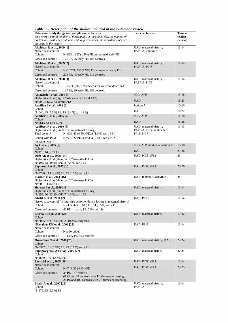
Table 3 – Description of the studies included in the systematic review. Reference, study design and sample characteristics
We report the total number of participants of the cohort (N), the number of
participants with each outcome and, in parentheses, the prevalence of each
outcome in the cohort.
Tests performed Time of
testing
(weeks)
Akolekar R et al., 2009 [1]
Nested case-control
UAD, maternal history,
PAPP-A, inhibin A
11-14
Cohort: N=8234, 147 (1.8%) PE, unreported early PE
Cases and controls: 121 PE, 26 early PE, 208 controls
Akolekar R et al., 2009 [2]
Nested case-control
UAD, maternal history,
PAPP-A, PP13
11-14
Cohort: N=15759, 298 (1.9%) PE, unreported early PE
Cases and controls: 208 PE, 48 early PE, 416 controls
Akolekar R et al., 2008 [3]
Nested case-control
UAD, maternal history,
PAPP-A, PlGF
11-14
Cohort: 1.8% PE, other characteristics were not described
Cases and controls: 127 PE, 29 early PE, 609 controls
Alkazaleh F et al., 2006 [4]
High-risk cohort (high 2nd trimester hCG and AFP)
N=50, 13 (26.0%) severe PE¶
hCG, AFP
UAD
15-18
19-23
Aquilina J et al., 2001 [5]
Cohort
N=640, 35 (5.5%) PE, 15 (2.3%) early PE§
Inhibin A
UAD
15-19
18-22
Audibert F et al., 2005 [7]
Cohort
N=2615, 51 (2.0%) PE
hCG, AFP
UAD
14-18
18-26
Audibert F et al., 2010 [8]
High-risk cohort (risk factors in maternal history)
UAD, maternal history,
PAPP-A, hCG, inhibin A,
PP13, PlGF
11-13
Total cohort** N=893, 40 (4.5%) PE, 9 (1.0%) early PE*
Cohort with PlGF
measurement**
N=531, 22 PE (4.1%), 4 (0.8%) early PE*
Ay E et al., 2005 [9]
Cohort
N=178, 14 (7.9%) PE
hCG, AFP, inhibin A, activin A
UAD
15-18
23-26
Diab AE et al., 2008 [14]
High-risk cohort (abnormal 2nd trimester UAD)
N=108, 33 (30.6%) PE, 8 (7.4%) early PE
UAD, PlGF, sFlt1 23
Espinoza J et al., 2007 [15]
Cohort
N=3296, 113 (3.4%) PE, 15 (0.5%) early PE
UAD, PlGF, sFlt1 22-26
Florio P et al., 2003 [16]
High-risk cohort (abnormal 2nd trimester UAD)
N=58, 18 (31.0%) PE
UAD, inhibin A, activin A
24
Herraiz I et al., 2009 [19]
High-risk cohort (risk factors in maternal history)
N=152, 20 (13.2%) PE, 7 (4.6%) early PE
UAD, maternal history 11-14
Khalil A et al., 2010 [21]
Nested case-control (in high-risk cohort with risk factors in maternal history)
UAD, PP13 11-14
Cohort: N=395, 42 (10.6%) PE, 14 (3.5%) early PE
Cases and controls: 42 PE, 14 early PE, 210 controls
Llurba E et al., 2009 [23]
Cohort
N=6035, 75 (1.2%) PE, 20 (0.3%) early PE†
UAD, maternal history 19-22
Nicolaides KH et al., 2006 [25]
Nested case-control
UAD, PP13 11-14
Cohort: Not described
Cases and controls: 10 early PE, 423 controls
Onwudiwe N et al., 2008 [26]
Cohort
N=3347, 101 (3.0%) PE, 23 (0.7%) early PE
UAD, maternal history, MAP 22-24
Papageorghiou AT et al., 2005 [27]
Cohort
N=16806, 369 (2.2%) PE
UAD, maternal history 22-24
Parra M et al., 2005 [28]
Nested case-control
UAD, PlGF, sFlt1
UAD, PlGF, sFlt1
11-14
22-25 Cohort: N=743, 33 (4.4%) PE
Cases and controls: 33 PE, 137 controls
(8 PE and 37 controls with 1st trimester screening;
26 PE and 100 controls with 2nd trimester screening)
Pilalis A et al., 2007 [29]
Cohort
N=878, 13 (1.5%) PE
UAD, maternal history,
PAPP-A
11-14

Plasencia W et al., 2007 [30]
Cohort
N=6015, 107 (1.8%) PE
UAD, maternal history 11-14
Plasencia W et al., 2008 [31]
Cohort
N=3107, 93 (3.0%) PE, 22 (0.7%) early PE
UAD, maternal history
UAD
11-14
21-25
Poon LC et al., 2009 [33]
Nested case-control
UAD, maternal history, MAP,
PAPP-A, PlGF
11-13
Cohort: N=7797, 157 (2.0%) PE, 34 (0.4%) early PE
Cases and controls: 127 PE, 29 early PE, 418 controls
Poon LC et al., 2009/2010 [34,36,37]
Cohort
N=8366, 165 (2.0%) PE, 37 (0.4%) early PE
UAD, maternal history, MAP,
PAPP-A
11-14
Poon LC et al., 2009 [35]
Cohort
N=8051, 156 (1.9%) PE, 32 (0.4%) early PE
UAD, maternal history,
PAPP-A
11-14
Simonazzi G et al., 2007 [39]
Cohort
N=152, 16 (10.5%) PE
UAD, maternal history 18-24
Spencer K et al., 2007 [40]
Nested case-control
UAD, PAPP-A, hCG, inhibin
A, activin A, PP13
22-24
Cohort: Not described
Cases and controls: 12 PE, 5 early PE‡, 73 controls
Spencer K et al., 2007 [41]
Nested case-control
PAPP-A, PP13
UAD
11-14
22-24 Cohort: N=5867, 88 (1.5%) PE, 44 (0.8%) early PE‡
Cases and controls: 88 PE, 44 early PE‡, 446 controls
Spencer K et al., 2008 [42]
Nested case-control
Inhibin A, activin A
UAD
11-14
22-24 Cohort: N=4390, 64 (1.5%) PE, 34 (0.8%) early PE‡
Cases and controls: 64 PE, 34 early PE‡, 240 controls
Spencer K et al., 2005 [43]
Cohort
N=4390, 64 (1.5%) PE
PAPP-A, hCG
UAD
11-14
22-24
Spencer K et al., 2006 [44]
Nested case-control
UAD, inhibin A, activin A 22-25
Cohort: Not described
Cases and controls: 24 PE, 144 controls
Stepan H et al., 2007 [46]
High-risk cohort (abnormal 2nd trimester UAD)
N=63, 12 (19.0%) PE, 9 (14.3%) early PE
UAD, PlGF, sFlt1 19-24
Yu CK et al., 2005 [49]
Cohort
UAD, maternal history 22-24
Total cohort†† N=30784, 612 (2.0%) PE, 144 (0.5%) early PE
“Model validation
group” ††
N=15392, 297 (1.9%) PE, 72 (0.5%) early PE
Yu J et al., 2010 [50]
Nested case-control
Inhibin A, activin A, PlGF
UAD
12-16
22-24 Cohort: N=613, 31 (5.1%) PE
Cases and controls: 31 PE, 93 controls
PE: pre-eclampsia; UAD: uterine artery Doppler; MAP: mean arterial pressure; PAPP-A: pregnancy-associated plasma protein-A; hCG: human
chorionic gonadotropin; AFP: alpha-fetoprotein; PP13: placental protein 13; PlGF: placental growth factor; sFlt1: soluble fms-like tyrosine kinase
1.
Unless otherwise specified, early PE refers to PE requiring delivery before 34 weeks’ gestation and late PE refers to PE requiring delivery at or
after 34 weeks’ gestation. *PE diagnosed before 34 weeks’ gestation. †PE requiring delivery before 32 weeks’ gestation. ‡PE requiring delivery
before 35 weeks’ gestation. §PE requiring delivery before 37 weeks’ gestation. ¶ Severe PE refers to PE with end-organ involvement or fetal
growth restriction.
**“PlGF could only be measured in 531 women, because this test was initially not scheduled and additional serum was not availab le for women
included in the first year of the study.” [8]
††“The cohort was divided into a model development group and a model validation group using a pseudo-random number for allocation”. [49]
The results of this study presented in our review were estimated in the “model validation group”.

Table 4 – Screening for PE with UAD and one other marker.
Test Type of PE AUC Sp % Sn % LR+ LR-
1st Trimester
Poon LC et al, 2009/2010 [34,36,37] | “significant contributions from a combination of (…) maternal risk factors with either the
lowest, mean or highest (…) PI” | “Although there was no significant difference (…) screening appeared to be best with the
lowest PI” L-PI + Maternal history Early 0.912 90.0 81.1 8.11 0.21
Late 0.812 90.0 45.3 4.53 0.61
Audibert F et al, 2010 [8] | “addition of Doppler did not improve (…) accuracy of screening by clinical characteristics alone”
L-PI + Maternal history Early* 0.738 90.0 50.0 5.00 0.56
Total 0.746 90.0 35.1 3.51 0.72
Pilalis A et al, 2007 [29] | “although the difference was not statistically significant (…) the combination of (…) PI and maternal
history (…) was better compared with (…) Doppler alone” M-PI + Maternal history Total 0.753 95.0 42.0 8.40 0.61
Plasencia W et al, 2008 [31] | “effective screening (…) can be achieved by a combination of maternal variables and (…)
Doppler” M-PI + Maternal history Early 0.931 90.0 77.3 7.73 0.25
Late 0.779 90.0 42.3 4.23 0.64
Poon LC et al, 2009/2010 [34,36,37] | See above.
M-PI + Maternal history Early 0.902 90.0 78.4 7.84 0.24
Late 0.813 90.0 46.9 4.69 0.59
Herraiz I 2009 [19] | “detection rates of the combination of (…) Doppler and maternal history (…) were considerably lower
than in the original study with low-risk pregnancies” [30] M-PI + Maternal history Early 0.779 90.0 42.9 4.29 0.63
Late 0.641 90.0 23.1 2.31 0.85
Poon LC et al, 2009/2010 [34,36,37] | See above.
H-PI + Maternal history Early 0.884 90.0 64.9 6.49 0.39
Late 0.810 90.0 46.1 4.61 0.60
Poon LC et al, 2009 [35] | “significant contributions from serum PAPP-A (…) PI (…) in the prediction of early PE”
M-PI + PAPP-A Early 0.852 90.0 59.4 5.94 0.45
Audibert F et al, 2010 [8] | “we did not confirm the predictive accuracy of (…) free ß-hCG”
L-PI + hCG Not available
Audibert F et al, 2010 [8] | “we did not confirm the predictive accuracy of (…) PP13”
L-PI + PP13 Not available
Nicolaides KH et al, 2006 [25] | “Effective screening (…) can potentially be provided by (…) PP-13 and (…) Doppler”
M-PI + PP13 Early ----- 90.0 90.0 9.00 0.11
M-PI followed by PP13 in 32% with highest risk Early ----- 84.0 90.0 5.63 0.12
PP13 followed by M-PI in 14% with highest risk Early ----- 94.0 90.0 15.00 0.11
Khalil A et al, 2010 [21] | “any pair combination provided better prediction than any individual marker”
M-PI + PP13 Early 0.900 90.0 78.6 7.86 0.24
Total 0.880 90.0 71.4 7.14 0.32
Parra M et al, 2005 [28] | “none of the parameters assessed during the first trimester was significantly associated with PE”
M-PI + PlGF Not available
M-PI + sFlt1 Not available
2nd
Trimester Llurba E et al, 2009 [23] | “combination of both methods did not significantly improve the sensitivity”
M-PI + Maternal history Not available
Yu CK et al, 2005 [49] | Early PE: “Ultrasound had an extremely high predictive value (…) which was not significantly
different from the combination of maternal and ultrasound prediction” | Late PE: “the combination of ultrasound and maternal
characteristics provided the best (…) model” M-PI + Bilateral diastolic notches + Maternal history Early 0.945 90.0 81.9 8.19 0.20
Late 0.798 89.8 47.6 4.67 0.58

Simonazzi G et al, 2007 [39] | “lower detection rate compared with the values stated by Yu et al.” [49]
M-PI + Bilateral diastolic notches + Maternal history Total 0.760 90.0 50.0 5.00 0.56
Spencer K et al, 2007 [40] | “combining (…) Doppler with (…) PAPP-A (…) did not improve detection”
M-PI + PAPP-A Early‡ 0.800 80.0 60.0 3.00 0.50
Late 0.520 80.0 43.0 2.15 0.71
Audibert F et al, 2005 [7] | “[UAD combined with hCG or AFP] despite a poor sensitivity, offers a very high PPV [positive
predictive value] in a low-risk population, multiplying the risk by 2-3 compared to Doppler alone” Diastolic notch + hCG Total ----- 99.53 7.84 16.68 0.93
Ay E et al, 2005 [9] | “addition of these hormonal measurements [AFP, hCG, inhibin A, activin A] to (…) Doppler (…) does not
cause a clinically significant improvement (…) over the use of Doppler (…) alone” Doppler + hCG Not available
Audibert F et al, 2005 [7] | See above.
Diastolic notch + AFP Total ----- 99.02 7.84 8.00 0.93
Bilateral diastolic notches + AFP Total ----- 99.57 5.88 13.67 0.95
Ay E et al, 2005 [9] | See above.
Doppler + AFP Not available
Spencer K et al, 2006 [44] | “screening can be improved by combining (…) Doppler scan with (…) biochemical analysis”
M-PI + Inhibin A Total 0.913 90.0 75.0 7.50 0.28
M-PI + Activin A Total 0.935 90.0 75.0 7.50 0.28
Aquilina J et al, 2001 [5] | “statistically significant improvement in the screening efficacy (…) when (…) Doppler studies (…)
are combined with inhibin-A” M-RI + Diastolic notch + Inhibin A Early§ ----- 97.0 60.0 20.00 0.41
Total ----- 93.4 71.4 10.82 0.31
Inhibin A followed by Doppler in 53% with highest risk Early§ ----- 93.0 73.0 10.43 0.29
Total ----- 93.0 70.0 10.00 0.32
Ay E et al, 2005 [9] | See above.
Diastolic notch + Inhibin A Total ----- 100.0 71.4 0.29
RI + Inhibin A Total ----- 100.0 71.4 0.29
Diastolic notch or high inhibin A Total ----- 93.9 85.7 14.05 0.15
High RI or high inhibin A Total ----- 82.9 78.6 4.60 0.26
Florio P et al, 2003 [16] | “activin A and inhibin A (…) may add significant prognostic information for predicting pre-eclampsia
among women with specific Doppler alterations” Inhibin A, in women with diastolic notches Total 0.555 92.0 39.0 4.88 0.66
Ay E et al, 2005 [9] | See above.
Diastolic notch + Activin A Total ----- 100.0 78.6 0.21
Diastolic notch or high activin A Total ----- 86.0 100.0 7.14 0.00
Florio P et al, 2003 [16] | See above.
Activin A, in women with diastolic notches Total 0.678 89.0 61.0 5.55 0.44
Spencer K et al, 2007 [40] | “combining (…) Doppler with (…) PP13 (…) did not improve detection”
M-PI + PP13 Early‡ 0.930 80.0 100.0 5.00 0.00
Late 0.620 80.0 29.0 1.45 0.89
Parra M et al, 2005 [28] | “Doppler is the best predictor of PE and none of biochemical markers significantly improved its
capacity to screen” M-PI + PlGF Not available
Espinoza J et al, 2007 [15] | “[the addition of PlGF to UAD] improved the positive predictive value (…) without a significant
reduction in the sensitivity” M-PI + Bilateral diastolic notches + PlGF Early ----- 96.4 73.3 20.36 0.28
Total ----- 96.4 27.3 7.58 0.75
Stepan H et al, 2007 [46] | “measurement of angiogenic factors has a useful predictive power when used in a risk group and
focused to severe and early presenting forms of preeclampsia” PlGF
in women with M-PI>1.45 and/or bilateral diastolic notches
Early ----- 62.0 83.0 2.18 0.27
Total ----- 62.0 77.0 2.03 0.37

M-PI + PlGF
in women with M-PI>1.45 and/or bilateral diastolic notches
Early ----- 76.0 83.0 3.46 0.22
Total ----- 68.0 77.0 2.41 0.34
Diab AE et al, 2008 [14] | “We found (…) angiogenic factors to be highly predictive in (…) women with high-risk pregnancies”
PlGF
in women with M-PI>1.45 and/or bilateral diastolic notches
Early 0.904 76.0 100.0 4.17 0.00
Total 0.897 81.0 88.0 4.63 0.15
Parra M et al, 2005 [28] | See above.
M-PI + sFlt-1 Not available
Espinoza J et al, 2007 [15] | “[sFlt1] did not improve the diagnostic indices of an abnormal [UAD]”
M-PI + Bilateral diastolic notches + sFlt1 Not available
Stepan H et al, 2007 [46] | See above.
sFlt1
in women with M-PI>1.45 and/or bilateral diastolic notches
Early ----- 89.0 67.0 6.09 0.37
Total ----- 70.0 62.0 2.07 0.54
M-PI + sFlt1
in women with M-PI>1.45 and/or bilateral diastolic notches
Early ----- 89.0 83.0 7.55 0.19
Total ----- 73.0 77.0 2.85 0.32
Diab AE et al, 2008 [14] | See above.
sFlt1
in women with M-PI>1.45 and/or bilateral diastolic notches
Early 0.956 87.0 100.0 7.69 0.00
Total 0.935 87.0 96.0 7.38 0.05
1st and 2
nd Trimesters
Plasencia W et al, 2008 [31] | “the ratio of uterine artery PI (…) improved significantly the prediction of pre-eclampsia”
M-PI (T1) + Ratio M-PI (T2/T1) + Maternal history Early 0.983 90.0 100.0 10.00 0.00
Late 0.783 90.0 46.5 4.65 0.59
M-PI (T1) + Maternal history
followed by Ratio M-PI (T2/T1) in 20% with highest risk
Early ----- 90.0 95.5 9.55 0.05
Late ----- 90.0 43.7 4.37 0.63
Spencer K et al, 2005 [43] | “The detection rate (…) in screening by (…) Doppler is improved by the inclusion of (…) PAPP-A”
M-PI (T2) + PAPP-A (T1) Total 0.853 95.0 62.1 12.42 0.40
Spencer K et al, 2007 [41] | “both PP-13 and PAPP-A when coupled with (…) PI (…) improve prediction over (…) Doppler
(…) alone” M-PI (T2) + PAPP-A (T1) Early‡ 0.860 80.0 76.0 3.80 0.30
Late 0.810 80.0 70.0 3.50 0.38
Spencer K et al, 2005 [43] | “levels of free β-hCG were not significantly altered in pregnancies that developed complications”
M-PI (T2) + hCG (T1) Not available
Spencer K et al, 2008 [42] | “When combined [inhibin-A and activin-A] with (…) PI some improvement (…) could be observed”
| “the predictive model was no better at identifying early- from late-onset pre-eclampsia” M-PI (T2) + Inhibin A (T1) Total ----- 95.0 67.5 13.50 0.34
Yu J et al, 2010 [50] | “inhibin A, activin A, PIGF and (…) PI may add further information for prediction of pre-eclampsia”
M-PI (T2) + Inhibin A (T1-T2) Total 0.813 90.0 47.0 4.70 0.59
Spencer K et al, 2008 [42] | See above.
M-PI (T2) + Activin A (T1) Total ----- 95.0 63.2 12.64 0.39
Yu J et al, 2010 [50] | See above.
M-PI (T2) + Activin A (T1-T2) Total 0.852 90.0 57.0 5.70 0.48
Spencer K et al, 2007 [41] | See above.
M-PI (T2) + PP13 (T1) Early‡ 0.900 80.0 79.0 3.95 0.26
Late 0.790 80.0 70.0 3.50 0.38
Yu J et al, 2010 [50] | See above.
M-PI (T2) + PlGF (T1-T2) Total 0.880 90.0 73.0 7.30 0.30
PE: pre-eclampsia; UAD: uterine artery Doppler; AUC: area under the ROC curve; Sp: specificity; Sn: sensitivity; LR+: likelihood ratio of the
positive result; LR-: likelihood ratio of the negative result; PI: pulsatility index; L-PI: lowest PI; M-PI: mean PI; H-PI: highest PI; RI: resistance
index; M-RI: mean RI; PAPP-A: pregnancy-associated plasma protein-A; hCG: human chorionic gonadotropin; AFP: alpha-fetoprotein; PP13:
placental protein 13; PlGF: placental growth factor; sFlt1: soluble fms-like tyrosine kinase 1; T1: first trimester; T2: second trimester.
Unless otherwise specified, early PE refers to PE requiring delivery before 34 weeks’ gestation and late PE refers to PE requ iring delivery at or
after 34 weeks’ gestation. *PE diagnosed before 34 weeks’ gestation. †PE requiring delivery before 32 weeks’ gestation. ‡PE requiring delivery
before 35 weeks’ gestation. §PE requiring delivery before 37 weeks’ gestation.

Table 5 – Screening for PE with UAD and two other markers.
Test Type of PE AUC Sp % Sn % LR+ LR-
1st Trimester
Poon LC et al, 2009/2010 [34,36,37] | Early PE: “significant contributions from PAPP-A (…) maternal factors, MAP and (…)
L-PI” | Late PE: “prediction from the combination of maternal factors (…) L-PI and MAP (…) was not improved by addition of
PAPP-A” L-PI + Maternal history + MAP Early 0.954 90.0 89.2 8.92 0.12
Late 0.863 90.0 57.0 5.70 0.48
L-PI + Maternal history + PAPP-A Early 0.925 90.0 81.1 8.11 0.21
Pilalis A et al, 2007 [29] | “We found increased (…) PI and maternal history (…) but not PAPP-A (…) to be independent risk
factors for pre-eclampsia” M-PI + Maternal history + PAPP-A Not available
Poon LC et al, 2009 [35] | Early PE: “[AUC] was significantly higher in screening by history with (…) PI than by history alone
(…) but there was no further improvement in screening if (…) PAPP-A was included” | Late PE: “[AUC] was not significantly
higher for screening by history with (…) PI than by history alone (…), or by history with serum PAPP-A than by history alone” M-PI + Maternal history + PAPP-A Early 0.905 90.0 71.9 7.19 0.31
Akolekar R et al, 2009 [1] | “Maternal plasma inhibin A in combination with (…) the maternal history and (…) PI could
provide effective first-trimester screening” M-PI + Maternal history + Inhibin A Early 0.938 90.0 88.5 8.85 0.13
Late 0.823 90.0 42.1 4.21 0.64
Akolekar R et al, 2009 [2] | Early PE: “PP13 (…) did provide significant contribution to prediction” but it “did not improve
the detection achieved by the combination of maternal factors, (…) L-PI and (…) PAPP-A” | Late PE: “PP13 (…) was not
significantly different from controls and therefore did not add value in screening” L-PI + Maternal history + PP13 Early 0.924 90.0 77.1 7.71 0.25
Akolekar R et al, 2008 [3] | Early PE: “significant contributions (…) from maternal factors, PlGF, PAPP-A and (…) PI” | Late
PE: “significant contributions (…) from maternal factors, PlGF and (…) PI but not PAPP-A” M-PI + Maternal history + PlGF Early 0.941 90.0 89.7 8.97 0.11
Late 0.817 90.0 49.0 4.90 0.57
2nd
Trimester Onwudiwe N et al, 2008 [26] | “maternal characteristics, (…) PI and (…) MAP provided significant independent contribution”
M-PI + Maternal history + MAP
Early 0.996 90.0 100.0 10.00 0.00
Late 0.830 90.0 56.4 5.64 0.48
Spencer K et al, 2007 [40] |“combining (…) Doppler with (…)PP13 or PAPP-A or all three together did not improve detection”
M-PI + PAPP-A + PP13 Early* 0.880 80.0 80.0 4.00 0.25
Late 0.610 80.0 43.0 2.15 0.71
Alkazaleh F et al, 2006 [4] | “these ultrasound tests [including UAD] did not identify women at greater (…) risk of severe
preeclampsia” M-PI, in women with hCG>2.5 MoM and AFP>2.0 MoM Severe† ----- 49.0 69.0 1.35 0.63
Spencer K et al, 2007 [40] | “combination [of PP13] with other biochemical or ultrasound markers did not improve the
detection” M-PI + hCG + PP13 Early* 0.930 80.0 100.0 5.00 0.00
Late 0.580 80.0 14.0 0.70 1.08
Spencer K et al, 2006 [44] | “screening can be improved by combining (…) Doppler scan with (…) biochemical analysis”
M-PI + Inhibin A + Activin A Total 0.970 90.0 92.0 9.20 0.09
Florio P et al, 2003 [16] | “combining both hormones may improve the predictive value of the test”
Inhibin A + Activin A, in women with diastolic notches Total ----- 97.5 33.3 13.32 0.68
High inhibin A or activin A, in women with diastolic
notches
Total ----- 72.5 66.7 2.43 0.46
Spencer K et al, 2007 [40] | See above.
M-PI + Inhibin A + PP13 Early* 0.910 80.0 100.0 5.00 0.00
Late 0.550 80.0 29.0 1.45 0.89
M-PI + Activin A + PP13 Early* 0.920 80.0 100.0 5.00 0.00
Late 0.840 80.0 71.0 3.55 0.36

Stepan H et al, 2007 [46] | “measurement of angiogenic factors has a useful predictive power when used in a risk group and
focused to severe and early presenting forms of preeclampsia” sFlt1/PlGF ratio
in women with M-PI>1.45 and/or bilateral diastolic notches
Early ----- 51.0 67.0 1.37 0.65
Total ----- 51.0 62.0 1.27 0.75
sFlt1 + PlGF
in women with M-PI>1.45 and/or bilateral diastolic notches
Early ----- 95.0 83.0 16.60 0.18
Total ----- 73.0 77.0 2.85 0.32
Diab AE et al, 2008 [14] | “We found (…) angiogenic factors to be highly predictive in (…) women with high-risk pregnancies”
sFlt1/PlGF ratio
in women with M-PI>1.45 and/or bilateral diastolic notches
Early 0.959 90.0 100.0 10.00 0.00
Total 0.937 85.0 100.0 6.67 0.00
1st and 2
nd Trimesters
Spencer K et al, 2007 [41] | “the use of all three markers had no advantage over using just two of them”
M-PI (T2) + PAPP-A (T1) + PP13 (T1) Early* 0.850 80.0 70.0 3.50 0.38
Late 0.820 80.0 73.0 3.65 0.34
Yu J et al, 2010 [50] | “The combination of activin A, inhibin A and (…) PI or activin A, PIGF and (…) PI provided a test with
high sensitivity and specificity” M-PI (T2) + Inhibin A (T1-T2) + Activin A (T1-T2) Total 0.907 90.0 83.0 8.30 0.19
M-PI (T2) + Inhibin A (T1-T2) + PlGF (T1-T2) Total 0.840 90.0 66.0 6.60 0.38
M-PI (T2) + Activin A (T1-T2) + PlGF (T1-T2) Total 0.925 90.0 84.0 8.40 0.18
PE: pre-eclampsia; UAD: uterine artery Doppler; AUC: area under the ROC curve; Sp: specificity; Sn: sensitivity; LR+: likelihood ratio of the
positive result; LR-: likelihood ratio of the negative result; PI: pulsatility index; L-PI: lowest PI; M-PI: mean PI; MAP: mean arterial pressure;
PAPP-A: pregnancy-associated plasma protein-A; hCG: human chorionic gonadotropin; AFP: alpha-fetoprotein; PP13: placental protein 13; PlGF:
placental growth factor; soluble fms-like tyrosine kinase 1; T1: first trimester; T2: second trimester.
Unless otherwise specified, early PE refers to PE requiring delivery before 34 weeks’ gestation and late PE refers to PE requiring delivery at or
after 34 weeks’ gestation. *PE requiring delivery before 35 weeks’ gestation. †Severe PE refers to PE with end-organ involvement or fetal growth
restriction.

Table 6 – Screening for PE with UAD and three other markers.
Test Type of PE AUC Sp % Sn % LR+ LR-
1st Trimester
Poon LC et al, 2009/2010 [34,36,37] | Early PE: “significant contributions from PAPP-A (…) maternal factors, MAP and (…)
L-PI” | Late PE: “prediction from the combination of maternal factors (…) L-PI and MAP (…) was not improved by addition of
PAPP-A”
L-PI + Maternal history + MAP + PAPP-A Early 0.960 90.0 94.6 9.46 0.06
Poon LC et al, 2009 [33] | Early PE: “significant contributions from maternal factors, (…) PI, MAP, PAPP-A, and PlGF” |
Late PE: “significant contributions from maternal factors, (…) PI, MAP, and PlGF but not PAPP-A” M-PI + Maternal history + MAP + PlGF Early ----- 95.0 82.8 16.56 0.18
Late ----- 95.0 44.9 8.98 0.58
Akolekar R et al, 2009 [2] | Early PE: “PP13 (…) did provide significant contribution to prediction” but it “did not improve
the detection achieved by the combination of maternal factors, (…) L-PI and (…) PAPP-A” | Late PE: “PP13 (…) was not
significantly different from controls and therefore did not add value in screening” L-PI + Maternal history + PAPP-A + PP13 Not available
Audibert F et al, 2010 [8] | “the addition of Doppler did not improve the diagnostic accuracy of screening by clinical
characteristics alone or combined with PAPP-A, PlGF and Inhibin A” L-PI + Maternal history + PAPP-A + Inhibin A Early* 0.834 90.0 37.5 3.75 0.69
Total 0.745 90.0 32.4 3.24 0.75
Akolekar R et al, 2009 [1] | Early PE: “significant contributions from (…) PI (…) PAPP-A (…) inhibin A (…)” and maternal
history | Late PE: “significant contributions from (…) PI (…) inhibin A” and maternal history “but not from (…) PAPP-A” M-PI + Maternal history + PAPP-A + Inhibin A Early 0.938 90.0 88.5 8.85 0.13
Akolekar R et al, 2008 [3] | Early PE: “significant contributions (…) from maternal factors, PlGF, PAPP-A and (…) PI” | Late
PE: “significant contributions (…) from maternal factors, PlGF and (…) PI but not PAPP-A” M-PI + Maternal history + PAPP-A + PlGF Early 0.936 90.0 86.2 8.62 0.15
1st and 2
nd Trimesters
Yu J et al, 2010 [50] | “Combination of the three serum markers [inhibin A, activin A, PIGF] and (…) PI has a higher
prediction value”
M-PI (T2) + Inhibin A (T1-T2) + Activin A (T1-T2) +
PlGF (T1-T2)
Total 0.941 90.0 90.0 9.00 0.11
PE: pre-eclampsia; UAD: uterine artery Doppler; AUC: area under the ROC curve; Sp: specificity; Sn: sensitivity; LR+: likelihood ratio of the
positive result; LR-: likelihood ratio of the negative result; PI: pulsatility index; L-PI: lowest PI; M-PI: mean PI; MAP: mean arterial pressure;
PAPP-A: pregnancy-associated plasma protein-A; PP13: placental protein 13; PlGF: placental growth factor; T1: first trimester; T2: second
trimester.
Unless otherwise specified, early PE refers to PE requiring delivery before 34 weeks’ gestation and late PE refers to PE requiring delivery at or
after 34 weeks’ gestation. *PE diagnosed before 34 weeks’ gestation.
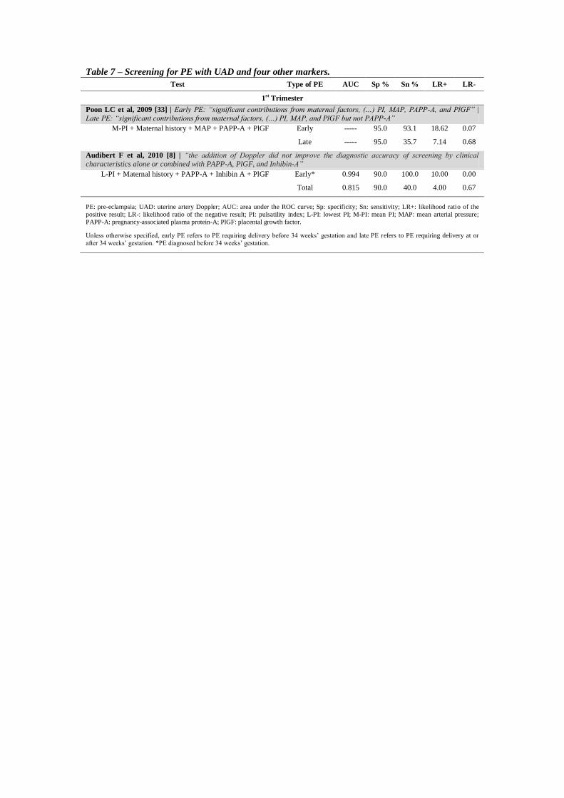
Table 7 – Screening for PE with UAD and four other markers.
Test Type of PE AUC Sp % Sn % LR+ LR-
1st Trimester
Poon LC et al, 2009 [33] | Early PE: “significant contributions from maternal factors, (…) PI, MAP, PAPP-A, and PlGF” |
Late PE: “significant contributions from maternal factors, (…) PI, MAP, and PlGF but not PAPP-A” M-PI + Maternal history + MAP + PAPP-A + PlGF
Early ----- 95.0 93.1 18.62 0.07
Late ----- 95.0 35.7 7.14 0.68
Audibert F et al, 2010 [8] | “the addition of Doppler did not improve the diagnostic accuracy of screening by clinical
characteristics alone or combined with PAPP-A, PlGF, and Inhibin-A” L-PI + Maternal history + PAPP-A + Inhibin A + PlGF Early* 0.994 90.0 100.0 10.00 0.00
Total 0.815 90.0 40.0 4.00 0.67
PE: pre-eclampsia; UAD: uterine artery Doppler; AUC: area under the ROC curve; Sp: specificity; Sn: sensitivity; LR+: likelihood ratio of the
positive result; LR-: likelihood ratio of the negative result; PI: pulsatility index; L-PI: lowest PI; M-PI: mean PI; MAP: mean arterial pressure;
PAPP-A: pregnancy-associated plasma protein-A; PlGF: placental growth factor.
Unless otherwise specified, early PE refers to PE requiring delivery before 34 weeks’ gestation and late PE refers to PE requiring delivery at or
after 34 weeks’ gestation. *PE diagnosed before 34 weeks’ gestation.

Early PE
1st Trimester Screening 2
nd Trimester Screening 1
st and 2
nd Trimester Screening
UAD + 1 other marker
M-PI + PP1325
M-PI followed by PP1325
PP13 followed by M-PI25
UAD + 2 other markers
UAD + 3 other markers
L-PI + History + MAP34,36,37
M-PI + History + Inhibin A1
M-PI + History + PlGF3
L-PI + History + MAP + PAPP-A34,36,37
M-PI + History + MAP + PlGF33
M-PI + History + PAPP-A + Inhibin A1
M-PI + History + PAPP-A + PlGF3
UAD + 4 other markers
M-PI + History + MAP
+ PAPP-A + PlGF*33
L-PI + History + PAPP-A
+ Inhibin A + PlGF†8
UAD + 1 other marker
M-PI + Bilateral diastolic notches
+ History49
M-PI + PP1340
sFlt114
in women with M-PI>1.45
and/or bilateral diastolic notches
M-PI + sFlt146
in women with M-PI>1.45
and/or bilateral diastolic notches
UAD + 2 other markers
M-PI + History + MAP†26
M-PI + hCG + PP1340
M-PI + Inhibin A + PP1340
M-PI + Activin A + PP1340
sFlt1 + PlGF46
in women with M-PI>1.45
and/or bilateral diastolic notches
sFlt1/PlGF ratio†14
in women with M-PI>1.45
and/or bilateral diastolic notches
UAD + 1 other marker
M-PI (T1) + Ratio M-PI (T2/T1)
+ History†31
M-PI (T1) + History31
followed by Ratio M-PI (T2/T1)
Figure 3 – Screening tests which provided moderate or convincing prediction of early PE, according to
the likelihood ratio (LR) values.
PE: pre-eclampsia; UAD: uterine artery Doppler; PI: pulsatility index; L-PI: lowest PI; M-PI: mean PI; MAP: mean arterial pressure; PAPP-A: pregnancy-
associated plasma protein-A; hCG: human chorionic gonadotropin; PP13: placental protein 13; PlGF: placental growth factor; sFlt1: soluble fms-like
tyrosine kinase 1; T1: first trimester; T2: second trimester.
*LR+ > 10 and LR- < 0.1.
†LR+ = 10 and LR- < 0.1.

Total PE
2nd
Trimester Screening 1st and 2
nd Trimester Screening
UAD + 1 other marker
Diastolic notch or high inhibin A9
Diastolic notch or high activin A9
sFlt114
in women with M-PI>1.45
and/or bilateral diastolic notches
UAD + 2 other markers
M-PI + Inhibin A + Activin A44
sFlt1/PlGF ratio14
in women with M-PI>1.45
and/or bilateral diastolic notches
UAD + 2 other markers
M-PI (T2) + Inhibin A (T1-T2) +
Activin A (T1-T2)50
M-PI (T2) + Activin A (T1-T2) +
PlGF (T1-T2)50
UAD + 3 other markers
M-PI (T2) + Inhibin A (T1-T2) +
Activin A (T1-T2) + PlGF (T1-T2)50
Figure 4 – Screening tests which provided moderate or convincing prediction of total PE, according to the
likelihood ratio (LR) values.
PE: pre-eclampsia; UAD: uterine artery Doppler; PI: pulsatility index; M-PI: mean PI; PlGF: placental growth factor; sFlt1: soluble fms-like tyrosine kinase
1; T1: first trimester; T2: second trimester.

