screening key genes for abdominal aortic aneurysm based on
TRANSCRIPT

RESEARCH ARTICLE Open Access
Screening key genes for abdominal aorticaneurysm based on gene expressionomnibus datasetLi Wan1, Jingyong Huang2* , Haizhen Ni2 and Guanfeng Yu2
Abstract
Background: Abdominal aortic aneurysm (AAA) is a common cardiovascular system disease with high mortality.The aim of this study was to identify potential genes for diagnosis and therapy in AAA.
Methods: We searched and downloaded mRNA expression data from the Gene Expression Omnibus (GEO) databaseto identify differentially expressed genes (DEGs) from AAA and normal individuals. Then, Gene Ontology and KyotoEncyclopedia of Genes and Genomes pathway analysis, transcriptional factors (TFs) network and protein-proteininteraction (PPI) network were used to explore the function of genes. Additionally, immunohistochemical (IHC) stainingwas used to validate the expression of identified genes. Finally, the diagnostic value of identified genes was accessedby receiver operating characteristic (ROC) analysis in GEO database.
Results: A total of 1199 DEGs (188 up-regulated and 1011 down-regulated) were identified between AAA and normalindividual. KEGG pathway analysis displayed that vascular smooth muscle contraction and pathways in cancer weresignificantly enriched signal pathway. The top 10 up-regulated and top 10 down-regulated DEGs were used toconstruct TFs and PPI networks. Some genes with high degrees such as NELL2, CCR7, MGAM, HBB, CSNK2A2, ZBTB16and FOXO1 were identified to be related to AAA. The consequences of IHC staining showed that CCR7 and PDGFAwere up-regulated in tissue samples of AAA. ROC analysis showed that NELL2, CCR7, MGAM, HBB, CSNK2A2, ZBTB16,FOXO1 and PDGFA had the potential diagnostic value for AAA.
Conclusions: The identified genes including NELL2, CCR7, MGAM, HBB, CSNK2A2, ZBTB16, FOXO1 and PDGFA mightbe involved in the pathology of AAA.
Keywords: abdominal aortic aneurysm, gene expression, protein-protein interaction network, TFs regulatory network,biomarkers, therapy target
BackgroundAbdominal aortic aneurysm (AAA), defined as the aorticdiameter > 3.0 cm, is a cardiovascular system disease thatis characterized by aortic dilation that exceeds the nor-mal aortic diameter by more than 50%. AAA dilatationwill lead to rupture of the aorta, which results in bleed-ing. Generally, it is asymptomatic until the rupture eventoccurs [1]. Additionally, AAA is common in adult patients,especially elderly men, and leads to severe complications[2–4]. Up to now, the etiology of AAA remains unclear. It
is noted that some clinical risk factors including smokinghistory, advanced age, family history, hypertension, hyper-lipidaemia, atherosclerosis, chronic obstructive pulmonarydisease are remarkably related to AAA [3, 5–7]. It is alsoobserved that the intricate interplay of apoptosis, inflam-mation and matrix degradation is involved in the develop-ment of this disorder [8–10]. Anyway, the pathophysiologyof AAA is complex, but fundamentally aneurysm comesfrom the vessel wall structural integrity loss and the vesselwall weakening. It is pointed out that vascular smoothmuscle cells are the critical cell type involved in the devel-opment of AAA [11].In a word, AAA is a common and late onset disease,
which can rupture with a high mortality if not treated.
* Correspondence: [email protected] of vascular surgery, The First Affiliated Hospital of WenzhouMedical University, NO.3, YuanXi Lane, Lucheng District, Wenzhou, Zhejiang325000, ChinaFull list of author information is available at the end of the article
© The Author(s). 2018 Open Access This article is distributed under the terms of the Creative Commons Attribution 4.0International License (http://creativecommons.org/licenses/by/4.0/), which permits unrestricted use, distribution, andreproduction in any medium, provided you give appropriate credit to the original author(s) and the source, provide a link tothe Creative Commons license, and indicate if changes were made. The Creative Commons Public Domain Dedication waiver(http://creativecommons.org/publicdomain/zero/1.0/) applies to the data made available in this article, unless otherwise stated.
Wan et al. BMC Cardiovascular Disorders (2018) 18:34 https://doi.org/10.1186/s12872-018-0766-8
In some clinical practice, there is no effective treatmentother than surgical approaches to repair AAA [12]. Andendovascular aneurysm repair has improved detectionand lower mortality rates of AAA [13–17]. However,morbidity and mortality after surgery are still common[18, 19]. Therefore, understanding the genetic architec-ture and pathological mechanism of the disease mayprovide valuable information for elucidation of patho-genic mechanisms and signal pathways in AAA and thediscovery of potential biomarkers and drug targets inAAA diagnosis and non-surgical treatment therapy.In this study, we tried to find differentially expressed
genes (DEGs) in AAA by integrated analysis. Then, func-tional enrichment analysis including Gene Ontology (GO)and Kyoto Encyclopedia of Genes Genomes (KEGG) wasused to investigate the biological function of DEGsfollowed by transcriptional factors (TFs) network an dprotein-protein interaction (PPI) network construction oftop 20 DEGs (10 up-regulated and 10 down-regulated).Immunohistochemical (IHC) staining was applied to val-idate the expression of candidate DEGs. Finally, receiveroperating characteristic (ROC) analyses was applied toanalyze diagnosis ability of identified DEGs. Our studymay be helpful in understanding the pathogenic mechan-ism and finding valuable diagnosis biomarkers and ther-apy drug in AAA.
MethodsDatasetsIn this study, we searched datasets from the Gene Expres-sion Omnibus (GEO) database (http://www.ncbi.nlm.nih.gov/geo/) with the keywords abdominal aortic aneurysm[All Fields] AND (“gse”[Filter] AND “Homo sapiens”[Or-ganism]). The study type was described as “expressionprofiling by array.” All selected datasets were genome-wide expression data of AAA group and/or normal grouptissue samples. Those standardized or primary datasetswere included in this study. Finally, a total of 3 datasetsincluding GSE7084, GSE47472 and GSE57691 werescreened, which was shown in Table 1.
Analysis of DEGsRaw expression data of AAA patients in this study weredownloaded. Limma and metaMA packages were used toidentify the DEGs. And the inverse normal method wasused to combine the p value in metaMA. The false discoveryrate (FDR) was performed for multiple testing corrections ofraw p value through the Benjamin and Hochberg method[20, 21]. The threshold of DEGs was set as FDR < 0.01.
Functional annotation analyses of DEGsTo obtain the biological function and signaling pathwaysof DEGs, the Metascape software was used for Gene
Table 1 Three datasets in GEO
GEO accession Author Platform Samples(P:N) Year
GSE7084 Tromp G GPL570[HG-U133_Plus_2] Affymetrix Human GenomeU133 Plus 2.0 Array;GPL2507Sentrix Human-6 ExpressionBeadChip
7:8 2007
GSE47472 Biros E GPL10558Illumina HumanHT-12 V4.0 expression beadchip 14:8 2013
GSE57691 Biros E GPL10558Illumina HumanHT-12 V4.0 expression beadchip 49:10 2015
P patients, N normal individual
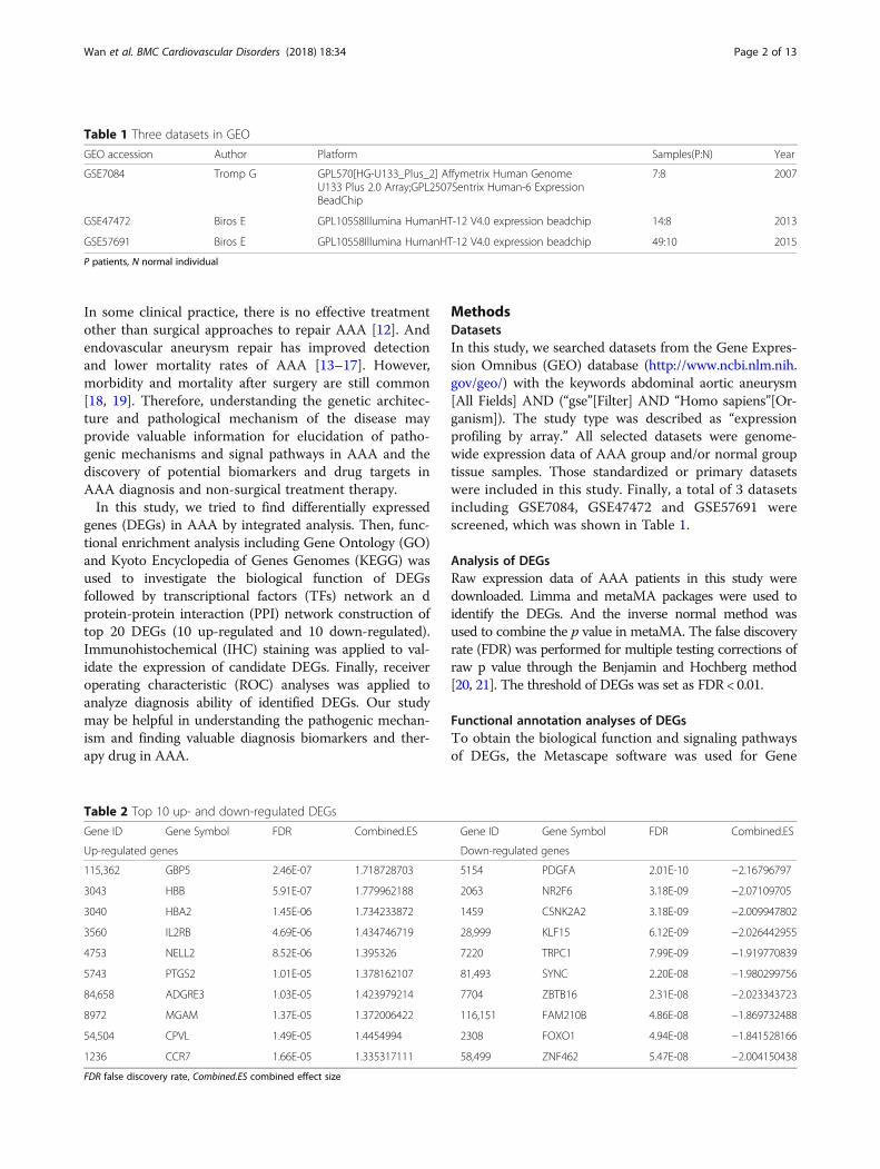
Table 2 Top 10 up- and down-regulated DEGs
Gene ID Gene Symbol FDR Combined.ES Gene ID Gene Symbol FDR Combined.ES
Up-regulated genes Down-regulated genes
115,362 GBP5 2.46E-07 1.718728703 5154 PDGFA 2.01E-10 −2.16796797
3043 HBB 5.91E-07 1.779962188 2063 NR2F6 3.18E-09 −2.07109705
3040 HBA2 1.45E-06 1.734233872 1459 CSNK2A2 3.18E-09 −2.009947802
3560 IL2RB 4.69E-06 1.434746719 28,999 KLF15 6.12E-09 −2.026442955
4753 NELL2 8.52E-06 1.395326 7220 TRPC1 7.99E-09 −1.919770839
5743 PTGS2 1.01E-05 1.378162107 81,493 SYNC 2.20E-08 −1.980299756
84,658 ADGRE3 1.03E-05 1.423979214 7704 ZBTB16 2.31E-08 −2.023343723
8972 MGAM 1.37E-05 1.372006422 116,151 FAM210B 4.86E-08 −1.869732488
54,504 CPVL 1.49E-05 1.4454994 2308 FOXO1 4.94E-08 −1.841528166
1236 CCR7 1.66E-05 1.335317111 58,499 ZNF462 5.47E-08 −2.004150438
FDR false discovery rate, Combined.ES combined effect size
Wan et al. BMC Cardiovascular Disorders (2018) 18:34 Page 2 of 13

Fig. 1 The heat map of top 50 DEGs. The diagram presents the result of a two-way hierarchical clustering of the top 50 DEGs and samples. Theclustering is constructed using the complete-linkage method together with the Euclidean distance. Each row represents a DEG and each column,a sample. The DEG clustering tree is shown on the right. The colour scale illustrates the relative level of DEG expression: red, below the referencechannel; green, higher than the reference
Fig. 2 Significantly enriched biological processes of DEGs. The x-coordinate axis presents the FDR value. FDR value is more highly, the colour ofthe bar is more deeply
Wan et al. BMC Cardiovascular Disorders (2018) 18:34 Page 3 of 13

Ontology (GO, http://www.geneontology.org/) annota-tion and Kyoto Encyclopedia of Genes Genomes (KEGG,http://www.genome. jp/kegg/pathway.html) pathway en-richment of DEGs. The threshold of GO function andKEGG pathway of DEGs was all set as FDR < 0.05.
PPI network constructionIt is useful for understanding the molecule mechanismof AAA to study the interactions between proteins. Inorder to gain insights into the interaction between pro-teins encoded by DEGs and other proteins, the databaseof BioGRID (http://thebiogrid.org) was used to retrievethe predicted interactions between top 20 proteins encodedby DEGs (10 up-regulated and 10 down-regulated) andother proteins. The PPI network was generated by theCytoscape Software (http://cytoscape.org/). A node inthe PPI network denotes protein, and the edge denotesthe interactions.
Analysis of potential TFs to target DEGsTFs play a critical role in regulating gene expression. Wedownloaded the TFs in the human genome and the motifs ofgenomic binding sites from the TRANSFAC. Moreover, the2 KB sequence in the upstream promoter region of DEGs
was downloaded from UCSC (http://www.genome.ucsc.edu/cgi-bin/hgTables). Target sites of potential TFs were thendistinguished. Finally, the transcriptional regulatory net-work was visualized by Cytoscape software.
Immunohistochemical (IHC) staining for CCR7 and PDGFAIn this study, a patient with AAA and a normal individualwas enrolled for the IHC experiment. The 5 μm slides wereincubated with anti CCR7 primary rabbit anti-human poly-clone antibody (1:500 dilution; abcam) and anti PDGFAprimary rabbit anti-human polyclone antibody (1:500dilution; Invitrogen) followed incubated with peroxidaseconjugated goat anti-rabbit secondary antibody (1:200 dilu-tion; Vector). For color visualization, diaminobenzidine(DAB) substrate (Vector) was applied. The staining areawas analyzed by the software of Image Pro-plus 6.0 (MediaCybernetics Corporation, arrendale, PA, USA), and quanti-fied by the IHC staining score (intensity score × positiverate score). The negative (−), positive (+), positive (++),positive (+++) of intensity scores represented 0, 1, 2 and 3,respectively. The positive rate score including negative,1–25%, 26–50%, 51–75% and 76–100% represented 0,1, 2, 3 and 4, respectively. IHC staining score of 0, 1~ 4,
Fig. 3 Significantly enriched molecular functions of DEGs. The x-coordinate axis presents the FDR value. FDR value is more highly, the colour ofthe bar is more deeply
Fig. 4 Significantly enriched cellular components of DEGs. The x-coordinate axis presents the FDR value. FDR value is more highly, the colour ofthe bar is more deeply
Wan et al. BMC Cardiovascular Disorders (2018) 18:34 Page 4 of 13

5~ 8 and 9~ 12 represented negative, slight positive,moderate positive and strong positive, respectively.All patients provided written informed consent with the
approval of the ethics committee of the First AffiliatedHospital of Wenzhou Medical University (2017147).
Receiver operating characteristic analysesBy using pROC package in R language we performed thereceiver operating characteristic (ROC) analyses to assess
the diagnostic value of DEGs (NELL2, CCR7, MGAM,HBB, CSNK2A2, ZBTB16, FOXO1 and PDGFA) in AAA.The area under the curve (AUC) was calculated and theROC curve was generated.
ResultsDEGs analysisRaw expression profiles of AAA patients were down-loaded from the data portal of the GEO database. A total
Table 3 Top 10 GO terms of DEGs
GO ID GO term List in term Log p
Biological process
GO:0072359 circulatory system development 101/913 −10.6462
GO:0061061 muscle structure development 67/563 −8.4846
GO:0006413 translational initiation 40/268 −7.9403
GO:0008285 negative regulation of cell proliferation 70/643 −7.2506
GO:0060548 negative regulation of cell death 89/910 −6.8694
GO:0003170 heart valve development 12/34 −6.8435
GO:0007169 transmembrane receptor protein tyrosine kinase signaling pathway 89/928 −6.4948
GO:0070372 regulation of ERK1 and ERK2 cascade 34/238 −6.3893
GO:0006935 chemotaxis 81/860 −5.6514
GO:0060485 mesenchyme development 32/237 −5.5128
Molecular function
GO:0016491 oxidoreductase activity 75/719 −6.9626
GO:0009055 electron carrier activity 21/112 −6.0854
GO:0019904 protein domain specific binding 61/623 −4.9011
GO:0008092 cytoskeletal protein binding 74/810 −4.7540
GO:0032403 protein complex binding 82/928 −4.6775
GO:0003735 structural constituent of ribosome 27/210 −4.3707
GO:1,901,681 sulfur compound binding 27/232 −3.6356
GO:0016453 C-acetyltransferase activity 3/4 −3.1832
GO:0016634 oxidoreductase activity, acting on the CH-CH group of donors, oxygen as acceptor 4/9 −3.0208
GO:0008565 protein transporter activity 14/98 −2.9919
GO:0016491 oxidoreductase activity 75/719 −6.9626
Cellular component
GO:0044429 mitochondrial part 109/943 −12.6878
GO:0005925 focal adhesion 55/391 −9.6768
GO:0030529 intracellular ribonucleoprotein complex 70/710 −5.6423
GO:0005759 mitochondrial matrix 45/404 −5.1349
GO:0015629 actin cytoskeleton 46/442 −4.4798
GO:0005901 caveola 14/76 −4.1752
GO:0030663 COPI-coated vesicle membrane 6/17 −3.6750
GO:0044455 mitochondrial membrane part 22/173 −3.6079
GO:0090665 glycoprotein complex 6/21 −3.1162
GO:0044451 nucleoplasm part 59/708 −2.9012
List in term: the number of DEGs on the total number of genes in GO termLog p logarithm processing of p value
Wan et al. BMC Cardiovascular Disorders (2018) 18:34 Page 5 of 13

of 1199 DEGs were identified as the threshold at FDR <0.01, consisting of 188 up-regulated genes and 1011 down-regulated genes. The top 10 up- and down-regulated DEGsare listed in Table 2. The heat map of the top 50 DEGs isshown in Fig. 1.
Functional and pathway enrichment analyses of DEGsTo investigate the biological function of the identifiedDEGs in AAA, GO term and KEGG pathway enrichmentanalyses was performed. In GO term and KEGG pathwayenrichment analyses, circulatory system development,muscle structure development and translational initiationwere the most significant enrichment in biological process(Fig. 2); Oxidoreductase activity, electron carrier activity,protein domain specific binding were the most notable en-richment in molecular function (Fig. 3); Mitochondrialpart, focal adhesion and intracellular ribonucleoproteincomplex were the most significant enrichment in cellularcomponent (Fig. 4). The top 10 GO terms of DEGs areshown in Table 3, and the KEGG enrichment signal path-ways of DEGs shown in Table 4. The vascular smooth
muscle contraction and pathways in cancer that were sig-nificantly related to AAA are shown in Fig. 5 and Fig. 6,respectively.
Establishment of TFs-target genes regulatory networkIn order to study the TFs-target genes regulatory networkfor AAA, we utilized TRANSFAC to identify TFs regulatingthe top ten up-regulated or down-regulated DEGs. In theend, we obtained transcriptional regulatory networks com-prised of 190 pairs of TFs-genes involving 40 TFs (Fig. 7).In this network, the top 7 downstream genes covered bymost TFs were neural EGFL like 2 (NELL2, degree = 13),C-C motif chemokine receptor 7 (CCR7, degree = 9),maltase-glucoamylase (MGAM, degree = 8), hemoglobinsubunit beta (HBB, degree = 8). Five hub TFs were HNF-4(degree = 10), Oct-1 (degree = 10), Pax-4 (degree = 8), Evi-1(degree = 6) and Nkx2–5 (degree = 6) (Table 5).
PPI networkTo obtain the interaction between the proteins encodedby DEGs and other proteins, PPI network was explored
Table 4 The KEGG enrichment signal pathways of DEGs
KEGG ID KEGG term List in term Log p Gene list
hsa03010 Ribosome 22/135 −6.5000 FAU,RPL7,RPL9,RPL24,RPL27,RPL30,RPL35A,RPS6,RPS21,UBA52,MRPL33,MRPL19,MRPL18,MRPL22,MRPS16,RSL24D1,MRPL20,MRPS15,MRPS6,MRPS5,MRPL1,MRPL24
hsa00640 Propanoate metabolism 10/32 −5.9097 ACAT1,ACAT2,LDHA,LDHB,ALDH6A1,MUT,PCCA,SUCLG2,HIBCH,ACSS2,ALDH2,ALDH3A2,HADH,HMGCL,ACO1,GCSH,HOGA1,ESD,PFKM,PRPS2,PHGDH,L2HGDH
hsa04510 Focal adhesion 23/202 −4.1028 ACTN1,CAPN2,CAV2,COL4A1,FLNC,HRAS,ITGA7,LAMA5,LAMC1,PPP1R12A,PDGFA,PDGFRB,MAPK3,PTEN,ROCK1,THBS2,ITGA10,ROCK2,ITGA11,PARVA,PDGFC,TLN2,SHC4,FGF13,MYH10,WASL,ARPC1A,ARHGEF12,GNG12,PIP4K2C
hsa04270 Vascular smooth muscle contraction 16/120 −3.8133 ADCY3,AGTR1,CALD1,EDNRA,GNA11,KCNMB1,MYH11,MYL6,PPP1R12A,MAPK3,PTGIR,ROCK1,ROCK2,RAMP1,ARHGEF12,PPP1R14A
hsa00071 Fatty acid degradation 9/44 −3.7701 ACADL,ACAT1,ACAT2,ADH1A,ADH1B,ALDH2,ALDH3A2,ECI1,HADH,ACYP2,LDHA,LDHB,ACSS2,PFKM,PGM1
hsa03020 RNA polymerase 7/31 −3.3221 POLR2C,POLR2F,POLR2G,POLR2H,POLR2I,POLR3F,POLR3C,ADCY3,AK1,GUK1,NME3,PGM1,PRPS2,ENPP4,NME7,AK3,NUDT9,POLE4,NT5C3B,CTPS1
hsa05016 Huntington’s disease 20/193 −3.1152 COX5B,COX6C,COX7A1,COX7B,COX7C,HDAC2,NDUFA4,NDUFA8,NDUFB10,NDUFC1,POLR2C,POLR2F,POLR2G,POLR2H,POLR2I,SOD1,ATP5H,UQCRQ,NDUFA12,NDUFA4L2,UBE2G2,SNCAIP,PINK1,COX17,ATP6V1D,CAPN2,MAPK3,RYR3
hsa05200 Pathways in cancer 33/397 −2.9576 ADCY3,AGTR1,AR,COL4A1,E2F3,EDNRA,MECOM,FGF13,FOXO1,FZD2,GNA11,GSTP1,MSH6,HDAC2,HRAS,LAMA5,LAMC1,SMAD4,PDGFA,PDGFRB,MAPK3,PTEN,ROCK1,SLC2A1,TCEB1,ZBTB16,FZD3,CCDC6,ROCK2,GNB5,RALBP1,ARHGEF12,GNG12,PDGFC
hsa05412 Arrhythmogenic right ventricularcardiomyopathy (ARVC)
10/74 −2.6366 ACTN1,CACNB3,CDH2,DAG1,GJA1,ITGA7,RYR2,SGCA,ITGA10,ITGA11
hsa00520 Amino sugar and nucleotide sugarmetabolism
7/48 −2.1749 CYB5R3,GMDS,PGM1,PMM1,UAP1,UGDH,UGP2
List in term: the number of DEGs on the total number of genes in GO termLog p logarithm processing of p value
Wan et al. BMC Cardiovascular Disorders (2018) 18:34 Page 6 of 13

and visualize by Cytoscape. PPI networks of the top 10up-regulated and the top 10 down-regulated DEGs werepresented in Fig. 8. As Fig. 8 shows, the network con-sisted of 539 nodes and 567 edges. The red and green di-amonds indicate the up- and down-regulated genes inAAA, respectively. The blue ellipses present the proteinsthat interacted with those proteins encoded by DEGs.The top three proteins with a high degree were caseinkinase 2 alpha 2 (CSNK2A2, degree = 184), zinc fingerand BTB domain containing 16 (ZBTB16, degree = 113)and forkhead box O1 (FOXO1, degree = 53).
Validation of CCR7 and PDGFA in AAAIn order to validate the expression of CCR7 and plateletderived growth factor subunit A (PDGFA), we assessedthe protein expression of CCR7 and PDGFA in AAAthrough the immunohistochemistry (Fig. 9 and Fig. 10).The result showed that CCR7 was obviously up-regulatedin AAA compared with the control, which was consist-ent with the bioinformatic consequence. However, PDGFAwas significantly up-regulated in AAA compared with thecontrol, which was not in line with the bioinformaticresult.
ROC curve analysisIn order to access the discriminatory ability of theNELL2, CCR7, MGAM, HBB, CSNK2A2, ZBTB16,FOXO1 and PDGFA among AAA tissues and adjacentnon-tumor tissues generated from GEO database, ROCcurve analyses were conducted and AUC were calcu-lated. As Fig. 11 shown, the AUC of all these genes wasmore than 0.8. For AAA diagnosis, the sensitivity andspecificity of these genes were very high.
DiscussionIn spite of improvement to surgical techniques that havebeen made in AAA treatment, morbidity and mortalityafter operations are still common. AAA seriously influ-ences the life quality of patients and brings a heavy bur-den on the family. Therefore, it is urgent to elucidateAAA pathogenesis mechanism for developing noveldiagnose and therapeutic target.TFs are key regulatory factors in gene expression. The
construction of regulatory networks between TFs andtarget genes is helpful in understanding the biologicalregulatory mechanism in the development of AAA. Inthis study, we found NELL2, CCR7, MGAM and HBBwere significantly expressed genes with the most degree
Fig. 5 Significantly enriched vascular smooth muscle contraction signal pathways of DEGs. The red and green diamond represents the up anddown-regulated DEGs
Wan et al. BMC Cardiovascular Disorders (2018) 18:34 Page 7 of 13

Fig. 6 Significantly enriched pathways in cancer signal pathways of DEGs. The red and green diamond represents the up and down-regulated DEGs
Fig. 7 The TFs networks of the top 20 DEGs. Diamonds and ellipses represent the TFs and target genes, respectively. The red and green colorsrepresent up-regulation and down-regulation, respectively
Wan et al. BMC Cardiovascular Disorders (2018) 18:34 Page 8 of 13

under the regulation of TFs including HNF-4, Oct-1 andPax-4. NELL2 is a neural tissue-enriched protein inmammal and it is a receptor for vascular endothelialgrowth factor-A, which plays an important role in angio-genesis. It is reported that the mRNA expression ofNELL2 is up-regulated in benign prostate hyperplasiaand prostate cancer [22]. In addition, NELL2 is regardedas the potential biomarker for bladder cancer [23]. CCR7is a pro-inflammatory cytokine and is found in humanatherosclerotic plaques [24]. It is found that expression ofCCR7 is dramatically down-regulated in human carotidatherosclerotic plaques [25]. MGAM is found down-regulated and considered as a candidate serum bio-marker in colorectal cancer [26]. Additionally, MGAMis a significantly mutated gene in lung adenocarcinoma[27]. HBB is suggested as a potential biomarker in theplasma sample of patients with AAA [28]. In this study,
we found that NELL2, CCR7, MGAM and HBB wereup-regulated in the AAA tissues, which played crucialroles in the carcinogenesis of AAA.The interaction among proteins determines the char-
acteristic of the cell, tissue and individual. The study ofPPI is a useful way to find the potential drug target ofAAA. Herein, we found three genes including CSNK2A2,ZBTB16 and FOXO1 were for the most degree in the PPInetwork. CSNK2A2 is found to be correlated with ovariancancer patient survival. Furthermore, the down-regulationof CSNK2A2 will decrease the proliferation of ovariancancer cells [29]. ZBTB16, also called PLZF, plays an im-portant role in oncogenesis and is first identified in acutepromyelocytic leukemia [30]. Based on a microarray study,ZBTB16 is found to be down-regulated in AAA [31].FOXO1 is a transcription factor and plays roles in diversephysiological processes including Akt-dependent cell pro-liferation and apoptosis [32]. Additionally, FOXO1 is alsoinvolved in energy metabolism and autophagy [33]. In ourstudy, we found down-regulated expression of CSNK2A2,ZBTB16 and FOXO1. It is worth mentioning thatZBTB16 and FOXO1 were also involved in the pathwaysin cancer according to the KEGG analysis. In addition,PDGFA was one of the top ten down-regulated genesand also involved in the pathways in cancer. PDGFA isexpressed in vascular smooth muscle cells and has beeninvolved in migration and proliferation of vascularsmooth muscle cells [34]. Moreover, the importance ofPDGFA in the arterial system has been demonstrated
Table 5 Top 5 TFs and target genes
TFs Number Target genes
Oct-1 10 CCR7, CPVL, CSNK2A2, HBB, IL2RB, MGAM, NELL2,TRPC1, ZBTB16, ZNF462
HNF-4 10 CCR7, CPVL, CSNK2A2, HBB, KLF15, MGAM, NELL2,NR2F6, PTGS2, ZNF462
Pax-4 8 ADGRE3, CSNK2A2, FAM210B, NELL2, NR2F6, PDGFA,SYNC, ZBTB16
Evi-1 6 CPVL, GBP5, HBB, NELL2, PTGS2, ZNF462
Nkx2–5 6 CCR7, GBP5, MGAM, NELL2, TRPC1, ZBTB16
Fig. 8 The PPI networks of the top 20 DEGs. All the diamonds are proteins encoded by the top 20 DEGs and the blue ellipses represent otherproteins. The red and green colors represent up-regulation and down-regulation, respectively
Wan et al. BMC Cardiovascular Disorders (2018) 18:34 Page 9 of 13

on account of that fact that the proliferation of arterialvascular smooth muscle cells was strongly stimulatedby PDGFA [35]. Moreover, an in situ hybridization studyhas demonstrated mRNA for PDGFA in atheroscleroticplaques [36]. In this study, we found that PDGFA wasdown-regulated in AAA. However, the IHC result was notconsistent with the bioinformatic analysis. The smallsample size may account for the discrepancy. In a word,CSNK2A2, ZBTB16 and FOXO1 played a crucial rolein the oncogenesis of AAA and could be considered asdrug targets of AAA.Apart from the cancer signal pathway, vascular smooth
muscle contraction was another signal pathway identifiedthat associated with AAA. Vascular smooth muscle cellshave been shown to play an important synthetic role invascular remodeling [37, 38]. It is pointed out that vascu-lar smooth muscle cells are the main component of theaortic media and the dysfunction plays a key role in
different arterial diseases, such as AAA [39]. In addition,vascular smooth muscle cell activation is the main hall-mark of atherosclerosis, which is a risk factor of AAA[40–43]. Several down-regulated genes were significantlyinvolved in the signal pathway, such as AGTR1, CALD1,EDNRA, MYH11, RAMP1, ROCK1 and ROCK2.Angiotensin II receptor type 1 (AGTR1) is a cardiovas-
cular risk gene. Jones, G. T et al. found that AGTR1 wasremarkably associated with AAA [44]. In addition, the1166A > C polymorphism in AGTR1 has been demon-strated to be associated with AAA [45, 46]. It is notedthat AGTR1 blockers (ARBs) have been investigated forprevention or delay of aortic dilatation [47]. It is re-ported that the expression of caldesmon 1 (CALD1) isincreased in aortas, which protects from aneurysm. Thissuggested that importance of CALD1 in maintainingvascular integrity in AAA. Endothelin receptor type A(EDNRA) is primarily located in the vascular smooth
Fig. 9 The IHC staining of CCR7 in AAA. CCR7 protein expression level detected by immunohistochemistry and photographs was amplified10 × 20 multiples. Bar = 100 μm. a and c were the case samples from two patients with AAA; b and d were the control sample from twonormal individuals. *P<0.05 vs control
Fig. 10 The IHC staining of PDGFA in AAA. PDGFA protein expression level detected by immunohistochemistry and photographs was amplified10 × 20 multiples. Bar = 100 μm. a and c were the case samples from two patients with AAA; b and d were the control sample from two normalindividuals. **P<0.01 vs control
Wan et al. BMC Cardiovascular Disorders (2018) 18:34 Page 10 of 13

muscle cells and mediates vasoconstriction and prolif-eration [48]. It has been reported that EDNRA onchromosome 4q31 is related to intracranial aneurysm[49]. It is found that heterozygous mutation of myosinheavy chain 11 (MYH11) results in the early and severedecrease in the aortic wall elasticity [50]. Additionally,it has been demonstrated the relationship between MYH11genetic and epigenetic and thoracic aortic aneurysms anddissections [51, 52]. Receptor activity modifying protein 1(RAMP1) is a member of a family of calcitonin receptormodifying proteins and is thought to play an importantrole in regulating blood pressure by vascular relaxation.Tsujikawa K et al. found that ramp1-deficient mice exhib-ited elevated blood pressure [53]. It is pointed out that themRNA levels of RAMP1 are decreased in AAA [31]. It isfound that the expression of Rho associated coiled-coilcontaining protein kinase 1 (ROCK1) and Rho associatedcoiled-coil containing protein kinase 1 (ROCK2) was in-creased at the AAA lesion compared with control [54].Thus it can be seen that AGTR1, CALD1, EDNRA,MYH11, RAMP1, ROCK1 and ROCK2 played an im-portant role in vascular smooth muscle contraction,which was significantly associated with AAA.In order to access the discriminatory ability of identified
genes in the TFs and PPI network, eight genes includingNELL2, CCR7, MGAM, HBB, CSNK2A2, ZBTB16,FOXO1 and PDGFA were applied to ROC curve ana-lyses among AAA tissues and adjacent non-tumor tis-sues in GEO database. Our result showed that the AUCof all these genes was more than 0.8, especially HBB(AUC: 0.906), CSNK2A2 (AUC: 0.945), ZBTB16 (AUC:
0.950), FOXO1 (AUC: 0.940) and PDGFA (AUC: 0.930).This suggested that NELL2, CCR7, MGAM, HBB,CSNK2A2, ZBTB16, FOXO1 and PDGFA may havevalue in diagnosis of the development of AAA.
ConclusionsIn summary, we found a series of DEGs in AAA. Amongwhich, eight genes including NELL2, CCR7, MGAM,HBB, CSNK2A2, ZBTB16, FOXO1 and PDGFA could beused for the diagnosis biomarkers of AAA. Especially,CSNK2A2, ZBTB16 and FOXO1 could be considered asdrug targets in the therapy of AAA. In addition, vascularsmooth muscle contraction was an important signal path-way identified in this study, which played crucial roles inthe aortic angiogenesis of AAA. There are limitations toour study. Firstly, the sample size in the IHC experimentis small and large numbers of tissue samples are needed tofurther validate the identified DEGs. Secondly, biologicalfunction of identified DEGs is not investigated, some invivo or in vitro experiments are needed to further studythe molecular mechanism of AAA. Thirdly, the samplesize of normal individuals in the selected dataset is lessthan that of the patient group. In further studies, it is bet-ter to sample equal numbers of individuals in both groupsin order to reduce the false positive/negative rate for up−/down-regulated DEGs detection.
AbbreviationsAAA: abdominal aortic aneurysm; AGTR1: angiotensin II receptor type 1;ARBs: AGTR1 blockers; AUC: area under the curve; CALD1: caldesmon 1;CCR7: C-C motif chemokine receptor 7; CSNK2A2: casein kinase 2 alpha 2;DAB: diaminobenzidine; DEGs: differentially expressed genes;
Fig. 11 The ROC curve analyses of NELL2, CCR7, MGAM, HBB, CSNK2A2, ZBTB16, FOXO1 and PDGFA between AAA and healthy controls
Wan et al. BMC Cardiovascular Disorders (2018) 18:34 Page 11 of 13

EDNRA: endothelin receptor type A; FDR: false discovery rate;FOXO1: forkhead box O1; GEO: gene expression omnibus; HBB: hemoglobinsubunit beta; IHC: immunohistochemical; MGAM: maltase-glucoamylase;MYH11: myosin heavy chain 11; NELL2: neural EGFL like 2; PDGFA: plateletderived growth factor subunit A; PPI: protein-protein interaction;RAMP1: receptor activity modifying protein 1; ROC: receiver operatingcharacteristic; ROCK1: Rho associated coiled-coil containing protein kinase 1;ROCK2: Rho associated coiled-coil containing protein kinase 1;TFs: transcriptional factors; ZBTB16: zinc finger and BTB domain containing 16
AcknowledgementsNot Applicable.
FundingNot applicable
Availability of data and materialsThe datasets used and/or analysed during the current study available fromthe corresponding author on reasonable request.
Authors’ contributionsLW and JH drafted and revised the manuscript. LW, HN and GY performedthe experiment and analyzed the data. JH designed the subject of themanuscript. All authors have read and agreed to the submission of themanuscript. All authors read and approved the final manuscript.
Ethics approval and consent to participateAll participating individuals provided written informed consent with theapproval of the ethics committee of the First Affiliated Hospital of WenzhouMedical University (2017147).
Consent for publicationNot applicable
Competing interestsThe authors declare that they have no competing interests.
Publisher’s NoteSpringer Nature remains neutral with regard to jurisdictional claims inpublished maps and institutional affiliations.
Author details1Department of pathology, The First Affiliated Hospital of Wenzhou MedicalUniversity, Wenzhou, Zhejiang, China. 2Department of vascular surgery, TheFirst Affiliated Hospital of Wenzhou Medical University, NO.3, YuanXi Lane,Lucheng District, Wenzhou, Zhejiang 325000, China.
Received: 13 October 2017 Accepted: 31 January 2018
References1. Weintraub NL. Understanding abdominal aortic aneurysm. N Engl J Med.
2009;361(11):1114–6.2. Hirsch AT, Haskal ZJ, Hertzer NR, Bakal CW, Creager MA, Halperin JL, Hiratzka LF,
Murphy WR, Olin JW, Puschett JB, et al. Acc/Aha 2005 Practice Guidelines ForThe Management Of Patients With Peripheral Arterial Disease (Lower Extremity,Renal, Mesenteric, And Abdominal Aortic): A Collaborative Report From TheAmerican Association For Vascular Surgery/Society For Vascular Surgery,Society For Cardiovascular Angiography And Interventions, Society ForVascular Medicine And Biology, Society Of Interventional Radiology, And TheAcc/Aha Task Force On Practice Guidelines (Writing Committee To DevelopGuidelines For The Management Of Patients With Peripheral Arterial Disease):Endorsed By The American Association Of Cardiovascular And PulmonaryRehabilitation; National Heart, Lung, And Blood Institute; Society For VascularNursing; Transatlantic Inter-Society Consensus; And Vascular DiseaseFoundation. Circulation. 2006;113(11):e463–654.
3. Lederle FA, Johnson GR, Wilson SE. Abdominal aortic aneurysm in women. JVasc Surg. 2001;34(1):122–6.
4. Katz DJ, Stanley JC, Zelenock GB. Gender differences in abdominal aorticaneurysm prevalence, treatment, and outcome. J Vasc Surg. 1997;25(3):561–8.
5. Lederle FA, Johnson GR, Wilson SE, Chute EP, Littooy FN, Bandyk D, KrupskiWC, Barone GW, Acher CW, Ballard DJ. Prevalence and associations ofabdominal aortic aneurysm detected through screening. Aneurysmdetection and management (ADAM) veterans affairs cooperative studygroup. Ann Intern Med. 1997;126(6):441–9.
6. Coady MA, Davies RR, Roberts M, Goldstein LJ, Rogalski MJ, Rizzo JA,Hammond GL, Kopf GS, Elefteriades JA. Familial patterns of thoracic aorticaneurysms. Archives of surgery (Chicago, Ill: 1960). 1999;134(4):361–7.
7. Lederle FA, Johnson GR, Wilson SE, Chute EP, Hye RJ, Makaroun MS, BaroneGW, Bandyk D, Moneta GL, Makhoul RG. The aneurysm detection andmanagement study screening program: validation cohort and final results.Aneurysm detection and management veterans affairs cooperative studyinvestigators. Arch Intern Med. 2000;160(10):1425–30.
8. Bobryshev YV, Lord RS, Parsson H. Immunophenotypic analysis of theaortic aneurysm wall suggests that vascular dendritic cells are involvedin immune responses. Cardiovascular surgery (London, England).1998;6(3):240–9.
9. Ernst CB. Abdominal aortic aneurysm. N Engl J Med. 1993;328(16):1167–72.10. Pearce WH, Koch AE. Cellular components and features of immune
response in abdominal aortic aneurysms. Ann N Y Acad Sci. 1996;800:175–85.11. Henderson EL, Geng YJ, Sukhova GK, Whittemore AD, Knox J, Libby P. Death of
smooth muscle cells and expression of mediators of apoptosis by T lymphocytesin human abdominal aortic aneurysms. Circulation. 1999;99(1):96–104.
12. Kniemeyer HW, Kessler T, Reber PU, Ris HB, Hakki H, Widmer MK. Treatmentof ruptured abdominal aortic aneurysm, a permanent challenge or a wasteof resources? Prediction of outcome using a multi-organ-dysfunction score.Eur J Vasc Endovasc Surg. 2000;19(2):190–6.
13. Brewster DC, Cronenwett JL, Hallett JW Jr, Johnston KW, Krupski WC,Matsumura JS. Guidelines for the treatment of abdominal aortic aneurysms.Report of a subcommittee of the joint Council of the American Associationfor vascular surgery and Society for Vascular Surgery. J Vasc Surg. 2003;37(5):1106–17.
14. Cowan JA Jr, Dimick JB, Henke PK, Rectenwald J, Stanley JC, Upchurch GRJr. Epidemiology of aortic aneurysm repair in the United States from 1993to 2003. Ann N Y Acad Sci. 2006;1085:1–10.
15. Lederle FA, Wilson SE, Johnson GR, Reinke DB, Littooy FN, Acher CW,Ballard DJ, Messina LM, Gordon IL, Chute EP, et al. Immediate repaircompared with surveillance of small abdominal aortic aneurysms. N Engl JMed. 2002;346(19):1437–44.
16. Mastracci TM, Cina CS. Screening for abdominal aortic aneurysm in Canada:review and position statement of the Canadian Society for Vascular Surgery.J Vasc Surg. 2007;45(6):1268–76.
17. Schermerhorn ML, O'Malley AJ, Jhaveri A, Cotterill P, Pomposelli F, LandonBE. Endovascular vs. open repair of abdominal aortic aneurysms in theMedicare population. N Engl J Med. 2008;358(5):464–74.
18. Moxon JV, Parr A, Emeto TI, Walker P, Norman PE, Golledge J. Diagnosis andmonitoring of abdominal aortic aneurysm: current status and futureprospects. Curr Probl Cardiol. 2010;35(10):512–48.
19. Mortality results for randomised controlled trial of early elective surgeryor ultrasonographic surveillance for small abdominal aortic aneurysms.The UK Small Aneurysm Trial Participants. Lancet (London, England)1998, 352(9141):1649–1655.
20. Reiner-Benaim A. FDR control by the BH procedure for two-sidedcorrelated tests with implications to gene expression data analysis. BiomJ. 2007;49(1):107–26.
21. Benjamini Y, Hochberg Y. Controlling the false discovery rate - a practicaland powerful approach to multiple testing. J R Stat Soc. 1995;57(1):289–300.
22. Shah US, Getzenberg RH. Fingerprinting the diseased prostate: associationsbetween BPH and prostate cancer. J Cell Biochem. 2004;91(1):161–9.
23. Osman I, Bajorin DF, Sun TT, Zhong H, Douglas D, Scattergood J, Zheng R,Han M, Marshall KW, Liew CC. Novel blood biomarkers of human urinarybladder cancer. Clin Cancer Res. 2006;12(11 Pt 1):3374–80.
24. Damas JK, Smith C, Oie E, Fevang B, Halvorsen B, Waehre T, Boullier A,Breland U, Yndestad A, Ovchinnikova O, et al. Enhanced expression of thehomeostatic chemokines CCL19 and CCL21 in clinical and experimentalatherosclerosis: possible pathogenic role in plaque destabilization.Arterioscler Thromb Vasc Biol. 2007;27(3):614–20.
25. Nickel T, Pfeiler S, Summo C, Kopp R, Meimarakis G, Sicic Z, Lambert M,Lackermair K, David R, Beiras-Fernandez A, et al. oxLDL downregulatesthe dendritic cell homing factors CCR7 and CCL21. Mediat Inflamm.2012;2012:320953.
Wan et al. BMC Cardiovascular Disorders (2018) 18:34 Page 12 of 13

26. Ivancic MM, Huttlin EL, Chen X, Pleiman JK, Irving AA, Hegeman AD, DoveWF, Sussman MR. Candidate serum biomarkers for early intestinal cancerusing 15N metabolic labeling and quantitative proteomics in the ApcMin/+mouse. J Proteome Res. 2013;12(9):4152–66.
27. Campbell JD, Alexandrov A. Distinct patterns of somatic genome alterationsin lung adenocarcinomas and squamous cell carcinomas. 2016;48(6):607–16.
28. Gamberi T, Puglia M, Guidi F, Magherini F, Bini L, Marzocchini R, Modesti A,Modesti PA. A proteomic approach to identify plasma proteins in patientswith abdominal aortic aneurysm. Mol BioSyst. 2011;7(10):2855–62.
29. Wang F, Chang JT, Kao CJ, Huang RS. High expression of miR-532-5p, atumor suppressor, leads to better prognosis in ovarian cancer both in vivoand in vitro. Mol Cancer Ther. 2016;15(5):1123–31.
30. Chen Z, Brand NJ, Chen A, Chen SJ, Tong JH, Wang ZY, Waxman S, ZelentA. Fusion between a novel Kruppel-like zinc finger gene and the retinoicacid receptor-alpha locus due to a variant t(11;17) translocation associatedwith acute promyelocytic leukaemia. EMBO J. 1993;12(3):1161–7.
31. Hinterseher I, Erdman R, Elmore JR, Stahl E, Pahl MC, Derr K, Golden A, LillvisJH, Cindric MC, Jackson K, et al. Novel pathways in the pathobiology ofhuman abdominal aortic aneurysms. Pathobiology. 2013;80(1):1–10.
32. Alikhani M, Alikhani Z, Graves DT. FOXO1 functions as a master switch thatregulates gene expression necessary for tumor necrosis factor-inducedfibroblast apoptosis. J Biol Chem. 2005;280(13):12096–102.
33. Stohr R, Kappel BA, Carnevale D, Cavalera M, Mavilio M, Arisi I, Fardella V,Cifelli G, Casagrande V, Rizza S, et al. TIMP3 interplays with apelin toregulate cardiovascular metabolism in hypercholesterolemic mice. Molecularmetabolism. 2015;4(10):741–52.
34. Cucina A, Pagliei S, Borrelli V, Corvino V, Stipa F, Cavallaro A, Sterpetti AV.Oxidised LDL (OxLDL) induces production of platelet derived growth factorAA (PDGF AA) from aortic smooth muscle cells. Eur J Vasc Endovasc Surg.1998;16(3):197–202.
35. Li L, Blumenthal DK, Terry CM, He Y, Carlson ML, Cheung AK. PDGF-inducedproliferation in human arterial and venous smooth muscle cells:molecular basis for differential effects of PDGF isoforms. J Cell Biochem.2011;112(1):289–98.
36. Wilcox JN, Smith KM, Williams LT, Schwartz SM, Gordon D. Platelet-derivedgrowth factor mRNA detection in human atherosclerotic plaques by in situhybridization. J Clin Invest. 1988;82(3):1134–43.
37. Touyz RM, Deng LY, He G, Wu XH, Schiffrin EL. Angiotensin II stimulates DNAand protein synthesis in vascular smooth muscle cells from human arteries:role of extracellular signal-regulated kinases. J Hypertens. 1999;17(7):907–16.
38. Takahashi T, Kawahara Y, Okuda M, Ueno H, Takeshita A, Yokoyama M,Angiotensin II. Stimulates mitogen-activated protein kinases and proteinsynthesis by a Ras-independent pathway in vascular smooth muscle cells. JBiol Chem. 1997;272(25):16018–22.
39. Lopez-Candales A, Holmes DR, Liao S, Scott MJ, Wickline SA, Thompson RW.Decreased vascular smooth muscle cell density in medial degeneration ofhuman abdominal aortic aneurysms. Am J Pathol. 1997;150(3):993–1007.
40. Randolph GJ. The fate of monocytes in atherosclerosis. J Thromb Haemost.2009;7(Suppl 1):28–30.
41. Libby P, Ridker PM, Hansson GK. Inflammation in atherosclerosis: frompathophysiology to practice. J Am Coll Cardiol. 2009;54(23):2129–38.
42. Rateri DL, Howatt DA, Moorleghen JJ, Charnigo R, Cassis LA, Daugherty A.Prolonged infusion of angiotensin II in apoE(−/−) mice promotesmacrophage recruitment with continued expansion of abdominal aorticaneurysm. Am J Pathol. 2011;179(3):1542–8.
43. Leeper NJ, Tedesco MM, Kojima Y, Schultz GM, Kundu RK, Ashley EA, Tsao PS,Dalman RL, Quertermous T. Apelin prevents aortic aneurysm formation byinhibiting macrophage inflammation. Am J Phys Heart Circ Phys. 2009;296(5):H1329–35.
44. Jones GT, Thompson AR, van Bockxmeer FM, Hafez H, Cooper JA, Golledge J,Humphries SE, Norman PE, van Rij AM. Angiotensin II type 1 receptor 1166Cpolymorphism is associated with abdominal aortic aneurysm in threeindependent cohorts. Arterioscler Thromb Vasc Biol. 2008;28(4):764–70.
45. Martin MM, Lee EJ, Buckenberger JA, Schmittgen TD, Elton TS. MicroRNA-155regulates human angiotensin II type 1 receptor expression in fibroblasts. J BiolChem. 2006;281(27):18277–84.
46. Martin MM, Buckenberger JA, Jiang J, Malana GE, Nuovo GJ, Chotani M,Feldman DS, Schmittgen TD, Elton TS. The human angiotensin II type 1receptor +1166 a/C polymorphism attenuates microRNA-155 binding. J BiolChem. 2007;282(33):24262–9.
47. Attenhofer Jost CH, Greutmann M, Connolly HM, Weber R, Rohrbach M,Oxenius A, Kretschmar O, Luscher TF, Matyas G. Medical treatment of aorticaneurysms in Marfan syndrome and other heritable conditions. Curr CardiolRev. 2014;10(2):161–71.
48. Low SK, Takahashi A, Cha PC, Zembutsu H, Kamatani N, Kubo M, NakamuraY. Genome-wide association study for intracranial aneurysm in the Japanesepopulation identifies three candidate susceptible loci and a functionalgenetic variant at EDNRA. Hum Mol Genet. 2012;21(9):2102–10.
49. Yasuno K, Bakircioglu M, Low SK, Bilguvar K, Gaal E, Ruigrok YM, Niemela M,Hata A, Bijlenga P, Kasuya H, et al. Common variant near the endothelinreceptor type a (EDNRA) gene is associated with intracranial aneurysm risk.Proc Natl Acad Sci U S A. 2011;108(49):19707–12.
50. Ikeda Y. Aortic aneurysm: Etiopathogenesis and Clinicopathologiccorrelations. Ann Vasc Dis. 2016;9(2):73–9.
51. Pannu H, Tran-Fadulu V, Papke CL, Scherer S, Liu Y, Presley C, Guo D, EstreraAL, Safi HJ, Brasier AR, et al. MYH11 mutations result in a distinct vascularpathology driven by insulin-like growth factor 1 and angiotensin II. HumMol Genet. 2007;16(20):2453–62.
52. Zhu L, Vranckx R, Khau Van Kien P, Lalande A, Boisset N, Mathieu F,Wegman M, Glancy L, Gasc JM, Brunotte F, et al. Mutations in myosin heavychain 11 cause a syndrome associating thoracic aortic aneurysm/aorticdissection and patent ductus arteriosus. Nat Genet. 2006;38(3):343–9.
53. Tsujikawa K, Yayama K, Hayashi T, Matsushita H, Yamaguchi T, Shigeno T,Ogitani Y, Hirayama M, Kato T, Fukada S, et al. Hypertension and dysregulatedproinflammatory cytokine production in receptor activity-modifyingprotein 1-deficient mice. Proc Natl Acad Sci U S A. 2007;104(42):16702–7.
54. Takahashi K, Matsumoto Y, Do e Z, Kanazawa M, Satoh K, Shimizu T, Sato A,Fukumoto Y, Shimokawa H. Combination therapy with atorvastatin andamlodipine suppresses angiotensin II-induced aortic aneurysm formation.PLoS One. 2013;8(8):e72558.
• We accept pre-submission inquiries
• Our selector tool helps you to find the most relevant journal
• We provide round the clock customer support
• Convenient online submission
• Thorough peer review
• Inclusion in PubMed and all major indexing services
• Maximum visibility for your research
Submit your manuscript atwww.biomedcentral.com/submit
Submit your next manuscript to BioMed Central and we will help you at every step:
Wan et al. BMC Cardiovascular Disorders (2018) 18:34 Page 13 of 13