screening ligands using large cterminus lac · 1866 biochemistry: cull et al. proc. natl. acad....
TRANSCRIPT

Proc. Nati. Acad. Sci. USAVol. 89, pp. 1865-1869, March 1992Biochemistry
Screening for receptor ligands using large libraries of peptideslinked to the C terminus of the lac repressor
(recombinant diversity/C-terminal peptides/DNA binding protein/affinity purification/antibody)
MILLARD G. CULL*, JEFF F. MILLERt, AND PETER J. SCHATZ*t*Affymax Research Institute, 4001 Miranda Avenue, Palo Alto, CA 94304; and tDepartment of Microbiology and Immunology, School of Medicine, andMolecular Biology Institute, University of California, Los Angeles, CA 90024
Communicated by Jonathan Beckwith, December 9, 1991 (received for review October 21, 1991)
ABSTRACT We have constructed a large library of ran-dom peptides fused to the C terminus of the lac repressor. TheDNA binding activity of the repressor protein physically linksthe peptides to the plasmid encoding them by binding to lacoperator sequences on the plasmid. This linkage allows efficientenrichment for specific peptide ligands in the random popu-lation of peptides by affinity purification of the peptide-repressor-plasmid complexes with an immobilized receptor.After transformation of Escherichia coli with recovered plas-mids, the library can be amplified for additional rounds ofaffinity enrichment or specific plasmids can be sequenced todetermine the primary structure of the peptides. We used amonoclonal antibody specific for the peptide dynorphin B as amodel receptor to screen a random dodecamer library. Afteronly two rounds of enrichment, the majority of the plasmids inthe selected population encoded fusion peptides that boundspecifically to the antibody. These peptides contain a consensussequence similar to a segment ofdynorphin B (RQFKW). Thistechnique should be useful to rmd peptide ligands for a varietyof biological receptors.
The isolation of ligands that bind biological receptors isfundamental to the understanding of signal transduction andin the search for therapeutics. The ability to synthesize DNAchemically has made possible the construction of extremelylarge collections of nucleic acid and peptide sequences aspotential ligands. Recently developed methods allow effi-cient screening of libraries for desired binding activities. Forexample, RNA molecules with the ability to bind a particularprotein (1) or a dye (2) have been selected by alternate roundsof affinity selection and PCR amplification. A similar tech-nique was used to determine the DNA sequences that bounda human transcription factor (3).
Application of efficient screening techniques to peptidesrequires the establishment of a physical or logical connectionbetween each peptide and the nucleic acid that encoded it.After rounds of affinity enrichment, such a connection allowsamplification and sequencing ofthe genetic material encodinginteresting peptides. Three groups, building on the fusionphage approach of Parmley and Smith (4), have describedsuch a system where the peptide is fused to the pIII coatprotein of filamentous phage (5-7). In these systems, thepeptide can be expressed at the N terminus of pIII or internalto the protein. The connection between peptide and thegenetic material that codes for it is established because thefusion protein is part of the capsid enclosing the phagegenomic DNA. Phage encoding peptide ligands for receptorsof interest can be isolated from libraries of greater than 108peptides after several rounds of affinity enrichment followedby phage growth. Other systems could be suggested for theconstruction ofpeptide libraries, including direct screening of
nascent peptides on polysomes (1), display of peptides onother phage, or display of peptides directly on the surface ofEscherichia coli. As in the filamentous phage system, all ofthese methods would rely on a physical association of thepeptide with the nucleic acid that encoded it.We have developed a system for generating peptide librar-
ies that relies on a DNA binding protein to establish theconnection between peptide and genetic material. Our tech-nique, "peptides-on-plasmids," allows the construction oflibraries of peptides linked to the genetic material encodingthem through the DNA binding activity of the lac repressorprotein (Lacd). Random peptides are fused to the C terminusofthe repressor by cloning degenerate oligonucleotides at the3' end of the repressor gene (lac!) present on a plasmid. Theplasmid also contains lac repressor binding sites, so thefusions bind the same plasmid that encodes them. After celllysis, libraries of peptide-LacI-plasmid complexes can bescreened by repeated rounds of affinity enrichment andamplification after transformation of E. coli (see Fig. 1).Although many DNA binding proteins could be used in the
construction of such a library, we chose the lac repressor forseveral reasons. Structure-function relationships in Lacdhave been studied extensively through the construction ofthousands of amino acid substitution variants of the protein(8). The repressor exists as a tetramer in its native form withtwo high-affinity DNA binding domains formed by the Ntermini of the subunits (9). The C-terminal domains form thedimer and tetramer contacts, but significantly, fusions ofproteins as large as /-galactosidase can be made to the Cterminus without eliminating the DNA binding activity of therepressor (10). Substitutions of other sequences, includingeukaryotic nuclear localization signals, transcriptional acti-vation domains, and nuclease domains, have been made atboth the N and C termini of repressor without seriousdisruption of specific DNA binding (11-13). The high stabilityof the repressor-DNA complex permits its use in methods foridentifying DNA binding proteins (14), for quantifying PCR-amplified DNA (15), and for cleavage of the E. coli and yeastgenomes at a single site (16). Here we show its use in theconstruction of peptide ligand libraries with greater than 108members.
MATERIALS AND METHODSBacterial Strains. The bacterial strains used were as fol-
lows: MC1061 [araD139 A(araABC-leu)7696 thr AlacX74galU galK hsdR mcrB rpsL(strA) thi], ARI20 [F' lac+ pro+laclqL8 lacIam74 1 A(lac-pro) thi rpsL(strA) recA::cat], XL1-Blue (F' proAB lacq lacZAM15 TnlO 11 recAl endAl gyrA96thi hsdRJ7 supE44 relAl lac).
Plasmids. The plasmids were constructed in vectorpBAD18 (Luz-Maria Guzman-Verduzco, Mike Carson, and
Abbreviation: BSA, bovine serum albumin.tTo whom reprint requests should be addressed.
1865
The publication costs of this article were defrayed in part by page chargepayment. This article must therefore be hereby marked "advertisement"in accordance with 18 U.S.C. §1734 solely to indicate this fact.

Proc. Natl. Acad. Sci. USA 89 (1992)
Jon Beckwith, personal communication). The lacI gene wasplaced under the control of the araB promoter with a strongconsensus ribosome binding site (17), an ATG start codon atthe 5' end, and cloning sites at the 3' end. Two lacO, sites,separated by unrelated sequence (the human D2 dopaminereceptor gene; ref. 18), were added to produce pMC5 (seeFig. 2; the complete sequence is available from the authors).Plasmid pMC3 includes sequence coding for dynorphin B(YGGFLRQFKVVT) linked to the end of the sequenceencoding the GADGA linker peptide in pMC5.
Construction of a Random Dodecamer Library. After syn-thesis and phosphorylation, 400 pmol of ON-332, 400 pmol ofON-369, and 400 pmol of ON-370 were annealed as shown inFig. 2 in 25-/l of reaction buffer (10 mM Tris HCI, pH 7.4/1mM EDTA/100 mM NaCl), by heating to 65TC for 10 min andcooling for 30 min to room temperature. The annealedoligonucleotides were ligated to 64 Ag of the large pMC5 SfiI-HindIll fragment at a 4:1 molar ratio in a 3.2-ml ligationreaction mixture containing 5% (wt/vol) PEG 8000, 3200units of HindIll, 194 Weiss units of T4 ligase (New EnglandBiolabs), 1 mM ATP, 20 mM Tris-HCI (pH 7.5), 10 mMMgCI2, 0.1 mM EDTA, bovine serum albumin (BSA; 50,ug/ml), and 2 mM dithiothreitol overnight at 15°C. Afterethanol precipitation, 4,ug of the ligated DNA was introducedinto MC1061 by electroporation (19), to yield 5.5 x 10ltransformants, which were amplified 1000-fold by growth toan A6N of 1 unit in 1 liter of LB medium (10 g of tryptone/5g of yeast extract/5 g of NaCI per liter) containing ampicillinat 100 j.g/ml. The cells were concentrated by centrifugationat 5500 x g for 6 min, washed once in ice-cold 50 mMTris HCI, pH 7.6/10 mM EDTA/100 mM KCI, followed by awash in ice-cold 10 mM Tris.HCI/0.1 mM EDTA/100 mMKCI. The final pellet was resuspended in 16 ml ofHEG buffer(35 mM Hepes'KOH, pH 7.5/0.1 mM EDTA/100 mM sodiumglutamate), distributed into 19 1-ml tubes, frozen on dry ice,and stored at -70°C.
Panning. One sample (1.0 ml) of the library was thawed onice and added to 9 ml of lysis buffer [35 mM Hepes (pH 7.5with KOH)/0.1 mM EDTA/100 mM sodium glutamate/5%(vol/vol) glycerol/BSA (0.3 mg/ml)/1 mM dithiothreitol/0.1mM phenylmethylsulfonyl fluoride]. Lysozyme was added(0.3 ml at 10 mg/ml in HEG) and the mixture was incubatedon ice for 1 hr. The lysate was centrifuged at 20,000 x g for15 min, and the supernatant was concentrated using a Cen-triprep-100 concentrator (Amicon). The concentrated super-natant (00.5 ml) was washed with 10 ml of HEG buffer andconcentrated as before. A sample (5%) of the total lysate wasremoved to determine the prepanned input of plasmid com-plexes. Half of the remaining concentrated lysate was addedto D32.39 (20)-antibody-coated sheep anti-mouse (Fc)-coupled magnetic beads (10 ,tg of D32.39 added to 5 mg ofDynal beads for 1 hr at 25°C followed by six washes withHEG), and half was added to uncoated beads. After incu-bating the lysates with the beads at 0°C for 1 hr with shaking,the beads were washed three times with 5 ml of cold HEG/0.1% BSA and then three times with HEG using a MACS 0.6tesla magnet (Miltenyi Biotec, Bergish Gladbach, Germany)to immobilize the beads. The plasmids were dissociated fromthe beads by phenol extraction, and after adding 20,g ofglycogen (Boehringer Mannheim), the DNA was precipitatedwith an equal volume of isopropanol. The DNA was resus-pended in either 4 ,ul (panned DNA) or 400 ,d (prepannedDNA) of H20. Strain MC1061 was transformed using 2 ,Al ofeach DNA solution to permit counts of recovered plasmidsand amplification of the selected plasmids.ELISA. Overnight cultures from single colonies were di-
luted 1:10 in 3 ml of LB ampicillin (100 ,ug/ml) and grown 1hr at 37TC. The expression of the LacI-peptide fusions wasinduced by the addition of arabinose to 0.2% for 3 hr. Thecells were lysed as described above in 1 ml of lysis buffer plus
lysozyme and stored at -70'C. Thawed crude lysate wasadded to 2 wells of a 96-well plate (100 Al per well), and theplate was incubated at 370C. After 45 min, 100 p.l of 1%BSA/PBS (10mM sodium phosphate, pH 7.4/120mM NaCl/2.7 mM KCI) was added for an additional 15 min at 370C,followed by three washes with PBS/0.05% Tween 20. Eachwell then was blocked with 1% BSA/PBS (200 A.l per well) for30 min at 370C and the wells were washed as before. Theprimary antibody D32.39 (100 A.l of antibody at 1 pug/ml inPBS/0.1% BSA) was added to each well; the plate wasincubated at 250C for 1 hr and then washed as before. Theplate was developed with alkaline phosphatase-conjugatedgoat anti-rabbit antibody (GIBCO/BRL) in PBS/0.1% BSA(100 ,1 per well for 1 hr at 250C) followed by a 6-min treatmentwith p-nitrophenyl phosphate (4 mg/ml) in 1 M diethanol-amine hydrochloride, pH 9.8/0.24 mM MgCl2 (200 ILI perwell, terminated with 2 M NaOH at 50 ,ul per well). The A405was measured on a plate reader (Biomek, Beckman, PaloAlto, CA). The positive control for the ELISA was MC1061transformed with pMC3, encoding the LacI-dynorphin Bfusion. The negative controls were wells not coated withIysate. Background variability was calculated from the wellscontaining lysates from 16 colonies selected at random fromthe library, none of which scored significantly above thenegative controls. Wells were scored as positive if themeasured absorbance was at least two standard deviationsabove background.DNA Sequencing. Double- and single-stranded plasmid
DNA, isolated from strain XL1-Blue (Stratagene), was se-quenced using Sequenase (United States Biochemical) ac-cording to the manufacturer's instructions.
RESULTSLibrary Design. The design of the peptides-on-plasmids
library is shown in Figs. 1 and 2. The library plasmid pMC5contains two major functional elements in a vector thatpermits replication and selection in E. coli. The lacI gene isexpressed under the control of the araB promoter and has aseries of restriction enzyme sites at the 3' end of the gene.Synthetic oligonucleotides cloned into these sites fuse therepressor protein to additional peptide sequence. Each mem-ber of a library is a plasmid encoding a peptide fused to theC terminus of repressor. Because the plasmid also containslacO sequences (binding sites for repressor), fusion proteinswill bind to the plasmids that encode them. After thesecomplexes form intracellularly, the cells containing a libraryare lysed and the complexes are partially purified away fromcell debris. In a process called panning, plasmid-peptidecomplexes that bind specifically to immobilized receptors areseparated from nonbinding complexes, which are washed
FIG. 1. Design of the peptides-on-plasmids library. The libraryplasmid carries the lacI gene with random coding sequence fused toits 3' end. The lac repressor-peptide fusions produced by the hybridgenes bind to the 1acO sites on the same plasmid that encodes them.After lysis of cells containing the random library, those plasmid-repressor-peptide complexes that specifically bind an immobilizedreceptor are enriched by affinity purification. Transformation of E.coli with recovered plasmids allows additional rounds of panning orsequencing of isolated clones.
1866 Biochemistry: Cull et al.

Proc. Natl. Acad. Sci. USA 89 (1992) 1867
insert for random ON-332dodecamer library I )
ON-369 ON-370lac-l
Xhol Sfii Pad Hindlil
FIG. 2. Construction of the peptides-on-plasmids library. Hy-bridization of ON-332 to ON-369 and ON-370 produces a fragmentwith cohesive ends compatible with Sfi I/HindIll-digested plasmidpMC5. The ligation product adds sequence coding for 12 randomamino acids to the end of lacI through a 6-codon linker. The libraryplasmid also contains the rrnB transcriptional terminator, the blagene to permit selection on ampicillin, the M13 phage intragenicregion to permit rescue of single-stranded DNA, a plasmid replica-tion origin (ori), two lacOs sequences (with their centers spaced326-base-pairs apart), and the araC gene to permit positive andnegative regulation of the araB promoter driving expression of thelacI fusion gene.
away. Finally, the plasmid DNA is released from boundcomplexes and reintroduced into E. coli by transformation.By using this population of transformants, additional cyclesof panning can be repeated to increase the proportion ofpeptides in the population that are specific for the receptor.The primary structure of the binding peptides can then bedetermined by sequencing the 3' region of the lacI fusiongene.For the panning process to succeed, the fusion protein
must remain connected to the plasmid that encoded itthroughout the procedure. Repressor binds to a single wild-type lacO with a dissociation constant of 1 x 10-13 M and ahalf-life of -30 min at 24TC (21). Two techniques have beenused to increase the stability of the plasmid-repressor com-plex. The repressor tetramer has two DNA binding sites andexhibits strong cooperativity of binding to DNA moleculeswith two lacO sequences. A single tetramer can bind tosuitably spaced sites on a plasmid, forming a loop of DNAbetween the two sites, and the resulting complex is stable fordays (22-27). Plasmid pMC5 has two lacO sequences to takeadvantage of this strong cooperativity. The complex is fur-ther stabilized by using a symmetric variant of the lacOsequence, lacOs, which has -10-fold higher affinity forrepressor than the wild-type sequence (28, 29).
It is also important to control the ratio of repressors toplasmids so that plasmids are saturated with repressors,without a vast excess of repressor. Too little repressor couldresult in plasmids with free binding sites that might be filledby repressors from other cells in the population during celllysis, thus scrambling the connection between the geneticinformation and the peptide ligand. Too much repressorcould lead to titration of available receptor sites duringpanning by repressor molecules not bound to plasmid. Tocontrol this ratio, we have used the inducible araB promoter,which is regulated both positively and negatively by the AraCprotein, also specified by the plasmid (30). This promoter canbe catabolite-repressed (e.g., by adding glucose to the growthmedium) and induced through the addition of L-arabinose tothe medium.A Test System. To test the peptides-on-plasmids system,
we chose monoclonal antibody D32.39, which binds to dynor-phin B, a 13-amino acid opioid peptide (20). As a positive
control, we constructed plasmid pMC3 by attaching anoligonucleotide to the lacI gene, which encodes a shortpeptide linker (GADGA) followed by dynorphin B (YGG-FLRRQFKVVT). Antibody D32.39 served two purposes.Initially, we used it, in combination with pMC3, as a reagentto measure expression ofthe lacl-dynorphin B fusion gene todetermine the growth conditions needed to maintain anacceptable ratio of repressors to plasmids. The antibodyserved subsequently as a test receptor in panning experi-ments.Western blot analysis with D32.39 revealed that the ex-
pression level of the lad fusion gene could be controlled overa very wide range through changes in the growth medium.Growth in LB allowed detection of a faint band of theexpected molecular weight, whereas addition of0.2% glucoserendered this band undetectable. Growth in LB/0.2% L-arab-inose led to the production of a very heavy band on a stainedgel, representing >10%o of the total cell protein, most ofwhich was present in insoluble inclusion bodies (data notshown).We estimated the lacI expression level necessary to fill the
available binding sites in the plasmid by observing thebehavior of strain AR120 (lacl lacZYA+) transformed withpMC3 or pMC5 (encoding only the linker peptide GADGA).Since the lacO sites in the plasmids have higher affinity thanthose in the lacZYA operon, the available repressor shouldfill them first. Substantial repression of lacZYA should beobserved only if there is an excess of repressor beyond theamount needed to fill the plasmid sites. As shown by colorlevel on indicator plates and direct assays of f3-galactosidase(31), the amount of repressor produced by pMC5 provedsufficient to fill the lacO sites and repress lacZYA in ARI20>200-fold during growth in normal LB medium (2.4 unitscompared to 500 units from ARI20 transformed with the lacI-vector pBAD18). The repressor encoded by pMC3 waspartially inactivated by the addition of the dynorphin B tail,allowing -10-fold higher expression of lacZYA (37 units).Because of the apparent excess production of repressorunder these conditions, we used LB for all subsequentpanning experiments.Antibody D32.39 and the pMC3 complex served next as a
receptor-ligand positive control in panning experiments todetermine our ability to recover plasmids based on thesequence of the fusion peptide. The negative controls werepMC5, which encodes only the linker fusion peptide(GADGA), and pMC1, which encodes the dynorphin Bpeptide but lacks the lacO sequences carried by pMC3 andpMC5. We panned lysates of strains carrying each plasmidusing the D32.39 antibody immobilized on polystyrene Petridishes. After washing, we recovered plasmids from com-plexes bound to the plates by phenol extraction, followed bytransformation of E. coli. Experiments with pure lysatesdemonstrated 100-fold more transformants recovered frompMC3 lysates compared to the negative controls. Experi-ments with mixed lysates revealed enrichment of pMC3 vs.controls among the population of recovered plasmids (datanot shown). Experiments in which cells were mixed beforelysis yielded similar results. We concluded that the plasmid-LacI-peptide complexes were sufficiently stable to allowenrichment of plasmids on the basis of the peptide that theyencode.
Screening of a Random Dodecapeptide Library. To deter-mine if the peptides-on-plasmids approach could be used toisolate additional ligands for a receptor, we constructed arandom dodecapeptide library and screened it with theD32.39 antibody. We constructed the library in pMC5 usingthe half-site cloning strategy of Cwirla et al. (5). The randomdodecamer peptide sequence, connected to the C terminus ofthe lac repressor through a linker peptide (GADGGA), isspecified by a degenerate oligonucleotide population con-
Biochemistry: Cull et al.
1868 Biochemistry: Cull et al.
taining 12 codons of the form NNK, where N is any base andK is G or T. There are 32 possible codons resulting from theNNK motif: 12 amino acids are encoded by unique codons,5 amino acids are each encoded by 2 codons, 3 amino acidsare each encoded by 3 codons, and only one of the three stopcodons is possible. Transformation of strain MC1061 using 4Ag of pMC5 ligated to a 4-fold molar excess of annealedoligonucleotides yielded a test library of 5.5 x 10⁸ clones.We panned the test library against D32.39 antibody cou-
pled to sheep anti-mouse-coated magnetic beads. At eachround of panning, we added plasmid complexes to the beadssufficient to yield 101o-1011 transformants (Table 1). Afterpanning, the recovered plasmids yielded transformants rang-ing in number from =108 in early rounds to -1011 in the fourthand final round (Table 1). Compared to the number oftransformants from antibody-panned complexes, panningagainst unmodified polystyrene beads produced orders ofmagnitude fewer transformants (Table 1).We used an ELISA to test MC1061 transformants from the
second, third, and fourth rounds for D32.39-specific ligands.Of randomly picked colonies, 35 of 58 (60%o) tested positiveby ELISA: 11 of 20 from round two, 12 of 16 from roundthree, and 12 of 22 from round four. None of 16 randomcolonies from the unpanned library scored significantlyabove background. These data demonstrate the rapid enrich-ment of specific ligands: after only two rounds ofpanning, themajority of plasmids encoded peptides with affinity for theD32.39 antibody.To determine the primary structure of the peptide ligands,
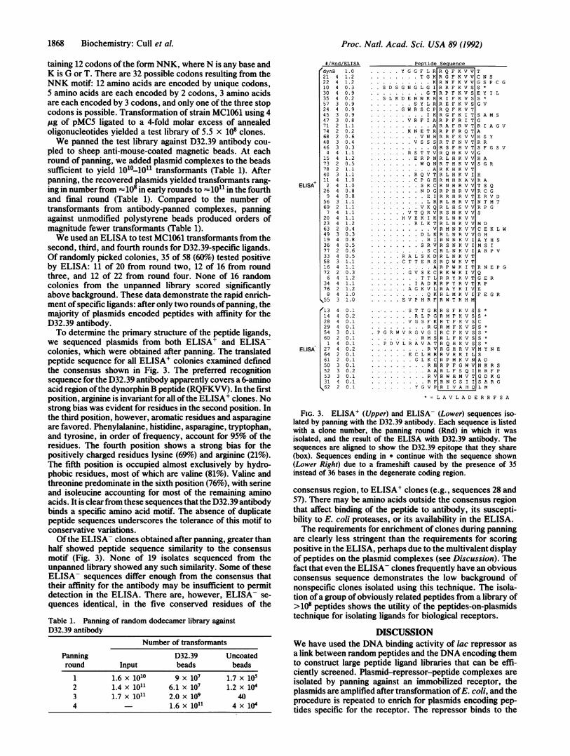
we sequenced plasmids from both ELISA+ and ELISA-colonies, which were obtained after panning. The translatedpeptide sequence for all ELISA+ colonies examined definedthe consensus shown in Fig. 3. The preferred recognitionsequence for the D32.39 antibody apparently covers a 6-aminoacid region ofthe dynorphin B peptide (RQFKVV). In the firstposition, arginine is invariant for all ofthe ELISA+ clones. Nostrong bias was evident for residues in the second position. Inthe third position, however, aromatic residues and asparagineare favored. Phenylalanine, histidine, asparagine, tryptophan,and tyrosine, in order of frequency, account for 95% of theresidues. The fourth position shows a strong bias for thepositively charged residues lysine (69%) and arginine (21%).The fifth position is occupied almost exclusively by hydro-phobic residues, most of which are valine (81%). Valine andthreonine predominate in the sixth position (76%), with serineand isoleucine accounting for most of the remaining aminoacids. It is clearfrom these sequences that the D32.39 antibodybinds a specific amino acid motif. The absence of duplicatepeptide sequences underscores the tolerance of this motif toconservative variations.Of the ELISA- clones obtained after panning, greater than
half showed peptide sequence similarity to the consensusmotif (Fig. 3). None of 19 isolates sequenced from theunpanned library showed any such similarity. Some of theseELISA- sequences differ enough from the consensus thattheir affinity for the antibody may be insufficient to permitdetection in the ELISA. There are, however, ELISA- se-quences identical, in the five conserved residues of the
Table 1. Panning of random dodecamer library againstD32.39 antibody
Number of transformants
Panning D32.39 Uncoatedround Input beads beads
1 1.6x10¹⁰ 9x10⁷ 1.7x10⁵
Proc. Natl. Acad. Sci. USA 89 (1992)
ELISA'
ELISA
# /Rnd/ELISA
dynB 1.021 4 1.222 4 1.210 4 0.330 4 0.935 4 0.257 3 0.924 4 0.945 3 0.947 3 0.871 2 1.174 2 0.268 2 0.648 3 0.446 3 0.34 4 1.1
15 4 1.273 2 0.578 2 1.140 3 1.111 4 1.02 4 1.0
26 4 0.89 4 0.8
56 3 1.169 2 1.17 4 1.1
20 4 1.123 4 1.263 2 0.449 3 0.319 4 0.836 4 0.577 2 0.633 4 0.558 3 1.116 4 1.172 2 0.36 4 1.2
34 4 1.176 2 1.28 4 1.0
s55 3 1.0
13 4 0.114 4 0.228 4 0.129 4 0.154 3 0.160 2 0.11 4 0.1
27 4 0.264 2 0.161 2 0.150 3 0.152 3 0.253 3 0.131 4 0.162 2 0.1
.... Y G G F L R..........T G K.~~~~~K
S D S G N G L G I................GT
S L K D E N N K R.....SY L R
.... G W R S C P.......1........IK
....VR F I A
.................A....KN E T R.............VNH
.....VS..................G
....RS T T V
.....ER P N.M...........WQ N.................A
......RQ V T
......CP G E
.............SR C
.H...........ND G
................EI
................LR
.............VK Q
....VT Q R V
.H V E K I K......RL K T
.................V......O......DL K................RI
.............SR V
................SCR A L S K D
.C T T E RSA
....GV S EC
.............T L
......IA DR
....AG K VL
................QEE V P H RF
SequenceR Q F K V VR G F K V VR N F K V VR R F K V SR P F K V SR I F K V SR E F K V SR Q F K V TR G F K I TR P F R I TR A F R V TR P F R Q TR R F S V VR T F N V TR S F H V TR Q H K V VR L H K V VR T H K V VR K H K V TR L H K V IR M H K A VR N H R V VR P H R V VR R H R V TR L H R V TR L H S V VR S N K V VR L N K V VR L N K V VR M N K V VR L N R V VR N N K V IR S N K V IR L N K V IR L N K V TR O W K V TR P W K I TR K W K I VR R Y K V TR P Y R V TR A Y K I VR L M K V IR W T K H M
TC N SG S P C GS*E Y I LS *G V
S A M SGR I A G VAH S YR RS F G S VGH AS G R
HR AT S QR C GE R V DN T M TR P GS
M DC E K L WG HA Y H SM S IA R P V
R N E P GQG E RR PEF E G R
........S T T G R R S F K V S S *
R L P G R M F K V S S *
V G S F K R T F K V S C
.P G R W V R G V
..P D V L R A.........E C I...................GI
.01..yc..
R G R M F K V S SV G I R C F K V S S*R M S R L F K V S SV A T R Q H K V S SR V R G H R V V M Y N E
L H R R V H K I L SL K C R P M K V N A DR H R P F G W V N K R SAA R L F S Q I R R F PRV R W H MVTG D GRFRCS I I S A R GVPR I V A H Q L M
=L A V L A D E R R F S A
FIG. 3. ELISA+ (Upper) and ELISA- (Lower) sequences iso-lated by panning with the D32.39 antibody. Each sequence is listedwith a clone number, the panning round (Rnd) in which it wasisolated, and the result of the ELISA with D32.39 antibody. Thesequences are aligned to show the D32.39 epitope that they share(box). Sequences ending in * continue with the sequence shown(Lower Right) due to a frameshift caused by the presence of 35instead of 36 bases in the degenerate coding region.
consensus region, to ELISA' clones (e.g., sequences 28 and57). There may be amino acids outside the consensus regionthat affect binding of the peptide to antibody, its suscepti-bility to E. coli proteases, or its availability in the ELISA.The requirements for enrichment of clones during panning
are clearly less stringent than the requirements for scoringpositive in the ELISA, perhaps due to the multivalent displayof peptides on the plasmid complexes (see Discussion). Thefact that even the ELISA- clones frequently have an obviousconsensus sequence demonstrates the low background ofnonspecific clones isolated using this technique. The isola-tion of a group of obviously related peptides from a library of>108 peptides shows the utility of the peptides-on-plasmidstechnique for isolating ligands for biological receptors.
DISCUSSIONWe have used the DNA binding activity of lac repressor asa link between random peptides and the DNA encoding themto construct large peptide ligand libraries that can be effi-ciently screened. Plasmid-repressor-peptide complexes areisolated by panning against an immobilized receptor, theplasmids are amplified after transformation ofE. coli, and the
procedure is repeated to enrich for plasmids encoding pep-tides specific for the receptor. The repressor binds to the
2 1.4 x 10¹¹ 6.1 x 10⁷ 1.2 x 10³ 1.7 x 10¹¹ 2.0 x 10⁹ 404 1.6x10¹¹ 4x 10
ereiniacer
"I '-'T
--I
II
41
'I
Pont i Hp

Proc. Natl. Acad. Sci. USA 89 (1992) 1869
library plasmid with sufficient avidity to allow panning of thelibrary on immobilized receptor without problematic levels ofdissociation. We have used this system to identify a series ofrelated peptides that bind to a monoclonal antibody whoseepitope was previously uncharacterized. The consensus se-
quence for binding is similar to sequences found using phagelibraries (ref. 5; Ron Barrett, personal communication), usingpeptides fused to maltose binding protein (Charles Hart,personal communication), and using light-directed spatiallyaddressable parallel chemical synthesis (ref. 32; ChrisHolmes, personal communication).
Several features of peptides-on-plasmids libraries distinguishthem from the phage libraries that have been described (5-7).The random peptides are displayed with a free C terminusinstead of at the N terminus or internal to the carrier protein,leaving a different end of the peptides free to interact withreceptors. Therefore, the use ofboth techniques in conjunctionwill increase the diversity of peptide structures that are avail-able for receptor binding. The C-terminal mode of display alsoensures that stop codons in the degenerate region, which occurwith increasing frequency in longer degenerate oligonucleo-tides, shorten rather than destroy individual clones. The lacrepressor fusions should allow the display of potential ligandswith a wide range of sizes, given the observation that proteinsas large as p-galactosidase can be fused to the repressor withouteliminating DNA binding activity (10).
Characteristics of the carrier proteins impose differentconstraints on the two methods. The repressor fusions are
cytoplasmic, unlike the phage fusions that are exported to theperiplasm. Thus there is no need for peptides fused torepressor to be compatible with the protein export apparatusand the formation of an intact phage coat. The peptides needsimply to be compatible with the formation of at least a
repressor dimer, which is the smallest form of the protein thatcan bind DNA (33, 34). In addition, the use of both methodsincreases total available peptide diversity because the twotypes of libraries are exposed to different cellular compart-ments and thus to different sets of E. coli proteases.The lac repressor displays multiple copies of the peptide on
each library particle. Each repressor tetramer, in principle,displays four peptides that are available for binding to recep-tors. In addition, each plasmid monomer can bind up to twotetramers (if no loop is formed), and tandemly duplicatedforms of the plasmid can display higher multiples of twotetramers. This multivalent display allows the isolation ofligands with moderate affinity (Kd 1 10-6 M; ref. 5). Under
multivalent conditions, these moderate-affinity ligands can
obscure less-numerous high-affinity ligands. The higher-affinity ligands (Kd 1 10-9 M) can be isolated from the
population by panning with receptor immobilized at lowdensity to ensure monovalent binding conditions (R. W. Bar-rett, S. E. Cwirla, M. S. Ackerman, A. E. Olson, E. A.Peters, and W. J. Dower, personal communication). For re-
ceptors whose normal ligands are not small peptides, thismultivalency of display will be an advantage for identifyinginitial families of moderate-affinity ligands. Screening variantsofthe moderate-affinity peptides under monovalent conditionswill allow selection of ligands with higher affinity.
In conclusion, we have developed a system that permitsthe construction of a wide variety of C-terminally exposedpeptide libraries through a direct linkage of the peptide to thegenetic material encoding it. This system should be useful todiscover ligands for a variety of biological receptors.
We thank Ramesh Bhatt for excellent sequence analysis andElizabeth Peters for oligonucleotide synthesis. We thank Luz-Maria
Guzman-Verduzco, Mike Carson, Alan Derman, and Jon Beckwithfor bacterial strains, plasmids, and advice. We thank Peter Mark-iewicz and Jeffrey H. Miller for strains and advice. We thank BillDower, Ron Barrett, Charles Hart, Bruce England, Steve Yanofsky,Erik Whitehorn, Steve Cwirla, and Martha Ackerman for advice andcomments.
1. Tuerk, C. & Gold, L. (1990) Science 249, 505-510.2. Ellington, A. D. & Szostak, J. W. (1990) Nature (London) 346,
818-822.3. Thiesen, H.-J. & Bach, C. (1990) Nucleic Acids Res. 18,
3203-3209.4. Parmley, S. F. & Smith, G. P. (1988) Gene 73, 305-318.5. Cwirla, S. E., Peters, E. A., Barrett, R. W. & Dower, W. J.
(1990) Proc. Natl. Acad. Sci. USA 87, 6378-6382.6. Devlin, J. J., Panganiban, L. C. & Devlin, P. E. (1990) Science
249, 404-406.7. Scott, J. K. & Smith, G. P. (1990) Science 249, 386-390.8. Kleina, L. G. & Miller, J. H. (1990) J. Mol. Biol. 212, 295-318.9. Beyreuther, K. (1980) in The Operon, eds. Miller, J. H. &
Reznikoff, W. S. (Cold Spring Harbor Lab., Cold Spring Har-bor, NY), pp. 123-154.
10. Muller-Hill, B. & Kania, J. (1974) Nature (London) 249,561-563.
11. Hu, M. C.-T. & Davidson, N. (1991) Gene 99, 141-150.12. Labow, M. A., Baim, S. B., Shenk, T. & Levine, A. J. (1990)
Mol. Cell. Biol. 10, 3343-3356.13. Panayotatos, N., Fontaine, A. & Bdckman, S. (1989) J. Biol.
Chem. 264, 15066-15069.14. Levens, D. & Howley, P. M. (1985) Mol. Cell. Biol. 5, 2307-
2315.15. Lundeberg, J., Wahlberg, J. & Uhldn, M. (1991) BioTechniques
10, 68-75.16. Koob, M. & Szybalski, W. (1990) Science 250, 271-273.17. Gold, L. & Stormo, G. D. (1990) Methods Enzymol. 185,
89-103.18. England, B. P., Ackerman, M. S. & Barrett, R. W. (1991)
FEBS Lett. 279, 87-90.19. Dower, W. J., Miller, J. F. & Ragsdale, C. W. (1988) Nucleic
Acids Res. 16, 6127-6145.20. Barrett, R. W. & Goldstein, A. (1985) Neuropeptides 6, 113-
120.21. Barkley, M. D. & Bourgeois, S. (1980) in The Operon, eds.
Miller, J. H. & Reznikoff, W. S. (Cold Spring Harbor Lab.,Cold Spring Harbor, NY), pp. 177-220.
22. Besse, M., von Wilcken-Bergmann, B. & Muller-Hill, B. (1986)EMBO J. 5, 1377-1381.
23. Flashner, Y. & Gralla, J. D. (1988) Proc. Natl. Acad. Sci. USA85, 8968-8972.
24. Hsieh, W.-T., Whitson, P. A., Matthews, K. S. & Wells, R. D.(1987) J. Biol. Chem. 262, 14583-14591.
25. Kramer, H., Niem6ller, M., Amouyal, M., Revet, B., Wilcken-Bergmann, B. & Muller-Hill, B. (1987) EMBO J. 6, 1481-1491.
26. Mossing, M. C. & Record, M. T., Jr. (1986) Science 233,889-892.
27. Whitson, P. A., Hsieh, W.-T., Wells, R. D. & Matthews, K. S.(1987) J. Biol. Chem. 262, 14592-14599.
28. Sadler, J. R., Sasmor, H. & Betz, J. L. (1983) Proc. Natl.Acad. Sci. USA 80, 6785-6789.
29. Simons, A., Tils, D., von Wilcken-Bergmann, B. & Muller-Hill,B. (1984) Proc. Natl. Acad. Sci. USA 81, 1624-1628.
30. Lee, N. (1980) in The Operon, eds. Miller, J. H. & Reznikoff,W. S. (Cold Spring Harbor Lab., Cold Spring Harbor, NY), pp.389-409.
31. Miller, J. H. (1972) Experiments in Molecular Genetics (ColdSpring Harbor Lab., Cold Spring Harbor, NY).
32. Fodor, S. P. A., Read, J. L., Pirrung, M. C., Stryer, L., Lu,A. T. & Solas, D. (1991) Science 251, 767-773.
33. Daly, T. J. & Matthews, K. S. (1986) Biochemistry 25, 5474-5478.
34. Kania, J. & Brown, D. T. (1976) Proc. Natl. Acad. Sci. USA73, 3529-3533.
Biochemistry: Cull et al.