secondary findings from non-invasive prenatal testing for ... · secondary findings from...
TRANSCRIPT
ORIGINAL ARTICLE
Secondary findings from non-invasive prenatal testing forcommon fetal aneuploidies by whole genome sequencing as aclinical serviceTze Kin Lau1, Fu Man Jiang2, Robert J. Stevenson3, Tsz Kin Lo4, Lin Wai Chan5, Mei Ki Chan2, Pui Shan Salome Lo1, Wei Wang2,Hong-Yun Zhang2, Fang Chen2 and Kwong Wai Choy6,7*
1Fetal Medicine Centre, Paramount Clinic, Hong Kong, China2BGI-Shenzhen, Shenzhen, China3Hong Kong Adventist Hospital, Hong Kong, China4Department of Obstetrics and Gynaecology, Kwong Wah Hospital, Hong Kong, China5Department of Obstetrics and Gynaecology, United Christian Hospital, Hong Kong, China6Department of Obstetrics and Gynaecology, Prince of Wales Hospital, The Chinese University of Hong Kong, Hong Kong, China7CUHK-Shenzhen Research Institute, Chinese University of Hong Kong, Shenzhen, China*Correspondence to: Kwong Wai Choy. E-mail: [email protected]
ABSTRACTObjective To report secondary or additional findings arising from introduction of non-invasive prenatal testing (NIPT)for aneuploidy by whole genome sequencing as a clinical service.
Methods Five cases with secondary findings were reviewed.
Results In Case 1, NIPT revealed a large duplication in chromosome 18p, which was supported by arrayCGH ofamniocyte DNA, with final karyotype showing mosaic tetrasomy 18p. In Case 2, a deletion in the proximal long arm ofchromosome 18 of maternal origin was suspected and confirmed by arrayCGH of maternal white cell DNA. In Case 3,NIPT was negative for trisomies 21 and 18. In-depth analysis for deletions/duplications was requested when fetalstructural anomalies were detected at routine scan. A deletion in the proximal long arm of chromosome 3 was foundand confirmed by karyotyping. In Case 4, NIPT correctly predicted confined placental mosaicism with triple trisomyinvolving chromosomes X, 7 and 21. In Case 5, NIPT correctly detected a previously unknown maternal mosaicismfor 45X.
Conclusion Non-invasive prenatal testing is able to detect a wide range of fetal, placental and maternal chromosomalabnormalities. This has important implications on patient counseling when an abnormality is detected by NIPT.© 2013 John Wiley & Sons, Ltd.
Funding sources: This work is partly supported by the National Basic Research Program of China (2012CB944600) and the Hong Kong General Research Grant.Conflicts of interest: The co-authors, Fuman Jiang, Mei Ki Chan, Hong-Yun Zhang, Fang Chen and Wei Wang are employees of BGI.
INTRODUCTIONNon-invasive prenatal testing (NIPT) for common fetalaneuploidies by massively parallel sequencing of maternalplasma DNA is a new technology detecting close to 100% oftrisomy 21 pregnancies with a false positive rate of <0.1%.1
There is much we do not yet know about this exciting newinvestigation, and there is much to learn and explore. Here,we report five cases of ‘unexpected’ secondary chromosomalabnormalities discovered after the implementation of this testin a clinical setting.
ABOUT THE TESTSFull details about the NIPT test were as reported previously.2 TheNIPT test was offered to pregnant women carrying singleton
pregnancies from 12weeks of gestation or beyond. Before the
blood test, each woman or couple had individual counseling by
an obstetrician and an ultrasound scan to confirm the number
of fetuses, fetal viability and fetal size and to exclude major fetal
structural abnormalities. A written informed consent was
obtained from all women. Five milliliters of maternal peripheral
blood was collected into an ethylene diamine tetraacetic acid
(EDTA) bottle. Samples were processed according to strict
protocol. All subsequent procedures and molecular tests,
including cell-free DNA isolation, library construction and
sequencing, were performed at the clinical laboratory of BGI-
Shenzhen, China, which had been ISO/IEC 17025 certified.Whole genome sequencing was used for the NIPT. For
aneuploidy detection, a binary hypothesis t-test and logarithmic
Prenatal Diagnosis 2013, 33, 602–608 © 2013 John Wiley & Sons, Ltd.
DOI: 10.1002/pd.4076
likelihood ratio (L-score) between the two t-tests were used toclassify whether the fetus had aneuploidy.1 This approach enablednot only the classification of pregnancies affected and unaffectedby trisomies but also the identification of cases with mosaicism.The test report initially included risk assessment for trisomy 21and trisomy 18 only, but was extended to include trisomy 13 sinceearly 2012. However, bioinformatics analysis actually includedaneuploidy detection of all 23 pairs of chromosomes, anddetection for chromosomal deletions and duplications by aspecially designed FCAPS (Fetal Copy Number Analysis throughMaternal Plasma Sequencing, see later for further details) pipelinewas routinely performed since July 2012. Before that, FCAPS wasperformed for caseswith t-score above 2.0, aiming at the detectionof partial trisomy. Referring clinicians were notified if any ofthese additional genome-wide abnormalities were suspected.Specifically, fetal sex was not reported, even on request, unlesssex chromosomal abnormalities were suspected.
The FCAPS pipeline is a special algorithm for the detection oflarge deletions and duplications. In NIPT for aneuploidies, therelative proportion of DNA fragments from a specificchromosome is estimated and compared against the normalrange. In FCAPS, the human genome was divided into a totalof 308 789 sliding, 99%-overlapping basic observation unitseach has 84 000 expected unique reads. Potential breakpointsof deletions/duplications are localized by comparing thedifference of read number, after correction for GC-bias,between the observational units on each side of the potentialbreakpoint. A binary segmentation algorithm with dynamicthreshold determination is used to determine whether thepotential breakpoint is significant or not. The detection powerof FCAPS increases with increasing cell-free fetal DNAconcentration and more sequencing reads. On the conditionof 10% cell-free fetal DNA concentration and a sequencingread number currently obtained with NIPT for aneuploidies,FCAPS is able to detect close to 100% of deletions/duplicationsof 10Mb or above. In-depth discussion on the FCAPSmethodology can be found in relevant publication.3
Conventional cytogenetic studies, quantitative fluorescentpolymerase chain reaction (QF-PCR) or arrayCGH (aCGH) studieswere performed at the laboratory of the Department of Obstetricsand Gynaecology, Chinese University of Hong Kong, or theDepartment of Obstetrics and Gynaecology, Tsan Yuk Hospital,University of Hong Kong. The aCGH test offered by the ChineseUniversity of Hong Kong was a custom-made panel for prenataldiagnosis that targeted at the loci of 100 common micro-deletionand micro-duplication syndromes of relevance in prenataldiagnosis at high resolution, plus whole genome coverage with abackbone resolution down to 100kb.4 The aCGH test offered bythe University of Hong Kong was NimbleGen CGX-135K wholegenome oligonucleotide microarray with resolution of 140kbacross the genome, and 40kb or less in regions of clinical relevance.The array can evaluate over 245 genetic syndromes and over 980gene regions of functional significance in human development.
CASE 1Case 1 was a 36-year-old woman with one previous miscarriage.She requested the NIPT as a primary screening test for fetal Downsyndrome at 13+4weeks. Pre-test sonogram was normal. NIPT
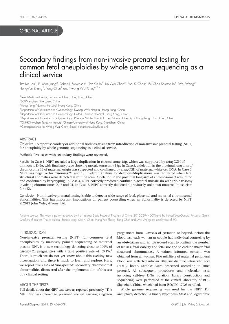
report at 15+3weeks showed normal numbers of chromosomes21 and 13. The t-score for chromosome 18 was 2.03, for whichthe FCAPS analysis was performed. The FCAPS suggestedthat there was an approximately 13Mb duplication involvingchromosome 18p (chr18:483517–14400897, corresponding to18p11.32–p11.21) (Figure 1a). Sonogram at 15+3weeks did notshow any structural anomalies. After extensive counseling, thecouple opted for amniocentesis and aCGH (to exclude possibleadditional deletions and duplications), which was performed at16+5weeks. aCGH showed a duplication of about 14Mb,suggesting partial trisomy 18p (Figure 1b). Final karyotypingshowed that it was a case of mosaic isochromosome tetrasomy18p at a ratio of 7:23 (abnormal to normal cells) (Figure 1c). Arepeat ultrasound scan did not show any structural abnormalities.The couple finally determined for pregnancy termination. Themedical termination was uncomplicated. The postmortemexamination showed no structural abnormalities.
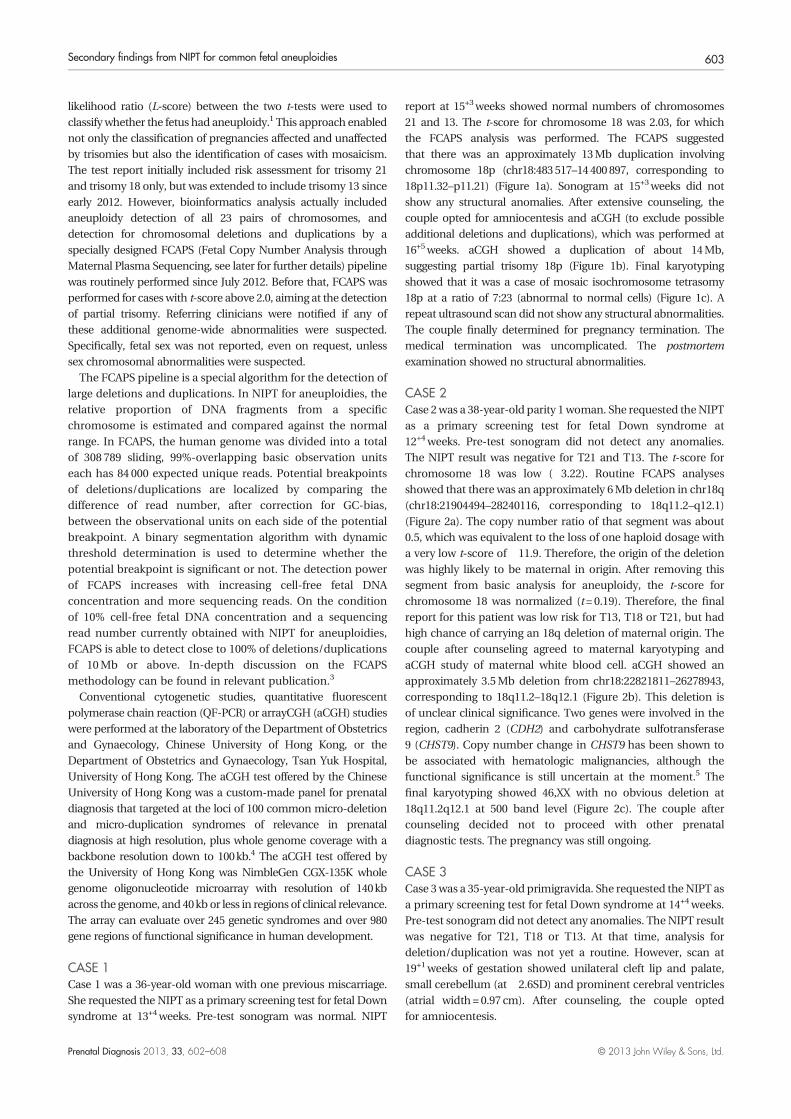
CASE 2Case 2was a 38-year-old parity 1woman. She requested theNIPTas a primary screening test for fetal Down syndrome at12+4weeks. Pre-test sonogram did not detect any anomalies.The NIPT result was negative for T21 and T13. The t-score forchromosome 18 was low (�3.22). Routine FCAPS analysesshowed that there was an approximately 6Mb deletion in chr18q(chr18:21904494–28240116, corresponding to 18q11.2–q12.1)(Figure 2a). The copy number ratio of that segment was about0.5, which was equivalent to the loss of one haploid dosage witha very low t-score of �11.9. Therefore, the origin of the deletionwas highly likely to be maternal in origin. After removing thissegment from basic analysis for aneuploidy, the t-score forchromosome 18 was normalized (t=0.19). Therefore, the finalreport for this patient was low risk for T13, T18 or T21, but hadhigh chance of carrying an 18q deletion of maternal origin. Thecouple after counseling agreed to maternal karyotyping andaCGH study of maternal white blood cell. aCGH showed anapproximately 3.5Mb deletion from chr18:22821811–26278943,corresponding to 18q11.2–18q12.1 (Figure 2b). This deletion isof unclear clinical significance. Two genes were involved in theregion, cadherin 2 (CDH2) and carbohydrate sulfotransferase9 (CHST9). Copy number change in CHST9 has been shown tobe associated with hematologic malignancies, although thefunctional significance is still uncertain at the moment.5 Thefinal karyotyping showed 46,XX with no obvious deletion at18q11.2q12.1 at 500 band level (Figure 2c). The couple aftercounseling decided not to proceed with other prenataldiagnostic tests. The pregnancy was still ongoing.
CASE 3Case 3was a 35-year-old primigravida. She requested theNIPT asa primary screening test for fetal Down syndrome at 14+4weeks.Pre-test sonogram did not detect any anomalies. TheNIPT resultwas negative for T21, T18 or T13. At that time, analysis fordeletion/duplication was not yet a routine. However, scan at19+1weeks of gestation showed unilateral cleft lip and palate,small cerebellum (at �2.6SD) and prominent cerebral ventricles(atrial width=0.97 cm). After counseling, the couple optedfor amniocentesis.
Secondary findings from NIPT for common fetal aneuploidies 603
Prenatal Diagnosis 2013, 33, 602–608 © 2013 John Wiley & Sons, Ltd.
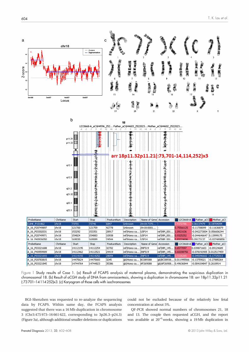
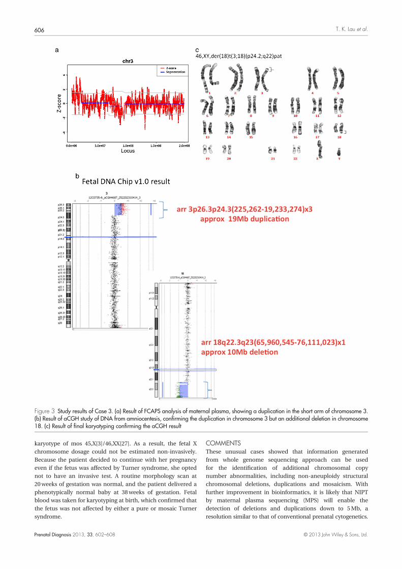
BGI-Shenzhen was requested to re-analyze the sequencingdata by FCAPS. Within same day, the FCAPS analysissuggested that there was a 16Mb duplication in chromosome3 (Chr3:475 973–16 661 622, corresponding to 3p26.3–p24.3)(Figure 3a), although additional smaller deletions or duplications
could not be excluded because of the relatively low fetalconcentration at about 5%.
QF-PCR showed normal numbers of chromosomes 21, 18and 13. The couple then requested aCGH, and the reportwas available at 20+0weeks, showing a 19Mb duplication in
Figure 1 Study results of Case 1. (a) Result of FCAPS analysis of maternal plasma, demonstrating the suspicious duplication inchromosomal 18. (b) Result of aCGH study of DNA from amniocentesis, showing a duplication in chromosome 18: arr 18p11.32p11.21(:73701–14114252)x3. (c) Karyogram of those cells with isochromosomes
T. K. Lau et al.604
Prenatal Diagnosis 2013, 33, 602–608 © 2013 John Wiley & Sons, Ltd.
chromosome 3p and a 10Mb deletion in chromosome18q (Figure 3b). The karyotype confirmed an unbalancedtranslocation of 46,XY, der(18)t(3:18)(p24:2,q22) (Figure 3c), whichwas subsequently found to be inherited from the father who wasa balanced translocation carrier. The couple finally decided tohave pregnancy termination, which was uncomplicated. Thecouple declined postmortem examination, but cleft lip and palatewas confirmed on external examination.
CASE 4Case 4 was a 37-year-old primigravida. She presented for theNIPT because of positive first trimester combined screeningtest. Pre-NIPT ultrasound showed the absence of nasal bonebut otherwise a normal singleton pregnancy with a nuchaltranslucency of 1.6mm. After counseling, the couple changedtheir mind to have a chorionic villus sampling (CVS) and agreedto donate a blood sample for the NIPT as a quality assurancesample (for that, BGI-Shenzhen was not aware of the scan orother laboratory test results until the NIPT report was issued).
The CVS was performed at 14+0weeks of gestation. QF-PCRwas available at 14+1weeks, showing normal copies ofchromosomes 21, 18 and 13, but suspected mosaic XXY.
The NIPT result was available at 15+5weeks. There was highsuspicion of mosaic 47,XXY (t-score = 4.06), mosaic trisomy 21(t-score = 3.32) and mosaic trisomy 7 (t-score = 6.32).
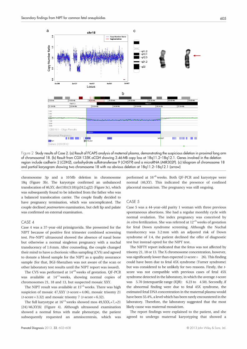
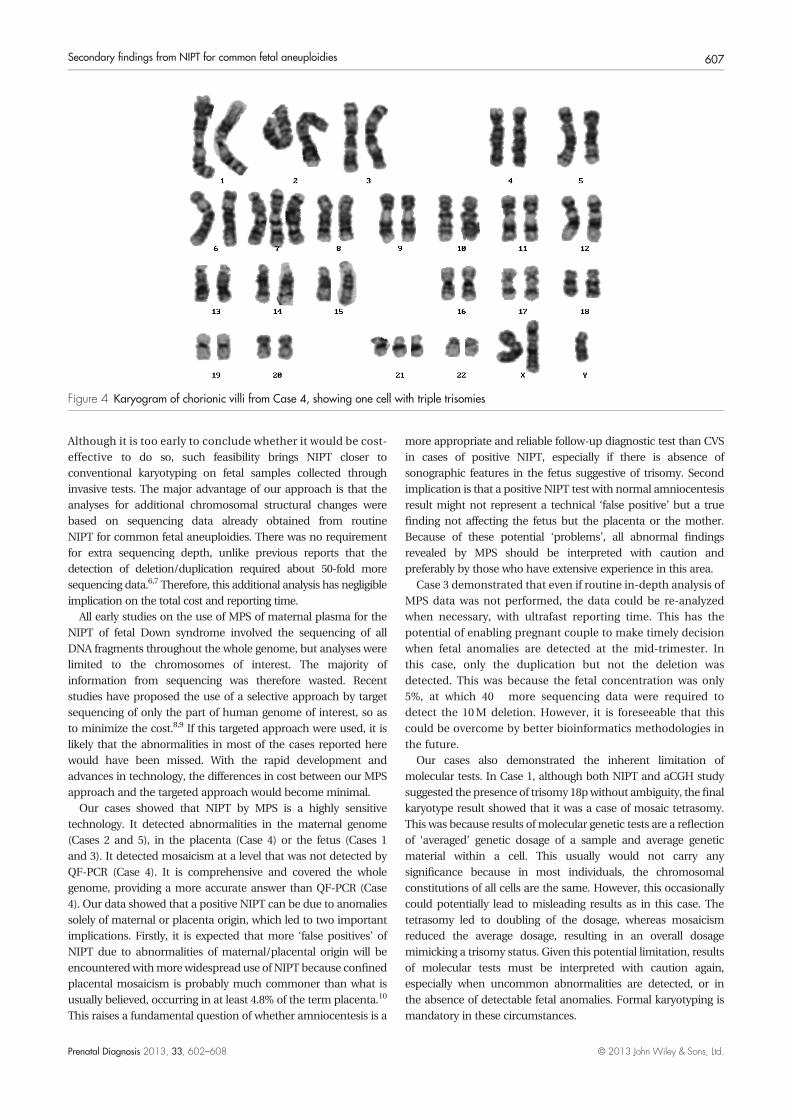
The full karyotype at 16+5weeks showed mos 49,XXX,+7,+21[24]/46,XY[6] (Figure 4). Although ultrasound examinationshowed a normal fetus with male phenotype, the patientsubsequently requested an amniocentesis, which was
performed at 16+6weeks. Both QF-PCR and karyotype werenormal (46,XY). This indicated the presence of confinedplacental mosaicism. The pregnancy was still ongoing.
CASE 5Case 5 was a 44-year-old parity 1 woman with three previousspontaneous abortions. She had a regular monthly cycle withnormal ovulation. The index pregnancy was conceived byin vitro fertilization. She was referred at 12+5weeks of gestationfor fetal Down syndrome screening. Although the Nuchaltranslucency was 3.2mm with an adjusted risk of Downsyndrome of 1:4, the patient declined the offer of diagnostictest but instead opted for the NIPT test.
The NIFTY report indicated that the fetus was not affected bytrisomy 21, 18 or 13. The X chromosome concentration, however,was significantly lower than expected (t-score=�26). This findingcould have been due to fetal 45X syndrome (Turner syndrome)but was considered to be unlikely for two reasons. Firstly, the t-score was not compatible with previous cases of fetal 45Xsyndrome detected in the laboratory, in which the average t-scorewas�5.70 (interquartile range (IQR):�6.23 to�4.50). Secondly, ifthe abnormal finding were due to fetal 45X syndrome, theestimated fetal DNA concentration in thematernal plasmawouldhavebeen 55.4%, a level which has been rarely encountered in thelaboratory. Therefore, the laboratory suggested that the mostlikely cause was maternal mosaicism.
The report findings were explained to the patient, and sheagreed to undergo maternal karyotyping that showed a
Figure 2 Study results of Case 2. (a) Result of FCAPS analysis of maternal plasma, demonstrating the suspicious deletion in proximal long armof chromosomal 18. (b) Result from CGX-135K aCGH showing 3.46MB copy loss at 18q11.2–18q12.1. Genes involved in the deletionregion include cadherin 2 (CDH2), carbohydrate sulfotransferase 9 (CHST9) and a microRNA (MIR302F). (c) Idiogram of chromosome 18and partial karyogram showing two chromosome 18 with no obvious deletion at 18q11.2–18q12.1 (arrow)
Secondary findings from NIPT for common fetal aneuploidies 605
Prenatal Diagnosis 2013, 33, 602–608 © 2013 John Wiley & Sons, Ltd.
karyotype of mos 45,X[3]/46,XX[27]. As a result, the fetal Xchromosome dosage could not be estimated non-invasively.Because the patient decided to continue with her pregnancyeven if the fetus was affected by Turner syndrome, she optednot to have an invasive test. A routine morphology scan at20weeks of gestation was normal, and the patient delivered aphenotypically normal baby at 38weeks of gestation. Fetalblood was taken for karyotyping at birth, which confirmed thatthe fetus was not affected by either a pure or mosaic Turnersyndrome.
COMMENTSThese unusual cases showed that information generatedfrom whole genome sequencing approach can be usedfor the identification of additional chromosomal copynumber abnormalities, including non-aneuploidy structuralchromosomal deletions, duplications and mosaicism. Withfurther improvement in bioinformatics, it is likely that NIPTby maternal plasma sequencing (MPS) will enable thedetection of deletions and duplications down to 5Mb, aresolution similar to that of conventional prenatal cytogenetics.
Figure 3 Study results of Case 3. (a) Result of FCAPS analysis of maternal plasma, showing a duplication in the short arm of chromosome 3.(b) Result of aCGH study of DNA from amniocentesis, confirming the duplication in chromosome 3 but an additional deletion in chromosome18. (c) Result of final karyotyping confirming the aCGH result
T. K. Lau et al.606
Prenatal Diagnosis 2013, 33, 602–608 © 2013 John Wiley & Sons, Ltd.
Although it is too early to conclude whether it would be cost-effective to do so, such feasibility brings NIPT closer toconventional karyotyping on fetal samples collected throughinvasive tests. The major advantage of our approach is that theanalyses for additional chromosomal structural changes werebased on sequencing data already obtained from routineNIPT for common fetal aneuploidies. There was no requirementfor extra sequencing depth, unlike previous reports that thedetection of deletion/duplication required about 50-fold moresequencing data.6,7 Therefore, this additional analysis has negligibleimplication on the total cost and reporting time.
All early studies on the use of MPS of maternal plasma for theNIPT of fetal Down syndrome involved the sequencing of allDNA fragments throughout the whole genome, but analyses werelimited to the chromosomes of interest. The majority ofinformation from sequencing was therefore wasted. Recentstudies have proposed the use of a selective approach by targetsequencing of only the part of human genome of interest, so asto minimize the cost.8,9 If this targeted approach were used, it islikely that the abnormalities in most of the cases reported herewould have been missed. With the rapid development andadvances in technology, the differences in cost between our MPSapproach and the targeted approach would become minimal.
Our cases showed that NIPT by MPS is a highly sensitivetechnology. It detected abnormalities in the maternal genome(Cases 2 and 5), in the placenta (Case 4) or the fetus (Cases 1and 3). It detected mosaicism at a level that was not detected byQF-PCR (Case 4). It is comprehensive and covered the wholegenome, providing a more accurate answer than QF-PCR (Case4). Our data showed that a positive NIPT can be due to anomaliessolely of maternal or placenta origin, which led to two importantimplications. Firstly, it is expected that more ‘false positives’ ofNIPT due to abnormalities of maternal/placental origin will beencounteredwithmorewidespread use of NIPT because confinedplacental mosaicism is probably much commoner than what isusually believed, occurring in at least 4.8% of the term placenta.10
This raises a fundamental question of whether amniocentesis is a
more appropriate and reliable follow-up diagnostic test than CVSin cases of positive NIPT, especially if there is absence ofsonographic features in the fetus suggestive of trisomy. Secondimplication is that a positive NIPT test with normal amniocentesisresult might not represent a technical ‘false positive’ but a truefinding not affecting the fetus but the placenta or the mother.Because of these potential ‘problems’, all abnormal findingsrevealed by MPS should be interpreted with caution andpreferably by those who have extensive experience in this area.
Case 3 demonstrated that even if routine in-depth analysis ofMPS data was not performed, the data could be re-analyzedwhen necessary, with ultrafast reporting time. This has thepotential of enabling pregnant couple to make timely decisionwhen fetal anomalies are detected at the mid-trimester. Inthis case, only the duplication but not the deletion wasdetected. This was because the fetal concentration was only5%, at which 40� more sequencing data were required todetect the 10M deletion. However, it is foreseeable that thiscould be overcome by better bioinformatics methodologies inthe future.
Our cases also demonstrated the inherent limitation ofmolecular tests. In Case 1, although both NIPT and aCGH studysuggested the presence of trisomy 18pwithout ambiguity, the finalkaryotype result showed that it was a case of mosaic tetrasomy.This was because results of molecular genetic tests are a reflectionof ‘averaged’ genetic dosage of a sample and average geneticmaterial within a cell. This usually would not carry anysignificance because in most individuals, the chromosomalconstitutions of all cells are the same. However, this occasionallycould potentially lead to misleading results as in this case. Thetetrasomy led to doubling of the dosage, whereas mosaicismreduced the average dosage, resulting in an overall dosagemimicking a trisomy status. Given this potential limitation, resultsof molecular tests must be interpreted with caution again,especially when uncommon abnormalities are detected, or inthe absence of detectable fetal anomalies. Formal karyotyping ismandatory in these circumstances.
Figure 4 Karyogram of chorionic villi from Case 4, showing one cell with triple trisomies
Secondary findings from NIPT for common fetal aneuploidies 607
Prenatal Diagnosis 2013, 33, 602–608 © 2013 John Wiley & Sons, Ltd.
Counseling for unexpected findings, especially with rareconditions, can be difficult. For example, case I was initiallysuspected to be trisomy 18p but ultimately was found to bemosaic tetrasomy 18p. Most publications on trisomy 18pstarted with a similar statement such as ‘Most of the patientshave either an apparently normal phenotype or only minoranomalies, and may or may not have mental retardation’.11,12
However, a more detailed analysis of reported cases showed thatmental retardation was present in eight of 14 (57.1%) reportedcases of pure trisomy 18p without associated abnormalities ofother chromosomes.13 The phenotype of tetrasomy 18p is moredistinct, including physical and growth abnormalities, anddevelopmental delay and cognitive impairment are universallypresent.14On the other hand, only a few cases ofmosaic tetrasomy18p have been reported, and therefore, no conclusion concerningphenotype can be drawn.15,16 Similar situation happens whenunexpected findings are detected by amniocentesis or chorionicvillus sampling. It is therefore important in the pre-test counselingto discuss with the couple whether they would like to be informedof such additional findings.
Case 5 is of particular relevancewhenNIPT is only limited to thedetection of common trisomies. It showed that maternalmosaicism for aneuploidy could result in a ‘false positive’ NIPTresult. Although the X chromosome was involved in this case, thisphenomenon of maternal mosaicism could occur in otherchromosomal aneuploidies including trisomy 21. A previous studyhas suggested that at the present sequencing depth, fetal trisomy21 can be detected by NIPT of maternal plasma when the fetalDNA concentration is above 3.9%.17 This means that if thepregnant woman has a low-level mosaic trisomy 21 of 4%, the
NIPT result will be ‘falsely’ positive. At such low-level mosaicism,it is very likely that the affected pregnant woman will have normalor mild phenotype that would escape clinical detection.18
In conclusion, NIPT is a new technology. It is a very sensitivetest for fetal common aneuploidies and other chromosomalabnormalities not confined to the fetus. It is expected that thisnew information will enhance our understanding of manypreviously undetected chromosomal aberrations; although atthis initial stage, this additional information must be interpretedwith caution
ACKNOWLEDGEMENTSWe would like to thank Dr Mary Tang, Dr Elizabeth Lau andMr. Tam Wai-keung of the Department of Obstetrics andGynaecology, Queen Mary Hospital, University of Hong Kong,for performing some of the chromosomal and genetic analysisreported in this study, and for their advice on the preparationof the manuscript.
WHAT’S ALREADY KNOWN ABOUT THIS TOPIC?
• Non-invasive prenatal testing (NIPT) by maternal plasma DNAsequencing is highly sensitive and specific in detecting fetalaneuploidies.
WHAT DOES THIS STUDY ADD?
• We report five cases of secondary findings of abnormalchromosome copy number when performing NIPT by maternalplasma sequencing for fetal aneuploidies.
REFERENCES1. Dan S, Wang W, Ren J. Clinical application of massively parallel
sequencing-based prenatal noninvasive fetal trisomy test for trisomies21 and 18 in 11 105 pregnancies with mixed risk factors. Prenat Diagn2012;1–8.
2. Lau TK, Chan MK, Lo PS. Clinical utility of noninvasive fetal trisomy(NIFTY) test – early experience. J Matern Fetal Neonatal Med2012;25:1856–9.
3. Chen S, Lau TK, Zhang C. A method for non-invasive detection of fetallarge deletions/duplications by low coverage massively parallelsequencing. Prenat Diagn Submitted
4. Leung TY, Vogel I, Lau TK. Identification of submicroscopicchromosomal aberrations in fetuses with increased nuchal translucencyand apparently normal karyotype. Ultrasound Obstet Gynecol2011;38:314–9.
5. Zhao X, Wu Q, Fu X. Examination of copy number variations of CHST9in multiple types of hematologic malignancies. Cancer Genet Cytogenet2010;203(2):176–9.
6. Peters D, Chu T, Yatsenko SA. Noninvasive prenatal diagnosis of a fetalmicrodeletion syndrome. N Engl J Med 2011;365:1847–8.
7. Jensen TJ, Dzakula Z, Deciu C. Detection of microdeletion 22q11.2 in afetus by next-generation sequencing of maternal plasma. Clin Chem2012;58:1148–51.
8. Ashoor G, Syngelaki A, Wagner M. Chromosome-selective sequencing ofmaternal plasma cell-free DNA for first-trimester detection of trisomy21 and trisomy 18. Am J Obstet Gynecol. 2012;206:322.e1-5
9. Sparks AB, Struble CA, Wang ET. Noninvasive prenatal detection andselective analysis of cell-free DNA obtained from maternal blood:evaluation for trisomy 21 and trisomy 18. Am J Obstet Gynecol2012;206:319.e1-9.
10. Artan S, Başaran N, Hassa H, et al. Confined placental mosaicismin term placentae: analysis of 125 cases. Prenat Diagn1995;15:1135–42.
11. Li S, Tuck-Muller CM, Martínez JE. Prenatal detection of de novoduplication of the short arm of chromosome 18 confirmed byfluorescence in situ hybridization (FISH). Am J Med Genet1998;80:487–90.
12. Marical H, Le Bris MJ, Douet-Guilbert N. 18p trisomy: a case of direct18p duplication characterized by molecular cytogenetic analysis. Am JMed Genet A 2007;143A:2192–5.
13. Mabboux P, Brisset S, Aboura A. Pure and complete trisomy 18p due toa supernumerary marker chromosome associated with moderatemental retardation. Am J Med Genet A 2007;143:727–33.
14. Sebold C, Roeder E, Zimmerman M. Tetrasomy 18p: report of themolecular and clinical findings of 43 individuals. Am J Med Genet A2010;152A:2164–72.
15. Göcke H, Muradow I, Zerres K, Hansmann M. Mosaicism ofisochromosome 18p. Cytogenetic and morphological findings in a malefetus at 21weeks. Prenat Diagn 1986;6:151–7.
16. Pinto MR, Silva ML, Ribeiro MC, Pina R. Prenatal diagnosis ofmosaicism for tetrasomy 18p: cytogenetic, fish and morphologicalfindings. Prenat Diagn 1998;18:1095–7.
17. Fan HC, Quake SR. Sensitivity of noninvasive prenatal detection of fetalaneuploidy from maternal plasma using shotgun sequencing is limitedonly by counting statistics. PLoS One 2010;5:e10439.
18. Papavassiliou P, York TP, Gursoy N. The phenotype of persons havingmosaicism for trisomy 21/Down syndrome reflects the percentage oftrisomic cells present in different tissues. Am J Med Genet A2009;149A:573–83.
T. K. Lau et al.608
Prenatal Diagnosis 2013, 33, 602–608 © 2013 John Wiley & Sons, Ltd.