secret talk between adipose tissue and central nervous
TRANSCRIPT
REVIEW Open Access
Secret talk between adipose tissue andcentral nervous system via secretedfactors—an emerging frontier in theneurodegenerative researchAvinash Parimisetty1,2, Anne-Claire Dorsemans1,2, Rana Awada3, Palaniyandi Ravanan4, Nicolas Diotel1,2†
and Christian Lefebvre d’Hellencourt1,2*†
Abstract
First seen as a storage organ, the white adipose tissue (WAT) is now considered as an endocrine organ. WAT canproduce an array of bioactive factors known as adipokines acting at physiological level and playing a vital role inenergy metabolism as well as in immune response. The global effect of adipokines in metabolic activities is wellestablished, but their impact on the physiology and the pathophysiology of the central nervous system (CNS)remains poorly defined. Adipokines are not only produced by the WAT but can also be expressed in the CNS wherereceptors for these factors are present. When produced in periphery and to affect the CNS, these factors may eithercross the blood brain barrier (BBB) or modify the BBB physiology by acting on cells forming the BBB. Adipokinescould regulate neuroinflammation and oxidative stress which are two major physiological processes involved inneurodegeneration and are associated with many chronic neurodegenerative diseases. In this review, we focus onfour important adipokines (leptin, resistin, adiponectin, and TNFα) and one lipokine (lysophosphatidic acid—LPA)associated with autotaxin, its producing enzyme. Their potential effects on neurodegeneration and brain repair(neurogenesis) will be discussed. Understanding and regulating these adipokines could be an interesting lead tonovel therapeutic strategy in order to counteract neurodegenerative disorders and/or promote brain repair.
Keywords: Diabetes, Obesity, White adipose tissue, Adipocytokines, Central nervous system, Neuroinflammation,Neurodegeneration, Neurogenesis
BackgroundObesity and type 2 diabetes mellitus (T2DM) are mainhealth issues in our modern societies and constitute veryimportant public health challenges [1–3]. The WorldHealth Organization (WHO) reported that worldwideobesity has more than doubled since 1980 and morethan 1.9 billion adults were overweight in 2014 [2]. Oneresult from excess body weight and physical inactivity isthe dramatic development of type 2 diabetes that WHOhas predicted to be the seventh leading cause of death in
2030 [3–5]. In parallel, 35.6 million people displaydementia and 7.7 million new cases are reported everyyear, Alzheimer’s disease (AD) being the main cause ofdementia [6, 7]. An increasing number of data recentlyhighlights that metabolic syndrome, notably obesity andtype 2 diabetes, are correlated with an increased risk todevelop dementia and/or neurodegenerative diseasessuch as AD, as well as neurological and neurovasculardisorders [8–11]. Consequently, adiposity has beenproposed as an independent factor favoring the devel-opment of AD [12–14]. However, breaking the para-digm, recent studies show that underweight people(BMI < 20 kg/m2) display higher risk of dementiawhile very obese people (BMI > 40 kg/m2) have lowerdementia risk than healthy weight people [15]. Simi-larly, a decrease in BMI from mid-life to late-life has
* Correspondence: [email protected]†Equal contributors1Université de La Réunion, UMR 1188, Sainte-Clotilde F-97490, France2Inserm, UMR 1188 Diabète athérothrombose Thérapies Réunion OcéanIndien (DéTROI), plateforme CYROI, Sainte-Clotilde F-97490, FranceFull list of author information is available at the end of the article
© 2016 Parimisetty et al. Open Access This article is distributed under the terms of the Creative Commons Attribution 4.0International License (http://creativecommons.org/licenses/by/4.0/), which permits unrestricted use, distribution, andreproduction in any medium, provided you give appropriate credit to the original author(s) and the source, provide a link tothe Creative Commons license, and indicate if changes were made. The Creative Commons Public Domain Dedication waiver(http://creativecommons.org/publicdomain/zero/1.0/) applies to the data made available in this article, unless otherwise stated.
Parimisetty et al. Journal of Neuroinflammation (2016) 13:67 DOI 10.1186/s12974-016-0530-x
been correlated with an increased risk of dementia[16]. Interestingly, it has been suggested that the mis-expression of adipose-derived factors called adipokinesor adipocytokines may disrupt directly or indirectlybrain homeostasis and functions.In this review, we aimed at first describing the links
between adiposity, adipokines levels, and neurological dis-orders. Furthermore, adipokine signaling in the centralnervous system (CNS), highlighting their potential effectson cognition, neurogenesis, and brain functioning, hasalso been explored. Finally, the possibilities of adipokinesto disturb brain physiology and functions through bloodbrain barrier disruption resulting from increased inflam-mation and oxidative stress have been discussed.
White adipose tissue: not just energy storageWhite adipose tissue secretes adipokinesWhite adipose tissue (WAT) was originally described tostore energy in the form of triglycerides. However, sincethe discovery of the leptin hormone in 1994, WAT isalso recognized as a major endocrine organ secreting awide variety of biologically active factors collectivelycalled adipokines or adipocytokines [17, 18]. To date,about hundred adipokines constituting the adipokinomehave been documented to be released from white adipo-cytes [19]. The most studied adipokines are leptin,adiponectin, apelin, resistin, monocytes, and macrophagechemotactic protein 1 (MCP1), interleukin-1β (IL-1β),interleukin-6 (IL-6), interleukin-10 (IL-10), tumor necro-sis factor-alpha (TNFα), and transforming growth factor(TGFβ). In addition to adipokines, lipid-derived factors(sometimes referred as lipokines) such as the lysopho-sphatidic acid are also important mediators produced bythe fat tissue [20, 21]. Pro-inflammatory factors includeadipokines such as leptin, TNFα, and IL-6, while anti-inflammatory ones include adiponectin and the secretedfrizzled-related protein 5 (sFRP5) [20, 22, 23]. Adipo-kines exert pleiotropic effects on different tissues suchas the lung, skeletal muscle, heart, liver, and blood ves-sels and regulate numerous physiological functions suchas appetite, energy expenditure, insulin sensitivity andsecretion, fat distribution, lipid and glucose metabolism,endothelial function, blood pressure, hemostasis, neuro-endocrine functions, and also immunity [18, 21, 24–28].The data generated over the last 20 years considerablychange our view on adipose tissue as WAT plays a wide-ranging role in metabolic regulation and physiologicalhomeostasis [17, 18].
Adipokines and diseases: focus on neurological disordersand diseasesThe dysregulation of adipokine production and/or levelshas been correlated with several diseases and could not-ably promote and/or result in obesity-linked metabolic
disorders [27, 29]. Thus, low plasmatic leptin concentra-tions are associated with an increased risk for cardiovascu-lar diseases [30]. In contrast, higher plasmatic adiponectinlevels seem to be associated with decreased risk for devel-oping type 2 diabetes mellitus (T2DM) [31]. Other dataalso show relationships between MCP-1 serum levels andinsulin resistance, as diabetic patients exhibit highestMCP-1 levels [32]. In the same line of evidences, itappears that the inflammatory status of WAT in obesepatients might be a key player linking high WAT mass toinsulin resistance. Interestingly, an increasing number ofstudies reported links between metabolic disorders (i.e.,T2DM and obesity) and brain homeostasis and function-ing [9, 12, 33–39]. Initial studies demonstrated that ahigher body mass index (BMI) and/or waist-to-hip ratio inmiddle-aged individuals is associated with a reduction inthe whole brain volume. Indeed, over the last decade, anumber of magnetic resonance imaging (MRI) andcomputed tomography (CT) studies also reported alter-ations in brain morphology of overweight/obese individuals[40–42]. Studies documented a link between abdominal fatand reduced brain volume in healthy middle-aged adultsnotably the temporal lobe volume and the hippocampus[43, 44]. In a cross-sectional study of normal elderly indi-viduals showing no sign of cognitive deficit, tensor-basedmorphometry also unveiled atrophy in the white and graymatter of the frontal lobes, anterior cingulate gyrus, hippo-campus, and thalamus in both male and female subjectswith a high BMI (BMI > 30) as compared to individualswith a normal BMI (18.5–25) [45]. Upon further investiga-tion, the brain volume reduction in gray and white matterwas found to be associated with a common variant ofthe fat mass and obesity-associated (FTO) gene [46].In addition, a growing body of studies also show thatobesity in mid-life is a predictor of mild cognitive im-pairment with aging and altered executive functionand short-term memory compared to normal weightcounterparts [39, 47–49]. Such data were also con-firmed in rodents for which high fat diets result inimpaired cognitive functions including a decrease inmemory performance, learning, and executive func-tions [39, 50, 51]. Furthermore, during the develop-ment of obesity in rodent models, it appears thatneurochemical changes occurs in the brain alteringcognition processes, reward neurocircuitry, and stressresponsiveness [52]. Consequently, numerous studiesdescribed association between rich diets (sugar and/or fat)and cognitive defects in rodents and humans [39, 52], andit seems that such effects of diets could occur through thedisruption of neurovascular function [52–54]. In addition,a linkage has been demonstrated between overweight,neuroinflammation, and neurodegenerative diseases namelyAD, Parkinson’s disease (PD), and autoimmune nervoussystem diseases such as multiple sclerosis [9, 12, 33–38].
Parimisetty et al. Journal of Neuroinflammation (2016) 13:67 Page 2 of 13
Similarly, T2DM is associated to impaired cognition, espe-cially learning and memory deficits such as shown inrodents and humans while such effects are rarely observedin type 1 diabetes [55, 56]. This is peculiarly interestinggiven that T2DM patients are mostly overweight or obesecompared to type 1 diabetic. In a recent study, working on80 T2DM patients and 80 healthy controls demonstrated acortical and subcortical atrophy and that cognition impair-ment was correlated with reduced hippocampal CA1 sizein the diabetic group [57]. Diabetes is associated with anincreased risk of AD and vascular dementia, supported byincreasing oxidative stress and inflammation and impairedinsulin and amyloid metabolisms [56, 58–62]. T2DM pa-tients also display lower cerebral blood flow and neuralslowing on recordings of sensory-evoked potentials [56].Numerous studies performed on rodents also show animpact of diabetes on neurogenesis, depression, andcognition [63].Taken together, these data show that obesity and
diabetes have negative effects on brain structures and/orfunctions. It also raises the question about the roles ofadipokines in such neurological disorders. A possibleexplanation could be that abnormal adipokine concen-trations, such as increase pro-inflammatory adipokinesTNFα, resistin, leptin, IL-1β, and also IL-6, could influ-ence the blood brain barrier integrity and disrupt brainhomeostasis through oxidative stress and inflammation[12, 20]. In the following part, we aim to describe the
effects of some adipokines in the brain, regarding theirtransport in the central nervous system and their signaling.
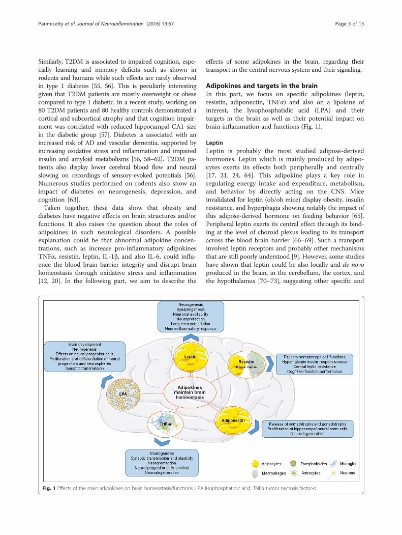
Adipokines and targets in the brainIn this part, we focus on specific adipokines (leptin,resistin, adiponectin, TNFα) and also on a lipokine ofinterest, the lysophosphatidic acid (LPA) and theirtargets in the brain as well as their potential impact onbrain inflammation and functions (Fig. 1).
LeptinLeptin is probably the most studied adipose-derivedhormones. Leptin which is mainly produced by adipo-cytes exerts its effects both peripherally and centrally[17, 21, 24, 64]. This adipokine plays a key role inregulating energy intake and expenditure, metabolism,and behavior by directly acting on the CNS. Miceinvalidated for leptin (ob/ob mice) display obesity, insulinresistance, and hyperphagia showing notably the impact ofthis adipose-derived hormone on feeding behavior [65].Peripheral leptin exerts its central effect through its bind-ing at the level of choroid plexus leading to its transportacross the blood brain barrier [66–69]. Such a transportinvolved leptin receptors and probably other mechanismsthat are still poorly understood [9]. However, some studieshave shown that leptin could be also locally and de novoproduced in the brain, in the cerebellum, the cortex, andthe hypothalamus [70–73], suggesting other specific and
Fig. 1 Effects of the main adipokines on brain homeostasis/functions. LPA lisophosphatidic acid, TNFα tumor necrosis factor-α
Parimisetty et al. Journal of Neuroinflammation (2016) 13:67 Page 3 of 13
local functions for leptin than those previously described.Leptin receptors belong to the family of cytokine recep-tors, and at least five different isoforms have been identi-fied in mouse: Ob-Ra to Ob-Re [65, 74]. In the CNS,leptin receptors (Ob-R or LepR) were first identified inchoroid plexus and in the hypothalamus [75, 76]. Amongall Ob-R isoforms, only the full-length isoform (Ob-Rb)appears to fully transduce the activation signal at least inthe brain and is essential for leptin’s weight-reducingeffects [65, 74]. Ob-Rb is expressed in the hypothalamicnuclei notably in the arcuate nucleus (ARC), the dorsome-dial nucleus (DMH), the paraventricular nucleus (PVN),the ventromedial hypothalamic nucleus (VMH), and thelateral hypothalamic nucleus (LH) [65, 77, 78] but is alsodetected in the neocortex, the hippocampus, the hind-brain (nucleus of the solitary tract), the ventral tegmentalarea, the medulla, and the cerebellum [77, 79–83]. Inaddition, a weaker expression was also detected by in situhybridization in the hippocampus and the thalamus [77].The expression of leptin receptors and leptin mRNAs isdocumented in the mouse brain and notably in the mainneurogenic niches, the subventricular zone of the lateralventricles, and the dentate gyrus of the hippocampus(Allen Brain Atlas [http://www.brain-map.org], [84]). Thiswork clearly illustrates the expression of leptin receptorsin the cortex, along the ventricular walls and also in thehippocampus. Leptin is expressed in the same regions atlower levels. In the hypothalamus, the primary leptintargets are the orexigenic agouti-related peptide (AgRP)neurons and the anorexigenic pro-opiomelanocortin(POMC) neurons that are involved in feeding behavior.Thus, in the CNS, leptin activates anorexigenic POMCneurons through a neural network in the arcuate nucleus[85]. The appetite-stimulating effects of AgRP/NPY areinhibited by leptin in the arcuate nucleus avoiding therelease of orexigenic factors [86, 87]. Furthermore, leptinreceptors were also expressed in glutamatergic andGABAergic neurons [78, 88, 89]. Vong and colleagues(2011) have shown that the main effects of leptin aremediated by GABAergic neurons and only barely by gluta-matergic neurons [88]. However, it was recently demon-strated that glutamate release mediates leptin action onenergy expenditure [89]. We realize now that the effectsof leptin on these different neuronal types and brainnuclei are not so easy to understand as originally thought.In homeostatic conditions, leptin inhibits food intake, andin extra-hypothalamic sites, leptin acts on neurogenesis,synaptogenesis, neuronal excitability, and neuroprotection[9, 90, 91]. Leptin was also shown to improve cognitionand mood in depressed and anxious animal models,notably by improving long-term potentiation [9]. Leptinlevels negatively correlated with the development ofAlzheimer’s disease in lean humans [91, 92], and leptinsignaling seems to be dysregulated in Alzheimer’s disease
brains [93]. Interestingly, there are also positive correlationsbetween plasma levels of leptin and body weight [94, 95].
ResistinResistin (or adipose tissue-specific secretory factor:ADSF or C/EBP-epsilon-regulated myeloid-specific se-creted cysteine-rich protein: XCP1) is a cysteine-richadipose-derived peptide hormone, encoded by the RETNgene, and known for its implication in inflammatoryprocesses [20, 96]. Its expression increases in parallel toadiposity [97–99] and is strongly related to insulin resist-ance in obese rodents [100]. Interestingly, in humans,resistin is mainly expressed and secreted by macro-phages while adipocytes are the main source in rodents[100]. Resistin is known to play a key role in the CNSnotably by regulating pituitary somatotrope cell func-tions [101], affecting hypothalamic and peripheral insu-lin responsiveness, thermogenesis, and feeding behavior,and also by enhancing renal sympathetic nerve activity[102–104]. However, the resistin receptor and the mo-lecular mechanisms sustaining such effects are poorlyunderstood and mainly unexplored until recently. Al-though resistin receptor has not been clearly identified,some potential candidate receptors have been proposedin different cell types such as an isoform of decorin (asmall proteoglycan associated with collagen fibrils) inadipose progenitor cells, tyrosine kinase-like orphanreceptor-1 (ROR1) in 3T3-L1 cells [105] or IGF-1R infibroblast [106]. Nevertheless, it has been shown thatresistin administration modulates or activates severalsignaling pathway involving Gs protein-dependentmechanisms, the adenylate cyclase/cAMP/protein kinaseA pathway, the phosphatidylinositol 3-kinase/Akt path-way, the protein kinase C, and extracellular Ca2+ sig-naling through L-type voltage-sensitive Ca2+ [102, 107].Such puzzling data strongly suggest that resistin couldpotentially interact with different receptors dependingon tissue and cell types. Furthermore, resistin also regu-lates the synthesis and secretion of the pro-inflammatorycytokines TNFα and IL-6 through nuclear factor-κB-dependent pathway in macrophage [108–110]. Recently,Toll-like receptor 4 (TLR-4) receptors were identified aspotential receptor for resistin in the hypothalamus,leading to the activation of JNK and p38/MAPK path-ways [111]. Interestingly, resistin was also reported to beexpressed in the hypothalamus and the cortex and toinactivate hypothalamic neurons [112–114]. In the ratbrain, resistin is de novo produced suggesting specificroles for this local synthesis [114]. Resistin gene expres-sion in the brain of mouse is reported in the cortexalong the walls of the lateral ventricles and also in thehippocampus (Allen Brain Atlas [http://www.brain-map.org], [84]). In rat, traumatic brain injury (TBI)increased resistin mRNA expression in the ipsilateral
Parimisetty et al. Journal of Neuroinflammation (2016) 13:67 Page 4 of 13
cortex without any effects on the contralateral hemi-sphere. However, resistin expression is upregulated afterTBI in the ipsi- and contralateral hippocampus [73].One explanation is that given TBI compromises the in-tegrity of the blood brain barrier, it could result in thechanges in gene expression in the contralateral side ofthe hippocampus by exposing the brain to circulatingfactors of peripheral origin (Brown et al., 2008). Therelatively rapid increase of resistin expression followingTBI (at 12 h post-injury), is in contrast to the delayedupregulation of resistin in hypoxic ischemic mouse brain(>7 days) [115]. Thus, resistin could participate in theacute responses to cerebral damage probably throughinflammatory mechanisms. A recent study suggestedthat resistin was not related to cognitive functionperformance [116].
AdiponectinAdiponectin was first characterized in 1995 in 3T3-L1adipocyte differentiation [117]. It is one of the mostabundant adipokines considering its concentration inplasma relative to many other hormones [118, 119]. Adi-ponectin self-associates into larger structures forminghomotrimers that also self-associate and form hexamersor dodecamers. A globular fraction, named globular adi-ponectin, resulting from the cleavage of the full-lengthmonomer, was also documented [120]. Adiponectin ismainly synthesized and secreted by adipocytes. However,it is now well admitted that adiponectin is expressed atthe mRNA and/or protein level by the placenta, the liver,epithelial cells, osteoblasts, myocytes, and also by pituit-ary cells [114, 119, 121]. Interestingly, some studiesdocumented adiponectin transcript expression in thediencephalon of chicken [114, 122] and in the humanpituitary [121]. In the pituitary, adiponectin could have arole in the release of somatotrophs and gonadotrophs[119]. It also modulates a wide range of metabolic pro-cesses such as body-weight regulation, glucose regulation,insulin sensitivity, lipid catabolism (fatty acid oxidation),endothelial function, and also anti-atherogenic process[119, 123–126]. Such effects are mediated by threedifferent receptor types: adiponectin receptor 1 (Adipo-R1), adiponectin receptor 2 (Adipo-R2), and T-cadherin(CDH13) and involved different signaling pathways in-cluding AMPK, p38-MAPK, JNK, PPAR-α, and NF-kB.These receptors appear to be widely expressed in themammalian brain including mouse, rat, pork and human.Their expression was documented in different brain struc-tures such as the pituitary, the hypothalamus, and incortical and subcortical neurons [97, 119, 121, 127–131].In their review, Thundyil and colleagues (2012) docu-mented adiponectin receptor expression in the centralnervous system showing that Adipo-R1 is mainly expressedin the hypothalamus, the brainstem, and the pituitary gland
while Adipo-R2 seems to be mostly expressed in the cortex.Furthermore, Adipo-R1 is strongly expressed in neuronsand to a lesser extent in astrocytes while Adipo-R2 isfigured to be only weakly expressed in astrocytes and neu-rons [119]. Adiponectin gene expression is widely expressedin the cortex and the hippocampus. Concerning T-cadherinreceptor, it seems to be temporally and spatially expressedin different neuronal populations during axon growth[132]. Furthermore, T-cadherin showed broad expression inthe cerebral cortex, basal ganglia, amygdala, and hippocam-pus in the developing postnatal telencephalon of marmoset(Callithrix jacchus) [133]. In mouse, CDH13 was alsoexpressed by projection neurons within the main andaccessory olfactory bulbs. Interestingly, adiponectindeficiency is associated with exaggerated inflammatoryresponse in critical illness or septic patients [134–136].Recently, Adipo-R1 and Adipo-R2 expression was de-scribed in both U373 MG (human glioblastoma astrocy-toma cell line) and primary human astrocytes [137]. Italso appears that adiponectin induces a pro-inflammatoryresponse in human astrocytes, increasing notably IL-6 andMCP-1 through NF-κB, p38MAPK, and ERK1/2 pathways(Wan et al., 2014). In contrast, adiponectin was describedto inhibit pro-inflammatory signal, notably by suppressingIL-6 release from blood brain barrier (BBB) endothelialcells [138]. It results that adiponectin indirectly modulatesinflammatory signaling across the BBB by negativelymodulating IL-6 and TNFα release. In vitro experiment ofhippocampal neurons reveals that adiponectin exertsneuroprotective effects through AMPK pathway [139].Such neuroprotective effects of adiponectin are furtherreinforced by the fact that knock-out mice for adiponectinexhibit more brain damages after ischemic stroke tocontrols [140]. This neuroprotective action is mediatedthrough an endothelial nitric oxide synthase (eNOS)-dependent mechanism [140].
Tumor necrosis factor αMany pro-inflammatory factors are produced in acti-vated WAT, such as TNFα, IL-1, and PGE2. We chooseto describe in more depth the prototype inflammatorycytokine TNFα. TNFα is a pro-inflammatory adipokinewell-known for its role in chronic peripheral and centralinflammation [9, 141]. TNFα is primarily produced as atransmembrane protein that self-associated into stablehomotrimers [142, 143]. Such homotrimers could becleaved by the TNFα-converting enzyme (TACE, alsocalled ADAM17), allowing the release of secreted formof TNFα [144]. In WAT, TNFα is produced by macro-phages as well as by adipocytes, and its expression is in-creased at the mRNA and protein levels in obese and inT2DM models [145]. TNFα actions are mediated by tworeceptors: TNF-R1 (TNF-RSF1a) and TNF-R2 (TNF-RSF1b). TNF-R1 is expressed in most tissues and can be
Parimisetty et al. Journal of Neuroinflammation (2016) 13:67 Page 5 of 13
fully activated by both the membrane-bound and solubletrimeric forms of TNF, while TNF-R2 is found in alimited cell types including cell of the immune system,oligodendrocytes, and certain neuron subtypes andresponds to the membrane-bound form of the TNFhomotrimer [9]. TNF-R1 and TNF-R2 are also expressedin the cortex, the subventricular zone of the lateralventricle, and the hippocampus (Allen Brain Atlas[http://www.brain-map.org], [84]). In homeostatic condi-tions, the TNFα gene expression is low. However, instress conditions (infection, trauma, pathologies), TNFαlevel can increase dramatically. As most informationregarding TNF signaling is derived from TNF-R1, therole of TNF-R2 is likely underestimated. In rodents,TNFα has been shown to be transported across the BBB,but to be also locally produced by microglia, astrocytesand neurons in the brain [146–148]. In the CNS, TNFαacts through TNF-receptors on neurons and astrocytesregulating a wide range of cellular processes such as cellsurvival [9, 149, 150]. Actually, TNFα exhibits pleio-tropic effects with positive and negative outcomes onthe brain. On the one hand, TNFα is considered as theprototypic inflammatory cytokine and elevated levels ofTNF have been described in many neurodegenerativesituations [151, 152]. For instance, we have demon-strated the role of TNF in chemically induced neurode-generation [153]. On the other hand, inhibition of TNFin inflammatory peripheral diseases induced CNS sideeffects including demyelination and neuropathies, sug-gesting a positive role for TNF maintaining the homeo-stasis in the CNS [154]. It acts on neurogenesis, synaptictransmission, and plasticity [9]. Thus, TNFα was describedfor its neuroprotective roles on hippocampal neurons bysuppressing the accumulation of reactive oxygen species(ROS) and by maintaining intracellular levels of calcium[155]. In addition, it modulates glutamatergic transmission[156]. Furthermore, TNFα favors neural progenitor cellsurvival by mediating anti-apoptotic signals via TNF-R2[157]. In rat, TNFα appears to promote the survival ofstroke-generated hippocampal and striatal neurons [158].In addition, TNFα knock-out mice show cognitive impair-ment (i.e., significant poorer learning, retention, andspatial learning), suggesting a strong role for TNFα onthese mechanisms [159]. However, TNFα also exhibits adark face, as reported in numerous other studies. It isnotably involved in myelin damages [160], in favoringglutamate excitotoxicity [161], in inhibition of long-termpotentiation in Cornu Ammonis area 1 (CA1) and in thedentate gyrus of the rat hippocampus [150, 162, 163] andin decreasing neurogenesis [164, 165].Altogether, these data established that the role of
TNFα is complex. TNFα could exhibit multiple facesexerting neuroprotective versus neurotoxic roles, pro-versus anti-neurogenic effects according to the conditions
(concentrations, physiological, or pathological condi-tions…). Neuroinflammation and metabolic disorderssuch as obesity could act on these mechanisms throughan excess of TNFα secretion.
Lysophosphatidic acidAmong the factors secreted by the adipose tissue, thereare many lipids from the lipokine family such as prosta-glandin E2 (PGE2), anandamide, and also lysophosphati-dic acid (LPA). LPA is a bioactive signaling phospholipidacting on a wide range of biological processes includingcell growth, migration, and morphology [166]. LPA isdetected in several biological fluids and tissues includingthe brain [167]. It is synthesized from different enzym-atic activities involving notably phospholipase A1 andA2, monoacylglycerol kinase, but the main enzyme lead-ing to LPA synthesis is autotaxin [168]. Autotaxin is amultifunctional phosphodiesterase that converts lyso-phospholipids into LPA through its lysophospholipase Dactivity. To date, LPA effects are mediated through fiveG protein coupled receptors. However, additional recep-tors have been identified for their potential responsive-ness to LPA [168–170]. Using knock-out mice for thefive most known LPA receptors (LPA-R), it was shownthat LPA plays key roles on inflammation [171], angio-genesis [172], reproduction [173–175], brain develop-ment, and neurogenesis [176, 177]. Indeed, LPA exertspleomorphic effects on neural progenitor cells from cor-tex, and notably calcium-mediated conductance [178].In the nervous system, neural progenitor cells, neurons,oligodendrocytes, Schwann cells, astrocytes, and micro-glia have been documented for expressing differentsubsets of LPA receptors [168]. It partially explains whyLPA exerts a wide variety of effects on these differentcell types. Thus, LPA can favor proliferation and differ-entiation of neural progenitor cells as shown by treat-ment on ex vivo embryonic brain slice cultures resultingin an increase cell survival and differentiation [179].Furthermore, LPA has been shown to promote prolifera-tion and differentiation in neurospheres [180, 181]. LPAalso displays effect on cell morphology and neurite for-mation in both neural progenitor cells and neurons[168]. It exhibits both cell death and survival propertieson neurons possibly due to differences in LPA concentra-tion or signaling through different receptors [182–184].For instance, it induces apoptosis and necrosis in hippo-campal neurons [182]. LPA also exerts various effects onglial and microglial cells, by modulating intracellularcalcium levels in oligodendrocytes, astrocytes, and micro-glia [168]. It notably favors astrocytes and microglia prolif-eration in vitro [185, 186]. Overexpression of autotaxin inmicroglia and by consequences, increased levels of LPA,protects the cells from an oxidative stress by increasingthe level of catalase [187] and decreases the inflammatory
Parimisetty et al. Journal of Neuroinflammation (2016) 13:67 Page 6 of 13
response at least partially through an upregulation of IL-10 [188]. Interestingly, following brain injury, in humanpostmortem brains, LPA receptors 1–3 and autotaxin areonly weakly expressed while LPA-R2 is increased andautotaxin transcripts are decreased. Such data alsoreinforce the fact that LPA signaling is involved in neuro-trauma [189]. During embryogenesis, LPA-R1 was de-tected in neural progenitors reinforcing a potential role ofLPA/LPA-R1 signaling in neurogenesis [190]. In addition,LPAR-1 knock-out reduces brain cell proliferation, differ-entiation, and cell survival in the mouse dentate gyrus,consequently strongly impairing neurogenesis [176].Autotaxin is widely expressed in the brain of mouse not-ably in neurogenic niches while LPA-R1 displays a lowerand more discrete expression (Allen Brain Atlas [http://www.brain-map.org], [84]).
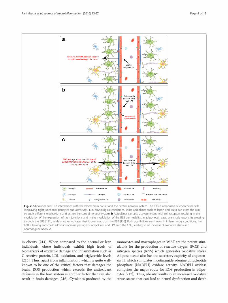
Blood brain barrier, adipokines, inflammation,and oxidative stressThe effects and actions of adipokines on the CNS aredependent on their capacity to interact with the cells ofthe BBB and eventually to enter the CNS (Fig. 2). Asmentioned above about the TNFα, some adipokinesincluding leptin can cross the BBB [9]. Concerningadiponectin, its capacity to cross the BBB is question-able. In one study, the results based on the measurementof radioactive labeled adiponectin and concentration inthe cerebrospinal fluid indicate that adiponectin doesnot cross the BBB but will affect the cells of the BBBand indirectly affect the CNS acting via AMPK pathway[9, 138]. In another work using adiponectin KO miceand recombinant adiponectin, it is shown that adiponec-tin crosses the BBB [191]. As the conclusion, these twopublications proposed opposite results concerning thepossibility of adiponectin to cross the BBB, and moreconclusive results will be needed to clarify this point.The BBB is a key player in the adipokine signaling
from the periphery to the CNS. Inflammation and oxida-tive stress associated with pathologies impact neurode-generation and neurogenesis but also affect the BBB.In this review, we highlighted the striking correlations
between metabolic syndrome and the prevalence ofneurological disorders and dementia including AD. Whiteadipose tissue was not initially envisioned as a source ofinflammatory factors. However, it is now well acceptedthat WAT is a key player in the development of a chroniclow-grade inflammation associated with adiposity andconsequently obesity [192, 193] with elevated productionof pro-inflammatory cytokines, such as TNFα, IL-6, andIL-1 [194, 195]. In contrast, loss of WAT is associatedwith a decrease in inflammation markers [196, 197]. Inter-estingly, chronic and low-grade inflammation has beenproposed to negatively favor neurodegenerative diseasesthrough the disruption of the BBB. Indeed, the blood brain
barrier is a key interface linking systemic inflammation,neuroinflammation, and neurodegeneration [198], inflam-matory factors being a main cause of the BBB disruption[199]. For instance, studies established positive correla-tions between mid-life adiposity in women with disruptionof BBB integrity, showing that overweight/obesity couldfavor the onset of vascular disorders increasing BBB per-meability later in life [200]. In the same line of evidence,rats fed with Western diet, known for promoting diabetesand obesity, display a leakier BBB due to the decreasedexpression of tight junctions [201]. Kanoski and colleagueshave also shown that a primary cerebral target followingBBB disruption is the hippocampus, well-known for itsinvolvement in cognitive processes [201]. This is of pecu-liar interest given that AD patients display hippocampalatrophy and disruption of fronto-hippocampal connec-tions early in the course of the disease [202–204]. This isfurther reinforced by the fact that AD in human androdent models is strongly linked to an increased perme-ability of the BBB [205, 206]. Consequently, the chroniclow-grade inflammation that takes place in obese and dia-betic people could negatively favor brain inflammationand degeneration through BBB disruption.Some interesting links exist between dietary factors
displaying anti-inflammatory properties, inflammation,and disease outcomes. For instance, polyphenols such asflavonoids and curcumin and spices such as cinnamonhave been suggested to decrease pro-inflammatory cyto-kines, inflammation, and cardiovascular diseases andtype 2 diabetes which are known to be risk factors forAD (for a recent review on this topic see ref [207]).
ConclusionsWhile the causal nature of all the processes leading toneurodegeneration has not been definitively established, itis widely accepted that neuroinflammation and oxidativestress responses occur with clinical manifestation of thedisease. In this review, we described the impact of pro-inflammatory adipokines (TNFα and leptin) on brainhomeostasis and functions. In addition, pro-inflammatoryadipokines play a major role in the production of reactiveoxygen species (ROS) [208, 209]. Due to its ability tosecrete adipokines that promote ROS production, WAThas been regarded as an independent factor provokingoxidative stress [210–212]. Exposure to obesity for a longtime in a host system downregulates and depletes theactivity of antioxidant enzymes such as superoxide dis-mutase (SOD), catalase (CAT), and glutathione peroxidase(GPx); these enzymes being found to be significantlylowered compared with healthy persons which in turnlead to the development of obesity-related health prob-lems [213]. In addition to this, levels of vitamin A andlevels of serum antioxidants, such as vitamin E, vitamin C,and β-carotene, as well as glutathione, are also decreased
Parimisetty et al. Journal of Neuroinflammation (2016) 13:67 Page 7 of 13
in obesity [214]. When compared to the normal or leanindividuals, obese individuals exhibit high levels ofbiomarkers of oxidative damage and inflammation such asC-reactive protein, LDL oxidation, and triglyceride levels[215]. Thus, apart from inflammation, which is quite well-known to be one of the critical factors that damages thebrain, ROS production which exceeds the antioxidantdefenses in the host system is another factor that can alsoresult in brain damages [216]. Cytokines produced by the
monocytes and macrophages in WAT are the potent stim-ulators for the production of reactive oxygen (ROS) andnitrogen species (RNS) which generates oxidative stress.Adipose tissue also has the secretory capacity of angioten-sin II, which stimulates nicotinamide adenine dinucleotidephosphate (NADPH) oxidase activity. NADPH oxidasecomprises the major route for ROS production in adipo-cytes [217]). Thus, obesity results in an increased oxidativestress status that can lead to neural dysfunction and death
Fig. 2 Adipokines and LPA interactions with the blood brain barrier and the central nervous system. The BBB is composed of endothelial cells(displaying tight junctions), pericytes and astrocytes. a In physiological conditions, some adipokines such as leptin and TNFα can cross the BBBthrough different mechanisms and act on the central nervous system. b Adipokines can also activate endothelial cell receptors resulting in themodulation of the expression of tight junctions and in the modulation of the BBB permeability. In adiponectin case, one study reports its crossingthrough the BBB [191], while another indicates that it does not cross the BBB [138]. Both possibilities are shown. In inflammatory conditions, theBBB is leaking and could allow an increase passage of adipokines and LPA into the CNS, leading to an increase of oxidative stress andneurodegeneration (c)
Parimisetty et al. Journal of Neuroinflammation (2016) 13:67 Page 8 of 13

[218, 219]. It has been reported that obesity may inducesystemic oxidative stress and, in turn, oxidative stress isassociated with an irregular production of adipokines,which contributes to the development of the metabolicsyndrome [220]. In parallel, oxidative stress is implicatedin numerous neurological diseases and/or disorders suchas AD, PD, amyotrophic lateral sclerosis (ALS), multiplesclerosis (MS), cerebral ischemia/reperfusion injury, andTBI, promoting neurodegeneration [221]. An increasingnumber of studies using in vitro models and knock-outanimals demonstrate that oxidative stress disrupts theBBB permeability [221–223].Taken together, these data suggest that in pathological
conditions, adipokines released by WAT promote inflam-mation and ROS production that may disrupt the BBBpermeability and could directly or indirectly act on diff-erent brain structures, the hippocampus being one of themost sensitive areas. It could explain why metabolicsyndrome is associated with hippocampus atrophy and anincrease risk to develop dementia such as AD. One mainissue in people suffering from metabolic syndrome shouldbe to struggle against inflammation and reduce oxidativestress in order to decrease their potential effects on brainneurodegeneration and their adverse effects.
Competing interestsThe authors declare that they have no competing interests.
Authors’ contributionsCLH and ND contributed equally to this work. AP carried out the writing ofthe study. ACD and ND contributed to the figures. CLH conceived the study.CLH and ND participated in its coordination and helped to draft themanuscript. AP, ACD, RA, RP, ND, and CLH contributed to the revision andedition of the study. All authors read and approved the final manuscript.
FundingThis work was supported by the grants from Conseil Régional de LaRéunion and Europe (CPER/FEDER). ACD is funded by a fellowship fromUniversity of La Réunion. AP was funded by a fellowship from “ConseilRégional de La Réunion.”
Author details1Université de La Réunion, UMR 1188, Sainte-Clotilde F-97490, France.2Inserm, UMR 1188 Diabète athérothrombose Thérapies Réunion OcéanIndien (DéTROI), plateforme CYROI, Sainte-Clotilde F-97490, France.3Lebanese University, Faculty of Sciences, Beirut, Lebanon. 4Apoptosis andCell Death Research Lab, School of Biosciences and Technology, VelloreInstitute of Technology University, Vellore, India.
Received: 7 December 2015 Accepted: 15 March 2016
References1. Awada R, Parimisetty A, Lefebvre d’Hellencourt C. Influence of obesity on
neurodegenerative diseases. Neurodegener Dis. 2013;Chapter 16:381–401.2. World Health Organisation W: obesity and overweight. http://www.who.int/
mediacentre/factsheets/fs311/en/. Accessed 22 Mar 2016.3. World Health Organisation W: diabetes. http://www.who.int/mediacentre/
factsheets/fs312/en/. Accessed 22 Mar 2016.4. Alberti KG, Zimmet PZ. Definition, diagnosis and classification of
diabetes mellitus and its complications. Part 1: diagnosis andclassification of diabetes mellitus provisional report of a WHOconsultation. Diabet Med. 1998;15:539–53.
5. Roglic G, Unwin N, Bennett PH, Mathers C, Tuomilehto J, Nag S, et al. Theburden of mortality attributable to diabetes: realistic estimates for the year2000. Diabetes Care. 2005;28:2130–5.
6. Reisberg B, Burns A, Brodaty H, Eastwood R, Rossor M, Sartorius N,et al. Diagnosis of Alzheimer’s disease. Report of an InternationalPsychogeriatric Association Special Meeting Work Group under thecosponsorship of Alzheimer’s Disease International, the EuropeanFederation of Neurological Societies, the World Health Organization,and the World Psychiatric Association. Int Psychogeriatr. 1997;9 Suppl 1:11–38.
7. World Health Organisation W: dementia. http://www.who.int/mediacentre/factsheets/fs362/en/. Accessed 22 Mar 2016.
8. Nguyen S, Major K, Demonet JF, Smith C, Rubli E, Humbert M, et al.[Diabetes and dementia: the dangerous liaisons?]. Rev Med Suisse. 2014;2090-2092(10):2094–6.
9. Arnoldussen IA, Kiliaan AJ, Gustafson DR. Obesity and dementia: adipokinesinteract with the brain. Eur Neuropsychopharmacol. 2014;24:1982-1999.
10. Kiliaan AJ, Arnoldussen IA, Gustafson DR. Adipokines: a link between obesityand dementia? Lancet Neurol. 2014;13:913–23.
11. Gustafson DR, Backman K, Waern M, Ostling S, Guo X, Zandi P, et al.Adiposity indicators and dementia over 32 years in Sweden. Neurology.2009;73:1559–66.
12. Letra L, Santana I, Seica R. Obesity as a risk factor for Alzheimer’s disease:the role of adipocytokines. Metab Brain Dis. 2014;29:563–8.
13. Beydoun MA, Beydoun HA, Wang Y. Obesity and central obesity as riskfactors for incident dementia and its subtypes: a systematic review andmeta-analysis. Obes Rev. 2008;9:204–18.
14. Whitmer RA, Gustafson DR, Barrett-Connor E, Haan MN, Gunderson EP, YaffeK. Central obesity and increased risk of dementia more than three decadeslater. Neurology. 2008;71:1057–64.
15. Qizilbash N, Gregson J, Johnson ME, Pearce N, Douglas I, Wing K, et al. BMIand risk of dementia in two million people over two decades: aretrospective cohort study. Lancet Diabetes Endocrinol. 2015;3:431–6.
16. Tolppanen AM, Ngandu T, Kareholt I, Laatikainen T, Rusanen M, Soininen H, etal. Midlife and late-life body mass index and late-life dementia: results from aprospective population-based cohort. J Alzheimers Dis. 2014;38:201–9.
17. Adamczak M, Wiecek A. The adipose tissue as an endocrine organ. SeminNephrol. 2013;33:2–13.
18. Lehr S, Hartwig S, Sell H. Adipokines: a treasure trove for the discovery ofbiomarkers for metabolic disorders. Proteomics Clin Appl. 2012;6:91–101.
19. Chaldakov GN. The adipobiology of disease. In: Immunology, endocrine &metabolic agents in medicinal chemistry (formerly current medicinalchemistry—immunology, endocrine and metabolic agents), vol. Volume 7,Number 2, April 2007. 2007. p. 105–5.
20. Ouchi N, Parker JL, Lugus JJ, Walsh K. Adipokines in inflammation andmetabolic disease. Nat Rev Immunol. 2011;11:85–97.
21. Trayhurn P, Wood IS. Adipokines: inflammation and the pleiotropic role ofwhite adipose tissue. Br J Nutr. 2004;92:347–55.
22. Maeda N, Shimomura I, Kishida K, Nishizawa H, Matsuda M, Nagaretani H, etal. Diet-induced insulin resistance in mice lacking adiponectin/ACRP30. NatMed. 2002;8:731–7.
23. Xu A, Wang Y, Keshaw H, Xu LY, Lam KS, Cooper GJ. The fat-derivedhormone adiponectin alleviates alcoholic and nonalcoholic fatty liverdiseases in mice. J Clin Invest. 2003;112:91–100.
24. Trayhurn P, Beattie JH. Physiological role of adipose tissue: whiteadipose tissue as an endocrine and secretory organ. Proc Nutr Soc.2001;60:329–39.
25. Leal Vde O, Mafra D. Adipokines in obesity. Clin Chim Acta. 2013;419:87–94.26. Ahima RS, Saper CB, Flier JS, Elmquist JK. Leptin regulation of
neuroendocrine systems. Front Neuroendocrinol. 2000;21:263–307.27. Bluher M, Mantzoros CS. From leptin to other adipokines in health and
disease: facts and expectations at the beginning of the 21st century.Metabolism. 2015;64:131–45.
28. Bluher M. Adipokines—removing road blocks to obesity and diabetestherapy. Mol Metab. 2014;3:230–40.
29. Maury E, Brichard SM. Adipokine dysregulation, adipose tissue inflammationand metabolic syndrome. Mol Cell Endocrinol. 2010;314:1–16.
30. Ku IA, Farzaneh-Far R, Vittinghoff E, Zhang MH, Na B, Whooley MA.Association of low leptin with cardiovascular events and mortality inpatients with stable coronary artery disease: the Heart and Soul Study.Atherosclerosis. 2011;217:503–8.
Parimisetty et al. Journal of Neuroinflammation (2016) 13:67 Page 9 of 13

31. Spranger J, Kroke A, Mohlig M, Bergmann MM, Ristow M, Boeing H, etal. Adiponectin and protection against type 2 diabetes mellitus. Lancet.2003;361:226–8.
32. Herder C, Baumert J, Thorand B, Koenig W, de Jager W, Meisinger C, et al.Chemokines as risk factors for type 2 diabetes: results from the MONICA/KORA Augsburg study, 1984-2002. Diabetologia. 2006;49:921–9.
33. Lee EB. Obesity, leptin, and Alzheimer’s disease. Ann N Y Acad Sci.2011;1243:15–29.
34. Gustafson D. Adiposity indices and dementia. Lancet Neurol. 2006;5:713–20.35. Zhang P, Tian B. Metabolic syndrome: an important risk factor for
Parkinson’s disease. Oxid Med Cell Longev. 2014;2014:729194.36. Gianfrancesco MA, Acuna B, Shen L, Briggs FB, Quach H, Bellesis KH, et al.
Obesity during childhood and adolescence increases susceptibility tomultiple sclerosis after accounting for established genetic andenvironmental risk factors. Obes Res Clin Pract. 2014;8:e435–47.
37. Versini M, Jeandel PY, Rosenthal E, Shoenfeld Y. Obesity in autoimmunediseases: not a passive bystander. Autoimmun Rev. 2014;13:981–1000.
38. Gustafson D, Rothenberg E, Blennow K, Steen B, Skoog I. An 18-year follow-upof overweight and risk of Alzheimer disease. Arch Intern Med. 2003;163:1524–8.
39. Nguyen JC, Killcross AS, Jenkins TA. Obesity and cognitive decline: role ofinflammation and vascular changes. Front Neurosci. 2014;8:375.
40. Bruce-Keller AJ, Keller JN, Morrison CD. Obesity and vulnerability of the CNS.Biochim Biophys Acta. 1792;2009:395–400.
41. Taki Y, Kinomura S, Sato K, Inoue K, Goto R, Okada K, et al. Relationshipbetween body mass index and gray matter volume in 1,428 healthyindividuals. Obesity (Silver Spring). 2008;16:119–24.
42. Ward MA, Carlsson CM, Trivedi MA, Sager MA, Johnson SC. The effect ofbody mass index on global brain volume in middle-aged adults: a crosssectional study. BMC Neurol. 2005;5:23.
43. Debette S, Beiser A, Hoffmann U, Decarli C, O’Donnell CJ, Massaro JM, et al.Visceral fat is associated with lower brain volume in healthy middle-agedadults. Ann Neurol. 2010;68:136–44.
44. Gustafson D, Lissner L, Bengtsson C, Bjorkelund C, Skoog I. A 24-yearfollow-up of body mass index and cerebral atrophy. Neurology.2004;63:1876–81.
45. Raji CA, Ho AJ, Parikshak NN, Becker JT, Lopez OL, Kuller LH, et al. Brainstructure and obesity. Hum Brain Mapp. 2010;31:353–64.
46. Ho AJ, Stein JL, Hua X, Lee S, Hibar DP, Leow AD, et al. A commonly carriedallele of the obesity-related FTO gene is associated with reduced brainvolume in the healthy elderly. Proc Natl Acad Sci U S A. 2010;107:8404–9.
47. Cournot M, Marquie JC, Ansiau D, Martinaud C, Fonds H, Ferrieres J, et al.Relation between body mass index and cognitive function in healthymiddle-aged men and women. Neurology. 2006;67:1208–14.
48. Lokken KL, Boeka AG, Austin HM, Gunstad J, Harmon CM. Evidence ofexecutive dysfunction in extremely obese adolescents: a pilot study. SurgObes Relat Dis. 2009;5:547–52.
49. Sabia S, Kivimaki M, Shipley MJ, Marmot MG, Singh-Manoux A. Body massindex over the adult life course and cognition in late midlife: the WhitehallII cohort study. Am J Clin Nutr. 2009;89:601–7.
50. McNeilly AD, Williamson R, Sutherland C, Balfour DJ, Stewart CA. High fatfeeding promotes simultaneous decline in insulin sensitivity and cognitiveperformance in a delayed matching and non-matching to position task.Behav Brain Res. 2011;217:134–41.
51. Murray AJ, Knight NS, Cochlin LE, McAleese S, Deacon RM, Rawlins JN, et al.Deterioration of physical performance and cognitive function in rats withshort-term high-fat feeding. FASEB J. 2009;23:4353–60.
52. Morris MJ, Beilharz JE, Maniam J, Reichelt AC, Westbrook RF. Why is obesitysuch a problem in the 21st century? The intersection of palatable food,cues and reward pathways, stress, and cognition. Neurosci Biobehav Rev.2015;58:36-45.
53. Lynch CM, Kinzenbaw DA, Chen X, Zhan S, Mezzetti E, Filosa J, et al.Nox2-derived superoxide contributes to cerebral vascular dysfunction indiet-induced obesity. Stroke. 2013;44:3195–201.
54. Li W, Prakash R, Chawla D, Du W, Didion SP, Filosa JA, et al. Early effects ofhigh-fat diet on neurovascular function and focal ischemic brain injury. AmJ Physiol Regul Integr Comp Physiol. 2013;304:R1001–8.
55. Zhou H, Liu J, Ren L, Liu W, Xing Q, Men L, et al. Relationship between[corrected] spatial memory in diabetic rats and protein kinase Cgamma,caveolin-1 in the hippocampus and neuroprotective effect of catalpol. ChinMed J (Engl). 2014;127:916–23.
56. McCrimmon RJ, Ryan CM, Frier BM. Diabetes and cognitive dysfunction.Lancet. 2012;379:2291–9.
57. Zhang YW, Zhang JQ, Liu C, Wei P, Zhang X, Yuan QY, et al. Memorydysfunction in type 2 diabetes mellitus correlates with reduced hippocampalCA1 and subiculum volumes. Chin Med J (Engl). 2015;128:465–71.
58. Biessels GJ, Deary IJ, Ryan CM. Cognition and diabetes: a lifespanperspective. Lancet Neurol. 2008;7:184–90.
59. Haan MN. Therapy insight: type 2 diabetes mellitus and the risk oflate-onset Alzheimer’s disease. Nat Clin Pract Neurol. 2006;2:159–66.
60. Cheng G, Huang C, Deng H, Wang H. Diabetes as a risk factor for dementiaand mild cognitive impairment: a meta-analysis of longitudinal studies.Intern Med J. 2012;42:484–91.
61. Whitmer RA. Type 2 diabetes and risk of cognitive impairment anddementia. Curr Neurol Neurosci Rep. 2007;7:373–80.
62. MacKnight C, Rockwood K, Awalt E, McDowell I. Diabetes mellitus andthe risk of dementia, Alzheimer’s disease and vascular cognitiveimpairment in the Canadian Study of Health and Aging. DementGeriatr Cogn Disord. 2002;14:77–83.
63. Ho N, Sommers MS, Lucki I. Effects of diabetes on hippocampal neurogenesis:links to cognition and depression. Neurosci Biobehav Rev. 2013;37:1346–62.
64. Zhang Y, Proenca R, Maffei M, Barone M, Leopold L, Friedman JM.Positional cloning of the mouse obese gene and its humanhomologue. Nature. 1994;372:425–32.
65. Friedman JM, Halaas JL. Leptin and the regulation of body weight inmammals. Nature. 1998;395:763–70.
66. Banks WA, Kastin AJ, Huang W, Jaspan JB, Maness LM. Leptin enters the brainby a saturable system independent of insulin. Peptides. 1996;17:305–11.
67. Banks WA, Clever CM, Farrell CL. Partial saturation and regional variation inthe blood-to-brain transport of leptin in normal weight mice. Am J PhysiolEndocrinol Metab. 2000;278:E1158–65.
68. Devos R, Richards JG, Campfield LA, Tartaglia LA, Guisez Y, van der HeydenJ, et al. OB protein binds specifically to the choroid plexus of mice and rats.Proc Natl Acad Sci U S A. 1996;93:5668–73.
69. Zlokovic BV, Jovanovic S, Miao W, Samara S, Verma S, Farrell CL. Differentialregulation of leptin transport by the choroid plexus and blood-brain barrierand high affinity transport systems for entry into hypothalamus and acrossthe blood-cerebrospinal fluid barrier. Endocrinology. 2000;141:1434–41.
70. Wilkinson M, Morash B, Ur E. The brain is a source of leptin. Front Horm Res.2000;26:106–25.
71. Morash B, Li A, Murphy PR, Wilkinson M, Ur E. Leptin gene expression in thebrain and pituitary gland. Endocrinology. 1999;140:5995–8.
72. Brown R, Imran SA, Belsham DD, Ur E, Wilkinson M. Adipokine geneexpression in a novel hypothalamic neuronal cell line: resistin-dependentregulation of fasting-induced adipose factor and SOCS-3.Neuroendocrinology. 2007;85:232–41.
73. Brown R, Thompson HJ, Imran SA, Ur E, Wilkinson M. Traumatic brain injuryinduces adipokine gene expression in rat brain. Neurosci Lett. 2008;432:73–8.
74. Gorska E, Popko K, Stelmaszczyk-Emmel A, Ciepiela O, Kucharska A, Wasik M.Leptin receptors. Eur J Med Res. 2010;15 Suppl 2:50–4.
75. Tartaglia LA. The leptin receptor. J Biol Chem. 1997;272:6093–6.76. Tartaglia LA, Dembski M, Weng X, Deng N, Culpepper J, Devos R, et al.
Identification and expression cloning of a leptin receptor, OB-R. Cell.1995;83:1263–71.
77. Mercer JG, Hoggard N, Williams LM, Lawrence CB, Hannah LT, Trayhurn P.Localization of leptin receptor mRNA and the long form splice variant(Ob-Rb) in mouse hypothalamus and adjacent brain regions by in situhybridization. FEBS Lett. 1996;387:113–6.
78. Yi CX, Meyer CW, Jastroch M. Leptin action in the brain: How (and when) itmakes fat burn. Mol Metab. 2013;2:63–4.
79. Burguera B, Couce ME, Long J, Lamsam J, Laakso K, Jensen MD, et al. Thelong form of the leptin receptor (OB-Rb) is widely expressed in the humanbrain. Neuroendocrinology. 2000;71:187–95.
80. Savioz A, Charnay Y, Huguenin C, Graviou C, Greggio B, Bouras C. Expressionof leptin receptor mRNA (long form splice variant) in the humancerebellum. Neuroreport. 1997;8:3123–6.
81. Shanley LJ, O’Malley D, Irving AJ, Ashford ML, Harvey J. Leptin inhibitsepileptiform-like activity in rat hippocampal neurones via PI 3-kinase-drivenactivation of BK channels. J Physiol. 2002;545:933–44.
82. Scott MM, Williams KW, Rossi J, Lee CE, Elmquist JK. Leptin receptorexpression in hindbrain Glp-1 neurons regulates food intake and energybalance in mice. J Clin Invest. 2011;121:2413–21.
Parimisetty et al. Journal of Neuroinflammation (2016) 13:67 Page 10 of 13
83. Scott MM, Lachey JL, Sternson SM, Lee CE, Elias CF, Friedman JM, et al.Leptin targets in the mouse brain. J Comp Neurol. 2009;514:518–32.
84. Lein ES, Hawrylycz MJ, Ao N, Ayres M, Bensinger A, Bernard A, et al.Genome-wide atlas of gene expression in the adult mouse brain. Nature.2007;445:168–76.
85. Cowley MA, Smart JL, Rubinstein M, Cerdan MG, Diano S, Horvath TL, et al.Leptin activates anorexigenic POMC neurons through a neural network inthe arcuate nucleus. Nature. 2001;411:480–4.
86. Baskin DG, Hahn TM, Schwartz MW. Leptin sensitive neurons in thehypothalamus. Horm Metab Res. 1999;31:345–50.
87. Enriori PJ, Evans AE, Sinnayah P, Jobst EE, Tonelli-Lemos L, Billes SK, et al.Diet-induced obesity causes severe but reversible leptin resistance inarcuate melanocortin neurons. Cell Metab. 2007;5:181–94.
88. Vong L, Ye C, Yang Z, Choi B, Chua Jr S, Lowell BB. Leptin action onGABAergic neurons prevents obesity and reduces inhibitory tone to POMCneurons. Neuron. 2011;71:142–54.
89. Xu Y, Kim ER, Zhao R, Myers Jr MG, Munzberg H, Tong Q. Glutamaterelease mediates leptin action on energy expenditure. Mol Metab.2013;2:109–15.
90. Bouret SG. Neurodevelopmental actions of leptin. Brain Res. 2010;1350:2–9.91. Paz-Filho G, Wong ML, Licinio J. The procognitive effects of leptin in the
brain and their clinical implications. Int J Clin Pract. 2010;64:1808–12.92. Paz-Filho G, Wong ML, Licinio J. Leptin levels and Alzheimer disease. JAMA.
2010;303:1478. author reply 1478-1479.93. Bonda DJ, Stone JG, Torres SL, Siedlak SL, Perry G, Kryscio R, et al.
Dysregulation of leptin signaling in Alzheimer disease: evidence forneuronal leptin resistance. J Neurochem. 2014;128:162–72.
94. Fleisch AF, Agarwal N, Roberts MD, Han JC, Theim KR, Vexler A, et al.Influence of serum leptin on weight and body fat growth in children athigh risk for adult obesity. J Clin Endocrinol Metab. 2007;92:948–54.
95. Salbe AD, Weyer C, Lindsay RS, Ravussin E, Tataranni PA. Assessing riskfactors for obesity between childhood and adolescence: I. Birth weight,childhood adiposity, parental obesity, insulin, and leptin. Pediatrics. 2002;110:299–306.
96. Wang H, Chu WS, Hemphill C, Elbein SC. Human resistin gene: molecularscanning and evaluation of association with insulin sensitivity and type 2diabetes in Caucasians. J Clin Endocrinol Metab. 2002;87:2520–4.
97. Degawa-Yamauchi M, Bovenkerk JE, Juliar BE, Watson W, Kerr K, Jones R, etal. Serum resistin (FIZZ3) protein is increased in obese humans. J ClinEndocrinol Metab. 2003;88:5452–5.
98. Lee JH, Bullen Jr JW, Stoyneva VL, Mantzoros CS. Circulating resistinin lean, obese, and insulin-resistant mouse models: lack of associationwith insulinemia and glycemia. Am J Physiol Endocrinol Metab. 2005;288:E625–32.
99. Vendrell J, Broch M, Vilarrasa N, Molina A, Gomez JM, Gutierrez C, et al.Resistin, adiponectin, ghrelin, leptin, and proinflammatory cytokines:relationships in obesity. Obes Res. 2004;12:962–71.
100. Park HK, Ahima RS. Resistin in rodents and humans. Diabetes Metab J.2013;37:404–14.
101. Broglio C, Gómez A, Durán E, Ocaña FM, Jiménez-Moya F, Rodríguez F, et al.Hallmarks of a common forebrain vertebrate plan: specialized pallial areasfor spatial, temporal and emotional memory in actinopterygian fish. BrainRes Bull. 2005;66:277–81.
102. Kosari S, Camera DM, Hawley JA, Stebbing M, Badoer E. ERK1/2 in thebrain mediates the effects of central resistin on reducingthermogenesis in brown adipose tissue. Int J Physiol PathophysiolPharmacol. 2013;5:184–9.
103. Kosari S, Rathner JA, Badoer E. Central resistin enhances renal sympatheticnerve activity via phosphatidylinositol 3-kinase but reduces the activity tobrown adipose tissue via extracellular signal-regulated kinase 1/2.J Neuroendocrinol. 2012;24:1432–9.
104. Yi CX, Tschop MH. Brain-gut-adipose-tissue communication pathways at aglance. Dis Model Mech. 2012;5:583–7.
105. Sanchez-Solana B, Laborda J, Baladron V. Mouse resistin modulatesadipogenesis and glucose uptake in 3T3-L1 preadipocytes through theROR1 receptor. Mol Endocrinol. 2012;26:110–27.
106. Böstrom EA, Svensson M, Andersson S, Jonsson IM, Ekwall AK, Eisler T, et al.Resistin and insulin/insulin-like growth factor signaling in rheumatoidarthritis. Arthritis Rheum. 2011;63:2894–904.
107. Rodriguez-Pacheco F, Vazquez-Martinez R, Martinez-Fuentes AJ, Pulido MR,Gahete MD, Vaudry H, et al. Resistin regulates pituitary somatotrope cell
function through the activation of multiple signaling pathways.Endocrinology. 2009;150:4643–52.
108. Bokarewa M, Nagaev I, Dahlberg L, Smith U, Tarkowski A. Resistin, anadipokine with potent proinflammatory properties. J Immunol. 2005;174:5789–95.
109. Olefsky JM, Glass CK. Macrophages, inflammation, and insulin resistance.Annu Rev Physiol. 2010;72:219–46.
110. Silswal N, Singh AK, Aruna B, Mukhopadhyay S, Ghosh S, Ehtesham NZ.Human resistin stimulates the pro-inflammatory cytokines TNF-alpha andIL-12 in macrophages by NF-kappaB-dependent pathway. Biochem BiophysRes Commun. 2005;334:1092–101.
111. Benomar Y, Gertler A, De Lacy P, Crepin D, Ould Hamouda H, Riffault L, etal. Central resistin overexposure induces insulin resistance throughToll-like receptor 4. Diabetes. 2013;62:102–14.
112. Brown R, Wiesner G, Ur E, Wilkinson M. Pituitary resistin gene expression isupregulated in vitro and in vivo by dexamethasone but is unaffected byrosiglitazone. Neuroendocrinology. 2005;81:41–8.
113. Morash BA, Willkinson D, Ur E, Wilkinson M. Resistin expression andregulation in mouse pituitary. FEBS Lett. 2002;526:26–30.
114. Wilkinson M, Brown R, Imran SA, Ur E. Adipokine gene expression in brainand pituitary gland. Neuroendocrinology. 2007;86:191–209.
115. Wiesner G, Brown RE, Robertson GS, Imran SA, Ur E, Wilkinson M. Increasedexpression of the adipokine genes resistin and fasting-induced adiposefactor in hypoxic/ischaemic mouse brain. Neuroreport. 2006;17:1195–8.
116. Miralbell J, Lopez-Cancio E, Lopez-Oloriz J, Arenillas JF, Barrios M,Soriano-Raya JJ, et al. Cognitive patterns in relation to biomarkers ofcerebrovascular disease and vascular risk factors. Cerebrovasc Dis. 2013;36:98–105.
117. Scherer PE, Williams S, Fogliano M, Baldini G, Lodish HF. A novel serumprotein similar to C1q, produced exclusively in adipocytes. J Biol Chem.1995;270:26746–9.
118. Matsuzawa Y. Adiponectin: identification, physiology and clinical relevancein metabolic and vascular disease. Atheroscler Suppl. 2005;6:7–14.
119. Thundyil J, Pavlovski D, Sobey CG, Arumugam TV. Adiponectin receptorsignalling in the brain. Br J Pharmacol. 2012;165:313–27.
120. Waki H, Yamauchi T, Kamon J, Ito Y, Uchida S, Kita S, et al. Impairedmultimerization of human adiponectin mutants associated with diabetes.Molecular structure and multimer formation of adiponectin. J Biol Chem.2003;278:40352–63.
121. Psilopanagioti A, Papadaki H, Kranioti EF, Alexandrides TK, Varakis JN.Expression of adiponectin and adiponectin receptors in human pituitarygland and brain. Neuroendocrinology. 2009;89:38–47.
122. Maddineni S, Metzger S, Ocon O, Hendricks 3rd G, Ramachandran R.Adiponectin gene is expressed in multiple tissues in the chicken: fooddeprivation influences adiponectin messenger ribonucleic acid expression.Endocrinology. 2005;146:4250–6.
123. Berg AH, Combs TP, Scherer PE. ACRP30/adiponectin: an adipokine regulatingglucose and lipid metabolism. Trends Endocrinol Metab. 2002;13:84–9.
124. Okamoto Y, Kihara S, Ouchi N, Nishida M, Arita Y, Kumada M, et al.Adiponectin reduces atherosclerosis in apolipoprotein E-deficient mice.Circulation. 2002;106:2767–70.
125. Stefan N, Stumvoll M. Adiponectin—its role in metabolism and beyond.Horm Metab Res. 2002;34:469–74.
126. Whitehead JP, Richards AA, Hickman IJ, Macdonald GA, Prins JB.Adiponectin—a key adipokine in the metabolic syndrome. Diabetes ObesMetab. 2006;8:264–80.
127. Fry M, Smith PM, Hoyda TD, Duncan M, Ahima RS, Sharkey KA, et al. Areapostrema neurons are modulated by the adipocyte hormone adiponectin.J Neurosci. 2006;26:9695–702.
128. Hoyda TD, Fry M, Ahima RS, Ferguson AV. Adiponectin selectively inhibitsoxytocin neurons of the paraventricular nucleus of the hypothalamus.J Physiol. 2007;585:805–16.
129. Thundyil J, Tang SC, Okun E, Shah K, Karamyan VT, Li YI, et al. Evidence thatadiponectin receptor 1 activation exacerbates ischemic neuronal death. ExpTransl Stroke Med. 2010;2:15.
130. Repunte-Canonigo V, Berton F, Cottone P, Reifel-Miller A, Roberts AJ,Morales M, et al. A potential role for adiponectin receptor 2 (AdipoR2) inthe regulation of alcohol intake. Brain Res. 2010;1339:11–7.
131. Yamauchi T, Kamon J, Ito Y, Tsuchida A, Yokomizo T, Kita S, et al. Cloning ofadiponectin receptors that mediate antidiabetic metabolic effects. Nature.2003;423:762–9.
Parimisetty et al. Journal of Neuroinflammation (2016) 13:67 Page 11 of 13
132. Ranscht B, Dours-Zimmermann MT. T-cadherin, a novel cadherin celladhesion molecule in the nervous system lacks the conserved cytoplasmicregion. Neuron. 1991;7:391–402.
133. Matsunaga E, Nambu S, Oka M, Iriki A. Differential cadherin expression inthe developing postnatal telencephalon of a New World monkey. J CompNeurol. 2013;521:4027–60.
134. Venkatesh B, Hickman I, Nisbet J, Cohen J, Prins J. Changes in serumadiponectin concentrations in critical illness: a preliminary investigation.Crit Care. 2009;13:R105.
135. Hillenbrand A, Knippschild U, Weiss M, Schrezenmeier H, Henne-BrunsD, Huber-Lang M, et al. Sepsis induced changes of adipokines andcytokines—septic patients compared to morbidly obese patients. BMCSurg. 2010;10:26.
136. Hillenbrand A, Weiss M, Knippschild U, Wolf AM, Huber-Lang M.Sepsis-induced adipokine change with regard to insulin resistance.Int J Inflam. 2012;2012:972368.
137. Wan Z, Mah D, Simtchouk S, Klegeris A, Little JP. Globular adiponectininduces a pro-inflammatory response in human astrocytic cells. BiochemBiophys Res Commun. 2014;446:37–42.
138. Spranger J, Verma S, Gohring I, Bobbert T, Seifert J, Sindler AL, et al.Adiponectin does not cross the blood-brain barrier but modifies cytokineexpression of brain endothelial cells. Diabetes. 2006;55:141–7.
139. Qiu G, Wan R, Hu J, Mattson MP, Spangler E, Liu S, et al. Adiponectinprotects rat hippocampal neurons against excitotoxicity. Age (Dordr).2011;33:155–65.
140. Nishimura M, Izumiya Y, Higuchi A, Shibata R, Qiu J, Kudo C, et al.Adiponectin prevents cerebral ischemic injury through endothelial nitricoxide synthase dependent mechanisms. Circulation. 2008;117:216–23.
141. Thaler JP, Yi CX, Schur EA, Guyenet SJ, Hwang BH, Dietrich MO, et al.Obesity is associated with hypothalamic injury in rodents and humans.J Clin Invest. 2012;122:153–62.
142. Kriegler M, Perez C, DeFay K, Albert I, Lu SD. A novel form of TNF/cachectinis a cell surface cytotoxic transmembrane protein: ramifications for thecomplex physiology of TNF. Cell. 1988;53:45–53.
143. Tang P, Hung MC, Klostergaard J. TNF cytotoxicity: effects of HER-2/neuexpression and inhibitors of ADP-ribosylation. Lymphokine Cytokine Res.1994;13:117–23.
144. Black RA, Rauch CT, Kozlosky CJ, Peschon JJ, Slack JL, Wolfson MF, et al.A metalloproteinase disintegrin that releases tumour-necrosis factor-alphafrom cells. Nature. 1997;385:729–33.
145. Hotamisligil GS, Shargill NS, Spiegelman BM. Adipose expression of tumornecrosis factor-alpha: direct role in obesity-linked insulin resistance. Science.1993;259:87–91.
146. Morganti-Kossman MC, Lenzlinger PM, Hans V, Stahel P, Csuka E, AmmannE, et al. Production of cytokines following brain injury: beneficial anddeleterious for the damaged tissue. Mol Psychiatry. 1997;2:133–6.
147. Chung IY, Benveniste EN. Tumor necrosis factor-alpha production byastrocytes. Induction by lipopolysaccharide, IFN-gamma, and IL-1 beta.J Immunol. 1990;144:2999–3007.
148. Lieberman AP, Pitha PM, Shin HS, Shin ML. Production of tumor necrosisfactor and other cytokines by astrocytes stimulated with lipopolysaccharideor a neurotropic virus. Proc Natl Acad Sci U S A. 1989;86:6348–52.
149. Montgomery SL, Bowers WJ. Tumor necrosis factor-alpha and the roles itplays in homeostatic and degenerative processes within the central nervoussystem. J Neuroimmune Pharmacol. 2012;7:42–59.
150. Pickering M, Cumiskey D, O’Connor JJ. Actions of TNF-alpha onglutamatergic synaptic transmission in the central nervous system. ExpPhysiol. 2005;90:663–70.
151. Clark IA, Vissel B. A neurologist’s guide to TNF biology and to the principlesbehind the therapeutic removal of excess TNF in disease. Neural Plast. 2015;2015:358263.
152. Fischer R, Maier O. Interrelation of oxidative stress and inflammation inneurodegenerative disease: role of TNF. Oxid Med Cell Longev. 2015;2015:610813.
153. Harry GJ, Funk JA, Lefebvre d’Hellencourt C, McPherson CA, Aoyama M. The type1 interleukin 1 receptor is not required for the death of murine hippocampaldentate granule cells and microglia activation. Brain Res. 2008;1194:8–20.
154. Probert L. TNF and its receptors in the CNS: the essential, the desirable andthe deleterious effects. Neuroscience. 2015;302:2–22.
155. Barger SW, Horster D, Furukawa K, Goodman Y, Krieglstein J, Mattson MP.Tumor necrosis factors alpha and beta protect neurons against amyloid
beta-peptide toxicity: evidence for involvement of a kappa B-binding factorand attenuation of peroxide and Ca2+ accumulation. Proc Natl Acad Sci US A. 1995;92:9328–32.
156. Beattie EC, Stellwagen D, Morishita W, Bresnahan JC, Ha BK, VonZastrow M, et al. Control of synaptic strength by glial TNFalpha.Science. 2002;295:2282–5.
157. Marchetti L, Klein M, Schlett K, Pfizenmaier K, Eisel UL. Tumor necrosis factor(TNF)-mediated neuroprotection against glutamate-induced excitotoxicity isenhanced by N-methyl-D-aspartate receptor activation. Essential role of aTNF receptor 2-mediated phosphatidylinositol 3-kinase-dependentNF-kappa B pathway. J Biol Chem. 2004;279:32869–81.
158. Heldmann U, Thored P, Claasen JH, Arvidsson A, Kokaia Z, Lindvall O.TNF-alpha antibody infusion impairs survival of stroke-generatedneuroblasts in adult rat brain. Exp Neurol. 2005;196:204–8.
159. Baune BT, Wiede F, Braun A, Golledge J, Arolt V, Koerner H. Cognitivedysfunction in mice deficient for TNF- and its receptors. Am J Med Genet BNeuropsychiatr Genet. 2008;147B:1056–64.
160. Selmaj K, Raine CS. Tumor necrosis factor mediates myelin damage inorganotypic cultures of nervous tissue. Ann N Y Acad Sci. 1988;540:568–70.
161. Hermann GE, Rogers RC, Bresnahan JC, Beattie MS. Tumor necrosis factor-alphainduces cFOS and strongly potentiates glutamate-mediated cell death in therat spinal cord. Neurobiol Dis. 2001;8:590–9.
162. Butler MP, O’Connor JJ, Moynagh PN. Dissection of tumor-necrosis factor-alphainhibition of long-term potentiation (LTP) reveals a p38 mitogen-activatedprotein kinase-dependent mechanism which maps to early-but not late-phaseLTP. Neuroscience. 2004;124:319–26.
163. Cunningham AJ, Murray CA, O’Neill LA, Lynch MA, O’Connor JJ. Interleukin-1beta (IL-1 beta) and tumour necrosis factor (TNF) inhibit long-termpotentiation in the rat dentate gyrus in vitro. Neurosci Lett. 1996;203:17–20.
164. Lan X, Chen Q, Wang Y, Jia B, Sun L, Zheng J, et al. TNF-alpha affectshuman cortical neural progenitor cell differentiation through the autocrinesecretion of leukemia inhibitory factor. PLoS ONE. 2012;7:e50783.
165. Iosif RE, Ekdahl CT, Ahlenius H, Pronk CJ, Bonde S, Kokaia Z, et al. Tumornecrosis factor receptor 1 is a negative regulator of progenitor proliferationin adult hippocampal neurogenesis. J Neurosci. 2006;26:9703–12.
166. Frisca F, Sabbadini RA, Goldshmit Y, Pebay A. Biological effects oflysophosphatidic acid in the nervous system. Int Rev Cell Mol Biol.2012;296:273–322.
167. Tokumura A. Metabolic pathways and physiological and pathologicalsignificances of lysolipid phosphate mediators. J Cell Biochem. 2004;92:869–81.
168. Noguchi K, Herr D, Mutoh T, Chun J. Lysophosphatidic acid (LPA) and itsreceptors. Curr Opin Pharmacol. 2009;9:15–23.
169. Kotarsky K, Boketoft A, Bristulf J, Nilsson NE, Norberg A, Hansson S, et al.Lysophosphatidic acid binds to and activates GPR92, a G protein-coupledreceptor highly expressed in gastrointestinal lymphocytes. J Pharmacol ExpTher. 2006;318:619–28.
170. Noguchi K, Ishii S, Shimizu T. Identification of p2y9/GPR23 as a novel Gprotein-coupled receptor for lysophosphatidic acid, structurally distant fromthe Edg family. J Biol Chem. 2003;278:25600–6.
171. Zhao Y, Natarajan V. Lysophosphatidic acid (LPA) and its receptors:role in airway inflammation and remodeling. Biochim Biophys Acta.1831;2013:86–92.
172. Chen Y, Ramakrishnan DP, Ren B. Regulation of angiogenesis byphospholipid lysophosphatidic acid. Front Biosci (Landmark Ed).2013;18:852–61.
173. Ye X, Skinner MK, Kennedy G, Chun J. Age-dependent loss of spermproduction in mice via impaired lysophosphatidic acid signaling. BiolReprod. 2008;79:328–36.
174. Ye X, Hama K, Contos JJ, Anliker B, Inoue A, Skinner MK, et al. LPA3-mediatedlysophosphatidic acid signalling in embryo implantation and spacing.Nature. 2005;435:104–8.
175. Ye X, Chun J. Lysophosphatidic acid (LPA) signaling in vertebratereproduction. Trends Endocrinol Metab. 2010;21:17–24.
176. Matas-Rico E, Garcia-Diaz B, Llebrez-Zayas P, Lopez-Barroso D, Santin L,Pedraza C, et al. Deletion of lysophosphatidic acid receptor LPA1reduces neurogenesis in the mouse dentate gyrus. Mol Cell Neurosci.2008;39:342–55.
177. Estivill-Torrus G, Llebrez-Zayas P, Matas-Rico E, Santin L, Pedraza C, De DiegoI, et al. Absence of LPA1 signaling results in defective cortical development.Cereb Cortex. 2008;18:938–50.
Parimisetty et al. Journal of Neuroinflammation (2016) 13:67 Page 12 of 13
178. Dubin AE, Herr DR, Chun J. Diversity of lysophosphatidic acidreceptor-mediated intracellular calcium signaling in early corticalneurogenesis. J Neurosci. 2010;30:7300–9.
179. Kingsbury MA, Rehen SK, Contos JJ, Higgins CM, Chun J. Non-proliferativeeffects of lysophosphatidic acid enhance cortical growth and folding. NatNeurosci. 2003;6:1292–9.
180. Fukushima N, Shano S, Moriyama R, Chun J. Lysophosphatidic acidstimulates neuronal differentiation of cortical neuroblasts through theLPA1-G(i/o) pathway. Neurochem Int. 2007;50:302–7.
181. Svetlov SI, Ignatova TN, Wang KK, Hayes RL, English D, Kukekov VG.Lysophosphatidic acid induces clonal generation of mouse neurospheresvia proliferation of Sca-1- and AC133-positive neural progenitors. Stem CellsDev. 2004;13:685–93.
182. Holtsberg FW, Steiner MR, Keller JN, Mark RJ, Mattson MP, Steiner SM.Lysophosphatidic acid induces necrosis and apoptosis in hippocampalneurons. J Neurochem. 1998;70:66–76.
183. Zheng ZQ, Fang XJ, Zhang Y, Qiao JT. Neuroprotective effect oflysophosphatidic acid on AbetaP31-35-induced apoptosis in culturedcortical neurons. Sheng Li Xue Bao. 2005;57:289–94.
184. Zheng ZQ, Fang XJ, Qiao JT. Dual action of lysophosphatidic acid incultured cortical neurons: survival and apoptogenic. Sheng Li Xue Bao.2004;56:163–71.
185. Keller JN, Steiner MR, Holtsberg FW, Mattson MP, Steiner SM.Lysophosphatidic acid-induced proliferation-related signals in astrocytes.J Neurochem. 1997;69:1073–84.
186. Moller T, Contos JJ, Musante DB, Chun J, Ransom BR. Expression andfunction of lysophosphatidic acid receptors in cultured rodent microglialcells. J Biol Chem. 2001;276:25946–52.
187. Awada R, Rondeau P, Gres S, Saulnier-Blache JS, Lefebvre d’Hellencourt C,Bourdon E. Autotaxin protects microglial cells against oxidative stress. FreeRadic Biol Med. 2012;52:516–26.
188. Awada R, Saulnier-Blache JS, Gres S, Bourdon E, Rondeau P, Parimisetty A, etal. Autotaxin downregulates LPS-induced microglia activation andpro-inflammatory cytokines production. J Cell Biochem. 2014;115:2123–32.
189. Frugier T, Crombie D, Conquest A, Tjhong F, Taylor C, Kulkarni T, et al.Modulation of LPA receptor expression in the human brain followingneurotrauma. Cell Mol Neurobiol. 2011;31:569–77.
190. Hecht JH, Weiner JA, Post SR, Chun J. Ventricular zone gene-1 (vzg-1)encodes a lysophosphatidic acid receptor expressed in neurogenic regionsof the developing cerebral cortex. J Cell Biol. 1996;135:1071–83.
191. Yau SY, Li A, Hoo RL, Ching YP, Christie BR, Lee TM, et al. Physicalexercise-induced hippocampal neurogenesis and antidepressant effectsare mediated by the adipocyte hormone adiponectin. Proc Natl AcadSci U S A. 2014;111:15810–5.
192. Weisberg SP, McCann D, Desai M, Rosenbaum M, Leibel RL, Ferrante Jr AW.Obesity is associated with macrophage accumulation in adipose tissue.J Clin Invest. 2003;112:1796–808.
193. Greenberg AS, Obin MS. Obesity and the role of adipose tissue ininflammation and metabolism. Am J Clin Nutr. 2006;83:461S–5.
194. Moschen AR, Kaser A, Enrich B, Mosheimer B, Theurl M, Niederegger H, et al.Visfatin, an adipocytokine with proinflammatory and immunomodulatingproperties. J Immunol. 2007;178:1748–58.
195. Tilg H, Moschen AR. Adipocytokines: mediators linking adipose tissue,inflammation and immunity. Nat Rev Immunol. 2006;6:772–83.
196. Clement K, Viguerie N, Poitou C, Carette C, Pelloux V, Curat CA, et al. Weightloss regulates inflammation-related genes in white adipose tissue of obesesubjects. FASEB J. 2004;18:1657–69.
197. Berg AH, Scherer PE. Adipose tissue, inflammation, and cardiovasculardisease. Circ Res. 2005;96:939–49.
198. Takeda S, Sato N, Morishita R. Systemic inflammation, blood-brain barriervulnerability and cognitive/non-cognitive symptoms in Alzheimer disease:relevance to pathogenesis and therapy. Front Aging Neurosci. 2014;6:171.
199. Mauro C, De Rosa V, Marelli-Berg F, Solito E. Metabolic syndromeand the immunological affair with the blood-brain barrier. FrontImmunol. 2014;5:677.
200. Gustafson DR, Karlsson C, Skoog I, Rosengren L, Lissner L, Blennow K. Mid-life adiposity factors relate to blood-brain barrier integrity in late life. J InternMed. 2007;262:643–50.
201. Kanoski SE, Zhang Y, Zheng W, Davidson TL. The effects of a high-energydiet on hippocampal function and blood-brain barrier integrity in the rat.J Alzheimers Dis. 2010;21:207–19.
202. Wisse LE, Reijmer YD, ter Telgte A, Kuijf HJ, Leemans A, Luijten PR, Koek HL,Geerlings MI, Biessels GJ, Utrecht Vascular Cognitive Impairment Study G.Hippocampal disconnection in early Alzheimer's disease: a 7 tesla MRI study.J Alzheimers Dis. 2015;45:1247-1256.
203. Remy F, Vayssiere N, Saint-Aubert L, Barbeau E, Pariente J. White matterdisruption at the prodromal stage of Alzheimer’s disease: relationships withhippocampal atrophy and episodic memory performance. Neuroimage Clin.2015;7:482–92.
204. Whitwell JL. Progression of atrophy in Alzheimer’s disease and relateddisorders. Neurotox Res. 2010;18:339–46.
205. Bowman GL, Kaye JA, Moore M, Waichunas D, Carlson NE, Quinn JF.Blood-brain barrier impairment in Alzheimer disease: stability andfunctional significance. Neurology. 2007;68:1809–14.
206. Ujiie M, Dickstein DL, Carlow DA, Jefferies WA. Blood-brain barrierpermeability precedes senile plaque formation in an Alzheimer diseasemodel. Microcirculation. 2003;10:463–70.
207. Gardener SL, Rainey-Smith SR, Martins RN. Diet and inflammation inAlzheimer’s disease and related chronic diseases: a review. J AlzheimersDis. 2015;50:301–34.
208. Matsuda M, Shimomura I. Roles of adiponectin and oxidative stress inobesity-associated metabolic and cardiovascular diseases. Rev Endocr MetabDisord. 2014;15:1–10.
209. Laurikka A, Vuolteenaho K, Toikkanen V, Rinne T, Leppanen T, Tarkka M,et al. Adipocytokine resistin correlates with oxidative stress andmyocardial injury in patients undergoing cardiac surgery. EurJ Cardiothorac Surg. 2014;46:729–36.
210. Furukawa S, Fujita T, Shimabukuro M, Iwaki M, Yamada Y, Nakajima Y, et al.Increased oxidative stress in obesity and its impact on metabolic syndrome.J Clin Invest. 2004;114:1752–61.
211. Vachharajani V, Granger DN. Adipose tissue: a motor for the inflammationassociated with obesity. IUBMB Life. 2009;61:424–30.
212. Fernandez-Sanchez A, Madrigal-Santillan E, Bautista M, Esquivel-Soto J,Morales-Gonzalez A, Esquivel-Chirino C, et al. Inflammation, oxidative stress,and obesity. Int J Mol Sci. 2011;12:3117–32.
213. Ozata M, Mergen M, Oktenli C, Aydin A, Sanisoglu SY, Bolu E, et al.Increased oxidative stress and hypozincemia in male obesity. Clin Biochem.2002;35:627–31.
214. Vincent HK, Vincent KR, Bourguignon C, Braith RW. Obesity and postexerciseoxidative stress in older women. Med Sci Sports Exerc. 2005;37:213–9.
215. Pihl E, Zilmer K, Kullisaar T, Kairane C, Magi A, Zilmer M. Atherogenicinflammatory and oxidative stress markers in relation to overweight valuesin male former athletes. Int J Obes (Lond). 2006;30:141–6.
216. Gandhi S, Abramov AY. Mechanism of oxidative stress inneurodegeneration. Oxid Med Cell Longev. 2012;2012:428010.
217. Morrow JD. Is oxidant stress a connection between obesity andatherosclerosis? Arterioscler Thromb Vasc Biol. 2003;23:368–70.
218. Rodriguez-Rodriguez A, Egea-Guerrero JJ, Murillo-Cabezas F, Carrillo-Vico A.Oxidative stress in traumatic brain injury. Curr Med Chem. 2014;21:1201–11.
219. Ruszkiewicz J, Albrecht J. Changes in the mitochondrial antioxidantsystems in neurodegenerative diseases and acute brain disorders.Neurochem Int. 2015;88:66-72.
220. Esposito K, Ciotola M, Schisano B, Misso L, Giannetti G, Ceriello A, etal. Oxidative stress in the metabolic syndrome. J Endocrinol Invest.2006;29:791–5.
221. Freeman LR, Keller JN. Oxidative stress and cerebral endothelial cells:regulation of the blood-brain-barrier and antioxidant based interventions.Biochim Biophys Acta. 1822;2012:822–9.
222. Lochhead JJ, McCaffrey G, Quigley CE, Finch J, DeMarco KM, Nametz N, etal. Oxidative stress increases blood-brain barrier permeability and inducesalterations in occludin during hypoxia-reoxygenation. J Cereb Blood FlowMetab. 2010;30:1625–36.
223. Enciu AM, Gherghiceanu M, Popescu BO. Triggers and effectors of oxidativestress at blood-brain barrier level: relevance for brain ageing andneurodegeneration. Oxid Med Cell Longev. 2013;2013:297512.
Parimisetty et al. Journal of Neuroinflammation (2016) 13:67 Page 13 of 13