section 1 chromosomes - jourdanton isd · paper with the titles of each section of the chapter,...
TRANSCRIPT

Overview Before beginning this sectionreview with your students theobjectives listed in the StudentEdition. In this section, students willlearn that cells must divide in orderto grow, replace worn-out cells, orreproduce asexually. It is vitallyimportant that each new cell receivesthe proper set of chromosomes tofunction normally. Students willstudy the structure of chromosomes,the role of chromosomes, and learnthat each organism has a charac-teristic number of chromosomes.
Write the words haploid, diploid,and zygote on the board. Ask stu-dents to write a single sentence thatreflects an understanding of eachterm. (Answers will vary. For example:During fertilization, haploid gametescombine to form a diploid zygote.)
DemonstrationShow the class two pictures orphotos. One picture should show aperson without any visible geneticdisorders. The second pictureshould show a person with recog-nizable physical abnormalities dueto a genetic disorder caused by thepresence or absence of a chromo-some (such as Klinefelter’s orTurner’s syndrome). Discuss withstudents how a single chromosomecontains thousands of genes thatcode for proteins involved indetermining how a person’s bodydevelops and functions. VisualTAKS 2 Bio 6C; 6F
LS
GENERAL
MotivateMotivate
Bio 6E
Bellringer
FocusFocus
Section 1
118 Chapter 6 • Chromosomes and Cell Reproduction
• Reading Organizers • Reading Strategies• Basic Skills Worksheet
Length, Area and Volume• Occupational Applications
Medical Sonographer GENERAL
Planner CD-ROM StrategiesStrategiesINCLUSIONINCLUSION
Ask students to label three sheets of notebookpaper with the titles of each section of thechapter, Chromosomes, The Cell Cycle, andMitosis and Cytokinesis. Have students takenotes as they read each section. The notesshould be divided into three parts, key terms,notes, and questions. In a small group or withthe teacher, students may share what theylearned in each section and ask for clarifica-tion on their questions.
• Learning Disability • English as a SecondLanguage
Section 1 Chromosomes
Formation of New Cells by Cell DivisionAbout 2 trillion cells are produced by an adult human body everyday. This is about 25 million new cells per second! These new cellsare formed when older cells divide. Cell division, also called cellreproduction, occurs in humans and other organisms at differenttimes in their life. In Figure 1, the cells of the fawn that is growingand developing and the cells in the wound that is healing are under-going cell division. The type of cell division differs depending on theorganism and why the cell is dividing. For example, bacterial cellsundergoing reproduction divide by one type of cell division. Eukary-otic organisms undergoing growth, development, repair, or asexualreproduction divide by a different type of cell division. And the for-mation of gametes involves yet a third type of cell division. are an organism’s reproductive cells, such as sperm or egg cells.
Regardless of the type of cell division that occurs, all of the infor-mation stored in the molecule DNA (deoxyribonucleic acid) mustbe present in each of the resulting cells. Recall from Chapter 3 thatDNA stores the information that tells cells which proteins to makeand when to make them. This information directs a cell’s activitiesand determines its characteristics. Thus, when a cell divides, theDNA is first copied and then distributed. Each cell ends up with acomplete set (copy) of the DNA.
Gametes
Objectives● Identify four examples of
cell division in eukaryotesand one example inprokaryotes.
● Differentiate betweena gene, a DNA molecule,a chromosome, and achromatid.
● Differentiate betweenhomologous chromosomes,autosomes, and sexchromosomes.
● Compare haploid anddiploid cells.
● Predict how changes inchromosome numberor structure can affectdevelopment.
Key Terms
gamete binary fissiongenechromosomechromatidcentromerehomologouschromosome
diploidhaploidzygoteautosomesex chromosomekaryotype
The cells of these organisms are undergoing some type of cell division.
Figure 1 Cell division
Growth and developmentRepair
4A 4B 6E
6A 6E
6A 6E
6E
6C 6F
118
Student Edition TAKS Obj 2 Bio 4B TAKS Obj 2 Bio 6A TEKS Bio 4A, 4B, 6A, 6B, 6E, 6F
Teacher Edition TAKS Obj 2 Bio 6A, 6CTAKS Obj 3 Bio 4DTEKS Bio 4D, 6A, 6C, 6E, 6F, 11D
pp. 118–119
TAKS 2
TAKS 2
TAKS 2
TAKS 2
Interactive Reading AssignChapter 6 of Holt Biology GuidedAudio CD Program to help stu-dents achieve greater success inreading the chapter.
DemonstrationUse a microprojector or microscopewith a video camera to show theclass a cross section of an onionroot tip. The root grows becausecells in the root tip undergorepeated mitotic divisions. Pointout chromosomes that are visible.Tell students that before each roottip cell divides, the chromosomesare replicated and then dividedequally into the two resulting cells.Explain that chromosomes consistof genes, which are segments ofDNA. Visual
Reading Organizer Havestudents create a reading
organizer to compare reproductionin bacterial cells and eukaryoticcells. A description of reproductionin eukaryotic cells is given inSections 2 and 3. VerbalLS
Writing
GENERALSKILLBUILDER
READINGREADING
TAKS 2 Bio 6A; Bio 6ELS
SKILLBUILDER
READINGREADING
TeachTeach
Chapter 6 • Chromosomes and Cell Reproduction 119
Answer
Answers will vary by community.The strain was first identified in1982. Between 1992 and 1998,30 outbreaks each year werereported in various communities,including areas in the PacificNorthwest; Alpine, Wyoming;Milwaukee, Wisconsin; andIndianapolis, Indiana.
TAKS 3 Bio 4D; Bio 11D
Real Life
• Lesson Plan• Directed Reading• Active Reading• Data Sheet for Quick Lab GENERAL
GENERAL
GENERAL
Chapter Resource File Transparencies
TT BellringerTT Chromosome StructureTT Chromosome Number of
Various OrganismsTT Karyotype
• Unit 4—Cell Reproduction: Topics 1–4This engaging tutorial introduces stu-dents to principles of chromosomereplication and cell division
BIOLOGYBIOLOGY
Prokaryotic Cell ReproductionA prokaryote’s single DNA molecule is circular and is attached to theinner cell membrane. Prokaryotes reproduce by a type of cell divisioncalled binary fission. is a form of asexual reproductionthat produces identical offspring. In asexual reproduction, a singleparent passes exact copies of all of its DNA to its offspring.
Binary fission occurs in two stages: first, the DNA is copied (sothat each new cell will have a copy of the genetic information), andthen the cell divides. The prokaryote divides by adding a new cellmembrane to a point on the membrane between the two DNAcopies. As new material is added, the growing cell membranepushes inward and the cell is constricted in the middle, like a longballoon being squeezed near the center. A new cell wall formsaround the new membrane. Eventually the dividing prokaryote ispinched into two independent cells. Each cell contains one of thecircles of DNA and is a complete functioning prokaryote.
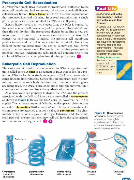
Eukaryotic Cell ReproductionThe vast amount of information encoded in DNA is organized intounits called genes. A is a segment of DNA that codes for a pro-tein or RNA molecule. A single molecule of DNA has thousands ofgenes lined up like train cars. Genes play an important role in deter-mining how a person’s body develops and functions. When genesare being used, the DNA is stretched out so that the information itcontains can be used to direct the synthesis of proteins.
As a eukaryotic cell prepares to divide, the DNA and the proteinsassociated with the DNA coil into a structure called a ,as shown in Figure 2. Before the DNA coils up, however, the DNA iscopied. The two exact copies of DNA that make up each chromosomeare called (KROH muh tihdz). The two chromatids of achromosome are attached at a point called a . The chro-matids, which become separated during cell division and placed intoeach new cell, ensure that each new cell will have the same geneticinformation as the original cell.
centromerechromatids
chromosome
gene
Binary fission
Real LifeEscherichia coli cellscan produce 1 millionnew cells in less than 7 hours. A variety of E. coli knownas O157:H7 is sometimesfound in raw or under-cooked meat. When suchmeat is eaten, this bacteriacan cause life-threateningintestinal bleeding andkidney failure. Thoroughcooking is necessary to destroy the bacteria.Finding Information Research out-breaks of E. coliO157:H7 in yourcommunity orstate.
DNA doublehelix
DNA andproteins
Further coilingwithin supercoil
Supercoil withinchromosome
Chromosome(made of 2 chromatids)
Centromere
Figure 2 Chromosomestructure. A chromosomeconsists of DNA tightlycoiled around proteins. Thechromosomes are formed as a cell prepares to divide.
119

Teaching TipMissing Homologue Ask stu-dents to hypothesize what mighthappen if a human sperm or eggcell did not contain one member ofeach homologous pair. (The result-ing zygote would not have a full setof chromosomes. The zygote mightfail to develop. If the zygote diddevelop, the individual would not benormal because its cells would lackthe important information containedin the missing genes.)
Math Skills Prokaryotic chromo-somes are hundreds of times longerthan the cell that contains them.For example, if a chromosome inan E. coli bacterium were fullyextended, it would measure about1 mm in length. The cell itself isonly about 0.002 mm in length.Have students calculate how muchlonger E. coli’s chromosome is thanthe cell itself. (1 mm/0.002 mm �500 times longer than the cell)
Logical
Using the FigureUse Figure 3 to point out howfertilization restores the diploidnumber of chromosomes. Ask stu-dents why sexually reproducingorganisms possess an even numberof chromosomes in the diploidstate. (If students do not realize that2n must be an even number, then askthem to calculate the haploid numberin each gamete as if the diploid num-ber in humans were 47 and not 46. Itcannot be done without a “half”chromosome.) Visual Bio 6ELS
GENERAL
Bio 4ALS
GENERALBUILDERSKILL
TAKS 2 Bio 6C; Bio 6E
GENERAL
Teach, continuedTeach, continued
120 Chapter 6 • Chromosomes and Cell Reproduction
CulturalAwarenessCulturalAwareness
Mormons and Gene Mapping The Human Genome Project is an effort tomap the over 100,000 genes in human cellsto each chromosome and to sequence eachgene. Scientists throughout the world arenow engaged in this effort. One group hastraced genetic markers through three genera-tions of 60 Mormon families living in Utah.Their work with the Mormon families hasresulted in the mapping of almost 500 genes.Bio/IPC 3C
www.scilinks.orgTopic: ChromosomesKeyword: HX4042
How Chromosome Number andStructure Affect DevelopmentEach human somatic cell (any cell other than a sperm or egg cell)normally has two copies of 23 different chromosomes, for a total of46 chromosomes. The 23 chromosomes differ in size, shape, andset of genes. Each chromosome contains thousands of genes thatplay important roles in determining how a person’s body developsand functions. For this reason, a complete set of all chromosomesis essential to survival.
Sets of ChromosomesEach of the 23 pairs of chromosomes consists of two homologous(hoh MAHL uh gus) chromosomes, or homologues (HOH muh logs).
are chromosomes that are similar insize, shape, and genetic content. Each homologue in a pair of ho-mologous chromosomes comes from one of the two parents, asshown in Figure 3. Thus, the 46 chromosomes in human somaticcells are actually two sets of 23 chromosomes. One set comes fromthe mother, and one set comes from the father. A human chromo-some is shown in Figure 4.
Homologous chromosomes
When haploid gametes fuse, they produce a diploid zygote.
Figure 3 Fertilization
Fertilization
Sperm celln = 23
Egg celln = 23
Zygote2n = 46
120
Student Edition TAKS Obj 2 Bio 4B TAKS Obj 2 Bio 6A TEKS Bio 4B, 6A
Teacher Edition TAKS Obj 1 Bio/IPC 2C TAKS Obj 2 Bio 6C TEKS Bio 3D, 3F, 4A, 6C, 6E, 6FTEKS Bio/IPC 3C
pp. 120–121
IPC Benchmark Mini Lesson
Biology/IPC Skills TAKS 1 Bio/IPC 2C Organize,analyze, evaluate, make inferences, and predicttrends from data. Activity Have students imagineseveral possible uses for the information gained bythe Human Genome Project. Have students write anessay describing their ideas and how they feel aboutresearch on the human genome.
Teaching TipPrefixes Have students use theprefixes in the words haploid anddiploid to help them remember themeanings of these words. Havethem relate the words to the alge-braic terms n and 2n. Haploid (n)represents one set of chromosomes,as seen in gametes. Diploid (2n)represents two sets of chromo-somes, as in somatic cells. Tellstudents that the prefix hapl-means “single” and that the prefixdipl- means “double.” Add that theprefix poly- means “many.” Thenhave students predict the nature ofa polyploid cell. (It would have multiple sets of chromosomes.)
ActivityKaryotypes Obtain copies ofnormal human karyotypes from thelibrary or on-line resources. Enlargethem so that each chromosome is nosmaller than 2.5 cm (1 in.) in length.Paste the copies of the karyotype ona poster board. Cut out each chro-mosome and mix up the pieces.Allow the students to sort the chro-mosomes into pairs using size,length, position of centromere, andbanding patterns. Have them com-pare the karyotypes with each otherand with the karyotype shown inFigure 5.
LogicalBio 6FLS
GENERAL
Bio 6E
Chapter 6 • Chromosomes and Cell Reproduction 121
English Language Learners
English Language Learners
HISTORYHISTORYCONNECTIONCONNECTION
In the 1920s, a biologist reported the diploidnumber in human cells to be 48. For manyyears following this report, textbooks describedthe normal diploid number of human chromo-somes as 48. It was not until 1956 that J. H.Tjio and A. Levan showed 46 to be the correctnumber. These two scientists worked out a procedure to culture white blood cells. Theirmethods increased the number of cells under-going mitosis and improved the spreading ofthe chromosomes, which allowed the scientiststo accurately count the chromosomes.Bio 3F, Bio/IPC 3C
Invite a genetic counselor to speak to theclass. Before the speaker attends, have thestudents research chromosomal abnormali-ties that result from too few or too manychromosomes. Have the students prepare alist of questions based on their findings. Askthe speaker to bring sample karyotypes ofthe abnormalities the students researched, ifpossible. Interpersonal Bio 3D, 6FLS
REAL WORLDREAL WORLDCONNECTIONCONNECTION
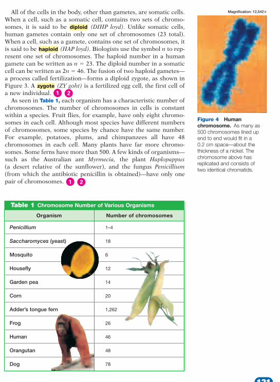
All of the cells in the body, other than gametes, are somatic cells.When a cell, such as a somatic cell, contains two sets of chromo-somes, it is said to be (DIHP loyd). Unlike somatic cells,human gametes contain only one set of chromosomes (23 total).When a cell, such as a gamete, contains one set of chromosomes, itis said to be (HAP loyd). Biologists use the symbol n to rep-resent one set of chromosomes. The haploid number in a humangamete can be written as n � 23. The diploid number in a somaticcell can be written as 2n � 46. The fusion of two haploid gametes—a process called fertilization—forms a diploid zygote, as shown inFigure 3. A (ZY goht) is a fertilized egg cell, the first cell ofa new individual.
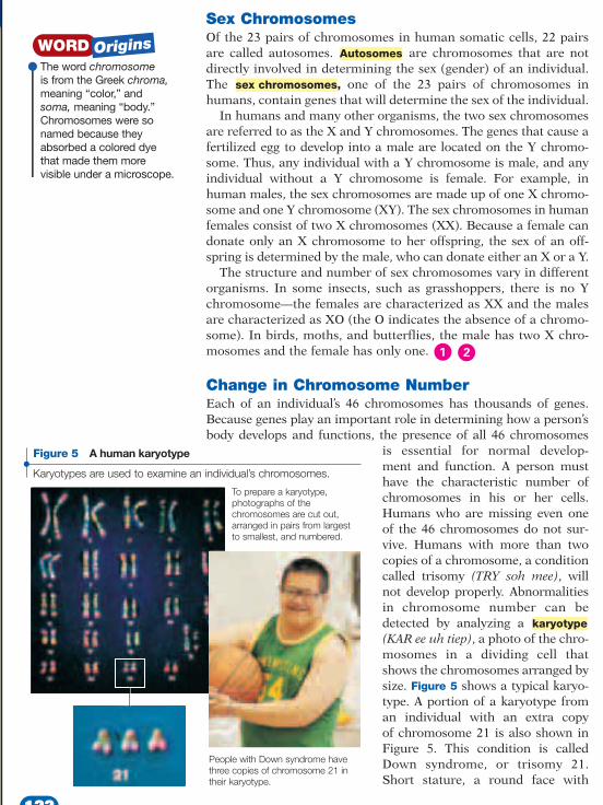
As seen in Table 1, each organism has a characteristic number ofchromosomes. The number of chromosomes in cells is constantwithin a species. Fruit flies, for example, have only eight chromo-somes in each cell. Although most species have different numbersof chromosomes, some species by chance have the same number.For example, potatoes, plums, and chimpanzees all have 48chromosomes in each cell. Many plants have far more chromo-somes. Some ferns have more than 500. A few kinds of organisms—such as the Australian ant Myrmecia, the plant Haplopappus(a desert relative of the sunflower), and the fungus Penicillium(from which the antibiotic penicillin is obtained)—have only onepair of chromosomes.
zygote
haploid
diploid
Figure 4 Human chromosome. As many as500 chromosomes lined upend to end would fit in a 0.2 cm space—about thethickness of a nickel. Thechromosome above hasreplicated and consists of two identical chromatids.
Table 1 Chromosome Number of Various Organisms
Organism Number of chromosomes
Penicillium 1–4
Saccharomyces (yeast) 18
Mosquito 6
Housefly 12
Garden pea 14
Corn 20
Adder’s tongue fern 1,262
Frog 26
Human 46
Orangutan 48
Dog 78
Magnification: 12,542�
121
Math Skills Combined, the 22somatic chromosomes and one sex chromosome consist of3,000,000,000 DNA nucleotidepairs. Ask students how many totalnucleotides are in a human diploidcell? (6,000,000,000) LogicalLS
GENERALBUILDERSKILL
Teach, continuedTeach, continued
122 Chapter 6 • Chromosomes and Cell Reproduction
On the Trail of aChromsomal DeletionTeaching StrategiesResearchers have found that lifethreatening defects in the aortacan result when a patient hastwo defective copies of the geneTbx1. Patients with one normaland one defective copy of Tbx1usually experience less seriousabnormalities.
DiscussionHow did researchers narrowthe search from 25 to 15 genes?(They found out that the chro-mosome in mice with the samedisorder only contained 15genes.)Researchers now know that thegene Tbx1 controls other genes.What types of genes might itcontrol? (those involved in devel-opment of the heart, blood, faceand immune system)
CareerCareerCytogenetic Technologist Cytogenetic tech-nologists aid physicians in diagnosis of geneticdisorders by preparing specimens, such as kary-otypes. Have students use library references oron-line resources to research the trainingrequirements and duties of a cytogenetic tech-nologist. (Technologist may prepare cells, prepareslides in the correct mitotic stage, stain slides, andprepare accurate karyotypes from photographicprints or computer images.) Bio 3D
GENERAL
Sex ChromosomesOf the 23 pairs of chromosomes in human somatic cells, 22 pairsare called autosomes. are chromosomes that are notdirectly involved in determining the sex (gender) of an individual.The , one of the 23 pairs of chromosomes inhumans, contain genes that will determine the sex of the individual.
In humans and many other organisms, the two sex chromosomesare referred to as the X and Y chromosomes. The genes that cause afertilized egg to develop into a male are located on the Y chromo-some. Thus, any individual with a Y chromosome is male, and anyindividual without a Y chromosome is female. For example, inhuman males, the sex chromosomes are made up of one X chromo-some and one Y chromosome (XY). The sex chromosomes in humanfemales consist of two X chromosomes (XX). Because a female candonate only an X chromosome to her offspring, the sex of an off-spring is determined by the male, who can donate either an X or a Y.
The structure and number of sex chromosomes vary in differentorganisms. In some insects, such as grasshoppers, there is no Ychromosome—the females are characterized as XX and the malesare characterized as XO (the O indicates the absence of a chromo-some). In birds, moths, and butterflies, the male has two X chro-mosomes and the female has only one.
Change in Chromosome NumberEach of an individual’s 46 chromosomes has thousands of genes.Because genes play an important role in determining how a person’sbody develops and functions, the presence of all 46 chromosomes
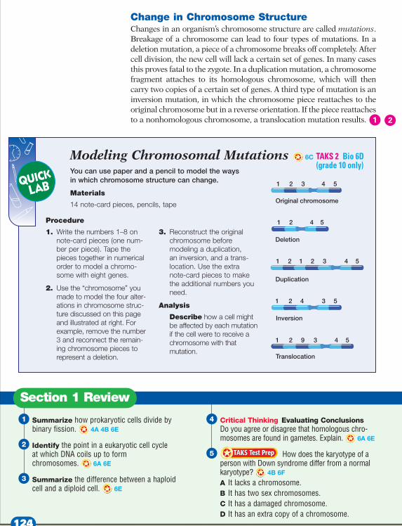
is essential for normal develop-ment and function. A person must have the characteristic number of chromosomes in his or her cells.Humans who are missing even oneof the 46 chromosomes do not sur-vive. Humans with more than twocopies of a chromosome, a conditioncalled trisomy (TRY soh mee), willnot develop properly. Abnormalitiesin chromosome number can bedetected by analyzing a (KAR ee uh tiep), a photo of the chro-mosomes in a dividing cell thatshows the chromosomes arranged bysize. Figure 5 shows a typical karyo-type. A portion of a karyotype froman individual with an extra copyof chromosome 21 is also shown inFigure 5. This condition is calledDown syndrome, or trisomy 21.Short stature, a round face with
karyotype
sex chromosomes
Autosomes
Karyotypes are used to examine an individual’s chromosomes.
Figure 5 A human karyotype
To prepare a karyotype, photographs of the chromosomes are cut out,arranged in pairs from largestto smallest, and numbered.
People with Down syndrome havethree copies of chromosome 21 intheir karyotype.
The word chromosomeis from the Greek chroma,meaning “color,” andsoma, meaning “body.”Chromosomes were sonamed because theyabsorbed a colored dye that made them more visible under a microscope.
122
Student Edition TAKS Obj 2 Bio 4B TAKS Obj 2 Bio 6A TAKS Obj 2 Bio 6C, 6D TEKS Bio 4A, 4B, 6A, 6C, 6D, 6E, 6F
Teacher Edition TAKS Obj 2 Bio 6B TAKS Obj 2 Bio 6C, 6D TAKS Obj 5 IPC 5B TEKS Bio 3D, 3F, 6C, 6D
pp. 122–123
IPC Benchmark Fact
Tell students the colored dye that makes chromo-somes more visible is the result of the reflection oflight. Indeed it is the bouncing back of waves to oureyes that makes any object visible. Although there isa tendency to think of reflection as images we cansee in a mirror, any object that we can observe withour eyes, whether or not that image is aided with ascientific instrument, is caused by light that bouncesback or is reflected from an object. In this instance,the dye increases the amount of reflected light thusmaking the chromosomes more visible.IPC 5B (grade 11 only)
TAKS 5
TAKS 2Bio 6C, 6D(grade 10 only)

Chapter 6 • Chromosomes and Cell Reproduction 123
ModelingChromosomalMutationsSkills AcquiredModeling, sequencing,interpreting
Teacher’s NotesCut note card pieces prior tothe lab.
Answers to AnalysisAnswers will vary based on thetype of mutation: deletion: cellwould be missing a gene, whichcould prove fatal; duplication:cell would have an extra gene,which could prove fatal or resultin malfunctioning of the cell;inversion: cell may not be able touse gene because it is located in adifferent area on the chromo-some, which could prove fatal;translocation: cell may not beable to use the gene because it islocated on a different chromo-some, which could prove fatal.
CulturalAwarenessCulturalAwareness
Cultural Attitudes Toward Genetic DefectsFrom the Middle Ages through the nineteenthcentury, most Europeans believed that geneticdefects reflected inner corruption. However,many other cultures, including the Celtic peo-ple of Europe and Native Americans, thoughtindividuals with such defects had a specialinsight and a closer connection with nature.
These unique members of the community weregiven responsibility as tribal leaders or healers.Many physically disabled leaders made carefulastronomical observations of the sun andmoon that helped them advise their communi-ties on the optimum times for planting, har-vesting, or migrating to another area.Bio 3F, TAKS 2 Bio 6C
Teaching TipEveryone Needs an X Tell stu-dents that most nondisjunctionsinvolving autosomes are lethal,whereas most involving sex chro-mosomes are not. However, a YOcombination in humans is lethal,leading biologists to suspect that atleast one X chromosome is neces-sary for development and survival.TAKS 2 Bio 6C; Bio 6D (grade 10 only)
GENERAL
upper eyelids that cover the inner corners of the eyes, and varyingdegrees of mental retardation are characteristics of people withDown syndrome.
In mothers younger than 30, Down syndrome occurs in about 1in 1,500 births. In mothers 37 years old, the incidence doubles to 1in 290 births. In mothers over 45, the risk is as high as 1 in 46births. Older mothers are more likely to have a baby with Downsyndrome because all the eggs a female will ever produce are pre-sent in her ovaries when she is born, unlike males who produce newsperm throughout adult life. As a female ages, her eggs can accu-mulate an increasing amount of damage. Because of this risk, apregnant woman over the age of 35 may be advised to undergo pre-natal testing that includes fetal karyotyping.
What events can cause an individual to have an extra copy of achromosome? When sperm and egg cells form, each chromosomeand its homologue separate, an event called disjunction (dihsJUHNK shuhn). If one or more chromosomes fail to separate prop-erly—an event called nondisjunction—one new gamete ends upreceiving both chromosomes and the other gamete receives none.Trisomy occurs when the gamete with both chromosomes fuseswith a normal gamete during fertilization, resulting in offspringwith three copies of that chromosome instead of two. In Down syn-drome, nondisjunction involves chromosome 21.
On the Trail of a Chromosomal Deletion
One of every 4,000 babies isborn with a genetic disorder
called DiGeorge syndrome. Thisdisorder causes serious heartdefects that must be surgicallycorrected within a few days afterbirth. Children born with DiGeorgesyndrome can also have bloodailments, facial abnormalities, adeficient immune system, andother problems.
A Faulty Chromosome Karyotypes of people with Di-George syndrome show thatthey have one normal and onefaulty 22nd chromosome. Thefaulty chromosome is missing asmall region that contains 25genes. To understand how thischromosomal deletion results in
DiGeorge syndrome, researchersat Baylor College of Medicinein Houston have been studyingthe disorder in mice.
First, the researchers foundthat they could produce similarheart defects in mice by deletinga part of mouse chromosome 16.When these mice were bred withmice that had a duplication of thesame part of chromosome 16,their offspring had no heartdefects. Because the deletedpart contained only 15 genes, thesearch for the cause of DiGeorgesyndrome was narrowed from 25genes to 15 genes.
Finding the Crucial Gene Using a technology called chro-mosome engineering, the Baylor
researchers eventually zeroed inon a gene called Tbx1. Theyshowed that deleting Tbx1 onone chromosome 16 in micecauses the heart defects ofDiGeorge syndrome. Tbx1 is alsorequired for the development ofother embryonic structuresbesides the heart. Thus, theresults of research on DiGeorgesyndrome may provide cluesabout the genetic causes ofother birth defects.
www.scilinks.orgTopic: Genetic DisordersResearch in TexasKeyword: HXX4008
123
TAKS 2
TAKS 2Bio 6C;Bio 6D(grade 10 only)
ReteachingAsk students to differentiate each ofthe following terms by defining andsketching them: chromosomes, chro-matids, DNA, and genes. Studentsshould label their sketches. (chromo-somes: coiled DNA and associatedproteins; chromatids: identical copiesof a given chromosome; DNA: chainof nucleic acids containing geneticcode; genes: sequences of DNA that code for specific proteins or RNA.)
Visual
QuizTrue or False:
1. At cell division, each chromo-some consists of two chromatidsattached at the centromere.(True)
2.The normal diploid number forhumans is 23. (False)
3. A person with the sex chromo-somes XX would be female.(True)
AlternativeAssessmentHave each student prepare a com-plete human karyotype based onFigure 5. Each chromosome shouldbe drawn and then cut out. Allowstudents to choose the sex of theirkaryotype and determine whetherthere are mutations, deletions, orextra chromosomes. Have studentsexchange their diagrams with apartner. Each student must nowmatch homologues and determinethe sex of the individual, as well asidentify any possible abnormalities.
Interpersonal TAKS 2 Bio 6C, Bio 6D(grade 10 only); Bio 6F
LS
GENERAL
GENERAL
LS
CloseClose
Answers to Section Review
1. DNA is first copied. Then the cell divides intoequal halves by adding new cell membranebetween the two DNA copies. The growing cellmembrane pushes inward, and the cell con-stricts to form two new, identical cells. A newcell wall forms around each new membrane.
2. Chromosomes become visible in a eukaryoticcell when the cell prepares to divide (duringprophase).
3. A haploid cell (n) contains one set of chromo-somes. A diploid cell (2n) contains two sets ofchromosomes. Bio 6E
TAKS 2 Bio 6A
TAKS 2 Bio 4B
4. Students should disagree. Homologous chromo-somes are pairs of similar chromosomes. Becausegametes are haploid (n) they contain only one setof chromosomes. Thus, homologous chromo-somes are not normally found in gametes.
5. A. Incorrect. Lacking a chromosome is usually a fatal abnormality. B. Incorrect. All normal individuals have twosex chromosomes. C. Incorrect. Damaged chro-mosomes can lead to death or abnormalities,but do not cause Down syndrome. D. Correct.People with Down syndrome have an extracopy of chromosome 21. TAKS 2 Bio 4B
TAKS 2 Bio 6A
124 Chapter 6 • Chromosomes and Cell Reproduction
English Language Learners
Change in Chromosome StructureChanges in an organism’s chromosome structure are called mutations.Breakage of a chromosome can lead to four types of mutations. In adeletion mutation, a piece of a chromosome breaks off completely. Aftercell division, the new cell will lack a certain set of genes. In many casesthis proves fatal to the zygote. In a duplication mutation, a chromosomefragment attaches to its homologous chromosome, which will thencarry two copies of a certain set of genes. A third type of mutation is aninversion mutation, in which the chromosome piece reattaches to theoriginal chromosome but in a reverse orientation. If the piece reattachesto a nonhomologous chromosome, a translocation mutation results.
Modeling Chromosomal MutationsYou can use paper and a pencil to model the ways in which chromosome structure can change.
Materials
14 note-card pieces, pencils, tape
Procedure
1. Write the numbers 1–8 onnote-card pieces (one num-ber per piece). Tape thepieces together in numericalorder to model a chromo-some with eight genes.
2. Use the “chromosome” youmade to model the four alter-ations in chromosome struc-ture discussed on this pageand illustrated at right. Forexample, remove the number3 and reconnect the remain-ing chromosome pieces torepresent a deletion.
3. Reconstruct the originalchromosome before modeling a duplication, an inversion, and a trans-location. Use the extra note-card pieces to makethe additional numbers youneed.
Analysis
Describe how a cell mightbe affected by each mutationif the cell were to receive achromosome with that mutation.
Original chromosome
Deletion
Translocation
1
1
1 2
2
2 3
4
4
3
1 2 4 5
1 2 3 4 5
5
5
Duplication
Inversion
91 2 3 4 5
Summarize how prokaryotic cells divide bybinary fission. 4A 4B 6E
Identify the point in a eukaryotic cell cycle at which DNA coils up to form chromosomes. 6A 6E
Summarize the difference between a haploidcell and a diploid cell. 6E
Critical Thinking Evaluating ConclusionsDo you agree or disagree that homologous chro-mosomes are found in gametes. Explain. 6A 6E
How does the karyotype of aperson with Down syndrome differ from a normalkaryotype? 4B 6F
A It lacks a chromosome.B It has two sex chromosomes.C It has a damaged chromosome.D It has an extra copy of a chromosome.
TAKS Test PrepTAKS Test Prep
Section 1 Review
6C
124
Student Edition TAKS Obj 2 Bio 4B TAKS Obj 2 Bio 6A, 6DTAKS Obj 2 Bio 6C TEKS Bio 4A, 4B, 6A, 6C, 6E, 6F
Teacher EditionTAKS Obj 2 Bio 4B, 6A, 6B, 6C, 6D TEKS Bio 4A, 4B, 6A, 6B, 6C, 6D,6E, 6F
pp. 124–125
TAKS 2 Bio 6A; Bio 4A
TAKS 2 Bio 6D(grade 10 only)

Section 2
OverviewBefore beginning this sectionreview with your students theobjectives listed in the StudentEdition. In this section, studentswill examine the three phases thattake place in the life cycle of a cell,which are collectively known as thecell cycle. They will learn that cellsspend most of their time in inter-phase, during which cells grow andDNA is replicated. The cell entersmitosis as it prepares to divide,then divides into two cells duringcytokinesis. Students will learn thatcancer may result if the controls forthe cell cycle break down.
Ask students to finish the followingsentence: A typical eukaryotic cellspends 90 percent of its time in________. Write the followingchoices on the board: mitosis,anaphase, interphase, andcytokinesis. (interphase)
DemonstrationDisplay either a model or a photo-graph of a human brain. Point outthat once the brain is fully formed,most of the nerve cells do notdivide, again. These cells remain inthe G1 phase of the cell cycle. Thendisplay either a model or a photo-graph of a human bone. Point outthat red blood cells are producedfrom cells in the marrow of longbones. An average red blood celllives for about 120 days. Each sec-ond, about 2 million red blood cellsare produced by cell division in thebone marrow. Cells in the marrow,unlike those in the brain, continuegoing through the cell cycle as longas a person lives. TAKS 2 Bio 4B
MotivateMotivate
Bio 4A
Bellringer
FocusFocus
Chapter 6 • Chromosomes and Cell Reproduction 125
• Reading Organizers• Reading Strategies• Portfolio Project
Genetics Project GENERAL
Planner CD-ROM
• Lesson Plan• Directed Reading• Active Reading GENERAL
GENERAL
Chapter Resource File
Transparencies
TT Bellringer
The Life of a Eukaryotic CellCell division in eukaryotic cells is more complex than cell divisionin bacteria because it involves dividing both the cytoplasm and thechromosomes inside the nucleus. Many internal organelles must becorrectly rearranged before the eukaryotic cell can properly divideand form two fully functioning cells.
The Cell Cycle The life of a eukaryotic cell is traditionally shown as a cycle, asillustrated in Figure 6. The is a repeating sequence of cel-lular growth and division during the life of an organism. A cellspends 90 percent of its time in the first three phases of the cycle,which are collectively called . A cell will enter the lasttwo phases of the cell cycle only if it is about to divide. The fivephases of the cell cycle are summarized below:
1. First growth (G1) phase. During the G1 phase, a cell growsrapidly and carries out its routine functions. For most organ-isms, this phase occupies the major portion of the cell’s life. Cellsthat are not dividing remain in the G1 phase. Some somatic cells,such as most muscle and nerve cells, never divide. Therefore, ifthese cells die, the body cannot replace them.
2. Synthesis (S) phase. A cell’s DNA is copied during this phase.At the end of this phase, each chromosome consists of two chro-matids attached at the centromere.
3. Second growth (G2) phase. In the G2 phase,preparations are made for the nucleus todivide. Hollow protein fibers called micro-tubules are assembled. The microtubules areused to move the chromosomes during mitosis.
4. Mitosis. The process during cell division inwhich the nucleus of a cell is divided into twonuclei is called (mie TOH sihs). Eachnucleus ends up with the same number andkinds of chromosomes as the original cell.
5. Cytokinesis. The process during cell divisionin which the cytoplasm divides is called
(SIET oh kih nee sihs).
Mitosis and cytokinesis produce new cellsthat are identical to the original cells and alloworganisms to grow, replace damaged tissues,and, in some organisms, reproduce asexually.
cytokinesis
mitosis
interphase
cell cycle
The Cell Cycle Section 2
Objectives● Identify the major
events that characterizeeach of the five phases ofthe cell cycle.
● Describe how the cellcycle is controlled ineukaryotic cells.
● Relate the role of the cell cycle to the onset of cancer.
Key Terms
cell cycleinterphasemitosiscytokinesiscancer
G1(Cell growth)
G2(Growth and
preparation formitosis)
Mitosis
Cytokinesis
S(DNA synthesis)
INTERPHASE
Figure 6 The eukaryoticcell cycle. The cell cycleconsists of phases of growth,DNA replication, preparationfor cell division, and division ofthe nucleus and cytoplasm.
4B 6E
4B 6E
4B 6C
125
TAKS 2
TAKS 2
TAKS 2

Teaching TipInterphase Have students draw a Graphic Organizer like the onebelow to summarize the events thatoccur during the phases of inter-phase. Have them use arrowsbetween the phases, pointing to the right, to indicate the sequenceof events.
Teaching TipBusy Cells Remind students thatduring interphase, a cell is not onlygrowing, it is also producing theproteins and carrying out thefunctions that are characteristic ofthat type of cell.
Group ActivityCell Cycle PhasesHave students read Section 2,
then write all the activities andevents described during the cellcycle on index cards (one event percard). Draw a large version ofFigure 6 at the front of the room.Then collect the students’ cards.Allow one student at a time tocome to the front of the room, reada card out loud, and then tape thecard to the proper phase of the cellcycle. Verbal TAKS 2 Bio 4BLS
Writing
TAKS 2 Bio 4B
GENERAL
TAKS 2 Bio 4B; Bio 3E
GENERAL
TeachTeach
126 Chapter 6 • Chromosomes and Cell Reproduction
Graphic Organizer
Use this graphic organizer withTeaching Tip: Interphase onthis page.
G2
Cellgrowth
DNAsynthesis
Growth andpreparation for cell division
Interphase
SG1
Control of the Cell Cycle If a cell spends 90 percent of its time in interphase, how do cells“know” when to divide? How is the cycle controlled? Just as trafficlights control the flow of traffic, cells have a system that controlsthe phases of the cell cycle. Cells have a set of “red light–green light”switches that are regulated by feedback information from the cell.The cell cycle has key checkpoints (inspection points) at whichfeedback signals from the cell can trigger the next phase of the cellcycle (green light). Other feedback signals can delay the next phaseto allow for completion of the current phase (yellow or red light).
The cell cycle in eukaryotes is controlled by many proteins. Con-trol occurs at three principal checkpoints, as shown in Figure 7.
1. Cell growth (G1) checkpoint. This checkpoint makes the deci-sion of whether the cell will divide. If conditions are favorablefor division and the cell is healthy and large enough, certainproteins will stimulate the cell to begin the synthesis (S) phase.During the S phase, the cell will copy its DNA. If conditions arenot favorable, cells can typically stop the cell cycle at this check-point. The cell cycle will also stop at this checkpoint if the cellneeds to pass into a resting period. Certain cells, such as somenerve and muscle cells, remain in this resting period perma-nently and never divide.
2. DNA synthesis (G2) checkpoint. DNA replication is checked atthis point by DNA repair enzymes. If this checkpoint is passed,proteins help to trigger mitosis. The cell begins the many mo-lecular processes that are needed to proceed into mitosis.
3. Mitosis checkpoint. This checkpoint triggers the exit frommitosis. It signals the beginning of the G1 phase, the majorgrowth period of the cell cycle.
When Control Is Lost: CancerCertain genes contain the information nec-essary to make the proteins that regulate cellgrowth and division. If one of these genes ismutated, the protein may not function, andregulation of cell growth and division can bedisrupted. , the uncontrolled growthof cells, may result. Cancer is essentially adisorder of cell division. Cancer cells do notrespond normally to the body’s controlmechanisms.
Some mutations cause cancer by over-producing growth-promoting molecules,thus speeding up the cell cycle. Otherscause cancer by inactivating the controlproteins that normally act to slow or stopthe cell cycle.
Cancer
Reviewing InformationLearn the stages of inter-phase by reviewing thesteps numbered 1–5 on theprevious page. You can seein Figures 6 and 7 that thecell cycle is a repeatingseries of three stepsfollowed by mitosis and cytokinesis.
G1
G1checkpoint
G2checkpoint
Mitosischeckpoint
G2
MitosisCytokinesis
S
INTERPHASE
Figure 7 Control of thecell cycle. The cell cycle ineukaryotes is controlled atthree inspection points, orcheckpoints. Many proteinsare involved in the control ofthe cell cycle.
126
Student Edition TAKS Obj 2 Bio 4B TAKS Obj 2 Bio 6C TEKS Bio 4A, 4B, 6C, 6E, 6F
Teacher Edition TAKS Obj 2 Bio 4B, 6C TEKS Bio 3E, 4B, 6C
pp. 126–127
Answers to Section Review
1. G1 phase: cell grows rapidly and carries outroutine functions; G2 phase: mitochondria andother organelles replicate, microtubules areassembled; S phase: DNA is copied
2. Cell growth checkpoint (G1): determines if cellis ready to undergo division; DNA synthesischeckpoint (G2): DNA repair enzymes checkprogression; mitosis checkpoint: triggers exitfrom mitosis at the beginning of the G1 cycle.TAKS 2 Bio 4B
TAKS 2 Bio 4B
3. Chromosomes are formed right before a celldivides in mitosis. During most of interphase,the DNA exists as chromatin, which is moreelongated and harder to see with a microscope.
4. A. Incorrect. Cancer is associ-ated with accelerated cell growth. B. Incorrect.Cancer does alter the cell cycle, but not byhalting mitosis. C. Correct. Uncontrolled cellgrowth is the defining characteristic of cancer.D. Incorrect. Cancer cells continue to divide.TAKS 2 Bio 4B, 6C
TAKS 2 Bio 4B
ReteachingAssign students to groups. Haveeach group visually interpret astage in the cell cycle. Have eachgroup display their graphics in theappropriate space in a circle graphthat represents the cell cycle.
Visual
Quiz1. The three phases of the cell cycle
are interphase, mitosis, and________. (cytokinesis)
2.When normal control of the cellcycle fails, ________ maydevelop. (cancer)
AlternativeAssessmentHave students write and illustrate adescription of the cell cycle in thestyle of a children’s book called “ACell’s Life.” Be sure students includeeach phase, including G1, S, G2,mitosis, and cytokinesis. VerbalLS
TAKS 2 Bio 4B
TAKS 2 Bio 4B
GENERAL
Co-op LearningLS
CloseClose
Chapter 6 • Chromosomes and Cell Reproduction 127
Understanding CancerTeaching Strategies• Tell students that most anti-
cancer drugs interfere withthe cell cycle of cancer cells.Unfortunately, the drugs alsointerfere with healthy cells,which is why the patientsuffers side effects from themedications.
Discussion• Where in the cell cycle could
scientists target anticancerdrugs? (Answers will vary butmay mention proteins involvedin the three checkpoints, DNAreplication, or cytokinesis.)
• What type of environmentalfactors have been associatedwith the onset of cancer?(Answers may include diet,UV radiation, hormones, andenvironmental pollution.)
Cancer
Although all cancers are not curable, greatprogress has been made in cancer research overthe last 30 years. We now know that cancerresults from damage to a small set of genes that,in normal cells, limits the ability of cells to divide. What causes this damage? Certain environmentalfactors appear to be associated with cancer. Forexample, the incidence of cancer per thousandpeople is not uniform throughout the UnitedStates. Rather, it is higher in cities and in theMississippi delta, suggesting that pollution andpesticide runoff may contribute to cancer. Whenpollutants, radiation, and other environmental fac-tors associated with cancer are analyzed, a clearpattern emerges. Most cancer-causing agents arepowerful mutagens—that is, they readily damageDNA. The conclusion that cancer is caused bymutation of a cell’s DNA is now supported by avery large body of evidence.How many mutations are required to producecancer? Research in the last several years indicatesthat mutation of only a few genes can transformnormal cells into cancerous ones. All of thesecancer-causing genes are involved with regulatinghow fast cells grow and divide. How is cell divisionregulated? As a crude analogy, imagine a carparked on the side of a road. To get it going, youmust step on the accelerator and release the brake.
Stepping on the AcceleratorA cell divides when it receives a signal to do so. A“divide” signal is usually in the form of a chemicalsubstance released by another cell. The sub-stance is bound by a protein on the surface of the receiving cell. This binding activates a secondprotein inside the cell—relaying the signal fromthe outside of the cell to the inside. Here, a familyof proteins then relay the signal inward to the
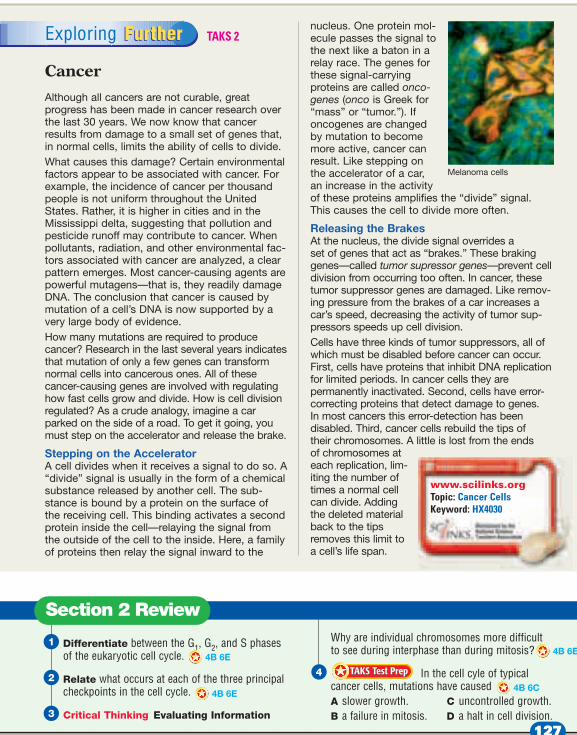
nucleus. One protein mol-ecule passes the signal tothe next like a baton in arelay race. The genes forthese signal-carrying proteins are called onco-genes (onco is Greek for“mass” or “tumor.”). Ifoncogenes are changedby mutation to becomemore active, cancer canresult. Like stepping onthe accelerator of a car,an increase in the activityof these proteins amplifies the “divide” signal. This causes the cell to divide more often.
Releasing the Brakes At the nucleus, the divide signal overrides a set of genes that act as “brakes.” These brakinggenes—called tumor supressor genes—prevent celldivision from occurring too often. In cancer, thesetumor suppressor genes are damaged. Like remov-ing pressure from the brakes of a car increases acar’s speed, decreasing the activity of tumor sup-pressors speeds up cell division.Cells have three kinds of tumor suppressors, all ofwhich must be disabled before cancer can occur.First, cells have proteins that inhibit DNA replicationfor limited periods. In cancer cells they are permanently inactivated. Second, cells have error-correcting proteins that detect damage to genes. In most cancers this error-detection has been disabled. Third, cancer cells rebuild the tips of their chromosomes. A little is lost from the ends of chromosomes ateach replication, lim-iting the number oftimes a normal cellcan divide. Addingthe deleted materialback to the tipsremoves this limit toa cell’s life span.
FurtherExploring Further
Section 2 Review
Differentiate between the G1, G2, and S phasesof the eukaryotic cell cycle.
Relate what occurs at each of the three principalcheckpoints in the cell cycle.
Critical Thinking Evaluating Information
Why are individual chromosomes more difficultto see during interphase than during mitosis?
In the cell cyle of typicalcancer cells, mutations have caused A slower growth. C uncontrolled growth.B a failure in mitosis. D a halt in cell division.
TAKS Test PrepTAKS Test Prep
4B 6C
4B 6E
Melanoma cells
www.scilinks.orgTopic: Cancer CellsKeyword: HX4030
4B 6E
4B 6E
127
TAKS 2Bio 4B, 6C
TAKS 2

OverviewBefore beginning this sectionreview with your students theobjectives listed in the StudentEdition. In this section, studentswill examine the processes of mito-sis and cytokinesis in more detail.During mitosis, spindle fibers pullthe chromatids to opposite ends ofthe cell, ensuring that each new cellreceives the proper assortment ofchromosomes. During cytokinesis,the cytoplasm is divided by a newcell membrane or cell wall.
Draw a football field on the boardwith the players lined up at mid-field and the goal posts at each end. Have students write a shortparagraph describing how the play-ers, the midfield line, and the goalposts compare to the structures of a cell involved in mitosis and celldivision. (The players represent thechromosomes, the midfield line repre-sents the equator of the cell, and thegoal posts represent the centrioles atthe poles of the cell.)
ActivityMitosis Set up several micro-scopes with prepared slides of cellsat various stages of mitosis, butpresented out of order. Have stu-dents examine the slides and try toplace each slide into the properorder of events. If microscopes and slides are not available, thevarious stages can be drawn on theboard, unlabeled and out of order.TAKS 2 Bio 4B; Bio 6E
GENERAL
MotivateMotivate
TAKS 2 Bio 4B; Bio 3E, 6E
Bellringer
FocusFocus
Section 3
128 Chapter 6 • Chromosomes and Cell Reproduction
English Language Learners • Lesson Plan
• Directed Reading• Active Reading• Data Sheet for Math Lab• Data Sheet for Quick Lab GENERAL
GENERAL
GENERAL
GENERAL
Chapter Resource File Transparencies
TT BellringerTT Stages of Mitosis
• Reading Organizers• Reading Strategies• Supplemental Reading
The Lives of a Cell
Planner CD-ROM
Each centriole is composed of nine triplets of microtubules arranged in a circle.
Centrioles
Cell
Spindlefibers
Centromere
Chromatids
Microtubuletriplets
Section 3 Mitosis and Cytokinesis
Chromatid Separation in MitosisEvery second about 2 million new red blood cells are produced inyour body by cell divisions occurring in the bone marrow. These cellshave received the signal to divide. The cells continue past the G2 phaseand enter into the last two phases of the cell cycle—mitosis andcytokinesis. During mitosis the nucleus divides to form two nuclei,each containing a complete set of the cell’s chromosomes. Duringcytokinesis the cytoplasm is divided between the two resulting cells.
During mitosis, the chromatids on each chromosome are physi-cally moved to opposite sides of the dividing cell with the help ofthe spindle, shown in Figure 8. are cell structures made upof both centrioles and individual microtubule fibers that areinvolved in moving chromosomes during cell division.
Forming the SpindleAnimal cells usually have one pair of centrioles, with the centrioles atright angles to each other. During the G2 phase of the cell cycle, thecentriole pair is replicated so that the cell has two pairs of centriolesas it enters the mitotic phase. When a cell enters the mitotic phase,the centriole pairs start to separate, moving toward opposite poles ofthe cell. As the centrioles move apart, the spindle begins to form.
Centrioles and spindle fibers are both made of hollow tubes of pro-tein called microtubules. Each spindle fiber is made of an individualmicrotubule. Each centriole, however, is made of nine triplets of
Spindles
Objectives● Describe the structure and
function of the spindleduring mitosis.
● Summarize the events of the four stages of mitosis.
● Differentiate cytokinesis inanimal and plant cells.
Key Terms
spindle
The spindle, made up of centrioles and spindle fibers, helps move chromosomes apart during mitosis.
Figure 8 The spindle
4B 6E
4B 6E
4B 6E
128
Student Edition TAKS Obj 2 Bio 4B TEKS Bio 4B, 6E
Teacher’s Edition TAKS Obj 2 Bio 4B TEKS Bio 3E, 3F, 4B, 6E
pp. 128–129
TAKS 2
TAKS 2
TAKS 2

TeachTeach
Chapter 6 • Chromosomes and Cell Reproduction 129
Calculating theNumber of CellsResulting fromMitosisSkills AcquiredCalculating, predicting
Teacher’s NotesReview numbers in scientificnotation before starting the lab.Review how to cancel units andset up equal unit proportions.For example, 60 seconds/1 minute is an equal proportion.
Analysis Answers1. 9.0 � 1010 cells �
90,000,000,000 cells2. 2.16 � 1012 cells3. Factors that might increase or
decrease the rate of mitosisinclude mutated genes, diet,and exposure to ultravioletlight and tobacco products.
TAKS 2 Bio 4B; Bio 6E
<x + 6x - 7 - 02
18
49376
0
52
did you know?Fly Chromosomes The fruit fly Drosophilamelanogaster has been used to study chromoso-mal mutations since 1933. The salivary glandsof these flies, as in many insects, are composedof cells that do not divide during the larvalstage. However, the chromosomes continue toreplicate, producing many copies. The copies ofeach chromosome are closely aligned, resultingin thick chromosomes that are easy to studywith a microscope. Bio 3F
microtubules arranged in a circle. Unlike animal cells, plant cells donot have centrioles, but they form a spindle that is almost identicalto that of an animal cell.
Separation of Chromatids by Attaching Spindle Fibers Some of the microtubules in the spindle interact with each other.Others attach to a protein structure found on each side of the centro-mere. The two sets of microtubules extend out toward opposite polesof the cell. Once the microtubules attach to the centromeres andpoles, the two chromatids in each chromosome can be separated.
The chromatids are moved to each pole of the cell in a mannersimilar to bringing in a fish with a fishing rod and reel. When themicrotubule “fishing line” is “reeled in,” the chromatids aredragged to opposite poles. The reeling in occurs because the endsof the spindle fibers are broken down bit by bit at each of the poles.As the fibers become shorter, the chromatids they are pulling movecloser and closer to the poles.
As soon as the chromatids separate from each other they arecalled chromosomes. When the chromosomes finally arrive, eachpole has one complete set of chromosomes.
Analysis
1. Calculate the number ofcells that would be producedin 1 hour.
2. Calculate the number ofcells that would be producedin 1 day.
3. Critical Thinking Predict-ing Patterns Identify factorsthat might increase or decreasethe rate of mitosis.
Calculating the Number of CellsResulting from MitosisBackground
Scientists investigating cancer might need to know thenumber of cells produced in a certain amount of time. In the human body the rate of mitosis is about 25 million (2.5 � 107) cells produced every second! You can calculatethe number of cells produced by mitosis in a given amountof time.
<x + 6x - 7 - 02
8
493 0
52
1. Calculate the number of cells produced by mitosis in the given time. For example, to findthe number of cells produced in 3 minutes, determine how many seconds there are in 3 minutes (sincethe rate is given in seconds).
� 3 minutes � 180 seconds
2. Multiply the rate of mitosis by the time (in seconds) asked for in the problem (180 seconds).
� 180 seconds � 4.5 � 109 cells (4,500,000,000 cells) 2.5 � 107 cellssecond
60 seconds1 minute
4B 6E
129
TAKS 2
MATH TAKS Obj 9 8.3B; Obj 10 8.14A

Vocabulary To help students mas-ter vocabulary, relate the wordmitosis to its Greek origin, mitos,meaning “thread.” Ask students toexplain why mitosis is associatedwith thread. (The hereditary materialconsists of long, threadlike molecules.)
Using the FigureLead students througheach stage shown inFigure 9, focusing on the
behavior of chromosomes. Pointout that the various stages are notof equal duration. Using movie filmas an analogy, help students avoidthe misconception that mitosis“jumps” from stage to stage.Although a movie consists of indi-vidual frames of film, the imageson the film appear to change con-tinuously. Explain that mitosisprogresses in a similar fashion.
Teaching TipPrefixes Tell students that the pre-fixes of the different stages of mito-sis describe the order of events orthe events themselves. Pro- means“earlier than;” meta- means “laterthan” or “after;” ana- means “up”or “back” and describes the move-ment of the chromosomes “up”toward the poles; and telo- means “end.”
Teaching TipHave students hypothesize abouthow a cell’s ATP use changes dur-ing mitosis. (The events of mitosisrequire a lot of additional energy,which is supplied by ATP.)TAKS 2 Bio 4B; Bio 6E
GENERAL
TAKS 2 Bio 4B; Bio 6E
BUILDERSKILL
Teach, continuedTeach, continued
130 Chapter 6 • Chromosomes and Cell Reproduction
English Language Learners
When cultured, cancer cells can divide indefi-nitely if they are given a continual supply ofnutrients. One cell line has been continuouslycultured since 1951. The cells in this cell lineare called HeLa cells because they were origi-nally from a tumor removed from a womannamed Henrietta Lacks. The patient sufferedfrom uterine cervical carcinoma. Cells fromthis cell line are still used by researchersaround the world, especially in research on viruses. Bio/IPC 3C; Bio 3F
MEDICINEMEDICINECONNECTIONCONNECTION
BIOgraphic
Mitosis and Cytokinesis Although mitosis is a continuous process, biologists traditionallydivide it into four stages, as shown in Figure 9.
Mitosis
Step Prophase Chromosomes coil up and become visible duringprophase. The nuclear envelope dissolves and a spindle forms.
Step Metaphase During metaphase the chromosomes move to thecenter of the cell and line up along the equator. Spindle fiberslink the chromatids of each chromosome to opposite poles.
Step Anaphase Centromeres divide during anaphase. The twochromatids (now called chromosomes) move toward oppo-site poles as the spindle fibers attached to them shorten.
Step Telophase A nuclear envelope forms around the chromo-somes at each pole. Chromosomes, now at opposite poles,
BIOgraphic
INTERPHASE
Figure 6-9
Stages of Mitosis
The chromosome copies in the nucleus of a dividing cell are separated into two nuclei.
Prophase1 Metaphase2
• Chromosomesbecome visible
• Nuclear envelopedissolves
• Spindle forms
• Chromosomesline up along equator
Nucleus
Chromosome(already copied)
Centrioles
Spindle fibers
The chromosomesreplicate during
interphase.
Magnification: 567�
G1
G2
Mitosis
Cytok
ines
is
S
Figure 9
130
Student Edition TAKS Obj 2 Bio 4B
Teacher Edition TAKS Obj 2 Bio 4BTEKS Bio/IPC 3CTEKS Bio 3E, 3F, 4A, 4B, 6E
pp. 130–131

Trends in Cell BiologyTurning Off Cancer Currently, researchers atthe National Cancer Institute are working tofind the proteins that “turn on” or “turn off”gene ECT2, an oncogene that appears to be acritical regulator of cytokinesis. Eventually,researchers may be able to “turn off” themechanism that causes cancer cells to divideuncontrollably. Bio/IPC 3C
Teaching TipStages of Mitosis Have studentsdraw pictures of each stage ofmitosis on four note cards. On oneside, have students write the nameof the stage. On the opposite side,have them draw an accurate sketchof the stage. They can then usethese as flash cards to help eachother learn the stages of mitosis.
Visual
Using the FigureTell students that the belt of protein threads that are labeled inFigure 10 is a ring of the proteinsactin and myosin, the same twoproteins that interact to contractmuscle cells. The furrowing of thecell membrane occurs perpendicu-lar to the long axis of the spindle.Cytokinesis usually begins inanaphase but is not completed untilafter the two nuclei have formed.
Group ActivityPerforming Mitosis andCytokinesis Have students work ingroups of three or four students.Ask each group to design a “per-formance” using the entire class asthe cast, as well as props, thatwould demonstrate the process ofmitosis and cytokinesis. Ask groupsto present their designs to the class,and have the class decide which“show” is best. If time and materi-als permit, allow students to stagetheir mitosis and cytokinesis show.
Interpersonal
TAKS 2 Bio 4B; Bio 3E, 6E
Co-op LearningLS
GENERAL
TAKS 2 Bio 4B; Bio 4A, 6E
TAKS 2 Bio 4B; Bio 6ELS
GENERAL
Chapter 6 • Chromosomes and Cell Reproduction 131
MISCONCEPTION ALERT
Mitosis vs. Cytokinesis Students often thinkthat mitosis is the same as cell division. Besure they understand that mitosis refersstrictly to the division of the chromosomes,whereas cytokinesis refers to the division ofthe cytoplasm. Remind students that celldivision is just one of the four events thatmake up the cell cycle. TAKS 2 Bio 4B; Bio 6E
uncoil and the spindle dissolves.The spindle fibers break down anddisappear. Mitosis is complete.
CytokinesisAs mitosis ends, cytokinesis begins. Dur-ing cytokinesis, the cytoplasm of the cellis divided in half, and the cell membranegrows to enclose each cell, forming twoseparate cells as a result. The end resultof mitosis and cytokinesis is two geneti-cally identical cells where only one cellexisted before.
During cytokinesis in animal cells andother cells that lack cell walls, the cell ispinched in half by a belt of proteinthreads, as shown in Figure 10.
Anaphase3 Telophase4
• Centromeres divide• Chromatids (now
called chromosomes)move toward oppositepoles
• Nuclear envelopeforms at each pole
• Chromosomes uncoil• Spindle dissolves• Cytokinesis beginsTwo genetically
identical cells
Belt of proteinthreads
Figure 10 Cytokinesisin animal cells. The cellmembrane is pinched inhalf by a belt of proteinthreads.
131

ReteachingProvide students with yarn torepresent chromosomes, nuclearenvelopes, and cell membranes; tietabs to represent centromeres; andstring to represent spindle fibers.Have them recreate on their desk-tops a cell undergoing mitosis.
Kinesthetic
Quiz1. In mitosis, the chromatids move
toward opposite poles during________. (anaphase)
2.During mitosis, the chromo-somes line up along the equatorduring ________. (metaphase)TAKS 2 Bio 4B
TAKS 2 Bio 4B
GENERAL
LS
CloseClose
Teach, continuedTeach, continued
Answers to Section Review
1. The microtubules, attached to the centromeres,shorten and pull the chromatids to oppositepoles, similar to a fishing line reeling in a fish.
2. Prophase: chromosomes become visible,nuclear envelope dissolves, spindle forms;metaphase: chromosomes line up at the equa-tor, spindle fibers attach to each chromatid;anaphase: centromeres divide, chromatidsmove to opposite poles due to shortening spin-dle fibers; telophase: nuclear envelope formsaround the chromatids at each pole, chromo-somes uncoil, spindle fibers break down anddisappear TAKS 2 Bio 4B
TAKS 2 Bio 4B
132 Chapter 6 • Chromosomes and Cell Reproduction
Observing Mitosisand CytokinesisSkills AcquiredComparing, inferring
Answers to Analysis1. Prophase: chromosomes are
visible as dark threads;metaphase: chromatids line upalong the equator; anaphase:chromosomes appear to pulltoward opposite poles;telophase: chromosomes are at opposite poles
2. Answers will vary but shouldindicate more cells in inter-phase than in the other stages.
3. Cells spend the majority oftheir time in interphase.
English Language Learners
3. In plant cells, vesicles formed by the Golgiapparatus fuse at the equator and form the cellplate. A new cell wall forms on both sides ofthe cell plate. In animal cells, the cell is pinchedin half by a belt of protein threads.
4. A. Incorrect. The cell wall doesnot play a key role in mitosis. B. Correct.Without the spindle fibers, chromosomes can-not segregate properly into two completenuclei. C. Incorrect. The cell membrane doesnot play a key role in mitosis. D. Incorrect. Thenuclear envelope breaks down before mitosisand reforms after the completion of mitosis.TAKS 2 Bio 4B, 6C
TAKS 2 Bio 4B
In plant cells and other cells that have rigid cell walls, thecytoplasm is divided in a different way. In plant cells, vesiclesformed by the Golgi apparatus fuse at the midline of thedividing cell and form a cell plate. A cell plate is a membrane-
bound cell wall that forms across the middle ofthe plant cell. A new cell wall then forms on bothsides of the cell plate, as shown in Figure 11. Whencomplete, the cell plate separates the plant cellinto two new plant cells.
In both animal and plant cells, offspring cells areabout equal in size. Each offspring cell receives anidentical copy of the original cell’s chromosomes.Each offspring cell also receives about one-half ofthe original cell’s cytoplasm and organelles.Forming
cell plate
Cell wall
Nucleus
Figure 11Cytokinesis inplant cells. A cell wall forms in the center of the dividing cell.
Observing Mitosis and Cytokinesis You can identify the stages of mitosis and the process of cytokinesis by observing slides of tissues undergoing mitosis using a compound microscope.
Materials
compound microscope, prepared slide of mitosis, paper, pencil
Procedure
1. View a prepared slide of cellsundergoing mitosis under lowpower of a compound micro-scope.
2. Move the slide until you find asection where different stagesof mitosis are visible.
3. Switch to high power. Usethe photos in Figure 9 to helpyou locate and identify cellsin interphase and in eachstage of mitosis.
4. On a separate piece of paper,sketch an example of eachstage. Label each sketch withthe following terms whereappropriate: chromosomes,cell membrane, cytoplasm,nucleus, spindle, and cell wall.
5. Switch to low power, andestimate how many cells areclearly in interphase and howmany cells are in one of thestages of mitosis.
Analysis
1. Describe the activity ofchromosomes in each stageof mitosis.
2. Compare the number ofcells in interphase with thenumber of cells in one of thestages of mitosis.
3. Critical Thinking Infer-ring Relationships Whatdoes your answer to item 2indicate about the relativelength of interphase?
Describe the function of the microtubulesduring anaphase. 4B 6E
Describe the events that occur during each ofthe four stages of mitosis. 4B 6E
Compare how cytokinesis occurs in plant cellswith how it occurs in animal cells. 4B 6E
Mitosis could not proceed if amutation interrupted the assembly of A the cell wall. C the cell membrane.B spindle fibers. D the nuclear envelope.
TAKS Test PrepTAKS Test Prep
Section 3 Review
4B 6C
132
TAKS 2Bio 4B; Bio 6E
Student Edition TAKS Obj 2 Bio 4B TAKS Obj 2 Bio 6CTEKS Bio 4B, 6C, 6E
Teacher Edition TAKS Obj 2 Bio 4B, 6CTEKS Bio 4B, 6C
pp. 132–133
TAKS 2
TAKS 2Bio 4B
AlternativeAssessmentSet up a lab practical with variousstations. Include microscopes withslides at various stages of mitosis,pictures of chromosomes, andkaryotypes for interpretation.
GENERAL
Answer to Concept Map
The following is one possible answer to Performance Zone item 15.
Chapter 6 • Chromosomes and Cell Reproduction 133
• Science Skills Worksheet• Critical Thinking Worksheet• Test Prep Pretest• Chapter Test GENERAL
GENERAL
GENERAL
Chapter Resource File
Cell cycle
includes
dividesincludes divides
chromosomes cytoplasm
cytokinesismitosisinterphase
first growth phase
synthesis phase
second growth phase
Key Concepts
Study CHAPTER HIGHLIGHTS
ZONEKey Terms
Section 1gamete (118) binary fission (119)gene (119)chromosome (119)chromatid (119)centromere (119)homologous chromosome (120)diploid (121)haploid (121)zygote (121)autosome (122)sex chromosome (122)karyotype (122)
Section 2cell cycle (125)interphase (125)mitosis (125)cytokinesis (125)cancer (126)
Section 3spindle (128)
BIOLOGYBIOLOGYUnit 4—Cell Reproduction Use Topics 1–4in this unit to review the key concepts andterms in this chapter.
Chromosomes
● Cell division allows organisms to reproduce asexually, grow,replace worn-out or damaged tissues, and form gametes.
● Bacteria reproduce by binary fission. ● Before cell division, DNA coils tightly around proteins and
forms chromosomes. At cell division, each chromosomeconsists of two chromatids attached at the centromere.
● Each organism has a characteristic number of chromosomes. ● Human somatic cells are diploid, with 23 pairs of homolo-
gous chromosomes. Human gametes are haploid, with 23chromosomes.
● Sex chromosomes carry information that determines anorganism’s sex.
● Changes in chromosome number or structure can causeabnormal development. Karyotypes are used to examine anindividual’s chromosomes.
The Cell Cycle
● The life of a eukaryotic cell—the cell cycle—includesinterphase, mitosis, and cytokinesis.
● Interphase consists of 3 phases: growth, DNA synthesis (repli-cation), and preparation for cell division. A cell about to divideenters the mitosis and cytokinesis phases of the cell cycle.
● The cell cycle is carefully controlled; failure of cellularcontrol can result in cancer.
Mitosis and Cytokinesis
● During mitosis, spindle fibers drag the chromatids to oppositepoles of the cell. A nuclear envelope forms. Each resultingnucleus contains a set of the original cell’s chromosomes.
● Cytokinesis in animal cells occurs when a belt of proteinthreads pinches the cell membrane in half. Cytokinesis inplant cells occurs when vesicles from the Golgi apparatusfuse to form a cell plate.
3
2
1
133
IPC BenchmarkReview
To prepare students for the TAKS, havestudents review Changes in Matter:Physical Changes and ChemicalChanges TAKS Obj 4 IPC 8A on p. 1052of the IPC Refresher in the TexasAssessment Appendix of this book.

ANSWERS
Using Key Terms
1. b2. a3. c4. d5. a. Gametes, or reproductive cells,
are haploid, or n, and containone set of chromosomes.When gametes unite, theyform a zygote, which isdiploid, or 2n, and containstwo sets of chromosomes.
b. Of the 23 pairs of chromo-somes, 22 pairs are calledautosomes. The individual’ssex is determined by the 23rdpair, the sex chromosomes. Akaryotype is a photograph ofchromosomes arranged by size.
c. A person’s sex is determinedby their sex chromosomes. Anindividual with two X chro-mosomes (XX) is female; anindividual with XY chromo-somes is male.
Understanding Key Ideas
6. b7. d8. c9. a
10. b11. a12. Normal cells can become cancer
cells if a genetic mutation impairsa cell’s ability to regulate cellgrowth and division. Themutation leads to cancer, oruncontrolled cell growth.TAKS 2 Bio 6C
TAKS 2 Bio 4B; Bio 6ETAKS 2 Bio 4B; Bio 6EBio 6ETAKS 2 Bio 4BTAKS 2 Bio 6CTAKS 2 Bio 4B
TAKS 2 Bio 4B; Bio 6EBio 4A, 6ETAKS 2 Bio 4B; Bio 6EBio 6E
Section Questions1 1, 3, 5 ,6, 14, 17, 192 2, 7, 8, 12, 15, 17, 18, 20, 21, 223 4, 9, 10, 11, 13
Assignment Guide
134 Chapter 6 • Chromosomes and Cell Reproduction
CHAPTER 6
13. Answers will vary, but should includeorganelles such as lysosomes, the Golgi appa-ratus, the endoplasmic reticulum, ribosomes,and vacuoles.
14. When the mice had two copies of those genes,DiGeorge syndrome did not appear.
15. The answer to the concept map is found atthe bottom of the Study Zone page. Bio 3E
TAKS 2 Bio 6C
TAKS 2 Bio 4B; Bio 6E
PerformanceZONE
CHAPTER REVIEW
Using Key Terms 1. Prokaryotes reproduce asexually by 6E
a. disjunction. c. cytokinesis.b. binary fission. d. mitosis.
2. The stage of the cell cycle in which a cell’sDNA is copied is called the _______ phase.a. S c. G2b. G1 d. mitosis
3. Chromatids are 4A 6Ea. dense patches within the nucleus.b. prokaryotic chromosomes.c. two exact copies of DNA that make up
each chromosome.d. structures that move chromosomes
during mitosis.
4. The process in which the cytoplasm of acell is divided is called 4B 6Ea. disjunction. c. binary fission.b. interphase. d. cytokinesis.
5. For each set of terms, write one or moresentences summarizing informationlearned in this chapter.a. haploid, diploid, gametes, zygoteb. autosome, sex chromosome, karyotypec. XX chromosomes, XY chromosomes
Understanding Key Ideas 6. In humans, females have _______ sex
chromosomes. 4B a. XY c. YY b. XX d. XO
7. The diagram below represents a(n) _______mutation. 6Ca. deletion c. inversionb. translocation d. duplication
8. When the cell cycle is not controlled,_______ may result. 4Ba. Down syndrome b. binary fission c. cancer d. a spindle
9. As a result of mitosis, each resulting cella. receives an exact copy of all of the chro-
mosomes present in the original cell.b. receives most of the chromosomes from
the original cell.c. donates a chromosome to the original
cell.d. receives exactly half the chromosomes
from the original cell.
10. During the metaphase stage of mitosis,a. the cell membrane folds inward.b. chromosomes line up at the cell’s equator.c. spindle fibers shorten, pulling chromo-
somes to the poles of the cell.d. chromosomes are at opposite ends of
the cell.
11. How does cell division differ betweenanimal and plant cells? 4B 6Ea. Plant cells do not have centrioles.b. Animal cells form a cell plate.c. Plant cells are always haploid.d. Animal cells do not have centrioles.
12. Summarize how normal cells can becomecancer cells. 6C
13. List five organelles that must divide orfragment before the cytoplasm divides.(Hint: See Chapter 3, Section 2.) 4B 6E
14. How did researchers narrowthe search for genes involved in DiGeorgesyndrome to 15 genes? 6C
15. Concept Mapping Make a conceptmap that shows the events in the cell cycle.Try to include the following words in yourmap: cell cycle, interphase, synthesis phase,chromosomes, cytokinesis, mitosis, secondgrowth phase, and first growth phase. 3E
Original chromosome
11 22 3 4
1 2 3 4 5
5
? mutation
4B 6E6E
4B 6E
134
Review and AssessTAKS Obj 1 Bio/IPC 2B, 2DTAKS Obj 2 Bio 3D, 4B, 6C, 6ETEKS Bio 3D, 4B, 6C, 6ETEKS Bio/IPC 2B, 2D
pp. 134–135
Critical Thinking
16. Without cytokinesis, the cellwould not divide into twodaughter cells.
17. Both result in new cells with thesame number of chromosomes asin the original cell.
18. Most nerve cells are permanentlyresting in interphase G1. Becausethey do not undergo mitosis,most damaged nerve cells are not replaced.
19. Answers will vary, but shouldinclude statistics about how riskfactors change with increasingmaternal age.
Alternative Assessment
20. Answers will vary. Cells fromlarge tumors often have unusuallyshort telomeres. Telomerase, anenzyme that catalyzes the length-ening of telomeres, stabilizestelomere length, especially in can-cer cells. Researchers are focusingon telomerase as a target for can-cer diagnosis and chemotherapy.
21. Oncologists treat and manage can-cer patients by using radiationtherapy or drugs. Training requiresan M.D. or D.O. degree, passing alicensing exam, an internship, anda residency. Employers includesolo, partnership, or group prac-tices, as well as universities andgovernment agencies. Growthprospects are excellent. Startingsalary varies by region.
22. Answers will vary. Examplesinclude vincristine and taxol,which prevent the mitosis spindlemicrotubules from functioning.Cancer occurs when cell divisiondoes not respond to the normalsignals that regulate the cell cycle.TAKS 1 Bio/IPC 2B; TAKS 2 Bio 4B; 6E
TAKS 1 Bio/IPC 2B; Bio 3D
TAKS 1 Bio/IPC 2B, 2D; TAKS 2 Bio 4B; Bio 6E
TAKS 2 Bio 4B, 6C
TAKS 2 Bio 4B; Bio 6E
TAKS 2 Bio 4B; Bio 4A, 6E
TAKS 2 Bio 4B; Bio 6E
Standardized Test Prep1. A. Incorrect. Cells spend the majority of time
in interphase. B. Incorrect. Cells spend themajority of time in interphase. C. Correct.Cells spend most of their time in interphase. D. Incorrect. While cells spend most of theirtime in interphase, the processes of mitosis andcytokinesis take longer than 1/10 of the totalcell cycle.
2. F. Correct. The cell cycle can be divided intorecognized phases that complete the continu-ous process. G. Incorrect. While the phases arepresented in proper order, cytokinesis does notimmediately follow cell division. H. Incorrect.
TAKS 2 Bio 4B; Bio 6E
While the phases are presented in proper order,S does not immediately follow cell division. J. Incorrect. The phases here are written back-wards of their proper order.
3. A. Incorrect. The S phase would not exist,because DNA synthesis takes place during theS phase. B. Incorrect. Cytokinesis would con-tinue to follow mitosis. C. Incorrect. G1 wouldcontinue to immediately follow cytokinesis. D. Correct. G2 normally follows S, the phaseduring which DNA synthesis takes place.TAKS 2 Bio 4B; Bio 6E
TAKS 2 Bio 4B; Bio 6E
Chapter 6 • Chromosomes and Cell Reproduction 135
Test
The illustration below shows the events of the cell cycle. Use the illustration and yourknowledge of science to answer questions 1–3.
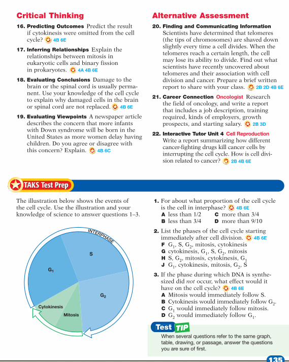
1. For about what proportion of the cell cycleis the cell in interphase? 4B 6EA less than 1/2 C more than 3/4B less than 3/4 D more than 9/10
2. List the phases of the cell cycle startingimmediately after cell division. 4B 6EF G1, S, G2, mitosis, cytokinesisG cytokinesis, G1, S, G2, mitosis H S, G2, mitosis, cytokinesis, G1J G1, cytokinesis, mitosis, G2, S
3. If the phase during which DNA is synthe-sized did not occur, what effect would ithave on the cell cycle? 4B 6EA Mitosis would immediately follow S.B Cytokinesis would immediately follow G2.C G1 would immediately follow mitosis.D G2 would immediately follow G1.
Critical Thinking16. Predicting Outcomes Predict the result
if cytokinesis were omitted from the cellcycle?
17. Inferring Relationships Explain therelationships between mitosis in eukaryotic cells and binary fission in prokaryotes.
18. Evaluating Conclusions Damage to thebrain or the spinal cord is usually perma-nent. Use your knowledge of the cell cycleto explain why damaged cells in the brainor spinal cord are not replaced.
19. Evaluating Viewpoints A newspaper articledescribes the concern that more infantswith Down syndrome will be born in theUnited States as more women delay havingchildren. Do you agree or disagree withthis concern? Explain.
Alternative Assessment20. Finding and Communicating Information
Scientists have determined that telomeres(the tips of chromosomes) are shaved downslightly every time a cell divides. When thetelomeres reach a certain length, the cellmay lose its ability to divide. Find out whatscientists have recently uncovered abouttelomeres and their association with celldivision and cancer. Prepare a brief writtenreport to share with your class.
21. Career Connection Oncologist Researchthe field of oncology, and write a reportthat includes a job description, trainingrequired, kinds of employers, growthprospects, and starting salary.
22. Interactive Tutor Unit 4 Cell ReproductionWrite a report summarizing how differentcancer-fighting drugs kill cancer cells byinterrupting the cell cycle. How is cell divi-sion related to cancer?
TAKS Test PrepTAKS Test Prep
G1
G2
Mitosis
Cytokinesis
S
INTERPHASE
When several questions refer to the same graph,table, drawing, or passage, answer the questionsyou are sure of first.
4B 6E
4A 4B 6E
4B 6E
2B 2D 4B 6E
2B 3D
2B 4B 6E
4B 6C
135