section 3 organogenesis - uv
TRANSCRIPT

SectionOrganogenesis
3
EDBC13 28/07/2005 13:29 Page 177

EDBC13 28/07/2005 13:30 Page 178
The chapters of this section will describe the development ofvarious selected organ systems, mostly in higher vertebrates,although certain aspects are illuminated by studies on the lowervertebrate and invertebrate models.
The techniques used in organogenesis research are essentiallysimilar to those used in early development. In addition, muchuse is made of in vitro organ cultures of rudiments from mouseand chick embryos as these are more accessible than the samestage organ in vivo. Knockout mouse strains provide most of theloss-of-function data, and it is often possible to grow an organculture from a knockout embryo beyond the stages at which thewhole embryo remains viable. Expression studies are done by in situ hybridization and immunostaining. Biological activitydata are acquired from addition of proteins to the organ cul-tures, or introduction of the genes encoding them by electropo-ration or viral infection.
This chapter deals with the chief tissue types found in the vertebrate body with special attention to their cellular renewal.On the basis of light microscopy there are about 200 types of differentiated cell, although in situ hybridization and immunos-taining reveal many more. They are arranged in tissues, each ofwhich contains several different cell types. An organ or body part contains several tissue types arranged to fulfill a commonfunction, and they are usually derived from more than oneembryonic cell lineage.
Types of tissue
Epithelia
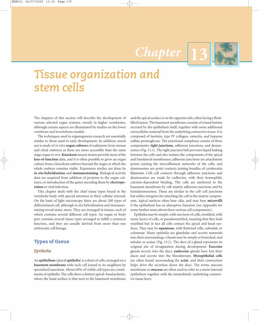
An epithelium (plural epithelia) is a sheet of cells, arranged on abasement membrane with each cell joined to its neighbors byspecialized junctions. About 60% of visible cell types are consti-tuents of epithelia. The cells show a distinct apical–basal polarity,where the basal surface is that next to the basement membrane
and the apical surface is on the opposite side, often facing a fluid-filled lumen. The basement membrane consists of a basal laminasecreted by the epithelium itself, together with some additionalextracellular material from the underlying connective tissue. It iscomposed of laminin, type IV collagen, entactin, and heparansulfate proteoglycan. The junctional complexes consist of threecomponents: tight junctions, adherens junctions, and desmo-somes (Fig. 13.1). The tight junction belt prevents liquid leakingbetween the cells and also isolates the components of the apicaland basolateral membranes; adherens junctions are attachmentpoints joining the microfilament networks of the cells; anddesmosomes are point contacts joining bundles of cytokeratinfilaments. Cell–cell contacts through adherens junctions anddesmosomes are made by cadherins, with their homophilic calcium-dependent binding. The cells are anchored to the basement membrane by cell–matrix adherens junctions and byhemidesmosomes. These are similar to the cell–cell junctionsbut utilize integrins for attaching the cell to the matrix compon-ents. Apical surfaces often bear cilia, and may bear microvilliif the epithelium has an absorptive function (see Appendix forsome further notes about these various cell components).
Epithelia may be simple, with one layer of cells, stratified, withmany layers of cells, or pseudostratified, meaning that they lookstratified but in fact all cells contact the apical and basal sur-faces. They may be squamous, with flattened cells, cuboidal, orcolumnar. Many epithelia are glandular and secrete materialsinto their surroundings. Glands may be simple or branched, andtubular or acinar (Fig. 13.2). The duct of a gland represents itsoriginal site of invagination during development. Exocrineglands secrete into the duct, endocrine glands have lost theirducts and secrete into the bloodstream. Myoepithelial cellsare often found surrounding the acini, and their contractionhelps drive the secretion down the duct. The terms mucousmembrane or mucosa are often used to refer to a moist internalepithelium together with the immediately underlying connect-ive tissue layer.
Tissue organization and stem cells
Chapter 13
EDBC13 28/07/2005 13:30 Page 179

Although commonly thought to be ectodermal in origin,epithelia are, in fact, derived from all three of the germ layers ofthe embryo. The organization and cell renewal in epidermis andintestinal epithelium are described below, and of neuroepithe-lium in Chapter 14.
180 u Chapter 13
Connective tissues
The term connective tissue refers to those tissues dominated by fibroblasts, such as the dermis of the skin and the fibrous capsules surrounding most organs. In some histology or biology
Microfilamentbundle
Cytokeratinfilaments
Hemidesmosome Basement membrane
Apical specialization
Tightjunction
Adherensjunction
Desmosome
Fig. 13.1 Diagram of an epithelial cellshowing the types of cell junction.
Simple squamous Simple columnar Stratified squamous
Branched acinar gland
Simple acinar glandSimple tubular gland
Fig. 13.2 Types of epithelium.
EDBC13 28/07/2005 13:30 Page 180

textbooks it may be used in a wider sense to include the skeletaltissues, muscle, and even the cells of the blood.
Much of the connective tissue is derived from the mesodermof the embryo, although some is also formed by the neural crest.Mature connective tissue consists of fibroblasts embedded in anextracellular matrix. Fibroblasts are cells specialized to secrete thematrix components which include hyaluronan, proteoglycans,fibronectin, type I collagen, type III collagen (reticulin), andelastin. Also found in connective tissue are histiocytes, which are macrophages resident in the tissue, and mast cells, which arehistamine-secreting cells similar to the basophils of the bloodbut also resident in the tissues. Both these types originate fromthe bone marrow. Adipose tissue is closely related to loose connective tissue, as fibroblasts can become adipocytes underappropriate conditions.
The skeletal tissues are composed of cartilage and bone andarise both from embryonic mesoderm and neural crest. Much of the skeleton is formed initially as cartilage which is then gradually replaced by bone. Skeletal parts formed in cartilage are known as cartilage models. Some parts, particularly in theskull, differentiate directly from mesenchyme into bone, andthese are known as membrane bones. Skeletal tissues are dis-cussed further in Chapter 18.
Tissue organization and stem cells u 181
A term causing much confusion is mesenchyme. This is not a synonym for connective tissue nor for mesoderm. It is a de-scriptive term for scattered stellate cells embedded in a looseextracellular matrix (see also Chapter 2). Mesenchyme, derivedeither from mesoderm or from neural crest, fills up much of theembryo and forms fibroblasts, adipose tissue, smooth muscle,and skeletal tissues, however these tissues should not be referredto as “mesenchymal” once they are differentiated.
Muscle
There are three main types of muscle (Fig. 13.3): skeletal muscleis composed of elongated multinucleate cells called myofibers.Bundles of myofibers are gathered together in fascicles sur-rounded by a fibrous sheath, the perimysium, and the wholemuscle is surrounded by another sheath called the epimysium.Skeletal muscle is derived from the myotomes of the somites andits development is further described in Chapter 15. Smooth (=visceral) muscle exists as bundles of individual spindle-shapedmononuclear cells. These contain a similar contractile apparatusto skeletal muscle but it is not arranged as visible sarcomeres.Smooth muscle is derived from the lateral plate of the embryo
(a) Striated muscle
Muscle
Epimysium Perimysium
Fascicle
(b) Smooth muscle (c) Cardiac muscle
Myofibrils
Individualmyofiber
Nucleus Sarcomere
Intercalated disk
Fig. 13.3 Types of muscle.
EDBC13 28/07/2005 13:30 Page 181

and is found mainly around the gut, blood vessels, and the ductsof glands, where inherent rhythmic contraction is required.Smooth muscle is usually mitotically quiescent but can be stimu-lated to grow following tissue damage. Cardiac muscle occursonly in the heart. It derives from the anteroventral margin of thelateral plate mesoderm of the embryo. Like skeletal muscle it hasvisible myofibrils, but like smooth muscle it remains as indi-vidual cells. The cells are joined end to end by intercalated discswhich contain structural junctions (adherens and desmosomes),together with gap junctions that allow rapid spread of electricalsignals through the myocardium. Cardiac muscle, like skeletalmuscle, is postmitotic, although some growth can occur by cellenlargement. Development of the skeletal muscle and heart aredescribed in Chapter 15.
Neural tissues
Neural tissues comprise those cell types formed from the neuraltube and some of those formed from the neural crest. The neuraltube is composed of a specialized epithelium, the neuroepithe-lium, and produces both central neurons and glial cells, whilethe neural crest produces autonomic neurons of the peripheralnervous system, together with Schwann cells and pigment cells.Development of neural tissues is described in Chapter 14.
Blood and blood vessels
Blood contains a variety of cell types. In addition to the red cells, there are granulocytes, monocytes, and lymphocytes. Allthese, as well as other cells such as histiocytes, osteoclasts, andLangerhans cells of the skin, arise from hematopoietic tissue in the bone marrow. The hematopoietic system is a state of continuous cell production and renewal throughout life, and is further described below.
The circulatory system consists of arteries taking blood fromthe heart to the tissues, capillaries supplying the tissues, andveins returning blood to the heart (Fig. 13.4). All blood vesselshave three layers. The inner layer is composed of a single layer of endothelial cells sometimes with a little underlying con-nective tissue. The middle layer is composed of smooth muscle,which may be very thick in arteries and thinner in veins, and theouter layer is composed of fibrous connective tissue.
The capillaries consist of a single layer of endothelial cells with a basal lamina on the exterior surface. There is no smoothmuscle but there may be some associated contractile cells calledpericytes. Usually the capillary wall is continuous but some-times, as in the sinusoids of the liver, it contains gaps. Endothe-lial cells can divide throughout life and there is usually a low levelof growth associated with tissue remodeling. The formation ofnew capillaries by endothelial cell division and cell movement isknown as angiogenesis. A number of growth factors are activein promoting angiogenesis, particularly vascular endothelial cell
182 u Chapter 13
growth factor (VEGF) and the fibroblast growth factors (FGFs).The embryonic development of blood vessels is considered inChapter 15.
Tissue renewal
Measurement of cell turnover
The brief sketch above focused on the visible appearance of tissues. But overall morphology tells us little about the cell turn-over which is so critical to their maintenance. Although tissueculture cells in optimal medium may grow exponentially, this israrely true of cells within the body. Usually cell turnover is slow,and particularly in epithelia it is often compartmentalized withseparate proliferative and differentiating zones.
Capillaries
Arteriole
Venule
Fig. 13.4 A microcirculatory unit, showing joining of terminal arterioleand venule by capillaries. (After Gray’s Anatomy, 35th edn, 1973. Longman,figure 6.10, p. 595.)
EDBC13 28/07/2005 13:30 Page 182

Chapter 5). This will reveal which cells were in S phase at thetime that the label was administered. It makes it possible to labelat one time and then to trace the subsequent position and differ-entiation class of the cells that were labeled. If the cells continuedividing, then the incorporated BrdU will be diluted out after afew rounds of division and will no longer be detectable, there-fore long-term retention of the label is taken to indicate that thecells underwent their final S phase at the time of administration,and divided only once before becoming postmitotic. This finaldivision is sometimes called the cell’s “birthday.” Before BrdUbecame available, many similar studies were conducted using3H-thymidine (3HTdR), which is also incorporated into DNAin S phase and can subsequently be localized by autoradio-graphy (see Chapter 5).
Tissue types in the body can be classified on the basis of theirproliferative behavior, visualized with BrdU or 3H-thymidine:1 Postmitotic, such as neurons and skeletal or cardiac muscle.Once formed these cells do not divide again, although it is nowknown that their numbers can be replaced to a small extent fromundifferentiated progenitors. There is a limited new formationof neurons of the olfactory bulbs and the hippocampus fromneuronal stem cells in the ependyma. There is also some newformation of myofibers from satellite cells.2 “Expanding.” These tissues grow while the animal is growingand stop when adult size is attained. In the adult such tissues aremostly quiescent although there may be a slow turnover of cells.In addition they remain capable of growth to a greater or lesserdegree when stimulated by wounding. In this category are con-nective tissue, smooth muscle, and liver.
Tissue organization and stem cells u 183
3 Renewal. Here the tissue is in a constant state of cell turnover.There is an active proliferative zone containing stem cells (seebelow) and this feeds a population of differentiated cells, whichitself has a finite lifetime and is constantly dying and beingrepopulated. Examples are the hematopoietic system, the epider-mis of the skin, and the epithelium of the gut.
Much of the developmental biology of postnatal life concernsthe behavior of the renewal tissues, some of which are describedbelow. Of course renewal involves cell death as well as cell birth.The index of apoptotic cell death can be measured by severalmethods. The most popular are immunostaining for the pres-ence of one of the apoptosis-associated proteins such as the caspase enzymes, and the detection of DNA breaks by a methodcalled TUNEL (TdT-mediated dUTP nick end labeling). Herethe enzyme terminal nucleotidyl transferase is used to add amodified nucleotide, usually biotin-labeled, to the fragementedDNA of the dying cell. This is then detected with a fluorescent orenzyme-linked streptavidin.
Although most measurements of cell turnover look at the pro-portion of cells in cycle, or the proportion of cells in apoptosis,what is really required to understand the situation is a meas-ure of cell production rate and cell removal rate. To obtain a cellproduction rate it is necessary to know, as well as the S-phaselabeling index, the duration of the cell cycle and the proportionof the cycle spent in S phase. For example if cells divide on aver-age once per 24 hours and the S phase lasts 6 hours, then a shortpulse of BrdU would enable the observation of about 6/24, orone quarter, of the cells in cycle. So in this case the cell produc-tion rate is about 4× the cell labeling index. The cell removal rate
T H E H E M AT O P O I E T I C S T E M C E L L
A N D I T S C E L L L I N E A G E
In the 1940s it was known that irradiatedmice could be rescued by a bone marrowgraft. But it was thought that somesubstance or hormone present in themarrow was responsible. The paper by Ford et al. showed by identification of achromosomal marker that the rescueactivity of the marrow graft was actually due to colonization by blood-forming cells.
The second paper describes the ability ofhematopoietic cells to form monoclonalcolonies in the spleen of irradiated animals.This provided a method for quantifyingnumbers of particular types of progenitorand showed the existence of multipotentcells forming clones of mixed cell type.
The third paper established an in vitroassay for colony formation which resulted in
the characterization of further multipotentcell types and was used to purify thehematopoietic growth factors.
Finally, the paper of Spangrude et al.describes the isolation of pure HSCs frommouse bone marrow by cell sorting, usingthe criterion of Sca1+Lin−Thy1lo.Ford, C.E., Hamerton, J.L., Barnes, D.W.H. &
Loutit, J.F. (1956) Cytological identification
of radiation chimaeras. Nature 177, 452–454.
Till, J.E. & McCulloch, E.A. (1961) A direct
measurement of the radiation sensitivity of
normal mouse bone marrow cells. Radiation
Research 14, 213–222.
Bradley, T.R. & Metcalf, D. (1966) The growth of
mouse bone marrow cells in vitro. Australian
Journal of Experimental Biology and Medical
Science 44, 287–300.
Spangrude, G.J., Heimfeld, S. & Weissman, I.L.
(1988) Purification and characterization of
mouse hematopoietic stem-cells. Science 241,
58–62.
The multiplication of cell popula-tions can be estimated by countingthe proportion of visible mitoses toobtain a mitotic index. However,mitosis usually occupies a short per-iod within the cell cycle, and a popu-lation has to be growing fast to show a significant mitotic index. Moresensitive are methods that identifycells in S phase, which represents alonger fraction of the cell cycle andhence enables more cycling cells tobe observed. One simple method isimmunostaining for a protein asso-ciated with DNA replication: prolifer-ating cell nuclear antigen (PCNA).This will give an estimate of the proportion of cells in S phase at thetime of fixation. Alternatively, cells,tissues, or whole animals can belabeled by administration of a DNAprecursor, usually bromodeoxyurid-ine (BrdU), a thymidine analog thatis incorporated into DNA and can bedetected with a specific antibody (see
EDBC13 28/07/2005 13:30 Page 183

is rarely calculated, and often underestimated. Because the dura-tion of apotosis is quite short (1–4 hours) the flux to cell deathper 24 hours is a high multiple of the apoptotic labeling index.For example, if the apoptotic index is 1% and the dying cells areobservable for only 2 hours, then the flux to cell death is actuallyabout 1× (24/2) or 12% per day.
Stem cells
Stem cells (Fig. 13.5) are defined as cells:1 that can divide without limit;2 that are visibly undifferentiated;3 whose progeny include both further stem cells and cells destined to differentiate.
In addition, there are other properties characteristic of somebut not all types of stem cell:1 some can give rise to more than one type of differentiatedprogeny (pluripotent);2 some undergo obligatory asymmetrical division to yield onestem cell daughter and one daughter destined to differentiate.
Embryonic stem cells (ES cells, see Chapter 10) are usually con-sidered to be similar to the cells of the mammalian embryonicepiblast or inner cell mass and are capable of forming any celltype in the body if reimplanted into an embryo. Tissue stemcells, sometimes called adult stem cells, are found in renewaltissues of the postnatal animal and are normally thought to becommitted to form one particular tissue type. Thus, an intestinalstem cell can form only intestinal types and an epidermal stemcell can form only keratinocytes. This corresponds to the ideathat embryonic development is hierarchical, with the cells of theearly blastula or epiblast being able to form anything, and thenbeing progressively restricted in their potency by a succession of inductive signals. For example, in the course of developmentthe precursors of an intestinal stem cell would have been com-mitted to endoderm and then to intestine, while the embryonic
184 u Chapter 13
precursors of a hematopoietic stem cell would have been com-mitted to mesoderm and then to the blood-forming tissue.According to this view, the stem cell for each tissue type may bequite similar to the cells of the appropriate organ rudiment atthe phylotypic stage. This conception of stem cells resemblingembryonic tissue rudiments has recently been challenged byexperiments showing repopulation of many tissues by a singletype of stem cell, but this area remains controversial (see below).
By definition, stem cells are capable of unlimited division.However, they are by no means the only dividing cells in the tissue because their direct progeny are usually transit amplify-ing cells capable of dividing only a few times but whose divisioncan be regulated to correspond to the demand for new differ-entiated cells. The stem cells are usually a minority among thedividing population and generally grow more slowly than the transit amplifying cells. Although they must repopulate thestem-cell compartment as well as feed cells to the transit amplify-ing and differentiated compartments, this does not necessarilymean that every individual cell division need be an asymmetricalone. It is simply required that, on average, the progeny of the stem cell consists of 50% stem cells and 50% cells destined todifferentiate (Fig. 13.6). This is important, for example, whenconsidering the acquisition of monoclonality in intestinal crypts(see below).
In the adult body, cell growth is mainly confined to theregions of the renewal tissues containing the stem cells and thetransit amplifying cells, and elsewhere most cells are quiescent. It is to be expected that cell division should be under strictinhibitory control, as otherwise the unregulated growth of evenone single cell could easily become a macroscopic cancer anddestroy the organism in a few months.
Stem cell niches
Many tissues have a histological substructure such that they consist of many small repeating modules or units, for exampleglandular acini or intestinal crypts. These units are not only thefunctional units of the tissue, but are also often the units withinwhich cell proliferation and turnover are organized. The placeswhere stem cells are found contain a specific microenvironmentknown as the stem cell niche suitable for their persistence andgrowth. If the stem cells are removed from this microenviron-ment they will grow no more. Conversely if they are reintro-duced to the niche then they will grow again.
The molecular identity of stem cell niches is now becomingknown. One well characterized example lies in the germariumregions of the Drosophila ovary (Fig. 13.7). The ovary comprisesa set of ovarioles, each of which contains a string of egg cham-bers with the germarium at the proximal end. The egg chambersconsist of one oocyte and 15 nurse cells surrounded by folliclecells, as previously described in Chapter 11. The oocyte andnurse cells of each egg chamber are formed from four divisionsof a single germ cell in the germarium region. This cell is known
Fig. 13.5 Cell lineage in a renewal tissue, showing stem cells, transitamplifying cells, and differentiated cells.
Differen
tiated cells
Renewal
Stem cell
Transit amplifying cells
EDBC13 28/07/2005 13:30 Page 184

Tissue organization and stem cells u 185
absence of bam the the germinal stem cells do not differentiatebut continue to proliferate, forming a germ cell tumor. Con-versely the overexpression of bam under control of a heat shockpromoter causes premature cystoblast formation in all the ger-minal stem cells. Evidence that the repression of bam dependson the dpp signal from the somatic cap cells comes from experi-ments showing that overexpression of dpp has a similar effect toloss of function of bam, and loss of function of dpp has the sameeffect as overexpression of bam. Normally the germinal stem celldivisions are asymmetrical, because the daughter not contactingthe cap cells turns on bam and becomes a cystoblast. But one of the properties of the niche is that if some stem cells areremoved by genetic means then this creates some space adjacentto the cap cells. This space can become occupied by progenyfrom the surviving stem cell(s) that would normally havebecome cystoblasts, and thus the production of cystoblasts issuspended while the normal number of stem cells is restored.This behavior resembles the repopulation of mammalian bonemarrow, following irradiation and grafting of marrow from ahealthy donor (see below).
Stem cell
Stem cell
Transit amplifying cell
Stem cell
Stem cell
Transitamplifying cell
Stem cell
Stem cell
Transitamplifying cell
(a)
(b)
Two stem cells
Two transitamplifying cells
Fig. 13.6 Stem cells can maintain themselves either (a) by repeatedasymmetrical division or (b) by generating stem cell and transit amplifyingdaughters with equal frequency but at separate divisions.
Germinalstem cells
Cap cells
Terminalfilament
Fusome
Cystoblast
Stage of egg chamber maturation
Pre-follicle cells
16-cell cyst
Follicle cells
1 2a 2b 3
Fig. 13.7 The stem cell niche in the Drosophila ovary germarium.
C E L L T U R N O V E R I N T I S S U E S
The first paper is a study of mitoses in theepithelium of the small intestine and arrivesat the conclusion that cells must be beingcontinuously produced in the crypts andshed from the villi. The second paper usesthe incorporation of radioactive debris from3H-thymidine-labeled cells that have died asa cell marker for neighboring cells. Fromstudying the subsequent distribution of
these radioactive debris it is postulated that asingle type of stem cell produces all four celltypes of the small intestinal epithelium.Leblond, C.P. & Stevens, C.E. (1948) The constant
renewal of the intestinal epithelium in the
albino rat. Anatomical Record 100, 357–377.
Cheng, H. & Leblond, C.P. (1974) Origin,
differentiation and renewal of the four main
epithelial cell types in the mouse small intestine.
V. Unitarian theory of the origin of the four
epithelial cell types. American Journal of
Anatomy 141, 537–562.
as a cystoblast, and its precursors arethe germinal stem cells (= oogonia)which divide mitotically about onceper day. Each germarium containstwo or three germinal stem cells,adjacent to five or six somatic capcells and it is these somatic cap cellsthat define the niche. They do so bysecretion of the decapentaplegic(dpp) protein. This represses expres-sion of the bag of marbles (bam) genein the adjacent cells. bam encodes acytoplasmic protein which interactswith a germ cell-specific organellecalled the fusome and is needed forthe cystoblast maturation. In the
EDBC13 28/07/2005 13:30 Page 185

Skin
Skin consists of a squamous stratified epithelium, the epidermis,on top of a connective tissue, the dermis (Fig. 13.8). The maincell type in the epidermis is the keratinocyte. Cell division isconfined to the basal layer. This contains stem cells, which canboth renew themselves and generate keratinocytes, and alsotransit amplifying cells, which are formed from the stem cellsand have a finite division potential before they differentiate. Inhumans the epidermis is renewed from the basal layer aboutevery 2 weeks. The entire basal layer of the epidermis, and ofother squamous epithelia such as those of the esophagus orvagina, depends on the activity of a transcription factor calledp63. This is expressed throughout the basal layer and is switchedoff when cells migrate upwards. The knockout mouse lackingp63 is unable to form any squamous epithelia and dies soon after birth.
The dermis is a dense fibroelastic connective tissue derivedfrom the dermatome and neural crest of the embryo. At deeperlevels it is largely adipose tissue. The junction between dermisand epidermis is marked by a basement membrane and, inhumans, this is wrinkled with epidermal ridges and corres-ponding dermal papillae (note that the specialized dermal coreof the hair bulb is also called a dermal papilla) . The dermis con-tains the usual nerves and blood vessels, and pressure receptorscalled Pacinian corpuscles, as well as the epidermis-derived specializations.
The factors required for proliferation of the epidermal basallayer include both factors produced by the epidermis itself, suchas TGFα, and factors secreted by the underlying dermis, includ-ing keratinocyte growth factor (KGF), a member of the FGFfamily. Keratinocyte cultures can be grown in vitro and form thesame stratified arrangement as the natural skin. There is no needfor the dermis in such cultures as the growth factors which itnormally supplies are present in the medium. Labeling studiesshow that about 60% of basal layer cells are in cycle, but only afraction of these are stem cells. The functional test for a stem cellis that it can form a large self-sustaining epidermal colony eitherin culture, or after grafting to a nude mouse (a type of mousethat cannot reject tissue grafts). Transit amplifying cells, by contrast, can only form small colonies of a few cells and thenstop growing. By this criterion about 10% of basal layer cells arethought to be stem cells. A similar proportion, presumably the
186 u Chapter 13
same stem cells, are capable of forming large colonies that canrepopulate the epidermis following severe radiation damage.
The stem cells defined by these criteria are characterized by a higher level of β-1 integrin than the transit amplifying cells.This is a cell adhesion molecule involved in the recognition ofcollagen, laminin, and fibronectin. In vivo, in human foreskin,the high-integrin cell clusters are found at the tips of the dermalpapillae, suggesting that this may define the stem cell niche forthe epidermis. The stem cells also show an enhanced level ofnuclear β-catenin, indicating a possible role for Wnt signaling in the maintenance of the niche. If either β-1 integrin or β-catenin are introduced into keratinocyte cultures by retroviralinfection, then the cells receiving the gene will acquire stem cellproperties. The stem cells, both in vivo and in vitro, also con-tain an elevated level of the Notch ligand delta-1. If delta-1 isintroduced into cells of a keratinocyte culture by retroviralinfection, then the high delta-expressing cells tend to cluster and to be inhibited from differentiation, while their neighborsare stimulated to differentiate. This suggests that the distinctionbetween stem cell clusters and the surrounding transit cells maybe maintained by a lateral inhibition mechanism similar to thatinvolved in neurogenesis and pancreatic differentiation (seeChapters 4, 14, and 16). Although this picture is incomplete, itseems likely that the dermal papillae emit a Wnt signal, and thatthe adjacent basal layer cells respond by increasing synthesis ofβ-1 integrin. This helps maintain the stem cells as a cluster andalso has additional intracellular signaling effects leading to the in-crease of delta-1 expression and the spatial segregation of the basallayer into stem cell and transit amplifying cell zones (Fig. 13.9).
Once cells leave the basal layer they stop dividing and enter aprogram of further differentiation. The progress of maturationis reflected by the names given to successive layers of the epider-mis: stratum germinativum (the basal layer), stratum spinosum(the “prickle” layer – the apparent prickles are abundant desmo-somes), stratum granulosum (with granules), and stratumcorneum (cells have lost nuclei and have become flat sacs of keratin). Keratin is a generic name for the large family of fibrousproteins forming the cytokeratin intermediate filament familyand found in all epithelial cells. There are many different keratinscoded by different genes, and the repertoire expressed changesas cells move up from the basal layer. In the granular andcornified layers the cells also contain a tough internal sheath ofan insoluble protein called involucrin.
Stratum corneum
Post-mitotic cells
Basal layer
Basement membrane
Dermis
Fig. 13.8 Organization of the epidermis.All keratinocytes are born in the basal layerand differentiate progressively as they moveup to the surface.
EDBC13 28/07/2005 13:30 Page 186

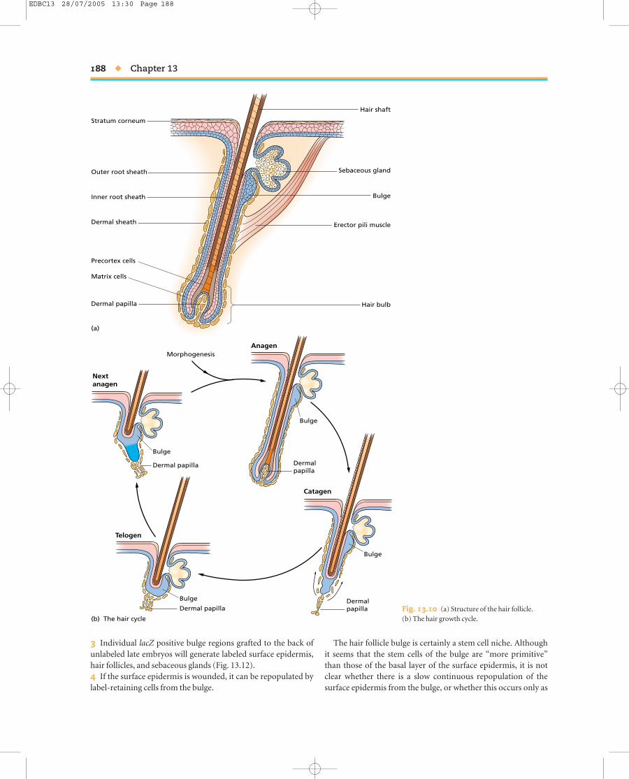
Hair follicles
The structure of the hair follicle is shown in Fig. 13.10a. The hairshaft is composed of dead keratinocytes, which are produced inthe epidermal matrix region at the base. This lies in close prox-imity to the dermal papilla, a projecting bud of fibroblastic cells,and also contains melanocytes which transfer pigment granulesto the keratinocytes of the hair. Surrounding the whole is a layerof cells continuous with the surface epidermis called the outerroot sheath. High up, near the junction with the surface epider-mis, lies a sebaceous gland. The entire region at the base of thefollicle comprising the dermal papilla, the proliferative epider-mal zone, and the outer layers, is known as the hair bulb. Hairdoes not grow continuously but in a cycle. The active growthphase is known as anagen, which lasts about 3 weeks in mice butcan be much longer in humans. The period of regression of thefollicle is called catagen, and the period of quiescence is calledtelogen (Fig. 13.10b).
Hair follicles start life in the late mammalian embryo as epi-dermal invaginations. Their formation requires both Wnt sig-naling and also inhibition of BMP signaling from the dermis.BMPs are produced by the epidermis, and noggin by the dermis.A combination of Wnt3A and noggin will induce new folliclebuds. The BMP inhibition causes transcription of Lef1 while the Wnt signal stabilizes β-catenin and the combination carriesthe Lef1 into the nucleus to control target genes (see Appendix).One target is E-cadherin, whose expression becomes repressed,thus reducing the mutual adhesion of the cells and leading to theinvagination behavior to form the bud. Evidence for this mech-anism is based on the the following:1 Invaginating buds are produced by adding Wnt3A + nogginto keratinocyte cultures, or in transfecting in Lef1 plus a con-sititutive form of β-catenin.2 Knockout mice lacking either noggin or Lef1 have few hair follicles.3 The Wnt signaling pathway is active in the early buds. Thismay be seen in a reporter strain of transgenic mice which con-tains lacZ driven by a Wnt-sensitive promoter.4 Ectopic hair follicles are formed in mice transgenic for con-stitutive β-catenin, driven by the keratin 14 promoter which isactive only in the basal layer of the epidermis.
Tissue organization and stem cells u 187
5 Bud formation is suppressed in mice transgenic for produc-tion of the Wnt-inhibitor Dickkopf, also driven in the basal layerby the keratin 14 promoter.
The understanding of hair follicle initiation makes it possibleto contemplate a possible “cure” for human baldness. Howeverthe overexpression of Wnt pathway components in humanpatients would probably not be acceptable because of the risk ofinducing cancers.
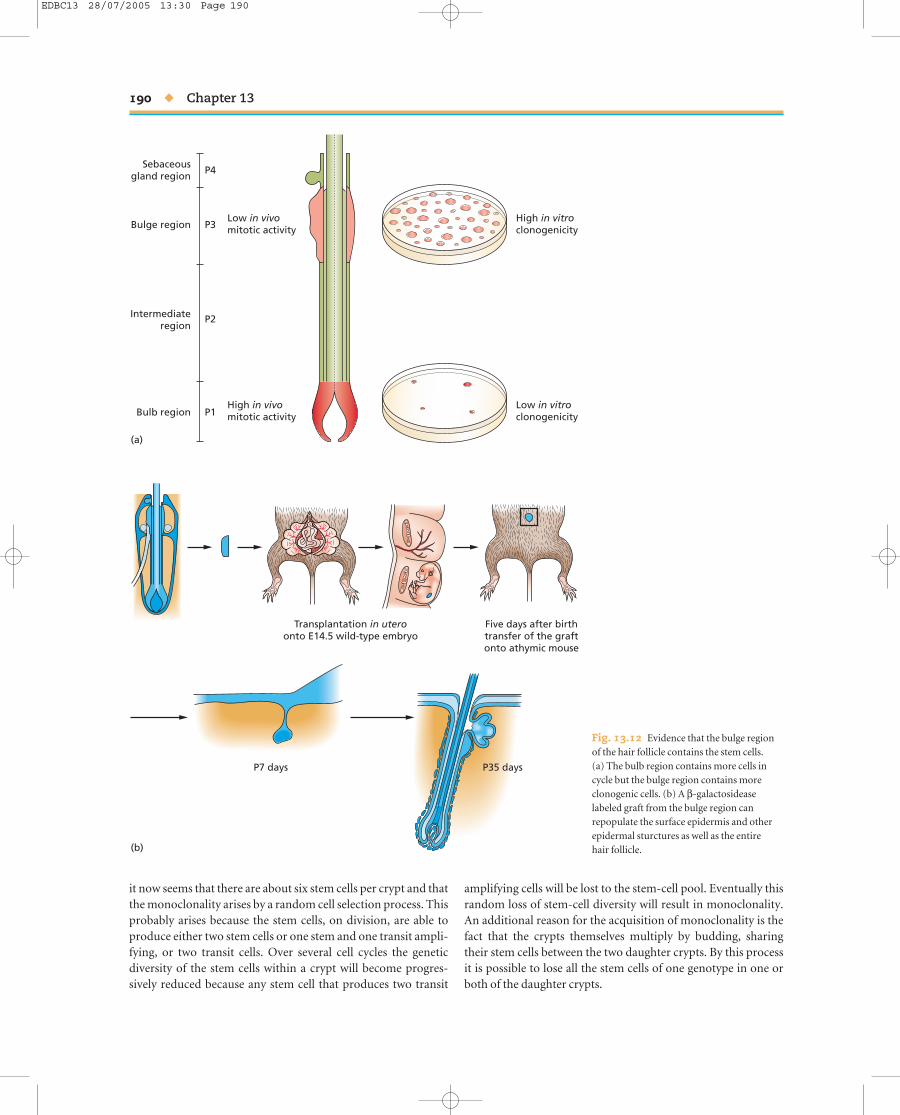
Although the initial signal for bud formation comes from thedermis, the formation of the dermal papilla depends on a sec-ond signal from the invaginating bud to the dermis (Fig. 13.11).The dermal papilla secretes growth factors needed by the pro-liferative zone of the epidermal matrix region. Isolated papillaewill induce new epidermal proliferative zones from the upperhalves of follicles, and in some situations can induce completenew follicles from epidermis. Examination of mouse aggrega-tion chimeras in which one component is labeled and the other unlabeled suggests that each follicle contains about fourepidermal stem cells, all of which contribute to all the layers ofthe hair shaft. In aggregation chimeras the cells derived from thetwo embryos are intimately mixed and so, allowing for similaradjacent clones, the number of labeled and unlabeled patches in a small structure like a hair approximate to the number ofclones, and therefore the number of stem cells (see also Chap-ter 10).
As far as stem cells are concerned, attention in the past con-centrated on the epidermal matrix region and the dermal papillabecause of their obvious role in hair shaft formation. However itis now thought that the real epidermal stem cell population ofthe hair follicle lies not in the bulb but in a lateral bulge half wayup the outer root sheath. The evidence for this is as follows:1 A 3-day label with BrdU will label many cells in the epidermisof the hair follicle. But a long chase period shows that the cellsthat retain the label, and are thus very slowly dividing, are con-centrated in the bulge region rather than in the bulb. In the nextactive growth cycle (anagen) some of these labeled cells are seento have migrated into the bulb.2 If a bulge region from a lacZ positive (Rosa26) mouse isgrafted to a large vibrissal follicle and then cultured under thekidney capsule of a nude mouse, the β-galactoside expressingcells are seen to populate the entire follicle.
Wnt? Wnt? Wnt?
delta delta delta
integrin
β-cateninFig. 13.9 The stem cell niche in theepidermis. Signals from the dermis, probablyincluding Wnt, maintain groups of stemcells. The signals increase the level of β1-integrin, which causes the stem cells toremain as a small cluster. The stem cellsdisplay delta-1 on their surfaces, therebyrepressing the surrounding cells from stemcell behavior.
EDBC13 28/07/2005 13:30 Page 187
188 u Chapter 13
MorphogenesisAnagen
Catagen
Telogen
Nextanagen
Bulge
Bulge
Bulge
Bulge
Outer root sheath
Inner root sheath
Precortex cells
Hair shaft
Stratum corneum
Erector pili muscle
Sebaceous gland
Dermal sheath
Matrix cells
Dermal papilla
Dermalpapilla
Hair bulb
Bulge
(a)
(b) The hair cycle
DermalpapillaDermal papilla
Dermal papilla
Fig. 13.10 (a) Structure of the hair follicle. (b) The hair growth cycle.
3 Individual lacZ positive bulge regions grafted to the back ofunlabeled late embryos will generate labeled surface epidermis,hair follicles, and sebaceous glands (Fig. 13.12).4 If the surface epidermis is wounded, it can be repopulated bylabel-retaining cells from the bulge.
The hair follicle bulge is certainly a stem cell niche. Althoughit seems that the stem cells of the bulge are “more primitive”than those of the basal layer of the surface epidermis, it is notclear whether there is a slow continuous repopulation of the surface epidermis from the bulge, or whether this occurs only as
EDBC13 28/07/2005 13:30 Page 188

a specific response to wounding. The bulge region does, in asense, represent the most secluded part of the epidermis as it iscontinuous with the surface epidermis and is the lowest level ofthe follicle that persists throughout the hair cycle. In the humanthere is no visible bulge, but the stem cells also reside in the lowest permanent part of the outer root sheath.
Intestine
The gastrointestinal tract of vertebrates consists of a musculartube running from the mouth to the anus. It is lined by a num-ber of different epithelia: pharyngeal, esophageal, gastric, small
Tissue organization and stem cells u 189
intestinal, and colonic, separated by abrupt discontinuities ofcell type. This epithelium is derived from the endoderm of theearly embryo, while the other cell layers of the gut are derivedfrom the splanchnic mesoderm. The epithelium, together withunderlying connective tissue called the lamina propria and athin muscle layer called the muscularis mucosa, is often knownas the mucosa. Outside the mucosa lie further thick layers ofconnective tissue and smooth muscle.
The program of cell renewal is best understood for the smallintestine. This contains regions called the duodenum, jejunum,and ileum, although the difference of cell type between them is not great. On a microscopic scale the intestinal epithelium is arranged on finger-like villi projecting into the lumen, andbetween the villi lie crypts of Lieberkuhn sunk below the surface(Fig. 13.13a,b). Cell proliferation takes place only in the crypts,and differentiated cells are continuously moving out from thecrypts, moving up the villi, then dropping off into the gut lumen.The main cell types are enterocytes (absorptive cells) and gobletcells. The absorptive cells are characterized by a brush border at their apical surface, consisting of numerous close-packedmicrovilli. Goblet cells contain a large vesicle filled with mucins.In addition there are Paneth cells located at the base of thecrypts, which secrete antibacterial substances, and several typesof enteroendocrine cells each secreting a particular peptide hormone. The crypts themselves are set up shortly before birthby folding of the endodermal epithelium which before this stageis a simple columnar epithelium. During the growth of the animalthey can divide by budding, starting at the base (Fig. 13.13c).The signal for crypt division is not known but may be an increasein the number of stem cells. Some experimental studies have alsobeen performed on the colon because of its importance in termsof colonic cancer. Its structure is similar to that of the smallintestine, but without villi and without Paneth cells.
Cell division in the crypts is rapid. The stem cells are locatednear the crypt base, above the Paneth cells, with several layers of transit amplifying cells lying above them. A mouse crypt contains about 250 cells of which roughly 160 are dividing, witha cycle time of about 13 hours. The progeny move up and out of the crypts and the tissue is arranged such that each crypt feedsmore than one villus, and each villus draws cells from severalcrypts. A small proportion of cells also move down the crypt toreplenish the Paneth cells at the crypt base.
It has been possible to examine the clonal composition of thecrypts by making aggregation chimeras between mouse strainsthat differ in the expression of a marker, Dolichos lectin receptor(Fig. 13.14a). This carbohydrate is expressed by intestinal cells in some mouse strains and is absent in others. In an aggrega-tion chimera the cells of the two donor embryos are intimatelymixed, so at the time of crypt formation most crypts will receivecells of both donor types. However, over the first 1–2 weeks of postnatal life the crypts lose one of the two types, such that all cells in each crypt are either one type or the other. In otherwords, the crypts become monoclonal. Initially it was thoughtthat this meant there was only one stem cell per crypt. However,
Epidermis
First mesenchymal signal
noggin, Wnt
Epidermal signal
Second mesenchymal signal
Dermis
Dermis
Dermis
Dermal condensate
Dermal papilla
Epidermis
Epidermis
Fig. 13.11 Initial formation of hair follicles. The first phase involvesinduction by noggin and Wnt from the dermis. This is followed by a signalfrom the epidermal bud inducing a specialized dermal papilla.
EDBC13 28/07/2005 13:30 Page 189
it now seems that there are about six stem cells per crypt and thatthe monoclonality arises by a random cell selection process. Thisprobably arises because the stem cells, on division, are able toproduce either two stem cells or one stem and one transit ampli-fying, or two transit cells. Over several cell cycles the geneticdiversity of the stem cells within a crypt will become progres-sively reduced because any stem cell that produces two transit
190 u Chapter 13
amplifying cells will be lost to the stem-cell pool. Eventually thisrandom loss of stem-cell diversity will result in monoclonality.An additional reason for the acquisition of monoclonality is thefact that the crypts themselves multiply by budding, sharingtheir stem cells between the two daughter crypts. By this processit is possible to lose all the stem cells of one genotype in one orboth of the daughter crypts.
Sebaceousgland region
Bulge region
Intermediateregion
Bulb region
P4
P3Low in vivomitotic activity
High in vivomitotic activity
High in vitroclonogenicity
Low in vitroclonogenicity
P2
P1
(a)
(b)
Transplantation in uteroonto E14.5 wild-type embryo
Five days after birthtransfer of the graftonto athymic mouse
P7 days P35 days
Fig. 13.12 Evidence that the bulge regionof the hair follicle contains the stem cells. (a) The bulb region contains more cells incycle but the bulge region contains moreclonogenic cells. (b) A β-galactosideaselabeled graft from the bulge region canrepopulate the surface epidermis and otherepidermal sturctures as well as the entire hair follicle.
EDBC13 28/07/2005 13:30 Page 190

Estimates of the proportion of stem cells among the dividingcells of the crypts have been made by several methods includingmodeling of the cell-cycle kinetics of the whole crypt. The esti-mate of six is based mainly on mutagenesis studies (Fig. 13.14b).If a mouse of a suitable strain is mutagenized, then a proportion of cells will acquire reactivity for Dolichos lectin. Shortly after themutagenesis, many crypts containing mutant clones are seen.Most of the clones arise in transit amplifying cells and are soonlost. But if they arise in stem cells they are retained in the longterm. In principle, if there are n stem cells per crypt, then a muta-tion in a stem cell should appear as a labeled sector within thecrypt occupying about 1/n of its circumference. In fact, after a few weeks many mutant crypts are uniformly composed ofmutant cells, showing the same drift to monoclonality as aggre-gation chimeras, probably for the same reasons. Mutagenesisalso enables the study of the contribution of each crypt to thevilli, as a labeled crypt will emit a stream of labeled cells thatform a strip running up each of the villi to which it contributes(Fig. 13.14b).
A different type of estimate of stem-cell number can be madebased on radiation toxicity (Fig. 13.14c). A given dose of X- orgamma rays will sterilize a proportion of cells in the epithelium.Cells that are capable of growing and repopulating the tissue areknown as clonogenic cells. It is assumed that a crypt can only
Tissue organization and stem cells u 191
regenerate if it includes at least one clonogenic cell that survivedthe radiation. Measurements of crypt survival following variousdose regimes suggest that there are about 80 clonogenic cells percrypt. This is substantially greater than the likely number of stemcells per crypt calculated from mutagenesis, and the difference inthe two estimates suggests that a proportion of the transit ampli-fying cells are capable of becoming stem cells again under condi-tions of severe tissue damage. This makes sense if it assumed thata transit amplifying cell will become promoted to stem cell statusif it can enter the stem cell niche. The mutagenesis and radiationexperiments also provide data to support the idea that the stemcells of the small intestine are pluripotent, being able to gener-ate all four of the usual cell types: columnar, goblet, Paneth, and enteroendocrine. Crypts entirely populated by one mutantclone contain all four cell types, so one cell must have generatedthem all. Similarly, after high doses of radiation from which onlya minority of crypts survive, these will mostly have regeneratedfrom a single clonogenic cell, but will nevertheless all acquire thefour cell types.
Knowledge of the molecular characteristics of intestinal stem cells remains limited. An RNA-binding protein calledMusashi-1 is expressed in a few cells between and just above thePaneth cells, and may be a stem cell marker. There is an increasein the number of Musashi-1 positive cells during the recovery
Villus
Crypt
Goblet cell
Mitotic cells
Paneth cells(a)
(b) (c)
Absorptive cell
Enteroendocrine cell
Fig. 13.13 Diagrammatic organization of the small intestinal epithelium. (a) Longitudinal section of two crypts and a villus. (b) Transverse section of a crypt. (c) Multiplication of crypts by budding from the base.
EDBC13 28/07/2005 13:30 Page 191

192 u Chapter 13
from radiation damage, which is consistent with the increase in the number of clonogenic cells in this situation. As in the epi-dermis, there is good evidence that the intestinal proliferativecompartment depends on the Wnt signal transduction path-way (Fig. 13.15). It is known that the mouse knockout of thetranscription factor gene tcf4 fails to form any proliferative compartment. TCF4 is one of the HMG-type transcription factors that is activated by β-catenin and nuclear β-catenin isnormally found in the cells of the bottom third of the crypt,which represents the proliferative compartment. So it is likelythat Wnt signaling from the lamina propria is needed to main-tain proliferation in the epithelium, and something additional isneeded to define the much smaller stem cell niche.
Mouse embryos Aggregationchimera
Polyclonal crypts Monoclonal crypts
Mutant crypt
(b) Mutagenesis
(c) Regeneration
Normal crypts
Newborn Later
(a) Chimera
Mutagenesis
Radiation Budding
Villi
Mouse
Most cells killed Regeneration ofisolated crypts
Full regeneration
+
Fig. 13.14 Methods for studying intestinal crypt organization. (a) Aggregation chimeras. Early crypts are polyclonal but later become monoclonal. (b) Mutagenesis produces occasional cells that can be visualized by binding of Dolichos lectin. One mutant stem cell may often come to populate an entirecrypt, and its progeny form streams up to the tips of the adjacent villi. (c) A dose of X-radiation that destroys most cells leads to regeneration of whole cryptsfrom individual clonogenic survivors.
Wnt Wnt
Wnt
Ephrin B1
Eph B2, B3Proliferative zone:nuclear β-cateninand TCF4
Fig. 13.15 Role of the Wnt pathway in controlling the proliferativestructure of the intestine.
EDBC13 28/07/2005 13:30 Page 192

a cytoplasmic protein required to enable the phosphorylation of β-catenin by GSK3. In loss-of-function mutants β-catenin isnot inactivated, and is therefore constitutively active. This leadsto the inability to shut off Eph B2 and B3, and to the formationof polyps, which are projections into the lumen of differentiatedbut abnormally organized intestinal tissue. Human patients suffering from adenomatous polyposis coli have many suchpolyps and a high risk of a polyp developing to cancer (see below).The disease is hereditary and due to loss of one copy of the APCgene. When the other, good, copy is lost from an individual celldue to occasional somatic mutations, then that cell will acquireconstitutively active β-catenin and develop into a polyp.
In the intestine, as is generally the case for renewal tissues, the stem cells are responsible for producing several types of differentiated cell. It now seems that this is achieved using theDelta-Notch lateral inhibition mechanism (see also Chapters 4,14, and 16). There is a “master switch” at the level of the decisionwhether to become an ordinary absorptive cell or one of thethree specialized cell types (goblet, enteroendocrine, or Paneth),and this is controlled by a bHLH type transcription factor calledMath1, which also promotes the formation of Delta (Fig. 13.16).All the cells express Notch and initially have a similar level ofMath1. Cells which by chance have a slightly higher level ofMath1 produce a little more Delta and signal to surroundingcells. Notch is stimulated in these surrounding cells leading toinhibition of expression of Math1, and hence reduction of Delta.The process will run on until there are a few high Math1-highDelta cells surrounded by a larger number of low Math1-lowDelta cells. The cells with low Math1 become absorptive cells,while those with high Math1 become either goblet or enteroen-docrine or Paneth, the subsequent decisions depending on fur-ther unknown mechanisms. The main evidence for this processis that the knockout of math1 has an intestinal epithelium which
Tissue organization and stem cells u 193
is normal in overall structure, and which contains normalabsorptive cells, but totally lacks all the types of specialized cell.Another knockout, of the transcription factor gene hes1, showsan opposite phenotype with an elevation of Math1-expressingcells and of the proportion of specialized cell types in the epithelium. Hes1 is on the Notch signaling pathway and its loss will reduce the inhibition of Math1 expression by Notch signaling.
Stem cell research is thought to be rich in practical applications, mostlyhaving the aim of repairing damaged tissues and organs by grafts of stemcells. In recent years many biotech companies have been founded on thebasis of these opportunities.
In terms of basic knowledge it is important to establish exactly howtissue stem cells arise during development and how similar they really are to the whole embryonic rudiment from which the tissue develops.
We need to know if Wnt signaling really controls stem cell behavior, or just proliferative behavior generally.
We also need to understand whether the Notch lateral inhibition systemcontrols the differentiation of multiple cell in all cases, or whether there are other similar systems.
Ultimately, understanding the signals that control the self-renewal and differentiation of the stem cells should enable the design of cultureconditions for growing tissue stem cells without limit in vitro.
There is also an intimate connec-tion between the control of prolif-eration and the actual structure ofthe epithelium, because the crypts arecharacterized by expression of theadhesion molecules Eph B2 and B3,while the villi are characterized byexpression of their ligand ephrin B1.Transcription of these molecules is,respectively, activated and repressedby β-catenin. If both Eph B2 and B3 are removed by targeted mutagenesisthen the intestine loses its structureand both dividing and differentiatedcells are found mixed together. Ifonly EphB3 is removed then thestructure is normal but Paneth cellsare found all over the epitheliuminstead of being confined to the crypt bases. APC, the product of theadenomatous polyposis coli gene, is
Deltaslightlyhigher
Deltaslightlylower
Deltaslightlylower
Delta NotchNotch
Math1
NeuroD
Math1Math1
Specialized cell(goblet, enteroendocrine,
Paneth)Absorptive cell
Fig. 13.16 Control of cell differentiation in the intestinal epithelium bylateral inhibition.
EDBC13 28/07/2005 13:30 Page 193

Hematopoietic system
In the adult mammal, the hematopoietic system resides in the bone marrow within the larger bones of the skeleton. In theembryo it is found at various other sites. Initially it is extra-embryonic, in the yolk sac, then in the AGM (aorta-gonad-mesonephros) region of the mesoderm, then in the liver, thenin the spleen and lymph nodes, and finally in the bone marrow.The first two of these sites of production arise in situ while the later ones are colonized by cell migration from the earliersites. This process of cell migration has been established by thegrafting of marked cells in amphibian and avian embryos andlocalizing the progeny of the grafted cells at a later stage.
Like the skin and gut epithelium, the hematopoietic system isa state of continuous cell production and renewal throughoutlife. There exists a hematopoietic stem cell (HSC) that can bothrenew itself and also differentiate into a wide variety of cell types.These include all cells of the blood and immune system togetherwith histiocytes, osteoclasts, and Langerhans cells of the skin.
The cellular components of the blood are as follows:1 Erythrocytes (red cells).2 Granulocytes, comprising neutrophils (phagocytes), eosino-phils, and basophils (similar to mast cells).3 Monocytes (similar to macrophages/histiocytes).4 Megakaryocytes, giant cells that break up to become platelets.The above four cell types are collectively known as myeloid cells.5 Lymphocytes (T and B cells).
Much of the evidence for the existence of HSCs comes fromreconstitution experiments. The bone marrow is the most sens-itive tissue in the body to irradiation, and so there is a dose rangethat will kill by bone marrow failure while other tissues are stillpotentially able to recover. If a mouse, lethally irradiated withsuch a dose, is given a graft of marrow cells by injection into thebloodstream, then the graft will colonize the marrow of the host,proliferate extensively, and enable survival of the host. After afew weeks the counts of the various cell types mentioned abovehave returned to normal and the observation of genetic markersshows that they are all derived from the graft. The ability per-manently to rescue lethally irradiated mice is often taken to be the defining feature of the HSC. The use of reconstitutionassays to examine the hematopoietic populations in the mouseembryo shows that the earliest HSCs are found in the AGMregion at 10–11 days of gestation and in the yolk sac and liverfrom about 12 days of gestation. This suggests that the hemato-poietic population seen in the yolk sac at earlier stages does notcontain HSCs capable of long term repopulation.
It is now possible to isolate HSCs directly from bone mar-row using the technique of fluorescence-activated cell sorting(FACS, see Chapter 5). Mouse HSCs are characterized by highlevels of Sca1, low but finite levels of Thy1, and the absence of allother differentiation markers (Sca1+, Thy1lo, Lin1−). Sca1 (stemcell antigen 1) and Thy1 (thymus 1) are both cell surface glyco-proteins, Thy1 being abundant on mature T cells. The study ofmice transgenic for GFP driven by the Sca1 promoter shows
194 u Chapter 13
that the very first HSCs arise in the endothelium of the dorsalaorta. Mouse HSCs can also be isolated because they preferenti-ally exclude certain fluorescent dyes such as Hoechst 33324 orRhodamine 123, so after exposure to these dyes give a lowerfluorescence signal than all other cells in the marrow. The self-renewing properties of the HSC seem to depend on the proto-oncogene bmi-1, which encodes a polycomb type transcriptionalrepressor. Knockouts for bmi-1 develop HSCs but the numbersare greatly reduced postnatally and they have very little recon-stitution activity. HSCs are difficult to grow in culture but thenumbers, measured by the mouse reconstitution assay, can beincreased by introduction of certain genes using retroviruses.These include stabilized (constitutive) β-catenin, suggestingthat Wnt signaling may be necessary for HSC self-renewal, as it isfor the stem cells of the skin and the intestine.
In addition to the HSC, the marrow contains other cells that can be isolated by different combinations of cell surfacemarkers. In the reconstitution assay they give rise to only a sub-set of the complete HSC repertoire. This shown the existence of various multipotent progenitors including the common lymphoid progenitor, the common myeloid progenitor, thegranulocyte-macrophage progenitor, and the megakaryocyte-erythrocyte progenitor. There are also pluripotent stem cellsthat have only a temporary repopulating ability, which arebelieved to represent the next step of maturation after the per-manent HSC. A current consensus model for the cell lineage ofthe hematopoietic system is shown in Fig. 13.17.
A second line of evidence for this model comes from in vitrocolony assays. It is possible to obtain clones of hematopoieticcells in vitro by plating marrow cells in soft agar or methyl cellu-lose in the presence of the appropriate growth factors. Sincemost single colonies will be clones derived from a single cell, theproduction of multiple cell types by a single colony indicates theexistence of a multipotent progenitor, and the same potencyclasses are recovered in these assays as in the whole mousereconstitution assay. Although the model is generally accepted,it has not yet been confirmed by prospective labeling, whichwould require the insertion of a permanent genetic marker intoHSCs in vivo, followed by identification of each type of progenycell as it is formed.
The in vitro colony formation assay has been used to isolate anumber of colony-stimulating factors (CSFs), otherwise knownas hematopoietic growth factors. Interleukin 3 and stem cell factor (steel factor) can stimulate proliferation of the HSCs,while granulocyte–macrophage CSF (GM-CSF), granulocyte CSF(G-CSF), macrophage CSF (M-CSF), and erythropoietin workin combinations to stimulate the division of the various transitamplifying lineages. Most of the feedback control over the pro-duction of the various cell types is exerted by varying the growthrate at the transit amplifying cell level. This is because there arelarge numbers of such cells and a rapid response can be obtainedto changing demand. By contrast, it would take several weeks toalter the rate of production by regulation at the level of the HSC.Several of the hematopoietic growth factors have been prepared
EDBC13 28/07/2005 13:30 Page 194

in therapeutic quantities by recombinant DNA methods and are now very useful in clinical practice, particularly for the treat-ment of various types of anemia and for enabling people torebuild their marrow after cancer therapy.
Tissue organization and stem cells u 195
The transplantable properties of the HSC have also mademarrow grafting one of the earliest types of cell therapy to beadopted in human medicine. Indeed the first human bone mar-row grafts between identical twins were performed in the 1950s.
BFU–E Erythroblast
CFU–GM
CFC–Meg
Granulocyteprogenitor
Pre-T T cell
Pre-B B cell
Eosinophil
Neutrophil
Basophil
Reticulocyte Erythrocyte
Nucleus
MacrophageMonocyte
Megakaryocyte
Platelets
Commonlymphoid
progenitor
Commonmyeloid
progenitor
Erythrocyte–megakaryocyte
precursor
Hematopoieticstem cell
(HSC)
Short-termHSC
Self-renewal
Fig. 13.17 A consensus model for the cell lineage for the cells of the blood and immune system.
EDBC13 28/07/2005 13:30 Page 195

Grafting of the patient’s own marrow (autologous graft) is nowroutinely used in cancer therapy. Some of the patient’s bonemarrow is extracted before treatment, the patient is then given alethal dose of radio- or chemotherapy to kill the cancer, and thenthe marrow is re-infused to rescue the patient from the radia-tion. One of the limitations of this method is that there are oftencancer cells in the graft which escape the treatment and are rein-troduced into the patient. In principle the effectiveness of suchgrafts could be increased by using pure HSCs instead of wholemarrow. It is also possible to graft bone marrow between indi-viduals (allograft). However, because the bone marrow is also afactory for the production of immunoactive lymphocytes it isnecessary to have a match of major histocompatibility (HLA)antigens in donor and host to avoid both rejection of the graft bythe host, and also graft-versus-host disease caused by reaction of lymphocytes in the graft against the host. Even matched graftsare likely to need immunosuppressive treatment to limit thereaction to minor histocompatibility antigens.
Various types of gene therapy have been proposed whichdepend on the possibility of inserting a missing gene into HSCs and then introducing them into the patient. This is likelyto be appropriate in cases where the missing gene has a metab-olic function, and so would be effective regardless of the tissue in which it is expressed. These methods are still experimentalbecause of the difficult of obtaining enough HSCs, of efficientlyintroducing genes, and the safety problems associated with random gene insertion events which can sometimes produce cancer-causing mutations.
The reason that the reconstitution assay for HSCs works is that there are a number of stem cell niches for the HSC within the marrow. These are now known to be formed by theosteocytes lining the trabecular bone surfaces in the marrowcavity, to which the HSCs attach via N-cadherin (Fig. 13.18).Various treatments that increase the number of trabecularosteoblasts, including downregulation of BMP receptor 1A or injection of parathyroid hormone, also increase the numberof HSCs.
Mesenchymal stem cells and“transdifferentiation”
In addition to the HSCs, the bone marrow contains another typeof stem cell. These are called mesenchymal stem cells, or marrowstromal cells, both conveniently abbreviating to MSC. It is notyet possible to purify them by cell sorting but they adhere toplastic and long-term cultures can be grown in which the othercell types of the marrow are selected out and disappear. MSCswill form adipocytes, chondrocytes, or osteocytes in vitro whencultured in appropriate media. The normal function of MSCs is to produce the various nonhematopoietic cell types found inthe bone marrow.
A number of recent studies have suggested that bone marrowcells are capable of colonizing a wide variety of other tissue types
196 u Chapter 13
when transplanted into irradiated hosts. Some of these are per-formed with unfractionated marrow, some with enriched orpurified HSCs or MSCs. The tissues colonized can include virtu-ally everything including epithelia, muscle, and neurons. Thiswork has generated considerable controversy because it suggestsa very different model of development from the conventionalone. Instead of cell populations undergoing a series of decisionsduring embryonic development, in each of which their com-petence is restricted, the idea is that the whole body is continu-ously being renewed by highly pluripotent cells from the bonemarrow. The phenomenon is known as “transdifferentiation”although this is unfortunate as the term was previously used in amore restricted sense to refer to the rare but well-establishedcases of direct transformation between differentiated cell types.It now appears that some of the results were due to cell fusion,whereby the genetic markers from donor cells became incor-porated into host cells by direct fusion. Other results appear to indicate genuine reprogramming of marrow-derived cells to various other tissue types, but only at very low frequency.Because the hosts are nearly always irradiated, and therefore haveconsiderable tissue damage and tissue regeneration all over thebody, it is thought that this situation allows favorable circum-stances for the occasional reprogramming event. It is not, how-ever, likely that reprogramming can occur on a large scale, orthat the bone marrow is a repository for cells that can regeneratethe rest of the body.
Bone marrow
Hematopoieticstem cell (HSC)
Cancellous bone/trabecular bone
OsteocyteN-cadherin
Fig. 13.18 The niche for the hematopoietic stem cell in the bone marrow.
EDBC13 28/07/2005 13:30 Page 196
Further reading
HistologyLe Gros Clark, W.E. (1971) The Tissues of the Body. Oxford: ClarendonPress.Cormack, D.H. (1987) Ham’s Histology. Philadelphia: LippincottWilliams and Wilkins.Wheatear P.R. & Burkitt, H.G., eds (2000) Wheater’s FunctionalHistology: a text and colour atlas. Edinburgh: Churchill Livingstone.Alberts, B., Johnson, A., Lewis, J., et al. (2002) Histology: the lives anddeath of cells in tissues. In: The Molecular Biology of the Cell, 4th edn,chapter 22. New York: Garland.Kaye, G.I., Ross, M.H. & Pawlina, W. (2002) Histology: a text and atlas.Philadelphia: Lippincott Williams and Wilkins.
Stem cells and their nichesPotten, C.S. & Loeffler, M. (1990) Stem cells: attributes, cycles, spirals,pitfalls and uncertainties. Lessons for and from the crypt. Development110, 1001–1020.Slack, J.M.W. (2000) Stem cells in epithelial tissues. Science 287, 1431–1433.Marshak, D.R., Gardner, R.L. & Gottlieb, D., eds (2001) Stem CellBiology. Cold Spring Harbor, NY: Cold Spring Harbor Laboratory Press.Spradling, A., Drummond-Barbosa, D. & Kai, T. (2001) Stem cells findtheir niche. Nature 414, 98–104.Raff, M. (2003) Adult stem cell plasticity: fact or artifact. Annual Reviewsof Cell and Developmental Biology 19, 1–22.Fuchs, E., Tumbar, T. & Gunsch, G. (2004) Socializing with the neigh-bors: stem cells and their niche. Cell 116, 769–778.Wagers, A.J. (2004) Plasticity of adult stem cells. Cell 116, 639–648.
Tissue organization and stem cells u 197
EpidermisWatt, F.M. (1998) Epidermal stem cells. Markers, patterning and thecontrol of stem cell fate. Philosophical Transactions of the Royal Society ofLondon Series B: Biological Sciences 353, 831–837.Gat, U., Dasgupta, R., Degenstein, L. & Fuchs, E. (1998) De novo hairfollicle morphogenesis and hair tumors in mice expressing a truncatedβ-catenin in skin. Cell 95, 605–614.Alonso, L. & Fuchs, E. (2003) Stem cells in the skin: waste not Wnt not.Genes and Development 17, 1189–2000.McKeon, F. (2004) p63 and the epithelial stem cell: more than statusquo. Genes and Development 18, 465–469.
IntestineWinton, D.J. & Ponder, B.A.J. (1990) Stem cell organization in mousesmall intestine. Proceedings of the Royal Society of London Series B: Biological Sciences 241, 13–18.Gordon, J.I., Schmidt, G.H. & Roth, K.A. (1992) Studies of intestinalstem cells using normal, chimeric and transgenic mice. FASEB Journal6, 3039–3050.Wright, N.A. (2000) Epithelial stem cell repertoire in the gut: clues tothe origin of cell lineages, proliferative units and cancer. InternationalJournal of Experimental Pathology 81, 117–143.Marshman, E., Booth, C. & Potten, C.S. (2002) The intestinal epithelialstem cell. Bioessays 24, 91–98.Vidrich, A., Buzan, J.M. & Cohn, S.M. (2003) Intestinal stem cells andmucosal gut development. Current Opinion in Gastroenterology 19,583–590.
Bone marrow stem cellsProckop, D.J. (1997) Marrow stromal cells as stem cells for non-hematopoietic tissues. Science 276, 71–74.
• The organs of the body are mostly composedof several tissue layers. Each tissue layercontains multiple cell types.• Tissues may be classified as postmitotic,quiescent but capable of growth, and renewal.In renewal tissues there is a balance of cellproduction and cell death.• Stem cells are cells that can both renewthemselves and produce offspring destined to differentiate. They are normally found in“niches” defined by signals from surroundingcells.• The epidermis is a stratified squamousepithelium. It has proliferative cells only in thebasal layer, and the successive layers representdegrees of postmitotic cell maturation. Thestem cells are found in integrin-rich clusters.
• Hair follicles contain stem cells in the bulge of the outer root sheath that can populate the entire epidermis and itsspecializations.• The small intestine has its proliferative zonein the lower part of the crypts. The stem cellsare found just above the Paneth cells at thecrypt base. Four types of differentiated cells are produced which move continuously to theupper crypt and villi.• The bone marrow contains hematopoieticstem cells (HSCs) and mesenchymal stem cells(MSCs). The HSCs continuously renew the cellsof the blood and immune system. Their niche isin association with osteocytes on the trabecularbone of the marrow cavity.
EDBC13 28/07/2005 13:30 Page 197

Weissman, I.L. (2000) Translating stem and progenitor cell biology tothe clinic: barriers and opportunities. Science 287, 1442–1446.Orkin, S.H. (2001) Hematopoietic stem cells: molecular diversificationand developmental interrelationships. In: Marshak, D.R., Gardner, R.L.& Gottlieb, D, eds. Stem Cell Biology, pp. 289–306. Cold Spring Harbor,NY: Cold Spring Harbor Laboratory Press.
198 u Chapter 13
Pittenger, M.F. & Marshak, D.R. (2001) Mesenchymal stem cells of adult human bone marrow. In: Marshak, D.R., Gardner, R.L. &Gottlieb, D., eds. Stem Cell Biology, pp. 349–373. Cold Spring Harbor,NY: Cold Spring Harbor Laboratory Press.Zhu, J. & Emerson, S.G. (2004) A new bone to pick: osteoblasts and thehematopoietic stem cell niche. Bioessays 26, 595–599.
EDBC13 28/07/2005 13:30 Page 198