section 34-1
TRANSCRIPT

8/14/2019 Section 34-1
http://slidepdf.com/reader/full/section-34-1 1/40
Section 34-1
The Circulatory System
Bio 30 NWRC
8/14/2019 Section 34-1
http://slidepdf.com/reader/full/section-34-1 2/40
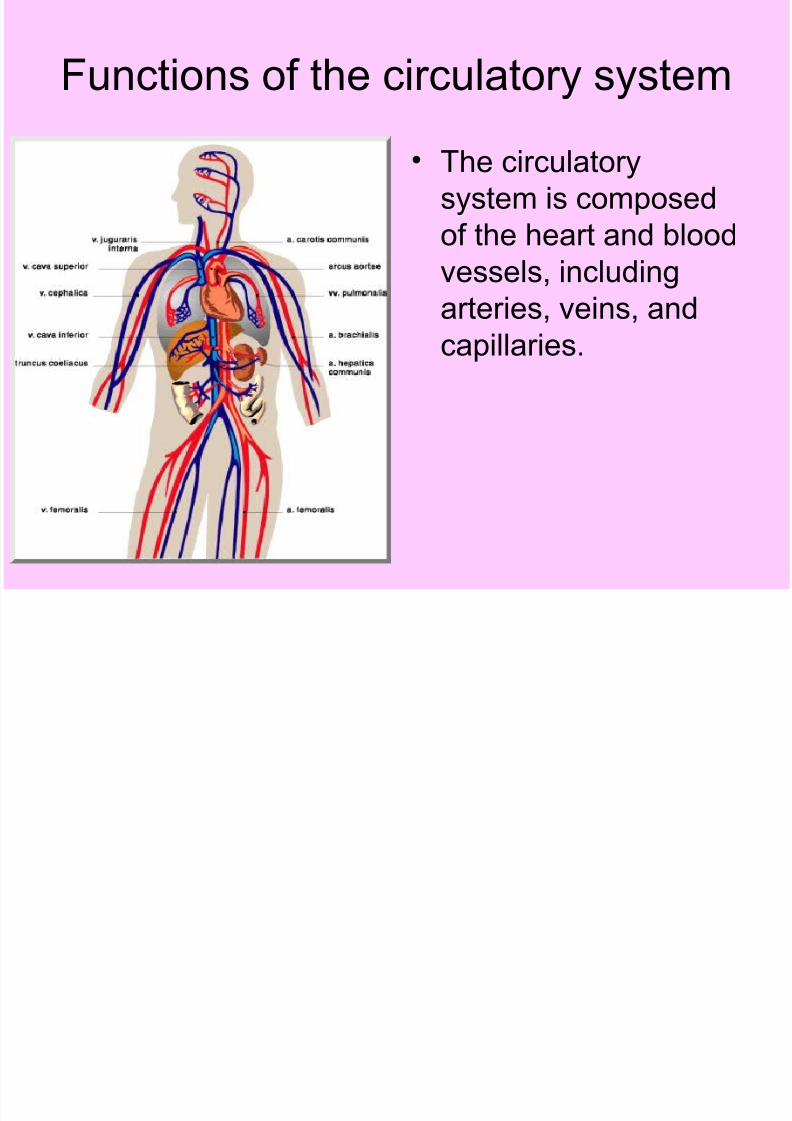
Functions of the circulatory system
• The circulatory
system is composed
of the heart and blood
vessels, includingarteries, veins, and
capillaries.
8/14/2019 Section 34-1
http://slidepdf.com/reader/full/section-34-1 3/40
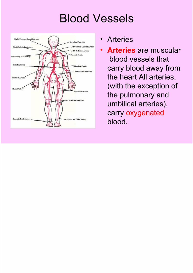
Blood Vessels
• Arteries
• Arteries are muscular
blood vessels that
carry blood away fromthe heart All arteries,
(with the exception of
the pulmonary and
umbilical arteries),
carry oxygenated
blood.
8/14/2019 Section 34-1
http://slidepdf.com/reader/full/section-34-1 4/40
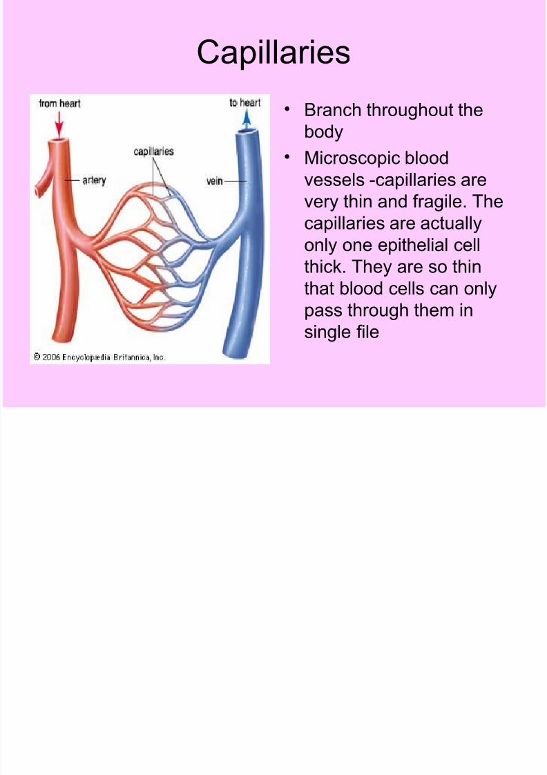
Capillaries
• Branch throughout the
body
• Microscopic blood
vessels -capillaries are
very thin and fragile. The
capillaries are actually
only one epithelial cell
thick. They are so thin
that blood cells can onlypass through them in
single file

8/14/2019 Section 34-1
http://slidepdf.com/reader/full/section-34-1 5/40
Capillaries
• The exchange of oxygenand carbon dioxide takesplace through the thincapillary wall. The redblood cells inside the
capillary release their oxygen which passesthrough the wall and intothe surrounding tissue.The tissue releases its
waste products, likecarbon dioxide, whichpasses through the walland into the red bloodcells.
8/14/2019 Section 34-1
http://slidepdf.com/reader/full/section-34-1 6/40
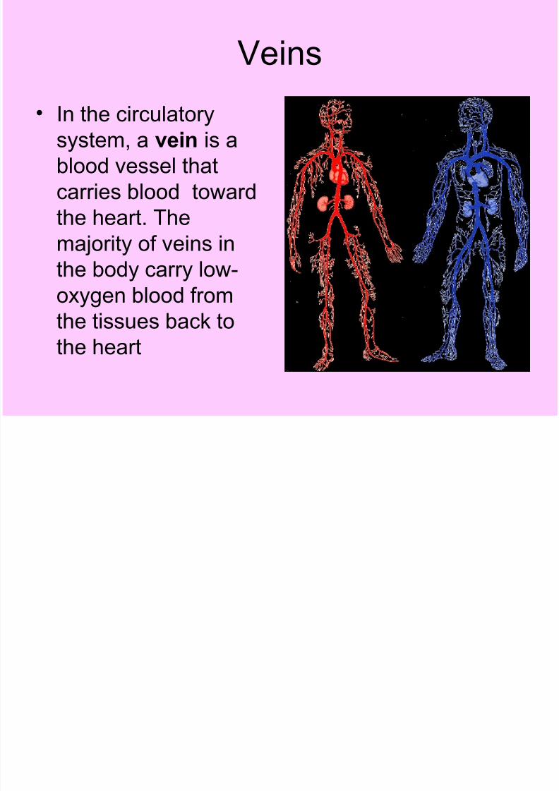
Veins
• In the circulatory
system, a vein is a
blood vessel that
carries blood towardthe heart. The
majority of veins in
the body carry low-
oxygen blood fromthe tissues back to
the heart

8/14/2019 Section 34-1
http://slidepdf.com/reader/full/section-34-1 7/40
The Heart- watch video
• Muscular organ about as
large as your fist
• The heart is the key
organ in the circulatorysystem. As a hollow,
muscular pump, its main
function is to propel blood
throughout the body. Itusually beats from 60 to
100 times per minute

8/14/2019 Section 34-1
http://slidepdf.com/reader/full/section-34-1 8/40
The Heart is divided into 4
chambers
Right Ventricle Left Ventricle
Right Atrium Left Atrium
8/14/2019 Section 34-1
http://slidepdf.com/reader/full/section-34-1 9/40
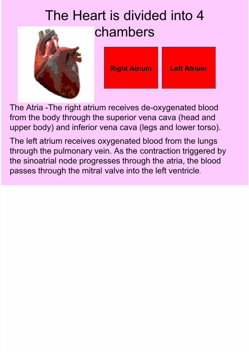
The Heart is divided into 4
chambers
Right Atrium Left Atrium
The Atria -The right atrium receives de-oxygenated blood
from the body through the superior vena cava (head and
upper body) and inferior vena cava (legs and lower torso).The left atrium receives oxygenated blood from the lungs
through the pulmonary vein. As the contraction triggered by
the sinoatrial node progresses through the atria, the blood
passes through the mitral valve into the left ventricle.

8/14/2019 Section 34-1
http://slidepdf.com/reader/full/section-34-1 10/40
The Heart is divided into 4
chambers
Right Ventricle Left Ventricle
The right ventricle is one of four chambers (two
atria and two ventricles) in the human heart. It
receives de-oxygenated blood from the right atrium
via the tricuspid valve, and pumps it into the
pulmonary artery via the pulmonary valve.
Left ventricle: The left lower chamber of the heart that receives blood from the
left atrium and pumps it out under high pressure through the aorta to the body.

8/14/2019 Section 34-1
http://slidepdf.com/reader/full/section-34-1 11/40
The chambers
• Right Atrium
• Left atrium
• Left ventricle
• Right ventricle

8/14/2019 Section 34-1
http://slidepdf.com/reader/full/section-34-1 12/40
8/14/2019 Section 34-1
http://slidepdf.com/reader/full/section-34-1 13/40
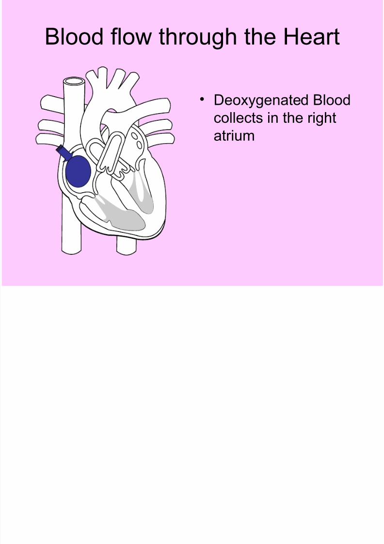
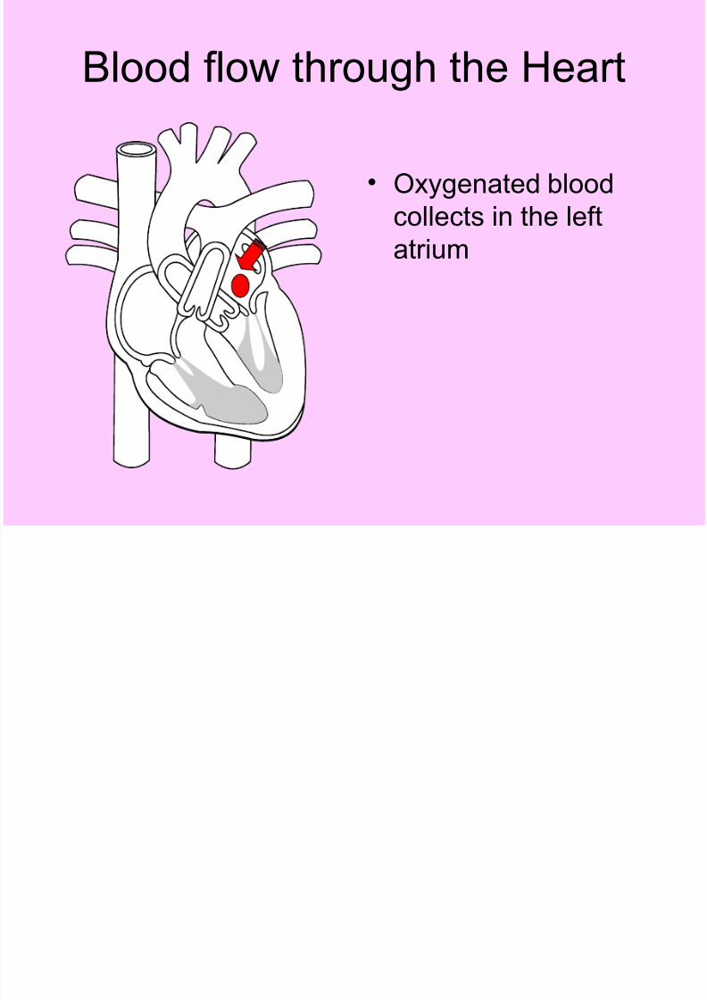
Blood flow through the Heart
• Deoxygenated Blood
collects in the right
atrium
8/14/2019 Section 34-1
http://slidepdf.com/reader/full/section-34-1 14/40
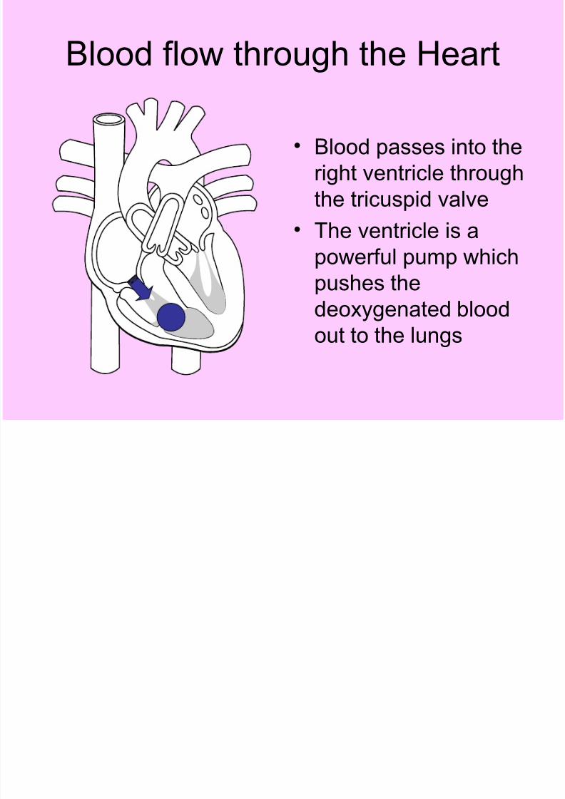
Blood flow through the Heart
• Blood passes into the
right ventricle through
the tricuspid valve• The ventricle is a
powerful pump which
pushes the
deoxygenated blood
out to the lungs
8/14/2019 Section 34-1
http://slidepdf.com/reader/full/section-34-1 15/40
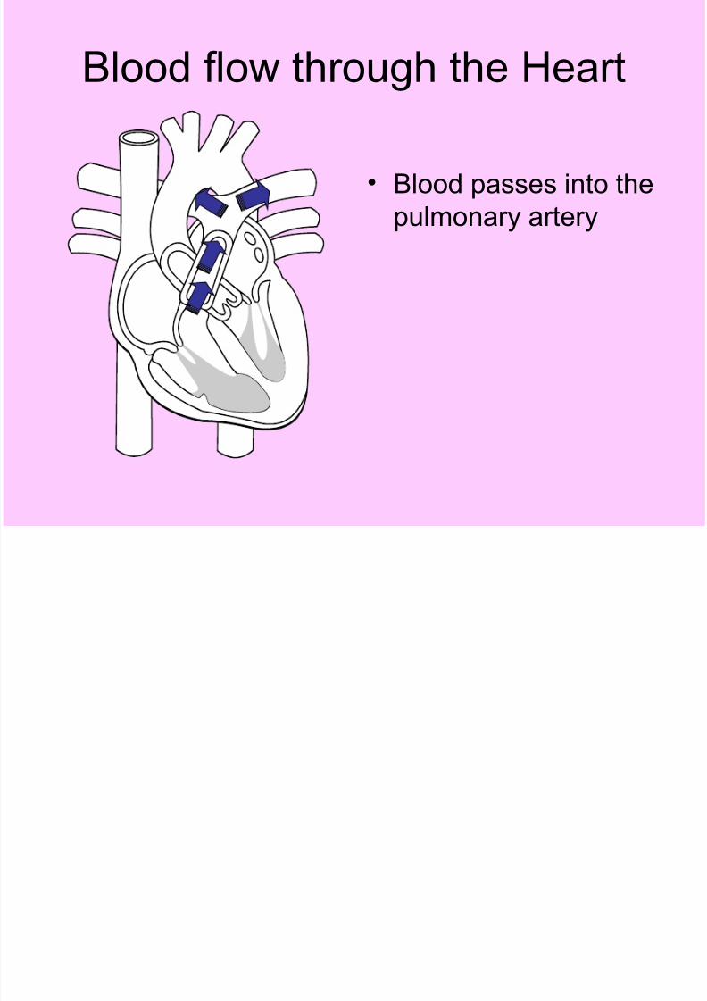
Blood flow through the Heart
• Blood passes into the
pulmonary artery

8/14/2019 Section 34-1
http://slidepdf.com/reader/full/section-34-1 16/40
Blood flow through the Heart
• Oxygenated blood
returns to the heart
from the lungs
• Pulmonary veins
return oxygenatedblood from the lungs
to the heart
8/14/2019 Section 34-1
http://slidepdf.com/reader/full/section-34-1 17/40
Blood flow through the Heart
• Oxygenated blood
collects in the left
atrium

8/14/2019 Section 34-1
http://slidepdf.com/reader/full/section-34-1 18/40

8/14/2019 Section 34-1
http://slidepdf.com/reader/full/section-34-1 19/40
Blood flow through the Heart
• Blood passes into the
aorta and out to body

8/14/2019 Section 34-1
http://slidepdf.com/reader/full/section-34-1 20/40

8/14/2019 Section 34-1
http://slidepdf.com/reader/full/section-34-1 21/40
Valves of the heart
• The valves of the heart arelocated within the chambers of the heart and are critical to theproper flow of blood throughthe heart. All of the valves,
when functioning normally, actas one-way valves, allowingblood to flow either from onechamber to another, or allowing blood to flow out of the heart, in only one direction.
The valves control the flow of blood through the heart byopening and closing during thecontractions of the heart.

8/14/2019 Section 34-1
http://slidepdf.com/reader/full/section-34-1 22/40
The “Pacemaker”
• Acting as the heart's naturalpacemaker, the SA node"fires" at regular intervals tocause the heart of beat with arhythm of about 60 to 70 beats
per minute for a healthy,resting heart. The electricalimpulse from the SA nodetriggers a sequence of electrical events in the heart tocontrol the orderly sequence of
muscle contractions that pumpthe blood out of the heart.

8/14/2019 Section 34-1
http://slidepdf.com/reader/full/section-34-1 23/40
The “Pacemaker”• The sinus rhythm
normally controls bothatrial and ventricular rhythm. Action potentialsgenerated by the SAnode spread throughoutthe atria, depolarizing this
tissue and causing atrialcontraction. The impulsethen travels into theventricles via theatrioventricular node (AV
node). This causes theventricles to contract –the 2 step process makesa complete heartbeat

8/14/2019 Section 34-1
http://slidepdf.com/reader/full/section-34-1 24/40
Pulse
• a person's pulse is the
throbbing of their arteries
as an effect of the
heartbeat. It can be felt in
any place that allows for an artery to be
compressed against a
bone, such as at the neck
at the wrist ,behind theknee, and on the inside of
the elbow , or near the
ankle joint

8/14/2019 Section 34-1
http://slidepdf.com/reader/full/section-34-1 25/40
Blood Pressure
• Blood pressure is thepressure of the bloodagainst the walls of thearteries.
• Blood pressure resultsfrom two forces. One iscreated by the heart as itpumps blood into thearteries and through the
circulatory system. Theother is the force of thearteries as they resist theblood flow.

8/14/2019 Section 34-1
http://slidepdf.com/reader/full/section-34-1 26/40
Blood Pressure
• The higher (systolic)number represents thepressure while the heartcontracts to pump bloodto the body.
• The lower (diastolic)number represents thepressure when the heartrelaxes between beats.
• The systolic pressure is
always stated first.Normal is 120/80although that is variablewith different sources

8/14/2019 Section 34-1
http://slidepdf.com/reader/full/section-34-1 27/40
Blood Components
• Blood plasma is the
liquid component of
blood, in which the
blood cells aresuspended. It makes
up about 55% of total
blood volume.

8/14/2019 Section 34-1
http://slidepdf.com/reader/full/section-34-1 28/40
Blood Components
• Red Blood Cells. A
single drop of blood
contains millions of
red blood cells whichare constantly
traveling through your
body delivering
oxygen and removingwaste.

8/14/2019 Section 34-1
http://slidepdf.com/reader/full/section-34-1 29/40

8/14/2019 Section 34-1
http://slidepdf.com/reader/full/section-34-1 30/40
Blood Components• Platelets :Platelets are
irregularly-shaped,colourless cell fragments that
are present in blood. Their
sticky surface lets them,
along with other substances,form clots to stop bleeding.
The platelets release a
chemical called FIBRIN
which weaves a network of
fibres across a cut andenables healing

8/14/2019 Section 34-1
http://slidepdf.com/reader/full/section-34-1 31/40
Blood Components• White Blood Cells
• White blood cells or
leukocytes are cells of
the immune system
defending the bodyagainst both infectious
disease and foreign
materials.
8/14/2019 Section 34-1
http://slidepdf.com/reader/full/section-34-1 32/40
Blood Types
• See Table 34-1 on page 998
• As you know there are 4 possible blood
types: A B O or AB
• There are several possible markers on
your blood cells if you have type A you
have A markers, B you have B markers
• And if you have AB you have both and
neither if you have type O blood
8/14/2019 Section 34-1
http://slidepdf.com/reader/full/section-34-1 33/40
Blood Types
• The plasma (liquid part of the blood) hasproteins called ANTIBODIES which attackforeign markers and cause the blood to
clump.• TYPE A blood has anti-B proteins
• TYPE B blood has anti-A proteins
• TYPE AB blood has no anti -proteins• TYPE O blood has both anti- A and anti-B
proteins

8/14/2019 Section 34-1
http://slidepdf.com/reader/full/section-34-1 34/40
Blood Types
• People with blood group 0 are called
"universal donors" (they can safely give
blood to anyone because their blood has
no markers) and people with blood groupAB are called "universal receivers." (they
can receive blood from anyone because
they have no antibodies)

8/14/2019 Section 34-1
http://slidepdf.com/reader/full/section-34-1 35/40
Blood Types• RH factor Many people
also have a Rh factor
on the red blood cell's
surface. This is also an
antigen and those whohave it are called Rh+.
Those who haven't are
called Rh-.
8/14/2019 Section 34-1
http://slidepdf.com/reader/full/section-34-1 36/40
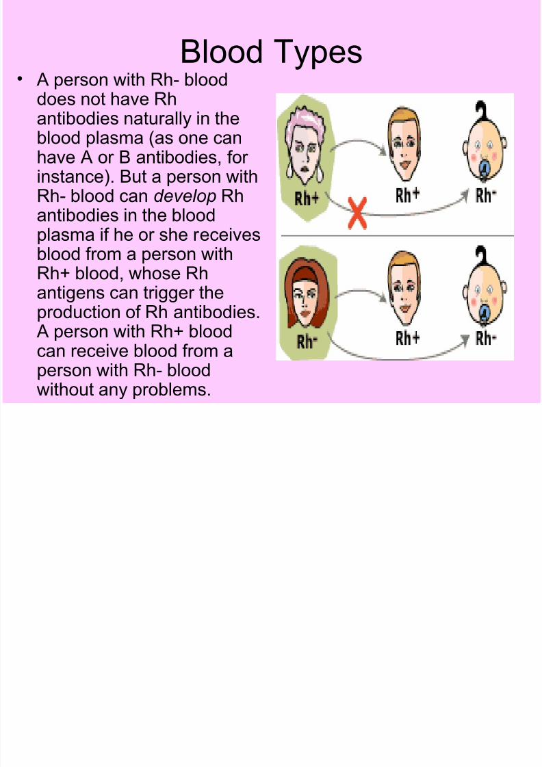
Blood Types• A person with Rh- blood
does not have Rhantibodies naturally in theblood plasma (as one canhave A or B antibodies, for instance). But a person with
Rh- blood can develop Rhantibodies in the bloodplasma if he or she receivesblood from a person withRh+ blood, whose Rhantigens can trigger theproduction of Rh antibodies.A person with Rh+ bloodcan receive blood from aperson with Rh- bloodwithout any problems.
8/14/2019 Section 34-1
http://slidepdf.com/reader/full/section-34-1 37/40
Assessment
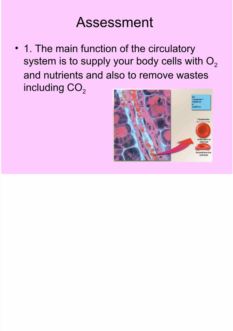
• 1. The main function of the circulatory
system is to supply your body cells with O2
and nutrients and also to remove wastes
including CO2
8/14/2019 Section 34-1
http://slidepdf.com/reader/full/section-34-1 38/40
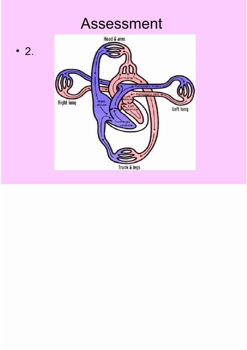
Assessment
• 2.

8/14/2019 Section 34-1
http://slidepdf.com/reader/full/section-34-1 39/40
Assessment
• 3. Arteries have a thicker wall than veins
have – veins have valves and arteries do
not

8/14/2019 Section 34-1
http://slidepdf.com/reader/full/section-34-1 40/40
Assessment
• 3. For every 100 white blood cells there
would be about 50-100,000 red blood cells