section 4 - who
TRANSCRIPT

Section 4.5
Genotoxicity
Second edition
(2020)

Hazard Identification and Characterization
4-1
CONTENTS
LIST OF ABBREVIATIONS 4-3
LIST OF CONTRIBUTORS 4-6
4.5 Genotoxicity 4-8 4.5.1 Introduction 4-8
Risk analysis context and problem formulation 4-10
Decision-tree for assessing the mutagenicity of substances that can be found in food 4-12 4.5.2 Tests for genotoxicity 4-19
Bacterial mutagenicity 4-23 In vitro mammalian cell mutagenicity 4-23
(a) Forward gene mutation tests using the
Tk gene 4-24
(b) Forward gene mutation tests using the
Hprt and Xprt genes 4-24
In vivo mammalian cell mutagenicity 4-25 (a) Somatic cell assays 4-25
(b) Germ cell assays 4-26
In vitro chromosomal damage assays 4-27 (a) Chromosomal aberration assay 4-27
(b) Micronucleus (MN) assay 4-28
(c) TK assay in mammalian cells 4-28
In vivo chromosomal damage assays 4-29 (a) Chromosomal aberration assay 4-29
(b) Micronucleus (MN) assay 4-29
In vitro DNA damage/repair assays 4-30 In vivo DNA damage/repair assays 4-30
This text updates section 4.5 of Chapter 4, Hazard Identification and
Characterization: Toxicological and Human Studies, of Environmental
Health Criteria 240 (EHC 240), which was originally published in 2009. It
was developed through an expert consultation and further advanced
following comments received through a public consultation in December
2019.
For abbreviations used in the text, the reader may refer to the list of
abbreviations at the front of this section. Definitions of select terms may be
found in the glossary in Annex 1 of EHC 240 (http://www.inchem.org/
documents/ehc/ehc/ehc240_annex1.pdf).

EHC 240: Principles for Risk Assessment of Chemicals in Food
4-2
(a) Comet (single-cell gel electrophoresis)
assay 4-30
(b) DNA adduct assays 4-32
(c) Unscheduled DNA synthesis (UDS)
assay in mammalian liver 4-32
4.5.3 Identification of relevant studies 4-33 4.5.4 Interpretation of test results 4-34
Presentation and categorization of results 4-35 (a) Assessing whether results of an assay
are positive, negative or equivocal for
genotoxicity 4-37
(b) Assessing data quality 4-38
Weighting and integration of results 4-46 Adequacy of the genotoxicity database 4-49 Mutagenic mode of action and adverse
outcomes 4-50 Integration of carcinogenicity and
mutagenicity 4-53 4.5.5 Approaches for evaluating data-poor substances 4-56
In silico approaches 4-56 (a) Available tools (QSARs, SARs/
structural alerts) for mutagenicity 4-57
(b) Confidence in approaches 4-57
(c) Mutagenicity assessment 4-63
Threshold of toxicological concern (TTC) 4-65 Grouping and read-across approaches 4-68
4.5.6 Considerations for specific compounds 4-71 Mixtures 4-71 Flavouring agents 4-73 Metabolites in crops/food-producing
animals, degradation products and impurities 4-75
Secondary metabolites in enzyme preparations 4-79 4.5.7 Recent developments and future directions 4-81
Novel in vivo genotoxicity approaches 4-82 Novel in vitro genotoxicity approaches 4-82 Adverse outcome pathways for
mutagenicity 4-90 Quantitative approaches for safety
assessment 4-92 4.5.8 References 4-93

Hazard Identification and Characterization
4-3
List of abbreviations
ACD Advanced Chemistry Development, Inc.
ADI acceptable daily intake
AOP adverse outcome pathway
ARfD acute reference dose
ATSDR Agency for Toxic Substances and Disease
Registry (USA)
BMD benchmark dose
CAS Chemical Abstracts Service
CCRIS Chemical Carcinogenesis Research Information
System
CEBS Chemical Effects in Biological Systems
Cefic European Chemical Industry Council
CHL Chinese hamster lung
CHO Chinese hamster ovary
CTD Comparative Toxicogenomics Database
DDI DNA damage–inducing
DNA deoxyribonucleic acid
ECHA European Chemicals Agency
EFSA European Food Safety Authority
EHC Environmental Health Criteria
EU European Union
EURL ECVAM European Union Reference Laboratory for
alternatives to animal testing
FAO Food and Agriculture Organization of the United
Nations
GENE-TOX Genetic Toxicology Data Bank
GLP Good Laboratory Practice
gpt glutamic–pyruvic transaminase
HBGV health-based guidance value
Hprt/HPRT hypoxanthine–guanine phosphoribosyl transferase
HTRF Homogeneous Time-Resolved Fluorescence
IATA Integrated Approaches to Testing and Assessment
ICH International Council for Harmonisation of
Technical Requirements for Registration of
Pharmaceuticals for Human Use
INCHEM Internationally Peer Reviewed Chemical Safety
Information
IPCS International Programme on Chemical Safety
IRIS Integrated Risk Information System (USA)
ISS Istituto Superiore di Sanità (Italy)

EHC 240: Principles for Risk Assessment of Chemicals in Food
4-4
ISSMIC Istituto Superiore di Sanità database on in vivo
mutagenicity (micronucleus test)
ISSSTY Istituto Superiore di Sanità database on in vitro
mutagenicity in Salmonella typhimurium (Ames
test)
JECDB Japanese Existing Chemical Data Base
JECFA Joint FAO/WHO Expert Committee on Food
Additives
JMPR Joint FAO/WHO Meeting on Pesticide Residues
LRI Long-range Research Initiative
MAK maximum workplace concentration
MN micronucleus/micronuclei
MOA mode of action
MOE margin of exposure
NGS next-generation DNA sequencing
NIHS National Institute of Health Sciences (Japan)
NOAEL no-observed-adverse-effect level
NOGEL no-observed-genotoxic-effect level
NTP National Toxicology Program (USA)
OECD Organisation for Economic Co-operation and
Development
PAH polycyclic aromatic hydrocarbon
PCR polymerase chain reaction
Pig-a phosphatidylinositol glycan complementation
group A
qPCR quantitative polymerase chain reaction
QSAR quantitative structure–activity relationship
REACH Registration, Evaluation, Authorisation and
Restriction of Chemicals
RNA ribonucleic acid
RT-qPCR reverse transcription quantitative polymerase
chain reaction
S9 9000 × g supernatant fraction from rat liver
homogenate
SAR structure–activity relationship
SciRAP Science in Risk Assessment and Policy
SYRCLE Systematic Review Centre for Laboratory Animal
Experimentation
Td threshold dose
TDI tolerable daily intake
T.E.S.T. Toxicity Estimation Software Tool
TG test guideline; thioguanine
TIMES tissue metabolism simulator

Hazard Identification and Characterization
4-5
Tk/TK thymidine kinase
ToxRTool Toxicological data Reliability Assessment Tool
TTC threshold of toxicological concern
UDS unscheduled DNA synthesis
USA United States of America
USEPA United States Environmental Protection Agency
USFDA United States Food and Drug Administration
WHO World Health Organization
WOE weight of evidence
Xprt/XPRT xanthine–guanine phosphoribosyl transferase

EHC 240: Principles for Risk Assessment of Chemicals in Food
4-6
List of contributors
Dr Virunya Bhat
PAHO/WHO Collaborating Centre on Food Safety, Water Quality and
Indoor Environment, NSF International, Ann Arbor, Michigan, United
States of America (USA)
Emeritus Professor Alan R. Boobis (co-lead author)
National Heart & Lung Institute, Faculty of Medicine, Imperial College
London, London, United Kingdom
Dr Riccardo Crebelli
Istituto Superiore di Sanità, Rome, Italy
Dr Nathalie Delrue
Test Guidelines Programme, Environment, Health and Safety Division,
Environment Directorate, Organisation for Economic Co-operation and
Development, Paris, France
Professor David Eastmond (co-lead author)
Department of Molecular, Cell, and Systems Biology, University of
California, Riverside, California, USA
Dr Susan Page Felter
Mason, Ohio, USA
Dr Rainer Guertler
Federal Institute for Risk Assessment (BfR), Berlin, Germany
Professor Andrea Hartwig
Karlsruhe, Germany
Dr Frank Le Curieux
European Chemicals Agency, Helsinki, Finland
Professor Angelo Moretto
Department of Biomedical and Clinical Sciences, University of Milan, and
International Centre for Pesticides and Health Risk Prevention, Luigi Sacco
Hospital, Milan, Italy
Professor Pasquale Mosesso
Department of Ecological and Biological Sciences, Università degli Studi
della Tuscia, Viterbo, Italy
Dr Utz Mueller
Australian Pesticides and Veterinary Medicines Authority, Kingston,
Australian Capital Territory, Australia

Hazard Identification and Characterization
4-7
Dr Takehiko Nohmi
Biological Safety Research Center, National Institute of Health Sciences,
Kamiyoga, Setagaya-ku, Tokyo, Japan
Dr Grace Patlewicz
National Center for Computational Toxicology, United States
Environmental Protection Agency, Durham, North Carolina, USA
Professor David H. Phillips
King’s College London, London, United Kingdom
Dr Andrea Richarz
Institute for Health and Consumer Protection, Joint Research Centre,
European Commission, Ispra, Italy
Dr Raymond R. Tice
National Institutes of Environmental Health Sciences, Research Triangle
Park, North Carolina, USA
Dr Paul A. White
Genetic Toxicology Group, Environmental Health Sciences & Research
Bureau, Environmental & Radiation Health Sciences Directorate, Healthy
Environments & Consumer Safety Branch, Health Canada, Ottawa,
Ontario, Canada
Dr Kristine L. Witt
National Institutes of Environmental Health Sciences, Research Triangle
Park, North Carolina, USA

EHC 240: Principles for Risk Assessment of Chemicals in Food
4-8
4.5 Genotoxicity
4.5.1 Introduction
The study of toxic effects on the inherited genetic material in
cells originated with the experiments of Muller (1927), who observed
“artificial transmutation of the gene” by ionizing radiation in the fruit
fly, Drosophila melanogaster. Chemically induced mutation also has
a long history, with the first scientific publication, using Muller’s fruit
fly model, describing mutations arising from exposure to sulfur
mustard (Auerbach, Robson & Carr, 1947). A key event stimulating
the development and validation of genetic toxicity tests occurred in
1966, when geneticists recommended at a conference sponsored by
the United States National Institutes of Health that food additives,
drugs and chemicals with widespread human exposure be routinely
tested for mutagenicity (see next paragraph for definitions) (Zeiger,
2004).
The term “mutation” refers to permanent changes in the structure
or amount of the genetic material of an organism that can lead to
heritable changes in its function; these changes include gene
mutations as well as structural and numerical chromosomal
alterations. The term “mutagen” refers to a chemical that induces
heritable genetic changes, most commonly through interaction with
DNA,1 and “mutagenicity” refers to the process of inducing a
mutation. The broader terms “genotoxicity” and “genetic toxicity”,
which are synonymous, include mutagenicity, but also include DNA
damage, which may be reversed by DNA repair processes or other
known cellular processes or result in cell death and may not result in
permanent alterations in the structure or information content of the
surviving cell or its progeny (OECD, 2017a). When reference is made
to genotoxicity testing, often what is meant is mutagenicity testing.
More properly, genotoxicity testing also includes tests that measure
the capability of substances to damage DNA or cellular components
regulating the fidelity of the genome – such as the spindle apparatus,
topoisomerases, DNA repair systems and DNA polymerases – and
encompasses tests of a broad range of adverse effects on genetic
components of the cell. Although such information can be of value in
interpreting the results of mutagenicity tests, it should be considered
supplementary data when assessing mutagenic potential. Therefore,
1 Pro-mutagens are mutagens requiring metabolic activation for
mutagenesis.

Hazard Identification and Characterization
4-9
the broader term “genotoxicant” is used to refer to a chemical that
induces adverse effects on genetic components via any of a variety of
mechanisms, including mutation, but does not necessarily connote the
ability to cause heritable changes. The purpose of mutagenicity
testing is to identify substances that can cause genetic alterations in
somatic or germ cells, and this information is used in regulatory
decision-making (OECD, 2017a).
The overview presented in this section focuses on the
identification of mutagens and on the use of such information in
assessing the role of DNA-reactive gene mutation in the adverse
effects of chemicals, consistent with the World Health Organization
(WHO)/International Programme on Chemical Safety (IPCS)
harmonized scheme for mutagenicity testing (Eastmond et al., 2009).
National and international regulatory agencies historically have
used genotoxicity information as part of a weight-of-evidence (WOE)
approach to evaluate potential human carcinogenicity and its
corresponding mode of action (MOA; discussed further in section
4.5.4.4). A conclusion on the genotoxic potential of a chemical – and,
more specifically, on a mutagenic MOA for carcinogenicity – can be
made on the basis of the results of only a few specific types of study,
if properly conducted and well reported.
Information on mutagenicity is also of value in assessing the risk
of other adverse effects, particularly developmental effects occurring
through mutation of germ cells or genotoxicity occurring in somatic
cells during embryogenesis and fetal development (Meier et al.,
2017).
A chemical could be acknowledged as having genotoxic
potential but low concern for a mutagenic MOA in its carcinogenicity
or other adverse effects because of mitigating factors, such as
toxicokinetics (e.g. phenol and hydroquinone; UKCOM, 2010) or
overwhelming toxicity (e.g. dichlorvos; FAO/WHO, 2011).
Some regulatory agencies, such as those within the USA,
Canada, the United Kingdom and the European Union (EU), consider
heritable mutation a regulatory end-point. Mutations in germ cells
may be inherited by future generations and may contribute to genetic
disease. Germline (or germ cell) or somatic cell mutations are
implicated in the etiology of some disease states, such as cancer,
sickle cell anaemia and neurological diseases (Youssoufian &

EHC 240: Principles for Risk Assessment of Chemicals in Food
4-10
Pyeritz, 2002; Erickson, 2003, 2010; Lupski, 2013; D’Gama et al.,
2015). Inherited mutations linked to human diseases are compiled in
the Human Gene Mutation Database (HGMD, 2017).
Testing for mutagenicity should utilize internationally
recognized protocols, where they exist. For example, mutagenicity
(gene mutation and structural and numerical chromosomal
alterations) is one of six basic testing areas that have been adopted by
the Organisation for Economic Co-operation and Development
(OECD, 2011) as the minimum required to screen high-production-
volume chemicals in commerce for toxicity.
Safety assessments of chemical substances with regard to
mutagenicity are generally based on a combination of tests to assess
three major end-points of genetic damage associated with human
disease:
1) gene mutation (i.e. point mutations or deletions/insertions that
affect single or blocks of genes);
2) clastogenicity (i.e. structural chromosome changes); and
3) aneuploidy (i.e. the occurrence of one or more extra or missing
chromosomes, leading to an unbalanced chromosome
complement).
Existing evaluation schemes tend to focus on single chemical
entities with existing data. However, there are scenarios that do not
involve single chemicals, such as enzyme preparations used in food
production that are mixtures including proteins and one or more low-
molecular-weight chemicals, or that involve chemicals, such as minor
plant and animal metabolites of pesticides or veterinary drugs, that
lack empirical data. Special considerations related to these scenarios,
including the evaluation of the mutagenicity of food extracts obtained
from natural sources, which are often complex botanical mixtures that
may not be fully characterized, are also discussed in this section.
4.5.1.1 Risk analysis context and problem formulation
The identification of compounds to which exposure may lead to
cancer (or other adverse effect) via a mutagenic MOA affects how
these compounds are handled within regulatory paradigms. A
distinction is often made between substances that require regulatory
approval before use (e.g. pesticides, veterinary drugs, food additives)
and those to which exposure is unavoidable (e.g. contaminants,
natural constituents of the diet). In practice, this distinction affects the

Hazard Identification and Characterization
4-11
nature of information provided to risk managers. For substances
intentionally added to or used in food that require regulatory
approval, key outputs of the hazard characterization are health-based
guidance values (HBGVs) (e.g. acceptable daily intake [ADI],
tolerable daily intake [TDI], acute reference dose [ARfD]). Intrinsic
to the establishment of such a value is that there is negligible concern
when exposure is below the HBGV, and implicit in this is that there
are biological and population thresholds for the adverse effect.
Mutagenicity, particularly gene mutation, is often assumed to lack a
threshold, in part due to uncertainty related to human exposure levels
and the assumption that even one molecule of a DNA-reactive
mutagen could theoretically induce heritable changes leading to an
adverse effect. Consequently, for substances considered to act
through a mutagenic MOA, it may not be possible to establish with
confidence an HBGV below which concern is considered negligible;
under such circumstances, in the context of the work of the Joint
FAO/WHO Expert Committee on Food Additives (JECFA) and the
Joint FAO/WHO Meeting on Pesticide Residues (JMPR), it is
generally understood that it would be inappropriate to establish an
HBGV. Nevertheless, risk managers may still require an indication
of the degree of health concern, and this should be reflected in the
problem formulation, which is a key component of risk analysis that
involves consideration of the risk management scope and goals in
relation to relevant exposure scenarios, available resources, urgency
of the assessment and the level of uncertainty that is acceptable
(Meek et al., 2014). In practice, in the international context in which
JECFA and JMPR work, rather than a detailed problem formulation,
the general question to be addressed is whether the compound poses
a significant mutagenic hazard and, if so, whether there is a concern
at estimated dietary exposures.
Most currently approved (e.g. by OECD) tests for mutagenicity,
both in vitro and in vivo, are designed to identify a mutagenic hazard
and in general are used for a simple yes/no answer for risk
management purposes (see section 4.5.2). Such a dichotomous
approach is useful for managing substances intentionally permitted in
food, such as food additives, pesticides and veterinary drugs, for
which regulatory approval is often required. Qualitative,
semiquantitative and non-testing approaches useful for managing
data-poor substances, such as unavoidable contaminants and plant
and animal metabolites, include:

EHC 240: Principles for Risk Assessment of Chemicals in Food
4-12
in silico approaches, such as (quantitative)structure–activity
relationship [(Q)SAR] models (see section 4.5.5.1);
the threshold of toxicological concern (TTC) approach (see
section 4.5.5.2); and
grouping and read-across approaches (see section 4.5.5.3).
Quantitative dose–response approaches for genotoxicity may
also be appropriate for unavoidable contaminants (see section
4.5.7.4). However, as this is a deviation from current practice, the
acceptability of such approaches should be indicated in the problem
formulation (see, for example, MacGregor et al., 2015a,b; UKCOM,
2018).
JECFA and JMPR do not set data requirements for their food
additive, veterinary drug and pesticide residue evaluations, although
there is a minimum data set expected in order to conduct an
assessment. In the case of mutagenicity, the nature of and guidance
to interpret the information are described in this section. In general,
JECFA and JMPR evaluate the available data, most often generated
in support of regulatory submissions elsewhere. Data requirements
set by a regulatory agency for a chemical evaluation can vary
substantially, depending on the chemical’s use and potential for
human exposure.
4.5.1.2 Decision-tree for assessing the mutagenicity of substances that can be found in food
Fig. 4.1 is a decision-tree illustrating issues to be considered in
assessing the mutagenic potential of different types of substances that
can be found in food. Subsequent subsections will describe the
process of identifying relevant and reliable mutagenicity data and,
depending on the regulatory jurisdiction, determining whether the
data and WOE are adequate to conclude on mutagenic potential. If a
substance is shown to possess mutagenic potential, the process of
discerning the likelihood of a mutagenic MOA for carcinogenicity
and other adverse effects is also discussed, in conjunction with
repeated-dose toxicity or carcinogenicity data, if available.

Hazard Identification and Characterization
4-13
Fig. 4.1. Decision-tree illustrating issues to be considered in assessing the mutagenic potential of different types of substances that can be found in food
1. Is there adequate evidence to exclude any possible
concerns for mutagenicity?
While it may be rare to exclude possible concerns for
mutagenicity a priori, occasionally the nature of the substance or its
production process may provide sufficient assurance that substance-
NO YES
O
2. No assessment of
mutagenicity necessary
4. Defined substance?
YES
YES
20. Are all
components known?
3. Subject to approval?
YES
NO
O
5. Mutagenicity testing
adequate?
17. Sufficient information to assess
dietary risk of mutagenicity (e.g. SAR)? If
mixture, include considerations from
box 20
23. Use component-
based approach
NO
O
NO
O
24. Use whole
mixture approach
as necessary
NO
Ocv
cv 19. Not possible to
conclude on
mutagenicity risk
YES
18. Proceed
with risk
assessment
7. Data beyond
core testing?
YES
8. Apply hierarchical
evaluation
YES
9. Does compound
show evidence of
mutagenicity?
NO
Ocv
cv
YES NO
Ocv
cv
15. Non-DNA-reactive mutagen with known
mode of action
10. Proceed with
risk assessment
11. Mutagenicity based on DNA interactions?
NO
Ocv
cv
YES
16. Proceed with
risk assessment
14. Not possible to exclude
risk of mutagenicity
12. Is there sufficient mechanistic evidence for a
threshold?
YES NO
13. Proceed with
risk assessment
NO
Ocv
cv
6. Not possible to
conclude on
mutagenicity risk
21. Does the mixture
contain known
mutagens(s)?
NO
O YES
22. Use TTC
approach
1. Is there adequate evidence to exclude
any possible concerns for mutagenicity?

EHC 240: Principles for Risk Assessment of Chemicals in Food
4-14
specific mutagenicity data are not necessary. One example is a natural
constituent of the diet produced by a fully controlled process (e.g.
invertase derived from Saccharomyces cerevisiae fermentation;
FAO/WHO, 2002). [See section 4.5.6.4.]
2. No assessment of mutagenicity necessary
If the answer to the question in box 1 is YES, no further
consideration of mutagenic potential is necessary, and risk
assessment of non-genotoxic (non-mutagenic) effects can proceed.
[See other sections of chapter 4.]
3. Subject to approval?
If concerns about potential mutagenicity cannot be excluded a
priori (i.e. the answer to the question in box 1 is NO), does the
substance require regulatory approval in Member States prior to uses
that could knowingly result in its presence in food (i.e. pesticides,
veterinary drugs and food additives, including flavouring agents)?
Excluded are contaminants and natural constituents of the diet (e.g.
mycotoxins), for which there are different considerations for tolerated
concentration limits. [See section 4.5.1.1.]
4. Defined substance?
If the answer to the question in box 3 is YES, does the substance
comprise a single chemical or a small number (e.g. stereoisomers) of
chemicals of known structure? In other words, is it chemically
defined? If not, the substance is considered a mixture. Included in this
group are single substances of unknown structure. Note that a critical
consideration is the purity of the substance. Expert judgement is
needed to decide whether, based on analytical or other relevant data,
a substance that nominally is a single chemical is so impure that it
should be considered a mixture with uncharacterized constituents
(e.g. <90% purity). [See section 4.5.6.1.]
5. Mutagenicity testing adequate?
For substances subject to regulatory approval in some
jurisdictions and where the answer to the question in box 4 is YES,
are the available data adequate to conclude whether the substance is
likely to pose a mutagenic risk in vivo at dietary levels of exposure?
[See sections 4.5.2 and 4.5.4.4.]
6. Not possible to conclude on mutagenicity risk
If mutagenic potential has not been adequately tested (i.e. the
answer to the question in box 5 is NO), it is not possible to conclude
on the likelihood of mutagenic risk in vivo at dietary levels of

Hazard Identification and Characterization
4-15
exposure. As such, it may be inappropriate to establish HBGVs that
encompass potential mutagenicity. The main data gaps precluding a
conclusion on mutagenic potential should be clearly articulated. [See
section 4.5.4.5.]
7. Data beyond core testing?
For some compounds, particularly newer ones, mutagenicity
testing may be adequate (i.e. the answer to the question in box 5 is
YES) based on available data from a small range of relevant and
reliable “standard” mutagenicity tests. [See section 4.5.4.2.]
However, for others, particularly those in use for some time or about
which there are specific concerns (e.g. bisphenol A; EFSA, 2015), the
available data may be much more extensive, including a variety of
test systems with a range of quality (i.e. in design, conduct or
reporting), and the results may be contradictory. It should be noted if
the genotoxicity database is considered to fall into this category. [See
section 4.5.3.]
8. Apply hierarchical evaluation
When the genotoxicity database is complex or contradictory (i.e.
the answer to the question in box 7 is YES), a WOE approach that
considers factors such as the results of in vivo versus in vitro testing,
the relevance of the test or end-point to humans and the relevance of
the route of exposure and dose is used to weight the studies. [See
sections 4.5.4.1 and 4.5.4.2.]
9. Does compound show evidence of mutagenicity?
Regardless of how extensive the database is (i.e. the answer to
the question in box 7 is NO or after application of the hierarchical
evaluation in box 8), a WOE conclusion should be reached on
whether the substance shows evidence of mutagenicity for relevant
end-points. For example, as defined by the OECD, an isolated
positive result at high, cytotoxic concentrations in vitro, without
evidence of mutagenicity in numerous guideline studies conducted to
an appropriate standard, is insufficient to conclude that, overall, there
is concern for mutagenicity. As the objective is not a hazard
classification, reaching a conclusion requires expert judgement,
which should be clearly explained and can often be the most difficult
aspect of the assessment. [See sections 4.5.3, 4.5.4.1 and 4.5.4.2.]

EHC 240: Principles for Risk Assessment of Chemicals in Food
4-16
10. Proceed with risk assessment
If the WOE does not suggest mutagenicity (i.e. the answer to the
question in box 9 is NO), no further consideration of the mutagenic
potential of the substance is necessary, and risk assessment of non-
genotoxic (non-mutagenic) effects can proceed. [See other sections
of chapter 4.]
11. Mutagenicity based on DNA interactions?
If there is evidence of mutagenicity (i.e. the answer to the
question in box 9 is YES), the nature of the mutagenicity should be
determined – specifically, whether the mutagenicity is based on the
parent compound or a metabolite interacting with DNA, thereby
resulting in heritable DNA changes. This evidence should come
primarily from appropriate tests for gene mutation, clastogenicity and
aneuploidy, and supporting evidence may include a variety of non-
standard tests, such as DNA reactivity/adduct formation. [See section
4.5.2.]
12. Is there sufficient mechanistic evidence for a threshold?
For a mutagenic chemical (i.e. the answer to the question in box
11 is YES), the relevance of the dose/concentration used in testing to
the estimated dietary exposure should be considered. For the majority
of mutagens, there may be little or no evidence for an effect threshold.
Hence, in the absence of such evidence, it is assumed that even high-
dose effects are relevant for assessing mutagenic potential in humans.
For a few substances, however, there may be clear mechanistic
evidence in vitro and in vivo for a biological threshold. Hence, in
theory, it may be possible to discount effects seen only at doses that
are irrelevant to conceivable human dietary exposure (or even a
multiple of that exposure) (e.g. dichlorvos; FAO/WHO, 2011). [See
also section 4.5.7.4.]
13. If there is sufficient mechanistic evidence for a threshold
for mutagenicity, proceed with risk assessment If it is concluded that a biological threshold exists for the
mutagenicity observed experimentally (i.e. the answer to the question
in box 12 is YES) and, after allowing for interspecies and intraspecies
differences, the estimated human dietary exposure is clearly well
below this, risk assessment based on the critical effect(s) can proceed.
[See other sections of chapter 4.]

Hazard Identification and Characterization
4-17
14. Not possible to exclude risk of mutagenicity
If it is concluded that the mutagenicity observed experimentally
is, or might be, relevant, considering conceivable human dietary
exposure levels (i.e. the answer to the question in box 12 is NO), it
will ordinarily be inappropriate to establish an HBGV. [See section
4.5.4.5.]
15. Non-DNA-reactive mutagen with known mode of action
For mutagenic compounds in which a DNA-reactive MOA can
be excluded (i.e. the answer to the question in box 11 is NO), the
nature of the mutagenicity, its molecular mechanism and the dose–
response relationship should be characterized. For some mechanisms,
there is evidence for a biological threshold – for example, aneuploidy
due to spindle disruption or mutagenicity secondary to inflammation
that generates reactive oxygen species. [See section 4.5.4.4.]
16. Proceed with risk assessment
The output of the mutagenic hazard characterization (i.e. output
from the question in box 15) can be used in the risk assessment, as
appropriate. For example, if mutagenicity is considered to exhibit a
threshold, the “normal” approach to establishing HBGVs and to risk
characterization can be applied. In many cases, this would mean that
the critical effect was other than mutagenicity, as it occurred at lower
exposure levels. In some cases, it might not be possible to conclude
that mutagenicity exhibits a threshold, in which case a margin of
exposure (MOE) approach may be appropriate. In either case, a
concluding statement regarding the potential risk of mutagenicity in
vivo at dietary levels of exposure should be provided. [See section
4.5.4.5.]
17. Sufficient information to assess dietary risk of
mutagenicity (e.g. SAR)?
For substances not subject to regulatory approval (i.e. the answer
to the question in box 3 is NO) that have unavoidable dietary
exposure, such as contaminants or natural dietary constituents (e.g.
mycotoxins), it should be assessed whether there is sufficient
information to reach a conclusion about potential mutagenicity.
When existing empirical mutagenicity data are insufficient to reach a
conclusion, additional information from the substance, from related
analogues (i.e. read-across) or from in silico approaches, such as
(Q)SARs, should also be considered in an overall WOE for the

EHC 240: Principles for Risk Assessment of Chemicals in Food
4-18
mutagenic potential of the substance. [See sections 4.5.5.1 and
4.5.5.3.]
18. Proceed with risk assessment
Where sufficient information is available to conclude on the
mutagenic potential of the substance (i.e. the answer to the question
in box 17 is YES), a risk assessment can proceed. This may justify
establishing an HBGV, such as a TDI, or the use of an MOE
approach. Where exposures are likely to be very low and the
compound is a potential mutagen, the TTC approach can be used. If
exposure is below the mutagenicity TTC value (0.0025 µg/kg body
weight per day for chemicals with structural alerts for DNA
reactivity), there is low concern for effects on human health. [See
section 4.5.5.2 and other sections of chapter 4.]
19. Not possible to conclude on mutagenicity risk
When it is not possible to conclude on potential mutagenicity
(i.e. the answer to the question in box 17 is NO), advice should be
provided on the assumption that the substance might be a mutagen.
Hence, the TTC for such compounds (0.0025 µg/kg body weight per
day) could be used, recognizing the considerable uncertainty in such
an assessment and that the risk may be appreciably overestimated.
Alternatively, it may be concluded that it is not possible to provide
any advice on potential human risk without additional data.
20. Are all components known?
For substances that are not composed of a single defined
chemical or a small number of defined chemical entities (i.e. the
answer to the question in box 4 is NO), are all of the components of
the mixture known? If all of the components are known and have
established chemical structures and concentrations, the mixture is
considered “simple”, whereas if a significant fraction of components
are of unknown structure or concentration, the mixture is considered
“complex”. [See section 4.5.6.1.]
Although there is no explicit question in the decision-tree as to
whether mixtures are subject to approval, a number of the
considerations for defined substances will also apply to mixtures.
That is, for those mixtures subject to approval, consideration will
need to be given to the adequacy of mutagenicity testing (of the
components or of the mixture as a whole). For those that are not, a
WOE approach using information on direct testing, read-across and
(Q)SAR can be applied, to the extent possible.

Hazard Identification and Characterization
4-19
21. Does the mixture contain known mutagen(s)?
Where all of the components in a “simple” mixture above a
minimum level of concern (as determined by expert judgement) are
known (i.e. the answer to the question in box 20 is YES), each
component should be assessed for its mutagenicity, on the basis of
prior knowledge. Are one or more known mutagens present? If so,
these should be assessed before considering the potential
mutagenicity of other components.
22. Use TTC approach
For mutagenic substances known to be present in a defined
mixture (i.e. the answer to the question in box 21 is YES), the TTC
approach can be applied. If estimated human exposure is below the
mutagenicity (DNA-reactive gene mutation) TTC, there is low
concern for mutagenicity in exposed individuals from these
substances, and the remaining components can then be assessed
individually, as described under the component-based approach in
box 23. If the estimated exposure exceeds the mutagenicity (DNA-
reactive gene mutation) TTC, additional information will be needed
to determine if there is concern for possible mutagenicity in exposed
individuals. [See section 4.5.6.3.]
23. Use component-based approach
For a “simple” mixture in which none of the components is
known to be mutagenic (i.e. the answer to the question in box 21 is
NO), each component should be assessed for potential mutagenicity,
as described for defined chemicals. [See section 4.5.6.1.]
24. Use whole mixture approach as necessary
For a “complex” mixture in which a significant fraction of the
mixture is unknown (i.e. the answer to the question in box 20 is NO),
extracts, subfractions or the whole mixture should be tested for
mutagenicity, depending on the nature of the mixture, the information
available and the mixture’s intended use. [See section 4.5.6.]
4.5.2 Tests for genotoxicity
More than 100 different in vitro and in vivo genotoxicity test
methods exist. Given the high degree of overlap, a much smaller
number of methods, most of which have OECD test guidelines (TGs),
although some are in an earlier stage of development, are commonly
used (Table 4.1) and can be grouped according to the test system (e.g.

EHC 240: Principles for Risk Assessment of Chemicals in Food
4-20
Table 4.1. Examples of assays for genotoxicity
Gene mutation Chromosomal damage DNA damage/repair
In vitro assays
Bacterial tests [see section 4.5.2.1]
Reversion to a specific nutrient independence in Salmonella typhimurium and Escherichia coli (OECD TG 471)
Mammalian tests [see section 4.5.2.2]
Forward mutation at the TK/Tk gene (OECD TG 490) in cell lines such as mouse lymphoma L5178Y and human TK6
Forward mutation at the Hprt/HPRT gene (OECD TG 476) in primary cells or cell lines such as mouse lymphoma (L5178Y), Chinese hamster ovary (CHO), Chinese hamster lung (V79), human TK6 and human lymphocytes
Sister chromatid exchange (OECD TG 479)a
Chromosomal aberrations (OECD TG 473) in CHO, CHL or V79 cell lines and human cells (lymphocytes and TK6) [see section 4.5.2.4(a)]
MN (resulting from clastogenicity and aneuploidy) (OECD TG 487) in CHO, CHL or V79 cell lines and human cells (lymphocytes and TK6) [see section 4.5.2.4(b)]
Chromosomal aberrations (OECD TG 490) in mouse lymphoma L5178Y and human TK6 cells [see section 4.5.2.4(c)]
UDS in primary cultures (often hepatocytes; OECD TG 482)a
DNA strand breakage and alkali-labile sites monitored by single-cell gel electrophoresis (comet assay) or by sucrose gradient, filter elution or alkaline unwinding, in cell cultures [see section 4.5.2.6]
Upregulation or stabilization of DNA damage responses (e.g. p53, ATAD5, pH2AX)
DNA adduct measurement in cell cultures

Hazard Identification and Characterization
4-21
Gene mutation Chromosomal damage DNA damage/repair
In vivo assays
Somatic cell assays [see section 4.5.2.3(a)]
Transgenic rodent assays: gpt, Spi− (gpt delta mouse or rat), lacZ plasmid, bacteriophage or cII (Muta™Mouse) or lacI or cII (Big Blue® mouse or rat) (OECD TG 488)
Pig-a gene mutation assay (mouse, rat, human)
Germ cell assays [see section 4.5.2.3(b)]
Specific locus test (mouse)
Dominant lethal assay (rodents) (OECD TG 478)
Transgenic rodent assays: gpt, Spi− (gpt delta mouse or rat), lacZ or cII (Muta™Mouse) or lacI or cII (Big Blue® mouse or rat) (OECD TG 488)
Somatic cell assays
Sister chromatid exchange (OECD TG 482)a in bone marrow (rodent)
Chromosomal aberrations (OECD TG 475) [see section 4.5.2.5(a)]
MN (resulting from clastogenicity and aneuploidy) (OECD TG 474) in erythrocytes (rodent) [see section 4.5.2.5(b)]
Germ cell assays
Chromosomal aberrations (OECD TG 483) (rodent) [see section 4.5.2.5(a)]
Dominant lethal mutations (OECD TG 478) (rodent)
Strand breakage and alkali-labile sites monitored by single-cell gel electrophoresis (comet assay) in nuclear DNA in various tissues (OECD TG 489) [see section 4.5.2.7(a)]
DNA adduct measurement [see section 4.5.2.7(b)]
UDS (liver; OECD TG 486) [see section 4.5.2.7(c)]
CHL: Chinese hamster lung; CHO: Chinese hamster ovary; DNA: deoxyribonucleic acid; gpt: glutamic–pyruvic transaminase; Hprt: hypoxanthine–guanine phosphoribosyl transferase; MN: micronuclei; OECD: Organisation for Economic Co-operation and Development; TG: Test Guideline; Tk: thymidine kinase; UDS: unscheduled DNA synthesis a OECD TGs for these assays were deleted in 2014; legacy data may be used in a comprehensive assessment of genotoxicity, but new tests of this nature should
not be conducted.

EHC 240: Principles for Risk Assessment of Chemicals in Food
4-22
in vitro or in vivo) and the genetic end-point assessed for genetic
damage:
Gene mutations:
– gene mutation in bacteria;
– gene mutation in mammalian cell lines; and
– gene mutation in rodents in vivo using constitutive or
transfected genes;
Clastogenicity and aneuploidy:
– chromosomal aberrations in cultured mammalian cells (to
assess structural chromosome changes);
– micronucleus (MN) induction in cultured mammalian cells
(to assess structural and numerical chromosome changes);
– chromosomal aberration in vivo in mammalian
haematopoietic cells (to assess structural chromosome
changes); and
– MN induction in vivo in mammalian haematopoietic cells
(to assess structural and numerical chromosome changes);
DNA damage/repair:
– DNA damage in vitro (e.g. formation of DNA adducts, DNA
strand breaks/alkali-labile sites);
– end-points related to damage/repair (e.g. unscheduled DNA
synthesis [UDS]; gamma-H2AX);
– DNA damage in vivo (e.g. DNA binding, DNA strand
breaks/alkali-labile sites, UDS in liver cells).
Complete consistency among the results of different classes of
assays is generally not expected, as the assays measure different end-
points. In addition to the commonly used tests in Table 4.1, there are
numerous methods with more limited validation, such as those in
which yeast, moulds and insects (e.g. Drosophila) are used as test
organisms.
Identification of germ cell mutagens is difficult, and studies in
rodents to identify these agents historically required large numbers of
animals. In contrast, identification of somatic cell mutagens can be
accomplished in vitro or with fewer animals in vivo. To date, all
identified germ cell mutagens are also somatic cell mutagens. Thus,
in risk assessment, a default assumption is that a somatic cell mutagen
may also be a germ cell mutagen. Regulatory decisions declaring that
such hazards exist would not ordinarily have different consequences,
unless there are demonstrated differences in potency between the

Hazard Identification and Characterization
4-23
doses causing somatic versus germ cell mutagenicity, which, for
example, may result in differential advice to pregnant women and the
general population. For the majority of known germ/somatic cell
mutagens, if the individual is protected from the genotoxic and
carcinogenic effects of a substance, then that individual would also
be protected from the heritable genetic effects. Although national
regulatory authorities might take a different view, this is the practical
viewpoint of JMPR and JECFA at this time, as information on
developmental and reproductive toxicity is often available
(particularly for chemicals subject to authorization in Member
States).
The following text provides a brief description of the main tests
for genotoxicity. For full details of test design and data interpretation,
and for information on less commonly used tests, the reader is
referred to the respective OECD TG (available at https://www.oecd-
ilibrary.org/environment/oecd-guidelines-for-the-testing-of-
chemicals-section-4-health-effects_20745788).
4.5.2.1 Bacterial mutagenicity
As one of the original mutagenicity assays (Ames, Lee &
Durston, 1973) to be required for regulatory submissions, the
bacterial reverse mutation assay (OECD TG 471) remains the most
frequently conducted of all current assays. The test uses several
strains of Salmonella typhimurium that carry different mutations in
various genes of the histidine operon, in which form it is widely
referred to as the “Ames test”, and some strains of Escherichia coli,
which carry the AT base pair mutation at a critical site in the trpE
gene. Among these strains, multiple modes of mutation induction
(e.g. base substitution or frameshift mutation) can be detected. When
these auxotrophic bacterial strains are grown on a minimal agar
containing only a trace of the required amino acid (histidine or
tryptophan, respectively), only those bacteria that revert by mutation
to amino acid independence will grow to form visible colonies.
Metabolic activation is provided by exogenous mammalian enzymes
– for example, liver post-mitochondrial (S9) fraction from rats
induced with Aroclor 1254 or phenobarbital/5,6-benzoflavone.
4.5.2.2 In vitro mammalian cell mutagenicity
Currently, two in vitro assays for the induction of mammalian
cell gene mutation have formal OECD TGs, as described below.

EHC 240: Principles for Risk Assessment of Chemicals in Food
4-24
(a) Forward gene mutation tests using the Tk gene
The mammalian cell TK gene mutation assay (OECD TG 490)
detects mutagenic and clastogenic events at the thymidine kinase (Tk)
locus of L5178Y mouse lymphoma Tk+/− cells (Lloyd & Kidd, 2012).
Although less frequently used, the human lymphoblastoid cell line
TK6 is also used for evaluating mutations induced at the TK locus.
Exogenous S9 provides metabolic activation. Cells that remain Tk+/−
after chemical exposure die in the presence of the lethal nucleoside
analogue trifluorothymidine, which becomes incorporated into DNA
during cell replication, but the lethal analogue cannot be incorporated
into the DNA of mutated Tk−/− (and Tk−/0) cells, which survive and
form colonies; large colonies often result from gene mutation (point
mutations or base deletions that do not affect the rate of cell
doubling), whereas small colonies often result from chromosomal
mutation (chromosomal rearrangements or translocations that result
in slow growth and extended cell doubling times). Similarly, TK−\−
(and TK−/0) mutants in TK6 cells can be selected with
trifluorothymidine, and early-appearing and late-appearing colonies
often indicate gene mutation and chromosome mutation, respectively.
(b) Forward gene mutation tests using the Hprt and Xprt genes
OECD TG 476 describes a test method that measures mutations
at the hypoxanthine–guanine phosphoribosyl transferase (Hprt) gene
on the X chromosome of mammalian cells or at a transgene of
xanthine–guanine phosphoribosyl transferase (Xprt) on a somatic
chromosome. Male cells possess a single copy of the Hprt gene, and
one copy of the gene is inactivated in female cells, resulting in one
functional allele. Mutation of the single copy makes the cells unable
to incorporate lethal 6-thioguanine (6-TG) into their DNA; therefore,
mutant cells will survive when cultured in the presence of 6-TG,
whereas Hprt+ cells will incorporate 6-TG into their DNA during
replicative synthesis and die (Dewangan et al., 2018). A number of
different cell lines can be used for the HPRT assay (e.g. Chinese
hamster ovary [CHO], Chinese hamster lung [V79], mouse
lymphoma L5178Y, human TK6), whereas CHO-derived AS52 cells
containing the glutamic–pyruvic transaminase (gpt) transgene (and
having the Hprt gene deleted) are used for the XPRT test (OECD TG
476), either directly or in the presence of S9-mix for metabolic
activation, or with the use of genetically modified cell lines that stably
express metabolic enzymes.

Hazard Identification and Characterization
4-25
Thus, the TK and HPRT/XPRT assays measure mutant
frequencies at the named genes in mammalian cells following
chemical exposure, but each genetic target detects a different
spectrum of mutational events. Mutant frequency is measured by
counting mutant colonies arising on plates with selective media. The
mouse lymphoma TK assay (OECD TG 490) is used rather than the
HPRT/XPRT assay (OECD TG 476) when an investigator wants to
detect a broader range of mutagenic events.
4.5.2.3 In vivo mammalian cell mutagenicity
(a) Somatic cell assays
Transgenic rodent assays. The OECD TG 488 assays employ
transgenic mice or rats harbouring lambda phage (or plasmid) DNA
carrying reporter genes in all cells (Nohmi, Suzuki & Masumura,
2000; Thybaud et al., 2003; Nohmi, Masumura & Toyodo-
Hokaiwado, 2017). After chemical treatment, the transgenes are
rescued from the DNA as phage particles by in vitro packaging
reactions and introduced into E. coli cells to detect mutations fixed in
vivo as bacterial colonies or phage plaques. These assays are
advantageous for further evaluation of rodent carcinogens because
gene mutations can be detected in almost any organ or tissue, aiding
evaluation of the target organs for carcinogenesis, and because of the
ability to distinguish DNA-reactive genotoxic carcinogens from
DNA-non-reactive (or non-genotoxic) carcinogens. Transgenic
rodent assays – such as the gpt, lacI, lacZ and cII assays that detect
point mutations (base substitution or frameshift) and the Spi− and
lacZ plasmid methods that detect deletion mutations – can be
integrated into 28-day repeated-dose toxicity studies with other
genotoxicity assays, such as the in vivo MN assay (see section
4.5.2.5(b)), Pig-a assay (see below) or comet assay (see section
4.5.2.7(a)). DNA sequencing of mutants can be useful to examine
chemical MOA by comparing the mutation spectrum with those of
other known mutagens and to identify duplicate mutants generated by
clonal expansion of single mutants.
Pig-a assay in rats or mice (or humans). This assay uses the
constitutive phosphatidylinositol glycan complementation group A
(Pig-a) gene as a reporter for mutation (Miura et al., 2008a,b;
Gollapudi et al., 2015). Mutations in the Pig-a gene result in the loss
of glycosylphosphatidylinositol-anchored proteins in the cell surface,
and thus the mutant cells fail to express surface markers such as the

EHC 240: Principles for Risk Assessment of Chemicals in Food
4-26
CD59 or CD24 antigens and be labelled by antibodies targeting these
antigens. The absence of these cell surface antigens, which is easily
detected by flow cytometry, is a direct reporter of Pig-a mutation.
The assay is rapid and low cost, requiring only a small volume of
blood, and can be conveniently integrated into rodent 28-day
repeated-dose toxicity studies along with other genotoxicity assays
(Dertinger et al., 2011a; Khanal et al., 2018). This assay can be
conducted in rats, mice and humans, because the Pig-a gene is
conserved. Currently, detection of the Pig-a mutant phenotype is
limited to erythrocytes (mature and immature) in peripheral blood
(Kimoto et al., 2016), which necessitates similar considerations of
target tissue exposure as those for the in vivo MN test (see section
4.5.2.5(b)). Other cell types are being investigated for suitability in
this assay, such as T-lymphocytes. An OECD TG for this assay is
under development (as of July 2020). An in vitro version of the Pig-
a assay amenable to scoring by flow cytometry is described in section
4.5.7.2.
(b) Germ cell assays
Mouse specific locus test. The specific locus test for
mutagenicity in germ cells is rarely used because of its cost and the
large number of animals needed (Russell & Shelby, 1985). In a
typical specific locus test, chemically exposed male mice are mated
with unexposed females that are homozygous for recessive alleles at
seven loci (Russell, 2004). If a mutation is induced in one of these
loci of male germ cells, the offspring will express altered phenotypes
for traits such as eye or coat colour. The interval between chemical
treatment and conception is used to identify the stage in
spermatogenesis when the mutation was induced. For example,
mutations detected in offspring born 49 days after the last treatment
are derived from exposed spermatogonial stem cells. About 30
chemicals have been examined by the specific locus test, and several
chemicals (e.g. ethyl nitrosourea) were detected as mutagenic in
spermatogonial stem cells (Shelby, 1996). Novel approaches, such as
Trio analysis, in which direct comparison of DNA sequences is made
between parents and offspring (Masumura et al., 2016a,b; Ton et al.,
2018), the expanded simple tandem repeats assay (Yauk, 2004) or the
transgenic rodent assays described below, have also shown some
success in detecting germ cell mutations.
Rodent dominant lethal assay. The dominant lethal assay
investigates whether a chemical induces mutations associated with

Hazard Identification and Characterization
4-27
embryo or fetal death. The mutations originate primarily from
chromosomal aberrations in germ cells (OECD TG 478). Although
the assay has advantages, such as in vivo metabolism,
pharmacokinetics and DNA repair processes that contribute to the
response, it requires a large number of animals. To conserve animals,
this assay can be integrated with other bioassays, such as
developmental, reproductive or somatic cell genotoxicity studies.
Transgenic rodent assays. The OECD TG 488 transgenic rodent
assays can, with some modifications, also be applicable to the
examination of germ cell mutagenesis (Douglas et al., 1995). The
transgenes are rescued from male germ cells collected from the cauda
epididymis and the vas deferens, where mature sperm are present.
Female germ cells are usually precluded because there is no DNA
synthesis in the oocyte in adult animals. Unlike somatic cell
mutations, where cells are collected shortly after the last treatment of
test chemical, sperm cells are collected 49 days (mice) or 70 days
(rats) after the last treatment, because those periods are necessary for
spermatogonial stem cells to mature into sperm and for the cells to
reach the vas deferens and cauda epididymis (Marchetti et al., 2018).
Mutations are induced during the proliferation phase of
spermatogenesis. A recent evaluation indicates that treatment for 28
days followed by a 28-day expression period allows mutagenic and
non-mutagenic chemicals to be distinguished in both rats and mice
(Marchetti et al., 2018).
4.5.2.4 In vitro chromosomal damage assays
(a) Chromosomal aberration assay
The in vitro chromosomal aberration assay (OECD TG 473)
assesses chemical-induced structural chromosomal damage in
cultured mammalian cells (e.g. CHO cells, human lymphocytes), but
is time-consuming, requires skilled and experienced scorers and does
not accurately measure aneuploidy (i.e. changes in chromosome
number). In the early years of conducting this assay, excessive
cytotoxicity affecting data interpretation was a major confounding
factor in many laboratories. As a result, updated guidelines have been
established identifying acceptable cytotoxicity levels (OECD, 2016a)
and have improved the reliability of the test.

EHC 240: Principles for Risk Assessment of Chemicals in Food
4-28
(b) Micronucleus (MN) assay
The in vitro chromosomal aberration assay has gradually been
replaced by the in vitro MN assay (OECD TG 487), which is less
expensive, faster, less subjective and amenable to automation using
flow cytometry or high-content screening; automation allows a far
greater number of cells to be scored, thus increasing the statistical
power of the assay (Bryce et al., 2010, 2011; Avlasevich et al., 2011).
Another feature of the MN assay is its capability to detect both
clastogenic and aneugenic events.
Both the in vitro chromosomal aberration assay (see section
4.5.2.4(a) above) and the in vitro MN assay must be conducted under
strict conditions limiting cytotoxicity to acceptable levels (defined in
the OECD TGs). When these in vitro tests for chromosomal damage
are conducted with appropriate bioactivation, more compounds are
detected as active for chromosomal damage than in the in vivo tests,
leading to suggestions that they produce many positives of limited or
questionable relevance. The increased sensitivity may involve factors
such as enhanced exposure of cells in culture compared with target
cells in vivo, higher achievable concentrations of the test article in
cultures and cytotoxicity-related DNA damage. Positive results in the
in vitro assay are typically followed by an in vivo test for
chromosomal damage (e.g. an in vivo rodent MN assay; see section
4.5.2.5(b)) to evaluate potential in vivo mutagenicity (Kirkland et al.,
2007).
(c) TK assay in mammalian cells
The TK assay in mouse lymphoma or TK6 (human) cells (OECD
TG 490), described above in section 4.5.2.2(a) for its ability to detect
changes in the nucleotide sequence in the Tk/TK gene (gene
mutations), is also used as an assay for chromosomal damage.
Compared with the other chromosomal damage assays, it has a much
lower background and much wider dynamic range, which can make
it easier in practice to differentiate a modest increase in damage from
background. Some regulatory agencies, such as the United States
Food and Drug Administration (USFDA, 2007), prefer this assay to
other mammalian cell assays for evaluating the mutagenicity of food
additives.

Hazard Identification and Characterization
4-29
4.5.2.5 In vivo chromosomal damage assays
(a) Chromosomal aberration assay
The in vivo chromosomal aberration assay (OECD TG 475)
detects structural chromosomal aberrations induced by chemical
exposure in target tissues of rodents (e.g. rats, mice), most commonly
the bone marrow, because of its high proliferative capacity. However,
mitogen-stimulated peripheral blood lymphocytes in whole blood or
as an isolated population from rodents have also been used (e.g. Au
et al., 1991; Kligerman et al., 1993). The test provides an accurate
assessment of induced chromosomal damage, but, like the in vitro
chromosomal aberration assay (OECD TG 473; see section
4.5.2.4(a)), is labour-intensive, requiring skilled and experienced
scorers, and, as commonly performed, does not accurately measure
aneuploidy, a core mutagenicity end-point.
A modified version of this assay can also be performed in
mammalian spermatogonial cells (OECD TG 483). The germ cell test
measures chromosome- and chromatid-type structural chromosomal
aberrations in dividing spermatogonial cells, but, as normally
performed, is not suitable for the detection of aneuploidy. The assay
is used to identify chemicals capable of inducing heritable mutations
in male germ cells.
(b) Micronucleus (MN) assay
The in vivo MN test (OECD TG 474) is the most commonly used
in vivo assay for chromosomal damage, as it can capture numerical
and structural chromosomal changes, is not technically exacting and
can be manually scored. It also lends itself to automation (flow
cytometry), which speeds up data acquisition and increases the
statistical power of the assay, as more cells can be readily counted
(Torous et al., 2000; Dertinger et al., 2006, 2011b; MacGregor et al.,
2006; Kissling et al., 2007). The standard assay evaluates MN
formation in newly formed bone marrow erythrocytes of mice and
rats. Modified versions of the assay can also be used in other tissues,
such as the liver, spleen and colon (Morita, MacGregor & Hayashi,
2011). In most species, except mice, the spleen sequesters and
destroys micronucleated erythrocytes entering the circulation,
limiting the use of this assay in peripheral blood. However, this
potential limitation has been overcome in a new flow cytometry
version of the MN assay, which employs fluorescent dyes to identify

EHC 240: Principles for Risk Assessment of Chemicals in Food
4-30
cell surface markers (transferrin receptors) specific to immature
erythrocyte populations. This ability to distinguish erythrocytes by
maturation stage allows the peripheral blood MN assay to be
conducted in mice, rats and a variety of other species. MN are formed
primarily by direct DNA damage, although formation through
indirect mechanisms resulting from cytotoxicity and hypothermia can
also occur. Positive results in in vivo chromosomal damage assays
correlate with rodent (and human) carcinogenicity (Witt et al., 2000).
However, the standard in vivo MN assay is limited to assessing events
occurring in the rapidly dividing pro-erythrocyte population in the
bone marrow, so negative results should be supported by evidence
that this target cell population was adequately exposed to the putative
reactive parent compound or metabolite (see subsection on
“Relevance” in 4.5.4.1(b)).
4.5.2.6 In vitro DNA damage/repair assays
In vitro DNA damage/repair assays have historically assessed
DNA damage and repair by measuring unscheduled DNA synthesis
(UDS) in cultured mammalian cells (OECD TG 482); however, based
on the observation that certain OECD TGs, including OECD TG 482,
are rarely used in various legislative jurisdictions and have been
superseded by more sensitive tests, OECD TG 482 has been deleted
by the OECD. Although information from such assays can still
contribute to a WOE assessment of mutagenicity, testing of chemicals
using these assays is not now recommended by the OECD (2017a).
JECFA and JMPR would expect information on new substances to be
based on the most up-to-date tests.
The in vitro comet assay is another approach to measuring DNA
damage in vitro, although a validated OECD TG does not currently
exist. Future, extended applications of the in vitro comet assay are
described in section 4.5.7.2.
4.5.2.7 In vivo DNA damage/repair assays
(a) Comet (single-cell gel electrophoresis) assay
The comet assay (OECD TG 489) detects DNA damage in the
form of breaks that may occur endogenously through the normal
action of enzymes involved in maintaining DNA integrity, such as
DNA repair processes, or may be induced by exposure to DNA-
damaging agents, either directly or indirectly (through the action of
DNA repair processes on chemical-induced damage). The assay

Hazard Identification and Characterization
4-31
detects overt double-strand and single-strand breaks as well as alkali-
labile lesions (e.g. oxidized bases, alkylations, bulky adducts,
crosslinks that can be converted to single-strand breaks under alkaline
[pH > 13] conditions) that are visualized following electrophoresis.
Furthermore, DNA strand break assays such as alkaline elution or
alkaline unwinding in combination with specific DNA repair
enzymes may be used to quantify specific DNA lesions, such as 8-
oxoguanine. Some types of DNA breaks can be rapidly repaired, so
tissues should be harvested shortly (usually 2–6 hours) after the last
dose of chemical has been administered.
The comet assay is increasingly employed as a second in vivo
assay to accompany the in vivo MN assay (see section 4.5.2.5(b)), as
the comet assay is not limited to a rapidly dividing cell population
and can be conducted with cells from virtually any tissue. For
example, site-of-contact tissues can be assessed for DNA damage that
depends on route of administration. There is another important
distinction between in vivo chromosomal damage assays (e.g. the
MN assay) and the comet assay: MN are biomarkers of chromosomal
damage, which is associated with a number of adverse health
outcomes in humans, and positive results correlate well with cancer
in rodents and an elevated risk of cancer in humans (positive
predictivity is high, but sensitivity is low). The comet assay, in
contrast, is an indicator test for genotoxicity, as there are multiple
fates of the DNA damage detected in this assay: accurate repair of the
damage, cell death due to inability to repair, or incorrect repair, which
may lead to mutation or chromosomal damage (i.e. permanent,
viable, heritable change). Hence, there may be no heritable
consequences of a positive finding in this assay.
The standard comet assay has a low capability of detecting some
types of DNA damage (e.g. oxidative damage, crosslinks, bulky
adducts). When the type of damage can be predicted, suitable
modifications can be made to the assay protocol to enable the
detection of such lesions. This makes the assay much more sensitive
and provides additional mechanistic information. Some organs may
exhibit relatively high backgrounds and variability in DNA
fragmentation, and experimental conditions need to be refined for
these tissues (OECD, 2014a). It should also be noted that OECD TG
489 was updated in 2016 (OECD, 2016b) to improve the reliability
and robustness of this assay.
EHC 240: Principles for Risk Assessment of Chemicals in Food
4-32
(b) DNA adduct assays
The detection and characterization of DNA adducts can provide
mechanistic information on the MOA of mutagenic agents.
Numerous methods can be employed, with varying degrees of
specificity, and thus the choice of method should be considered on a
case-by-case basis (Phillips et al., 2000; Brown, 2012). A broadly
applicable and nonspecific, but highly sensitive, method is the 32P-
postlabelling assay (e.g. Phillips, 1997; Jones, 2012). This involves
labelling of adducted nucleosides from digested DNA with 32P and
their quantification following chromatographic separation. A number
of physical detection methods may be suitable for agents with the
physicochemical properties necessary for the detection method used
(e.g. fluorescence or electrochemical detection, coupled with high-
performance liquid chromatography). Immunological methods have
been used where antisera have been raised against carcinogen-
modified DNA or against a specific adduct. Mass spectrometry has
the ultimate ability to characterize and identify DNA adducts. Where
it is possible to investigate radiolabelled compounds (usually with 14C), accelerator mass spectrometry offers the highest sensitivity in
detection, but does not provide structural information. As with the
comet assay (see section 4.5.2.7(a)), there can be different fates of
adducted DNA, not all of which lead to heritable changes in the cell.
(c) Unscheduled DNA synthesis (UDS) assay in mammalian liver
The UDS assay (OECD TG 486) is an indicator test that
measures the synthesis of DNA outside of normal S-phase synthesis
and reflects the repair of DNA damage (mainly bulky adducts
repaired by nucleotide excision repair) induced by chemical or
physical agents. Synthesis is commonly measured by the
incorporation of tritiated thymidine into the DNA of liver cells
obtained from treated and untreated rats. Although the assay has a
long history of use, concerns continue to be raised about it,
particularly its sensitivity to detect mutagenic agents (Eastmond et
al., 2009). As explained in ECHA (2017a):
the UDS test can detect some substances that induce in vivo gene
mutation because this assay is sensitive to some (but not all) DNA repair
mechanisms. However not all gene mutagens are positive in the UDS test
and it is thus useful only for some classes of substances. A positive result
in the UDS assay can indicate exposure of the liver DNA and induction
of DNA damage by the substance under investigation but it is not
sufficient information to conclude on the induction of gene mutation by

Hazard Identification and Characterization
4-33
the substance. A negative result in a UDS assay alone is not a proof that
a substance does not induce gene mutation.
4.5.3 Identification of relevant studies
As the assessment of mutagenicity is preferably based on all
available data, an appropriate literature search should be performed.
WHO (2017) guidance on systematic literature searches can be
consulted for general aspects, such as selection of the database,
inclusion and exclusion criteria (e.g. language(s)), documentation of
search strategy and screening of the results.
Generally, information on the chemical of interest is obtained
using a database such as ChemIDplus,2 which enables combining the
Chemical Abstracts Service (CAS) number, chemical names and
literature search terms from databases such as PubMed. Structure
searches should be performed with care and should consider
stereochemistry, tautomerism, salt form and counterions, if
applicable.
At a minimum, the following search terms should be used with
the chemical identifier:
aneugen* aneuploid*
“chromosom* aberration*” clastogen*
“DNA adduct*” “DNA damage*”
“DNA strand break*” “gene mutation*”
“genetic damage*” “genetic toxicity”
“genetic toxicology” genotox*
micronucle* mutagen*
mutation* polyploid*
Search terms for specific tests may also be used (e.g. “in vivo
comet assay*”). In addition, depending on the problem formulation,
further non-pivotal assays could provide supporting information,
such as:
2 https://chem.nlm.nih.gov/chemidplus/.

EHC 240: Principles for Risk Assessment of Chemicals in Food
4-34
“unscheduled DNA synthesis” “DNA repair”
“sister chromatid exchange*” “cell transformat*”
Search terms with an asterisk (*) cover all expansions of a term
(e.g. mutagen* covers mutagens, mutagenicity, mutagenic, etc.).
Quotation marks can be used to search for a specific term comprising
two or more words (e.g. “DNA damage*”).
The main focus of the literature search is to identify the most
relevant and reliable studies from those available. At a minimum, the
identified data should assess gene mutations, structural chromosomal
aberrations or aneuploidy. Lacking these data, the chemical is
considered data poor. For data-poor chemicals with known chemical
structures, read-across, structural alert, QSAR or TTC-based
approaches can be considered for the evaluation and are discussed in
section 4.5.5.
It may be appropriate to further limit the search, such as by
language and time period, for chemicals with previous evaluations.
Exclusion criteria, if applied, should be clearly described, and
justification should be provided for excluded publications, for the
purposes of transparency. For example, a publication lacking original
data could be appropriately excluded.
Additional information sources include commercial and public
databases with chemical-specific empirical data that may include
associated mechanistic information or information on structurally
related compounds. Some useful open-access databases are shown in
Table 4.2.
For details of a testing scheme for the three mutagenicity end-
points (i.e. gene mutation, clastogenicity and aneuploidy), reference
should be made to the updated WHO/IPCS harmonized scheme for
mutagenicity testing, described in Eastmond et al. (2009).
4.5.4 Interpretation of test results
Mutagenicity can be a hazard end-point of concern per se or a
potential key event in the MOA for an adverse outcome such as
carcinogenicity or developmental toxicity. Assessment of
mutagenicity, both qualitatively and quantitatively, can therefore be
of great value in interpreting the toxicological consequences of such
adverse outcomes. Quantitatively, the potency of the response could

Hazard Identification and Characterization
4-35
inform the nature of the overall dose–response relationship and the
implications for establishing HBGVs based on these or other effects.
Qualitatively, it can add to the WOE for mutagenicity as a key event
in an adverse outcome, in different species, tissues, life stages, etc.
4.5.4.1 Presentation and categorization of results
Criteria for the evaluation of the results of a genotoxicity test,
similar to those described in the respective OECD guidelines, should
be used to judge a study result as positive, negative or equivocal. In
general, the result should be considered clearly positive if all three of
the following criteria are fulfilled:
Table 4.2. Open-access sources of genotoxicity data (non-exhaustive list)
Database Description
ATSDR United States Agency for Toxic Substances and Disease Registry (ATSDR) chemical database with genotoxicity information
https://www.atsdr.cdc.gov
CCRIS Chemical Carcinogenesis Research Information System (CCRIS) database with summary carcinogenicity and genotoxicity results of studies conducted in 1985–2011
https://www.nlm.nih.gov/toxnet/Accessing_CCRIS_Content_from_PubChem.html
CTD Comparative Toxicogenomics Database (CTD) with chemical–gene/protein interactions and gene–disease relationships
http://ctdbase.org
ECHA European Chemicals Agency (ECHA) database with summary carcinogenicity and genotoxicity study results
https://echa.europa.eu
EFSA European Food Safety Authority (EFSA) genotoxicity database for pesticide residues (290+ active substances and ~600 metabolites)
https://data.europa.eu/euodp/data/dataset/database-pesticide-genotoxicity-endpoints
EURL ECVAM Genotoxicity and Carcinogenicity Consolidated Database of Ames Positive Chemicals of the European Union Reference Laboratory for alternatives to animal testing (EURL ECVAM)
http://data.jrc.ec.europa.eu/dataset/jrc-eurl-ecvam-genotoxicity-carcinogenicity-ames

EHC 240: Principles for Risk Assessment of Chemicals in Food
4-36
Table 4.2 (continued)
Database Description
GENE-TOX Externally peer-reviewed data from the Genetic Toxicology Data Bank (GENE-TOX) from literature published in 1991–1998
https://www.nlm.nih.gov/toxnet/Accessing_GENETOX_Content_from_PubChem.html
IPCS INCHEM International Programme on Chemical Safety (IPCS) database of summary documents including genotoxicity via Internationally Peer Reviewed Chemical Safety Information (INCHEM)
http://www.inchem.org
IRIS Integrated Risk Information System (IRIS) database from the United States Environmental Protection Agency (USEPA) with chemical risk assessments, including genotoxicity
https://www.epa.gov/iris
ISSSTY, ISSMIC
In vitro Salmonella typhimurium mutagenicity (ISSSTY) and in vivo MN test results (ISSMIC) from Istituto Superiore di Sanità
http://old.iss.it/meca/index.php?lang=1
Japanese NIHS Ames list
Japanese National Institute of Health Sciences (NIHS): Ames mutagenicity data for approximately 12 000 new chemicals, list of strongly positive chemicals
http://www.nihs.go.jp/dgm/amesqsar.html
JECDB Japanese Existing Chemical Data Base (JECDB) of high-production-volume chemicals, including genotoxicity studies
http://dra4.nihs.go.jp/mhlw_data/jsp/SearchPageENG.jsp
MAK Maximum workplace concentration (MAK) value documentations for chemical substances at the workplace, including data on genotoxicity and carcinogenicity
https://onlinelibrary.wiley.com/doi/book/10.1002/3527600418
NTP-CEBS Chemical Effects in Biological Systems (CEBS) database of United States National Toxicology Program (NTP) study results, including genotoxicity
https://tools.niehs.nih.gov/cebs3/ui/
NTP-Tox21 Toolbox
Tox21 Toolbox, including the DrugMatrix toxicogenomics database and its companion ToxFX database of the United States NTP
https://ntp.niehs.nih.gov/whatwestudy/tox21/toolbox/index.html
USEPA CompTox Chemicals Dashboard
Web-based dashboard integrating diverse data types with cheminformatics, with links to other sources, including genotoxicity data (e.g. USEPA IRIS, GENE-TOX, ECHA)
https://comptox.epa.gov
Source: Modified from Amberg et al. (2016)

Hazard Identification and Characterization
4-37
1) At least one of the test concentrations (or doses) results in a
statistically significant increase compared with the concurrent
negative control.
2) The increase is dose related when evaluated with an appropriate
trend test.
3) Any of the results are outside the distribution of the historical
negative control data (e.g. statistically based control limits).
In contrast, results are considered clearly negative if none of the three
criteria is fulfilled, given a lack of major methodological deficiencies.
Expert judgement or additional studies are recommended if only one
or two criteria are fulfilled (i.e. the result is equivocal). Whereas these
criteria could generally be applied to results from unpublished
studies, which may or may not conform to an OECD TG, historical
control data are rarely reported in published studies. In such cases,
the reproducibility of the result should be considered when separate
experiments were performed in the same study. The magnitude of the
effect may also be considered. If a study result cannot be evaluated
based on these three criteria, the limitations and potential
uncertainties should be described.
The distinction between the terms “equivocal” and
“inconclusive” by EFSA (2011) may be informative to assist in an
evaluation. The term “equivocal” usually refers to a situation where
not all the requirements for a clear positive or clear negative result
have been met. In contrast, an “inconclusive” result is one where the
lack of a clear result may have been a consequence of some limitation
of the test. In this case, repeating the test under the correct conditions
may produce a clear result. Similarly, the OECD (2017a)
recommends that when, even after further investigations, the data set
precludes a definitive positive or negative call, the test chemical
response should be concluded to be equivocal (interpreted as equally
likely to be positive or negative).
(a) Assessing whether results of an assay are positive, negative or equivocal for genotoxicity
Specific aspects that should be considered for the evaluation of
positive and negative findings in mutagenicity/genotoxicity studies
have been addressed by the European Chemicals Agency (ECHA,

EHC 240: Principles for Risk Assessment of Chemicals in Food
4-38
2017a). These are recommended for use in JECFA and JMPR
assessments, as described below.
Particular considerations when evaluating positive results
include:
testing conditions (e.g. pH, osmolality, precipitates) in in vitro
mammalian cell assays and their relevance to in vivo conditions;
factors such as the cell line, the maximum concentration tested,
the measure of cytotoxicity and the metabolic activation system,
which can influence specificity for in vitro mammalian cell
assays;
responses generated only at highly toxic doses or highly
cytotoxic concentrations, which should be interpreted with
caution (i.e. based on criteria defined in OECD TGs);
the presence or absence of a dose (concentration)–response
relationship; and
the presence of known genotoxic impurities.
Particular considerations when evaluating negative results
include:
testing conditions (e.g. solubility of test agent, precipitates in the
medium), degree of variability between replicates, high
concurrent control value and widely dispersed historical control
data;
whether the doses or concentrations tested were adequately
spaced and sufficiently high to elicit signs of (cyto)toxicity or
reach the assay limit concentration;
whether the test system was adequately sensitive (e.g. some in
vitro assays are sensitive to point mutations and small but not
large deletions);
concerns about test substance stability or volatility;
use of proper metabolic activation and vehicles – for example,
some common diluents, such as dimethyl sulfoxide, methanol
and ethanol, inhibit CYP2E1 (Busby, Ackermann & Crespi,
1999) and thus may interfere with bioactivation; and
excessive cytotoxicity, particularly in bacterial mutation assays.
(b) Assessing data quality
Evaluation of data quality for hazard/risk assessment includes
the evaluation of the adequacy, relevance and reliability of the data

Hazard Identification and Characterization
4-39
(Klimisch, Andreae & Tillmann, 1997; OECD, 2005; ECHA, 2011).
Relevance and reliability of study results and relevance of the test
system, as they relate specifically to genotoxicity data, are described
further below, as their combination helps define the adequacy of the
genotoxicity database to support a conclusion on mutagenic potential
for hazard/risk assessment purposes. Adequacy is discussed in
section 4.5.4.3; weighting and integration of available information,
which are pivotal to determining adequacy, are discussed in section
4.5.4.2. A genotoxicity database may also include specific
mechanistic or MOA studies, particularly if the substance is
carcinogenic or causes other relevant effects, such as developmental
toxicity; these are discussed in sections 4.5.4.4 and 4.5.4.5.
Relevance of study results for a conclusion on mutagenicity.
The relevance of available genotoxicity data should be evaluated
based on whether the data inform one of the three mutagenicity end-
points (i.e. gene mutation, clastogenicity and aneuploidy) or other
genotoxic effects, with the former being more relevant and the latter
considered supporting information. Some considerations that could
have an impact on the relevance of the study results include the
following (EFSA, 2011):
Purity of test substance: Generally, test substances should have
high purity, unless a substance of lower purity is more relevant
to food and dietary exposures.
Uptake/bioavailability under testing conditions: In certain cases,
standard testing protocols (e.g. OECD TGs) may not ensure the
bioavailability of test substances – for example, of poorly water-
soluble substances or nanomaterials.
High cytotoxicity: A positive result in mammalian cells in vitro
is of limited or no relevance if observed only at highly cytotoxic
concentrations.
Metabolism: A negative result in an in vitro assay in which the
exogenous metabolizing system does not adequately reflect
metabolic pathways in vivo is of low relevance (e.g. azo-
compounds, which require reduction for their activation; Suzuki
et al., 2012).

EHC 240: Principles for Risk Assessment of Chemicals in Food
4-40
Target tissue exposure: A negative result from an in vivo study
may have limited or no relevance if supporting information that
the test substance reached the target tissue (e.g. cytotoxicity or
reduced proliferation) is lacking and if there are no other data
(e.g. plasma concentrations or toxicokinetics data) on which such
an assumption could be based (ICH, 2011; Kirkland et al., 2019).
Problem formulation: Problem formulation – that is, whether the
assessment is being conducted as part of hazard classification or
risk characterization – also needs to be taken into consideration
here. For example, if the acceptable maximum oral dose does not
give rise to significant exposure of the target tissue to either the
parent compound or a bioactive metabolite, there will be no risk
of mutagenicity in that tissue in vivo from dietary exposure (e.g.
phenol, which undergoes efficient first-pass metabolism when
administered orally; UKCOM, 2010).
Inconclusive results: Inconclusive results are generally less
relevant than clearly positive results; however, they may suggest
mutagenic potential, which should be clarified by further testing,
as recommended by OECD TGs. Some modification of the
experimental conditions may be necessary when repeating the
study – for example, to allow for the possible absence of enzymes
of activation in the original test.
When the available data preclude an assessment of the potential
to induce gene mutations, clastogenicity and aneuploidy, the outcome
of the literature search may be described narratively, with the most
notable limitations specified.
Reliability of study results for a conclusion on mutagenicity.
Factors to be considered in assessing the reliability of a study include
the following:
Were the results with concurrent positive and negative controls,
cell growth characteristics, etc., consistent with expectations
based on published ranges (Lorge et al., 2016; Levy et al., 2019)?
Was the highest dose/concentration adequate based on the upper
concentration or cytotoxicity limit described in the relevant TGs?

Hazard Identification and Characterization
4-41
For mammalian cell assays limited by cytotoxicity, were data
available from concentrations at both low and moderate levels of
cytotoxicity, as described in the relevant TGs?
When the initial test result was inconclusive due to a modest
response near a limit dose/concentration, was the test repeated
using appropriate protocol modifications (OECD, 2017a; Levy
et al., 2019)?
Was the test conducted under currently acceptable protocols?
The OECD recommends consideration of results from any test
conforming to the TG in effect at the time the test was conducted,
but such data may be less reliable than those from studies
conducted according to current guidelines. This applies equally
to published studies.
Some approaches for evaluating reliability, although not specific
to genotoxicity, include the Systematic Review Centre for Laboratory
Animal Experimentation (SYRCLE) Risk of Bias tool for animal
studies (Hooijmans et al., 2014), the Toxicological data Reliability
Assessment Tool (ToxRTool) (Schneider et al., 2009) and Science in
Risk Assessment and Policy (SciRAP) (Molander et al., 2015;
Beronius & Ågerstrand, 2017). Klimisch, Andreae & Tillmann
(1997) provided a classification approach, including 1) Reliable
without restriction, 2) Reliable with restrictions, 3) Not reliable and
4) Reliability not assignable. The resulting classifications are often
referred to as “Klimisch scores”. The approaches described here may
be particularly helpful when assessing unpublished studies based on
secondary sources. However, the value of the information obtained
from their use for primary study reports, including peer-reviewed
literature, should be considered on a case-by-case basis, based on the
problem formulation and given the resource-intensive nature of such
approaches. The choice of whether to use a formal scoring system,
and, if so, which one, should be decided on a case-by-case basis, and
a clear explanation should be provided for the decisions made.
The type of document (e.g. published or unpublished study
report) and TG or GLP conformance do not necessarily have an
impact on reliability. Adequate data reporting is more relevant,
recognizing that the quality of articles published in peer-reviewed
journals is significantly higher than the quality of articles published
in non-peer-reviewed journals. It is also recognized that for regulated
EHC 240: Principles for Risk Assessment of Chemicals in Food
4-42
substances, such as food additives or pesticides, appropriate data can
be requested from the petitioner or producer; this is not possible for
substances such as food contaminants, for which the evaluation is
performed based on available data and assessment approaches such
as read-across from similar chemicals and (Q)SAR.
Relevance of the test system. The relevance of the test system
(high, limited or low) to conclusions on mutagenicity is based on the
genetic end-point, with gene mutations, clastogenicity and
aneuploidy considered of high relevance. The in vivo comet assay,
which detects DNA damage, is also generally considered to be of high
relevance as supporting information. Similarly, measurement of
DNA adducts, as supporting information, may be considered of high
(or lower) relevance, depending, for example, on the methodology
used to assess their occurrence and on the types of adducts induced
(e.g. bulky adduct). Other tests of limited or low(er) relevance may
also provide useful supporting information. The available studies
should be categorized according to the end-point assessed. For
chemicals in food, results from oral in vivo genotoxicity studies are
generally preferred to data obtained through exposure by non-oral
routes, such as intraperitoneal, dermal or inhalation routes.
Presentation of results. If data to assess gene mutations,
clastogenicity or aneuploidy are available, it is useful to tabulate the
results grouped by end-point, as described in the JMPR Guidance
document for WHO monographers and reviewers (WHO, 2015a),
with columns on 1) Reliability/comments, 2) Relevance of the test
system and 3) Relevance of the study result. Tables reporting in vivo
studies should include the test system (e.g. bone marrow MN assay;
10 12-week-old male B6C3F1 mice per dose), route (e.g. oral gavage,
feed, intraperitoneal), dose (in mg/kg body weight; if only the
concentration in feed or drinking-water is reported), result (as
reported by the study author(s)) and reference, as well as the three
additional columns mentioned above.
The result should be presented as judged by the genotoxicity
experts/reviewers, preferably as positive, negative, equivocal or
inconclusive. Discordance between judgements of the genotoxicity
experts/reviewers and those of the study authors should be described
(e.g. in the Comments section of JECFA/JMPR evaluations).
Generally, the quality of a study result is based on its reliability
and on the relevance of the test system. Conformance to Good

Hazard Identification and Characterization
4-43
Laboratory Practice (GLP) can also provide confidence related to
study protocol and standard operating procedure, but should not be a
reason for exclusion a priori. Only the relevant and reliable studies
should be tabulated, rather than an exhaustive list. Studies considered
to have low relevance of both the test system and the study result
should be omitted. The relevance of the study result is low if either
the reliability is low (e.g. a Klimisch score of greater than 2) or the
relevance of the test system is low (or both).
Any limitation that results in or contributes to a judgement of
limited or insufficient reliability should be described in the
“Reliability/comments” column. As an example of how studies might
be scored and the factors to be considered, in the Klimisch, Andreae
& Tillmann (1997) classification approach, a reliability score of 2
(Reliable with restrictions) indicates that although the results in
general are scientifically acceptable, the study does not conform to a
TG, and hence there will be some uncertainties in the methodology.
A score of 3 (Not reliable) indicates that there were either
methodological deficiencies or aspects of the study design that were
not appropriate, such as inappropriate doses, lack of appropriate
controls, inappropriate solvent/carrier, insufficient protocol details,
inappropriate data analysis, unreported source and purity of chemical,
use of a chemical mixture (unless target substance) and potential for
bias (e.g. samples not analysed blind); and, for human studies,
uncharacterized or mixed exposures, inappropriate sampling times,
etc. A score of 4 (Not assignable) indicates a report that provides
insufficient information for data assessment, such as a report with no
original data or a conference abstract without subsequent full
publication.
Conflicting results in more than one test with similar reliability
should be judged for whether the differences might be attributable to
different test conditions (e.g. concentrations, animal strains, cell
lines, exogenous metabolizing systems). Without a plausible
explanation, the data may be of limited use, and a further study may
provide clarification.
Recommended templates for the reliability and relevance of a
test system and study results are provided for in vitro studies (Table
4.3) and in vivo studies (Table 4.4).

EHC 240: Principles for Risk Assessment of Chemicals in Food
4-44
Table 4.3. In vitro study table showing recommended columns for reliability and relevance
Test system Concentrations Result Reference(s)
Klimisch reliability/ comments
Relevance of test system
Relevance of study result
1 High High
2 High Limited
3 High Low
4 High Low
1 Limited Limited
2 Limited Limited
3 Limited Low

Hazard Identification and Characterization
4-45
Table 4.4. In vivo study table showing recommended columns for reliability and relevance
Test system Route Doses Result Reference
Klimisch reliability/ comments
Relevance of test system
Relevance of study result
1 High High
2 High Limited
3 High Low
4 High Low
1 Limited Limited
2 Limited Limited
3 Limited Low

EHC 240: Principles for Risk Assessment of Chemicals in Food
4-46
A general footnote can be included to indicate that studies with
low relevance of both the test system and the study result have been
omitted. After the data are tabulated, the most notable data gaps,
whether in vitro or in vivo, that have an impact on the evaluation
should be discussed narratively.
4.5.4.2 Weighting and integration of results
In assessing mutagenicity specifically and the broader concept of
genotoxicity in general, a WOE approach should be used, with
considerations for elements such as relevance and reliability of study
results and relevance of the test system, as described in section
4.5.4.1(b) above, reproducibility and consistency, significance and
mechanism of the genetic alteration, phylogenetic relationship to
humans, study type (i.e. in vivo or in vitro) and physiological
relevance of the dose and route of administration with respect to
human exposures (see below in this section and Eastmond, 2017 for
additional details). In applying this guidance, reviewers should have
flexibility in evaluating all relevant scientific information in order to
apply best scientific judgement to reach conclusions about the
significance of the genotoxicity results. The WOE approach should
account for the key genetic end-points (i.e. gene mutations,
clastogenicity and aneuploidy) and the appropriateness of in vivo
follow-up for positive in vitro results.
Studies with the following characteristics are generally given the
greatest weight in assessing human health risks, although all
appropriate studies should be considered:
• highly relevant and reliable studies, as described in section
4.5.4.1(b); the studies should not be in draft form and should
have sufficient detail for a thorough review;
• results that have been independently reproduced;
• studies measuring key end-points of mutagenicity (i.e. gene
mutations, clastogenicity and aneuploidy);
• studies using accepted and validated models and protocols, with
proper negative and positive controls within historical ranges,
protections against bias (e.g. coding and blind scoring of slides,
randomization of animals for treatment), chemical purity known
and within an acceptable range, and proper statistical analyses;
• studies measuring genotoxicity in a known or suspected target
organ;

Hazard Identification and Characterization
4-47
• in vivo studies in humans, other mammals or other species
known or likely to respond similarly to humans;
• human studies with well-characterized exposures and an absence
of co-exposures or other potential confounders;
• studies conducted using an exposure route physiologically
relevant to the problem formulation (i.e. oral, dermal or
inhalation; studies by the oral route are preferred when
evaluating chemicals present in the diet) and under other
conditions (e.g. acceptable concentrations/doses, levels of
toxicity and diluents, absence of co-exposures) within generally
accepted guidelines;
• studies in which the damage has been well characterized or
identified (e.g. specific DNA adducts derived from the chemical
of interest have been identified); and
• studies involving bioactivation systems known or likely to mimic
bioactivation in humans or those known to be involved in the
bioactivation of similar compounds.
In contrast, little or no weight is given to DNA damage or other
types of genotoxicity occurring through mechanisms for which there
is sufficient evidence that these will not occur or are highly unlikely
to occur in humans. For example, DNA damage occurring in the
bladder of saccharin-treated rats secondary to urinary crystal
formation (USNTP, 2011) and DNA damage occurring as a
consequence of or secondary to toxicity, such as during the cytotoxic
phase in male rat kidney cells following exposure to a chemical that
binds to and induces α2u-globulin nephropathy (Swenberg, 1993),
are weighted less in an evaluation (Eastmond, 2017). Although the
comet assay can provide valuable information, positive results alone
(i.e. with no positive results in assays for any of the mutagenic end-
points) should be viewed with caution, given the fact that the assay
detects only overt or alkali-induced DNA strand breaks and, in itself,
is unable to establish the mechanism for the strand break (see also
section 4.5.2.7(a) above).
In many cases, substances exhibit a positive result in more than
one assay or test system. However, a single, clear positive
mutagenicity result in a relevant and reliable study may, at times, be
sufficient to conclude that a substance is mutagenic, without other
evidence of genotoxicity. This will depend on expert judgement.
Contrasting results for the same end-point in studies using

EHC 240: Principles for Risk Assessment of Chemicals in Food
4-48
comparable methodology should be evaluated on a case-by-case basis
using the weighting considerations outlined above.
As indicated above, assessing study quality includes determining
whether the study was conducted according to standard guidelines
and protocols, such as those published by the OECD (see
http://www.oecd-ilibrary.org/environment/oecd-guidelines-for-the-
testing-of-chemicals-section-4-health-effects_20745788). Guideline-
compliant studies are generally considered relevant and reliable and
weighted more in an evaluation. Conversely, deficiencies or other
limitations with respect to the guidelines should be noted. The
decisions on the relevance and acceptability of non-compliant or pre-
guideline studies may require particular attention and expert
judgement, particularly when guideline studies exist.
Another consideration is that, as noted above, the results should
be reproducible. The strength of a finding is increased if the same
result has been demonstrated in different laboratories. An observation
made in a single laboratory – even if repeated on separate occasions
– may be viewed with less confidence than one that has been
reproduced in other laboratories.
Another consideration is whether a consistent pattern exists. The
observed results should be plausible given the known mechanisms of
toxicity or action of the agent. It is anticipated that a substance that is
clastogenic in vivo would also be clastogenic in vitro and that an
agent that is clastogenic in somatic cells in vivo would also be
clastogenic in germ cells (with appropriate toxicokinetic or sex
considerations, if applicable). Deviations from the expected pattern
should be scrutinized with special care. Inferences with regard to
mutagenicity in vitro versus in vivo have been limited owing to the
few adequately validated in vivo mutagenicity tests. It is recognized
that this situation has improved in recent years with the increased use
of the transgenic and Pig-a mutation models.
An additional consideration is the purity of the substance used in
the different studies. The amount of impurity present in the material
tested should be compared with the amount specified in the technical
material. This information should be used when assessing the
relevance of the results from different studies. Where concern exists
about the mutagenicity of an impurity, approaches described
elsewhere in this document should be considered, including
application of a TTC approach.

Hazard Identification and Characterization
4-49
The WOE evaluation should also note whether evidence exists
to support a biological threshold or alternative, non-mutagenic MOAs
for the adverse effects observed, such as cancer or developmental
toxicity (discussed in further detail below in section 4.5.4.4), and
whether structural relationships to known mutagenic substances
exist, to identify data gaps and uncertainties. The evaluation should
ultimately enable a final conclusion on genotoxicity and, more
specifically, mutagenicity (described further in section 4.5.4.3).
4.5.4.3 Adequacy of the genotoxicity database
After a critical review of relevant and reliable genotoxicity data
has been completed, WHO (2015a) recommends that a conclusion on
the genotoxic risk to humans be included based on standard phrases
for defined scenarios. For example, when a compound “has been
tested for genotoxicity in an adequate range of in vitro and in vivo
assays” and “no evidence of genotoxicity is found”, it is acceptable
to conclude that the compound “is unlikely to be genotoxic”. Recent
examples are abamectin (WHO, 2016), tioxazafen (WHO, 2019) and
pyriofenone (WHO, 2019). It is important to note that when JMPR
and JECFA use the term genotoxic(ity), in most instances they are
referring to mutagenic(ity), as defined in this section of EHC 240.
Hence, it is recommended that the terms “genotoxic” and
“genotoxicity” in the above standard phrases be changed to
“mutagenic” and “mutagenicity”, as appropriate.
In contrast, the database can be considered “inadequate” to allow
a conclusion on genotoxicity after review of the available in vivo and
in vitro genotoxicity data for the compound. For example, JECFA
was unable to complete the evaluation of the copolymer food additive
anionic methacrylate copolymer (FAO/WHO, 2018); although the
copolymer itself was not a health concern, JECFA noted that there
were insufficient data to conclude on the genotoxic potential of the
residual monomer, methyl acrylate, and requested further studies to
clarify its in vivo carcinogenic potential.
For chemicals of interest (e.g. residues or contaminants) that lack
data from the minimum range of tests (i.e. an indication of their
ability to induce gene mutations, clastogenicity and aneuploidy), it is
necessary to evaluate their mutagenicity using (Q)SAR, read-across
or TTC-based approaches (see section 4.5.5).

EHC 240: Principles for Risk Assessment of Chemicals in Food
4-50
There is considerable flexibility in the description when positive
or equivocal test results exist (WHO, 2015a). For example, when
tested in an adequate range of in vitro and in vivo assays, the
compound “gave a positive/equivocal response in the in vitro [names
of end-point/assay], but it was negative in the in vivo [names of end-
point(s)/assay(s)]”. The data may also support a more specific
conclusion, such as the compound is “unlikely to be genotoxic in
vivo”, followed by the primary rationale. For example, JMPR found
no evidence of genotoxicity in numerous in vivo assays for acetochlor
(96% purity), despite weak mutagenicity in vitro with less pure
material (89.9% purity) and clastogenicity occurring at cytotoxic
concentrations; recognizing the lack of a specific assay for gene
mutations in vivo, JMPR concluded that, on the basis of the WOE,
acetochlor was unlikely to be genotoxic in vivo (WHO, 2016). It is
expected that positive results in vitro would be followed up by an
appropriate in vivo assay for the respective end-point. As mentioned
in section 4.5.2, the comet assay (OECD TG 489) and transgenic
rodent assays (OECD TG 488) are being increasingly employed as a
second in vivo assay to accompany the in vivo MN assay (OECD TG
474).
Exposure context, such as whether the observed mutagenicity
would be expected to occur in humans exposed to low-level pesticide
residues in food, should also be considered (Eastmond, 2017). It is
useful to specify the exposure route that was considered in the overall
evaluation, such as through the diet, by the dermal route or by
inhalation, when concluding on mutagenic potential.
4.5.4.4 Mutagenic mode of action and adverse outcomes
The WOE conclusion on mutagenicity can be used to help
interpret available data on specific adverse outcomes in humans or
laboratory animals, particularly carcinogenicity and developmental
toxicity. The default assumption in hazard and risk characterization
has been that if the substance is mutagenic, then this is its MOA as a
carcinogen. This policy decision has driven the manner in which
mutagenic carcinogens are dealt with in national and international
regulatory arenas and assumes that a single mutation in a single
relevant gene (e.g. oncogene) could cause oncogenic transformation;
therefore, it is reasoned, there can be no DNA damage threshold that
is without consequence and, hence, no safe level of exposure to a
mutagenic carcinogen. However, recent studies challenge this linear,
non-threshold or “one-hit” theory of carcinogenesis, and

Hazard Identification and Characterization
4-51
experimental thresholds have been observed for some DNA-reactive
mutagenic carcinogens (Kobets & Williams, 2019). For example,
studies on chromosomal damage and gene mutations in mice
repeatedly exposed to the mutagen ethyl methanesulfonate
demonstrated a clear, practical threshold or no-observed-genotoxic-
effect level (NOGEL) (Pozniak et al., 2009). Thus, even for DNA-
reactive mutagens, non-linear, threshold-type dose–response curves
can be seen. For all mutagens, there may be a level of exposure below
which chemical-induced mutation levels cannot be distinguished
from background (spontaneous) mutation levels, which are tightly
monitored by endogenous systems designed to control cellular
perturbations, including DNA damage, caused by exogenous and
endogenous stressors. In reaching a conclusion on the nature of the
dose–response relationship and its linearity or otherwise, all relevant
information on toxicokinetics and toxicodynamics should be
considered, as described by Dearfield et al. (2002, 2011, 2017). In
most cases, however, the available evidence is insufficient to enable
a conclusion on the existence of a threshold, and the risk assessment
should proceed as if there is no threshold. This is because even should
a threshold exist, there would be considerable uncertainty, potentially
by orders of magnitude, as to the dose at which it occurs.
For substances that do not react with DNA, such as those that
affect spindle function and organization, inducing aneuploidy, or
chromosome integrity through topoisomerase inhibition, threshold-
based mechanisms may be proposed. Other examples of mutagenic
mechanisms that may be characterized by non-linear or threshold
dose–response relationships include extremes of pH, ionic strength
and osmolarity, inhibition of DNA synthesis, alterations in DNA
repair, overloading of defence mechanisms (antioxidants or metal
homeostasis), high cytotoxicity, metabolic overload and
physiological perturbations (e.g. induction of erythropoiesis)
(Dearfield et al., 2011; OECD, 2011). Nevertheless, some indirect
interactions that may give rise to non-linear dose–response curves can
occur at very low exposures, such as for arsenite carcinogenicity,
where DNA repair inhibition has been reported to occur at very low,
environmentally relevant concentrations (Hartwig, 2013).
Determining that a substance is mutagenic is not sufficient to
conclude that it has a mutagenic MOA for an adverse outcome
(Cimino, 2006). A WOE approach that applies various weights to
different end-points or assays is recommended when evaluating

EHC 240: Principles for Risk Assessment of Chemicals in Food
4-52
whether a substance is likely to act via a mutagenic MOA. The level
of evidence is specific to the end-point that the assay is evaluating
and thus needs to be considered along with all available evidence to
conclude on the overall likelihood of a mutagenic MOA. Expert
judgement is necessary with respect to the data quality described in
section 4.5.4.2 (i.e. relevance, reliability, adequacy). For example,
some factors that provide more weight include the following:
The substance is mutagenic in the target organ or system in
which the adverse outcome was observed.
The substance is DNA reactive, or there is significant conversion
to a DNA-reactive intermediate that is confirmed to be associated
with the adverse outcome.
There is evidence of substantial covalent binding to DNA,
preferably in vivo in the target tissue or system.
The substance is a multiroute, multisite and multispecies
carcinogen in animal bioassays, particularly if tumours arise in
tissues that do not have high spontaneous incidences or are not
hormonally sensitive.
There is evidence that the substance acts as an initiator in a well-
conducted rodent tumour initiation:promotion assay.
Highly similar structural analogues produce the same, or a
pathologically closely related, adverse outcome via a mutagenic
MOA; the WOE is increased if the substance contains structural
alerts for DNA mutagenicity and reactivity.
Factors that stimulate cell replication (e.g. classical tumour
promoters in the case of carcinogenicity, which stimulate growth of
initiated cells), epigenetic alterations (e.g. DNA/histone methylation)
and non-mutagenic or indirectly mutagenic (i.e. non-DNA-reactive)
events are important in certain adverse outcomes (e.g. cancer,
developmental toxicity) in both experimental animals and humans.
Indirectly mutagenic MOAs that are particularly relevant involve
interactions with proteins (including enzymes) involved in
maintaining genomic stability, such as inhibition of DNA repair
processes, tumour suppressor functions, cell cycle regulation and
apoptosis. Some of these mechanisms may lead indirectly to an
increase in mutant frequency – for example, by an accumulation of

Hazard Identification and Characterization
4-53
DNA lesions induced by endogenous processes or by exogenous
DNA-reactive agents due to diminished repair. Also, accelerated cell
cycle progression due to impaired cell cycle control may reduce the
time for DNA repair and thus increase the risk of mutations during
DNA replication. For some classes of compounds, such as some
carcinogenic metal compounds, such interactions have been observed
at particularly low concentrations and thus appear to be relevant
under low-exposure conditions (e.g. Hartwig, 2013).
Epigenetic alterations refer to changes in gene expression
without alterations in DNA sequences. They include alterations in
DNA methylation patterns, in histone and chromatin modifications,
in histone positioning and in non-coding RNAs. Disruption can lead
to altered gene function, such as activation of proto-oncogenes or
inactivation of tumour suppressor genes. Thus, epigenetic alterations
can contribute to the initiation and progression of some adverse
outcomes, such as cancer (for review, see Kanwal, Gupta & Gupta,
2015). Again, for carcinogenic metal compounds such as arsenic,
nickel and chromium, epigenetic alterations appear to be a major
mechanism contributing to carcinogenicity (e.g. Beyersmann &
Hartwig, 2008; Chervona, Arita & Costa, 2012; Costa, 2019). From
a risk assessment point of view, these MOAs are usually thought to
exhibit a threshold, which, in principle, would, at low doses, protect
against the respective adverse outcome. However, the no-observed-
adverse-effect level (NOAEL) in humans is frequently unknown and
may be very low, occurring sometimes even at background exposure
levels of the general population, as is believed to be the case for
arsenic (e.g. Langie et al., 2015). In general, however, such
information would more inform the WOE than contribute directly to
the risk assessment.
DNA-reactive, epigenetic and non-DNA-reactive mechanisms
can cooperate in inducing an adverse outcome. Indeed, epigenetic
changes often occur as a result of initial mutagenic events (see Nervi,
Fazi & Grignani, 2008).
4.5.4.5 Integration of carcinogenicity and mutagenicity
JECFA and JMPR integrate information on mutagenicity and
carcinogenicity, together with all other relevant data, to reach an
overall conclusion on carcinogenic risk. Similar to the standard
phrases for mutagenic potential mentioned in section 4.5.4.3,

EHC 240: Principles for Risk Assessment of Chemicals in Food
4-54
standard phrases with defined scenarios for chemicals with
mutagenicity and carcinogenicity evaluations may include the
following (adapted from WHO, 2015a, to reflect the updated
guidance in this section of EHC 240). It should be noted that the
wording for the conclusions on specific substances is taken from the
respective meeting reports. It is anticipated that future conclusions of
JMPR and JECFA will reflect the recommendations in this section of
EHC 240:
[compound not carcinogenic or mutagenic]
In view of the lack of mutagenicity and the absence of carcinogenicity
in mice and rats, it is concluded that [compound] is unlikely to pose
a carcinogenic risk to humans.
For example, the evaluation of chlormequat by JMPR in 2017
(FAO/WHO, 2017a) noted that “In view of the lack of genotoxic
potential and absence of carcinogenicity in mice and rats, the Meeting
concluded that chlormequat is unlikely to pose a carcinogenic risk to
humans.”
or
[compound not carcinogenic or mutagenic in vivo with positive in
vitro mutagenicity]
In view of the lack of mutagenicity in vivo and the absence of
carcinogenicity in mice and rats, it is concluded that [compound] is
unlikely to pose a carcinogenic risk to humans at levels occurring in
the diet.
For example, the evaluation of flufenoxuron by JMPR in 2014
(WHO, 2015b) noted that “In view of the lack of genotoxicity in vivo
and the absence of carcinogenicity in mice and rats at exposure levels
that are relevant for human dietary risk assessment, the Meeting
concluded that flufenoxuron is unlikely to pose a carcinogenic risk to
humans from the diet.”
or
[compound carcinogenic but not mutagenic]
In view of the lack of mutagenicity, the absence of carcinogenicity in
[species] and the fact that only [tumours] were observed and that
these were increased only in [sex] [species] at the highest dose tested,

Hazard Identification and Characterization
4-55
it is concluded that [compound] is unlikely to pose a carcinogenic risk
to humans from the diet. [There is considerable flexibility in wording
here.]
For example, the evaluation of ethiprole by JMPR in 2018 (WHO,
2019) noted that “In view of the lack of genotoxicity and the fact that
tumours were observed only at doses unlikely to occur in humans, the
Meeting concluded that ethiprole is unlikely to pose a carcinogenic
risk to humans via exposure from the diet.”
or
[compound carcinogenic with positive in vitro mutagenicity]
As [compound] was not mutagenic in vivo and there is a clear
NOAEL for [tumour type] in [sex] [species], it is concluded that
[compound] is unlikely to pose a risk for carcinogenicity to humans
from the diet. [There is considerable flexibility in wording here.]
For example, the evaluation of fenpicoxamid by JMPR in 2018
(WHO, 2019) noted that “As fenpicoxamid is unlikely to be
genotoxic in vivo and there is a clear threshold for liver adenomas in
male mice and ovarian adenocarcinomas in female rats, the Meeting
concluded that fenpicoxamid is unlikely to pose a carcinogenic risk
to humans from the diet.”
or
[compound carcinogenic with positive in vitro and in vivo
mutagenicity]
As [compound] is mutagenic in a variety of in vivo and in vitro tests
and there is no clear NOAEL for [tumour type] in [sex] [species], it
is concluded that [compound] should be considered a carcinogen
acting by a mutagenic MOA.
or
[compound lacks carcinogenicity data]
If a compound lacks carcinogenicity data or has carcinogenicity data
with major limitations, with or without adequate genotoxicity data, it
should be noted that a conclusion on carcinogenic potential cannot be
reached, and the major limitations of the existing database should be

EHC 240: Principles for Risk Assessment of Chemicals in Food
4-56
specified. In such a case, establishment of an HBGV may not be
appropriate if adequate genotoxicity data are available to support a
WOE conclusion that the substance is mutagenic in vivo.
For example, the evaluation of natamycin by JMPR in 2017
(FAO/WHO, 2017b) noted that “In view of the limitations in the
available database on carcinogenicity and genotoxicity, the Meeting
determined that no conclusions can be drawn on the carcinogenic risk
to humans from the diet.” JMPR did not establish an ADI or an ARfD
for natamycin owing to the inadequate database available to the
Meeting. Alternatively, if adequate data on genotoxicity are
available, it may be possible to use a WOE approach to reach a
conclusion on risk of carcinogenicity from exposure via the diet, even
in the absence of data from carcinogenicity bioassays.
The above phrases are intended to cover all standard scenarios
that might be encountered in evaluating the carcinogenic potential of
a substance. Where no suitable phrase exists, additional phrases will
be developed by JMPR and JECFA as necessary.
As with any outcome addressed by JECFA or JMPR, due
consideration should be given to the evaluation and communication
of major sources of uncertainty in the assessment of mutagenicity.
Guidance is available in section 7.2.2 and elsewhere in EHC 240 and
in IPCS (2018).
4.5.5 Approaches for evaluating data-poor substances
4.5.5.1 In silico approaches
In the regulatory arena, QSAR methods are used to predict
bacterial mutagenicity (as well as other end-points). These have been
used for drug impurities lacking empirical data, as described in the
International Council for Harmonisation of Technical Requirements
for Registration of Pharmaceuticals for Human Use (ICH) M7
guidelines (ICH, 2014, 2017) (see Sutter et al., 2013; Amberg et al.,
2016; Wichard, 2017). (Q)SAR and read-across approaches3 have
been used (see WHO, 2015a), or have been proposed for use, to assess
the genotoxicity of pesticide residues (degradation products and
metabolites) for dietary risk assessment (see Worth et al., 2010;
EFSA, 2016a). QSAR models are also applied under the aegis of the
3 For a more detailed explanation of these terms, see Patlewicz &
Fitzpatrick (2016).

Hazard Identification and Characterization
4-57
EU Registration, Evaluation, Authorisation and Restriction of
Chemicals (REACH) regulation, most commonly, although not
exclusively, to support WOE approaches for mutagenicity prediction
(e.g. REACH Annex VII).
(a) Available tools (QSARs, SARs/structural alerts) for mutagenicity
In silico approaches pertaining to genotoxicity typically
comprise QSARs, SARs (often referred to as structural alerts) and
“expert systems”, the last comprising QSARs, SARs or both. Expert
systems are categorized as statistical (QSAR) or knowledge based
(SAR) or hybrids (Patlewicz et al., 2014).
Relative to other hazard end-points, structural alerts for
mutagenicity, particularly for DNA-reactive gene mutagenicity, are
the most established, and many software tools exist to identify them.
The breadth and scope of structural alert schemes may differ between
different tools, with the quantity of alerts within a given tool not
necessarily being the best or most useful measure of the coverage of
the alerts or their performance. The majority of structural alerts
available have been derived from Ames test data, although alerts and
QSARs are also available for gene mutations in mammalian cells,
chromosomal aberrations, MN formation and DNA binding, all of
which contribute to mutagenicity assessment – for example, to
determine the TTC tier (see section 4.5.5.2). In silico models and
tools and the data availability for model development for different
mutagenicity end-points have been recently reviewed (Benigni et al.,
2019; Hasselgren et al., 2019; Tcheremenskaia et al., 2019). Table
4.5 provides examples of genotoxicity assessment approaches within
commercial, open-source or freely available software.
(b) Confidence in approaches
When applying (Q)SAR models, an important consideration is
the decision context that will inform the level of confidence needed
from one or more models. For example, a different degree of
confidence may be required for:
screening and prioritization of chemicals for further evaluation;
hazard characterization or risk assessment;
classification and labelling (under the Globally Harmonized
System of Classification and Labelling of Chemicals); and

EHC 240: Principles for Risk Assessment of Chemicals in Food
4-58
Table 4.5. Examples of commercial, freely available or open-source in silico tools
Type of model Effects Software/availability Link/reference
Expert system – knowledge based
Alerts for mutagenicity, also subcategorized for chromosomal effects and gene mutations
Derek Nexus – commercial https://www.lhasalimited.org/products/derek-nexus.htm
https://www.lhasalimited.org/products/ICH-M7-assessment-using-derek-nexus.htm
Alerts to assign concern levels for carcinogenicity
USEPA OncoLogic cancer tool – freely available
https://www.epa.gov/tsca-screening-tools/oncologictm-computer-system-evaluate-carcinogenic-potential-chemicals (USEPA, 2019)
Expert system – hybrid – mix of QSARs and knowledge underpinned by a metabolism simulator
Ames mutagenicity
In vitro chromosomal aberration
In vivo MN induction
In vivo liver genotoxicity
In vivo liver transgenic rodent mutagenicity
In vivo liver clastogenicity
Comet genotoxicity
TIMES – commercial http://oasis-lmc.org/products/models/human-health-endpoints/?page=2& (Patlewicz et al., 2007)

Hazard Identification and Characterization
4-59
Type of model Effects Software/availability Link/reference
Expert system – statistical
Various genotoxicity end-points
Leadscope Model Applier – commercial
http://www.leadscope.com/product_info.php?products_id=67
Various genotoxicity end-points
CASE Ultra – commercial http://www.multicase.com/case-ultra-models
Various genotoxicity end-points (“Impurity Profiling Module”)
ACD/Percepta – commercial https://www.acdlabs.com/products/percepta/index.php
Various genotoxicity end-points
ChemTunes ToxGPS – commercial
https://www.mn-am.com/products/chemtunestoxgps
Ames mutagenicity Biovia Discovery Studio –
commercial https://www.3ds.com/products-services/biovia/products/molecular-modeling-simulation/biovia-discovery-studio/
Ames mutagenicity LAZAR – freely available https://openrisknet.org/e-infrastructure/services/110/
Ames mutagenicity USEPA T.E.S.T. – freely available
https://cfpub.epa.gov/si/si_public_record_report.cfm?Lab=NRMRL&dirEntryId=232466

EHC 240: Principles for Risk Assessment of Chemicals in Food
4-60
Table 4.5 (continued)
Type of model Effects Software/availability Link/reference
Expert system – statistical (continued)
Ames mutagenicity Sarah Nexus – commercial https://www.lhasalimited.org/products/sarah-nexus.htm
Ames mutagenicity VEGA – freely available https://www.vegahub.eu/
Chromosomal aberration ADMET Predictor –
commercial https://www.simulations-plus.com/software/admetpredictor/toxicity/
Read-across tools – also incorporate WOE QSAR results
Ames mutagenicity ToxRead – open source https://www.vegahub.eu/download/toxread-download/
Chemoinformatics system with databases, in silico models and supporting read-across
Prediction tools integrated (e.g. Ames mutagenicity, Toxtree, VEGA models)
AMBIT (Cefic-LRI) – freely available
http://cefic-lri.org/toolbox/ambit/

Hazard Identification and Characterization
4-61
Type of model Effects Software/availability Link/reference
SARs/structural alerts
Carcinogenicity rules based by ISS (incorporates Ashby– Tennant rules), ISS in vitro Ames test alerts and in vivo mutagenicity (MN); DNA binding alerts (also implemented as DNA binding for OECD in QSAR Toolbox)
Toxtree – open source https://ec.europa.eu/jrc/en/scientific-tool/toxtree-tool
Profilers – rule based on structural alerts to facilitate grouping of substances for read-across
DNA binding for OECD, DNA binding for OASIS, DNA alerts for Ames, chromosomal aberrations and MN by OASIS, Benigni/Bossa (ISS) alerts for in vitro mutagenicity Ames and in vivo mutagenicity (MN)
OECD QSAR Toolbox – freely available
https://qsartoolbox.org/
ACD: Advanced Chemistry Development, Inc.; Cefic: European Chemical Industry Council; DNA: deoxyribonucleic acid; ISS: Istituto Superiore di Sanità; MN: micronucleus/micronuclei; OECD: Organisation for Economic Co-operation and Development; QSAR: quantitative structure–activity relationship; T.E.S.T.: Toxicity Estimation Software Tool; TIMES: tissue metabolism simulator; USEPA: United States Environmental Protection Agency; WOE: weight of evidence

EHC 240: Principles for Risk Assessment of Chemicals in Food
4-62
addressing specific information requirements depending on
regulatory jurisdiction (e.g. EU REACH vs Korea REACH).
(Q)SAR models should follow the OECD (2007) principles for
validation to be considered of high quality. When applying a (Q)SAR,
it is important that the substance being assessed is within the intended
scope of the model – that is, the model is underpinned by substances
of like chemistry. Generally, the predictivity of (Q)SAR models is
closely related to the data available for model development and their
quality. The aim of a recent project was to improve the quality of
Ames data as the basis of related (Q)SAR models by extending the
data sets with new data and re-evaluating historic Ames test results
(Honma et al., 2019).
The performance of different in silico approaches for
mutagenicity prediction has been reviewed elsewhere (see Netzeva et
al., 2005; Serafimova, Fuart-Gatnik & Worth, 2010; Hanser et al.,
2016), including analyses specifically for food ingredients, food
contact materials and pesticides (e.g. Worth et al., 2010; Bakhtyari et
al., 2013; Cassano et al., 2014; Greene et al., 2015; Vuorinen, Bellion
& Beilstein, 2017; Van Bossuyt et al., 2018; Benigni et al., 2019).
General aspects of confidence in and applicability of (Q)SAR models
have also been reviewed recently, providing a list of guiding
assessment criteria (Bossa et al., 2018; Cronin, Richarz & Schultz,
2019).
Quantitative consensus models and expert judgement can be
used to deal with multiple QSAR predictions by leveraging the
strengths and compensating for the weaknesses of any individual
model and quantifying uncertainties in the predictions. For instance,
Cassano et al. (2014) evaluated the performance of seven freely
available QSAR models for predicting Ames mutagenicity and found
that a consensus model outperformed individual models in terms of
accuracy. A strategy for integrating different QSAR models for
screening and predicting Ames mutagenicity in large data sets of
plant extracts has recently been proposed (Raitano et al., 2019).
Large-scale, collaborative, consensus model–building efforts have
also been undertaken for other end-points, substantiating the benefits
of improved performance of consensus models over individual
models and the use of a common, harmonized training data set – for
example, in vitro estrogenic activity (Mansouri et al., 2016) and acute
oral toxicity (Kleinstreuer et al., 2018).

Hazard Identification and Characterization
4-63
Different perspectives exist on how to combine predictions from
one or more models and how to resolve discordant predictions, with
some form of expert review and judgement applied to conclude on
divergent results (Greene et al., 2015; Powley, 2015; Wichard, 2017).
Expert review can also be applied to resolve cases of equivocal and
out-of-domain predictions (see Amberg et al., 2019) and is discussed
generally in Dobo et al. (2012), Barber et al. (2015), Powley (2015),
Amberg et al. (2016) and Myatt et al. (2018). The expert review in a
WOE approach can include analogue information (i.e. read-across;
see section 4.5.5.3) (Amberg et al., 2019; Petkov et al., 2019).
A decision workflow has been proposed by the international In
Silico Toxicology Protocol initiative led by Leadscope Inc. (see
Myatt et al., 2018; Hasselgren et al., 2019), which is based on a
combination of different experimental and in silico evidence lines to
arrive at an overall conclusion about the mutagenic hazard of a
substance. This approach includes Klimisch scores extended to more
general reliability scores in order to include assessment of in silico
results, taking account of consistency of prediction and expert review.
In this scheme, in silico results cannot be assigned a score better than
3 (i.e. <3) (Table 4.6).
(c) Mutagenicity assessment
In the context of the present guidance, in silico approaches for
mutagenicity assessment can be used (see also Fig. 4.1, boxes 17 and
22):
When empirical data on a compound are insufficient to reach a
conclusion on mutagenicity, additional information should be
sought from related analogues (i.e. read-across; see section
4.5.5.3) and in silico approaches (e.g. (Q)SARs) and considered
in an overall WOE evaluation of mutagenic potential (see also
section 4.5.4.2).
In silico approaches can be used as the basis for application of
the TTC approach, depending on the presence or absence of
structural alerts for DNA-reactive mutagenicity (or WOE that the
substance might be mutagenic) to determine the TTC tier applied
(see section 4.5.5.2).

EHC 240: Principles for Risk Assessment of Chemicals in Food
4-64
Table 4.6. Reliability of (geno)toxicity assessments based on in silico models and experimental data
Reliability score
Klimisch scorea Description Summary
1 1 Data reliable without restriction
Well-documented study from published literature
Performed according to valid/accepted TG (e.g. OECD) and preferably according to GLP
2 2 Data reliable with restriction
Well-documented study/data partially compliant with TG and may not have been GLP compliant
3 – Expert review Read-across
Expert review of in silico result(s)b or Klimisch 3 or 4
4 – Multiple concurring prediction results
5 – Single acceptable in silico result
5 3 Data not reliable Inferences between test system and substance
Test system not relevant to exposure
Method not acceptable for the end-point
Not sufficiently documented for an expert review
5 4 Data not assignable
Lack of experimental details
Referenced from short abstract or secondary literature
ECHA: European Chemicals Agency; GLP: Good Laboratory Practice; OECD: Organisation for Economic Co-operation and Development; TG: test guideline
a For an explanation of the Klimisch scores, see “Reliability” in section 4.5.4.1(b). b In silico results in this case are broadly intended to capture expert systems, whereas
read-across makes reference to expert-driven read-across – e.g. per the ECHA Read-across Assessment Framework.
Source: Modified from Myatt et al. (2018)

Hazard Identification and Characterization
4-65
When using in silico models for mutagenicity assessment, it is
recommended that two complementary models (e.g. a statistics-based
model and an expert rule–based system) be applied, as recommended
in ICH guideline M7(R1) (ICH, 2017) and EFSA (2016a). As stated
by Barber et al. (2017), “the impact of a second system will be
dependent upon not only its performance but also on its orthogonality
to the first system, particularly in terms of training data, descriptors
used and learning methods”, in order to allow a WOE evaluation of
two independent approaches (see also Greene et al., 2015). Practical
application of QSAR models to predict mutagenicity is discussed in
Sutter et al. (2013), Barber et al. (2015), Greene et al. (2015), Amberg
et al. (2016), Mombelli, Raitano & Benfenati (2016) and Wichard
(2017). In particular, the study by Greene et al. (2015) investigated
how to best combine existing statistical and rule-based systems to
enhance the detection of DNA-reactive mutagenic chemicals.
4.5.5.2 Threshold of toxicological concern (TTC)
Whereas an understanding of the potential for a chemical in the
diet to pose a mutagenic hazard is an important element of the overall
safety assessment of the chemical in food, it is also recognized that
food can contain many contaminants and other constituents at very
low levels. These can enter through natural sources (e.g. naturally
present in plants or animals or taken up through the environment),
through food processing or via migration from storage or packaging
materials; they can also be formed during food processing and
cooking. Analytical chemists are now able to routinely detect
chemicals at sub–parts per billion levels, and, as analytical tools
continue to improve, the detection limits will continue to be lowered.
At some point, one could consider exposure to a constituent to be so
low that it does not pose a safety concern, and testing is not needed.
This is the principle behind the TTC concept.
The TTC is a screening tool that can be used to decide whether
experimental mutagenicity testing is required for compounds present
in the diet at very low levels. However, the TTC approach should not
be used to replace data requirements for products, such as pesticides,
subject to authorization by regulatory agencies. The TTC is defined
as “a pragmatic risk assessment tool that is based on the principle of
establishing a human exposure threshold value for all chemicals,
below which there is a very low probability of an appreciable risk to
human health” (Kroes et al., 2004). The origins of the TTC stem from

EHC 240: Principles for Risk Assessment of Chemicals in Food
4-66
the USFDA’s threshold of regulation (USFDA, 1995), which was
developed as a tool to facilitate the safety evaluation of food
packaging materials, components of which have the potential to
migrate into food at very low levels.
The TTC is used widely to assess low-level exposures to
substances with insufficient toxicity data; it was reviewed most
recently by EFSA & WHO (2016). It has been expanded from a single
value (the USFDA’s threshold of regulation) to encompass a range of
exposure limits based on potency bins for chemicals. Substances
posing a real or potential hazard from DNA-reactive mutagenicity are
assigned to the bin with the most stringent exposure limit of 0.0025
µg/kg body weight per day (0.15 µg/day for a 60 kg adult). This
exposure limit, first published by Kroes et al. (2004), was based on
the distribution of cancer potencies for over 730 carcinogens and has
been widely accepted in regulatory opinions on the TTC. Work is
ongoing to further substantiate the TTC exposure limit for
compounds considered to pose a possible hazard from DNA-reactive
mutagenicity (Boobis et al., 2017; Cefic-LRI, 2020). This review is
updating the existing database of carcinogens that was evaluated
when this exposure limit was first established and will update
methods using the state-of-the-science for the safety assessment of
(mutagenic) carcinogens. It is also recognized that there are
opportunities to refine the 0.0025 µg/kg body weight per day
exposure limit for the TTC DNA-reactive mutagenicity tier, which
currently assumes daily lifetime exposure, when it is generally
recognized that higher exposures can be supported for shorter
durations (Felter et al., 2009; Dewhurst & Renwick, 2013). This
assumption has been accepted in guidance for mutagenic (DNA-
reactive gene mutagens) impurities in pharmaceuticals (ICH, 2017),
but is handled on a case-by-case basis in other sectors. It is also
recognized that evaluations by the USFDA (Cheeseman, Machuga &
Bailey, 1999) have shown that, on average, Ames-positive
carcinogens are more potent than Ames-negative carcinogens (see
sections 4.5.6.3 and Chapter 9, section 9.1.1, for further details of the
TTC approach).
Chemicals are assigned to the “genotox tier” based on existing
data (e.g. from mutagenicity assays) and evaluation of chemical
structure. The latter is done based on the presence of structural alerts
for DNA reactivity, which have been encoded in a number of
software programs (e.g. Toxtree, OECD QSAR Toolbox, Derek
Nexus; see section 4.5.5.1). Although this approach is generally

Hazard Identification and Characterization
4-67
considered to be robust, it is also recognized that different software
programs can result in binning chemicals differently, such that EFSA
& WHO (2016) concluded that “a transparent, consistent and reliable
source for identifying structural alerts needs to be produced.” In the
absence of a single globally accepted tool to identify structural alerts,
it is generally recognized that the existing tools are adequate to
identify the alerts of greatest concern and that discordant results from
different software programs do not necessarily raise a concern. As an
example, an alert triggered by Toxtree based solely on the presence
of a structural alert may be “overridden” by Derek Nexus, which
evaluates the entire structure and may recognize that another part of
the molecule renders that alert inactive. For example, Solvent Yellow
93 (CAS No. 4702-90-3), an azomethine dye, triggers an alert for
genotoxic (DNA-reactive) carcinogenicity based on the presence of
an α,β-unsaturated carbonyl. Derek Nexus also triggers this alert, but
not if an aryl group is attached to the α,β-bond, as is the case for this
chemical. Information available on this substance in a REACH
dossier4 confirms that “The test item did not induce mutagenicity in
bacteria and in mammalian cell culture. It did furthermore not induce
micronuclei in human lymphocytes.” In addition, many scientists
have emphasized the role of expert review when using in silico tools
(e.g. Barber et al., 2015; Powley, 2015; Amberg et al., 2016). A WOE
approach should be taken when binning chemicals into the
genotoxicity tier for the TTC. This could be based on a combination
of available data, structural similarity to other chemicals with data,
evaluation of structural alerts from one or more software programs
and expert judgement. Although there remains more work to do on
the TTC approach, this is true for all safety assessment approaches.
The TTC remains an important tool for evaluating low-level
exposures to chemicals in food and can be used as an initial screen to
determine whether mutagenicity testing or evaluation is needed. This
would be the case when a plausible estimate of exposure to a
substance with a clear structural alert for DNA-reactive gene
mutagenicity exceeds the respective TTC.
To date, JMPR has applied the TTC approach to single
metabolites of pesticides. The issue of how to deal with multiple
metabolites that are considered potential DNA-reactive mutagens is
4 https://echa.europa.eu/registration-dossier/-/registered-dossier/19812/7/
7/1.

EHC 240: Principles for Risk Assessment of Chemicals in Food
4-68
under active discussion by the OECD Residue Chemistry Expert
Group’s Drafting Group on Definition of Residues at the time of
writing (mid-2020). Once agreed, the recommendations of that group
should be adopted in this guidance. The TTC approach is used by
JECFA as part of its procedure for assessing the safety of flavouring
agents (see section 4.5.6.2).
4.5.5.3 Grouping and read-across approaches
For substances lacking empirical data, grouping approaches can
be used to find similar substances for which data exist, which can then
be used to infer properties of the data-poor substances (“read-
across”). The WOE for evaluating mutagenic potential may come
from read-across, structural alerts or QSAR models, using expert
judgement on all available information, including empirical data, if
limited data exist.
Groups of substances with similar human health or
environmental toxicological properties, typically based on an aspect
of chemical similarity, are known as chemical categories. When a
category comprises two substances (an untested target substance of
interest and a source analogue with data from which to read across),
the approach is referred to as an analogue approach. Hanway & Evans
(2000) were among the first to report read-across as part of the
regulatory process for new substances in the United Kingdom.
Concerted efforts have since sought to clarify terminology and
formalize the linkages between read-across and (Q)SAR approaches,
such as in the EU REACH guidance (ECHA, 2008, 2017a), which
was developed in collaboration with the OECD to ensure broad
consensus of the way in which read-across frameworks were outlined.
Read-across, one of the main data gap–filling techniques, can be
qualitative or quantitative. Other data gap–filling techniques include
trend analysis and (Q)SARs (see also ECHA, 2008; ECETOC, 2012;
OECD, 2014b).
The two main approaches to grouping similar chemicals together
are “top down” and “bottom up”. In a top-down approach, a large
inventory of substances is subcategorized into smaller pragmatic
groups. In some decision contexts, these “assessment groups” might
take on specific context, such as to allow for the consideration of
cumulative effects. Examples of a top-down approach are the
grouping of food flavouring agents based on chemical structure by
JECFA (see section 9.1.2.1) and the grouping of pesticides based
either on phenomenological effects by EFSA (2013) or on common

Hazard Identification and Characterization
4-69
MOAs by the USEPA (Leonard et al., 2019). Top-down groupings
might also be used to prioritize large numbers of substances based on
specific risk assessment concerns, such as persistence,
bioaccumulation and toxicity or carcinogenicity, mutagenicity and
reproductive toxicity. In contrast, the bottom-up approach tends to
encompass scenarios in which a single target substance is being
assessed based on source analogues identified as relevant to infer
hazard properties lacking empirical data. In either the top-down or
bottom-up approach, the grouping performed is intended to enable
the inference of properties between group members (i.e. “reading
across” these properties).
In the context of the EU REACH regulation, 63% of the
substances submitted for registration used read-across as part of the
hazard characterization (ECHA, 2020). In the USA, application of
read-across varies widely between and within regulatory agencies and
decision contexts (Patlewicz et al., 2019). For example, applications
within the USEPA vary from the use of established chemical
categories to identify potential concerns and testing expectations as
part of the New Chemicals Program to the use of expert-driven read-
across to inform screening-level provisional peer review toxicity
value derivation in quantitative risk assessments for chemicals of
interest to the USEPA Superfund programme (Wang et al., 2012).
Critical aspects in a read-across determination are the
identification and evaluation of analogues (i.e. the definition of
similarity), which depend on their chemistry and biological activity.
In the mutagenicity field, these aspects are facilitated by the
understanding of the MOAs and the associated test systems that
characterize them. As such, the existence of structural alerts for
mutagenicity, clastogenicity and DNA reactivity (see section 4.5.5.1)
informs initial chemical categories.
There is a wide range of publicly accessible read-across tools
(see Table 4.5 for examples and Patlewicz et al., 2017, for a detailed
review), databases with genotoxicity or mutagenicity data (see, for
example, Worth et al., 2010; Benigni, Bossa & Battistelli, 2013;
Amberg et al., 2016; Corvi & Madia, 2018; Hasselgren et al., 2019;
Table 4.2) and other data resources (Pawar et al., 2019) that can help
establish sufficient similarity and compile a data matrix for the source
and target substances.

EHC 240: Principles for Risk Assessment of Chemicals in Food
4-70
Defining adequate similarity or dissimilarity requires a rational
hypothesis with empirical evidence and depends on the end-point of
concern, decision context and similarity metric chosen. Similarity
should be based not only on structural and physicochemical
properties, which tend to have been overemphasized (see Mellor et
al., 2019, for recommendations on optimal use of molecular
fingerprint–derived similarity measures), but also on toxicological
(i.e. toxicodynamics and toxicokinetics) similarity (Schultz et al.,
2015) supported by biological data (Zhu, Bouhifd & Donley, 2016).
It is crucial to reflect on the boundaries of a category and whether
specific structural dissimilarities have an impact on category
membership.
Existing read-across frameworks rely on expert judgement to
assess similarity in structure, reactivity, metabolism and
physicochemical properties (Wu et al., 2010; Wang et al., 2012;
Patlewicz et al., 2018) and can include a quantitative similarity score
between analogues (Lester et al., 2018) or physicochemical similarity
thresholds to assess performance (Helman, Shah & Patlewicz, 2018).
Reporting templates for read-across assessments also help to identify
uncertainties that concern the similarity argumentation and read-
across rationale, and also whether the underlying data are of sufficient
quality (see, for example, Blackburn & Stuard, 2014; Patlewicz et al.,
2015; Schultz et al., 2015; Schultz, Richarz & Cronin, 2019). The
ECHA Read-Across Assessment Framework (ECHA, 2017b), which
also has been implemented in the OECD QSAR Toolbox (Kuseva et
al., 2019), formulates a series of assessment criteria to establish
confidence in the prediction and what information might be needed
to reduce the uncertainties. New approach methodologies such as
high-throughput or high-content screening data and linkages to
adverse outcome pathways (AOPs) may help reduce uncertainty in
read-across evaluations (see Wetmore, 2015; Zhu et al., 2016; OECD,
2017b,c, 2018a, 2019; Nelms et al., 2018). More recently, efforts to
systematize read-across have sought to quantify the performance and
uncertainty of the predictions akin to a QSAR-like approach (Shah et
al., 2016; Zhu et al., 2016; Helman, Shah & Patlewicz, 2018;
Patlewicz et al., 2018).
Read-across and (Q)SAR approaches are underpinned by the
same principles and continuum of relating property or activity to a
chemical structure, but boundaries between the two approaches are
being challenged. (Q)SAR approaches are a more formal means of

Hazard Identification and Characterization
4-71
characterizing the relationship, whereas read-across approaches tend
to be more case by case, based on expert review and judgement.
4.5.6 Considerations for specific compounds
4.5.6.1 Mixtures
Extracts from raw natural sources (e.g. plants, animals, algae,
fungi, lichens) may be added to food for various purposes – for
example, as supplements, flavouring agents or colouring agents. Such
extracts are generally complex chemical mixtures, often including
many uncharacterized components, rather than simple mixtures that
comprise relatively fewer constituents, all with known identities.
Natural extracts from food-grade material generally do not raise
safety concerns, based on a history of safe use, unless their use
significantly increases exposure to any ingredient above average
dietary exposure. In some cases, however, the safety of natural
extracts added to food should be evaluated based on experimental or
in silico data. Mutagenicity testing, in particular, is complicated by
the dilution of individual components, which may hinder their
identification using conventional test guidelines.
It is recommended that the selection (i.e. extraction) of test
materials for mutagenicity testing follow the suggestions given by the
European Medicines Agency’s Committee on Herbal Medicinal
Products (EMA, 2009). Extracts should be prepared with extremes of
extraction solvents in order to maximize the spectrum of materials
extracted, assuming that the mutagenicity of any extract produced
with intermediate extraction solvents would be represented by the test
results of the extremes tested.
Mutagenicity testing of mixtures may apply the tiered approach
recommended by EFSA (2019a). The mixture should be chemically
characterized as far as possible, providing critical quantitative
compositional data, including stability and batch-to-batch variability,
to ensure that the test material is representative of the mixture added
to food. Useful guidelines exist for the chemical characterization of
botanicals (e.g. EFSA, 2009), novel foods (e.g. EFSA, 2016b) and
herbal medicinal products (e.g. EMA, 2011; USFDA, 2016) and for
assessing the combined exposure to multiple chemicals (e.g. Meek et
al., 2011; OECD, 2018b; EFSA, 2019b). Analytical methods to
identify and control mutagenic impurities and degradation products

EHC 240: Principles for Risk Assessment of Chemicals in Food
4-72
of pharmaceuticals (e.g. Görög, 2018; Teasdale & Elder, 2018),
although not directly applicable to food, could also be consulted.
For a well-characterized mixture (i.e. a simple mixture in which
all components above a certain level5 are identified and quantified),
the mutagenic hazard of the mixture can be evaluated with a
component-based approach that assesses all components
individually, or at least representative substances for structurally
related groups, using existing mutagenicity data and, if limited,
supplemental (Q)SAR models. Where appropriate, a quantitative
approach can be used for risk characterization, assuming dose
addition (Ohta, 2006; EFSA, 2019a).
If the mixture contains a significant fraction of unidentified
substances (i.e. complex mixture) or substances lacking empirical
data, the chemically identified substances are first assessed
individually for potential mutagenicity. If none of the identified
substances is mutagenic or likely to be mutagenic, the mutagenic
potential of the unidentified fraction should be evaluated. If possible,
the unidentified fraction should be isolated for testing (e.g. Guo et al.,
2014). Further fractionation of the unidentified material could be
considered on a case-by-case basis to remove inert, toxicologically
irrelevant components (e.g. high-molecular-weight polymers) in
order to minimize the dilution of the components of interest or to
remove highly toxic components (e.g. surface-active substances),
which may prevent the testing of adequately high doses of the mixture
owing to (cyto)toxicity. Testing of the whole mixture can be
considered when isolation of the unidentified fraction is not feasible.
The testing strategy for mixtures or their fractions is similar to
that for chemically defined constituents. However, as mentioned in
OECD TGs 473, 476, 487 and 490, the top concentration may need
to be higher than recommended for individual chemicals, in the
absence of sufficient cytotoxicity, to increase the concentration of
each component. The limit concentration recommended by the
OECD for mixtures is 5 mg/mL, compared with 2 mg/mL for single
substances (see, for example, OECD TG 473).
5 Determining an appropriate level for this purpose relies on expert
judgement, on a case-by-case basis, as it will depend on several factors, such
as the source, process of production and formation of the mixture.

Hazard Identification and Characterization
4-73
If testing of the whole mixture or fractions thereof in an
adequately performed range of in vitro mutagenicity assays provides
clearly negative results, the mixture could be considered to lack
mutagenicity, and no further testing (e.g. by in vivo assays) would be
needed. If testing of the whole mixture or fractions thereof in an
adequately performed range of in vitro assays provides one or more
positive results, in vivo follow-up testing should be considered on a
case-by-case basis, based on the activity profile or MOA observed in
vitro, following the same criteria applied to chemically defined
substances.
Regulatory guidelines for the assessment of the potential
mutagenicity of botanical or herbal medicinal products (EMA, 2006;
USFDA, 2016) may also be useful when evaluating complex
mixtures used in food.
4.5.6.2 Flavouring agents
The Codex Alimentarius Commission guidelines define a
flavour as being the sum of those characteristics of any material taken
in the mouth, perceived principally by the senses of taste and smell,
and also the general pain and tactile receptors in the mouth, as
received and interpreted by the brain. The perception of flavour is a
property of flavourings (traditionally referred to as flavouring agents
by JECFA). Flavourings represent a variety of liquid extracts,
essences, natural substances and synthetic substances that are added
to natural food products to impart taste and aroma or enhance taste
and aroma when they are lost during food processing. Flavourings do
not include substances that have an exclusively sweet, sour or salty
taste (e.g. sugar, vinegar and table salt) (Codex Alimentarius
Commission, 2008).
Depending on the origin and means of production, flavourings
identified as a single constituent include those obtained by chemical
synthesis or isolated through chemical processes as well as natural
substances. Alternatively, flavourings derived from materials of
vegetable, animal or microbiological origin by appropriate physical,
enzymatic or microbiological processes are usually complex
chemical mixtures that contain many different agents, including
volatile substances. Constituents that occur naturally in flavourings,
owing to their presence in the source materials (e.g. intrinsic fruit
water) as well as foods or food ingredients used during the

EHC 240: Principles for Risk Assessment of Chemicals in Food
4-74
manufacturing process (e.g. ethanol, edible oil, acetic acid), can be
considered to be part of the flavouring.
A category of complex flavourings is smoke flavourings and
thermal process flavourings. Smoke flavourings include primary
smoke condensates and primary tar fractions, flavourings produced
by further processing of primary products, the purified water-based
part of condensed smoke and the purified fraction of the water-
insoluble high-density tar phase of condensed smoke. Thermal
process flavourings are obtained by heating a blend of a nitrogen
source (e.g. amino acids and their salts, peptides, proteins from foods)
and a reducing sugar (e.g. dextrose/glucose, xylose). Owing to the
intrinsic chemical complexity of flavourings (e.g. essential oils) that
may consist of a number of organic chemical components, such as
alcohols, aldehydes, ethers, esters, hydrocarbons, ketones, lactones,
phenols and phenol ethers, mutagenicity testing, if needed, should be
tailored accordingly. Benzo(a)pyrene, a DNA-reactive genotoxic
carcinogen, is one of several polycyclic aromatic hydrocarbons
(PAHs) that may occur in liquid smoke flavourings and is an indicator
of PAH levels in liquid smoke flavourings. Current JECFA
specifications limit the total PAH concentration to no more than 2
µg/kg, the lowest practical limit of measurement (FAO, 2001). After
reviewing toxicological and carcinogenicity studies on smoke
condensates and liquid smoke preparations, JECFA (FAO/WHO,
1987) concluded that such a complex group of products might not be
amenable to the allocation of an ADI and that smoke flavourings of
suitable specifications could be used provisionally to flavour foods
traditionally treated by smoking; however, as the safety data on
smoke flavourings were limited, novel uses of smoke flavourings
should be approached with caution (FAO/WHO, 1987).
Currently, the JECFA Procedure for the Safety Evaluation of
Flavouring Agents considers whether the WOE from empirical
mutagenicity data or structural alerts suggests that the flavouring is
potentially a DNA-reactive carcinogen (although this should more
properly be DNA-reactive in vivo mutagen). If the answer is
affirmative, then the Procedure for the Safety Evaluation of
Flavouring Agents (described in Chapter 9, section 9.1.2.1, and
updated in FAO/WHO, 2016) cannot be applied.
Flavourings that are complex mixtures should be tested
according to the procedure recommended for extracts from natural
sources (see section 4.5.6.1).

Hazard Identification and Characterization
4-75
4.5.6.3 Metabolites in crops/food-producing animals, degradation products and impurities
Substances considered here include metabolites of pesticide or
veterinary drug active ingredients found as residues in food of plant
and animal origin, impurities of the active ingredients, degradation
products of pesticides or veterinary drugs due to non-enzymatic
processes during food preparation or degradation products found in
food commodities following application of pesticides or veterinary
drugs.
A stepwise approach to evaluate the mutagenicity of these often
minor components is suggested and begins with a non-testing phase.
In fact, in many instances, experimental data are limited, but
preliminary consideration of available data and information in
conjunction with estimated exposure might suffice to reach a
conclusion on safety with regard to mutagenicity. Whereas the
scheme was first developed by JMPR for metabolites and degradation
products of pesticides, the same principles should be applicable to
impurities and contaminants in, or derived from, other substances.
The evaluation of (DNA-reactive) mutagenic potential is part of
the general toxicological evaluation of such impurities or degradation
products, as illustrated in Fig. 4.2. Sections of the assessment scheme
pertaining to mutagenicity are described below, assuming that, for the
compound under evaluation, there are no empirical mutagenicity data
available:
Step 1: Is toxicological information on the compound of interest
available? If so, evaluate the available toxicological information
to determine potency relative to that of the parent.
Step 2: If substance-specific data are available on the compound,
determine appropriate HBGVs for use in risk assessment. If not,
evaluate whether the compound of interest is formed in mice, rats
or dogs, and hence whether the compound has been tested for
DNA-reactive mutagenicity in tests with the parent compound. As
a general rule, the compound is considered to have been tested in
studies of the parent compound if urinary levels of the compound
of interest represent at least 10% of the absorbed dose. Conjugates
and downstream metabolites that derive only from the compound
of interest are also included in the total.

EHC 240: Principles for Risk Assessment of Chemicals in Food
4-76
Fig. 4.2. Assessment scheme for the safety of plant and animal metabolites/degradation products
* Note: For compounds already included in residue definition.
1. Is toxicological information on compound of interest available?
Evaluate available acute and/or repeated-dose
toxicity studies
YES
Likely more
toxic than
parent
Calculate relative
potency or set separate
reference values
Likely same
toxicity as parent
Apply ADI-ARfD of
parent
Likely less
toxic than
parent
Concern
2. Is the compound present in mouse/rat/dog metabolism?
If inconclusive
NO
3. Evaluate possible role of
the compound in parent
toxicity; provide qualitative
and quantitative
assessment to the extent
possible
Is conclusion possible?
YES NO
No concern*
* Note: For compounds already included in residue definition.
4. Is read-across possible with parent?
Establish ADI-ARfD
of parent, if needed
YES NO
5. Are specific residue data available?
YES
NO
Provide summary of available
information: read-across with
known substances; alert for
DNA-reactive mutagenicity;
Cramer class; estimate of
upper bound of exposure, if
available; other data. Provide
summary conclusions.
NO
6. Is the compound suitable for assessment using the TTC approach?
YES

Hazard Identification and Characterization
4-77
ADI: acceptable daily intake; ARfD: acute reference dose; bw: body weight; TTC: threshold of toxicological concern
Source: Adapted from WHO (2015a)
6. Is the compound suitable for assessment using the TTC approach?
YES
YES
8. Are there alerts that raise concern for
potential DNA-reactive mutagenicity?
NO
NO
Negligible risk - at such an
intake there would be a low
probability that the lifetime
cancer risk would exceed
one in a million; in addition,
the intake is >100-fold less
than the TTC values given
in steps 13, 15, 16 and 17
NO
9. Are chemical-specific genotoxicity data, such
as DNA binding and Ames tests, available?
10. Are the results of genotoxicity tests and/or the weight of
evidence for mutagenicity negative, and do they indicate that
the chemical would NOT be a DNA-reactive carcinogen?
YES
NO
NO
YES
Risk assessment possible only
with chemical-specific toxicity
data
7. Does estimated intake exceed TTC of 0.0025 µg/kg bw per
day (0.15 µg/person per day) for possible DNA-reactive
mutagenicity?
YES
NO
11. Is the compound a carbamate or organophosphate that would inhibit acetylcholinesterase?
12. Is the compound in Cramer class III?
NO
14. Is the compound
in Cramer class II?
NO
Substance would not be
expected to be a safety concern
YES
16. Does estimated intake
exceed TTC of 9 µg/kg bw per
day (540 µg/person per day)?
17. Does estimated intake
exceed TTC of 30 µg/kg bw per
day (1800 µg/person per day)?
NO
15. Does estimated intake
exceed TTC of 1.5 µg/kg bw per
day (90 µg/person per day)?
Risk assessment possible
only with chemical-specific
toxicity data
YES
YES
13. Does estimated intake
exceed TTC of 0.3 µg/kg bw per
day (18 µg/person per day)?
NO
NO
Risk assessment possible only with
chemical-specific toxicity data
Substance would not be expected to be a
safety concern
YES
YES
NOYES
NO
YES

EHC 240: Principles for Risk Assessment of Chemicals in Food
4-78
Step 3: Evaluate the possible role of the metabolite in the DNA-
reactive mutagenicity, if any, of the parent compound. If
conclusions cannot be drawn, proceed to step 5.
Step 4: For compounds that are unique plant or livestock
metabolites or degradation products, the read-across approach is
applied to use the mutagenicity information of compounds,
including the parent compound, considered to have sufficient
structural similarities to the compound of interest to permit read-
across (see section 4.5.5.3 for details). If read-across is not
deemed possible, owing to, for example, the lack of sufficiently
similar tested analogues, proceed to step 5.
Step 5: This step starts with consideration of whether specific
residue data are available, such that dietary exposure can be
estimated.6 If estimation of dietary exposure is possible, proceed
to step 6. If not, list all available relevant information, such as:
– read-across from related substance(s),
– structural alerts for DNA-reactive mutagenicity,
– Cramer class,
– estimate of upper bound of dietary exposure, if available,
and
– other relevant information,
then determine whether the metabolite is of potential DNA-
reactive mutagenicity concern, if possible, and provide advice for
further assessment.
Step 6: Determine whether the compound is suitable for
assessment using the TTC approach. Substances currently not
suitable (see section 4.5.5.2) are non-essential metals or metal-
containing compounds, aflatoxin-like, azoxy-, benzidine- or N-
nitroso- compounds, polyhalogenated dibenzodioxins,
dibenzofurans or biphenyls, other chemicals that are known or
predicted to bioaccumulate, proteins, steroids, insoluble
nanomaterials, radioactive chemicals or mixtures of chemicals
containing unknown chemical structures.
Step 7: If the compound does not exceed the TTC for DNA-
reactive mutagenic compounds (0.0025 µg/kg body weight per
6 Dietary exposure assessment is detailed in Chapter 6.

Hazard Identification and Characterization
4-79
day), the evaluation can be terminated with low concern for
carcinogenicity from dietary exposure. Otherwise, proceed to step
8. See section 4.5.5.2 for more details on application of the TTC.
Step 8: A number of models, including structural alert models (see
section 4.5.5.1), are available that are suitable for this step. If there
are no alerts for DNA-reactive mutagenicity, it can be concluded
that there is low concern for this end-point. Similarly, if the only
alert is also present in the parent compound, there is no evidence
for a differential influence (compared with the parent compound)
of the rest of the molecule on its mutagenic potential and the
parent compound was negative in an adequate range of
mutagenicity tests, it can be concluded that there is low concern
for DNA-reactive mutagenicity. Otherwise, proceed to steps 9/10.
Steps 9/10: Adequate in vitro or in vivo mutagenicity data are
required to assure that DNA-reactive mutagenicity,
carcinogenicity or developmental toxicity is unlikely despite the
presence of structural alerts, based on a WOE evaluation (see
sections 4.5.4.2 and 4.5.4.5).
Note that, based on structural considerations, if there are several
compounds for which read-across would be possible, testing might
be limited to one or a few representative compounds.
4.5.6.4 Secondary metabolites in enzyme preparations
Many commercial food enzymes are synthesized by
microorganisms, which have been improved through classical
enhancement techniques, such as mutagenesis and selection, or
recombinant DNA technology. The process of manufacturing these
food enzymes usually involves large-scale fermentations that
necessitate large numbers of microorganisms. The enzymes
synthesized de novo by these microorganisms either accumulate
inside the cells or are secreted into the culture media of the
fermentation tanks. In subsequent steps, the disrupted cells (or the
culture media including the enzymes) are subjected to a range of
purification processes using chemical, mechanical and thermal
techniques (i.e. concentration, precipitation, extraction,
centrifugation, filtration, chromatography, etc.).
The issue that is of interest from a safety assessment perspective
is the presence of microorganism-derived secondary metabolites in

EHC 240: Principles for Risk Assessment of Chemicals in Food
4-80
the enzyme-purified extract. This material or extract, which also
includes the food enzyme of interest, has traditionally been used in
mutagenicity tests. Food enzymes (i.e. proteins) are heteropolymers
of amino acids with high molecular weight (>1000 daltons), and they
have poor cell membrane penetration potential. Furthermore, most
proteins, excluding some allergens, are rapidly hydrolysed to their
constituent amino acids in the gastrointestinal tract, so they are
unlikely to come into direct contact with the DNA in a cell. Important
information about microorganism-synthesized enzymes usually
involves a consideration of their susceptibility to degradation in the
gastrointestinal tract and the likelihood of them showing
immunological cross-reactivity with known allergenic proteins.
The JECFA General Specifications and Considerations for
Enzyme Preparations Used in Food Processing (FAO, 2006) are
based on Pariza & Foster (1983) and guidelines of Europe’s Scientific
Committee for Food (SCF, 1991). A decision-tree approach is used
for determining the safety of microbial enzyme preparations derived
from non-pathogenic and non-toxigenic microorganisms and enzyme
preparations derived from recombinant DNA microorganisms (Pariza
& Foster, 1983; Pariza & Johnson, 2001) (see also Chapter 9, section
9.1.4.2).
To evaluate the safety of an enzyme preparation, a key initial
consideration is an assessment of the production strain, in particular
its capacity to synthesize potentially mutagenic secondary
metabolites. Microbial secondary metabolites are low-molecular-
weight entities that are not essential for the growth of producing
cultures. JECFA (FAO, 2006), based on SCF (1991), recommended
that the following tests be performed:
a test for gene mutation in bacteria; and
a test for chromosomal aberrations (preferably in vitro).
These tests should, where possible, be performed on a batch from
the final purified fermentation product (i.e. before the addition of
carriers and diluents). It was emphasized that these tests were
intended to reveal mutagenic effects of unknown compounds
synthesized during the fermentation process. It is recommended that
the choice of test to assess these end-points should follow the
guidance provided in this section of EHC 240. Hence, the preferred
test for chromosomal aberrations would be an in vitro mammalian
cell MN assay (OECD TG 487), which will also detect aneugenicity.

Hazard Identification and Characterization
4-81
However, if the microorganism used in the production has a long
history of safety in food use and belongs to a species about which it
has been documented that no toxins are produced, and if the actual
strain used has a well-documented origin, then it is possible to use the
enzyme preparation from such an organism without any mutagenicity
testing.
In such situations, a confirmed identification of the
microorganism is very important. One example is S. cerevisiae (SCF,
1991). An invertase preparation derived from S. cerevisiae
fermentation did not require toxicity testing (FAO/WHO, 2002)
based on a JECFA (FAO/WHO, 1972) conclusion that enzymes from
microorganisms traditionally accepted as natural food constituents or
normally used in food preparation should themselves be regarded as
foods. By 2018, JECFA had evaluated over 80 food enzyme
preparations from microorganisms such as Trichoderma reesei,
Bacillus subtilis, B. amyloliquefaciens, B. licheniformis, Aspergillus
niger and A. oryzae, but had never recorded a positive result in any
mutagenicity assay (FAO/WHO, 2019). These data suggest that there
are several strains of microorganisms that could constitute safe strain
lineages for food enzyme production and would therefore not require
mutagenicity testing.
Alternatives to mutagenicity testing for secondary metabolites in
fermentation extracts could be chemical characterization of the
extracts supported by detailed knowledge of the genomic sequence of
any genetically modified microorganisms to exclude the possibility
of secondary metabolite toxin genes.
4.5.7 Recent developments and future directions
The need to evaluate the potential mutagenicity posed by
thousands of chemicals in commerce remains an urgent priority.
There is also a need for the quantitative assessment of the risk
associated with realistic environmental exposures. The former
necessitates the development and validation of novel, high-
throughput tools for mutagenicity/genotoxicity assessment, including
in vitro tools that are aligned with the demand to replace and reduce
animal use for toxicity assessment (Richmond, 2002; Pfuhler et al.,
2014; Burden et al., 2015; Beken, Kasper & Van der Laan, 2016;
Riebeling, Luch & Tralau, 2018). The latter will require the
establishment of a computational framework for dose–response

EHC 240: Principles for Risk Assessment of Chemicals in Food
4-82
analysis that includes point of departure determinations for the
interpretation of mutagenicity test data in the context of risk
assessment (White & Johnson, 2016).
Recently developed high-throughput tools exploit advances in
informatics and instrumentation technologies to rapidly assess
traditional mutagenicity end-points (e.g. mutations and chromosome
damage) and molecular end-points indicative of DNA damage or a
DNA damage response. Additionally, (Q)SAR-based models
developed by commercial (e.g. Leadscope, MultiCase, Lhasa Ltd) or
public sector (e.g. OECD) organizations are increasingly being used
for predicting bacterial mutagenicity and chromosomal damage (see
Table 4.5 and section 4.5.5.1). High-throughput and in silico methods
can rapidly screen and prioritize potential mutagens, but their direct
utility for establishing HBGVs (e.g. ADI, ARfD, MOE) is currently
limited.
4.5.7.1 Novel in vivo genotoxicity approaches
High-throughput technologies such as flow cytometry and
automated microscopy permit the rapid detection and quantification
of induced gene mutations and chromosomal aberrations in vivo (see
section 4.5.2.3). As many of these assays evaluate mutagenicity
biomarkers in peripheral blood, they can be readily integrated into
ongoing repeated-dose toxicity studies, thus reducing the need for
independent mutagenicity tests (Dertinger et al., 2002; Witt et al.,
2007, 2008). Additionally, some methods are amenable to evaluating
mutagenicity biomarkers in humans (Witt et al., 2007; Fenech et al.,
2013; Collins et al., 2014; Dertinger et al., 2015; Olsen et al., 2017).
In addition to the high-throughput approaches highlighted
previously (see section 4.5.2.3), novel in vivo approaches (Table 4.7)
can measure MN frequency in liver and, with modification, in small
intestine and colon (Uno et al., 2015a,b). Additional novel
approaches can measure homologous recombination in virtually any
tissue of interest (e.g. FYDR, RaDR mouse; Hendricks et al., 2003;
Sukup-Jackson et al., 2014). No international guidelines yet exist for
these approaches, but data from these approaches could be used in
support of TG data.
4.5.7.2 Novel in vitro genotoxicity approaches
The last few years have seen the development of a range of novel,
high-throughput in vitro tools for assessing genotoxicity. Despite

Hazard Identification and Characterization
4-83
Table 4.7. Novel approaches for genotoxicity assessment
Test system Principle Advantages Disadvantages, limitations Key reference(s)
In vivo assays
Liver MN assay MN frequency in hepatic tissue
Traditional end-point; metabolically competent tissue; can be adapted to other tissues (e.g. colon, intestine)
Technically challenging; not high throughput
Uno et al. (2015a,b)
Recombo-Mouse Integrated, direct repeat reporter to score homologous recombination events
Flow cytometry or automated imaging to score fluorescent signal; can examine almost any tissue
Rarity of recombinant cells in quiescent tissues; not high throughput
Hendricks et al. (2003); Sukup-Jackson et al. (2014)
Adductomics Rapid assessment of type and frequency of DNA adducts
Combined with stable isotopes; can differentiate between endogenous and exogenous DNA lesions; can be applied in vivo or in vitro
Indicator test detecting pre-mutagenic lesions; interpretation of results can be complicated, particularly if endogenous and exogenous adducts are not distinguished; no standardized protocols
Rappaport et al. (2012); Balbo, Turesky & Villalta (2014); Hemeryck, Moore & Vanhaecke (2016); Lai et al. (2016); Yao & Feng (2016); Chang et al. (2018); Yu et al. (2018); Takeshita et al. (2019)

EHC 240: Principles for Risk Assessment of Chemicals in Food
4-84
Table 4.7 (continued)
Test system Principle Advantages Disadvantages, limitations Key reference(s)
In vitro assays that assess the frequency of mutations or DNA damage
Pig-a mutagenicity assay
Flow cytometric detection of Pig-a mutant phenotype
Analogous to in vivo assay; automated detection of cells with mutant phenotype; flow cytometry scoring
No consensus on protocol Krüger, Hofmann & Hartwig (2015); Krüger et al. (2016); Bemis & Heflich (2019)
Transgenic rodent reporter mutagenicity assays
Positive selection assay to detect mutations at a variety of transgenic loci (e.g. lacI, lacZ, cII, gpt, Spi−)
Scoring protocol identical to in vivo version (i.e. OECD TG 488); scores actual mutations; numerous cell systems available; detects a variety of mutation types; does not require laborious clonal selection; some versions partially validated
Laborious compared with high-throughput reporter-based assays; transgenes, not endogenous loci; no consensus regarding assay protocol; not high throughput
White et al. (2019)
Hupki Mouse Immortalization of primary embryonic fibroblasts
Measures mutation in human p53; in vitro scoring
Continuous culture maintenance for an extended period (8–12 weeks); not high throughput
Luo et al. (2001); Besaratinia & Pfeifer (2010); Kucab, Phillips & Arlt (2010)
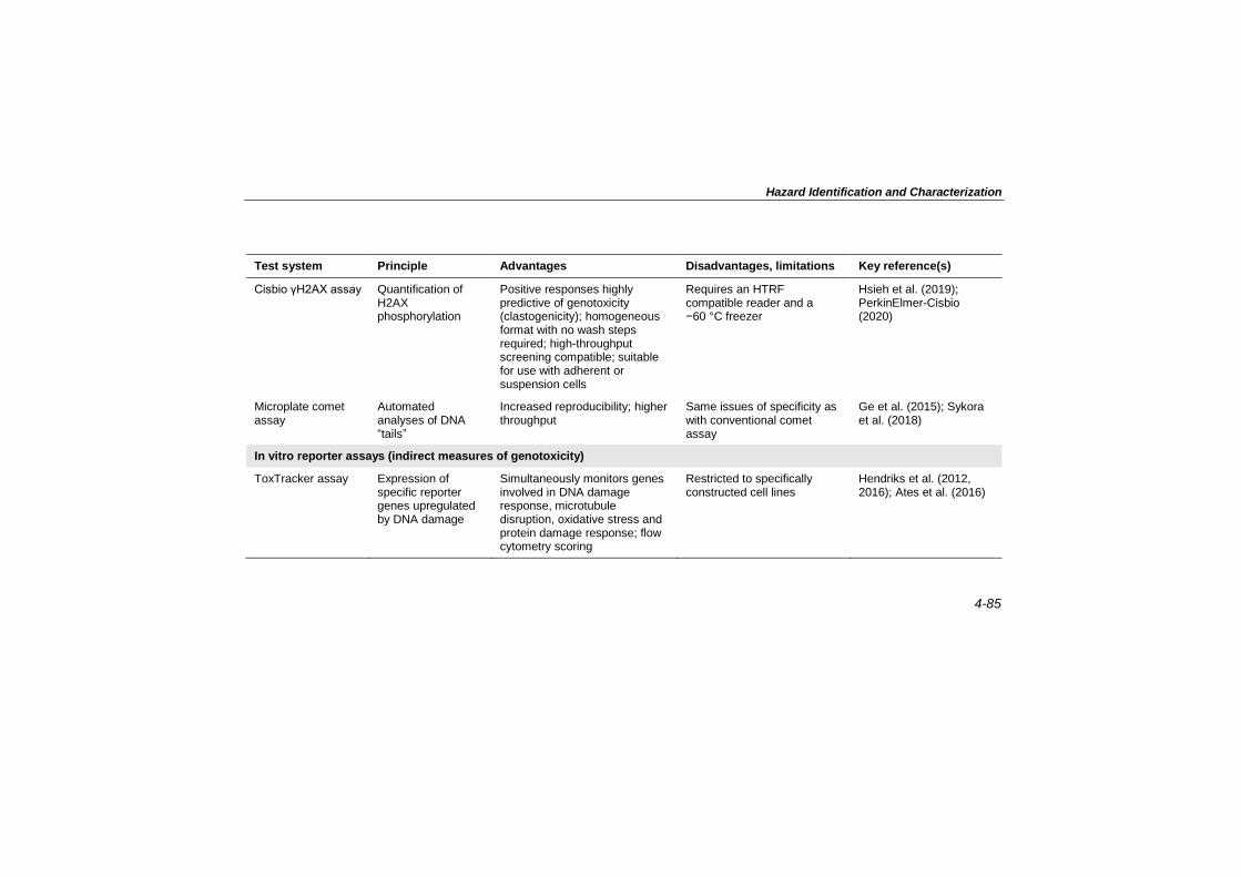
Hazard Identification and Characterization
4-85
Test system Principle Advantages Disadvantages, limitations Key reference(s)
Cisbio γH2AX assay Quantification of H2AX phosphorylation
Positive responses highly predictive of genotoxicity (clastogenicity); homogeneous format with no wash steps required; high-throughput screening compatible; suitable for use with adherent or suspension cells
Requires an HTRF compatible reader and a −60 °C freezer
Hsieh et al. (2019); PerkinElmer-Cisbio (2020)
Microplate comet assay
Automated analyses of DNA “tails”
Increased reproducibility; higher throughput
Same issues of specificity as with conventional comet assay
Ge et al. (2015); Sykora et al. (2018)
In vitro reporter assays (indirect measures of genotoxicity)
ToxTracker assay Expression of specific reporter genes upregulated by DNA damage
Simultaneously monitors genes involved in DNA damage response, microtubule disruption, oxidative stress and protein damage response; flow cytometry scoring
Restricted to specifically constructed cell lines
Hendriks et al. (2012, 2016); Ates et al. (2016)

EHC 240: Principles for Risk Assessment of Chemicals in Food
4-86
Table 4.7 (continued)
Test system Principle Advantages Disadvantages, limitations Key reference(s)
MultiFlow DNA Damage assay
In vitro high-content assays for multiple end-points
Determines MOA for MN induction; flow cytometry scoring
Method developed for suspension cell lines only
Bryce et al. (2013); Bemis et al. (2016a); Smith-Roe et al. (2018)
MultiFlow Aneugen Molecular Initiating Event Kit
In vitro follow-up assay for determining MOA of aneugens identified in the MultiFlow assay
Identifies tubulin binders and inhibitors of Aurora B kinase; flow cytometry scoring
Not yet commercially available
Bernacki et al. (2019)
p53-RE assay Reporter gene assay to assess activation of p53 response element
Assay for cellular signalling pathways activated by DNA damage; automated scoring
Currently limited to a single cell line (HCT-116); can respond to non-genotoxic stressors
Witt et al. (2017)
DT40 differential cytotoxicity assay
Enhanced cytotoxicity in cell lines lacking specific DNA repair enzymes
Highly specific for DNA repair pathways; automated scoring
Limited to isogenic chicken cell lines
Yamamoto et al. (2011); Nishihara et al. (2016)

Hazard Identification and Characterization
4-87
Test system Principle Advantages Disadvantages, limitations Key reference(s)
GreenScreen, BlueScreen
GADD45a-based reporter system; green fluorescent protein (GreenScreen) or Gaussia Luciferase (BlueScreen) detection
Highly specific for DNA repair pathways; automated scoring
Currently limited to a single cell line; may respond to non-genotoxic stressors
Hastwell et al. (2006); Simpson et al. (2013)
High-throughput real-time RT-qPCR
Gene expression assessment of 95 genes involved in genomic stability
Can be used for cell lines, primary cells, three-dimensional cultures
Limited to a few cell types, each requiring response characterization
Fischer et al. (2016); Strauch et al. (2017)
TGx-DDI Gene expression assessment of 64 DNA damage/repair genes
Prediction of DNA-damaging potential
Limited to a few cell types, each requiring response characterization
Li et al. (2015, 2017); Williams et al. (2015); Yauk et al. (2016a); Corton, Williams & Yauk (2018)
DDI: DNA damage–inducing; DNA: deoxyribonucleic acid; HTRF: Homogeneous Time-Resolved Fluorescence; MN: micronucleus; MOA: mode of action; RT-qPCR: reverse transcription quantitative polymerase chain reaction

EHC 240: Principles for Risk Assessment of Chemicals in Food
4-88
noteworthy advantages related to the cost, throughput and
information content of these assays, incorporation of realistic and
effective xenobiotic metabolism is a concern. Nevertheless, high-
throughput assays are now available to rapidly assess the induction
of DNA damage and repair, gene mutations, chromosomal damage or
DNA strand breaks (Table 4.7). As mutagenicity screening for
regulatory purposes generally requires the assessment of gene
mutations and chromosomal damage, assays that streamline detection
of these end-points are particularly noteworthy. In vitro versions of
the flow cytometric Pig-a gene mutation assay and the Transgenic
Rodent Somatic and Germ Cell Mutation Assays (OECD TG 488)
permit enumeration of mutations at a variety of endogenous and
transgenic loci (e.g. Pig-a, lacI, lacZ, cII, gpt, Spi−). These assays do
not require clonal selection and can measure mutagenicity more
efficiently than, for example, traditional Tk and Hprt locus mutation
assays.
Some of the high-throughput in vitro assays summarized in Table
4.7 exploit cellular pathways to rapidly measure biomarkers of DNA
damage or repair; most are based on genetically engineered cell lines
containing a promoter activated by genotoxic insult (e.g. p53
response element) fused to one or more reporter genes (e.g. β-
lactamase). Reporter gene activation is visualized via, for example,
automated micro-confocal imaging, fluorescent or luminescent
readouts, or flow cytometry. Examples include the ToxTracker
(Hendriks et al., 2012, 2016; Ates et al., 2016), GreenScreen
(Hastwell et al., 2006; Simpson et al., 2013) and several reporter gene
and antibody assays (e.g. p53RE, γH2AX, ATAD5) used by the
United States Tox21 Program (https://ntp.niehs.nih.gov/
whatwestudy/tox21/toolbox/index.html) or the USEPA’s ToxCast
Program (https://comptox.epa.gov/dashboard/chemical_lists/
toxcast). Importantly, in addition to mutagenic hazard, the
simultaneous or sequential examination of multiple end-points
representing several distinct pathways permits delineation of the
mutagenic MOA. Related assays, such as the MultiFlow DNA
Damage assay, assess the presence and localization of proteins (e.g.
γH2AX, nuclear p53, phospho-histone H3) indicative of DNA
damage and alterations in chromosome structure or number (Bryce et
al., 2016, 2017, 2018). Proteins are targeted by fluorescently labelled
antibodies, and cellular phenotype is scored using flow cytometry. In
addition to reporter-based approaches that track and quantify DNA
damage response activation, gene expression–based strategies, such
as DNA microarray, quantitative polymerase chain reaction (qPCR)

Hazard Identification and Characterization
4-89
and RNA sequencing approaches, have been used as high-throughput
approaches for measuring DNA damage signalling. For example, the
TGx-DDI assay monitors genes involved in genomic stability (e.g.
generalized stress responses, DNA repair, cell cycle control,
apoptosis and mitotic signalling) to identify DNA damage–inducing
(DDI) substances (Li et al., 2015, 2017; Williams et al., 2015; Yauk
et al., 2016a; Corton, Williams & Yauk, 2018; Corton, Witt & Yauk,
2019). Similarly, a high-throughput real-time reverse transcription
quantitative polymerase chain reaction (RT-qPCR) assay rapidly
scores 95 genes active in maintaining genomic integrity (Fischer et
al., 2016; Strauch et al., 2017). These reporter systems rapidly track
DNA damage and repair as indirect measures of genotoxicity.
To date, none of the high-throughput tools listed in Table 4.7
have OECD TGs, nor have they been incorporated into widely
accepted genotoxicity assessment platforms, such as those
recommended by ICH (2011), USFDA (2007) and ECHA (2017a). A
future role for these tools in regulatory decision-making would be
consistent with global trends to modernize the current mutagenicity
assessment frameworks, to reduce and replace the use of
experimental animals and to generate mutagenicity MOA
information. For example, Dearfield et al. (2017) outlined a paradigm
shift whereby a variety of mechanistic end-points indicative of
genomic damage are incorporated into a “next-generation testing
strategy”. Indeed, high-throughput tools are already supporting
regulatory evaluations based on traditional in vitro assays. For
example, the European Commission’s Scientific Committee on
Consumer Safety considers additional in vitro tests that include gene
expression and recombinant cell reporter assays (SCCS, 2018).
Similarly, Corton, Williams & Yauk (2018) outlined how the TGx-
DDI assay can be used for regulatory screening of chemicals. Buick
et al. (2017) used a TGx-DDI biomarker to evaluate two data-poor
substances prioritized by Health Canada for regulatory decision-
making due to structural similarity to known mutagens (i.e. Disperse
Orange and 1,2,4-benzenetriol), resulting in compound classification
consistent with more traditional end-points (e.g. in vitro MN
formation). Private sector organizations are now routinely using high-
throughput in vitro assays to evaluate the mutagenicity of products in
development, such as therapeutic candidates and industrial chemicals
(Thougaard et al., 2014; International Antimony Association, 2018;
Motoyama et al., 2018; Dertinger et al., 2019; Pinter et al., 2020).

EHC 240: Principles for Risk Assessment of Chemicals in Food
4-90
The in vitro tools and approaches summarized in Table 4.7
employ standard cultures of mammalian cells (e.g. two-dimensional
attached cultures, suspension cultures). To acquire data that might be
deemed more relevant to humans, while also reducing the use of
animals in research, three-dimensional cell culture systems have been
developed to score end-points such as chromosomal (i.e. MN) and
DNA damage (i.e. comet assay). Several novel assays are
summarized in Table 4.8.
Another alternative to traditional in vivo testing involves the use
of chicken eggs to assess chromosomal damage based on the
frequency of MN in extraembryonic peripheral blood (Wolf &
Luepke, 1997; Wolf, Niehaus-Rolf & Luepke, 2003; Hothorn et al.,
2013).
Advances in high-throughput detection of DNA damage and
repair, chromosomal aberrations and gene mutations may soon be
eclipsed by error-corrected, next-generation DNA sequencing (NGS)
approaches. Whereas previous NGS technologies did not permit
detection of rare, exposure-induced mutations (i.e. <10−5) in the
absence of clonal expansion, recent computational and experimental
innovations now allow detection of such rare mutations (<10−8) (Salk,
Schmitt & Loeb, 2018), with the precision and accuracy required to
assess genetic alterations in only a few DNA molecules within a cell
population. Although error-corrected NGS technologies are not yet
fully validated or widely applied, the technology is rapidly advancing
and may soon be routinely available, particularly because it does not
require specialized cells, loci or reporters, can score mutations at
virtually any locus in any tissue, organism or cells in culture, and can
readily be integrated into repeated-dose or translational studies
linking observations to humans.
4.5.7.3 Adverse outcome pathways for mutagenicity
The OECD AOP framework organizes diverse toxicological data
from different levels of biological complexity in order to increase
confidence in mechanistic relationships between key events leading
to adverse health outcomes. The AOP Knowledge Base,7 which
includes several modules, supports AOP construction to improve
application of mechanistic information for both chemical testing and
assessment (OECD, 2017d). AOPs also feed into Integrated
7 https://aopkb.oecd.org/index.html.

Hazard Identification and Characterization
4-91
Table 4.8. Novel in vitro genotoxicity assessment systems based on multicellular, three-dimensional constructs
Test system Principle Advantages Disadvantages, limitations Key reference(s)
Three-dimensional MN test
MN frequency in reconstructed skin model
Traditional end-point; simple to score; application in reconstructed skin models
Questions remain concerning metabolism
Aardema et al. (2010); Kirsch-Volders et al. (2011); Chapman et al. (2014); Pfuhler et al. (2014)
Three-dimensional comet assay
DNA damage assay in reconstructed skin model
Traditional end-point; simple to score; application in reconstructed skin models
Questions remain concerning metabolism
Pfuhler et al. (2014); Reisinger et al. (2018)
Hen’s egg MN assay
MN frequency in extraembryonic peripheral blood of fertilized hen eggs
Traditional end-point; some metabolic capacity
Non-mammalian test; limited metabolism
Wolf & Luepke (1997); Wolf, Niehaus-Rolf & Luepke (2003); Hothorn et al. (2013)
Avian egg genotoxicity assay
Comet assay and 32P-postlabelling of adducts in hepatocytes isolated from turkey or hen eggs treated ex vivo
Some metabolic activity; traditional end-points; studies of some MOAs
Non-mammalian test; limited metabolism; postlabelling with 32P
Williams, Deschl & Williams (2011); Kobets et al. (2016, 2018, 2019)
DNA: deoxyribonucleic acid; MN: micronucleus; MOA: mode of action

EHC 240: Principles for Risk Assessment of Chemicals in Food
4-92
Approaches to Testing and Assessment (IATA), a pragmatic
approach to hazard characterization that integrates in silico, in vitro
and in vivo assessment tools, including high-throughput in vitro tools
based on toxicogenomic or recombinant cell reporter technologies
(Sakuratani, Horie & Leinala, 2018). The OECD IATA Case Studies
Project reviews case-studies related to different end-points, including
mutagenicity and genotoxicity,1 and publishes the learnings and areas
identified where additional guidance is needed (OECD, 2017a,b,
2018a, 2019). The AOP on “alkylation of DNA in male pre-meiotic
germ cells leading to heritable mutations” was the first AOP on
mutagenicity published in the OECD AOP series (Yauk et al.,
2016b). To date, several other AOPs related to mutagenicity are under
development in the AOP-Wiki2 (one module of the AOP Knowledge
Base), and several ongoing initiatives should contribute to populating
the AOP Knowledge Base with more AOPs on mutagenicity in the
near future, increasing the development of AOP networks and
supporting further tiered testing and IATA strategies.
4.5.7.4 Quantitative approaches for safety assessment
National and international mutagenicity evaluation committees
have highlighted a desire to employ quantitative methods for
regulatory interpretation of mutagenicity dose–response data
(MacGregor et al., 2015a,b; UKCOM, 2018). Lacking
carcinogenicity data, quantitative analysis of in vivo mutagenicity
dose–response data could be used for deriving MOEs (White &
Johnson, 2016). This is particularly relevant for risk assessment and
management of unavoidable food contaminants with positive results
for gene mutation or DNA-reactive mutagenicity structural alerts and
exposures exceeding the TTC of 0.0025 µg/kg body weight per day
(see section 4.5.5.2). Moving to a quantitative approach requires a
paradigm shift from hazard identification of mutagens and recognizes
that compensatory cellular responses (i.e. DNA damage processing)
are quantitatively manifested as mechanistically plausible dose–
response thresholds (Parry, Fielder & McDonald, 1994; Nohmi,
2008, 2018; Carmichael, Kirsch-Volders & Vrijhof, 2009; Johnson et
al., 2014; Nohmi & Tsuzuki, 2016). With respect to threshold
determination, this is still under debate, and there is currently no
international consensus.
1 h t tp : / /www.oecd .o rg/chemicalsafety/ r isk -assessment/ iata -
in tegrated-approaches- to-tes t ing-and-assessment.h tm. 2 https://aopwiki.org/.

Hazard Identification and Characterization
4-93
Several researchers have employed dose–response point of
departure values, such as the benchmark dose (BMD), the threshold
dose (Td) and the NOGEL, for quantitative interpretation of in vitro
and in vivo mutagenicity dose–response data. With respect to in vitro
dose–response data, the BMD approach has been used for MOE
determinations and to rank potency across test substances, cell types
and experimental protocols (Bemis et al., 2016b; Benford, 2016;
Tweats et al., 2016; Wills et al., 2016; Verma et al., 2017; Guo et al.,
2018). However, it should be noted that not all in vitro guideline
mutagenicity tests are suitable for dose–response assessment, as they
are optimized to discriminate between “positive” and “negative”
compounds. The mutagenicity of ethyl methanesulfonate, an impurity
detected in Viracept, an antiretroviral drug, was shown to exhibit a
threshold, both in vitro and in vivo. In vivo mutagenicity data were
then used to determine a permissible daily exposure to the compound
(Gocke & Wall, 2009; Müller & Gocke, 2009). Although the
regulatory utility of quantitative interpretation of in vivo dose–
response data is increasingly recognized, use of mutagenicity-based
BMD values to estimate MOEs for mutagenic food contaminants will
require consensus regarding, for example, choice of test/end-point, an
appropriate benchmark response for mutagenicity end-points, and
appropriate safety factors for exposure limit determination (Ritter et
al., 2007; Nielsen, Ostergaard & Larsen, 2008; Dankovic et al., 2015;
IPCS, 2018).
4.5.8 References1
Aardema MJ, Barnett BC, Khambatta Z, Reisinger K, Ouedraogo-Arras G, Faquet B et al. (2010). International prevalidation studies of the EpiDerm™ 3D human reconstructed skin micronucleus (RSMN) assay: transferability and reproducibility. Mutat Res. 701(2):123–31. doi:10.1016/j.mrgentox.2010.05.017.
Amberg A, Beilke L, Bercu J, Bower D, Brigo A, Cross KP et al. (2016). Principles and procedures for implementation of ICH M7 recommended (Q)SAR analyses. Regul Toxicol Pharmacol. 77:13–24. doi:10.1016/j.yrtph.2016.02.004.
Amberg A, Andaya RV, Anger LT, Barber C, Beilke L, Bercu J et al. (2019). Principles and procedures for handling out-of-domain and indeterminate
1 Internet links provided in these references were active as of the date of
final editing.

EHC 240: Principles for Risk Assessment of Chemicals in Food
4-94
results as part of ICH M7 recommended (Q)SAR analyses. Regul Toxicol Pharmacol. 102:53–64. doi:10.1016/j.yrtph.2018.12.007.
Ames BN, Lee FD, Durston WE (1973). An improved bacterial test system for the detection and classification of mutagens and carcinogens. Proc Natl Acad Sci U S A. 70(3):782–6. doi:10.1073/pnas.70.3.782.
Ates G, Favyts D, Hendriks G, Derr R, Mertens B, Verschaeve L et al. (2016). The Vitotox and ToxTracker assays: a two-test combination for quick and reliable assessment of genotoxic hazards. Mutat Res. 810:13–21. doi:10.1016/j.mrgentox.2016.09.005.
Au WW, Ramanujam VMS, Ward JB, Legator MS (1991). Chromosome aberrations in lymphocytes of mice after sub-acute low-level inhalation exposure to benzene. Mutat Res. 260(2):219–24. doi:10.1016/0165-1218(91)90011-a.
Auerbach C, Robson JM, Carr JG (1947). The chemical production of mutations. Science. 105(2723):243–7. doi:10.1126/science.105.2723.243.
Avlasevich S, Bryce S, de Boeck M, Elhajouji A, Van Goethem F, Lynch A et al. (2011). Flow cytometric analysis of micronuclei in mammalian cell cultures: past, present and future. Mutagenesis. 26(1):147–52. doi:10.1093/mutage/geq058.
Bakhtyari NG, Raitano G, Benfenati E, Martin T, Young D (2013). Comparison of in silico models for prediction of mutagenicity. J Environ Sci Health C Environ Carcinog Ecotoxicol Rev. 31(1):45–66. doi:10.1080/10590501.2013.763576.
Balbo S, Turesky RJ, Villalta PW (2014). DNA adductomics. Chem Res
Toxicol. 27(3):356–66. doi:10.1021/tx4004352.
Barber C, Amberg A, Custer L, Dobo KL, Glowienke S, Van Gompel J et al. (2015). Establishing best practise in the application of expert review of mutagenicity under ICH M7. Regul Toxicol Pharmacol. 73(1):367–77. doi:10.1016/j.yrtph.2015.07.018.
Barber C, Hanser T, Judson P, Williams R (2017). Distinguishing between expert and statistical systems for application under ICH M7. Regul Toxicol Pharmacol. 84:124–30. doi:10.1016/j.yrtph.2016.12.012.
Beken S, Kasper P, Van der Laan JW (2016). Regulatory acceptance of alternative methods in the development and approval of pharmaceuticals. Adv Exp Med Biol. 856:33–6. doi:10.1007/978-3-319-33826-2_3.
Bemis JC, Heflich RH (2019). In vitro mammalian cell mutation assays based on the Pig-a gene: a report of the 7th International Workshop on Genotoxicity Testing (IWGT) workgroup. Mutat Res. 847:403028. doi:10.1016/j.mrgentox.2019.03.001.
Bemis JC, Bryce SM, Nern M, Raschke M, Sutter A (2016a). Best practices for application of attachment cells to in vitro micronucleus assessment by flow cytometry. Mutat Res. 795:51–9. doi:10.1016/j.mrgentox.2015.10.007.

Hazard Identification and Characterization
4-95
Bemis JC, Wills JW, Bryce SM, Torous DK, Dertinger SD, Slob W (2016b). Comparison of in vitro and in vivo clastogenic potency based on benchmark dose analysis of flow cytometric micronucleus data. Mutagenesis. 31(3):277–85. doi:10.1093/mutage/gev041.
Benford DJ (2016). The use of dose–response data in a margin of exposure approach to carcinogenic risk assessment for genotoxic chemicals in food. Mutagenesis. 31(3):329–31. doi:10.1093/mutage/gev064.
Benigni R, Bossa C, Battistelli CL (2013). ISSTOX Chemical Toxicity Databases [online databases]. Rome: Istituto Superiore di Sanità (http://old.iss.it/meca/index.php?lang=1&anno=2013&tipo=25).
Benigni R, Battistelli CL, Bossa C, Giuliani A, Fioravanzo E, Bassan A et al. (2019). Evaluation of the applicability of existing (Q)SAR models for predicting the genotoxicity of pesticides and similarity analysis related with genotoxicity of pesticides for facilitating of grouping and read across. Parma: European Food Safety Authority (EFSA supporting publications EN-1598; https://efsa.onlinelibrary.wiley.com/doi/epdf/10.2903/sp.efsa.2019.EN-1598).
Bernacki DT, Bryce SM, Bemis JC, Dertinger SD (2019). Aneugen molecular mechanism assay: proof-of-concept with 27 reference chemicals. Toxicol Sci. 170(2):382–93. doi:10.1093/toxsci/kfz123.
Beronius A, Ågerstrand M (2017). Making the most of expert judgment in hazard and risk assessment of chemicals. Toxicol Res. 6(5):571–7. doi:10.1039/c7tx00114b.
Besaratinia A, Pfeifer GP (2010). Applications of the human p53 knock-in (Hupki) mouse model for human carcinogen testing. FASEB J. 24(8):2612–9. doi:10.1096/fj.10-157263.
Beyersmann D, Hartwig A (2008). Carcinogenic metal compounds: recent insight into molecular and cellular mechanisms. Arch Toxicol. 82(8):493–512. doi:10.1007/s00204-008-0313-y.
Blackburn K, Stuard SB (2014). A framework to facilitate consistent characterization of read across uncertainty. Regul Toxicol Pharmacol. 68(3):353–62. doi:10.1016/j.yrtph.2014.01.004.
Boobis A, Brown P, Cronin MTD, Edwards J, Lodovico Galli C, Goodman J et al. (2017). Origin of the TTC values for compounds that are genotoxic and/or carcinogenic and an approach for their re-evaluation. Crit Rev Toxicol. 47(8):710–32. doi:10.1080/10408444.2017.1318822.
Bossa C, Benigni R, Tcheremenskaia O, Battistelli CL (2018). (Q)SAR methods for predicting genotoxicity and carcinogenicity: scientific rationale and regulatory frameworks. In: Nicolotti O, editor. Computational toxicology. Methods in molecular biology, vol. 1800. New York (NY): Humana Press; 447–73. doi:10.1007/978-1-4939-7899-1_20.

EHC 240: Principles for Risk Assessment of Chemicals in Food
4-96
Brown K (2012). Methods for the detection of DNA adducts. In: Genetic toxicology. New York (NY): Springer; 207–30.
Bryce SM, Avlasevich SL, Bemis JC, Phonethepswath S, Dertinger SD (2010).
Miniaturized flow cytometric in vitro micronucleus assay represents an efficient
tool for comprehensively characterizing genotoxicity dose–response
relationships. Mutat Res. 703:191–9. doi:10.1016/j.mrgentox.2010.08.020.
Bryce SM, Avlasevich SL, Bemis JC, Dertinger SD (2011). Miniaturized flow cytometry–based CHO-K1 micronucleus assay discriminates aneugenic and clastogenic modes of action. Environ Mol Mutagen. 52(4):280–6. doi:10.1002/em.20618.
Bryce SM, Avlasevich SL, Bemis JC, Tate M, Walmsley RM, Saad F et al. (2013). Flow cytometric 96-well microplate–based in vitro micronucleus assay with human TK6 cells: protocol optimization and transferability assessment. Environ Mol Mutagen. 54(3):180–94. doi:10.1002/em.21760.
Bryce SM, Bernacki DT, Bemis JC, Dertinger SD (2016). Genotoxic mode of action predictions from a multiplexed flow cytometric assay and a machine learning approach. Environ Mol Mutagen. 57(3):171–89. doi:10.1002/em.21996.
Bryce SM, Bernacki DT, Bemis JC, Spellman RA, Engel ME, Schuler M et al. (2017). Interlaboratory evaluation of a multiplexed high information content in vitro genotoxicity assay. Environ Mol Mutagen. 58(3):146–61. doi:10.1002/em.22083.
Bryce SM, Bernacki DT, Smith-Roe SL, Witt KL, Bemis JC, Dertinger SD (2018). Investigating the generalizability of the MultiFlow® DNA Damage assay and several companion machine learning models with a set of 103 diverse test chemicals. Toxicol Sci. 162(1):146–66. doi:10.1093/toxsci/kfx235.
Buick JK, Williams A, Kuo B, Wills JW, Swartz CD, Recio L et al. (2017). Integration of the TGx-28.65 genomic biomarker with the flow cytometry micronucleus test to assess the genotoxicity of disperse orange and 1,2,4-benzenetriol in human TK6 cells. Mutat Res. 806:51–62. doi:10.1016/j.mrfmmm.2017.09.002.
Burden N, Mahony C, Müller BP, Terry C, Westmoreland C, Kimber I (2015). Aligning the 3Rs with new paradigms in the safety assessment of chemicals. Toxicology. 330:62–6. doi:10.1016/j.tox.2015.01.014.
Busby WF Jr, Ackermann JM, Crespi CL (1999). Effect of methanol, ethanol, dimethyl sulfoxide, and acetonitrile on in vitro activities of cDNA-expressed human cytochromes P-450. Drug Metab Dispos. 27(2):246–9.
Carmichael P, Kirsch-Volders M, Vrijhof H (2009). Thresholds for genotoxins and their application in risk assessment. Mutat Res. 678(2):71. doi:10.1016/j.mrgentox.2009.04.012.
Cassano A, Raitano G, Mombelli E, Fernández A, Cester J, Roncaglioni A et al. (2014). Evaluation of QSAR models for the prediction of Ames genotoxicity:

Hazard Identification and Characterization
4-97
a retrospective exercise on the chemical substances registered under the EU REACH regulation. J Environ Sci Health C Environ Carcinog Ecotoxicol Rev. 32(3):273–98. doi:10.1080/10590501.2014.938955.
Cefic-LRI (2020). B18: Carcinogen dose–response database for threshold of toxicological concern (CDRD-TTC). Brussels: European Chemical Industry Council Long-Range Research Initiative (http://cefic-lri.org/projects/b18-carcinogen-dose-response-database-for-threshold-of-toxicological-concern-cdrd-ttc/).
Chang YJ, Cooke MS, Hu CW, Chao MR (2018). Novel approach to integrated DNA adductomics for the assessment of in vitro and in vivo environmental exposures. Arch Toxicol. 92(8):2665–80. doi:10.1007/s00204-018-2252-6.
Chapman KE, Thomas AD, Wills JW, Pfuhler S, Doak SH, Jenkins GJS (2014). Automation and validation of micronucleus detection in the 3D EpiDerm™ human reconstructed skin assay and correlation with 2D dose responses. Mutagenesis. 29(3):165–75. doi:10.1093/mutage/geu011.
Cheeseman MA, Machuga EJ, Bailey AB (1999). A tiered approach to threshold of regulation. Food Chem Toxicol. 37(4):387–412. doi:10.1016/S0278-6915(99)00024-1.
Chervona Y, Arita A, Costa M (2012). Carcinogenic metals and the epigenome: understanding the effect of nickel, arsenic, and chromium. Metallomics. 4(7):619–27. doi:10.1039/c2mt20033c.
Cimino MC (2006). Comparative overview of current international strategies and guidelines for genetic toxicology testing for regulatory purposes. Environ Mol Mutagen. 47(5):362–90. doi:10.1002/em.20216.
Codex Alimentarius Commission (2008). Guidelines for the use of flavourings. Rome: Food and Agriculture Organization of the United Nations and World Health Organization, Joint FAO/WHO Food Standards Programme, Codex Alimentarius Commission (CAC/GL 66-2008; http://www.fao.org/input/download/standards/11020/cxg_066e.pdf).
Collins A, Koppen G, Valdiglesias V, Dusinska M, Kruszewski M, Møller P et al. (2014). The comet assay as a tool for human biomonitoring studies: the ComNet project. Mutat Res. 759:27–39. doi:10.1016/j.mrrev.2013.10.001.
Corton JC, Williams A, Yauk CL (2018). Using a gene expression biomarker to identify DNA damage–inducing agents in microarray profiles. Environ Mol Mutagen. 59(9):772–84. doi:10.1002/em.22243.
Corton JC, Witt KL, Yauk CL (2019). Identification of p53 activators in a human microarray compendium. Chem Res Toxicol. 32(9):1748–59. doi:10.1021/acs.chemrestox.9b00052.
Corvi R, Madia F (2018). EURL ECVAM Genotoxicity and Carcinogenicity Consolidated Database of Ames Positive Chemicals [online database].

EHC 240: Principles for Risk Assessment of Chemicals in Food
4-98
Brussels: European Commission, Joint Research Centre (http://data.europa.eu/89h/jrc-eurl-ecvam-genotoxicity-carcinogenicity-ames).
Costa M (2019). Review of arsenic toxicity, speciation and polyadenylation of canonical histones. Toxicol Appl Pharmacol. 375:1–4. doi:10.1016/j.taap.2019.05.006.
Cronin MTD, Richarz A-N, Schultz TW (2019). Identification and description of the uncertainty, variability, bias and influence in quantitative structure–activity relationships (QSARs) for toxicity prediction. Regul Toxicol Pharmacol. 106:90–104. doi:10.1016/j.yrtph.2019.04.007.
Dankovic DA, Naumann BD, Maier A, Dourson ML, Levy LS (2015). The scientific basis of uncertainty factors used in setting occupational exposure limits. J Occup Environ Hyg. 12(Suppl 1):S55–68. doi:10.1080/15459624.2015.1060325.
Dearfield KL, Cimino MC, McCarroll NE, Mauer I, Valcovic LR (2002). Genotoxicity risk assessment: a proposed classification strategy. Mutat Res. 521(1–2):121–35. doi:10.1016/S1383-5718(02)00236-X.
Dearfield KL, Thybaud V, Cimino MC, Custer L, Czich A, Harvey JS et al. (2011). Follow-up actions from positive results of in vitro genetic toxicity testing. Environ Mol Mutagen. 52(3):177–204. doi:10.1002/em.20617.
Dearfield KL, Gollapudi BB, Bemis JC, Benz RD, Douglas GR, Elespuru RK et al. (2017). Next generation testing strategy for assessment of genomic damage: a conceptual framework and considerations. Environ Mol Mutagen. 58(5):264–83. doi:10.1002/em.22045.
Dertinger SD, Torous DK, Hall NE, Murante FG, Gleason SE, Miller RK et al. (2002). Enumeration of micronucleated CD71-positive human reticulocytes with a single-laser flow cytometer. Mutat Res. 515(1–2):3–14. doi:10.1016/s1383-5718(02)00009-8.
Dertinger SD, Bishop ME, McNamee JP, Hayashi M, Suzuki T, Asano N et al. (2006). Flow cytometric analysis of micronuclei in peripheral blood reticulocytes: I. Intra- and interlaboratory comparison with microscopic scoring. Toxicol Sci. 94(1):83–91. doi:10.1093/toxsci/kfl075.
Dertinger SD, Phonethepswath S, Weller P, Nicolette J, Murray J, Sonders P et al. (2011a). International Pig-a gene mutation assay trial: evaluation of transferability across 14 laboratories. Environ Mol Mutagen. 52(9):690–8. doi:10.1002/em.20672.
Dertinger SD, Torous DK, Hayashi M, MacGregor JT (2011b). Flow cytometric scoring of micronucleated erythrocytes: an efficient platform for assessing in vivo cytogenetic damage. Mutagenesis. 26(1):139–45. doi:10.1093/mutage/geq055.
Dertinger SD, Avlasevich SL, Bemis JC, Chen Y, MacGregor JT (2015). Human erythrocyte PIG-A assay: an easily monitored index of gene mutation requiring low volume blood samples. Environ Mol Mutagen. 56(4):366–77. doi:10.1002/em.21924.

Hazard Identification and Characterization
4-99
Dertinger SD, Kraynak AR, Wheeldon RP, Bernacki D, Bryce SM, Hall N et al. (2019). Predictions of genotoxic potential, mode of action, molecular targets, and potency via a tiered Multiflow® assay data analysis strategy. Environ Mol Mutagen. 60(6):513–33. doi:10.1002/em.22274.
Dewangan J, Pandey PK, Divakar A, Mishra S, Srivastava S, Rath SK (2018). Detection of gene mutation in cultured mammalian cells. In: Kumar A, Dobrovolsky VN, Dhawan A, Shanker R, editors. Mutagenicity: assays and applications. London: Elsevier; 49–67.
Dewhurst I, Renwick AG (2013). Evaluation of the threshold of toxicological concern (TTC) – challenges and approaches. Regul Toxicol Pharmacol. 65(1):168–77. doi:10.1016/j.yrtph.2012.03.007.
D’Gama AM, Pochareddy S, Li M, Jamuar SS, Reiff RE, Lam AN et al. (2015). Targeted DNA sequencing from autism spectrum disorder brains implicates multiple genetic mechanisms. Neuron. 88:910–7. doi:10.1016/j.neuron.2015.11.009.
Dobo KL, Greene N, Fred C, Glowienke S, Harvey JS, Hasselgren C et al. (2012). In silico methods combined with expert knowledge rule out mutagenic potential of pharmaceutical impurities: an industry survey. Regul Toxicol Pharmacol. 62(3):449–55. doi:10.1016/j.yrtph.2012.01.007.
Douglas GR, Jiao J, Gingerich JD, Gossen JA, Soper LM (1995). Temporal and molecular characteristics of mutations induced by ethylnitrosourea in germ cells isolated from seminiferous tubules and in spermatozoa of lacZ transgenic mice. Proc Natl Acad Sci U S A. 92(16):7485–9. doi:10.1073/pnas.92.16.7485.
Eastmond DA (2017). Recommendations for the evaluation of complex genetic toxicity data sets when assessing carcinogenic risks to humans. Environ Mol Mutagen. 58:380–5. doi:10.1002/em.22078.
Eastmond DA, Hartwig A, Anderson D, Anwar WA, Cimino MC, Dobrev I et al. (2009). Mutagenicity testing for chemical risk assessment: update of the WHO/IPCS harmonized scheme. Mutagenesis. 24(4):341–9. doi:10.1093/mutage/gep014.
ECETOC (2012). Category approaches, read-across, (Q)SAR. Brussels: European Centre for Ecotoxicology and Toxicology of Chemicals (ECETOC Technical Report No. 116; http://www.ecetoc.org/wp-content/uploads/2014/08/ECETOC-TR-116-Category-approaches-Read-across-QSAR.pdf).
ECHA (2008). Chapter R.6: QSARs and grouping of chemicals. In: Guidance on information requirements and chemical safety assessment. Helsinki: European Chemicals Agency (https://echa.europa.eu/documents/10162/13632/information_requirements_r6_en.pdf).
ECHA (2011). Chapter R.4: Evaluation of available information. Version 1.1. In: Guidance on information requirements and chemical safety assessment. Helsinki: European Chemicals Agency (https://www.echa.europa.eu/documents/10162/13643/information_requirements_r4_en.pdf).

EHC 240: Principles for Risk Assessment of Chemicals in Food
4-100
ECHA (2017a). Chapter R.7a: Endpoint specific guidance. Version 6.0. In: Guidance on information requirements and chemical safety assessment. Helsinki: European Chemicals Agency (https://www.echa.europa.eu/documents/10162/13632/information_requirements_r7a_en.pdf).
ECHA (2017b). Read-Across Assessment Framework (RAAF). Helsinki: European Chemicals Agency (https://echa.europa.eu/documents/10162/13628/raaf_en.pdf).
ECHA (2020). The use of alternatives to testing on animals for the REACH regulation. Fourth report under Article 117(3) of the REACH regulation. Helsinki: European Chemicals Agency (https://echa.europa.eu/documents/10162/0/alternatives_test_animals_2020_en.pdf/db66b8a3-00af-6856-ef96-5ccc5ae11026).
EFSA (2009). European Food Safety Authority Scientific Committee; Guidance on safety assessment of botanicals and botanical preparations intended for use as ingredients in food supplements, on request of EFSA. EFSA J. 7(9):1249. doi:10.2093/j.efsa.2009.1249.
EFSA (2011). European Food Safety Authority Scientific Committee; Scientific Opinion on genotoxicity testing strategies applicable to food and feed safety assessment. EFSA J. 9(9):2379. doi:10.2903/j.efsa.2011.2379.
EFSA (2013). European Food Safety Authority Panel on Plant Protection Products and their Residues (PPR); Scientific Opinion on the identification of pesticides to be included in cumulative assessment groups on the basis of their toxicological profile. EFSA J. 11(7):3293. doi:10.2903/j.efsa.2013.3293.
EFSA (2015). European Food Safety Authority Panel on Food Contact Materials, Enzymes, Flavourings and Processing Aids (CEF); Scientific Opinion on the risks to public health related to the presence of bisphenol A (BPA) in foodstuffs. EFSA J. 13(1):3978. doi:10.2903/j.efsa.2015.3978.
EFSA (2016a). European Food Safety Authority Panel on Plant Protection Products and their Residues (PPR); Guidance on the establishment of the residue definition for dietary risk assessment. EFSA J. 14(12):e04549. doi:10.2903/j.efsa.2016.4549.
EFSA (2016b). European Food Safety Authority Panel on Dietetic Products, Nutrition and Allergies (NDA); Guidance on the preparation and presentation of an application for authorisation of a novel food in the context of Regulation (EU) 2015/2283. EFSA J. 14(11):4594. doi:10.2903/j.efsa.2016.4594.
EFSA (2019a). European Food Safety Authority Scientific Committee; Statement on genotoxicity assessment of chemical mixtures. EFSA J. 17:5519. doi:10.2903/j.efsa.2019.5519.
EFSA (2019b). European Food Safety Authority Scientific Committee; Guidance on harmonised methodologies for human health, animal health and ecological risk assessment of combined exposure to multiple chemicals. EFSA J. 17(3):5634. doi:10.2903/j.efsa.2019.5634.

Hazard Identification and Characterization
4-101
EFSA, WHO (2016). Review of the threshold of toxicological concern (TTC) approach and development of new TTC decision tree. Parma: European Food Safety Authority and World Health Organization (EFSA supporting publication 2016: EN-1006; https://efsa.onlinelibrary.wiley.com/doi/pdf/10.2903/sp.efsa.2016.EN-1006).
EMA (2006). Guideline on non-clinical documentation for herbal medicinal products in applications for marketing authorisation (bibliographical and mixed applications) and in applications for simplified registration. London: European Medicines Agency, Committee on Herbal Medicinal Products (HMPC) (http://www.ema.europa.eu/docs/en_GB/document_library/Scientific_guideline/2009/09/WC500003576.pdf).
EMA (2009). Guideline on selection of test materials for genotoxicity testing for traditional herbal medicinal products/herbal medicinal products. London: European Medicines Agency, Committee on Herbal Medicinal Products (HMPC) (http://www.ema.europa.eu/docs/en_GB/document_library/Scientific_guideline/2010/01/WC500059583.pdf).
EMA (2011). Guideline on specifications: test procedures and acceptance criteria for herbal substances, herbal preparations and herbal medicinal products/traditional herbal medicinal products. London: European Medicines Agency (http://www.ema.europa.eu/docs/en_GB/document_library/Scientific_guideline/2011/09/WC500113210.pdf).
Erickson RP (2003). Somatic gene mutation and human disease other than cancer. Mutat Res. 543(2):125–36. doi:10.1016/S1383-5742(03)00010-3.
Erickson RP (2010). Somatic gene mutation and human disease other than cancer: an update. Mutat Res. 705(2):96–106. doi:10.1016/j.mrrev.2010.04.002.
FAO (2001). Smoke flavourings. Prepared by the fifty-seventh meeting of JECFA for publication in FNP 52 Addendum 9. Rome: Food and Agriculture Organization of the United Nations (http://www.fao.org/fileadmin/user_upload/jecfa_additives/docs/Monograph1/Additive-386.pdf).
FAO (2006). General specifications and considerations for enzyme preparations used in food processing. Prepared by the sixty-seventh meeting of JECFA for publication in FAO JECFA Monographs 3. Rome: Food and Agriculture Organization of the United Nations (http://www.fao.org/docrep/009/a0691e/A0691E03.htm).
FAO/WHO (1972). Toxicological evaluation of some enzymes, modified starches and certain other substances. Prepared by the fifteenth meeting of the Joint FAO/WHO Expert Committee on Food Additives. Geneva: World Health Organization (FAO Nutrition Meetings Report Series, No. 50A; WHO Food Additives Series, No. 1; http://www.inchem.org/documents/jecfa/jecmono/v001je01.htm).
FAO/WHO (1987). Evaluation of certain food additives and contaminants. Thirty-first report of the Joint FAO/WHO Expert Committee on Food Additives.

EHC 240: Principles for Risk Assessment of Chemicals in Food
4-102
Geneva: World Health Organization (WHO Technical Report Series, No. 759; https://apps.who.int/iris/bitstream/handle/10665/39108/WHO_TRS_759.pdf?sequence=1).
FAO/WHO (2002). Invertase from Saccharomyces cerevisiae. In: Fifty-seventh report of the Joint FAO/WHO Expert Committee on Food Additives. Geneva: World Health Organization; 18 (http://apps.who.int/iris/bitstream/handle/10665/42578/WHO_TRS_909.pdf?sequence=1).
FAO/WHO (2011). Pesticide residues in food 2011 – Report of the Joint Meeting of the FAO Panel of Experts on Pesticide Residues in Food and the Environment and the WHO Core Assessment Group on Pesticide Residues. Rome: Food and Agriculture Organization of the United Nations and World Health Organization (FAO Plant Production and Protection Paper 211; http://www.fao.org/fileadmin/templates/agphome/documents/Pests_Pesticides/JMPR/Report11/JMPR_2011_Report.pdf).
FAO/WHO (2016). Evaluation of certain food additives. Eighty-second report of the Joint FAO/WHO Expert Committee on Food Additives. Geneva: World Health Organization (WHO Technical Report Series, No. 1000: https://apps.who.int/iris/bitstream/handle/10665/250277/9789241210003-eng.pdf?sequence=1).
FAO/WHO (2017a). Chlormequat. In: Pesticide residues in food – 2017. Report of the Joint Meeting of the FAO Panel of Experts on Pesticide Residues in Food and the Environment and the WHO Core Assessment Group on Pesticide Residues. Rome: Food and Agriculture Organization of the United Nations (FAO Plant Production and Protection Paper 232; http://www.fao.org/fileadmin/templates/agphome/documents/Pests_Pesticides/JMPR/Report2017/5.6_CHLORMEQUAT__015_.pdf).
FAO/WHO (2017b). Natamycin. In: Pesticide residues in food – 2017. Report of the Joint Meeting of the FAO Panel of Experts on Pesticide Residues in Food and the Environment and the WHO Core Assessment Group on Pesticide Residues. Rome: Food and Agriculture Organization of the United Nations (FAO Plant Production and Protection Paper 232; http://www.fao.org/fileadmin/templates/agphome/documents/Pests_Pesticides/JMPR/Report2017/5.25_NATAMYCIN__300_.pdf).
FAO/WHO (2018). Evaluation of certain food additives. Eighty-sixth report of the Joint FAO/WHO Expert Committee on Food Additives. Geneva: World Health Organization and Food and Agriculture Organization of the United Nations (WHO Technical Report Series, No. 1014; https://apps.who.int/iris/bitstream/handle/10665/279832/9789241210232-eng.pdf?sequence=1&isAllowed=y).
FAO/WHO (2019). Joint FAO/WHO Expert Committee on Food Additives: Expert meeting of Working Group established to consider the evaluation of enzyme preparations used in the manufacture of foods, Rome, 11–14 December 2018. Final report. Rome: Food and Agriculture Organization of the United Nations and World Health Organization (http://www.fao.org/3/ca7188en/ca7188en.pdf).

Hazard Identification and Characterization
4-103
Felter S, Lane RW, Latulippe ME, Llewellyn GC, Olin SS, Scimeca JA et al. (2009). Refining the threshold of toxicological concern (TTC) for risk prioritization of trace chemicals in food. Food Chem Toxicol. 47(9):2236–45. doi:10.1016/j.fct.2009.06.018.
Fenech M, Kirsch-Volders M, Rossnerova A, Sram R, Romm H, Bolognesi C et al. (2013). HUMN project initiative and review of validation, quality control and prospects for further development of automated micronucleus assays using image cytometry systems. Int J Hyg Environ Health. 216(5):541–52. doi:10.1016/j.ijheh.2013.01.008.
Fischer BM, Neumann D, Piberger AL, Risnes SF, Köberle B, Hartwig A (2016). Use of high-throughput RT-qPCR to assess modulations of gene expression profiles related to genomic stability and interactions by cadmium. Arch Toxicol. 90(11):2745–61. doi:10.1007/s00204-015-1621-7.
Ge J, Chow DN, Fessler JL, Weingeist DM, Wood DK, Engelward BP (2015). Micropatterned comet assay enables high throughput and sensitive DNA damage quantification. Mutagenesis. 30(1):11–9. doi:10.1093/mutage/geu063.
Gocke E, Wall M (2009). In vivo genotoxicity of EMS: statistical assessment of the dose response curves. Toxicol Lett. 190(3):298–302. doi:10.1016/j.toxlet.2009.03.008.
Gollapudi BB, Lynch AM, Heflich RH, Dertinger SD, Dobrovolsky VN, Froetschl R et al. (2015). The in vivo Pig-a assay: a report of the International Workshop on Genotoxicity Testing (IWGT) workgroup. Mutat Res. 783:23–35. doi:10.1016/j.mrgentox.2014.09.007.
Görög S (2018). Critical review of reports on impurity and degradation product profiling in the last decade. Trends Anal Chem. 101:2–16. doi:10.1016/j.trac.2017.09.012.
Greene N, Dobo KL, Kenyon MO, Cheung J, Munzner J, Sobol Z et al. (2015). A practical application of two in silico systems for identification of potentially mutagenic impurities. Regul Toxicol Pharmacol. 72(2):335–49. doi:10.1016/j.yrtph.2015.05.008.
Guo X, Zhang S, Dial SL, Boudreau MD, Xia Q, Fu PP et al. (2014). In vitro investigation of the mutagenic potential of Aloe vera extracts. Toxicol Res. 3(6):487–96. doi:10.1039/c4tx00053f.
Guo X, Heflich RH, Dial SL, De M, Richter PA, Mei N (2018). Quantitative differentiation of whole smoke solution–induced mutagenicity in the mouse lymphoma assay. Environ Mol Mutagen. 59(2):103–13. doi:10.1002/em.22151.
Hanser T, Barber C, Marchaland JFF, Werner S (2016). Applicability domain: towards a more formal definition. SAR QSAR Environ Res. 27:865–81. doi:10.1080/1062936X.2016.1250229.

EHC 240: Principles for Risk Assessment of Chemicals in Food
4-104
Hanway RH, Evans PF (2000). Read-across of toxicological data in the notification of new chemicals. Toxicol Lett. 116(Suppl 1):61. doi:10.1016/S0378-4274(00)80001-6.
Hartwig A (2013). Metal interaction with redox regulation: an integrating concept in metal carcinogenesis? Free Radic Biol Med. 55:63–72. doi:10.1016/j.freeradbiomed.2012.11.009.
Hasselgren C, Ahlberg E, Akahori Y, Amberg A, Anger LT, Atienzar F et al. (2019). Genetic toxicology in silico protocol. Regul Toxicol Pharmacol. 107:104403. doi:10.1016/j.yrtph.2019.104403.
Hastwell PW, Chai LL, Roberts KJ, Webster TW, Harvey JS, Rees RW et al. (2006). High-specificity and high-sensitivity genotoxicity assessment in a human cell line: validation of the GreenScreen HC GADD45a-GFP genotoxicity assay. Mutat Res. 607(2):160–75. doi:10.1016/j.mrgentox.2006.04.011.
Helman G, Shah I, Patlewicz G (2018). Extending the Generalised Read-Across approach (GenRA): a systematic analysis of the impact of physicochemical property information on read-across performance. Comp Toxicol. 8:34–50. doi:10.1016/j.comtox.2018.07.001.
Hemeryck LY, Moore SA, Vanhaecke L (2016). Mass spectrometric mapping of the DNA adductome as a means to study genotoxin exposure, metabolism, and effect. Anal Chem. 88(15):7436–46. doi:10.1021/acs.analchem.6b00863.
Hendricks CA, Almeida KH, Stitt MS, Jonnalagadda VS, Rugo RE, Kerrison GF et al. (2003). Spontaneous mitotic homologous recombination at an enhanced yellow fluorescent protein (EYFP) cDNA direct repeat in transgenic mice. Proc Natl Acad Sci U S A. 100(11):6325–30. doi:10.1073/pnas.1232231100.
Hendriks G, Atallah M, Morolli B, Calléja F, Ras-Verloop N, Huijskens I et al. (2012). The ToxTracker assay: novel GFP reporter systems that provide mechanistic insight into the genotoxic properties of chemicals. Toxicol Sci. 125(1):285–98. doi:10.1093/toxsci/kfr281.
Hendriks G, Derr RS, Misovic B, Morolli B, Calléja FMGR, Vrieling H (2016). The extended ToxTracker assay discriminates between induction of DNA damage, oxidative stress, and protein misfolding. Toxicol Sci. 150(1):190–203. doi:10.1093/toxsci/kfv323.
HGMD (2017). Human Gene Mutation Database [online database]. Cardiff: Institute of Medical Genetics (http://www.hgmd.cf.ac.uk/ac/index.php).
Honma M, Kitazawa A, Cayley A, Williams RV, Barber C, Hanser T et al. (2019). Improvement of quantitative structure–activity relationship (QSAR) tools for predicting Ames mutagenicity: outcomes of the Ames/QSAR International Challenge Project. Mutagenesis. 34(1):3–16. doi:10.1093/mutage/gey031.
Hooijmans CR, Rovers MM, de Vries RB, Leenaars M, Ritskes-Hoitinga M, Langendam MW (2014). SYRCLE’s risk of bias tool for animal studies. BMC Med Res Methodol. 14:43. doi:10.1186/1471-2288-14-43.

Hazard Identification and Characterization
4-105
Hothorn LA, Reisinger K, Wolf T, Poth A, Fieblinger D, Liebsch M et al. (2013). Statistical analysis of the hen’s egg test for micronucleus induction (HET-MN assay). Mutat Res. 757(1):68–78. doi:10.1016/j.mrgentox.2013.04.023.
Hsieh J-H, Smith-Roe SL, Huang R, Sedykh A, Shockley KR, Auerbach SS et al. (2019). Identifying compounds with genotoxicity potential using Tox21 high throughput screening assays. Chem Res Toxicol. 32(7):1384–1401. doi:10.1021/acs.chemrestox.9b00053.
ICH (2011). Guidance on genotoxicity testing and data interpretation for pharmaceuticals intended for human use. S2(R1). London: European Medicines Agency, International Conference on Harmonisation of Technical Requirements for Registration of Pharmaceuticals for Human Use (https://database.ich.org/sites/default/files/S2%28R1%29%20Guideline.pdf).
ICH (2014). ICH guideline M7 on assessment and control of DNA reactive (mutagenic) impurities in pharmaceuticals to limit potential carcinogenic risk. London: European Medicines Agency, International Conference on Harmonisation of Technical Requirements for Registration of Pharmaceuticals for Human Use (https://www.ema.europa.eu/en/documents/scientific-guideline/guideline-assessment-control-dna-reactive-mutagenic-impurities-pharmaceuticals-limit-potential_en.pdf).
ICH (2017). ICH guideline M7(R1) on assessment and control of DNA reactive (mutagenic) impurities in pharmaceuticals to limit potential carcinogenic risk. London: European Medicines Agency, International Conference on Harmonisation of Technical Requirements for Registration of Pharmaceuticals for Human Use (https://www.ema.europa.eu/en/documents/scientific-guideline/ich-guideline-m7r1-assessment-control-dna-reactive-mutagenic-impurities-pharmaceuticals-limit_en.pdf).
International Antimony Association (2018). Scientific opinion, provisional read-across justification, and further research opportunities – Human health: genotoxicity. Brussels: International Antimony Association (https://ntp.niehs.nih.gov/ntp/roc/pubcomm/01braibant20180809.pdf).
IPCS (2018). Guidance document on evaluating and expressing uncertainty in hazard characterization, second edition. Geneva: World Health Organization, International Programme on Chemical Safety (Harmonization Project Document 11; https://www.who.int/publications-detail/guidance-document-on-evaluating-and-expressing-uncertainty-in-hazard-characterization-2nd-ed).
Johnson GE, Soeteman-Hernandez LG, Gollapudi BB, Bodger OG, Dearfield KL, Heflich RH et al. (2014). Derivation of point of departure (PoD) estimates in genetic toxicology studies and their potential applications in risk assessment. Environ Mol Mutagen. 55(8):609–23. doi:10.1002/em.21870.
Jones NJ (2012). 32P-postlabelling for the sensitive detection of DNA adducts. Methods Mol Biol. 817:183–206. doi:10.1007/978-1-61779-421-6_10.
Kanwal R, Gupta K, Gupta S (2015). Cancer epigenetics: an introduction. Methods Mol Biol. 1238:3–25. doi:10.1007/978-1-4939-1804-1_1.

EHC 240: Principles for Risk Assessment of Chemicals in Food
4-106
Khanal S, Singh P, Avlasevich SL, Torous DK, Bemis JC, Dertinger SD (2018). Integration of liver and blood micronucleus and Pig-a gene mutation endpoints into rat 28-day repeat-treatment studies: proof-of-principle with diethylnitrosamine. Mutat Res. 828:30–5. doi:10.1016/j.mrgentox.2018.02.005.
Kimoto T, Horibata K, Miura D, Chikura S, Okada Y, Ukai A et al. (2016). The PIGRET assay, a method for measuring Pig-a gene mutation in reticulocytes, is reliable as a short-term in vivo genotoxicity test: summary of the MMS/JEMS-collaborative study across 16 laboratories using 24 chemicals. Mutat Res. 811:3–15. doi:10.1016/j.mrgentox.2016.10.003.
Kirkland D, Pfuhler S, Tweats D, Aardema M, Corvi R, Darroudi F et al. (2007). How to reduce false positive results when undertaking in vitro genotoxicity testing and thus avoid unnecessary follow-up animal tests: report of an ECVAM workshop. Mutat Res. 628(1):31–55. doi:10.1016/j.mrgentox.2006.11.008.
Kirkland D, Uno Y, Luijten M, Beevers C, Van Benthem J, Burlinson B et al. (2019). In vivo genotoxicity testing strategies: report from the 7th International Workshop on Genotoxicity Testing (IWGT). Mutat Res. 847:403035. doi:10.1016/j.mrgentox.2019.03.008.
Kirsch-Volders M, Decordier I, Elhajouji A, Plas G, Aardema MJ, Fenech M (2011). In vitro genotoxicity testing using the micronucleus assay in cell lines, human lymphocytes and 3D human skin models. Mutagenesis. 26(1):177–84. doi:10.1093/mutage/geq068.
Kissling GE, Dertinger SD, Hayashi M, MacGregor JT (2007). Sensitivity of the erythrocyte micronucleus assay: dependence on number of cells scored and inter-animal variability. Mutat Res. 634(1–2):235–40. doi:10.1016/j.mrgentox.2007.07.010.
Kleinstreuer NC, Karmaus A, Mansouri K, Allen DG, Fitzpatrick JM, Patlewicz G (2018). Predictive models for acute oral systemic toxicity: a workshop to bridge the gap from research to regulation. Comput Toxicol. 8:21–4. doi:10.1016/j.comtox.2018.08.002.
Kligerman AD, Allen JW, Erexson GL, Morgan DL (1993). Cytogenetic studies of rodents exposed to styrene by inhalation. In: Sorsa M, Peltonen K, Vainio H, Hemminki K, editors. Butadiene and styrene: assessment of health hazards. Lyon: International Agency for Research on Cancer; 217–24 (IARC Scientific Publications, No. 127).
Klimisch HJ, Andreae M, Tillmann U (1997). A systematic approach for evaluating the quality of experimental toxicological and ecotoxicological data. Regul Toxicol Pharmacol. 25(1):1–5. doi:10.1006/rtph.1996.1076.
Kobets T, Williams GM (2019). Review of the evidence for thresholds for DNA-reactive and epigenetic experimental chemical carcinogens. Chem Biol Interact. 301:88–111. doi:10.1016/j.cbi.2018.11.011.
Kobets T, Duan JD, Brunnemann KD, Etter S, Smith B, Williams GM (2016). Structure–activity relationships for DNA damage by alkenylbenzenes in turkey egg fetal liver. Toxicol Sci. 150(2):301–11. doi:10.1093/toxsci/kfv322.

Hazard Identification and Characterization
4-107
Kobets T, Duan JD, Brunnemann KD, Iatropoulos MJ, Etter S, Hickey C et al. (2018). In ovo testing of flavor and fragrance materials in Turkey Egg Genotoxicity Assay (TEGA), comparison of results to in vitro and in vivo data. Food Chem Toxicol. 115:228–43. doi:10.1016/j.fct.2018.03.015.
Kobets T, Cartus AT, Fuhlbrueck JA, Brengel A, Stegmüller S, Duan JD et al. (2019). Assessment and characterization of DNA adducts produced by alkenylbenzenes in fetal turkey and chicken livers. Food Chem Toxicol. 129:424–33. doi:10.1016/j.fct.2019.05.010.
Kroes R, Renwick AG, Cheeseman M, Kleiner J, Mangelsdorf I, Piersma A et al. (2004). Structure-based thresholds of toxicological concern (TTC): guidance for application to substances present at low levels in the diet. Food Chem Toxicol. 42(1):65–83. doi:10.1016/j.fct.2003.08.006.
Krüger CT, Hofmann M, Hartwig A (2015). The in vitro PIG-A gene mutation assay: mutagenicity testing via flow cytometry based on the glycosylphosphatidylinositol (GPI) status of TK6 cells. Arch Toxicol. 89(12):2429–43. doi:10.1007/s00204-014-1413-5.
Krüger CT, Fischer BM, Armant O, Morath V, Strähle U, Hartwig A (2016). The in vitro PIG-A gene mutation assay: glycosylphosphatidylinositol (GPI)-related genotype-to-phenotype relationship in TK6 cells. Arch Toxicol. 90(7):1729–36. doi:10.1007/s00204-016-1707-x.
Kucab JE, Phillips DH, Arlt VM (2010). Linking environmental carcinogen exposure to TP53 mutations in human tumours using the human TP53 knock-in (Hupki) mouse model. FEBS J. 277(12):2567–83. doi:10.1111/j.1742-4658.2010.07676.x.
Kuseva C, Schultz TW, Yordanova D, Tankova K, Kutsarova S, Pavlov T et al. (2019). The implementation of RAAF in the OECD QSAR toolbox. Regul Toxicol Pharmacol. 105:51–61. doi:10.1016/j.yrtph.2019.03.018.
Lai Y, Yu R, Hartwell HJ, Moeller BC, Bodnar WM, Swenberg JA (2016). Measurement of endogenous versus exogenous formaldehyde-induced DNA–protein crosslinks in animal tissues by stable isotope labeling and ultrasensitive mass spectrometry. Cancer Res. 76(9):2652–61. doi:10.1158/0008-5472.CAN-15-2527.
Langie SA, Koppen G, Desaulniers D, Al-Mulla F, Al-Temaimi R, Amedei A et al. (2015). Causes of genome instability: the effect of low dose chemical exposures in modern society. Carcinogenesis. 36(Suppl 1):S61–S88. doi:10.1093/carcin/bgv031.
Leonard JA, Nelms M, Craig E, Perron M, Pope-Varsalona H, Dobreniecki S et al. (2019). A weight of evidence approach to investigate potential common mechanisms in pesticide groups to support cumulative risk assessment: a case study with dinitroaniline pesticides. Regul Toxicol Pharmacol. 107:104419. doi:10.1016/j.yrtph.2019.104419.

EHC 240: Principles for Risk Assessment of Chemicals in Food
4-108
Lester C, Reis A, Laufersweiler M, Wu S, Blackburn K (2018). Structure activity relationship (SAR) toxicological assessments: the role of expert judgment. Regul Toxicol Pharmacol. 92:390–406. doi:10.1016/j.yrtph.2017.12.026.
Levy DD, Zeiger E, Escobar PA, Hakura A, Van der Leede BJM, Kato M et al. (2019). Recommended criteria for the evaluation of bacterial mutagenicity data (Ames test). Mutat Res. 848:403074. doi:10.1016/j.mrgentox.2019.07.004.
Li HH, Hyduke DR, Chen R, Heard P, Yauk CL, Aubrecht J et al. (2015). Development of a toxicogenomics signature for genotoxicity using a dose-optimization and informatics strategy in human cells. Environ Mol Mutagen. 56(6):505–19. doi:10.1002/em.21941.
Li HH, Chen R, Hyduke DR, Williams A, Frötschl R, Ellinger-Ziegelbauer H et al. (2017). Development and validation of a high-throughput transcriptomic biomarker to address 21st century genetic toxicology needs. Proc Natl Acad Sci U S A. 114(51):E10881–E10889. doi:10.1073/pnas.1714109114.
Lloyd M, Kidd D (2012). The mouse lymphoma assay. Methods Mol Biol. 817:35–54. doi:10.1007/978-1-61779-421-6_3.
Lorge E, Moore MM, Clements J, O’Donovan M, Fellows MD, Honma M et al. (2016). Standardized cell sources and recommendations for good cell culture practices in genotoxicity testing. Mutat Res. 809:1–16. doi:10.1016/j.mrgentox.2016.08.001.
Luo JL, Yang Q, Tong WM, Hergenhahn M, Wang ZQ, Holstein M (2001). Knock-in mice with a chimeric human/murine p53 gene develop normally and show wild-type p53 responses to DNA damaging agents: a new biomedical research tool. Oncogene. 20:320–8. doi:10.1038/sj.onc.1204080.
Lupski JR (2013). Genetics. Genome mosaicism – one human, multiple genomes. Science. 341(6144):358–9. doi:10.1126/science.1239503.
MacGregor JT, Bishop ME, McNamee JP, Hayashi M, Asano N, Wakata A et al. (2006). Flow cytometric analysis of micronuclei in peripheral blood reticulocytes: II. An efficient method of monitoring chromosomal damage in the rat. Toxicol Sci. 94(1):92–107. doi:10.1093/toxsci/kfl076.
MacGregor JT, Frötschl R, White PA, Crump KS, Eastmond DA, Fukushima S et al. (2015a). IWGT report on quantitative approaches to genotoxicity risk assessment. I. Methods and metrics for defining exposure–response relationships and points of departure (PoDs). Mutat Res. 783:55–65. doi:10.1016/j.mrgentox.2014.09.011.
MacGregor JT, Frötschl R, White PA, Crump KS, Eastmond DA, Fukushima S et al. (2015b). IWGT report on quantitative approaches to genotoxicity risk assessment. II. Use of point-of-departure (PoD) metrics in defining acceptable exposure limits and assessing human risk. Mutat Res. 783:66–78. doi:10.1016/j.mrgentox.2014.10.008.
Mansouri K, Abdelaziz A, Rybacka A, Roncaglioni A, Tropsha A, Varnek A et al. (2016). CERAPP: Collaborative Estrogen Receptor Activity Prediction Project. Environ Health Perspect. 124(7):1023–33. doi:10.1289/ehp.1510267.

Hazard Identification and Characterization
4-109
Marchetti F, Aardema MJ, Beevers C, Van Benthem J, Godschalk R, Williams A et al. (2018). Identifying germ cell mutagens using OECD test guideline 488 (Transgenic Rodent Somatic and Germ Cell Gene Mutation Assays) and integration with somatic cell testing. Mutat Res. 832–833:7–18. doi:10.1016/j.mrgentox.2018.05.021.
Masumura K, Toyoda-Hokaiwado N, Ukai A, Gondo Y, Honma M, Nohmi T (2016a). Estimation of the frequency of inherited germline mutations by whole exome sequencing in ethyl nitrosourea–treated and untreated gpt delta mice. Genes Environ. 38:10. doi:10.1186/s41021-016-0035-y.
Masumura K, Toyoda-Hokaiwado N, Ukai A, Gondo Y, Honma M, Nohmi T (2016b). Dose-dependent de novo germline mutations detected by whole-exome sequencing in progeny of ENU-treated male gpt delta mice. Mutat Res. 810:30–9. doi:10.1016/j.mrgentox.2016.09.009.
Meek ME, Boobis AR, Crofton KM, Heinemeyer G, Van Raaij M, Vickers C (2011). Risk assessment of combined exposure to multiple chemicals: a WHO/IPCS framework. Regul Toxicol Pharmacol. 60:S1–S14. doi:10.1016/j.yrtph.2011.03.010.
Meek ME, Palermo CM, Bachman AN, North CM, Lewis RJ (2014). Mode of action human relevance (species concordance) framework: evolution of the Bradford Hill considerations and comparative analysis of weight of evidence. J Appl Toxicol. 34(6):595–606. doi:10.1002/jat.2984.
Meier MJ, O’Brien JM, Beal MA, Allan B, Yauk CL, Marchetti F (2017). In utero exposure to benzo[a]pyrene increases mutation burden in the soma and sperm of adult mice. Environ Health Perspect. 125:82–8. doi:10.1289/EHP211.
Mellor CL, Marchese Robinson RL, Benigni R, Ebbrell D, Enoch SJ, Firman JW et al. (2019). Molecular fingerprint–derived similarity measures for toxicological read-across: recommendations for optimal use. Regul Toxicol Pharmacol. 101:121–34. doi:10.1016/j.yrtph.2018.11.002.
Miura D, Dobrovolsky VN, Kasahara Y, Katsuura Y, Heflich RH (2008a). Development of an in vivo gene mutation assay using the endogenous Pig-A gene: I. Flow cytometric detection of CD59-negative peripheral red blood cells and CD48-negative spleen T-cells from the rat. Environ Mol Mutagen. 49:614–21. doi:10.1002/em.20414.
Miura D, Dobrovolsky VN, Mittelstaedt RA, Kasahara Y, Katsuura Y, Heflich RH (2008b). Development of an in vivo gene mutation assay using the endogenous Pig-A gene: II. Selection of Pig-A mutant rat spleen T-cells with proaerolysin and sequencing Pig-A cDNA from the mutants. Environ Mol Mutagen. 49:622–30. doi:10.1002/em.20413.
Molander L, Ågerstrand M, Beronius A, Hanberg A, Rudén C (2015). Science in Risk Assessment and Policy (SciRAP): an online resource for evaluating and reporting in vivo (eco)toxicity studies. Hum Ecol Risk Assess. 21(3):753–62. doi:10.1080/10807039.2014.928104.

EHC 240: Principles for Risk Assessment of Chemicals in Food
4-110
Mombelli E, Raitano G, Benfenati E (2016). In silico prediction of chemically induced mutagenicity: how to use QSAR models and interpret their results. Methods Mol Biol. 1425:87–105. doi:10.1007/978-1-4939-3609-0_5.
Morita T, MacGregor JT, Hayashi M (2011). Micronucleus assays in rodent tissues other than bone marrow. Mutagenesis. 26(1):223–30. doi:10.1093/mutage/geq066.
Motoyama S, Takeiri A, Tanaka K, Harada A, Matsuzaki K, Taketo J et al. (2018). Advantages of evaluating γH2AX induction in non-clinical drug development. Genes Environ. 40:10. doi:10.1186/s41021-018-0098-z.
Muller HJ (1927). Artificial transmutation of the gene. Science. 66(1699):84–7. doi:10.1126/science.66.1699.84.
Müller L, Gocke E (2009). Considerations regarding a permitted daily exposure calculation for ethyl methanesulfonate. Toxicol Lett. 190(3):330–2. doi:10.1016/j.toxlet.2009.03.015.
Myatt GJ, Ahlberg E, Akahori Y, Allen D, Amberg A, Anger LT et al. (2018). In silico toxicology protocols. Regul Toxicol Pharmacol. 96:1–17. doi:10.1016/j.yrtph.2018.04.014.
Nelms MD, Mellow CL, Enoch SJ, Judson RS, Patlewicz G, Richard AM et al. (2018). A mechanistic framework for integrating chemical structure and high-throughput screening results to improve toxicity predictions. Comput Toxicol. 8:1–12. doi:10.1016/j.comtox.2018.08.003.
Nervi C, Fazi F, Grignani F (2008). Oncoproteins, heterochromatin silencing and microRNAs: a new link for leukemogenesis. Epigenetics. 3(1):1–4. doi:10.4161/epi.3.1.5651.
Netzeva TI, Worth A, Aldenberg T, Benigni R, Cronin MTD, Gramatica P et al. (2005). Current status of methods for defining the applicability domain of (quantitative) structure–activity relationships. The report and recommendations of ECVAM Workshop 52. Altern Lab Anim. 33(2):155–73. doi:10.1177/026119290503300209.
Nielsen E, Ostergaard G, Larsen J (2008). Toxicological risk assessment of chemicals: a practical guide. New York (NY): Informa Healthcare USA.
Nishihara K, Huang R, Zhao J, Shahane SA, Witt KL, Smith-Roe SL et al. (2016). Identification of genotoxic compounds using isogenic DNA repair deficient DT40 cell lines on a quantitative high throughput screening platform. Mutagenesis. 31(1):69–81. doi:10.1093/mutage/gev055.
Nohmi H (2008). Possible mechanisms of practical thresholds for genotoxicity. Genes Environ. 30(4):108–13. doi:10.3123/jemsge.30.108.
Nohmi T (2018). Thresholds of genotoxic and non-genotoxic carcinogens. Toxicol Res. 34(4):281–90. doi:10.5487/tr.2018.34.4.281.
Nohmi T, Tsuzuki T (2016). Possible mechanisms underlying genotoxic thresholds: DNA repair and translesion DNA synthesis. In: Nohmi T,

Hazard Identification and Characterization
4-111
Fukushima S, editors. Thresholds of genotoxic carcinogens. From mechanisms to regulation. Tokyo: Academic Press; 49–66.
Nohmi T, Masumura K, Toyoda-Hokaiwado N (2017). Transgenic rat models for mutagenesis and carcinogenesis. Genes Environ. 39:11. doi:10.1186/s41021-016-0072-6.
Nohmi T, Suzuki T, Masumura K (2000). Recent advances in the protocols of transgenic mouse mutation assays. Mutat Res. 455(1–2):191–215. doi:10.1016/S0027-5107(00)00077-4.
OECD (2005). 3.1 Guidance for determining the quality of data for the SIDS dossiers (reliability, relevance and adequacy). In: Manual for investigation of HPV chemicals. Paris: Organisation for Economic Co-operation and Development (http://www.oecd.org/chemicalsafety/risk-assessment/49191960.pdf).
OECD (2007). Guidance document on the validation of (quantitative)structure–activity relationships [(Q)SAR] models. Paris: Organisation for Economic Co-operation and Development (ENV/JM/MONO(2007)2; https://www.oecd.org/officialdocuments/publicdisplaydocumentpdf/?cote=env/jm/mono%282007%292&doclanguage=en).
OECD (2011). 4.3.6 Genetic toxicity. In: Manual for the assessment of chemicals. Paris: Organisation for Economic Co-operation and Development; 15 (https://www.oecd.org/chemicalsafety/risk-assessment/49188998.pdf).
OECD (2014a). Test No. 489: In vivo mammalian alkaline comet assay. Paris: Organisation for Economic Co-operation and Development (https://ntp.niehs.nih.gov/iccvam/suppdocs/feddocs/oecd/oecd-tg489-2014.pdf).
OECD (2014b). Guidance on grouping of chemicals, second edition. Paris: Organisation for Economic Co-operation and Development (ENV/JM/MONO(2014)4; OECD Series on Testing & Assessment, No. 194; https://www.oecd-ilibrary.org/docserver/9789264274679-en.pdf?expires=1594050117&id=id&accname=guest&checksum=02B76DA824F325DEFC826C2A1E14F463).
OECD (2016a). Test No. 473: In vitro mammalian chromosomal aberration test. Paris: Organisation for Economic Co-operation and Development (https://www.oecd-ilibrary.org/docserver/9789264264649-en.pdf?expires=1593166230&id=id&accname=guest&checksum=44D55AE3906A5EDAC33CCB80A3EF6A47).
OECD (2016b) Test No. 489: In vivo mammalian alkaline comet assay. Paris: Organisation for Economic Co-operation and Development (https://www.oecd-ilibrary.org/docserver/9789264264885-en.pdf?expires=1593166181&id=id&accname=guest&checksum=D4F62ED21A519E97F6CF00B745D868F8).
OECD (2017a). Overview of the set of OECD genetic toxicology test guidelines and updates performed in 2014–2015. Paris: Organisation for Economic Co-operation and Development (ENV/JM/MONO(2016)33/REV1; OECD Series on Testing & Assessment, No. 238, second edition; http://www.oecd.org/

EHC 240: Principles for Risk Assessment of Chemicals in Food
4-112
officialdocuments/publicdisplaydocumentpdf/?cote=ENV-JM-MONO(2016)33/rev1&doclanguage=en).
OECD (2017b). Report on considerations from case studies on integrated approaches for testing and assessment (IATA) – first review cycle (2015). Case studies on grouping methods as a part of IATA. Paris: Organisation for Economic Co-operation and Development (ENV/JM/MONO(2016)48; OECD Series on Testing & Assessment, No. 250; https://www.oecd-ilibrary.org/environment/report-on-considerations-from-case-studies-on-integrated-approaches-for-testing-and-assessment-iata-first-review-cycle-2015_9789264274815-en).
OECD (2017c). Report on considerations from case studies on integrated approaches for testing and assessment (IATA) – second review cycle (2016). Paris: Organisation for Economic Co-operation and Development (ENV/JM/MONO(2017)22; OECD Series on Testing & Assessment, No. 270; http://www.oecd.org/officialdocuments/publicdisplaydocumentpdf/?cote=env/jm/mono(2017)22&doclanguage=en).
OECD (2017d). Revised guidance document on developing and assessing adverse outcome pathways. Paris: Organisation for Economic Co-operation and Development (ENV/JM/MONO(2013)6; OECD Series on Testing & Assessment, No. 184; http://www.oecd.org/officialdocuments/publicdisplaydocumentpdf/?cote=env/jm/mono(2013)6&doclanguage=en).
OECD (2018a). Report on considerations from case studies on integrated approaches for testing and assessment (IATA) – third review cycle (2017). Paris: Organisation for Economic Co-operation and Development (ENV/JM/MONO(2018)25; OECD Series on Testing & Assessment, No. 289; http://www.oecd.org/officialdocuments/publicdisplaydocumentpdf/?cote=ENV/JM/MONO(2018)25&docLanguage=En).
OECD (2018b). Considerations for assessing the risks of combined exposure to multiple chemicals. Paris: Organisation for Economic Co-operation and Development (OECD Series on Testing and Assessment, No. 296; https://www.oecd.org/chemicalsafety/risk-assessment/considerations-for-assessing-the-risks-of-combined-exposure-to-multiple-chemicals.pdf).
OECD (2019). Report on considerations from case studies on integrated approaches for testing and assessment (IATA) – fourth review cycle (2018). Paris: Organisation for Economic Co-operation and Development (ENV/JM/MONO(2019)26; OECD Series on Testing & Assessment, No. 307; http://www.oecd.org/officialdocuments/publicdisplaydocumentpdf/?cote=ENV/JM/MONO(2019)26&docLanguage=en).
Ohta T (2006). Mutagenic activity of a mixture of heterocyclic amines at doses below the biological threshold level of each. Genes Environ. 28(4):181–4. doi:10.3123/jemsge.28.181.
Olsen AK, Dertinger SD, Kruger CT, Eide EM, Instanes C, Brunborg G et al. (2017). The Pig-a gene mutation assay in mice and human cells: a review. Basic Clin Pharmacol Toxicol. 121(Suppl 3):78–92. doi:10.1111/bcpt.12806.

Hazard Identification and Characterization
4-113
Pariza MW, Foster EM (1983). Determining the safety of enzymes used in food processing. J Food Prot. 46(5):453–68. doi:10.4315/0362-028X-46.5.453.
Pariza MW, Johnson EA (2001). Evaluating the safety of microbial enzyme preparations used in food processing: update for a new century. Regul Toxicol Pharmacol. 33(2):173–86. doi:10.1006/rtph.2001.1466.
Parry JM, Fielder RJ, McDonald A (1994). Thresholds for aneuploidy-inducing chemicals. Mutagenesis. 9(6):503–4. doi:10.1093/mutage/9.6.503.
Patlewicz G, Fitzpatrick JM (2016). Current and future perspectives on the development, evaluation, and application of in silico approaches for predicting toxicity. Chem Res Toxicol. 29(4):438–51. doi:10.1021/acs.chemrestox.5b00388.
Patlewicz G, Dimitrov SD, Low LK, Kern PS, Dimitrova GD, Comber MIH et al. (2007). TIMES-SS – a promising tool for the assessment of skin sensitization hazard. A characterization with respect to the OECD validation principles for (Q)SARs and an external evaluation for predictivity. Regul Toxicol Pharmacol. 48(2):225–39. doi:10.1016/j.yrtph.2007.03.003.
Patlewicz G, Ball N, Becker RA, Booth ED, Cronin MTD, Kroese D et al. (2014). Food for thought: read-across approaches – misconceptions, promises and challenges ahead. ALTEX. 31(4):387–96. doi:10.14573/altex.1410071.
Patlewicz G, Ball N, Boogaard PJ, Becker RA, Hubesch B (2015). Building scientific confidence in the development and evaluation of read-across. Regul Toxicol Pharmacol. 72(1):117–33. doi:10.1016/j.yrtph.2015.03.015.
Patlewicz G, Helman G, Pradeep P, Shah I (2017). Navigating through the minefield of read-across tools: a review of in silico tools for grouping. Comp Toxicol. 3:1–18. doi:10.1016/j.comtox.2017.05.003.
Patlewicz G, Cronin MTD, Helman G, Lambert JC, Lizarraga LE, Shah I (2018). Navigating through the minefield of read-across frameworks: a commentary perspective. Comp Toxicol. 6:39–54. doi:10.1016/j.comtox.2018.04.002.
Patlewicz G, Lizarraga LE, Rua D, Allen DG, Daniel AB, Fitzpatrick SC et al. (2019). Exploring current read-across applications and needs among selected U.S. federal agencies. Regul Toxicol Pharmacol. 106:197–209. doi:10.1016/j.yrtph.2019.05.011.
Pawar G, Madden JC, Ebbrell DJ, Firman JW, Cronin MTD (2019). In silico toxicology data resources to support read-across and (Q)SAR. Front Pharmacol. 10:561. doi:10.3389/fphar.2019.00561.
PerkinElmer-Cisbio (2020). Phospho-H2AX (Ser139) cellular kit. Waltham (MA): PerkinElmer-Cisbio (https://ca.cisbio.net/phospho-h2ax-ser139-cellular-kit-40571#product-insert-assets).
Petkov PI, Kuseva C, Kotov S, Honma M, Kitazawa A, Kulkarni S et al. (2019). Procedure for toxicological predictions based on mechanistic weight of

EHC 240: Principles for Risk Assessment of Chemicals in Food
4-114
evidences: application to Ames mutagenicity. Comp Toxicol. 12:100009. doi:10.1016/j.comtox.2017.02.004.
Pfuhler S, Fautz R, Ouedraogo G, Latil A, Kenny J, Moore C et al. (2014). The Cosmetics Europe strategy for animal-free genotoxicity testing: project status up-date. Toxicol In Vitro. 28(1):18–23. doi:10.1016/j.tiv.2013.06.004.
Phillips DH (1997). Detection of DNA modifications by the 32P-postlabelling assay. Mutat Res. 378(1–2):1–12. doi:10.1016/s0027-5107(97)00092-4.
Phillips DH, Farmer PB, Beland FA, Nath RG, Poirier MC, Reddy MV et al. (2000). Methods of DNA adduct determination and their application to testing compounds for genotoxicity. Environ Mol Mutagen. 35:222–33. doi:10.1002/(SICI)1098-2280(2000)35:3<222::AID-EM9>3.0.CO;2-E.
Pinter E, Rainer B, Czerny T, Riegel E, Schilter B, Marin-Kuan M et al. (2020). Evaluation of the suitability of mammalian in vitro assays to assess the genotoxic potential of food contact materials. Foods. 9(2):237. doi:10.3390/foods9020237.
Powley MW (2015). (Q)SAR assessments of potentially mutagenic impurities: a regulatory perspective on the utility of expert knowledge and data submission. Regul Toxicol Pharmacol. 71(2):295–300. doi:10.1016/j.yrtph.2014.12.012.
Pozniak A, Müller L, Salgo M, Jones JK, Larson P, Tweats D (2009). Elevated ethyl methanesulfonate (EMS) in nelfinavir mesylate (Viracept®, Roche): overview. AIDS Res Ther. 6:18. doi:10.1186/1742-6405-6-18.
Raitano G, Roncaglioni A, Manganaro A, Honma M, Sousselier L, Do QT et al. (2019). Integrating in silico models for the prediction of mutagenicity (Ames test) of botanical ingredients of cosmetics. Comput Toxicol. 12:100108. doi:10.1016/j.comtox.2019.100108.
Rappaport SM, Li H, Grigoryan H, Funk WE, Williams ER (2012). Adductomics: characterizing exposures to reactive electrophiles. Toxicol Lett. 213(1):83–90. doi:10.1016/j.toxlet.2011.04.002.
Reisinger K, Blatz V, Brinkmann J, Downs TR, Fischer A, Henkler F et al. (2018). Validation of the 3D Skin Comet assay using full thickness skin models: transferability and reproducibility. Mutat Res. 827:27–41. doi:10.1016/j.mrgentox.2018.01.003.
Richmond J (2002). Refinement, reduction, and replacement of animal use for regulatory testing: future improvements and implementation within the regulatory framework. ILAR J. 43(Suppl 1):S63–S68. doi:10.1093/ilar.43.Suppl_1.S63.
Riebeling C, Luch A, Tralau T (2018). Skin toxicology and 3Rs – Current challenges for public health protection. Exp Dermatol. 27(5):526–36. doi:10.1111/exd.13536.
Ritter L, Totman C, Krishnan K, Carrier R, Vézina A, Morisset V (2007). Deriving uncertainty factors for threshold chemical contaminants in drinking

Hazard Identification and Characterization
4-115
water. J Toxicol Environ Health B Crit Rev. 10(7):527–57. doi:10.1080/15287390600975178.
Russell LB (2004). Effects of male germ-cell stage on the frequency, nature, and spectrum of induced specific-locus mutations in the mouse. Genetica. 122(1):25–36. doi:10.1007/s10709-004-1443-7.
Russell LB, Shelby MD (1985). Tests for heritable genetic damage and for evidence of gonadal exposure in mammals. Mutat Res. 154(2):69–84. doi:10.1016/0165-1110(85)90020-X.
Sakuratani Y, Horie M, Leinala E (2018). Integrated approaches to testing and assessment: OECD activities on the development and use of adverse outcome pathways and case studies. Basic Clin Pharmacol Toxicol. 123(Suppl 5):20–8. doi:10.1111/bcpt.12955.
Salk JJ, Schmitt MW, Loeb LA (2018). Enhancing the accuracy of next-generation sequencing for detecting rare and subclonal mutations. Nat Rev Genet. 19(5):269–85. doi:10.1038/nrg.2017.117.
SCCS (2018). The SCCS notes of guidance for the testing of cosmetic ingredients and their safety evaluation, tenth revision. Luxembourg: European Commission, Scientific Committee on Consumer Safety (SCCS/1602/18; https://ec.europa.eu/health/sites/health/files/scientific_committees/consumer_safety/docs/sccs_o_224.pdf).
SCF (1991). Food science and techniques. Reports of the Scientific Committee for Food (twenty-seventh series). Luxembourg: Commission of the European Communities, Scientific Committee for Food (EUR 14181 EN; http://aei.pitt.edu/40836/1/27th_good.pdf).
Schneider K, Schwarz M, Burkholder I, Kopp-Schneider A, Edler L, Kinsner-Ovaskainen A et al. (2009). “ToxRTool”, a new tool to assess the reliability of toxicological data. Toxicol Lett. 189(2):138–44. doi:10.1016/j.toxlet.2009.05.013.
Schultz TW, Richarz A-N, Cronin MTD (2019). Assessing uncertainty in read-across: questions to evaluate toxicity predictions based on knowledge gained from case studies. Comp Toxicol. 9:1–11. doi:10.1016/j.comtox.2018.10.003.
Schultz TW, Amcoff P, Berggren E, Gautier F, Klaric M, Knight DJ et al. (2015). A strategy for structuring and reporting a read-across prediction of toxicity. Regul Toxicol Pharmacol. 72(3):586–601. doi:10.1016/j.yrtph.2015.05.016.
Serafimova R, Fuart Gatnik M, Worth A (2010). Review of QSAR models and software tools for predicting genotoxicity and carcinogenicity. Luxembourg: European Union, Joint Research Centre (EUR 24427 EN; https://publications.jrc.ec.europa.eu/repository/bitstream/JRC59068/jrc%20report_genotoxicity%20qsar.pdf).
Shah I, Liu J, Judson RS, Thomas RS, Patlewicz G (2016). Systematically evaluating read-across prediction and performance using a local validity

EHC 240: Principles for Risk Assessment of Chemicals in Food
4-116
approach characterized by chemical structure and bioactivity information. Regul Toxicol Pharmacol. 79:12–24. doi:10.1016/j.yrtph.2016.05.008.
Shelby MD (1996). Selecting chemicals and assays for assessing mammalian germ cell mutagenicity. Mutat Res. 352(1–2):159–67. doi:10.1016/0027-5107(95)00222-7.
Simpson K, Bevan N, Hastwell P, Eidam P, Shah P, Gogo E et al. (2013). The BlueScreen-384 assay as an indicator of genotoxic hazard potential in early-stage drug discovery. J Biomol Screen. 18(4):441–52. doi:10.1177/1087057112470858.
Smith-Roe SL, Swartz CD, Shepard KG, Bryce SM, Dertinger SD, Waidyanatha S et al. (2018). Black cohosh extracts and powders induce micronuclei, a biomarker of genetic damage, in human cells. Environ Mol Mutagen. 59(5):416–26. doi:10.1002/em.22182.
Strauch BM, Niemand RK, Winkelbeiner NL, Hartwig A (2017). Comparison between micro- and nanosized copper oxide and water soluble copper chloride: interrelationship between intracellular copper concentrations, oxidative stress and DNA damage response in human lung cells. Part Fibre Toxicol 14:28. doi:10.1186/s12989-017-0209-1.
Sukup-Jackson MR, Kiraly O, Kay JE, Na L, Rowland EA, Winther KE et al. (2014). Rosa26-GFP direct repeat (RaDR-GFP) mice reveal tissue- and age-dependence of homologous recombination in mammals in vivo. PLOS Genetics. 10(6):e1004299. doi:10.1371/journal.pgen.1004299.
Sutter A, Amberg A, Boyer S, Brigo A, Contrera JF, Custer LL et al. (2013). Use of in silico systems and expert knowledge for structure-based assessment of potentially mutagenic impurities. Regul Toxicol Pharmacol. 67(1):39–52. doi:10.1016/j.yrtph.2013.05.001.
Suzuki Y, Umemura T, Ishii Y, Hibi D, Inoue T, Jin M et al. (2012). Possible involvement of sulfotransferase 1A1 in estragole-induced DNA modification and carcinogenesis in the livers of female mice. Mutat Res. 749(1–2):23–8. doi:10.1016/j.mrgentox.2012.07.002.
Swenberg JA (1993). Alpha 2u-globulin nephropathy: review of the cellular and molecular mechanisms involved and their implications for human risk assessment. Environ Health Perspect. 101(Suppl 6):39–44. doi:10.1289/ehp.93101s639.
Sykora P, Witt KL, Revanna P, Smith-Roe SL, Dismukes J, Lloyd DF et al. (2018). Next generation high throughput DNA damage detection platform for genotoxic compound screening. Sci Rep. 8(1):2771. doi:10.1038/s41598-018-20995-w.
Takeshita T, Tao F, Kojima N, Kanaly RA (2019). Triple quadrupole mass spectrometry comparative DNA adductomics of Hep G2 cells following exposure to safrole. Toxicol Lett. 300:92–104. doi:10.1016/j.toxlet.2018.10.023.

Hazard Identification and Characterization
4-117
Tcheremenskaia O, Battistelli CL, Giuliani A, Benigni R, Bossa C (2019). In silico approaches for prediction of genotoxic and carcinogenic potential of cosmetic ingredients. Comput Toxicol. 11:91–100. doi:10.1016/j.comtox.2019.03.005.
Teasdale A, Elder DP (2018). Analytical control strategies for mutagenic impurities: current challenges and future opportunities? Trends Anal Chem. 101:66–84. doi:10.1016/j.trac.2017.10.027.
Thougaard AV, Christiansen J, Mow T, Hornberg JJ (2014). Validation of a high throughput flow cytometric in vitro micronucleus assay including assessment of metabolic activation in TK6 cells. Environ Mol Mutagen. 55(9):704–18. doi:10.1002/em.21891.
Thybaud V, Dean S, Nohmi T, de Boer J, Douglas GR, Glickman BW et al. (2003). In vivo transgenic mutation assays. Mutat Res. 540(2):141–51. doi:10.1016/j.mrgentox.2003.07.004.
Ton ND, Nakagawa H, Ha NH, Duong NT, Nhung VP, Hien LTT et al. (2018). Whole genome sequencing and mutation rate analysis of trios with paternal dioxin exposure. Hum Mutat. 39(10):1384–92. doi:10.1002/humu.23585.
Torous DK, Dertinger SD, Hall NE, Tometsko CR (2000). Enumeration of micronucleated reticulocytes in rat peripheral blood: a flow cytometric study. Mutat Res. 465(1–2):91–9. doi:10.1016/s1383-5718(99)00216-8.
Tweats DJ, Johnson GE, Scandale I, Whitwell J, Evans DB (2016). Genotoxicity of flubendazole and its metabolites in vitro and the impact of a new formulation on in vivo aneugenicity. Mutagenesis. 31(3):309–21. doi:10.1093/mutage/gev070.
UKCOM (2010). Guidance statement: thresholds for in vivo mutagens. Chilton: United Kingdom Committee on Mutagenicity of Chemicals in Food, Consumer Products and the Environment (https://assets.publishing.service.gov.uk/government/uploads/system/uploads/attachment_data/file/315698/assessment_of_threshold_for_in_vivo_mutagens.pdf).
UKCOM (2018). Statement on the quantitative approaches to the assessment of genotoxicity data. Chilton: United Kingdom Committee on Mutagenicity of Chemicals in Food, Consumer Products and the Environment (Statement 2018/S1; https://assets.publishing.service.gov.uk/government/uploads/system/uploads/attachment_data/file/704462/COM_statement_on_quantitative_approaches.pdf).
Uno Y, Morita T, Luijten M, Beevers C, Hamada S, Itoh S et al. (2015a). Recommended protocols for the liver micronucleus test: report of the IWGT working group. Mutat Res. 783:13–8. doi:10.1016/j.mrgentox.2014.10.010.
Uno Y, Morita T, Luijten M, Beevers C, Hamada S, Itoh S et al. (2015b). Micronucleus test in rodent tissues other than liver or erythrocytes: report of the IWGT working group. Mutat Res. 783:19–22. doi:10.1016/j.mrgentox.2015.03.001.

EHC 240: Principles for Risk Assessment of Chemicals in Food
4-118
USEPA (2019). OncoLogic™ – A computer system to evaluate the carcinogenic potential of chemicals. Washington (DC): United States Environmental Protection Agency (https://www.epa.gov/tsca-screening-tools/oncologictm-computer-system-evaluate-carcinogenic-potential-chemicals).
USFDA (1995). United States Food and Drug Administration; Food additives: threshold of regulation of substances used in food-contact articles: final rule. Fed Regist. 60(136):36582–96 (https://www.govinfo.gov/content/pkg/FR-1995-07-17/pdf/95-17435.pdf).
USFDA (2007). Toxicological principles for the safety assessment of food ingredients. Redbook 2000: IV.C.1. Short-term tests for genetic toxicity. Rockville (MD): United States Food and Drug Administration (https://www.fda.gov/regulatory-information/search-fda-guidance-documents/redbook-2000-ivc1-short-term-tests-genetic-toxicity).
USFDA (2016). Botanical drug development: guidance for industry. Silver Spring (MD): United States Food and Drug Administration (https://www.fda.gov/downloads/Drugs/Guidances/UCM458484.pdf).
USNTP (2011). 12th report on carcinogens. Research Triangle Park (NC): United States Department of Health and Human Services, Public Health Service, National Toxicology Program (https://oehha.ca.gov/media/downloads/proposition-65/crnr/comments/12throc-complete.pdf).
Van Bossuyt M, Van Hoeck E, Raitano G, Vanhaecke T, Benfenati E, Mertens B et al. (2018). Performance of in silico models for mutagenicity prediction of food contact materials. Toxicol Sci. 163(2):632–8. doi:10.1093/toxsci/kfy057.
Verma JR, Rees BJ, Wilde EC, Thornton CA, Jenkins GJS, Doak SH et al. (2017). Evaluation of the automated MicroFlow® and Metafer™ platforms for high-throughput micronucleus scoring and dose response analysis in human lymphoblastoid TK6 cells. Arch Toxicol. 91(7):2689–98. doi:10.1007/s00204-016-1903-8.
Vuorinen A, Bellion P, Beilstein P (2017). Applicability of in silico genotoxicity models on food and feed ingredients. Regul Toxicol Pharmacol. 90:277–88. doi:10.1016/j.yrtph.2017.09.026.
Wang NCY, Zhao QJ, Wesselkamper SC, Lambert JC, Petersen D, Hess-Wilson JK (2012). Application of computational toxicological approaches in human health risk assessment. I. A tiered surrogate approach. Regul Toxicol Pharmacol. 63(1):10–9. doi:10.1016/j.yrtph.2012.02.006.
Wetmore BA (2015). Quantitative in vitro-to-in vivo extrapolation in a high-throughput environment. Toxicology. 332:94–101. doi:10.1016/j.tox.2014.05.012.
White PA, Johnson GE (2016). Genetic toxicology at the crossroads – from qualitative hazard evaluation to quantitative risk assessment. Mutagenesis. 31(3):233–7. doi:10.1093/mutage/gew011.
White PA, Luijten M, Mishima M, Cox JA, Hanna JN, Maertens RM et al. (2019). In vitro mammalian cell mutation assays based on transgenic reporters:

Hazard Identification and Characterization
4-119
a report of the International Workshop on Genotoxicity Testing (IWGT). Mutat Res. 847:403039. doi:10.1016/j.mrgentox.2019.04.002.
WHO (2015a). Pesticide residues in food: Guidance document for WHO monographers and reviewers. Geneva: World Health Organization (https://www.who.int/foodsafety/publications/jmpr_guidance_document_1.pdf?ua=1).
WHO (2015b). Pesticide residues in food – 2014 evaluations. Part II – Toxicological. Geneva: World Health Organization (https://apps.who.int/iris/bitstream/handle/10665/164597/9789241665308_eng.pdf;jsessionid=65B92CA6D1D058F62646E2BCBBAB888D?sequence=1).
WHO (2016). Pesticide residues in food – 2015 evaluations. Part II – Toxicological. Geneva: World Health Organization (http://apps.who.int/iris/bitstream/10665/205165/1/9789241655316_eng.pdf?ua=1).
WHO (2017). Guidance to JECFA experts on systematic literature searches. Geneva: World Health Organization, JECFA Secretariat (https://www.who.int/foodsafety/chem/jecfa/Litertature_Search.pdf).
WHO (2019). Pesticide residues in food – 2018 evaluations. Part II – Toxicological. Geneva: World Health Organization (https://apps.who.int/iris/bitstream/handle/10665/325787/9789241655330-eng.pdf?ua=1).
Wichard J (2017). In silico prediction of genotoxicity. Food Chem Toxicol. 106(Part B):595–9. doi:10.1016/j.fct.2016.12.013.
Williams A, Buick JK, Moffat I, Swartz CD, Recio L, Hyduke DR et al. (2015). A predictive toxicogenomics signature to classify genotoxic versus non-genotoxic chemicals in human TK6 cells. Data Brief. 5:77–83. doi:10.1016/j.dib.2015.08.013.
Williams JG, Deschl U, Williams GM (2011). DNA damage in fetal liver cells of turkey and chicken eggs dosed with aflatoxin B1. Arch Toxicol. 85:1167–72. doi:10.1007/s00204-011-0653-x.
Wills JW, Johnson GE, Doak SH, Soeteman-Hernández LG, Slob W, White PA (2016). Empirical analysis of BMD metrics in genetic toxicology. Part I: In vitro analyses to provide robust potency rankings and support MOA determinations. Mutagenesis. 31(3):255–63. doi:10.1093/mutage/gev085.
Witt KL, Knapton A, Wehr CM, Hook GJ, Mirsalis J, Shelby MD et al. (2000). Micronucleated erythrocyte frequency in peripheral blood of B6C3F1 mice from short-term, prechronic, and chronic studies of the NTP carcinogenesis bioassay program. Environ Mol Mutagen. 36(3):163–94. doi:10.1002/1098-2280(2000)36:3<163::AID-EM1>3.0.CO;2-P.
Witt KL, Cunningham CK, Patterson KB, Kissling GE, Dertinger SD, Livingston E et al. (2007). Elevated frequencies of micronucleated erythrocytes in infants exposed to zidovudine in utero and postpartum to prevent mother-to-child

EHC 240: Principles for Risk Assessment of Chemicals in Food
4-120
transmission of HIV. Environ Mol Mutagen. 48(3–4):322–9. doi:10.1002/em.20266.
Witt KL, Livanos E, Kissling GE, Torous DK, Caspary W, Tice RR et al. (2008). Comparison of flow cytometry– and microscopy-based methods for measuring micronucleated reticulocyte frequencies in rodents treated with nongenotoxic and genotoxic chemicals. Mutat Res. 649(1–2):101–13. doi:10.1016/j.mrgentox.2007.08.004.
Witt KL, Hsieh JH, Smith-Roe SL, Xia M, Huang R, Zhao J et al. (2017). Assessment of the DNA damaging potential of environmental chemicals using a quantitative high-throughput screening approach to measure p53 activation. Environ Mol Mutagen. 58:494–507. doi:10.1002/em.22112.
Wolf T, Luepke N-P (1997). Formation of micronuclei in incubated hen’s eggs as a measure of genotoxicity. Mutat Res. 394(1–3):163–75. doi:10.1016/S1383-5718(97)00136-8.
Wolf T, Niehaus-Rolf C, Luepke N-P (2003). Investigating genotoxic and hematotoxic effects of N-nitrosodimethylamine, N-nitrosodiethylamine and N-nitrosodiethanolamine in the hen’s egg micronucleus test (HET-MN). Food Chem Toxicol. 41(4):561–73. doi:10.1016/S0278-6915(02)00281-8.
Worth A, Fuart-Gatnik M, Lapenna S et al. (2010). Applicability of QSAR analysis to the evaluation of the toxicological relevance of metabolites and degradates of pesticide active substances for dietary risk assessment. Scientific report submitted to the European Food Safety Authority. Ispra: European Commission, Joint Research Centre, Institute for Health & Consumer Protection (https://efsa.onlinelibrary.wiley.com/doi/pdf/10.2903/sp.efsa.2010.EN-50).
Wu S, Blackburn K, Amburgey J, Jaworska J, Federle T (2010). A framework for using structural, reactivity, metabolic and physicochemical similarity to evaluate the suitability of analogs for SAR-based toxicological assessments. Regul Toxicol Pharmacol. 56(1):67–81. doi:10.1016/j.yrtph.2009.09.006.
Yamamoto KN, Hirota K, Kono K, Takeda S, Sakamuru S, Xia M et al. (2011). Characterization of environmental chemicals with potential or DNA damage using isogenic DNA repair–deficient chicken DT40 cell lines. Environ Mol Mutagen. 52(7):547–61. doi:10.1002/em.20656.
Yao C, Feng YL (2016). A nontargeted screening method for covalent DNA adducts and DNA modification selectivity using liquid chromatography–tandem mass spectrometry. Talanta. 159:93–102. doi:10.1016/j.talanta.2016.05.074.
Yauk CL (2004). Advances in the application of germline tandem repeat instability for in situ monitoring. Mutat Res. 566(2):169–82. doi:10.1016/j.mrrev.2003.08.001.
Yauk CL, Buick JK, Williams A, Swartz CD, Recio L, Li HH et al. (2016a). Application of the TGx-28.65 transcriptomic biomarker to classify genotoxic and non-genotoxic chemicals in human TK6 cells in the presence of rat liver S9. Environ Mol Mutagen. 57(4):243–60. doi:10.100/Fem.22004.

Hazard Identification and Characterization
4-121
Yauk C, Lambert I, Marchetti F, Douglas G (2016b). Adverse outcome pathway on alkylation of DNA in male pre-meiotic germ cells leading to heritable mutations. Paris: OECD Publishing (OECD Series on Adverse Outcome Pathways, No. 3; https://doi.org/10.1787/5jlsvvxn1zjc-en).
Youssoufian H, Pyeritz RE (2002). Mechanisms and consequences of somatic mosaicism in humans. Nat Rev Genet. 3:748–58. doi:10.1038/nrg906.
Yu Y, Wang P, Cui Y, Wang Y (2018). Chemical analysis of DNA damage. Anal Chem. 90(1):556–76. doi:10.1021/acs.analchem.7b04247.
Zeiger E (2004). History and rationale of genetic toxicity testing: an impersonal, and sometimes personal, view. Environ Mol Mutagen. 44(5):363–71. doi:10.1002/em.20062.
Zhu H, Bouhifd M, Donley E, Egnash L, Kleinstreuer N, Kroese DE et al. (2016). Supporting read-across using biological data. ALTEX. 33(2):167–82. doi:10.14573/altex.1601252.