section of surgery - europe pubmed central
TRANSCRIPT
Sectional Proceedings of the Royal Society of Medicine Vol.44page 57 995
Section of SurgeryPresident-Sir STANFORD CADE, K.B.E., C.B., F.R.C.S.
[April 4, 19511
DISCUSSION ON THE SURGERY OF THE HEART AND GREAT VESSELSMr. R. C. Brock: Intracardiac surgery.-Heart disease may be caused by a lesion of the great
vessels near the heart and may be relieved by an operation upon the great vessels. Alternatively, theheart itself may be malformed or diseased and relief may be accorded it by some indirect proceduresuch as a systemic-pulmonary anastomosis. The heart lesion may also be operated upon directly bysome form of intracardiac surgery and f intend to confine my remarks almost entirely to this. I shallrefer only briefly to the indirect operations for heart disease in so far as they introduce the subject ofdirect cardiac surgery. It is at once significant that 1 am able to discuss the intracardiac operationsalone, because it would not have been possible three years ago. So far I have performed some 130intracardiac operations; thus it would seem that we have here a thriving new branch of surgery.
I shall not refer to operations for injuries to the heart nor to the treatment of cardiac ischTmia norto septal defects, but shall confine myself to the group ofcongenital lesions associated with pulmonarystenosis, and to mitral stenosis as representing acquired heart disease.
There are two important points:(1) Although intracardiac surgery is a somewhat spectacular procedure and appeals (sometimes
too much) to the popular imagination, it is not a stunt. It is here to stay and to develop, and it hasfully justified itself.
(2) Intracardiac surgery is not for the lone worker. Team work is essential and I am only too happyto acknowledge that success is due principally to the loyal and unstinted co-operation of my variouscolleagues who take part with me in this work both at Guy's and the Brompton Hospital. To giveone example, at Guy's there is a group of some 15 people actively engaged in the work, and as timepasses we find that more and more are drawn into the team. I particularly wish to acknowledge theinvaluable help given by Dr. Maurice Campbell, Dr. Charles Baker and Dr. Paul Wood.
CONGENITAL HEART DISEASEAlthough this is a rather specialized subject and the cases are not nearly so numerous as, for instance,
those of rheumatic heart disease, it is important to begin with it as it has served as a great stimulusto the expansion of cardiac surgery; apart from the suture of wounds and the removal of foreignbodies, the first elective intracardiac operations were done on the cyanotic group.Indirect SurgeryAlthough my main discourse is on intracardiac procedures, it is necessary first to consider briefly
the indirect operations for morbus caruleus, notably the Blalock-Taussig operation and its importantmodification introduced by Potts. The basic conception of these operations and their results arebrilliant and they have provided hope and relief to parents and children for what had been, untiltheir introduction, a hopeless condition. This type of operation rests upon the argument put forwardclearly and convincingly, by Taussig and Blalock, that the cyanosis and disability are due essentiallyto a diminished flow of blood to the lungs. Until then it had been assumed that arteriovenous mixingwas probably the more important factor. Blalock and Taussig argued that by making a systemic-pulmonary anastomosis the unoxygenated blood that had passed into the aorta could be re-directedinto the pulmonary circulation and its oxygenation would relieve cyanosis and disability. The excellentresults that follow the procedure bear testimony to the correctness of their argument. Blalock usesa branch of the aorta, preferably the subclavian, which is anastomosed end-to-side to the right orleft pulmonary artery. Blalock and Taussig also mentioned in their original paper that the aortaitself could be used and it remained for Potts and his colleagues to design the ingenious clamp which,by allowing only partial occlusion of the aorta, permits an aortic-pulmonary anastomosis to be made.Indirect versus Direct Surgery
Excellent as are the results of these operations, certain objections suggest themselves, at any ratein theory.
(I) Nothing is done to relieve the essential lesion within the heart; both the pulmonary obstructionand the venous-arterial shunt remain.
(2) An artificial ductus arteriosus is created. Patent ductus arteriosus, when it occurs naturally,is a condition for which operative closure is considered desirable. It is possible that the artificialductus in the presence of a pulmonary stenosis and interventricular septal defect, as in Fallot's tetralogyDEC.-SURG. 1
996 Proceedings of the Royal Society of Medicine 58
in which there is a primary diminution of blood flow to the lungs, is not strictly comparable to thenaturally occurring condition in which there is a greatly increased pulmonary blood flow whichdemands correction. However, even if the undesirability of the artificial ductus is not admitted, thethird objection is especially significant and is, moreover, a feature that is commonly overlooked.
(3) The obstruction to the pulmonary blood flow, whether it be valvular or infundibular, does notremain constant. The daily wear and tear of the blood stream causes a steady and progressive increasein the obstruction by deposition of platelets, fibrin, organization, fibrous contraction, verrucosevegetations and even calcification. Ultimately the obstruction becomes complete or so nearly completethat a condition of virtual or actual pulmonary atresia results. This means that the venous-arterial(right-to-left) shunt also steadily increases until ultimately all the blood leaving the right ventriclemust pass into the aorta. The pulmonary blood supply, and therefore life itself, must depend entirelyon the collateral bronchial circulation and the artificial stoma created by the original operation.The condition is, therefore, finally one of truncus arteriosus with a functional single ventricle; scarcelya condition compatible with a long and trouble-free life.
It should, however, be remembered that Blalock and Taussig rightly pointed out that, whateverthe final result of their operation may be, theoretical objections of this type are no argument forallowing a child to die of anoxemia in the present. The brilliance and success of their operation areunquestioned. The discussion now put forward is only given in ordereto consider whether it is possible,by a direct operation upon the heart, to obtain a similar relief of symptoms without the doubtfullater prognosis. It is not possible at this time to say whether the direct operations to relieve theobstruction itself within the heart will give better or worse results in future years than the indirectanastomotic operations on the great vessels. Only the passage of time can tell this. It will be admittedthat it is, in any case, necessary to find out whether direct operation to relieve the obstruction isfeasible and can give good immediate results. It can now be stated that the direct operations are nomore dangerous and can give results just as good as the Blalock-Taussig or Potts operations. Theultimate comparison of the two procedures, direct and indirect, lies in the future.
It is, perhaps, worth considering what would have been the position of the direct cardiac operationsfor morbus cxeruleus if they had been introduced first. The success of the indirect procedures is sostriking and so dramatic that it has led to their almost complete acceptance. In fact, I find I have topropound a somewhat elaborate argument to try and justify the development and application of thedirect, or intracardiac methods. If direct operations with the low mortality and good results, whichwe have had, had been introduced first it is possible that the anastomotic operations would not havebeen so 'completely accepted.
I have been asked, "Why try these new, unknown and possibly more dangerous direct procedureswhen there is already a well-tried and proven operation at your disposal?" This is a powerful andpertinent argument and one that arouses anxiety and great responsibility when one contemplatesintroducing new procedures so critical and delicate. Surgery, however, is not static; it must unfoldand advance and in this, f hope, lies the justification for my present work. In any case the importanceof the introduction and development of successful intracardiac operations far transcends theimportance of mere operations for congenital heart disease; it is representative of direct surgeryupon the heart itself and in this way links up with a whole new field of surgery. Perhaps the lastnew field of surgical endeavour that remains almost unchallenged in the body.
PULMONARY VALVULOTOMY FOR PULMONARY VALVULAR STENOSISThere is a condition of so-called "pure" pulmonary valvular stenosis in which the cusps of the
pulmonary valves are so fused as to create a severe obstruction, unassociated with the other lesionspresent in the tetralogy of Fallot. There is no interventricular septal defect nor is the aorta overriding.Sometimes a patent foramen ovale permits the passage of venous blood from the right atrium to theleft atrium when sufficient pressure builds up behind the pulmonary obstruction. As a result of thisshunt deep cyanosis may occur. Until quite recently this condition has been thought to be very rare,but as so often happens when a disease is recognized and sought for, it is now known to be relativelycommon. Its importance in relation to my argument lies first of all in the fact that it cannot besatisfactorily or safely treated by an indirect or anastomotic operation of the Blalock or Potts type;it must be treated by pulmonary valvulotomy.
If an anastomotic operation is done no relief is afforded to the obstructed right ventricle; insteadan extra strain is thrown on the left ventricle; in addition the extra blood directed into the pulmonarycirculation increases still further the congestion in the obstructed right heart and right-sided failureinevitably supervenes sooner or later-and usually sooner than later. In the earlier days of theBlalock and Potts operations quite a number of cases of pure pulmonary valvular stenosis wereoperated on and went into heart-failure in this way. I have had to re-operate on such cases and, afterperforming a pulmonary valvulotomy, have closed the anastomosis. In this way the right-sided failureis relieved and the primary pulmonary obstruction cured. The cyanosis and disability are, of course,also corrected.When one examines a specimen from a case of pure pulmonary valvular stenosis and contemplates
the minute hole, sometimes no more than 2-3 mm. in diameter, through which the whole of the blood

Section of Surgery
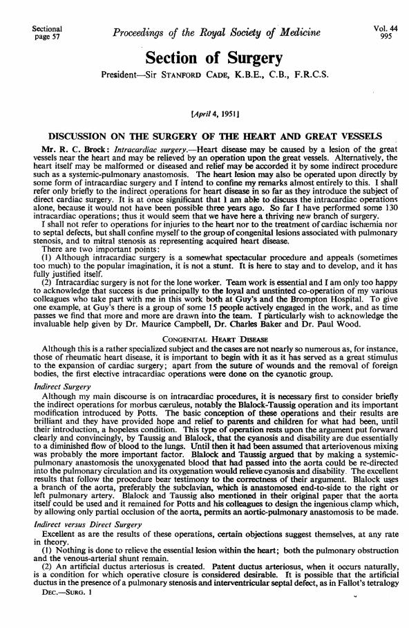
FIG. 1.-Pulmonary valvulotomy.
he body has to pass, the obstructing membrane formed by the fused valve-cusps presents anobvious challenge to surgery for its relief by direct operation. This can be done with relative ease andsafety and with a low mortality by means of instruments (valvulotome and various special dilators)passed through a small incision made in the wall of the right ventricle (Fig. 1) (Brock, 1948; Brockand Campbell, 1950a). I have also discussed and described the advarntages and disadvantages of analternative retrograde route through the left pulmonary artery (Brock, 1950).
In the very early days of the operation the mortality was high, as has usually been the case with allnew major procedures. This was chiefly due to the application of the operation to the very worsttype of case; late and neglected adults with years of increasingly severe disability and completeinvalidism and often with a huge heart or even in heart-failure. We now know that the chance ofsuccess in such cases is small. If, however, the operation is used on patients before these last stagesof grave deterioration have been reached the mortality is low and the results excellent, as will be shownlater from our mortality figures.One patient, a young woman in the middle twenties who before operation was deeply cyanosed and
so disabled that she could walk only a few yards and clearly had but a few weeks to live, improvedso much after operation that when she was found to be pregnant the pregnancy was allowed to proceedand she delivered herself without event eleven months after valvulotomy. She was soon pushing thebaby around in its pram without any distress, in marked contrast to her earlier condition of completeinvalidism. I have recently performed pulmonary valvulotomy in the fifth month of a pregnancy whichis being allowed to continue in the good hope that a live child will be safely delivered.
Pulmonary Valvulotomy in Fallot's TetralogyThe case for pulmonary valvulotomy in pure pulmonary stenosis, as I have just detailed, is
unanswerable and the operation is, indeed, now generally accepted throughout the surgical world asthe only one that should be used for the condition. The application of pulmonary valvulotomy tocases of Fallot's tetralogy has received less wide acceptance, and in fact the possibility of using theprocedure often has been much doubted in some quarters. It has been stated that valvular stenosisin Fallot's tetralogy is so uncommon that the operation is rarely applicable. Without elaborating theevidence here it can be stated that the obstruction in Fallot's tetralogy is valvular in at least 40%of cases. Thus Keith (1909) in examining 270 cases of congenital heart disease found a pulmonaryvalvular stenosis in 25/63 (40%) of cases of Fallot's tetralogy. Brown (1939) states that it is presentin "about half", and Sellors and Belcher (1950) found it in 20 out of 65 cases (30%). In my last 50cases of Fallot's tetralogy I found a predominating valvular stenosis, which was relieved by pulmonaryvalvulotomy, in 22 cases (44 %). It is clear, therefore, that with a valvular obstruction present in such
5t9 997
998 Proceedings of the Royal Society of Medicine
a high proportion of cases, and providing that it can be relieved safely and certainly by direct operation,we have a method of treatment that must commend itself since it corrects the chief lesion within theheart and does not merely circumvent it by the creation of an additional heart lesion, namely an artificialductus arteriosus. The functional results of the operation, as judged by the relief of cyanosis anddisability, are excellent and just as good as can be achieved by a successful Blalock's or Potts' operation.The mortality figures show that it can be an equally safe procedure.
RESULTS OF PULMONARY VALVULOTOMY
Total cases 57 [Pulmonary valvular stenosis 28Fallot's tetralogy ..29
Died . . . . . 10 f Pulmonary valvular stenosis 8DFallot's tetralogy 22In first 20 .. .. .. .. .. .. .. 8 diedIn remaining 37 .. .. .. .. .. 2 diedIn last 29 .. .. .. .. .. .. .. I died
The number of cases operated upon (57) alone indicates something of the importance of theoperation considering the short time that has elapsed since its introduction. The overall mortalityis high (nearly 18 %) but as can be seen this is chiefly due to the loss of 8 patients in the first 20; mostof whom were examples of late, neglected, almost hopeless, cases of pure pulmonary valvular stenosis.The very low mortality in the last 37 cases is noteworthy, as is also the loss of only 2 patients out of29 with Fallot's tetralogy; both of these patients were, moreover, adults. The one patient who diedin the last 29 cases was a woman aged 22 on whom an unsuccessful Blalock's operation had beenperformed two and a half years earlier.
Infuindibular Resectionfn some 600% of cases of Fallot's tetralogy the obstruction is not valvular but is below the valve
level and is situated in the infundibulum: infundibular stenosis. It has been, and in fact still is, statedthat the obstruction is long and tubular and in general unsuitable for direct relief by surgery. Thisis not the place to elaborate the arguments to refute this; some account has been given elsewhere(Brock, 1949; Brock and Campbell, 1950b), and it must suffice to say that, although in all cases theinfundibular portion of the right ventricle is essentially smaller than normal, the effective obstructiongenerally occurs at one level only and is usually linear or diaphragmatic. It may be high infundibular,intermediate, or low.
If the arguments for relief of the pulmonary obstruction by direct attack are correct, it must alsobe desirable to consider whether it is possible to divide, resect, or dilate the infundibular stenosis incases of Fallot's tetralogy. It can be said at once that it is feasible to do this and that good and evenexcellent results in the relief of cyanosis and disability can be obtained, just as with pulmonaryvalvulotomy.
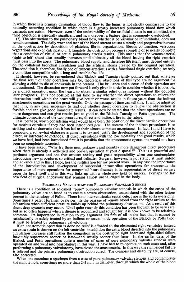
I have used the operation in 24 cases with 7 deaths; rather a high mortality but perhaps not excessiveconsidering its, as yet, pioneer nature and the fact that several of the deaths occurred in adults. In 6cases an infundibular and valvular stenosis co-existed and were dealt with by combined valvulotomyand infundibular resection, without mortality.The infundibular resection is carried out by introducing a special punch through an incision in the
wall of the right ventricle (Fig. 2).
2
FIG. 2.-Infundibular punch resection.

61 Section of Surgery 999
I have little hesitation in recommending valvulotomy in suitable cases of Fallot's tetralogy as asafe and logical operation giving good results. Infundibular resection can also give good results andis a logical operation; it is perhaps as yet a little too early to recommend it so unreservedly as valvu-lotomy. More cases must be done and more time must elapse before a careful assessment can be made.It is an operation that must, however, be fully applied and assessed because of the fundamental natureof its conception in regard to the direct relief of infundibular pulmonary stenosis.
Differential Diagnosis and Recognition of Valvular and Infundibular Stenosis in Fallot's TetralogyThe practice of many surgeons who use the anastomotic type of operation is to open the chest,
dissect out the two relevant vessels and anastomose them, and then close the chest. No attempt ismade to examine or explore the heart and there is but little confirmation of diagnosis. Certainly if aright-sided approach is used the pulmonary outflow tract cannot be examined. This is a policy towhich I do not subscribe. Before operation every effort is made by the basic clinical and radiologicalexamination, with the additional aid of cardiac catheterization and angiocardiography (usuallyselective), to achieve as accurate an anatomical diagnosis as possible of the exact site and nature ofthe obstruction. At operation (usually through a left postero-lateral thoracotomy done by resectionof the fifth rib) the pericardium is widely opened and a careful examination made of the heart. It isusually easy to recognize whether the stenosis is valvular or infundibular by studying certain local
A. B., 7 years. Operation Record. 1.5.51. Fallot's tetralogy.not.F9 BEFORE VALVULOTOtIY
R.V.
PRA.
n. e AFTER VALVULOTOMY
R.V.
A.
Before valvulotomy P.A. 18/10 R.V. 72/5 mm. HgAfter valvulotomy P.A. 30/15 R.V. 68/5 mm. Hg
FIG. 3.-Intracardiac pressure tracings taken at operation in a case of Fallot's tetralogy treated by pulmonaryvalvulotomy.
JD J. 1 351o00 mrn.Hg
R.\/. ?A. r ItF.CgM ER frVtCHInBER. I R.VC
50 _
PA. PA. . v veFilyn. vclve
FIG. 4.-Intracardiac pressure tracings taken at operation in a case of Fallot's tetralogy to show a doublepressure change indicating a combined valvular and infundibular stenosis. Treatment was by valvulotomy andinfundibular resection. The pressures are shown before and after operation.
1000 Proceedings of the Royal Society of Medicine 62
features. In valvular stenosis, for instance, the valve usually distends the first part of the artery andin systole can be felt as a tight hard cone or ball.from which a tiny jet issues. A detailed descriptionof the various types of obstruction cannot be given here. The diagnosis can also be made very exactby inserting a catheter through a small incision in the wall of the right ventricle and observing thepressure changes in various parts of the outflow tract. As can be seen from Figs. 3 and 4 it is possibleto reach a high standard of accuracy and to confirm the presence of a valvular stenosis or to identifythe presence of a combined infundibular and valvular obstruction. The exact level of the infundibularobstruction is also assessed.
In this way the correct operation for the relief of the condition disclosed can be selected.This policy has led to a steady increase in the proportion of direct operations being done, especially
as increasing confidence has been gained in their performance and in their safety and good results.The following figures indicate this trend towards a higher proportion of direct operations.
MORBUS Ck RULEUS (ALL TYPES). TOTAL 176 CASESIn last 100 cases In last 50 cases
Direct operation .. 70 Direct operation .. 59 Direct operation .. 33Anastomosis.. 106 Anastomosis.. .. 41 Anastomosis.. .. 17
FALIOTos TETRALOGY. TOTAT 149 CASESIn last 50 cases
Direct operation .. 43 Direct operation .. 27Anastomosis.. .. 106 Anastomosis.. .. 23
MrrRAL STENOSISCongenital heart disease is an important problem and has been dealt with first because it served
as the great stimulus of heart surgery. It is, however, rather a special problem since it constitutesonly 2% of all cases of heart disease. From its relative infrequency and the highly skilled methodsneeded for its full investigation and management it is fair to say that it is a condition best dealt within a few special centres. With rheumatic heart disease the position is different, seeing that it forms25% of all cases of heart disease (Wood, 1950). The surgical relief of mitral stenosis is much morein the nature of a national problem because the large number of cases suitable for operation can onlybe handled if they can be dealt with in numerous surgical centres. It may sound strange to manyto speak of the surgery of mitral stenosis in this way, but the success of mitral valvulotomy and thegreat and widespread interest and the large number of patients presenting themselves for operationproves it beyond doubt. Mitral valvulotomy has quickly established itself as a standard operation insurgery and is rapidly giving great benefit to many hitherto hopeless cardiac invalids and at a low risk.
It is instructive, interesting and not a little gratifying to consider the position of the operation atfour phases; twenty-five years ago, four years ago, one year ago, and to-day.
Twenty-five years ago, when the first attempts at mitral valvulotomy were made, the results wereso bad that the problem was considered to be insoluble and it was left as the great unsolved problemof surgery. Many things contributed to this early failure; no discredit was due to the early pioneersbecause in great part the time was not yet ripe for success. More recently, however, it became clearthat a re-trial of the issue was both justified and necessary. Some years ago we began our plans butfound it very difficult to obtain suitable cases owing to the extreme caution exhibited by most physiciansand cardiologists. Ultimately a suitable case was found and after nearly a year of observation thepatient was operated on, with success, in September 1948.During the succeeding twenty-one months, before our first paper on the subject was published, only
9 cases were operated upon, but with 7 successes (Baker, Brock and Campbell, 1950). It should bemade clear that although our work at Guy's was synchronous with that of Bailey, Glover and O'Neillof Philadelphia and proceeded along similar lines, it was quite independent of and uninfluenced bytheir experiences.The response to this paper published one year ago was immediate and gratifying; the operation
seemed to be enthusiastically accepted and the pressure from patients and from doctors became almostirresistible. Long waiting lists rapidly formed and the present position is not one of anxious searchingfor suitable cases but continuous hard work to examine and sort out the numerous applicants and totry and operate on the large number of suitable cases. The position to date is shown in the followingtable.
MITRAL VALVULOTOMYTotal operations, 47 In first 20 cases, 7 deaths Oldest patient, 50 years 21-30 3140 41-50
Died, 8 In last 27 cases, 1 death Youngest patient, 21 years 16 24 7
These figures show that operation can be offered with every chance of success and with a nowrelatively small risk; the series of 27 consecutive cases with only one death amply supports tbis.TJ roWIt in those who have survived the operation has been excellent in all but 3 cases.
63 Section of Surgery 1001
Without operation the plight of many patients is desperate; the husband and father is unable towork and support his family; the wife cannot manage to carry on with her essential household dutiesand, if she happens to be a mother, cannot look after her children; the spinster who is unable to worksoon finds herself destitute and an object of charity unless she has friends or relatives to care for her.In all, the state of disability and chronic invalidism holds no future except an early death. The patientssoon come to realize this and it is no wonder that they eagerly beg for operation. In discussing operationwith a bad risk case recently I pointed out that the risk was greater than normal; the woman's replywas "It is no risk to me; it is my chance". This sums up most aptly the patient's reaction to theprospect of relief by operation.
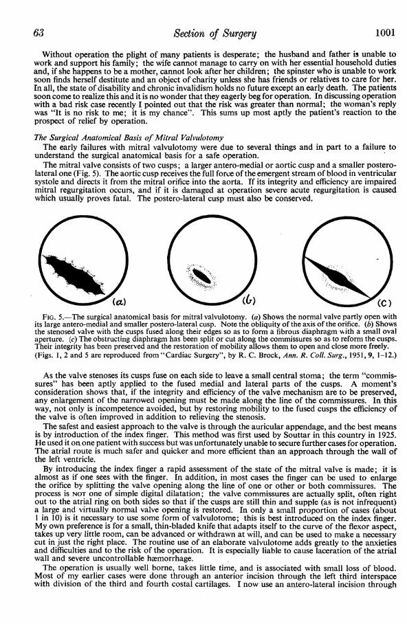
The Surgical Aaiatomical Basis of Mitral ValvulotomyThe early failures with mitral valvulotomy were due to several things and in part to a failure to
understand the surgical anatomical basis for a safe operation.The mitral valve consists of two cusps; a larger antero-medial or aortic cusp and a smaller postero-
lateral one (Fig. 5). The aortic cusp receives the full force of the emergent stream of blood in ventricularsystole and directs it from the mitral orifice into the aorta. If its integrity and efficiency are impairedmitral regurgitation occurs, and if it is damaged at operation severe acute regurgitation is causedwhich usually proves fatal. The postero-lateral cusp must also be conserved.
FIG. 5.-The surgical anatomical basis for mitral valvulotomy. (a) Shows the normal valve partly open withits large antero-medial and smaller postero-lateral cusp. Note the obliquity of the axis of the orifice. (b) Showsthe stenosed valve with the cusps fused along their edges so as to form a fibrouLs diaphragm vwith a small ovalaperture. (c) The obstructing diaphragm has been split or cut along the commissures so as to reform the cusps.Their integrity has been preserved and the restoration of mobility allows them to open and close more freely.(Figs. 1, 2 and 5 are reproduced from "Cardiac Surgery", by R. C. Brock, Ann. R. Coll. Surg., 1951, 9, 1-12.)
As the valve stenoses its cusps fuse on each side to leave a small central stoma; the term "commis-sures" has been aptly applied to the fused medial and lateral parts of the cusps. A moment'sconsideration shows that, if the integrity and efficiency of the valve mechanism are to be preserved,any enlargement of the narrowed opening must be made along the line of the commissures. In thisway, not only is incompetence avoided, but by restoring mobility to the fused cusps the efficiency ofthe valve is often improved in addition to relieving the stenosis.The safest and easiest approach to the valve is through the auricular appendage, and the best means
is by introduction of the index finger. This method was first used by Souttar in this country in 1925.He used it on one patient with success but was unfortunately unable to secure further cases for operation.The atrial route is much safer and quicker and more efficient than an approach through the wall ofthe left ventricle.By introducing the index finger a rapid assessment of the state of the mitral valve is made; it is
almost as if one sees with the finger. In addition, in most cases the finger can be used to enlargethe orifice by splitting the valve opening along the line of one or other or both commissures. Theprocess is NOT one of simple digital dilatation; the valve commissures are actually split, often rightout to the atrial ring on both sides so that if the cusps are still thin and supple (as is not infrequent)a large and virtually normal valve opening is restored. In only a small proportion of cases (about1 in 10) is it necessary to use some form of valvulotome; this is best introduced on the index finger.My own preference is for a small, thin-bladed knife that adapts itself to the curve of the flexor aspect,takes up very little room, can be advanced or withdrawn at will, and can be used to make a necessarycut in just the right place. The routine use of an elaborate valvulotome adds greatly to the anxietiesand difficulties and to the risk of the operation. It is especially liable to cause laceration of the atrialwall and severe uncontrollable hemorrhage.The operation is usually well borne, takes little time, and is associated with small loss of blood.
Most of my earlier cases were done through an anterior incision through the left third interspacewith division of the third and fourth costal cartilages. I now use an antero-lateral incision through

Proceedings of the Royal Society qf Medicine
the left fiftfi interspace with the patient lying on his right side and rotated 45 degrees backwards. Itis not necessary to divide a rib or cartilage and an excellent exposure is given. It is less disturbingand less extensive than a formal long postero-lateral incision.
Indications for Mitral ValvulotomyIt is not possible here to discuss fully the indications for mitral valvulotomy. The type of case
considered most favourable up to now is the one with a small heart and evidence (clinical, radiologicaland laboratory) of pulmonary hypertension with "pulmonary episodes" of pulmonary cedema,cardiac asthma, nocturnal dyspncea, orthopnoea, hmmoptysis, "bronchitis", &c. Another importantindication is disability. This is a feature about which we have hitherto been rather complacent,chiefly because once medical measures had failed to give relief, nothing more could be done for thepatient, who was condemned to progressive invalidism and early death. It is the recognition of thisdesperate and sorrowful state that urges the patient to grasp so eagerly at the chance of relief thatoperation offers. The young patient in the late twenties or thirties is particularly eager as he or shesees nothing but a blank and hopeless future. The patient between 25 and 35 with mitral stenosiswho is beginning to notice disability is bound to deteriorate steadily if unrelieved, and operationmust therefore properly be considered early when the risk is less and the avoidance of invalidism andfurther permanent cardiac deterioration is possible.
It is fair to say that the chief indication for mitral valvulotomy is mitral stenosis. This may sounda loose statement, and perhaps is liable to be misinterpreted, but it is one that demands our greatattention. It does not mean that every case of mitral stenosis should be operated upon; it merelyindicates that operation may need to be considered in each case. There may be reasons against it;the patient may be too young or too old; he may be too ill or not disabled enough; there may besome feature that is a grave contra-indication, such as severe aortic disease or unrelieved right-sidedfailure. One of the greatest contra-indications is active or recent rheumatic infection and for thisreason we feel that most patients under 20 must be assessed with the greatest care.
In many cases of mitral stenosis the question will not be whether operation is needed, but when itis needed. Our cardiological clinics doubtless have groups of these patients under regular observationand will be able to select the most favourable time for operation.
It is important to distinguish between contra-indications to operation and merely unfavourablefeatures. It has, for instance, been stated that auricular fibrillation and a history of embolism arecontra-indications; that calcification of the valve is also a barrier to successful surgery. These featuresmay increase the difficulties or the risk, but that they are not contra-indications is shown by the factthat many of our patients presented them.Thus auricular fibrillation was present in 20 cases; 5 deaths occurred among these in contrast to
3 among the 27 in normal rhythm. Incidentally 8 patients developed auricular fibrillation afteroperation and 5 of these soon reverted to normal rhythm.
Calcification of the valve was found in 19 cases. Embolism had occurred before operation in 5.Embolism may be rather an indication than a contra-indication.We have included cases with aortic disease when it has been considered not to be the dominant
heart lesion. 3 cases with tricuspid regurgitation and right-sided failure have been successfullyoperated on. Right-sided failure is not necessarily a contra-indication providing it shows response toroutine treatment. It may well be an extra indication that the heart is insistently demanding relieffrom the crippling mitral obstruction.
Mitral regurgitation, especially if associated with aneurysmal dilatation of the left atrium and a
large heart, we feel is probably not yet amenable to operative relief. The great difficulty is the pre-operative assessment or recognition of genuine, significant mitral incompetence. The mere findingof a systolic murmur is not in itself diagnostic or often even helpful.
In addition to satisfactory clinical improvement, and especially the relief of disability and of pul-monary episodes such as cardiac asthma or pulmonary cedema, confirmation of the good results ofoperation is also obtained by the lessening of the degree ofpulmonary hypertension measured at cardiaccatheterization before and after operation. Thus in one patient, a woman in the middle twentieswith accompanying aortic disease, the pulmonary artery pressure dropped from 130/56 mm. Hgto 40/10. In a man in the forties it dropped from 70/20 mm. Hg to 40/18. Cardiac catheterizationhas been used on all our cases; very often it furnishes evidence strongly in favour of operation, suchas severe pulmonary hypertension with a further rise on exercise and a low cardiac output. In othercases it does not materially influence the clinical assessment. On the whole the information obtainedfrom pre- and post-operative studies is more likely to be of greater use in the future than it is now;when a sufficient mass of data is assembled and studied with the related clinical features much of valueshould emerge.
It is felt that the operation of mitral valvulotomy is now basically established in principle and as avaluable procedure that can give real relief to many and with a relatively low risk. No doubt therewill be improvements in details of technique but the immediate concern for the future is not establish-ment of the operation; this has already been done. What is needed is careful selection of the type of
1002 64d

65 Section of Surgery 1003
cases suitable and, particularly, extension of the indications to embrace as many variants as possible.In addition it is important to apply and prosecute the various physiological methods of investigationand assessment in order that we may elucidate as much information as possible about the circulatorydisturbances present and their correction.
It is wrong now to limit operation to nothing but advanced and desperate cases. This was justifiablein the early days when the operation was considered hopelessly dangerous and was certainly untried.That stage has now been passed; the safety of the operation has been demonstrated and it is notonly justifiable to apply it to the earlier, less severe type of case in which a better result with less riskis likely, but it is no longer wise or fair to surgeon or cardiologist to embark on this field of surgeryby selecting the worst and most hopeless type of case. That way lies the great danger of high mortality,disappointment and discouragement, and retardation of the acceptance and development of thisvaluable addition to the treatment of rheumatic heart disease.
Mitral Stenosis and PregnancyIt has been the custom in many cases of pregnancy in mitral stenosis to terminate the pregnancy
and sterilize the patient. This practice has been in great part justified and even now may be the propercourse in certain cases. It is important to point out, so that it may be taken note of by gynecologists,cardiologists and others, that this policy now needs reconsideration. Apart from destroying the childand the hope of future children it may also destroy the marriage. Mitral valvulotomy may alter thesituation so much that safe child-bearing is possible. Often this can only be settled when the valve isactually examined at operation. If a tough fibrous valve is found and but little improvement in itsefficiency is achieved it may well be desirable to forbid child-bearing. If, on the other hand, a valvefavourable for much improvement of functi6n is disclosed then there may be no reason why pregnancyshould be interfered with or forbidden.The safety and success of permitting the child-bearing was mentioned in a case of pulmonary
valvulotomy, and also an example of pulmonary valvulotomy during actual pregnancy. We now havea similar patient on whom a mitral valvulotomy has been successfully done during early pregnancyand it is hoped that she will go safely to term and delivery. In this patient abdominal evacuation andsterilization had been planned elsewhere. This is a situation which is bound to arise with somefrequency in cases of mitral stenosis, and it is desirable that the changed position afforded bysuccessful mitral valvulotomy should be widely known and respected.
CONCLUSIONOur experience with direct heart operations is now sufficient to recommend their adoption in this
way with confidence and with the sure knowledge that good results can be obtained with a low mortality.It is now abundantly proven that if handled properly the heart, even the diseased heart, is just astolerant of interference as any other viscus in the body. Provided blood loss is corrected, skilledanesthesia is available, and the excitability of the heart is suitably controlled by the topical andsystemic use of procaine, it will be found that "running repairs" can quite safely be performed on theheart. This is an important and fundamental contribution to our knowledge and means that we cancontinue to apply and develop this type of surgery further, until such time as the successful introductionof an independent circulation may enable us to operate on the dry heart.
REFERENCESBAKER, C., BROCK, R. C., and CAMPBELL, M. (1950) Brit. med. J. (i), 1283.BROCK, R. C. (1948) Brit. med. J. (i), 1121.
(1949) Brit. med. J. (ii), 399.(1950) Guv's Hosp. Rep., 99, 236.and CAMPBELL, M. (1950a) Brit. Heart J., 12, 377.
(1950b) Brit. IHeart J., 12, 403.BROWN, J. W. (1939) Congenital Heart Disease. London.KEITH, A. (1909) Lancet (ii), 359.SELLORS, T. H., and BELCHER, J. R. (1950) Lancet (ii), 887.SOUrrAR, H. S. (1925) Brit. med. J. (ii), 603.WOOD, P. (1950) Diseases of the Heart and Circulation. London.
Dr. Maurice Campbell: The recent advances in cardiac surgery call for the closest co-operationbetween physicians and surgeons, and this, not only because diagnosis may be difficult, but becausethe prognosis may often be excellent without surgical intervention, in spite of the presence of somedegree of heart disease.When the diagnosis is simple an angiocardiogram will often confirm it with certainty and add
precision to the details, though in coarctation of the aorta and patent ductus, the information neededby the surgeon generally calls for aortograms or selective angiocardiograms, rather than ordinary
Proceedings of the Royal Society of Medicine
ones. When the diagnosis is not simple, the interpretation of the angiocardiogram often calls formuch experience, and is more likely to be helpful when there is some idea of exactly what is to belooked for.
The Surgery of Coarctation and Patent Ductus(a) I find it very difficult to advise about coarctation of the aorta. The patients have grave dangers
ahead but generally feel well and can do most, if not all, they want and may have twenty or thirtyyears of an active life before them. An operative mortality of 11 % with another 9% who cannothave successful anastomosis, and a further 9% who may not get the full benefits hoped for afteroperation (Gross, 1950) is a very heavy price to pay even for the hope of prolonging life and avoidingsome of the future risks.
It may be that in years to come, a lower mortality will make the decision for the physician lessdifficult, but the operation is one of great difficulty: it should, therefore, remain in the hands of thosethoracic surgeons with special experience. If, in a patient under 20, the heart is becoming larger, orthe blood pressure rising progressively, operation should generally be advised. Over 20 the risksare even greater and the indications should, therefore, be even more certain.
(b) The Surgery of Patent Ductus ArteriosusThis is an easier problem, but still not easy, because the patient is generally well and has a fairly
good prognosis. Most children with this lesion feel able to do most of what they want and have areasonable expectation of remaining in good health for twenty or thirty years. It is probably fair tosay that few of them can expect to be in as good health after 40, and when symptoms become worsein middle life they may go downhill quickly. If operation could be performed with a negligiblemortality, it would become the routine for all patients with patent ductus, for it is a complete cure,which is a higher standard than that for the rest of the surgery of the heart, as yet. In the hands ofexperts the operative mortality is probably 2 or 3%, and when it falls to 1 % for straightforwardcases as in the hands of Professor C. Crafoord and some others, the improved prospects for thefuture and the disappearance of slighter symptoms of which the patient was hardly aware, will bean adequate reason for all patients to be operated on. In my view, these facts should be put beforethe parents and the decision made in full consultation with them. If, in a child, the heart is alreadyenlarged and the pleonemic lungs and the peripheral signs show that there is a large amount of bloodpassing through the ductus, the operation should be advised at once. As safety is of such primeimportance, ligation should be the routine method for most surgeons, in spife of the small risk ofrecanalization.
Subclavian-pulmonary Anastomosis for Fallot's Tetralogyand other related conditions. There is already a good deal of experience of the results of this at manycentres. The operative mortality is higher for this more complicated procedure but not now above10%, and 80% of the patients can expect an enormous increase in their capacity and in their expectationof life. Here, the risk will be readily accepted, and the older patients who can discuss it themselvesare more than ready to face an even higher risk, owing to their great disability, often made worseby the realization that they are becoming less and less capable even of the limited life they have led.
Pulmonary ValvulotomyTurning to direct operations on theheart, and taking pulmonary valvulotomy first, this proved a
dangerous operation at first, owing to the very serious cases that were put before the surgeon. Mr.R. C. Brock has now shown that it can be reasonably safe in patients who have not reached the stageof very large hearts and congestive failure, and it is quite certain that the results can be as brilliantas those of the Blalock-Taussig operation.
In pulmonary stenosis with a closed inter-ventricular septum, valvulotomy is the only operationthat can be performed with any hope of lasting success. When the stenosis of Fallot's tetralogy isvalvular there is a choice between thisand the Blalock operation, and both can be very successful,but the final decision as to which should be advised can only come from a longer follow-up of morecases than are at present available. Pulmonary regurgitation has not been a complication as wasfeared, and no patient has developed any signs suggestingthis. (Since this meeting 2 patients havedeveloped signs suggesting regurgitation, one shortly after operation, where it is too early to say howfar this may mar his improvement; and one who is doing extremely well eighteen months afteroperation.)
Infundibular ResectionWith one exception, all these operations on the heart have become safer with increasing experience.
But infundibular resection for the cases of Fallot's tetralogy with infundibular stenosis has not producedsuch good results as in the first cases reported by Brock and Campbell(1950). This may prove atemporary phase but more experience is needed before the operation can be looked on as a routinealternative to a.subclavian-pulmonary anastomosis.
1004 AA
67 Section of Surgery 1005
Mitral StenosisIn mitral stenosis it has already been established that many patients can be greatly improved.
It is too early to answer the question as regards all types of mitral stenosis. There are two chiefpoints to consider: (1) how much of the disability is mechanical from the valvular obstruction;and (2) how much of it is due to myocardial disease. Obviously the patients likely to do best are thosewhere the disability is mainly due to the obstruction and only slightly to the damaged heart muscle,and these are particularly the patients about 25-40 with attacks of cardiac asthma and pulmonarycedema and with orthopnoea, but with a heart that is not very large. In these cases, one can confidentlyexpect relief and great improvement in their conditions without much risk. The most importantcontra-indication to valvulotomy is rheumatic activity or the chance of this recurring; and, for thisreason, patients under 20 are rarely suitable. Neither mild aortic valvular disease nor slight mitralincompetence is a contra-indication; gross aortic or mitral incompetence is another matter. Patientswith long-standing congestive failure have passed the stage when they can best be helped, but whetherthey can be helped with reasonable safety is a problem still to be solved.
Since the first successful mitral valvulotomy in 1948, two and a half years have elapsed, and it isalready clear that improvement has been maintained in all but one of the cases followed up. Theresults have been surprisingly good and there is no question of a quick relapse. Mitral incompetencedoes not generally result from the operation. There is some evidence that pulmonary hypertensionis reversible in these cases, and sometimes the fall in pulmonary artery pressure is gradual rather thanabrupt. Mitral stenosis will have to be a problem for many thoracic surgeons, as the numbers involvedare far too large for a few special centres.
REFERENCESBROCK, R. C., and CAMPBELL, M. (1950) Brit. Heart J., 12, 403.GROSS, R. E. (1950) Circulation, 1, 41.
[May 2, 1951]
DISCUSSION ON THE REPAIR OF INJURIES TO THE COMMON BILE DUCT
Benign Strictures of the Extrahepatic Bile DuctsBy HOWARD K. GRAY, M.D., F.A.C.S.
Division ofSurgery, Mayo Clinic, Rochester, Minnesota
IT is doubtful that anyone who has had the responsibility of caring for a patient with benignstricture of the extrahepatic bile ducts will disagree with the statement that there is no more distressingsituation in abdominal surgery. The situation is distressing for a variety of reasons, not the least ofwhich is the fact that it should and could have been avoided in the great majority of instances. Theunfortunate features of this condition may be emphasized further when it is considered thatapproximately 70% of these patients will be less than 50 years of age and approximately 25% willbe in the fourth decade of life. Recurrence of stricture in an appreciable number of patients,irrespective of the method by which the lesion may have been attacked initially, will impose subsequentoperation or operations in addition to the associated distress, risk, expense, prolonged morbidityand catastrophic disruption of normal domestic affairs of patients in this age-group in which suchresponsibilities frequently have increased to a point where the patient is indispensable. Thiscombination of circumstances makes it imperative that any surgical procedure on the biliary tractshall be approached and executed with the caution and the respect that it rightfully commands.That this concept of the problem is not appreciated widely is attested to by the all too frequent casualassignment of the patient with disease of the gall-bladder to the least qualified surgeon in a teachingprogramme and by the willingness of independent practitioners who have had minimal surgical trainingto assume such responsibility without evident twinge of conscience.
INCIDENCEIt is not possible to determine the incidence of the development of benign stricture of the extra-
hepatic bile ducts following operative procedures for lesions of the gall-bladder, owing to the dearthof any statistical material on the subject except from large institutions. For obvious reasons thesestatistics will not be representative of the general experience. It is important to note that lesions inthis category are being reported with what apparently is increased frequency. Whether or not this isan indication of a relative enlargement of the number of these unfortunate individuals or is evidenceof greater interest in the subject is an unanswerable question.