sections of the dolphin's head (stenella caeruleoalba) · sections of the dolphin's head...
TRANSCRIPT
SECTIONS OF THE DOLPHIN'S HEAD (STENELLA CAERULEOALBA) *
HIROSHI HOSOKA WA** AND TOSHIRO KAMIYA**
For the past years one of the authors, Hosokawa, has dissected several specimens of the Stenella (Prodelphinus) caeruleoalba (Meyen), paying speciel attention to the peripheral distribution of cranial nerves. In order to supplement the ordinary anatomical observations, a couple of heads of this animal were sectioned transversely or lengthwise with the hand-saw. Anatomical features of each section surface were very interesting and instructive for understanding the three-dimentional structures of the dolphin's head. Such cross-sectional observation was adopted partially by Slijper (1936) in his comparative-anatomical study of the cetacean blood vessels, by Lawrence and Schevill (1956) in their functional anatomical study of the delphinid nose as well as by Nakajima (1961) in his work on the cetacean rete mirabile, although they did not give detailed explanation of each structure on the section surfaces.
Materials used in the present study comprise two heads of adult Stenella caeruleoalba from Kawana, Shizuoka Prefecture, which had been soaked and fixed thoroughly in 10% solution of formalin. One of the specimens was sectioned transversely into six slices (Fig. 1), and each section surface was photographed and sketched from rostral direction. The total length of the head was 45 cm, and each slice was successively some 16, 5, 6, 4.5, 4.5 and 9 cm in thickness. The second specimen, 46 cm in length, was sectioned at first in the median plane. Then the right half was cut lengthwise 5 cm apart from the median surface. In the following pages each section surface will be shown with brief explanations. Since the cross-sections are seen from rostral, the right side of the head is on the left side of the figures.
CROSS-SECTIONS
Section I (Fig. 2-a, b)
The section passes through the root of the snout or rostrum. The upper and lower jaws are separated from each other by the gap of the oral cavity. The skeleton of the upper jaw is represented by the maxilla, premaxilla, vomer and mesethmoid cartilage. The last one lies on the median line, enclosed in the canal formed by the premaxillae of both sides and the vomer. The dorsal part of the premaxilla is composed of particularly compact bony substance. Inside the maxilla and premaxilla we see small canals for conducting the superior alveolar arteries and nerves, which supply small branches to the alveolar plexus around the root of the teeth. For reference, the peripheral distribution of the trigeminal nerve is shown in Fig. 13.
* To the memory of late Prof. emeritus Tsunetaro Fujita. ~ * Department of Anatomy, University of Tokyo School of Medicine, Hongo, Tokyo, Japan.
105

106 H. HOSOKA WA AND T. KAMIYA
Needless to say, the Stenella is, like all the other odontocetes, furnished with homodont teeth, of which the number amounting to some forty or fifty pairs in both upper and lower jaws. On the left side of the figure the whole contour of one tooth is shown. Approximately halfofthe total length is inside the maxilla (radix dentis), while the other half, conical in shape and somewhat curved mediad, is out of the gmg1va.
Dorsal to the premaxilla and maxilla is a white area occupied by adipose connective tissue. This is the anterior extremity of the so-called " melon". This peculiar structure, characteristic of most dolphins, lies on the snout and forms the prominence in front of the nostrils. Although there have been proposed many hypotheses about the function of this unique structure, we do not know yet anything definite. Probably the spermaceti organ, characteristic of the sperm whale, represents the highly differentiated " melon" of other odontocetes. The " melon" and spermaceti organ have been very often thought in relation to the respiration of the cetacea (Raven & Gregory, 1933; Slijper, 1962, etc.).
Under the melon we see the anterior end of the m. rostralis or the pars labialis of m. maxillonasolabialis (Huber, 1934). These muscle bundles arise from the upper surface of maxilla and run up- and forwards, so as to insert to the thin fibrous capsule of the melon. Some of the muscle bundles, which arise from the lateral border of the maxilla, are directed up- and backwards, so as to border the anterior end of the narial muscle group, pars nasalis of m. maxillonasolabialis (see Fig. 10).
The mandibula is cut on both sides in the lower jaw. This bone is furnished with a wide internal space filled with spongy, adipose tissue (mandibular canal or fossa), and the inferior alveolar vessels and nerves are seen to pass through this fatty substance (confer Fig. 13).
In the middle of the lower jaw is the cut surface of the tongue, representing a peculiar quadrangular shape. Inside the tongue we see the inverted V letter of the genioglossus muscle, as well as many internal lingual muscle bundles arranged irregularly. Beneath the tongue are sections of the mylohyoid muscle and of the panniculus carnosus, which covers the ventral surface of the lower jaw.
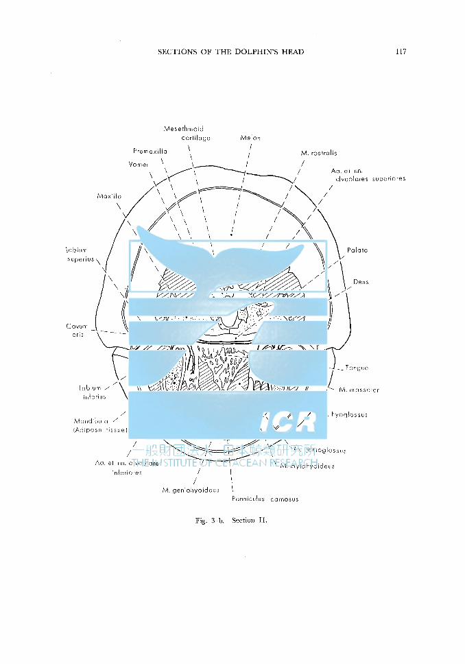
Section II (Fig. 3-a, b)
It represents a section through the middle of the " melon ". Anatomical features of this section do not show significant differences from those of the preceding one, while the melon and the rostral muscle have considerably increased in size.
In the lower jaw the tongue is cut at about the middle of the corpus linguae. In addition to mm. genioglossus and mylohyoideus, which were already observed in Section I, the geniohyoideus and hyoglossus are seen in the floor of the oral cavity. The arrangements of these muscles are shown in Figures 11 and 12. As seen in Fig. 11, the panniculus carnosus is considerably well-developed on the ventral surface of the lower jaw. Systematic description of the cetacean muscles is to be referred to Stannius (1849), Schulte (1916, 1918), Howell (1930), Slijper (1936), etc.

SECTIONS OF THE DOLPHIN'S HEAD 107
Section III (Fig. 4-a, b)
This is a section at the oral angle or the posterior end of the oral cleft. The bony framework of this section, shaped like a flying bird with extended wings, is composed of premaxilla, maxilla, vomer, os malare or jugale, os palatinum, os pterygoideum as well as of the mesethmoidal cartilage. The maxilla and os malare form a doublelayered bony plate (confer Fig. 9). On the left side of the figure the malar bone shows on its lower surface a pointed prominence representing the root of the zygomatic process.
Noteworthily there are peculiar cavernous sinuses inside or in close apposition to the skeleton. The largest one is inside the pterygoid fossa surrounded by os palatinum and pterygoideum (postpalatine air sinus of Flower, 1876). Hence it is called sinus pterygoideus. Other sinuses on the section are secondary expansions of the pterygoid sinus.
All the sinuses are traversed by fibrous trabeculae and give spongy appearances. These sinuses belong to the pneumatic sinus system, which characterizes especially the odontocetes. All the sinuses such as pterygoid, epitympanic, peripetrosal, etc. are continuous with the tympanic cavity. The pneumatic sinus system was described by Boenninghaus (1904), Gruhl (1911), Yamada (1953) etc., who did not however explain the physiological significance of this peculiar structure. Nerve bundles belonging to V 2 (n. maxillaris) are seen in the trabeculae of the frontal and maxillary sinus expansions (confer Fig. 13).
Above the bony frame the " melon " is seen in the middle of the section, being abutted on by the m. narialis on both sides (Fig. 10). The narial muscle of blow hole muscle, pars nasalis of m. maxillonasolabialis of Huber ( 1934), is formed by many layers and subdivisions, of which the direction of muscle bundles is somewhat different from one another. Lawrence and Schevill ( 1956) recognized six layers and called them posteroexternus (pe), intermedius (i), anteroexternus (ae), posterointernus (pi), anterointernus (ai), and profundus (pr).
At the center of the lower half of the section is a transverse slit which is lined with the thick wall of mucous membrane. This is the posterior part of the oral cavity, of which the floor is represented by the radix linguae (see Section VI). Ventral to the oral cavity we see m. hyoglossus, genioglossus, geniohyoideus, monogaster, etc. M. mylohyoideus is cut obliquely on each side of the oral cavity.
The pterygoideus medialis muscle is seen to occupy the space between the ptergoid process and mandibula, showing close relationship to the frontal sinus expansion. The n. facialis is cut under the bony shelter formed by the maxilla and os malare. This nerve trunk can be traced to the rostral until the antorbital notch, where it turns upwards, returns backward on the dorsal surface of the maxilla, so as to supply the whole musculature of the nostril (see Fig. 13).
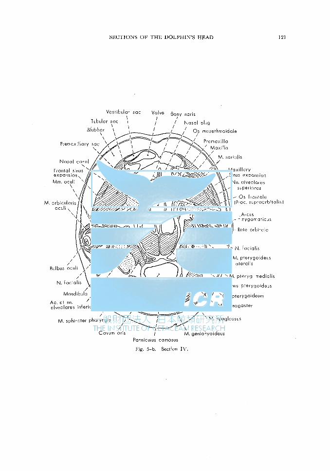
Section IV (Fig. 5-a, b)
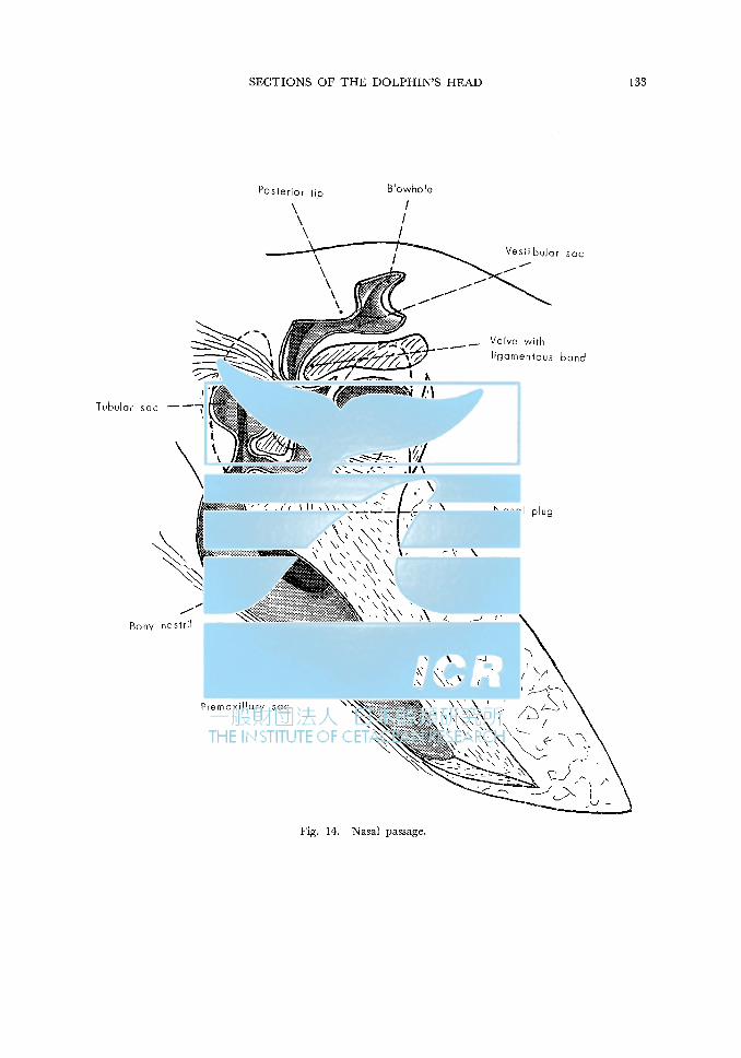
The section passes through the bony nasal canal, which courses along the anterior wall of the cranial case. The nasal canals of both sides are separated by the ovalshaped mesethmoid bone. Above the bony naris the nasal canal continues to the pre-

108 H. HOSOKAW A AND T. KAMIYA
maxillray sac and still further to the vestibular sac just under the thick blubber layer. The complicated structure of the dolphin's nasal passages has been subjected to
investigations of many scholars such as Rawitz ( 1900), Gruhl ( 1911), Burne ( 1952), Lawrence and Schevill ( 1958), etc. So the present authors do not intend to go into its details. A somewhat schematic illustration will outline this labyrinthous nasal passage (Fig. 14).
On both sides of the nasal ways we see massively developed narial muscle, pars nasalis of m. maxillonasolabialis. The intimate topographical relations between this muscle and nasal passage imply their close functional correlations.
The bony frame of the section is composed of premaxilla, maxilla, os frontale and os pterygoideum. The plate-like lateral projection is formed by the maxilla and os frontale. This peculiar modification and dislocation of the cranial bones are associated with the famous phenomenon called " telescoping " of the cetacean skull (Kernan, 1918; Kellog, 1928, etc.). Branches of nn. alveolares superiores are seen between these two bony laminae (see Fig. 13). Lateral to the pterygoid process is seen the sinus pterygoideus of the pneumatic sinus system. Upwards it is elongated and continuous to the maxillary and frontal expansions.
On the left side of the section we see the cut surface of the 9e ball as well as of extrinsic 9e muscles. The eye ball is characterized by the extraordinarily thick scleral wall, an adaptation to the hydrodynamic environment of the cetacea. The floor of the orbit is formed by a tough fibrous membrane, and the facial nerve runs just under this orbital floor. The :::ygomatic arc is also cut near the tip of the orbital cone.
It is noteworthy that the eye muscles are covered by a thick cavernous layer composed of spongy substance. This is nothing but the rete mirabile, or the peculiar network of arterial blood vessels. As well known, the extraordinary development of the rete mirabile is one of the most remarkable characteristics of the cetacean anatomy (Stannius, 1841; Wilson, 1879; Ommanney, 1932; Slijper, 1936; Kock, 1959; Nakajima, 1961; Slijper, 1962, etc.). The rete is distributed mainly in the trunk and neck. In the cranial region it is found in the orbit (rete orbitale) and in the floor of the cranial cavity (rete basis cranii) (see Section V).
In the lower part of the section we see the oral cavity, which is cut at its posterior end.
Section V (Fig. 6-a, b)
The cranial cavity is cut open and the brain, which is poorly fixed, is exposed. The brain lies on the cushion of the rete basis cranii. Meningeal coverings of the brain are quite same as those of man and other mammals.
The bony frame of the section is formed by the maxilla, os jrontale, os parietale, os ethmoidale and vomer. The lateral dislocation of os parietale is a part phenomenon of the " telescoping " of dolphin's skull. The zygomatic process of the squamosal is cut on the left side, while the zygomatic arc is still seen on the right.
The mandibula is cut near its posterior extremity. As seen already in the preceding section, the medial wall of the mandibula is lost and the mandibular fossa
SECTIONS OF THE DOLPHIN'S HEAD 109
is filled with massive fatty substance, which extends forward into the wide mandibular canal (Sections I-III). Medially the mandibular adipose substance is abutted on by the pterygoid muscles and pterygoid sinus. Because of these intimate topographical relationships, Yamada (1953) guessed that the movements of the lower jaw might effect strongly upon the distension of the pterygoid sinus.
The pharynx and larynx are shown on the median line. This peculiar situation that the pharynx is cut in front of the larynx can be understood by checking the guide line in Fig. 6-a. The pharnyx is surrounded by muscle groups such as m. monogaster, ~ylohyoideus, styloglossus, hyoideus, sternothyroideus, etc. Os hyoideum is cut at the basihyal as well as at the stylohyal.
The larynx is cut at the tip of the arytenoepiglottideal tube, which protrudes uniquely into the choana. This peculiar tube, characteristically well-developed in the odontocetes, was described by Hosokawa ( 1950) in the sperm whale (see Fig. 7).
LONGITUDINAL SECTIONS
Section VI (Fig. 7-a, b)
This is the median section of the Stenella's head. Because of the slight asymmetry of dolphin's skull, the right nasal passage is cut through, showing the strange disposition of the nostril (blowhole), vestibular sac, tubular sac, nasal plug, premaxillary sac, etc. The m. narialis is seen in close topographical relationship to the nasal passage, especially to the nasal plug. In front of the nasal passage we see the " melon ", a peculiar mass of adipose tissue on the dolphin's rostrum.
The skull is represented by the brain case and the long rostrum. The former is cut through osfrontale fused with mesethmoidale, os supraoccipitale, basisphenoidale and basioccipitale, while in the elongated snout we see os mesethmoidale, mesethmoid cartilage, premaxilla, maxilla, os palatinum, os pterygoideum, etc.
The brain shows a remarkable dorsal flexion in the pontine region, representing a noteworthy characteristic of the cetacean brain. As seen in this section the brain stem is relatively well developed. The cerebral hemisphere shows a very complicated pattern of convolutions and sulci. As well known, the cetacean brain stands highest in the mammalian kingdom, so far as the complexity of the cerebral fissural configuration is concerned. The corpus callosum is relatively small.
The tongue is very long and its greater part is attached to the floor of the oral cavity. Intrinsic and extrinsic lingual muscles are fairly well developed, implying the considerable motility of the tongue. The arytenoepiglottideal tube of the larynx is clearly shown at the left and lower end of the figure. As explained in Section V, this tubular structure formed by modified arytenoid and epiglottic cartilages protrudes into the choana. Probably the dolphin can breathe and swallow foods simultaneously.
Section VII (Fig. 8-a, b)
This section shows a sagittal plane 5 cm to the right side from the median plane. The medial wall of the section is seen from outside. The section passes through
110 H. HOSOKA WA AND T. KAMIYA
the foramen jugulare (foramen lacerum posterius), which is represented by a wide gap between the basi-exoccipital and alisphenoidal bones. The gap is filled with vascular plexus, rete mirabile, and nn. IX-XI are cut along the posterior bony wall, while the n. XII is seen inside the hypoglossal canal within the exoccipital bone.
The os tympanicum is seen under the jugular foramen, surrounded by remarkable spongy cavernae of the peripetrosal pneumatic sinus. The latter continues forward into the larger pterygoid sinus.
The mandibula is cut at the mandibular fossa, which is filled with adipose mass containing branches of the n. mandibularis. In the hyoid region we see os basihyale and stylohyale, surrounded by muscle groups such as m. monogaster, m. mylohyoideus, etc.
IN CONCLUSION
For the purpose of supplementing the ordinary anatomical dissections and observations of the dolphin's body, heads of the Stenella caeruleoalba (Meyen) were sectioned transversely or lengthwise. Then each section surface was photographed, sketched and described. Anatomical features of these sections are very interesting and instructive for understanding the three-dimensional structures of the dolphin's head.
REFERENCES
BoENNINGHAus, A. (1904). Das Ohr des Zahnwales. Z,ool. Jahrb., Abt. Anat. Ontog., Bd. 19. (Cited from Yamada, '53)
BURNE, R.H. (1952). Handbook of cetacean dissections. British Museum (Natural History). London. FLOWER, W. H. (1876). An introduction to the osteology of the mammalia. MacMillan Co., London. GRUHL, K. (1911). Beitriige zur Anatomie und Physiologie der Cetaceennase. Jena.<:,. f. Naturw., 47:
367-414. HosoKAWA, H. (1950). On the cetacean larynx, with special remarks on the laryngeal sack of the sei whale
and the aryteno-epiglottideal tube of the sperm whale. Sci. Repts. Whales Res. Inst., No. 3: 23-62. HowELL, A. B. (1930). Myology of the narwhal (Monodon monoceros). Amer.]. Anat., 46: 187-215. HUBER, E. (1934). Anatomical notes on pinnipedia and cetacea. Carnegie Inst. Washington,publ. No. 447:
105-36. (Cited from Lawrence & Schevill, '56) KELLOGG, R. (1928). The history of whales-Their adaptation to life in the water. Quart. Rev. Biol., 3:
29-76, 174-208. KERNAN, J. D. (1918). The skull of Ziphius cavirostris. Bull. Amer. Mus. Nat. Hist., 38: 349-94. KocK, L. L. de ( 1959). The arterial vessels of the neck in the pilot-whale and the porpoise in relation to the
carotid body. Acta Anat., 36: 274-92. LAWRENCE, B. & ScHEVILL, W. E. (1956). The functional anatomy of the delphinid nose. Bull. Mus. of
Comp. Z,ool. at Harvard College, 114: 103-51. NAKAJIMA, M. (1961). The study of the rete mirabile of the cetacea. (in Japanese) Toho-Igakukai-Z,asshi, 8:
1611-24. 0MMANNEY, F. D. (1932). The vascular networks (retia mirabilia) of the fin whale. DiscoveryRepts., 5: 327-
62. RAVEN, H. C. & GREGORY, W. K. (1933). The spermaceti organ and nasal passages of the sperm whale
(Physeter catodon) and other odontocetes. Amer. 1\1us. Novit., No. 677: 1-18. RAWITZ, B. (1900). Die Anatomie-des Kehlkopfes und der Nase von Phocaena communis Cuv. Internal.
Monatschr. f. Anat. Physiol., 17: 245-354. SCHULTE, H. von W. (1916). Anatomy of a foetus ofBalaenoptera borealis. Mem. Amer. Mus. Nat. Hist.,
New Ser., 1 (6): 389-502.
SECTIONS OF THE DOLPHIN'S HEAD 111
SCHULTE, H. von \'\'. & SMITH, M. de F. (1918). The external characters, skeletal muscles, and peripheral nerves of Kogia breviceps (Blainville). Bull. Amer. Mus. Nat. Hist., 38: 7-72.
SLIJPER, E. J. ( 1936). Die Cetaceen. Vergleichend-anatomisch und systematisch. Martin us Mijhoff, Haag. SLIJPER, E.J. (1962). Whales. (Translated into English by A.J. Pomerans) Hutchinson Co., London. STANNIUS, H. ( 1841). Ueber den Verlauf der Arterien bei Delphinus phocaena. Arch. Anat. Physiol., Jahrg.
1841: 379-402. STANNrus, H. (1849). Beschreibung der Muskeln des Ttimmlers (Delphinus phocaena). /bid.,Jahrg. 1849:
1-44. WILSON, H. S. (1879). The rete mirabile of the narwhal. J. Anat. Physiol., 14: 377-98. YAMADA, M. ( 1953). Contribution to the anatomy of the organ of hearing of whales. Sci. Repts. Whales Res.
Inst., No. 8: 1-79. Addendum
After this paper went to press, we had the opportunity to see the following article which gave brief outline of cross-sections of the Stenella graffmani.
BorcE, R. C., SWIFT, M. L., & ROBERTS, J.C. (1964). Cross-sectional anatomy of the dolphin. Norwegian
Whaling Gazette, 53 : 177-93.


"M;;nA jBS.iOp q 'M;)JA JB.i:llBJ B・SUOJjJ;)SJO S:ll!S dl{) MOLJS Ol S:lll!J ;ipmo ・ 1 "BlcJ:
A
Sil O¥i3.H g‘NIHd寸oa宜H.L丘OSNOI.L:::>ョs

114 H. HOSOKA WA AND T. KAMIYA
Fig. 2-a. Section I.

SECTIONS OF THF DOLPHIN'S HEAD 115
Fig. 2-b. Section I.

116 H. HOSOKAWA AND T. KAMIYA
Fig. 3-a. Section II.
Cavurn oris
SECTIONS OF T
Mesethmoid
Premaxilla
HE DOLPHIN'S H EAD
Melon
M. rostrolis
M. rnylohyoideus
Ponniculus carnosus
Fig. 3-b. Section II.
117

J 18 H. HOSOKA WA AND T. KA恥1IYA
Fig. 4-a. Section III.

Os polotinum-
M.
SECTIONS OF THE DOLPHIN'S HEAD
!Posterior end I
carnosus
Fig. 4-b. Section III.
119
mosseter
1nferiores

120 H. HOSOKAWA AND T. KAMIYA
Fig. 5-a. Section IV.
SECTIONS OF THE DOLPHIN'S HEAD
Tubular sac
Cavum oris
sac Valve I
Bony naris I
I
Panniculus carnosus
Fig. 5-b. Section IV.
121

122 H. HOSOKAWA AND T. KAMIYA
Fig. 6-a. Sect;on V.

SECTIONS OF THE DOLPHIN'S HEAD 123
Brain Os frontale cranii
Os cranii
M. pterygaid.
Fig. 6-b. Section V.

zoニUUTE05
』。EZ目ZEozm
凶
Eoe己〈
-(
5ニUEロ522)同〉
5
5U的信
1h
p
同
モー-==-
モーーーー
←-==
モーー==
=ー-モ一一圃ー
H. HOSOKA WA AND T. KAMIYA
一ーヤ
一→
124

Os
Cer
ebra
l
Bon
y
Ves
tibu
lar
sac
Os
nas
ale,
O
s lr
on
tole
et
\ m
eset
hm
oid
ale,
\
\ I I I
Nos
tril
I
ca
rno
su
s
Fig
. 7
-b.
Sec
tion
VI.
r:n
l:<1
0 >--3 0 z r:n
0 >rj
~ l:<1
t:i
0 r ~ z r)j
::i:::
l:<1 6 !..:>
(.
Jl

126 H. HOSOKA WA AND T. KAMIYA
(口。ニuum口同一℃
UEF石仏)
ロ〉ロEちお
付-∞・切一向

et
Ma
xilla
Os
su
pra
accip
ita
le
Ce
reb
ral
Ret
e b
asis
cr
an
ii "P,,~~,J '
\\ '?
-~ I
UJ~~
-·.:
.~f;
;:::
'~:"
' /
-;Y),_
__
,/'
~/ F
"" T
en
tari
um
ce
reb
elli
_ ~\\
;;
~~...._,7
)}:.·:-yt;$~~~~.:::~ ~//,f/,1/\
))
Ca
nd
ylu
s
accip
ita
lis-1
-• (:!~\ -,~~~ l/;/@~Yf:'~~ WA~ ~
Os al
isph
enai
dale
----
0/"'
:/:i
".:\
~T:'
d' ~T PJ;;;fJ~~_;::;:~_o;:~.
':'\'\
Y//:~/& ;;--
N n
. IX
. X.
XI --
--~~/,~~'9~·;:'<<"\_'..::."~~---
c:,<
:.V /
./
' ~
\ \
' F
ora
me
n
;,,,
...,
....
,,,l
riro
/.
V//
/"//
_,,
.r.f
///
/./'
>?
'/A
\"//
/// /\////v
-7
/////\
..,..
\ ,,,-
__,-
' ~
Os
Os
Os
ba
sih
ya
le
Fig
. 8
-b.
Sec
tion
VIL
l i :
.)
_/
rn
171
0 ..., >--< 0 z rn
0 >rj ..., ::r:
171 u 0 t""
"t:I ::r: z r.ii
::r:
171 >-- u - "" "

Os
Os
Os
exo
ccip
ita
le
Pro
c. z
ygo
ma
ticu
s !O
s sq
uo
mo
so
lel-
-
Co
nd
ylu
s occipit~:_
Fo
sso
m
on
dib
ulo
ris
Po
roo
ccip
ito
l proce~-
Os
bo
sio
ccip
ito
le -
-/
Ca
no
lis
n. h
ypo
g1
oss
i _.
.. . .-
/I
,,.. .....
,..
--F
ore
me
n
jug
ulo
re
,/"
,/'
Os
tym
po
nic
um
Fo
ra m
en
fro
nto
le I
Os
su
pro
occip
ita
le
Os
fro
nto
le
\ M
axilla
Co
na
lis
' \
' I I I I
co
roti
cu
s
Pte
ryg
oid
I l I I
l
I
I /
\ Y
orn
er
/ O
s .~J
isph
enoi
dale
rid
ge
Su
pro
orb
ito
l p
roce
ss
Pre
ma
xilla
/
/ p
roce
ss
Os
mo
lore
' '
An
torb
ito
l n
otc
h
',
Os
pa
loti
nu
m
' 'Arc
us
zyg
om
ot.
icu
s
Ca
na
l is
n. o
pti
ci
, '-
'O
orb
ito
sp
he
no
ido
lei?
I
\ O
s
pte
ryg
oid
eu
m
\ F
ore
me
n
rotu
nd
um
Sp
he
no
ida
l fi
ssu
re
Fig
. 9.
S
kull
of
the
Ste
nell
a.
See
n fr
om
ri
gh
t an
d
belo
w.
-!-.:> OJ ;:i: ~
0 ff] 0 ~ :E >
> z tJ
~
ii'!
>
~ ~

66f
~ ~ αq
~ ζ:p
!."' 炉晶、
戸
汚。句へ}
、J
玄-
oふ町内C一凸
30円cr
。、U 3
3
4
vi O
N
Pδ 0
74 H
2 α
o
y
vd
H
vI N
Pδ H
3
V
Q

Man
dib
ulo
M.
man
og
aste
r
Fig
. 11
. M
uscl
es (
2).
"° 0 ~
::r:: 0 [f! 0 :;>\ ~ ~
~
;p. z t:I
~
:;>\ >- ~ ::;: >--

M.
mo
no
go
ste
r
M.
orb
icu
\ari
s
ocu
\i
I I M
. m
asse
ter
I /
M.
sty
log
lossu
s
Att
ach
me
nt
are
a
of
m.
mo
no
ga
s te
r
12.
Mus
cles
(3)
.
ge
nio
hyo
ide
us
[fJ t:T1 0 ~ 0 z [f
J 0 ":I Sl t:T1 tl
0 r-' ~ z r)5
::r::
t:T1 > tl "°

Nn
. µ
tery
go
ide
i \
Nn
. te
rnµ
oro
les
\ \
\ \
N.
me
atu
s a
cu
sti
ci
ext
ern
i
N.
mo
sse
teri
cus
Nn
. a
lve
ola
res
sup
eri
ore
s.
alv
ea
lare
s
inle
rio
res
N
ma
na
ga
str
icu
s
Fig
. 13
. P
erip
her
al d
istr
ibu
tio
n o
f n.
m
axil
lari
s (V
2),
n.
man
dib
ula
ris
(V3
) an
d n
. fa
cial
is
(VII
).
- "° "" ~
::1:1 0 1
J)
0 ~ >
::;,;; >
> z tJ
;l
~ >
~ ~
Tubular sac
Bony nostril
SECTIONS OF THE DOLPHIN'S HEAD
Posterior lip
\
Prem axillary
\ \
Blowhole
I I I
Fig. 14. Nasal passage.
Vesti bulor sac
Valve with
ligomentous bond
-- Nosal plug
133