sedative load and dental caries and periodontal infection ...jultika.oulu.fi › files ›...
TRANSCRIPT
Sedative load and dental caries and periodontal infection among
community-dwelling older people
Antti Tiisanoja1, Anna-Maija Syrjälä1,2, Kaija Komulainen3,4, Sirpa Hartikainen5,6, Heidi Taipale5,6,
Matti Knuuttila1, Pekka Ylöstalo1,7,8,9
Keywords: aged, independent living, sedative load, dental caries, periodontitis
1 Department of Periodontology and Geriatric dentistry, Institute of Dentistry, University of Oulu, Finland
2 Dental training clinic, Social and Health services, City of Oulu, Oulu, Finland
3 School of Pharmacy, University of Eastern Finland, Kuopio, Finland
4 Social and Health Centre of Kuopio, Kuopio, Finland
5 Department of Social Pharmacy, School of Pharmacy, University of Eastern Finland, Kuopio, Finland
6 Unit of Clinical Pharmacology and Geriatric Pharmacotherapy, School of Pharmacy, University of Eastern Finland,
Kuopio, Finland
7 Institute of Dentistry, University of Eastern Finland, Kuopio, Finland
8 Oral and Maxillofacial Department, Oulu University Hospital, Oulu, Finland
9 Department of Oral and Maxillofacial Diseases, Kuopio University Hospital, Kuopio, Finland
Corresponding author:
Antti Tiisanoja
Department of Periodontology and Geriatric Dentistry
Institute of Dentistry
P.O. BOX 5281
FI-90401, University of Oulu
Finland

1
ABSTRACT
Objective: To study the relation of sedative load to carious teeth and periodontal pocketing—
indication of infectious periodontal disease—among older people.
Materials and Methods: This cross-sectional study was based on a subpopulation of 158
community-dwelling, dentate, non-smoking, 75-year-old or older people from the Oral Health
Geriatric Multidisciplinary Strategy study. The data were collected by interviews and clinical oral
examinations during 2004–2005. Sedative load was measured by means of the Sedative Load
Model and Poisson multivariate regression models were used to estimate relative risk (RR) with 95%
confidence intervals (CI).
Results: Participants with a sedative load of either 1–2 (n=31) or ≥ 3 (n=12) had an increased
likelihood of having carious teeth (RR: 1.8, CI: 1.2–2.6 and RR: 2.4, CI: 1.4–4.1, respectively)
compared to participants without a sedative load. There was an inverse association between sedative
load and the number of teeth with periodontal pockets.
Conclusions: Presence of dental caries was associated with the use of drugs with sedative properties.
The use of drugs with sedative properties was not associated with the presence of periodontal
pockets.
2
INTRODUCTION
The most commonly used sedatives and hypnotics are benzodiazepines or benzodiazepine-like
agents, and their effects include sedative, hypnotic, anticonvulsive and muscle-relaxant properties1.
Also other drugs, such as opioids, antipsychotics, antidepressants and beta-blockers, for example,
have sedative properties, but usually as an unwanted side effect. When a patient uses at least one of
the above-mentioned drugs, they create a sedative load for the patient, which can be assessed by
using the Sedative Load Model2.
Use of sedative and hypnotic drugs has been reported to be higher among older people than among
the general population3. Among older people, from 10 to 40 per cent of patients take sedative drugs
or drugs with sedative properties, and use of sedatives becomes more common with increasing age4-
6. It is also noteworthy that among community-dwelling older people who use psychotropics, about
30 per cent take these drugs without proper diagnosis of a psychic disorder7, which is related to the
fact that psychotropics are prescribed for treatment of unspecific side effects of other drugs or
nonspecific symptoms such as dizziness, malaise, headache or anxiety as well as behavioural and
psychological symptoms of dementia8, 9.
There are a number of studies reporting that use of medications with sedative properties–such as
antidepressants for example–is associated with an increased risk of having carious teeth10-13. On the
other hand, the study by Thompson et.al14 showed that drugs with sedative properties did not have
association with increased risk of having caries. Other drugs, possibly related to dental caries,
include antihistamines15, β-blockers and antiasthma drugs15. The possible effects of the total number
of drugs on dental caries have also been studied16, 17.
There is a lack of knowledge about the relation of cumulative exposure to drugs with sedative
properties to dental caries or periodontal diseases. To our knowledge, there are no studies on the
cumulative effect of drugs with sedative properties on dental caries or periodontal diseases. Hence,
3
the aim of this study was to examine whether sedative load, measured by using the Sedative Load
Model, is related to the number of carious teeth and periodontal pocketing—indicative of infectious
periodontal disease—among older people.
MATERIALS AND METHODS
This study is a secondary analysis of a larger Geriatric Multidisciplinary Strategy for the Good Care
of the Elderly -study (GeMS), which originally included 1000 randomly selected inhabitants of
Kuopio, aged 75 years or older. The aim of the GeMS study was to evaluate a model for geriatric
assessment, care and rehabilitation. 781 participants provided written informed consent to
participate in the original study (162 refused, 2 moved residence and 55 died before the scheduled
baseline examination). GeMS study population was divided into a control group (n=377) and to a
geriatric intervention group (n=404). Oral clinical examination was performed on the participants
belonging to the geriatric intervention group (n=354, 27 refused and 23 died before the oral
examination). In the present study, we restricted our study population to include community-
dwelling, dentate and non-smoking participants, who had an oral clinical examination during the
years 2004–2005 (n=158; 110 women and 48 men, with a mean age of 79.3 and SD± 3.7 years).
Written informed consent was obtained from the participants or their relatives. The study protocol
was approved by the ethics committee of Kuopio University Hospital and the University of Kuopio.
More information about the GeMS study can be found in the papers by Lampela et al.18 and
Tikkanen et al.19 and about the Oral Health GeMS study in the papers by Komulainen et al.20 and
Tiisanoja et al.21
Comprehensive Geriatric Assessment (CGA)
Information about the participants’ health status and health behaviour was collected with interviews
and clinical examinations, carried out by a multiprofessional team of two trained nurses, two trained
physiotherapists and two physicians specialising in geriatrics. If a participant was unable to answer

4
the questions due to his/her cognitive or other impairment, a caregiver or a close relative provided
the information. If the participant was unable to visit the local municipal health centre, the
interviewer and a physician visited his/her home to conduct the interview and geriatric clinical
examination. Medical records from local municipal health centres, home-nursing services, local
smaller hospitals and Kuopio University Hospital were also utilised in the GeMS study.
Clinical oral examination
The clinical oral examinations were performed in 2004–2005 by one of two examining dentists
during a dental appointment. The clinical oral examinations were performed in a dental unit
including a unit lamp, a dental chair, a syringe and saliva suction with a gauze pad, a WHO colour-
coded periodontal probe and a mouth mirror. The dentists were trained by examining seven
participants together, and because the examination was time-consuming, no repeated examinations
were done.
If the participant was unable or unwilling to visit a local dental clinic of the municipal health centre
of Kuopio, a dentist accompanied by a dental nurse or dental hygienist made a home visit. The rate
of participation in the clinical oral examinations, including home visits, was 70.8%. The clinical
oral examinations were performed an average of six months later than the collection of information
about the participants’ medication.
Outcome variables
The outcome variables were the number of teeth with caries lesions needing restorative treatment
and the number of teeth with pathologically deepened periodontal pockets (4 mm or more). The
presence of dental caries was detected by means of a visual and tactile examination on five surfaces
(occlusal, mesial, buccal, distal and lingual) of each tooth. Dental caries was recorded as: 1) crown
caries when the lesion reached the dentin layer on the clinical crown, 2) root caries if the root
surface was softened, 3) crown and root caries and 4) decayed dental root. The tooth was recorded

5
as a carious tooth if one of the above-mentioned criteria was fulfilled. Teeth with incipient and
arrested lesions were not considered as carious teeth.
The number of teeth with periodontal pockets 4 mm deep or deeper, i.e. periodontal pocketing was
used to measure the extent of infection in the periodontium. The periodontal pockets were probed
on the distopalatal/distolingual and mesiobuccal surfaces of each tooth. Only the deepest pocket
depth by each tooth was recorded.
Sedative load
Medication use was assessed in the Comprehensive Geriatric Assessment (CGA) by a study nurse
and verified by the examining physician on the basis of each participant’s actual pattern of use
rather than the clinician’s prescribed pattern of use. Sedative load was calculated from the
medication data (2004) according to the previously published Sedative Load Model2, 6, which is
designed especially for older patients and was updated on 200922. Each drug taken by the
participant was categorised into one of four groups based on its sedative properties. The first group
included primary sedative drugs (e.g., conventional antipsychotics, anxiolytics, hypnotics and
tricyclic antidepressants). The second group included drugs with sedation as a prominent side effect
and preparations with a sedating component (e.g., atypical antipsychotics, SSRIs, antiepileptics).
The third group included drugs with sedation as a potential but rare adverse reaction (e.g. second-
generation antihistamines) and the fourth group consisted of all other drugs with no known sedative
properties.
A sedative rating was assigned to each drug group. All the drugs in group one had a rating of 2, and
in group two the rating was 1. Drugs in groups three and four were assigned a sedative rating of
zero. To define the participant’s sedative load, the sedative ratings of all regularly used drugs were
summed up. Sedative load was classified into three categories: 1) 0, n = 115; 2) 1–2, n = 31 and 3) ≥

6
3, n = 12. Further information about sedative load in the GeMS study population has been published
previously by Taipale and co-workers23.
The total number of drugs used by the participant—including when-required drugs—was based on
the review of the patient’s actual pattern of use. The Carnahan's Anticholinergic Drug Scale24 (ADS)
was used to measure the anticholinergic burden caused by the medication.
Other variables
The presence of dental plaque was measured on the buccal and palatal surfaces of all teeth, based on
a visual examination after light drying with an air syringe. The amount of dental plaque was
classified into three categories: 1) dental plaque on ≤ 20%, 2) 21–50% and 3) more than 50% of the
examined teeth. The presence of dental calculus (both supra- and subgingival calculus) was
determined during the probing of periodontal pockets. This variable was classified as: 1) dental
calculus on ≤ 20%, 2) 21–50% and 3) >50% of the examined teeth.
Visits to a dentist were classified into two categories: regularly vs. symptom-based or never.
Toothbrushing and use of toothpaste (mostly fluoridated) were classified as at least twice a day vs.
more seldom. Consumption of pastilles or other sweets was classified as never or more seldom than
weekly vs. weekly or more often. Consumption of juices or soft drinks was classified as never or
more seldom than weekly vs. weekly or more often.
The participants’ cognitive function was assessed using the Mini-Mental State Examination
(MMSE)25, and scores less than 25 of 30 were considered indicative of cognitive impairment26.
Comorbidities were scored using a modified version of the Functional Comorbidity Index27 (FCI),
which was developed to assess physical function in older people. Medical diagnoses included in the
FCI were arthritis (rheumatoid arthritis and other connective tissue disorders), osteoporosis,
asthma/chronic obstructive pulmonary disease, coronary artery disease, congestive heart failure,
myocardial infarction, Parkinson’s disease, stroke, diabetes mellitus, depressive symptoms, visual

7
impairment, hearing impairment and obesity (body mass index > 30). Each diagnosis was assigned
a value of 1, and a value of 0 means the participant does not have any of the diagnoses included in
the FCI. Information about diagnoses was obtained from the participants themselves, the CGA,
medical records of primary health care or Kuopio University Hospital or data obtained from the
Finnish Special Reimbursement Registers maintained by the Social Insurance Institution of Finland.
The FCI was classified into two categories: 0–2 points vs. ≥ 3 points.
Functional ability was assessed using the Lawton-Brody Questionnaire on the Instrumental
Activities of Daily Living Scale (IADL) which included eight domains28. These domains were
ability to use a telephone, shop for groceries, prepare food, do housekeeping, do laundry, use
transportation, manage medication and handle finances. The sum IADL scores ranged from 0
(inability) to 8 (high ability) and were classified into two categories: a score of 0–6 vs. 7–8.
Body mass index (BMI) was classified into two categories: BMI < 30 vs. BMI ≥ 30. Diabetes was
determined from information obtained from the CGA, medical records of primary health care or
Kuopio University Hospital or data obtained from the Finnish Special Reimbursement Registers
maintained by the Social Insurance Institution of Finland. Diagnoses of rheumatoid diseases
(arthritis, polymyalgia rheumatica, Sjörgen’s syndrome, other rheumatoid disease) were also
obtained from medical records of primary health care or Kuopio University Hospital. Education was
classified by its duration as follows: 7 years or more vs. less than 7 years.
Statistical methods
We used Poisson multivariate regression models to estimate relative risk (RR) and their 95%
confidence intervals (CI). All models were adjusted for age, gender, education, FCI, MMSE, IADL,
diabetes, rheumatoid diseases and number of teeth (as an offset variable). We did additional
analyses where we also adjusted for toothbrushing frequency and toothpaste (Model 2), the
presence of dental plaque (Model 3) and ADS (Model 4). All except one of the models for dental

8
caries were also adjusted for the patient’s total number of drugs (Model 5). For dental caries we also
tested interactions between sedative load and oral health variables such as toothbrushing, the use of
toothpaste and dental visits. SPSS 22.0 software for Windows29 was used in the statistical analyses.
RESULTS
The characteristics of the study population according to categories of sedative load are presented in
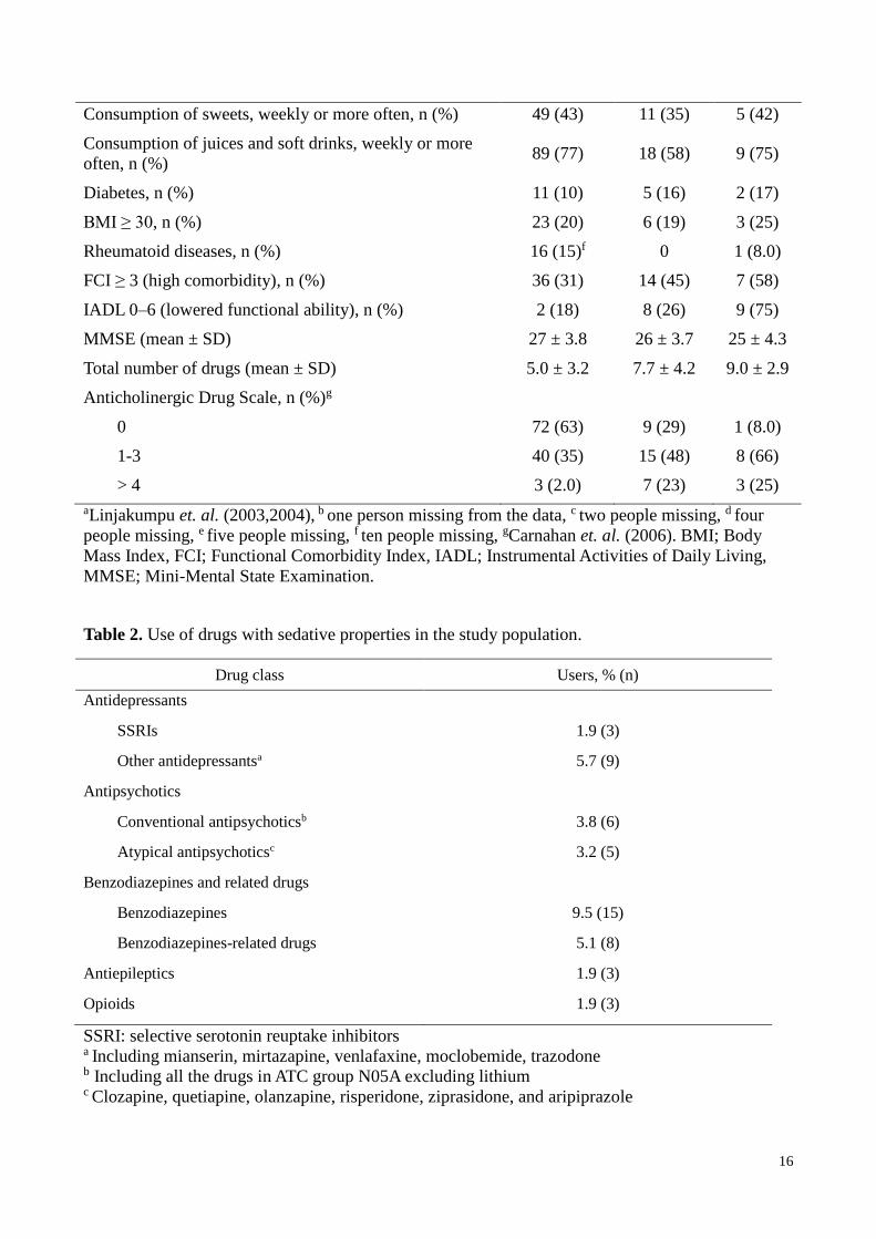
Table 1. The most frequently used drugs with sedative properties among the study population were
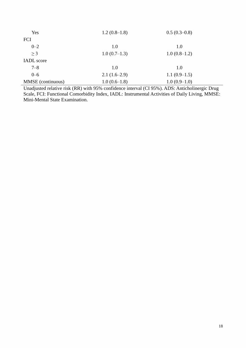
benzodiazepines and related drugs (Table 2). The unadjusted relative risks of the explanatory
variables are presented in Table 3. Distributions of dental caries and number of teeth with deepened
periodontal pockets in the study population are presented in Figure 1.
After adjusting for confounding factors (age, gender, education, FCI, MMSE, IADL, diabetes,
rheumatoid diseases and the patient’s total number of drugs), participants with either SL 1–2 or SL
≥ 3 showed an increased likelihood of having carious teeth (RR: 1.8, CI: 1.2–2.6 and RR: 2.4, CI:
1.4–4.1, respectively) compared to participants without any sedative load. Further adjustment for
toothbrushing frequency, use of toothpaste, the use of anticholinergic drugs (ADS) or the presence
of dental plaque did not essentially affect the risk estimates, except that the association with dental
caries was somewhat stronger when the model was also adjusted for the use of anticholinergic drugs
(ADS) (Table 4). To assess the magnitude of the effects of other medications, we created models
where the patient’s total number of drugs was not controlled for. The respective risk estimates for
these models were slightly higher: RR: 1.9, CI: 1.3–2.8 and RR: 2.9, CI: 1.8–4.7. The Pearson
correlation coefficient between sedative load and total number of drugs was 0.40 (p=0.01).
After adjustment for age, gender, education, FCI, MMSE, IADL, diabetes and rheumatoid diseases
there was no dose-dependent association between sedative load and the number of teeth with
deepened periodontal pockets, although the participants with a sedative load ≥ 3 had a decreased
likelihood of having teeth with deepened periodontal pockets (RR: 0.5, CI: 0.3–0.9) compared to

9
participants without any sedative load. Further adjustments for toothbrushing frequency, presence of
dental plaque or use of toothpaste did not change the risk estimates essentially (Table 4).
Adjustment for the use of anticholinergic drugs (ADS) did not have any essential effect on risk
estimates (Table 4).
Additional analyzes were performed to study whether there are any synergistic or antagonist effects
between dental caries and oral health behaviour variables, such as toothbrushing, the use of
toothpaste and dental visits. These analyses showed that there was a statistically significant product
term between the use of toothpaste and sedative load (p=0.024), non-significant product terms
between toothbrushing and sedative load (p= 0.249) and dental visits and sedative load (p= 0.139).
The results regarding the relation of the total number of drugs and both dental caries and
periodontal pocketing are shown in Table 4. The total number of drugs was not consistently
associated with dental caries. The overall association of the total number of drugs with periodontal
pocketing was opposite direction to that of sedative load.
DISCUSSION
To the best of our knowledge, this is the first study to analyse the relation of the cumulative effects
of multiple drugs with sedative properties to oral diseases, whereas all previous studies have
focused on the effect of a single category of drugs or alternatively the total amount of drugs on oral
diseases. The main finding of the present study was that participants who had higher sedative load
were more likely to have carious teeth but not deepened periodontal pockets.
10
Regarding dental caries, the association between sedative load and the number of carious teeth was
consistent and not essentially affected by further adjustment for variables describing oral hygiene,
such as the presence of dental plaque, toothbrushing frequency and use of toothpaste, in most cases
fluoridated. The association with dental caries concurs with previous studies, which have shown an
association between use of antidepressants and dental caries10-12. One possible and perhaps the most
likely explanation why sedative load was associated with carious teeth is that participants with a
higher sedative load had low salivary secretion–as seen in Table 1– which is known to be associated
with low intraoral pH, low buffer capacity of saliva30, 31 and changes in oral microflora32. This
observation is further supported with the fact that the anticholinergic burden, measured by
Carnahan's Anticholinergic Drug Scale24, was higher among those with the high sedative load
(Table 1). On the other hand, when the models were adjusted for ADS the risk estimates changed
only slightly (Table 4). This suggests that sedative properties of the drugs, rather than solely
anticholinergic properties, explain the association with dental caries. Such sedative drugs without
anticholinergic properties are benzodiazepines, hypnotic, or opioids, for example33.
Other mechanism which could explain the observed association with dental caries is that use of
drugs with sedative properties can deteriorate cognition and alter mood34, which in turn can cause
disregard for daily tasks such as toothbrushing. This explanation is supported by the observation
that the participants with SL ≥ 3 brushed their teeth less often than those with a lower sedative load,
but not by the fact that there was at the same time an inverse association of sedative load with
periodontal pocketing. This kind of inverse association with plaque related condition would not be
expected if the association was explained solely by poor oral hygiene.
Sedative load has been associated with impaired muscle strength35, which in turn may decrease the
participant’s capability to brush his/her teeth properly. However, a previous study based on the Oral
Health GeMS study suggests that impaired muscle strength is not a plausible explanation, at least
not in this home-dwelling population, as handgrip strength was not associated with oral self-care36.

11
On the other hand, additional analyses (interactions between sedative load and oral health behaviour)
suggest the possibility of a synergistic effect of poor health behaviour and high sedative load in the
development or progression of dental caries. However, the results are not fully consistent and are
subject to uncertainty.
Whether sedative load has any effect on the periodontium has not to our knowledge been studied
previously. In this study, we found that participants with the highest sedative load had the poorest
oral hygiene—whether measured by dental plaque or calculus—but despite this they had fewer
teeth with deepened periodontal pockets. This seemingly unexpected observation supports the
earlier-mentioned explanation that it is a question of qualitative changes in microbiota in the oral
cavity, most likely related to low salivary secretion, which seem to create favourable conditions for
cariogenic bacteria but not for periodontal pathogens37-39. The fact that sedative load was more or
less inversely associated with periodontal pocketing is in accordance with previous results from the
GeMS study showing that sedative load was associated with low salivary flow21 and that low
salivary secretion was inversely associated with periodontal pocketing40. Although this explanation
seems to be the most probable, there are other explanations as well. For example, it could be
speculated that certain sedative drugs can have immunomodulative properties which reduce tissue
destruction in the periodontium thus explaining the inverse association between sedative load and
periodontal pocketing.
Our paper stands out from previous papers because we were able to focus solely on drugs with
sedative properties. The advantages of our explanatory variable were that the Sedative Load Model
also includes drugs prescribed for somatic diseases and that the model depicts cumulative exposure
to drugs with sedative properties. Altogether, sedative load has been reported to be a valid
measurement of the total sedative load of all drugs used7. In spite of this, the use of medications
which have no sedative properties is one potential source of residual confounding. However, we
observed that adjustment with the total number of drugs changed the risk estimates only slightly,
12
indicating also that overall, non-specific use of drugs may not predispose teeth to dental caries and
that dental caries is specifically related to the use of drugs with sedative properties. This
interpretation is also supported by the finding that the total number of drugs was weakly associated
with carious teeth. Regarding effects on the periodontium, we found that the total number of drugs
showed effects opposite to those of sedative load; the total number of drugs was associated with the
number of teeth with deepened periodontal pockets, whereas sedative load was inversely associated
with the number of teeth with deepened periodontal pockets. These findings suggest that the
number of drugs is an inaccurate indicator of oral health risks.
Strengths and limitations
The Oral health GeMS study was designed to be an intervention study where the recording of
clinical parameters, such as dental caries and periodontal condition, was based on the participants’
need for restorative treatment and periodontal treatment, respectively. In addition, the registration
of dental caries and periodontal condition was done at tooth level. This robustness of measurements
may have attenuated the strength of the association between sedative load and outcome variables.
Due to the design of the GeMS study, the study population was homogeneous in terms of ethnic
origin and place of residence. Homogeneity was further increased by excluding smokers from the
study. The exclusions that were made in order to increase the validity of the study obviously meant
that the study population became smaller, which can be considered a limitation. We adjusted for a
number of potential determinants of oral diseases such as gender, education, FCI, MMSE, IADL,
ADS, diabetes and rheumatoid diseases (the most common general diseases, which have effect on
oral health) and took into account the number of remaining teeth in the analysis. However, it must
be remembered that the underlying reasons for taking medication and oral diseases may have
factors in common, which may not be totally controlled for using statistical methods. These
uncontrolled or partially controlled factors could be related to poor general health, for example. It

13
should be noted that in this data other diseases or conditions that can affect oral health, such as
Parkinson’s disease, depression, HIV and radiotherapy in the head-and-neck region, were rare or
non-existent.
Despite the fact that the participants were 75 years old or older, the fairly high participation rate
(70.8%) in the clinical oral examination was achieved by making visits outside of the dental clinic.
A limitation related to the clinical oral examination was a lack of assessment of repeatability (intra-
examiner kappa) and concordance between examiners (inter-examiner kappa), which could not be
assessed.
Another limitation in this study was the sedative load itself since it does not take into account drug
dosages22. It is commonly accepted that the dose-response relationship provides the evidence for
adverse drug reactions41. Due to the complexity of the situation, we are regrettable not able to study
the dosage of the drugs. This is due to the fact that, groups one (SL 2) and two (SL 1) alone in the
sedative load model include 120 different drugs, with individual doses.
It should be remembered that this is not a true follow-up study despite the six-month time interval
between the collection of the participant’s medication and the clinical oral examination, because the
participants were not free of diseases at the baseline of the study. In this sense, the study design is
cross-sectional, where the data about medications were collected on average six months earlier than
the clinical oral examination. Therefore, we cannot make any conclusions about the caries
increment or development of periodontal disease.
The fact that there was a six-month delay before the clinical oral examination was done means
periodontal condition or medications may have changed during this six-month period. However, it
is not unreasonable to make the assumption that this time interval did not have an essential effect on
the results due to the shortness of the time interval and also because, among older people,

14
medications for chronic diseases are fairly permanent and periodontal condition–in terms of
periodontal pocketing–is in most cases quite stable42.
Implications of the study
Based on the findings of this study, it is important that dentists and oral hygienists emphasize the
importance of regular dental prophylaxis and cariological maintenance care among patients using
multiple drugs with sedative properties. Instructions should be given to patients regarding proper
toothbrushing and interdental cleaning techniques and the use of fluoride toothpaste, fluoride rinses
and tablets. Also, clinicians caring for older patients should keep in mind that dental caries is a
serious problem among older people. Especially patients who are taking multiple drugs with
sedative properties should be referred to a dentist for assessment of dental prophylaxis need.
CONCLUSION
It can be concluded that presence of dental caries is associated to the use of drugs with sedative
properties. Another conclusion is that the use of drugs with sedative properties is not associated
with the presence of periodontal pockets.

15
Table 1. Basic descriptive statistics of the study population by different categories of sedative load.
Characteristics Sedative loada
0 1–2 ≥ 3
N 115 31 12
Age (mean ± SD) 78.8 ± 3.6 80.6 ± 3.7 80.5 ± 3.5
≥ 85 years, n (%) 7 (6.0) 4 (13) 0
Gender, proportion of women, n (%) 73 (64) 27 (87) 10 (83)
Education ≥ 7 years, n (%) 63 (56) 18 (58) 6 (50)
Number of teeth (mean ± SD) 15.1 ± 8.0 13.4 ± 8.0 12.3 ± 9.3
Number of teeth with periodontal pockets ≥ 4 mm (mean
± SD) 2.7 ± 3.5 2.8 ± 5.0 1.6 ± 1.9
Number of carious teeth (mean ± SD) 0.9 ± 1.4 1.8 ± 2.5 2.6 ± 4.6
Feeling of dry mouth
No or Occasional, n (%) 94 (82) 25 (81) 8 (67)
Often, n (%) 21 (18) 6 (19) 4 (33)
Stimulated salivary flow
< 1 ml/min, n (%) 28 (25)e 13 (43)b 7 (70)c
≥ 1 ml/min, n (%) 82 (75)e 17 (57)b 3 (30)c
Unstimulated salivary flow
< 0.1 ml/min, n (%) 26 (23)d 14 (45) 6 (55)b
≥ 0.1 ml/min, n (%) 85 (77)d 17 (55) 5 (45)b
Dental plaque
≤ 20% of teeth with dental plaque, n (%) 43 (37) 9 (29) 2 (17)
21–50% of teeth with dental plaque, n (%) 27 (24) 9 (29) 2 (17)
> 50% of teeth with dental plaque, n (%) 45 (39) 13 (42) 8 (66)
Dental calculus
≤ 20% of teeth with dental calculus, n (%) 28 (24) 10 (32) 4 (33)
21–50% of teeth with dental calculus, n (%) 41 (36) 9 (29) 2 (17)
> 50% of teeth with dental calculus, n (%) 46 (40) 12 (39) 6 (50)
Toothbrushing at least twice a day, n (%) 95 (83)b 25 (83)b 9 (75)
Use of toothpaste at least twice a day, n (%) 60 (52) 12 (39) 3 (25)
Regular dental visits, n (%) 68 (60)b 16 (53)b 5 (42)
16
Consumption of sweets, weekly or more often, n (%) 49 (43) 11 (35) 5 (42)
Consumption of juices and soft drinks, weekly or more
often, n (%) 89 (77) 18 (58) 9 (75)
Diabetes, n (%) 11 (10) 5 (16) 2 (17)
BMI ≥ 30, n (%) 23 (20) 6 (19) 3 (25)
Rheumatoid diseases, n (%) 16 (15)f 0 1 (8.0)
FCI ≥ 3 (high comorbidity), n (%) 36 (31) 14 (45) 7 (58)
IADL 0–6 (lowered functional ability), n (%) 2 (18) 8 (26) 9 (75)
MMSE (mean ± SD) 27 ± 3.8 26 ± 3.7 25 ± 4.3
Total number of drugs (mean ± SD) 5.0 ± 3.2 7.7 ± 4.2 9.0 ± 2.9
Anticholinergic Drug Scale, n (%)g
0 72 (63) 9 (29) 1 (8.0)
1-3 40 (35) 15 (48) 8 (66)
> 4 3 (2.0) 7 (23) 3 (25)
aLinjakumpu et. al. (2003,2004), b one person missing from the data, c two people missing, d four
people missing, e five people missing, f ten people missing, gCarnahan et. al. (2006). BMI; Body
Mass Index, FCI; Functional Comorbidity Index, IADL; Instrumental Activities of Daily Living,
MMSE; Mini-Mental State Examination.
Table 2. Use of drugs with sedative properties in the study population.
Drug class Users, % (n)
Antidepressants
SSRIs 1.9 (3)
Other antidepressantsa 5.7 (9)
Antipsychotics
Conventional antipsychoticsb 3.8 (6)
Atypical antipsychoticsc 3.2 (5)
Benzodiazepines and related drugs
Benzodiazepines 9.5 (15)
Benzodiazepines-related drugs 5.1 (8)
Antiepileptics 1.9 (3)
Opioids 1.9 (3)
SSRI: selective serotonin reuptake inhibitors a Including mianserin, mirtazapine, venlafaxine, moclobemide, trazodone b Including all the drugs in ATC group N05A excluding lithium c Clozapine, quetiapine, olanzapine, risperidone, ziprasidone, and aripiprazole

17
Table 3. Factors related to carious teeth and the number of teeth with periodontal pockets ≥ 4 mm
deep.
Outcome
Characteristics Number of carious
teeth
Number of teeth with periodontal pockets
≥ 4 mm
RR (CI 95%) RR (CI 95%)
Sedative load
0 1.0 1.0
1–2 2.2 (1.6–3.0) 1.2 (0.9–1.5)
≥ 3 3.5 (1.6–5.2) 0.7 (0.4–1.1)
Total number of drugs,
continuous 1.04 (1.01–1.08) 1.04 (1.01–1.08)
ADS, continuous 0.96 (0.87–1.07) 0.94 (0.87–1.07)
Age, continuous 1.1 (1.0–1.1) 1.0 (1.0–1.0)
Gender
Female 1.0 1.0
Male 0.9 (0.6–1.2) 0.9 (0.7–1.0)
Education
≥ 7 years 1.0 1.0
< 7 years 1.2 (0.9–1.6) 0.9 (0.7–1.0)
Dental plaque
≤ 20% of teeth with dental
plaque 1.0 1.0
21–50% of teeth with dental
plaque 1.2 (0.8–1.8) 1.6 (1.3–2.1)
> 50% of teeth with dental
plaque 1.8 (1.3–2.5) 1.7 (1.3–2.1)
Toothbrushing
At least twice a day 1.0 1.0
More seldom 1.8 (1.3–2.5) 0.7 (0.5–0.9)
Use of toothpaste
At least twice a day 1.0 1.0
More seldom 1.9 (1.4–2.6) 1.5 (1.3–1.9)
Dental visits
Regularly 1.0 1.0
Symptom-based, never 2.5 (1.9–3.3) 1.7 (1.4–2.0)
Diabetes
No 1.0 1.0
Yes 1.1 (0.7–1.6) 1.0 (0.7–1.4)
Rheumatoid disease
No 1.0 1.0
18
Yes 1.2 (0.8–1.8) 0.5 (0.3–0.8)
FCI
0–2 1.0 1.0
≥ 3 1.0 (0.7–1.3) 1.0 (0.8–1.2)
IADL score
7–8 1.0 1.0
0–6 2.1 (1.6–2.9) 1.1 (0.9–1.5)
MMSE (continuous) 1.0 (0.6–1.8) 1.0 (0.9–1.0)
Unadjusted relative risk (RR) with 95% confidence interval (CI 95%). ADS: Anticholinergic Drug
Scale, FCI: Functional Comorbidity Index, IADL: Instrumental Activities of Daily Living, MMSE:
Mini-Mental State Examination.

19
Table 4. Relation between sedative load and the total number of drugs used and both carious teeth
and the number of teeth with periodontal pockets ≥ 4 mm deep.
Outcome
Number of carious teeth
Number of teeth with
periodontal pockets ≥ 4 mm
RR (CI 95%) RR (CI 95%)
Model 1
Sedative load
0 1.0 1.0
1–2 1.8 (1.2–2.6) 0.9 (0.7–1.2)
≥ 3 2.4 (1.4–4.1) 0.5 (0.3–0.9)
continuous 1.21 (1.06–1.38) 0.92 (0.82–1.00)
Model 2a
Sedative load
0 1.0 1.0
1–2 1.5 (1.0–2.3) 0.9 (0.7–1.2)
≥ 3 1.4 (0.8–2.8) 0.5 (0.3–0.9)
continuous 1.09 (0.94–1.27) 0.91 (0.82–1.01)
Model 3b
Sedative load
0 1.0 1.0
1–2 1.8 (1.2–2.6) 0.9 (0.7–1.1)
≥ 3 2.4 (1.4–4.1) 0.5 (0.3–0.8)
continuous 1.21 (1.05–1.38) 0.89 (0.79–0.98)
Model 4c
0 1.0 1.0
1–2 1.9 (1.3–2.8) 0.9 (0.6–1.1)

20
≥ 3 3.0 (1.7–5.2) 0.5 (0.3–0.8)
continuous 1.27 (1.10–1.45) 0.87 (0.78–0.97)
Model 5d
Sedative load
0 1.0
1–2 1.9 (1.3–2.8)
≥ 3 2.9 (1.8–4.7)
continuous 1.29 (1.14–1.45)
Total number of drugse
0–3 1.0 1.0
4–6 0.9 (0.6–1.4) 1.3 (0.9–1.6)
7–9 1.9 (1.2–3.1) 1.0 (0.7–1.4)
≥ 10 1.1 (0.6–2.0) 1.6 (1.1–2.4)
continuous 1.04 (0.99–1.09) 1.02 (0.99–1.06)
Adjusted relative risk (RR) with 95% confidence interval (CI 95%).
All models were adjusted for age, gender, education, Functional Comorbidity Index, Mini-Mental
State Examination, Instrumental Activity of Daily Living, diabetes and rheumatoid diseases and the
number of teeth was used as an offset variable. Dental caries was also adjusted for the total number
of drugs.
a Adjusted for toothbrushing and the use of toothpaste b Adjusted for dental plaque c Adjusted for Anticholinergic Drug Scale d Adjusted without the total number of drugs e Adjusted for sedative load

21
Figure 1 Distribution of outcome variables in study population

22
References
1. Trevor AJ. Sedative-Hypnotic Drugs. In: Katzung BG ed. Basic and Clinical pharmacology, San
Francisco: The McGraw-Hill Companies, Inc.; 2007:347–362.
2. Linjakumpu T, Hartikainen S, Klaukka T, Koponen H, Kivela SL, Isoaho R. A model to
classify the sedative load of drugs. Int J Geriatr Psychiatry 2003; 18:542–544.
3. Blazer D, Hybels C, Simonsick E, Hanlon JT. Sedative, hypnotic, and antianxiety medication
use in an aging cohort over ten years: a racial comparison. J Am Geriatr Soc 2000; 48:1073–1079.
4. Beck CA, Williams JV, Wang JL, Kassam A, El-Guabaly N, Currie SR et al. Psychotropic
medication use in Canada. Can J Psychiatry 2005; 50:605–613.
5. Hollingworth SA, Siskind DJ. Anxiolytic, hypnotic and sedative medication use in Australia.
Pharmacoepidemiol Drug Saf 2010; 19:280–288.
6. Linjakumpu TA, Hartikainen SA, Klaukka TJ, Koponen HJ, Hakko HH, Viilo KM et al.
Sedative drug use in the home-dwelling elderly. Ann Pharmacother 2004; 38:2017–2022.
7. Skoog I, Nilsson L, Landahl S, Steen B. Mental disorders and the use of psychotropic drugs in
an 85-year-old urban population. Int Psychogeriatr 1993; 5:33–48.
8. Linjakumpu T, Hartikainen S, Klaukka T, Koponen H, Kivela SL, Isoaho R. Psychotropics
among the home-dwelling elderly--increasing trends. Int J Geriatr Psychiatry 2002; 17:874–883.
9. Llorente MD, David D, Golden AG, Silverman MA. Defining patterns of benzodiazepine use
in older adults. J Geriatr Psychiatry Neurol 2000; 13:150–160.
10. deVries MW, Peeters F. Dental caries with longterm use of antidepressants. Lancet 1995;
346:1640.
11. Peeters FP, deVries MW, Vissink A. Risks for oral health with the use of antidepressants. Gen
Hosp Psychiatry 1998; 20:150–154.
12. Rundegren J, van Dijken J, Mornstad H, von Knorring L. Oral conditions in patients
receiving long-term treatment with cyclic antidepressant drugs. Swed Dent J 1985; 9:55–64.
13. Thomson WM, Slade GD, Spencer AJ. Dental caries experience and use of prescription
medications among people aged 60+ in South Australia. Gerodontology 1995; 12:104–110.
14. Thomson WM. Spencer AJ, Slade GD, Chalmers JM. Is medication a risk factor for dental
caries among older people? Evidence from a longitudinal study in South Australia. Community Dent
Oral Epidemiol 2002; 30:224–32.
15. Lawrence HP, Hunt RJ, Beck JD. Three-year root caries incidence and risk modelling in older
adults in North Carolina. J Public Health Dent 1995; 55:69–78.
16. Närhi TO, Vehkalahti MM, Siukosaari P, Ainamo A. Salivary findings, daily medication and
root caries in the elderly. Caries Res 1998; 32:5–9.

23
17. Hawkins RJ, Jutai DKG, Brothwell DJ, Locker D. Threeyear coronal caries incidence in
older Canadian adults. Caries Res 1997;31:405–10.
18. Lampela P, Hartikainen S, Lavikainen P, Sulkava R, Huupponen R. Effects of medication
assessment as part of a comprehensive geriatric assessment on drug use over a 1-year period: a
population-based intervention study. Drugs Aging 2010; 27:507–521.
19. Tikkanen P, Nykänen I, Lönnroos E, Sipila S, Sulkava R, Hartikainen S. Physical Activity
at Age of 20-64 Years and Mobility and Muscle Strength in Old Age: A Community-Based Study.
Journals of Gerontology.Series A, Biological Sciences & Medical Sciences 2012; 67:905–910.
20. Komulainen K, Ylostalo P, Syrjälä AM, Ruoppi P, Knuutila M, Sulkava R et al. Preference
for dentist's home visits among older people. Community Dent Oral Epidemiol 2012; 40:89–95.
21. Tiisanoja A, Syrjälä AM, Komulainen K, Hartikainen S, Taipale H, Knuuttila M et al.
Sedative load and salivary secretion and xerostomia in community-dwelling older people.
Gerodontology 2014; doi;10.1111/ger.12129.
22. Taipale HT, Hartikainen S, Bell JS. A comparison of four methods to quantify the cumulative
effect of taking multiple drugs with sedative properties. Am J Geriatr Pharmacother 2010; 8:460–
471.
23. Taipale HT, Bell JS, Uusi-Kokko M, Lonnroos E, Sulkava R, Hartikainen S. Sedative load
among community-dwelling people aged 75 years and older: a population-based study. Drugs Aging
2011; 28:913–925.
24. Carnahan RM, Lund BC, Perry PJ, Pollock BG, Culp KR. The Anticholinergic Drug Scale
as a measure of drug-related anticholinergic burden: associations with serum anticholinergic
activity. J Clin Pharmacol 2006: 46: 1481–1486.
25. Crum RM, Anthony JC, Bassett SS, Folstein MF. Population-based norms for the Mini-
Mental State Examination by age and educational level. JAMA 1993; 269:2386–2391.
26. Dahl A, Berg S, Nilsson SE. Identification of dementia in epidemiological research: a study on
the usefulness of various data sources. Aging Clin Exp Res 2007; 19:381–389.
27. Groll DL, To T, Bombardier C, Wright JG. The development of a comorbidity index with
physical function as the outcome. J Clin Epidemiol 2005; 58:595–602.
28. Lawton MP, Brody EM. Assessment of older people: self-maintaining and instrumental
activities of daily living. Gerontologist 1969; 9:179–186.
29. SPSS Inc. SPSS 22.0 for Windows. 2013;22.0.0.
30. Dawes C. Salivary flow patterns and the health of hard and soft oral tissues. J Am Dent Assoc
2008; 139 (Suppl 1):18–14.
31. Ravald N, List T. Caries and periodontal conditions in patients with primary Sjogren's
syndrome. Swed Dent J 1998; 22:97–103.

24
32. Almstahl A, Wikstrom M. Oral microflora in subjects with reduced salivary secretion. J Dent
Res 1999; 78:1410–1416.
33. Scully C. (2003) Drug effects on salivary glands: dry mouth. Oral Dis 9:165-176.
34. Vermeeren A. Residual effects of hypnotics: epidemiology and clinical implications. CNS
Drugs 2004; 18:297–328.
35. Taipale HT, Bell JS, Gnjidic D, Sulkava R, Hartikainen S. Muscle strength and sedative load
in community-dwelling people aged 75 years and older: a population-based study. J Gerontol A Biol
Sci Med Sci 2011; 66:1384–1392.
36. Komulainen K, Ylöstalo P, Syrjälä AM, Ruoppi P, Knuutila M, Sulkava R et al.
Associations of instrumental activities of daily living and handgrip strength with oral self-care
among home-dwelling elderly 75+. Gerodontology 2012; 29:e135–42.
37. Bradshaw DJ, Marsh PD. Analysis of pH-driven disruption of oral microbial communities in
vitro. Caries Res 1998; 32:456–462.
38. Bradshaw DJ, McKee AS, Marsh PD. Effects of carbohydrate pulses and pH on population
shifts within oral microbial communities in vitro. J Dent Res 1989; 68:1298–1302.
39. Haase EM, Bonstein T, Palmer RJ,Jr, Scannapieco FA. Environmental influences on
Actinobacillus actinomycetemcomitans biofilm formation. Arch Oral Biol 2006; 51:299–314.
40. Syrjälä AM, Raatikainen L, Komulainen K, Knuutila M, Ruoppi, Hartikainen S et al.
Salivary flow rate and periodontal infection: a study among subjects aged 75 years or older. Oral
Dis 2011; 17:387–392.
41. Naranjo CA, Busto U, Sellers EM, Sandor P, Ruiz I, Roberts EA et al. A method for
estimating the probability of adverse drug reactions. Clin Pharmacol Ther 1981: 30: 239–245.
42. Thomson WM, Slade GD, Beck JD, Elter JR, Spencer AJ, Chalmers JM. Incidence of
periodontal attachment loss over 5 years among older South Australians. J Clin Periodontol 2004:
31: 119–125.