segmentation of infant brain mr images based on...
TRANSCRIPT
SEGMENTATION OF INFANT BRAIN MR IMAGES BASED ON ADAPTIVE SHAPE PRIORAND HIGHER-ORDER MGRF
M. Ismail1,M. Mostapha1, A. Soliman1,M. Nitzken1, F. Khalifa1,A. Elnakib1, G.Gimel’farb2, M. F. Casanova3, and A. El-Baz1
1BioImaging Laboratory, Bioengineering Department, University of Louisville, Louisville, KY, USA.2Department of Computer Science, University of Auckland, Auckland 1142, New Zealand.
3Department of of Psychiatry and Behavioral Science, University of Louisville, Louisville, KY, USA.
ABSTRACT
This paper introduces a new framework for the segmentation of dif-ferent brain structures from 3D infant MR brain images. The pro-posed segmentation framework is based on a shape prior built us-ing a subset of co-aligned training images that is adapted during thesegmentation process based on higher-order visual appearance char-acteristics of infant MRIs. These characteristics are described usingvoxel-wise image intensities and their spatial interaction features. Inorder to more accurately model the empirical grey level distributionof infant brain signals, a Linear Combination of Discrete Gaussians(LCDG) is used that has positive and negative components. Alsoto accurately account for the large inhomogeneity in infant MRIs,a higher-order Markov Gibbs Random Field (MGRF) spatial inter-action model that integrates third- and fourth-order families with atraditional second-order model is proposed. The proposed approachwas tested on 40 in-vivo infant 3D MR brain scans, having theirground truth created by an expert radiologist, using three metrics: theDice coefficient, the 95-percentile modified Hausdorff distance, andthe absolute brain volume difference. Experimental results promisean accurate segmentation of infant MR brain images compared tocurrent open source segmentation tools.
Index Terms— Adaptive Shape, Higher Order MGRF, InfantBrain Segmentation
1. INTRODUCTIONAccurate segmentation of brain tissues from Magnetic ResonanceImaging (MRI) data is an essential step in clinical diagnostics, ther-apy evaluation, human brain mapping, and neuroscience [1, 2]. Theanalysis and treatment of brain injuries and disorders rely on ac-curate brain segmentation that accounts for classifying its differenttissue types [3]. Brain MRI segmentation has many challenges, es-pecially with infant brains, such as image noise, inhomogeneities,and low contrast between tissue types. Also infant brains have areverse in contrast in the White Matter and Gray Matter [4], and ahigher amount of noise than adult brains [5]. This is due to the factthat the WM is unmyelinated and has water makeup that results in alow contrast between tissue classes, and an intensity level that is al-most identical to that of GM, which makes it difficult to distinguishthese 2 classes even by experts [6].
There has been extensive work in the literature that addressesadult brain segmentation, with fewer techniques suited for infantbrain segmentation. Probabilistic methods were adopted in [7] foradult brain segmentation, where a map classifier along with a prob-ability clustering method are employed for brain tissue classifica-tion. In [8], a pairwise joint MGRF interaction model was usedfor adult brain segmentation, however the pairwise MGRF failed to
capture large inhomogeneities of signals, and thus would not suit in-fant brain segmentation. Deformable models were used in [9, 10],where region-boundary models are employed to segment brain tis-sues. Atlas-based methods have also been adopted, as in [11, 12],where brain shape priors are registered with the subjects to be seg-mented to guide the segmentation process.
Since the intensity alone would fail to segment infant brain tis-sues due to the similar intensities of different structures and poorcontrast, fewer techniques can be found for infant brain segmenta-tion. Xue et al. [4] employed an Expectation-Maximization (EM)algorithm along with a Markov Random Field (MRF) prior for in-fant brain segmentation. Classifying brain structures, such as WM,CSF, central GM, and cortical GM was conducted by Abneek et al.in [13], where T2-weighted images of neonatal brains were used.Probability maps were used to segment each brain tissue class witha K-nearest neighbor classifier. Wang et al. [14] segmented T1,T2, and diffusion-weighted brain images using a sparse representa-tion of the complementary tissue distribution. In [15], the randomforest technique was used to integrate features from the differentmodalities for tissue segmentation along with probability maps ofGM, WM, and CSF. Some approaches use longitudinal scans at alate-time-point age, where the contrast is much better between dif-ferent tissue types, from which probabilistic atlases are constructedto guide segmentation of neonatal images,[16, 17]. Segmentationwith shape priors was also adopted as in [18, 19, 20].
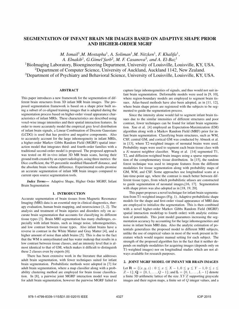
This paper proposes a novel technique for infant brain segmenta-tion from T1-weighted images (Fig. 1). Adaptive probabilistic shapemodels for the shape and first-order visual appearance of MRI dataare employed to initialize the segmentation. This is then combinedwith a novel higher-order Markov Gibbs Random Field (MGRF)spatial interaction model(up to fourth order) with analytic estima-tion of potentials. This joint model guarantees increasing the seg-mentation accuracy by accounting for the large inhomogeneities andnoise in infant brain MRI data. Also the analytic estimation of po-tentials generalizes the proposed model to different MRI subjects,unlike the use of empirical values in most of the work present in lit-erature which would require manual setting for each subject. Thestrength of the proposed algorithm lies in the fact that it neither de-pends on multiple modalities for acquiring images (depends only onT1-weighted images) nor on longitudinal studies which are not al-ways available for research purposes.
2. JOINT MGRF MODEL OF INFANT MR BRAIN IMAGES
Let R = {(x, y, z) : 0 ≤ x ≤ X − 1, 0 ≤ y ≤ Y − 1, 0 ≤ z ≤Z−1}; Q = {0, 1, . . . , Q−1}; and L = {0, 1, . . . , L− 1} denotea finite 3D arithmetic lattice of the size XY Z supporting grayscaleimages and their region maps, a finite set of Q integer values, and a
4327978-1-4799-8339-1/15/$31.00 ©2015 IEEE ICIP 2015
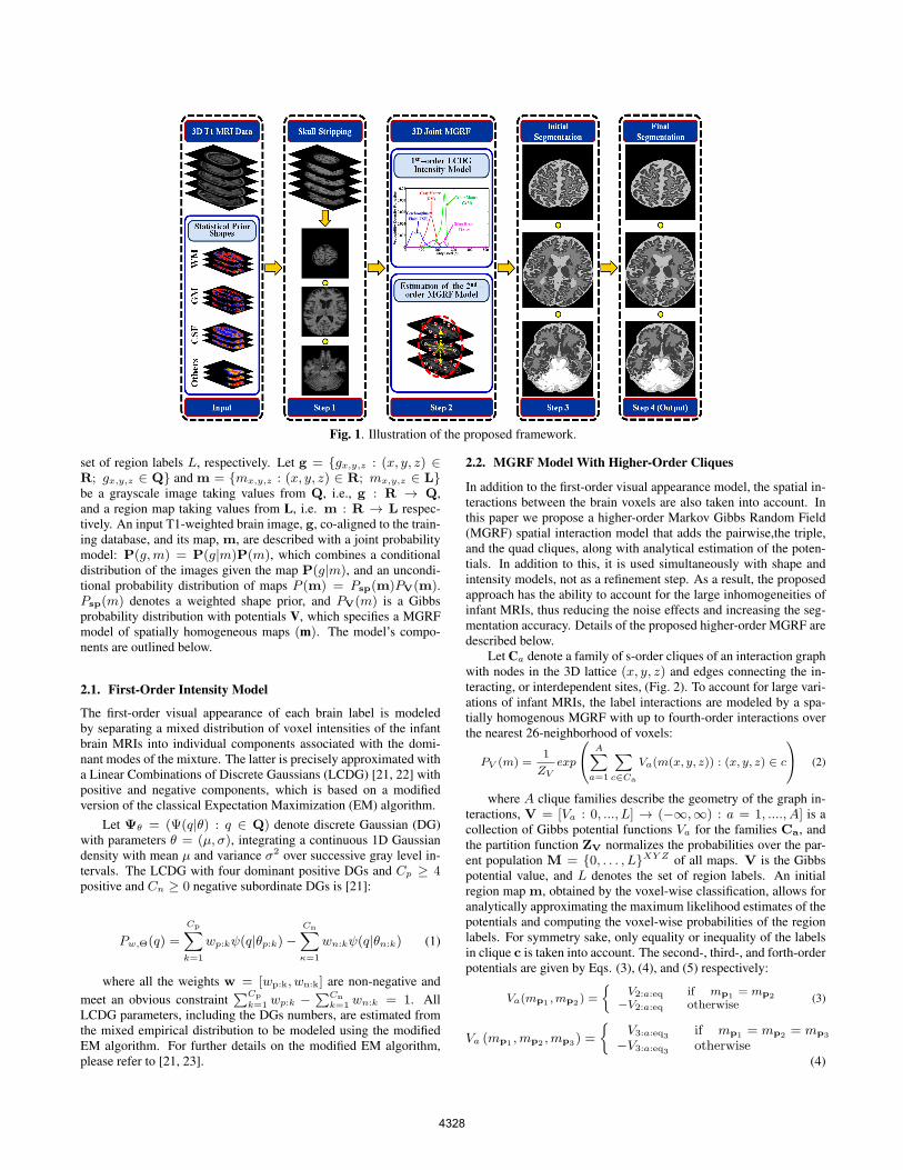
Fig. 1. Illustration of the proposed framework.
set of region labels L, respectively. Let g = {gx,y,z : (x, y, z) ∈R; gx,y,z ∈ Q} and m = {mx,y,z : (x, y, z) ∈ R; mx,y,z ∈ L}be a grayscale image taking values from Q, i.e., g : R → Q,and a region map taking values from L, i.e. m : R → L respec-tively. An input T1-weighted brain image, g, co-aligned to the train-ing database, and its map, m, are described with a joint probabilitymodel: P(g,m) = P(g|m)P(m), which combines a conditionaldistribution of the images given the map P(g|m), and an uncondi-tional probability distribution of maps P (m) = Psp(m)PV(m).Psp(m) denotes a weighted shape prior, and PV(m) is a Gibbsprobability distribution with potentials V, which specifies a MGRFmodel of spatially homogeneous maps (m). The model’s compo-nents are outlined below.
2.1. First-Order Intensity Model
The first-order visual appearance of each brain label is modeledby separating a mixed distribution of voxel intensities of the infantbrain MRIs into individual components associated with the domi-nant modes of the mixture. The latter is precisely approximated witha Linear Combinations of Discrete Gaussians (LCDG) [21, 22] withpositive and negative components, which is based on a modifiedversion of the classical Expectation Maximization (EM) algorithm.
Let Ψθ = (Ψ(q|θ) : q ∈ Q) denote discrete Gaussian (DG)with parameters θ = (µ, σ), integrating a continuous 1D Gaussiandensity with mean µ and variance σ2 over successive gray level in-tervals. The LCDG with four dominant positive DGs and Cp ≥ 4positive and Cn ≥ 0 negative subordinate DGs is [21]:
Pw,Θ(q) =
Cp∑k=1
wp:kψ(q|θp:k)−Cn∑κ=1
wn:kψ(q|θn:k) (1)
where all the weights w = [wp:k, wn:k] are non-negative andmeet an obvious constraint
∑Cp
k=1 wp:k −∑Cn
k=1 wn:k = 1. AllLCDG parameters, including the DGs numbers, are estimated fromthe mixed empirical distribution to be modeled using the modifiedEM algorithm. For further details on the modified EM algorithm,please refer to [21, 23].
2.2. MGRF Model With Higher-Order Cliques
In addition to the first-order visual appearance model, the spatial in-teractions between the brain voxels are also taken into account. Inthis paper we propose a higher-order Markov Gibbs Random Field(MGRF) spatial interaction model that adds the pairwise,the triple,and the quad cliques, along with analytical estimation of the poten-tials. In addition to this, it is used simultaneously with shape andintensity models, not as a refinement step. As a result, the proposedapproach has the ability to account for the large inhomogeneities ofinfant MRIs, thus reducing the noise effects and increasing the seg-mentation accuracy. Details of the proposed higher-order MGRF aredescribed below.
Let Ca denote a family of s-order cliques of an interaction graphwith nodes in the 3D lattice (x, y, z) and edges connecting the in-teracting, or interdependent sites, (Fig. 2). To account for large vari-ations of infant MRIs, the label interactions are modeled by a spa-tially homogenous MGRF with up to fourth-order interactions overthe nearest 26-neighborhood of voxels:
PV (m) =1
ZVexp
A∑a=1
∑c∈Ca
Va(m(x, y, z)) : (x, y, z) ∈ c
(2)
where A clique families describe the geometry of the graph in-teractions, V = [Va : 0, ..., L] → (−∞,∞) : a = 1, ...., A] is acollection of Gibbs potential functions Va for the families Ca, andthe partition function ZV normalizes the probabilities over the par-ent population M = {0, . . . , L}XY Z of all maps. V is the Gibbspotential value, and L denotes the set of region labels. An initialregion map m, obtained by the voxel-wise classification, allows foranalytically approximating the maximum likelihood estimates of thepotentials and computing the voxel-wise probabilities of the regionlabels. For symmetry sake, only equality or inequality of the labelsin clique c is taken into account. The second-, third-, and forth-orderpotentials are given by Eqs. (3), (4), and (5) respectively:
Va(mp1 ,mp2 ) =
{V2:a:eq if mp1 = mp2
−V2:a:eq otherwise(3)
Va (mp1 ,mp2 ,mp3) =
{V3:a:eq3 if mp1 = mp2 = mp3
−V3:a:eq3 otherwise(4)
4328
Va (mp1 ,mp2 ,mp3 ,mp4 ) =
V4:a:eq4 if 4 equal labelsV4:a:eq3 if 3 equal labels−
(V4:a:eq3 + V4:a:eq4
)otherwise
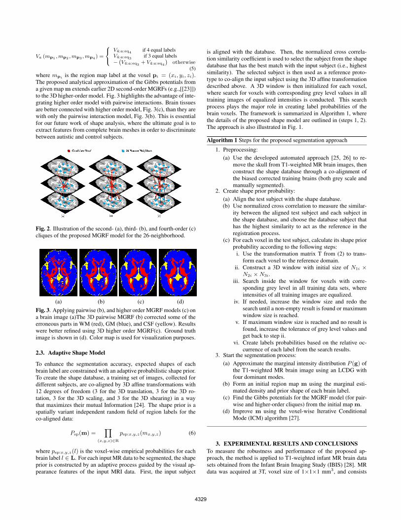
(5)where mpi is the region map label at the voxel pi = (xi, yi, zi).The proposed analytical approximation of the Gibbs potentials froma given map m extends earlier 2D second-order MGRFs (e.g.,[[23]])to the 3D higher-order model. Fig. 3 highlights the advantage of inte-grating higher order model with pairwise interactions. Brain tissuesare better connected with higher order model, Fig. 3(c), than they arewith only the pairwise interaction model, Fig. 3(b). This is essentialfor our future work of shape analysis, where the ultimate goal is toextract features from complete brain meshes in order to discriminatebetween autistic and control subjects.
Fig. 2. Illustration of the second- (a), third- (b), and fourth-order (c)cliques of the proposed MGRF model for the 26-neighborhood.
(a) (b) (c) (d)Fig. 3. Applying pairwise (b), and higher order MGRF models (c) ona brain image (a)The 3D pairwise MGRF (b) corrected some of theerroneous parts in WM (red), GM (blue), and CSF (yellow). Resultswere better refined using 3D higher order MGRF(c). Ground truthimage is shown in (d). Color map is used for visualization purposes.
2.3. Adaptive Shape Model
To enhance the segmentation accuracy, expected shapes of eachbrain label are constrained with an adaptive probabilistic shape prior.To create the shape database, a training set of images, collected fordifferent subjects, are co-aligned by 3D affine transformations with12 degrees of freedom (3 for the 3D translation, 3 for the 3D ro-tation, 3 for the 3D scaling, and 3 for the 3D shearing) in a waythat maximizes their mutual Information [24]. The shape prior is aspatially variant independent random field of region labels for theco-aligned data:
Psp(m) =∏
(x,y,z)∈R
psp:x,y,z(mx,y,z) (6)
where psp:x,y,z(l) is the voxel-wise empirical probabilities for eachbrain label l ∈ L. For each input MR data to be segmented, the shapeprior is constructed by an adaptive process guided by the visual ap-pearance features of the input MRI data. First, the input subject
is aligned with the database. Then, the normalized cross correla-tion similarity coefficient is used to select the subject from the shapedatabase that has the best match with the input subject (i.e., highestsimilarity). The selected subject is then used as a reference proto-type to co-align the input subject using the 3D affine transformationdescribed above. A 3D window is then initialized for each voxel,where search for voxels with corresponding grey level values in alltraining images of equalized intensities is conducted. This searchprocess plays the major role in creating label probabilities of thebrain voxels. The framework is summarized in Algorithm 1, wherethe details of the proposed shape model are outlined in (steps 1, 2).The approach is also illustrated in Fig. 1.
Algorithm 1 Steps for the proposed segmentation approach
1. Preprocessing:(a) Use the developed automated approach [25, 26] to re-
move the skull from T1-weighted MR brain images, thenconstruct the shape database through a co-alignment ofthe biased corrected training brains (both grey scale andmanually segmented).
2. Create shape prior probability:(a) Align the test subject with the shape database.(b) Use normalized cross correlation to measure the similar-
ity between the aligned test subject and each subject inthe shape database, and choose the database subject thathas the highest similarity to act as the reference in theregistration process.
(c) For each voxel in the test subject, calculate its shape priorprobability according to the following steps:
i. Use the transformation matrix T from (2) to trans-form each voxel to the reference domain.
ii. Construct a 3D window with initial size of N1i ×N2i ×N3i.
iii. Search inside the window for voxels with corre-sponding grey level in all training data sets, whereintensities of all training images are equalized.
iv. If needed, increase the window size and redo thesearch until a non-empty result is found or maximumwindow size is reached.
v. If maximum window size is reached and no result isfound, increase the tolerance of grey level values andget back to step ii.
vi. Create labels probabilities based on the relative oc-currence of each label from the search results.
3. Start the segmentation process:(a) Approximate the marginal intensity distribution P (g) of
the T1-weighted MR brain image using an LCDG withfour dominant modes.
(b) Form an initial region map m using the marginal esti-mated density and prior shape of each brain label.
(c) Find the Gibbs potentials for the MGRF model (for pair-wise and higher-order cliques) from the initial map m.
(d) Improve m using the voxel-wise Iterative ConditionalMode (ICM) algorithm [27].
3. EXPERIMENTAL RESULTS AND CONCLUSIONSTo measure the robustness and performance of the proposed ap-proach, the method is applied to T1-weighted infant MR brain datasets obtained from the Infant Brain Imaging Study (IBIS) [28]. MRdata was acquired at 3T, voxel size of 1×1×1 mm3, and consists
4329
A
C
S(a) (b) (c) (d) (e) (f)
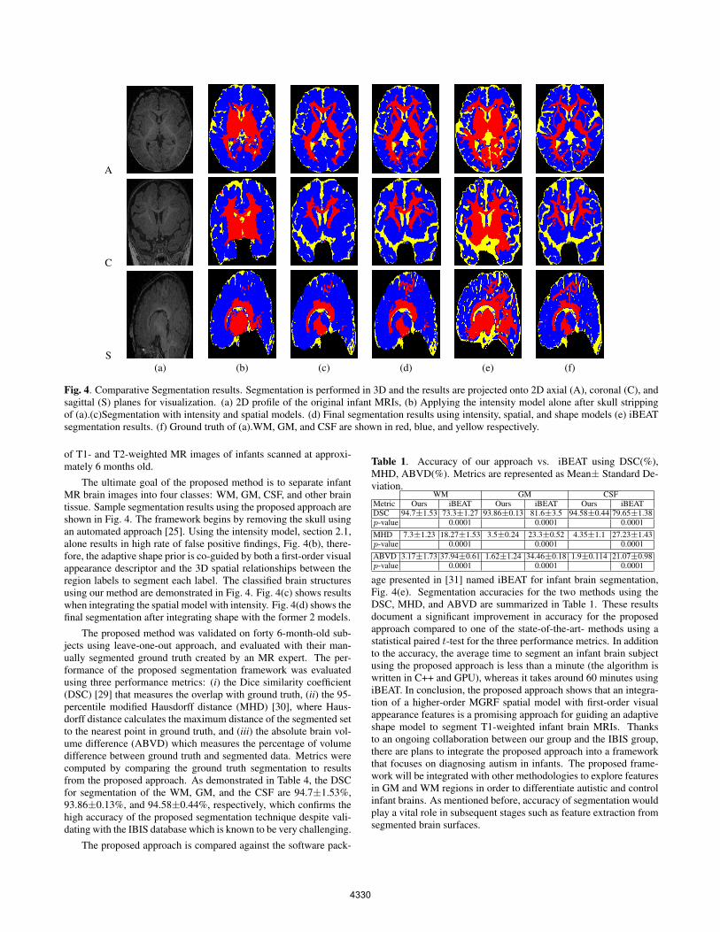
Fig. 4. Comparative Segmentation results. Segmentation is performed in 3D and the results are projected onto 2D axial (A), coronal (C), andsagittal (S) planes for visualization. (a) 2D profile of the original infant MRIs, (b) Applying the intensity model alone after skull strippingof (a).(c)Segmentation with intensity and spatial models. (d) Final segmentation results using intensity, spatial, and shape models (e) iBEATsegmentation results. (f) Ground truth of (a).WM, GM, and CSF are shown in red, blue, and yellow respectively.
of T1- and T2-weighted MR images of infants scanned at approxi-mately 6 months old.
The ultimate goal of the proposed method is to separate infantMR brain images into four classes: WM, GM, CSF, and other braintissue. Sample segmentation results using the proposed approach areshown in Fig. 4. The framework begins by removing the skull usingan automated approach [25]. Using the intensity model, section 2.1,alone results in high rate of false positive findings, Fig. 4(b), there-fore, the adaptive shape prior is co-guided by both a first-order visualappearance descriptor and the 3D spatial relationships between theregion labels to segment each label. The classified brain structuresusing our method are demonstrated in Fig. 4. Fig. 4(c) shows resultswhen integrating the spatial model with intensity. Fig. 4(d) shows thefinal segmentation after integrating shape with the former 2 models.
The proposed method was validated on forty 6-month-old sub-jects using leave-one-out approach, and evaluated with their man-ually segmented ground truth created by an MR expert. The per-formance of the proposed segmentation framework was evaluatedusing three performance metrics: (i) the Dice similarity coefficient(DSC) [29] that measures the overlap with ground truth, (ii) the 95-percentile modified Hausdorff distance (MHD) [30], where Haus-dorff distance calculates the maximum distance of the segmented setto the nearest point in ground truth, and (iii) the absolute brain vol-ume difference (ABVD) which measures the percentage of volumedifference between ground truth and segmented data. Metrics werecomputed by comparing the ground truth segmentation to resultsfrom the proposed approach. As demonstrated in Table 4, the DSCfor segmentation of the WM, GM, and the CSF are 94.7±1.53%,93.86±0.13%, and 94.58±0.44%, respectively, which confirms thehigh accuracy of the proposed segmentation technique despite vali-dating with the IBIS database which is known to be very challenging.
The proposed approach is compared against the software pack-
Table 1. Accuracy of our approach vs. iBEAT using DSC(%),MHD, ABVD(%). Metrics are represented as Mean± Standard De-viation.
WM GM CSFMetric Ours iBEAT Ours iBEAT Ours iBEATDSC 94.7±1.53 73.3±1.27 93.86±0.13 81.6±3.5 94.58±0.44 79.65±1.38p-value 0.0001 0.0001 0.0001MHD 7.3±1.23 18.27±1.53 3.5±0.24 23.3±0.52 4.35±1.1 27.23±1.43p-value 0.0001 0.0001 0.0001ABVD 3.17±1.73 37.94±0.61 1.62±1.24 34.46±0.18 1.9±0.114 21.07±0.98p-value 0.0001 0.0001 0.0001
age presented in [31] named iBEAT for infant brain segmentation,Fig. 4(e). Segmentation accuracies for the two methods using theDSC, MHD, and ABVD are summarized in Table 1. These resultsdocument a significant improvement in accuracy for the proposedapproach compared to one of the state-of-the-art- methods using astatistical paired t-test for the three performance metrics. In additionto the accuracy, the average time to segment an infant brain subjectusing the proposed approach is less than a minute (the algorithm iswritten in C++ and GPU), whereas it takes around 60 minutes usingiBEAT. In conclusion, the proposed approach shows that an integra-tion of a higher-order MGRF spatial model with first-order visualappearance features is a promising approach for guiding an adaptiveshape model to segment T1-weighted infant brain MRIs. Thanksto an ongoing collaboration between our group and the IBIS group,there are plans to integrate the proposed approach into a frameworkthat focuses on diagnosing autism in infants. The proposed frame-work will be integrated with other methodologies to explore featuresin GM and WM regions in order to differentiate autistic and controlinfant brains. As mentioned before, accuracy of segmentation wouldplay a vital role in subsequent stages such as feature extraction fromsegmented brain surfaces.
4330
4. REFERENCES
[1] M. A. Balafar, A. R. Ramli, A. Saripan, and S. Mashohor, “Re-view of brain MRI segmentation methods,” Artificial Intelli-gence Review, vol. 33, no. 3, pp. 261–274, 2010.
[2] A. Elnakib, G. Gimelfarb, J. S. Suri, and A. El-Baz, “Med-ical image segmentation: a brief survey,” in Multi ModalityState-of-the-Art Medical Image Segmentation and RegistrationMethodologies, pp. 1–39. Springer, 2011.
[3] N. Weisenfeld and S. K. Warfield, “Automatic segmentation ofnewborn brain MRI,” Neuroimage, vol. 47, no. 2, pp. 564–572,2009.
[4] H. Xue, L. Srinivasan, S. Jiang, M. Rutherford, A. Edwards,D. Rueckert, and J. Hajanl, “Automatic segmentation and re-construction of the cortex from neonatal MRI,” Neuroimage,vol. 38, no. 3, pp. 461–477, 2007.
[5] A. U. Mewes et al., “Regional brain development in serialmagnetic resonance imaging of low-risk preterm infants,” Pe-diatrics, vol. 118, no. 1, pp. 23–33, 2006.
[6] A. Barkovich, “Magnetic resonance techniques in the as-sessment of myelin and myelination,” Journal of inheritedmetabolic disease, vol. 28, no. 3, pp. 311–343, 2005.
[7] A. Ortiz, J. M. Grriz, J. Ramirez, and D. Salas-Gonzalez, “MRbrain image segmentation by growing hierarchical SOM andprobability clustering,” Electronics Letters, vol. 47, no. 10, pp.585–586, 2011.
[8] A. Alansary, A. Soliman, F. Khalifa, A. Elnakib, M. Mostapha,M. Nitzken, M. F. Casanova, and A. El-Baz, “MAP–basedframework for segmentation of mr brain images based on vi-sual appearance and prior shape,” MIDAS Journal [online].Available: http://hdl.handle.net/10380/3440, 2013.
[9] L. Wang, Y. Chen, X. Pan, X. Hong, and D. Xia, “Level setsegmentation of brain magnetic resonance images based on lo-cal Gaussian distribution fitting energy,” J. Neurosci. Methods,vol. 188, no. 2, pp. 316–325, 2010.
[10] S. Bourouis and K. Hamrouni, “3D segmentation of MRI brainusing level set and unsupervised classification,” Int. J. Image.Graph., vol. 10, no. 01, pp. 135–154, 2010.
[11] J.-P. Morin, C. Desrosiers, and L. Duong, “Atlas-based seg-mentation of brain magnetic resonance imaging using randomwalks,” in Computer Vision and Pattern Recognition Work-shops (CVPRW’12), 2012, pp. 44–49.
[12] C. Ledig, R. Wolz, P. Aljabar, J. Lotjonen, R. A. Heckemann,A. Hammers, and D. Rueckert, “Multi-class brain segmen-tation using atlas propagation and em-based refinement,” inproceedings of IEEE International Symposium on BiomedicalImaging (ISBI), 2012, pp. 896–899.
[13] P. Anbeek, K. L. Vincken, F. Groenendaal, A. Koeman, M. J.Van Osch, and J. Van der Grond, “Probabilistic brain tissuesegmentation in neonatal magnetic resonance imaging,” Pedi-atric Research, vol. 63, no. 2, pp. 158–163, 2008.
[14] L. Wang, F. Shi, G. Li, W. Lin, J. H. Gilmore, and D. Shen, “In-tegration of Sparse Multi-modality Representation and Geo-metrical Constraint for Isointense Infant Brain Segmentation,”in Medical Image Computing and Computer-Assisted Interven-tion (MICCAI), pp. 703–710. 2013.
[15] L. Wang, Y. Gao, F. Shi, G. Li, J. H. Gilmore, W. Lin, andD. Shen, “Links: Learning-based multi-source integrationframework for segmentation of infant brain image,” in MIC-CAI workshop on Medical Computer Vision, 2014.
[16] L. Wang, F. Shi, P. T. Yap, W. Lin, J. H. Gilmore, and D. Shen,“Longitudinally guided level sets for consistent tissue segmen-
tation of neonates,” Human Brain Mapping, vol. 34, no. 4, pp.956–972, 2013.
[17] F. Shi, Y. Fan, S. Tang, J. H. Gilmore, W. Lin, and D. Shen,“Neonatal brain image segmentation in longitudinal MRI stud-ies,” Neuroimage, vol. 49, no. 1, pp. 391–400, 2010.
[18] M. Altaye, S. K. Holland, M. Wilke, and C. Gaser, “Infantbrain probability templates for MRI segmentation and normal-ization,” Neuroimage, vol. 43, no. 4, pp. 721–730, 2008.
[19] Z. Song, S. P. Awate, D. J. Licht, and J. C. Gee, “Clinicalneonatal brain mri segmentation using adaptive nonparametricdata models and intensity-based markov priors,” in MedicalImage Computing and Computer-Assisted Intervention (MIC-CAI), pp. 883–890. 2007.
[20] Z. Song, N. Tustison, B. Avants, and J. Gee, “Adaptive graphcuts with tissue priors for brain MRI segmentation,” in Inproceedings of IEEE International Symposium on BiomedecialImaging (ISBI), 2006, pp. 762–765.
[21] A. El-Baz, A. Elnakib, F. Khalifa, M. A. El-Ghar, P. McClure,A. Soliman, and G. Gimel’farb, “Precise segmentation of 3-D magnetic resonance angiography,” IEEE Transaction onBiomedical Engineering, vol. 59, no. 7, pp. 2019–2029, 2012.
[22] A. El-Baz and G. Gimel’farb, “EM–based approximation ofempirical distributions with linear combinations of discreteGaussians,” in proceedings of IEEE International Conferenceon Image Processing (ICIP’07), San Antonio, Texas, USA,September 16–19, 2007.
[23] A. Farag, A. El-Baz, and G. Gimel’farb, “Precise segmentationof multimodal images,” IEEE Transactions on Image Process-ing, vol. 15, no. 4, pp. 952–968, 2006.
[24] P. A. Viola and W. M. Wells III, “Alignment by maximizationof mutual information,” International Journal of ComputerVision, vol. 24, no. 2, pp. 137–154, 1997.
[25] A. Alansary, A. Soliman, M. Nitzken, F. Khalifa, A. Elnakib,M. Mostapha, M. F. Casanova, and A. El-Baz, “An integratedgeometrical and stochastic approach for accurate infant brainextraction,” in In proceedings of International Conference onImage Processing (ICIP’14), 2014, pp. 3542–3546.
[26] A. Alansary, M. Ismail, A. Soliman, F. Khalifa, M. Nitzken,A. Elnakib, M. Mostapha, A. Black, K. Stinebruner, M. F.Casanova, J. M. Zurada, and A. El-Baz, “Infant brain ex-traction using BET and refinement using LCDG and MGRFmodels,” IEEE Journal of Biomedical and Health Informatics,2015, In Press.
[27] J. Besag, “On the statistical analysis of dirty pictures,” Journalor Royal Statistics Society: Series B, vol. 48, no. 3, pp. 259–302, 1986.
[28] H. C. Hazlett, H. Gu, R. C. McKinstry, D. W. Shaw, K. N.Botteron, S. R. Dager, M. Styner, C. Vachet, G. Gerig, S. J. Pa-terson, et al., “Brain volume findings in 6-month-old infants athigh familial risk for autism,” American Journal of Psychiatry,vol. 169, no. 6, pp. 601–608, 2012.
[29] L. Dice, “Measures of the amount of ecologic association be-tween species,” Ecology, vol. 26, pp. 297–302, 1945.
[30] G. Gerig, M. Jomier, and M. Chakos, “Valmet: A new valida-tion tool for assessing and improving 3D object segmentation,”in Medical Image Computing and Computer-Assisted Interven-tion (MICCAI), 2001, pp. 516–523.
[31] D. Yakang, F. Shi, L. Wang, G. Wu, and D. Shen, “”iBEAT: atoolbox for infant brain magnetic resonance image processing,”Neuroinformatics, vol. 11, no. 2, pp. 211–225, 2013.
4331