segmentation oral-osf image
DESCRIPTION
Segmentation Oral-OSF image. Segmentation of the images Analysis of the features of Oral Epithelium Identification of potential precancerous lesions and conditions. Original image. Segmentation using texture filter. Algorithm color_img=imread('c.jpg'); gray_img=rgb2gray(color_img); - PowerPoint PPT PresentationTRANSCRIPT

Segmentation Oral-OSF image
• Segmentation of the images• Analysis of the features of Oral Epithelium• Identification of potential precancerous
lesions and conditions

Original image

Segmentation using texture filter
Algorithm
color_img=imread('c.jpg');gray_img=rgb2gray(color_img);
% Convert the rgb image into gray scale image


E = entropyfilt(gray_img); Eim = mat2gray(E);
% The function entropyfilt returns an array where each output pixel contains the entropy value of the 9-by-9 neighborhood around the corresponding pixel in the input image.

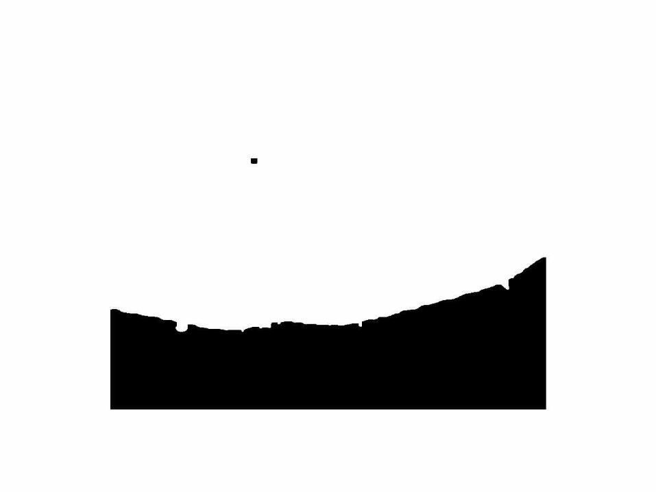
% Create a mask for the bottom texture
BW = im2bw(Eim, .6);


BWao = bwareaopen(BW,2000);
figure,imshow(BWao);
% removes the objects from the image whose total number of pixels is less than 2000.


nhood = true(9);
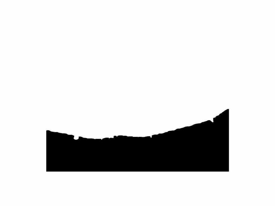
closeBWao = imclose(BWao,nhood);
% imclose is used to smooth the edges and to close any open holes in the object in BWao.
roughMask = imfill(closeBWao,'holes');
% imfill to fill holes in the object in closeBWao

I = gray_img;
I(roughMask) = 0;
I2=gray_img-I;
% the final segmented image of the epithelial layer

