selection of a multi-ligand capture agent for carbonic...
TRANSCRIPT
15
Chapter 2
Selection of a Multi-ligand Capture Agent for Carbonic Anhydrase II by Iterative In Situ
Click Chemistry
16 2.1 INTRODUCTION
Protein biomarkers comprise an important aspect of in vitro diagnostics. Most
protein detection methods rely upon antibody-based capture agents.1 A high-quality
antibody exhibits a high affinity and specificity for its cognate protein. However,
antibodies are expensive, and can be unstable toward dehydration, pH variation, thermal
shock, and many other chemical and biochemical processes.2,3 In addition, antibodies
are not available for many potential protein biomarkers. Thus, a major challenge is to
discover an efficient and general approach for producing protein capture agents that
display the positive attributes of antibodies, and exhibit a high level of chemical and
biochemical stability. This is becoming an increasingly important problem as single
protein-based diagnostics are being replaced by measurements of large panels of protein
biomarkers.4
Several alternative protein capture agents, including oligonucleotide aptamers
and phage display peptides, have been reported. Each of them have attributes as well as
significant limitations.5–11 A third alternative is to utilize one-bead one-compound
(OBOC) peptide or peptide mimetic libraries.12–16 An advantage of OBOC libraries is
that chemical stability, water solubility, and other desired properties may be achieved by
design. However, OBOC libraries are typically only 104–106 elements, and so
significant trade-offs are made between peptide length and library chemical diversity.
Phage display methods, by contrast, produce ~1012 element peptide libraries. As a result,
high-quality protein capture agents can be challenging to identify directly from standard
OBOC peptide libraries.
Herein, we combine the chemical flexibility of comprehensive, OBOC libraries
of oligopeptides with in situ click chemistry17–21 to yield a target-guided,22–24 potentially
17 general screening approach for building high-affinity protein capture agents. For this
selection scheme, the protein target replaces the role of a Cu(I) catalyst for promoting
the 1,3-dipolar “click” cycloaddition reaction between azide-functionalized and
acetylene-functionalized peptide affinity agents. First, an anchor (1°) ligand, containing
acetylene (or azido) functionality, is selected for specific binding to a protein target via
standard OBOC methods. Second, the same protein target is utilized to template the
covalent coupling between two peptide ligands, the pre-identified 1° ligand and a
secondary (2°) ligand, which is selected by the protein target and the 1° ligand from a
comprehensive OBOC library of 2° ligands displaying azido (or acetylene) functionality.
Synthetic scale-up yields a biligand composed of the 1° and 2° ligands, joined together
via the 1,2,3-triazole linker. This biligand can then be used as a new anchor ligand, and
the in situ click chemistry selection may be repeated to form a triligand, and so forth. As
the number of peptide ligands that comprise the multi-ligand capture agent increases, the
binding affinity and specificity rapidly increase.25,26 Thus, multivalent binding agents
can provide a potential shortcut to high affinity.27
By instituting iterative in situ click chemistry selections with OBOC, we exploit
both technologies to produce a triligand capture agent against human and bovine
carbonic anhydrase II (hCAII and bCAII, respectively). These two proteins are >80%
identical in sequence (PDB ID: 1CA2, 1V9E). Carbonic anhydrase II belongs to a
family of metalloenzymes that catalyze the reversible hydration of carbon dioxide. CA
II expression is induced in the endothelium of neovessels in melanoma, renal carcinoma,
and other cancers.28 Furthermore, CA II represents a major target antigen for stimulating
an autoantibody response in melanoma patients,29 and is potentially a therapeutic target

18 for glial tumors.30 It has served as a model protein to understanding protein-ligand
interactions, and is a demonstrated receptor for bivalent ligands.31–34
In this chapter, the discovery process for high-affinity protein capture agents is
discussed, using the triligand capture agent for b(h)CAII as the prototype. First, the
construction of OBOC libraries containing artificial amino acids is detailed. Through
iterative OBOC and in situ click chemistry selections, specific binders of b(h)CAII are
identified sequentially—1° ligands, then biligands, and finally a triligand capture agent
which displays ≥20 ng sensitivity for the protein target in dilute serum. The entire
screening approach is summarized in Figure 2.1.
2.2 MATERIALS AND EXPERIMENTAL METHODS
2.2.1 Materials
Fmoc-D-X-OH (Fmoc, fluoren-9-ylmethoxycarbonyl) (X = Ala, Arg(Pbf) (Pbf,
pentamethyldihydrobenzofuran-5-sulfonyl), Asn(Trt) (Trt, trityl), Asp(OtBu) (tBu, tert-
butyl), Glu(OtBu), Gln(Trt), Gly, His(Trt), Ile, Leu, Lys(Boc) (Boc, tert-
butyloxycarbonyl), Met, Phe, Pro, Ser(tBu), Thr(tBu), Trp(Boc), Tyr(tBu), and Val)
were purchased (Anaspec; San Jose, CA) and used as received. TentaGel S-NH2 resins
(90 μm, 0.31 mmol/g) (Rapp-Polymere; Tübingen, Germany) were utilized for OBOC
library construction. Amino acid coupling reactions were performed in 1-methyl-2-
pyrrolidinone (NMP, 99%) with HATU (2-(7-Aza-1H-benzotriazole-1-yl)-1,1,3,3-
tetramethylammonium hexafluorophosphate, ChemPep; Miami, FL) and N,N′-
diisopropylethylamine (DIEA). For removal of Nα-Fmoc protecting groups, a solution
of 20% piperidine in NMP was used. For final deprotection of the peptide libraries,
trifluoroacetic acid (TFA, 98% min. titration) and triethylsilane (TES) were used. All

19
Figure 2.1. A schematic representation of a method for preparing a multi-ligand capture
agent. (A) In the first step, a plurality of candidate oligopeptides in an OBOC library is
contacted with a labeled target to identify an anchor (1°) ligand. (B) In the second step,
a modified 1° ligand from the first step is contacted with the same OBOC library now
appended with an azide linker to identify a secondary (2°) ligand. A biligand, formed by
the 1° ligand of the first step and the 2° ligand, can be obtained. (C) In the third step, the
screen is repeated by employing the biligand formed from the second step as the new
primary ligand to allow identification of higher-order multi-ligands.
(A)
(B)
(C)
20 solvents and reagents were purchased from Sigma-Aldrich (St. Louis, MO) and used as
received, unless otherwise noted.
OBOC libraries were synthesized using a 180-degree variable-speed shaker,
fitted with small sample adapter (St. John Associates; Beltsville, MD). Fritted
polypropylene solid-phase synthesis tubes were used for repeated split-mix cycles. A
24-port SPE vacuum manifold system (Grace; Deerfield, IL) was used for exchanging
coupling solutions and washing the resins.
Bovine carbonic anhydrase II (bCAII, C2522), from bovine erythrocytes,
lyophilized powder, was obtained (Sigma-Aldrich; St. Louis, MO) and used as received.
To prepare the protein for screening, dye-labeling was accomplished with the Alexa
Fluor 647 Microscale Protein Labeling Kit (Invitrogen; Carlsbad, CA) following the
manufacturer’s protocol for a low degree of labeling (DOL). Protein (100 μg) was
incubated with 6 mol equiv Alexa Fluor 647 succinimidyl ester for 15 min at 25 °C.
Excess dye was removed by BioGel P-6 size exclusion resin (Bio-Rad; Hercules, CA).
The labeled protein (bCAII-Alexa Fluor 647) was characterized by UV-Vis and mass
spectrometry.
Human carbonic anhydrase II (hCAII, C6165), from human erythrocytes,
lyophilized powder, was obtained (Sigma-Aldrich; St. Louis, MO) and used in affinity
and specificity studies. Both bCAII and hCAII were tested by SDS gel electrophoresis,
and confirmed to display a single band corresponding to 29,000 Da.
2.2.2 Artificial Amino Acids
Fmoc-D-propargylglycine (Fmoc-D-Pra-OH) was acquired (Chem-Impex
International; Wood Dale, IL) and used as the acetylene handle for construction of
21 ligands. Azide-containing amino acids Fmoc-Az4-OH (and intermediates 1a-3a) and
Fmoc-Az8-OH (and intermediates 1b-3b) were synthesized using a modification of
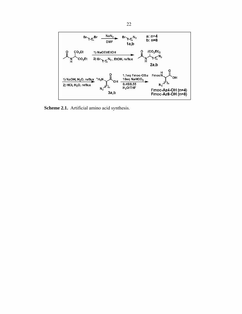
literature protocols (Scheme 2.1).35–37
Azidobutylbromide (1a). To a solution of 1,4-dibromobutane (123 mmol),
sodium azide (61.5 mmol) was added and stirred overnight in N,N′-dimethylformamide
(DMF) at 50 °C. The reaction was diluted with ethyl acetate, and the organic layer was
washed with water, then brine, and then dried over MgSO4. The crude residue was
purified by silica gel chromatography (100% hexanes) to give a product (80%) as a clear
oil. 1H NMR (300 MHz, CDCl3): δ 3.44 (2H, t, J = 6.3 Hz), 3.34 (2H, t, J = 6.6 Hz),
1.93-1.98 (2H, m), 1.74-1.79 (2H, m).
Azidooctylbromide (1b). Synthesis was carried out as described above, except
1,8-dibromooctane was used as the starting material. 1H NMR (300 MHz, CDCl3):
δ 3.41 (2H, t, J = 6.9 Hz), 3.26 (2H, t, J = 6.6 Hz), 1.86 (2H, p, J = 6.9 Hz), 1.60 (2H, p,
J = 8.7 Hz), 1.34-1.55 (4H, m).
Diethyl 2-acetamido-2-(4-azidobutyl)malonate (2a). To a solution of 0.598 g
(0.026 mol) sodium metal in 25 mL absolute EtOH, 5.65 g diethyl acetamidomalonate
(0.026 mol) was added, following previously published procedures.35 The mixture was
stirred for 30 min at room temperature. By dropwise addition, azidobutylbromide 1a
(4.82 g, 0.027 mol) was added with stirring. The reaction mixture was stirred for 2 h at
room temperature and refluxed for 6 h at 80 °C. After cooling overnight, the reaction
mixture was concentrated to dryness, and the residue was extracted with diethyl ether.
The combined ether extracts were washed with water, sat. NaHCO3, water, and brine,
and were dried over MgSO4 and then concentrated. Silica gel chromatography
(Hex:EtOAc = 1:1) gave a product (63%) as a clear, viscous oil. 1H NMR (300 MHz,
22
Scheme 2.1. Artificial amino acid synthesis.

23 CDCl3): δ 6.77 (1H, s), 4.24 (4H, q, J = 6.9 Hz), 3.26 (2H, t, J = 6.9 Hz), 2.31-2.37 (2H,
m), 2.04 (3H, s), 1.59 (2H, p, J = 7.5 Hz), 1.26 (6H, t, J = 6 Hz), 1.16-1.27 (2H, m).
ESI-MS m/e 315.
Diethyl 2-acetamido-2-(8-azidooctyl)malonate (2b). Similar synthetic protocol
as 2a was adopted, only with azidooctylbromide 1b serving as the starting material. 1H
NMR (300 MHz, CDCl3): δ 6.76 (1H, s), 4.24 (4H, q, J = 7.2 Hz), 3.24 (2H, t, J =
6.9 Hz), 2.27-2.33 (2H, m), 2.04 (3H, s), 1.56 (2H, p, J = 7.5 Hz), 1.25 (6H, t, J =
7.2 Hz), 1.06-1.16, 1.2-1.4 (10H, m). ESI-MS m/e 371.
2-Azidobutyl amino acid (3a). Following standard methods,36 the diester 2a
(2.8 mmol) in 25 mL of 10% NaOH solution was heated to reflux for 4 h. The solution
was then neutralized with concentrated HCl and evaporated. The residue was dissolved
in 25 mL of 1 M HCl and heated to reflux for 3 h. The solvent was reduced and
extraction with MeOH afforded amino acid 3a as the hydrochloride salt (85%). 1H
NMR (300 MHz, CD3OD): δ 3.98 (1H, t, J = 6.3 Hz), 3.35 (2H, t, J = 7.8 Hz), 1.45-1.7,
1.85-2.05 (6H, m). MALDI-MS m/e 173.
2-Azidooctyl amino acid (3b). Synthesis was carried out as described above,
using diester 2b as the starting material. 1H NMR (300 MHz, CD3OD): δ 3.94 (1H, t,
J = 6.3 Hz), 3.27 (2H, t, J = 6.9 Hz), 1.3-1.52, 1.52-1.62, 1.8-1.98 (14H, m). ESI-MS
m/e 229.
Fmoc-2-Azidobutyl amino acid (Fmoc-Az4-OH). The amino acid 3a
(26.3 mmol) was dissolved in 0.45:0.55 H2O:THF (150 mL), and NaHCO3 (22.1 g,
263 mmol) was added, following published methods.37 After the mixture was cooled to
0 °C, Fmoc-OSu (9.7 g, 28.9 mmol) was added dropwise over 5 min. The reaction
mixture was allowed to come to room temperature and stirred overnight. Evaporation of
24 THF was completed in vacuo and the aqueous residue was washed with diethyl ether
(2 × 200 mL). The aqueous layer was then collected and acidified with conc. HCl to
pH 2 before extraction with ethyl acetate (4 × 100 mL). The combined organic layers
were washed with brine, dried over MgSO4, filtered, and concentrated. The organic
residue was purified by column chromatography (2% MeOH in DCM) to yield a white
powder (48% yield). 1H NMR (300 MHz, CDCl3): δ 7.76 (2H, d, J = 7.5 Hz), 7.59 (2H,
d, J = 6.9 Hz), 7.40 (2H, t, J = 7.5 Hz), 7.31 (2H, t, J = 7.5 Hz), 5.34 (1H, d, J = 7.8 Hz),
4.49-4.59 (1H, m), 4.43 (2H, d, J = 6.6 Hz), 4.22 (1H, t, J = 6.6 Hz), 3.27 (2H, t, J =
6.6 Hz), 1.3-2.0 (6H, m). ESI-MS m/e 395.
Fmoc-2-Azidooctyl amino acid (Fmoc-Az8-OH). The amino acid 3b was
treated to Fmoc protection as described above. 1H NMR (300 MHz, CDCl3): δ 7.75 (2H,
d, J = 7.5 Hz), 7.57-7.61 (2H, m), 7.39 (2H, t, J = 7.5 Hz), 7.30 (2H, t, J = 7.2 Hz), 5.40
(1H, d, J = 8.1 Hz), 4.42-4.52 (1H, m), 4.40 (2H, d, J = 7.2 Hz), 4.21 (1H, t, J = 7.2 Hz),
3.23 (2H, t, J = 6.9 Hz), 1.18-1.98 (14H, m). ESI-MS m/e 450.
2.2.3 OBOC Oligopeptide Library Construction
Randomized OBOC libraries of penta- to heptapeptides were synthesized
manually via standard split-and-mix solid-phase peptide synthesis methods on 90 µm
polyethylene glycol-grafted polystyrene beads (TentaGel S-NH2, 0.31 mmol/g, 2.86 ×
106 beads/g).12–14 Non-natural D-stereoisomers (denoted by lowercase one-letter amino
acid code) were used at every possible position in the peptide sequence to infer intrinsic
biochemical stability. At least a 5-fold excess of beads was utilized in each library
synthesis to ensure adequate representation of each library element. A standard solid-
phase peptide synthesis method with Fmoc chemistry was used.38 All wash,

25 deprotection, and coupling steps were facilitated by 180-degree shaking of the resin.
The resin was pre-swelled in NMP in a plastic fritted reaction vessel, and was separated
into multiple aliquots. Each aliquot was reacted with 2-fold molar excess (relative to
resin) of a single Nα-Fmoc-amino acid. Amide coupling was initiated by addition of a 2-
fold molar excess of HATU and a 6-fold molar excess of DIEA.39 The coupling reaction
was run for 15 min. Another 2 equiv Nα-Fmoc-amino acid, 2 equiv HATU, and 6 equiv
DIEA were added, and allowed to react for 15 min (“double coupling”). In some cases,
“triple coupling” was performed with a third set of coupling reagents and Nα-Fmoc-
amino acids (Table 2.1, Libraries D, E, F, and G). Following coupling, the aliquots were
thoroughly washed (5 × NMP), mixed together into a single vessel, and deprotected with
20% piperidine in NMP (30 min). The resin was thoroughly washed (5 × NMP), dried
(5 × DCM), and re-divided into multiple equal-mass aliquots for the next cycle of
coupling. The procedures were repeated until the desired length of peptide was attained.
The amino acid side chain protecting groups were then removed by incubation in
trifluoroacetic acid (95%), water (5%), and triethylsilane (2-fold molar excess per
protected side chain) for 2 h at 25 °C. The library resin was then neutralized with DMF,
and washed thoroughly with DMF (5 ×), water (5 ×), methanol (MeOH, 5 ×), and
methylene chloride (DCM, 5 ×),40 and then dried under vacuum and stored in phosphate-
buffered saline [PBS (pH 7.4)] + 0.05% NaN3 at 25 °C.
2.2.4 Screening Procedures for Anchor Ligand
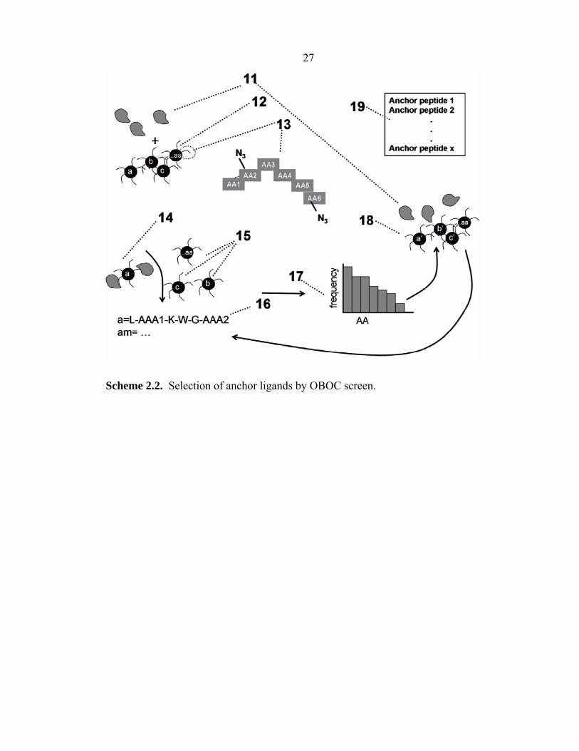
A method for identifying an anchor (1°) ligand is schematically illustrated in
Scheme 2.2. In particular, in the illustration of Scheme 2.2, a fluorescently labeled
protein of interest (11) is screened against an OBOC library of peptides (12). Each bead
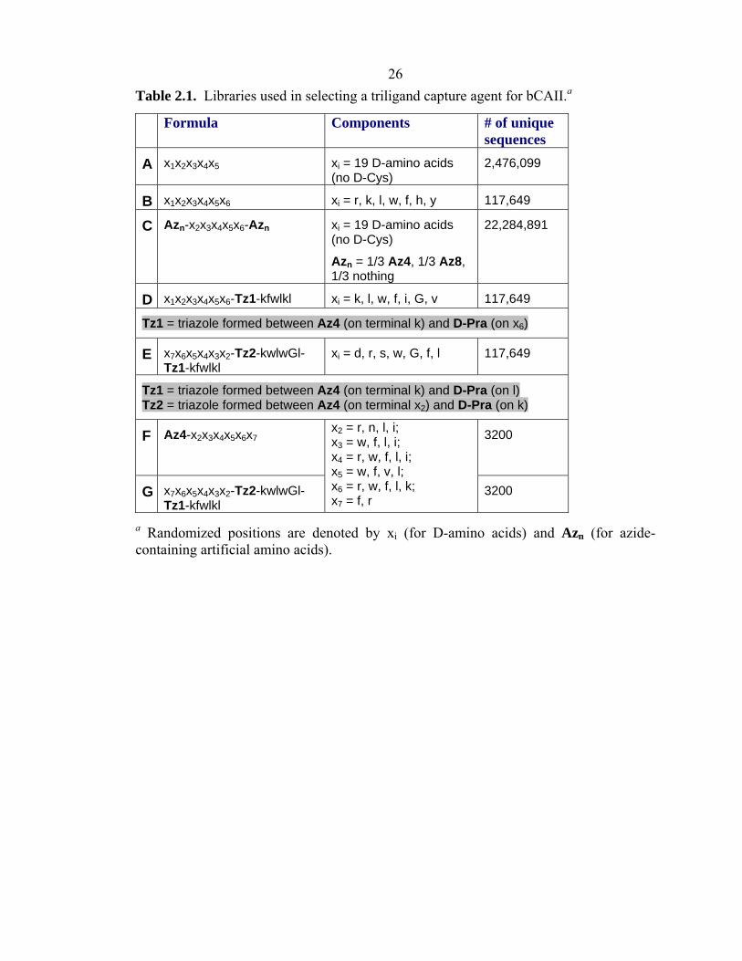
26 Table 2.1. Libraries used in selecting a triligand capture agent for bCAII.a
a Randomized positions are denoted by xi (for D-amino acids) and Azn (for azide-containing artificial amino acids).
Formula Components # of unique sequences
A x1x2x3x4x5 xi = 19 D-amino acids (no D-Cys)
2,476,099
B x1x2x3x4x5x6 xi = r, k, l, w, f, h, y 117,649
C Azn-x2x3x4x5x6-Azn xi = 19 D-amino acids (no D-Cys)
Azn = 1/3 Az4, 1/3 Az8, 1/3 nothing
22,284,891
D x1x2x3x4x5x6-Tz1-kfwlkl xi = k, l, w, f, i, G, v 117,649
Tz1 = triazole formed between Az4 (on terminal k) and D-Pra (on x6)
E x7x6x5x4x3x2-Tz2-kwlwGl-Tz1-kfwlkl
xi = d, r, s, w, G, f, l 117,649
Tz1 = triazole formed between Az4 (on terminal k) and D-Pra (on l) Tz2 = triazole formed between Az4 (on terminal x2) and D-Pra (on k)
F Az4-x2x3x4x5x6x7 3200
G x7x6x5x4x3x2-Tz2-kwlwGl-Tz1-kfwlkl
x2 = r, n, l, i; x3 = w, f, l, i; x4 = r, w, f, l, i; x5 = w, f, v, l; x6 = r, w, f, l, k; x7 = f, r
3200
27
Scheme 2.2. Selection of anchor ligands by OBOC screen.
28 contains a unique peptide (13) comprised of non-natural amino acids (D-stereoisomers)
or artificial amino acids (displaying azide or acetylene functionalities). The protein (11)
and the library (12) are incubated for a period of time at a particular protein
concentration (Table 2.2, Screen An1), and the “hit” beads (14) are identified by their
fluorescence using a GenePix 4200 array scanner (λex = 635 nm). Typically 0.1% or less
of the beads are identified as hit beads, and are separated manually from the non-hit
beads by micropipette (15). The protein is removed from the beads by incubation with
7.5 M guanidine hydrochloride (GuHCl, pH 2.0) for 1 h, and the peptides on single hit
beads are sequenced using Edman degradation41 (Procise cLC Sequencing System,
Applied BioSystems, Foster City, CA; see Appendix C) or MALDI-TOF/TOF mass
spectrometry.42
Once the hit peptide sequences (16) are identified, a histogram (17) that
correlates the amino acid frequency vs. amino acid identity is prepared. A second, more
focused library (18) that uses those most commonly identified amino acids can then be
prepared and re-screened against the protein (11) (Table 2.2, Screens An2a and An2b).
This focused library can contain slightly longer peptides, and the screening process can
involve a lower concentration of the protein (11). This process can then be repeated
until the desired affinity of peptide anchor ligand (19) is achieved. The affinity of the
peptide anchor ligand will depend upon the number of amino acids in the peptide, and
the three-dimensional structure of the peptide, among other factors. Affinities in the
order of 10–4–10–6 M are typically achievable.
2.2.5 In Situ Click Screening Procedures for Biligand
Identification of the secondary (2°) ligand and formation of a biligand then can
29
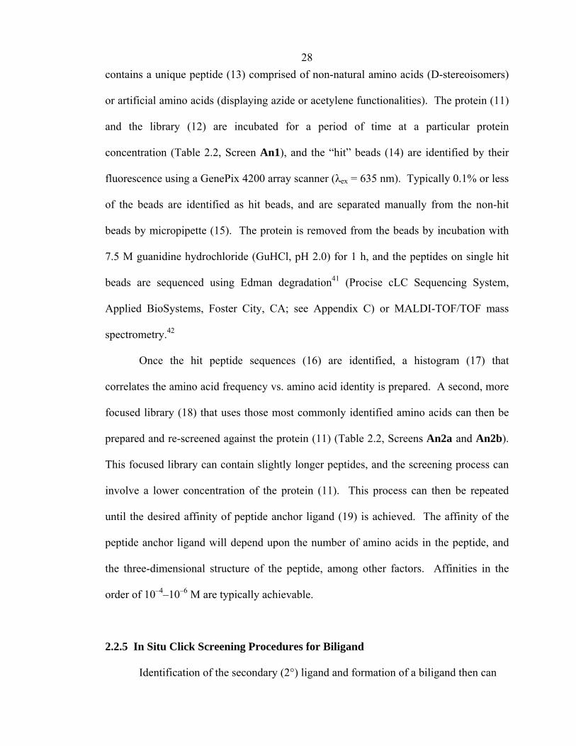
a All screens were conducted at pH = 7.4 and T = 25 °C, unless otherwise noted.
Table 2.2. Screening summary.a Screen Library [bCAII-
AF647] Time(h) % hit
beads Buffer Other
components
An1 A 100 nM 1 h 0.02% PBS N/A
An2a B 50 nM 1 h 0.09% PBS N/A
An2b B 8 nM 24 h 2 hits PBS N/A
Bi1 C 50 nM 2 h; 37o C (no beads) + 48 h; 37o C
0.007% PBS + 1% DMSO (v/v)
100 µM of lklwfk-(D-Pra)
Bi2a D 50 nM 17 h 0.07% PBSTBNaN3 N/A
Bi2b D 10 nM 17 h 0.008% PBSTBNaN3 N/A
Tri1 C 10 nM 2 h (no beads) +15 h
0.007% PBSTBNaN3 + 1% DMSO (v/v)
100 µM of (D-Pra)-kwlwGl-Tz1-kfwlkl
Tri2 E 10 nM 17 h 0.008% PBSTBNaN3 N/A
TriX A 10 nM 17 h 0.007% PBSTBNaN3 + 1% DMSO (v/v)
100 µM of (D-Pra)-kwlwGl-Tz1-kfwlkl
Tri3 F 0.5 nM 2 h (no beads) +18 h
0.005%-0.01%
PBSTBNaN3 + 1% DMSO (v/v)
100 µM of (D-Pra)-kwlwGl-Tz1-kfwlkl
Tri4 G 0.25 nM 18 h 0.005%-0.01%
PBSTBNaN3 N/A
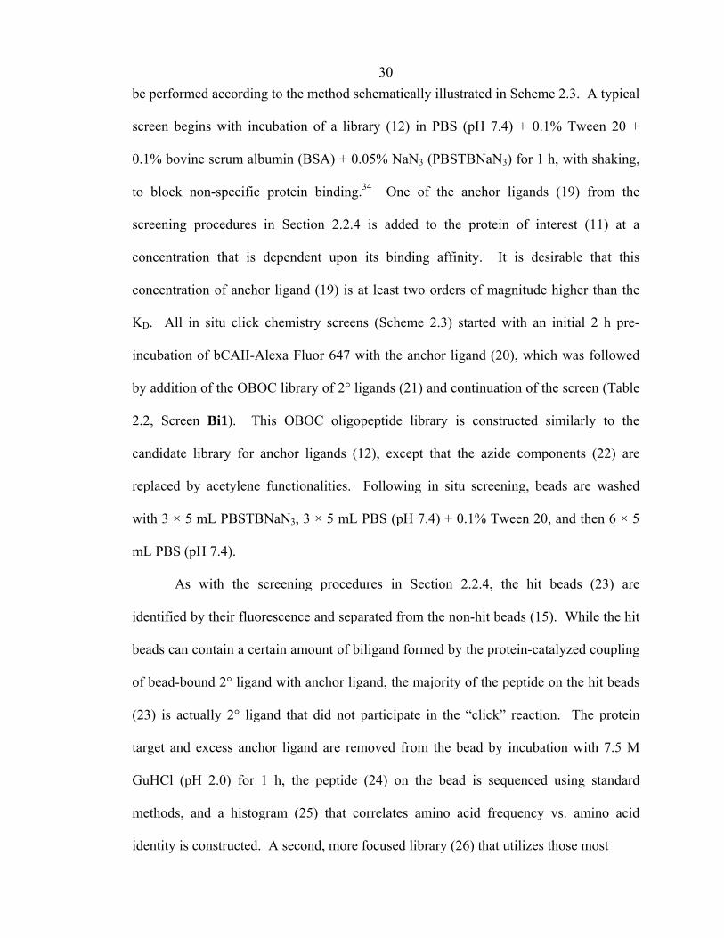
30 be performed according to the method schematically illustrated in Scheme 2.3. A typical
screen begins with incubation of a library (12) in PBS (pH 7.4) + 0.1% Tween 20 +
0.1% bovine serum albumin (BSA) + 0.05% NaN3 (PBSTBNaN3) for 1 h, with shaking,
to block non-specific protein binding.34 One of the anchor ligands (19) from the
screening procedures in Section 2.2.4 is added to the protein of interest (11) at a
concentration that is dependent upon its binding affinity. It is desirable that this
concentration of anchor ligand (19) is at least two orders of magnitude higher than the
KD. All in situ click chemistry screens (Scheme 2.3) started with an initial 2 h pre-
incubation of bCAII-Alexa Fluor 647 with the anchor ligand (20), which was followed
by addition of the OBOC library of 2° ligands (21) and continuation of the screen (Table
2.2, Screen Bi1). This OBOC oligopeptide library is constructed similarly to the
candidate library for anchor ligands (12), except that the azide components (22) are
replaced by acetylene functionalities. Following in situ screening, beads are washed
with 3 × 5 mL PBSTBNaN3, 3 × 5 mL PBS (pH 7.4) + 0.1% Tween 20, and then 6 × 5
mL PBS (pH 7.4).
As with the screening procedures in Section 2.2.4, the hit beads (23) are
identified by their fluorescence and separated from the non-hit beads (15). While the hit
beads can contain a certain amount of biligand formed by the protein-catalyzed coupling
of bead-bound 2° ligand with anchor ligand, the majority of the peptide on the hit beads
(23) is actually 2° ligand that did not participate in the “click” reaction. The protein
target and excess anchor ligand are removed from the bead by incubation with 7.5 M
GuHCl (pH 2.0) for 1 h, the peptide (24) on the bead is sequenced using standard
methods, and a histogram (25) that correlates amino acid frequency vs. amino acid
identity is constructed. A second, more focused library (26) that utilizes those most
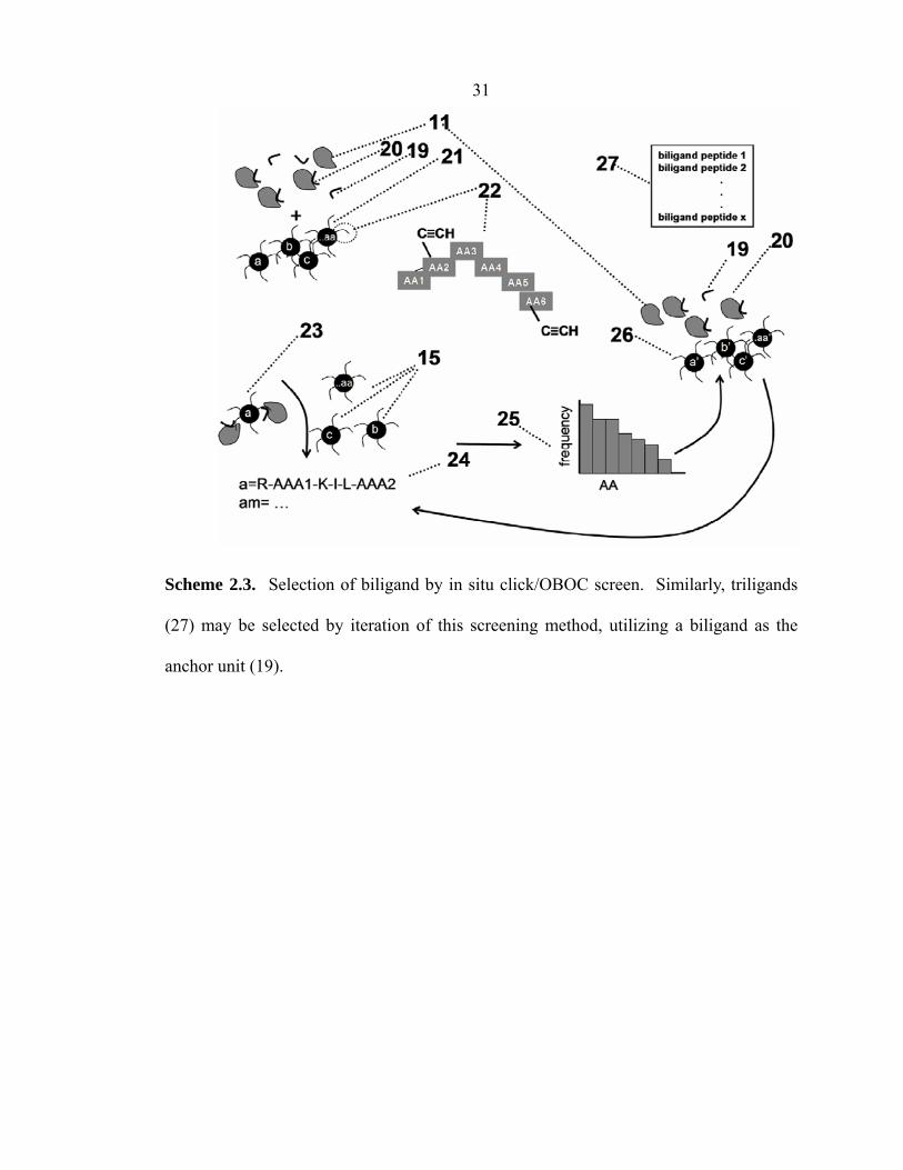
31
Scheme 2.3. Selection of biligand by in situ click/OBOC screen. Similarly, triligands
(27) may be selected by iteration of this screening method, utilizing a biligand as the
anchor unit (19).

32 commonly identified amino acids may then be prepared and re-screened against the
protein (11). Once again, the hit beads are identified via peptide sequencing (24). This
second library of 2° ligands can contain slightly longer peptides, and the screening
process can involve a lower concentration of the protein (11).
2.2.6 In Situ Click Screening Procedures for Higher-Order Multi-ligands
In situ click screening procedures operate similarly to Scheme 2.3 for
identification of higher-order multi-ligands such as the triligand in Figure 2.1. The in
situ click/OBOC screen for this triligand (Table 2.2, Screens Tri1 and Tri3) contained
an initial 2 h pre-incubation of bCAII-Alexa Fluor 647 with biligand anchor, which was
followed by addition of the OBOC library of 3° ligands and continuation of the screen.
As a negative control, screen TriX was performed with an azide-free OBOC library of
3° ligands.
2.2.7 Validation of In Situ Click/OBOC Multi-ligand Screening Procedures
Binary component screen for in situ biligand. Stock solutions of 2° ligand
(azide, Az4-kiwiG, 13.1 mM) and anchor ligand (acetylene, lklwfk-(D-Pra), 2.1 mM)
were prepared in DMSO. Stock solutions of bCAII and bovine serum albumin (BSA)
were prepared in PBS (pH 7.4). Each reaction contained 394 μM azide, 65 μM alkyne,
and 36 μM protein in 100 μL PBS (pH 7.4) + 6% DMSO (v/v). Reactions proceeded for
48 h at 37 °C, followed by 5 days at 25 °C. Reactions were quenched with 100 μL of
7.5 M GuHCl (pH 2.0), and proteins were subsequently removed by centrifugal filtration
(Microcon YM-3, Millipore, Billerica, MA).

33 The formation of in situ biligands was identified by MALDI-MS. Control
experiments were conducted (1) in the absence of bCAII, and (2) replacing bCAII with
BSA, to verify that the click reaction between the azide and alkyne is specific to the
bCAII protein target. A third control, performed in the absence of protein, represents the
slow thermally driven reaction between solutions of azide and alkyne.
On-bead biligand screen. Synthesis of Library D was achieved on bead via the
Cu(I)-catalyzed azide-alkyne cycloaddition (CuAAC),43–45 as described in Section 2.2.9.
Screens Bi2a and Bi2b (Table 2.2) were conducted using Library D following the
general OBOC screening protocol described in Section 2.2.4. After initial blocking with
PBSTBNaN3 for 1 h, 10 nM to 50 nM bCAII-Alexa647 in PBSTBNaN3 was incubated
with the library for 17 h at 25 °C, with shaking. The screened beads were washed with 3
× 5 mL PBSTBNaN3, then 3 × 5 mL PBS (pH 7.4) + 0.1% Tween 20, and finally 6 × 5
mL PBS (pH 7.4). The beads were imaged for fluorescence, and the hits were selected
by micropipette. After washing the hits to remove bound protein [7.5 M GuHCl (pH
2.0)], their sequences were determined by Edman degradation.
On-bead triligand screen. Synthesis of Libraries E and G was achieved on
bead via the CuAAC, as described in Section 2.2.9. Screens Tri2 and Tri4 (Table 2.2)
were conducted following the general OBOC screening protocol described in Section
2.2.4, using <10 nM bCAII-Alexa647 and fluorescent detection of hits.
2.2.8 Bulk Peptide Synthesis
Bulk synthesis of hit peptide sequences was performed on either Fmoc-Rink
amide MBHA (50 μm, 0.67 mmol/g, AnaSpec) or Biotin-PEG-NovaTag resin (0.48
mmol/g; Novabiochem), on a typical resin scale of 0.2 g per sequence. Crude peptides

34 were precipitated with ether, and then purified to >95% by HPLC (Beckman Coulter
System Gold 126 Solvent Module and 168 Detector, Fullerton, CA) on a C18 reversed-
phase semi-preparative column (Phenomenex Luna 10 µm, 250 × 10 mm). The pure
peptides were used for affinity measurements, in situ click/OBOC screens, and binding
assays. Hit peptide sequences were also re-synthesized on TentaGel S-NH2 on a similar
resin scale, and used for on-bead binding assays.
Installation of polyethylene glycol linkers (EG)n was achieved by Fmoc-NH-
(PEG)5-COOH (22 atoms) (Novabiochem) via SPPS with standard HATU/DIEA
coupling. N-terminal biotin labeling of certain sequences was achieved via SPPS with
standard HATU/DIEA coupling and overnight reaction.
It should be noted that the protein-templated in situ click reaction may yield
product regioisomers that are either anti (1,4), syn (1,5), or a mixture of the two
geometries. Although we have not yet determined which regioisomers of the in situ
click products were formed, the authentic multi-ligands synthesized by CuAAC to test
affinity and specificity were definitely the 1,4-triazole (see Chapter 3).
All anchor ligands, biligands, and triligands were prepared in bulk by solid-phase
synthesis, purified by HPLC, and analyzed by mass spectrometry prior to further study.
Their characterization is as follows:
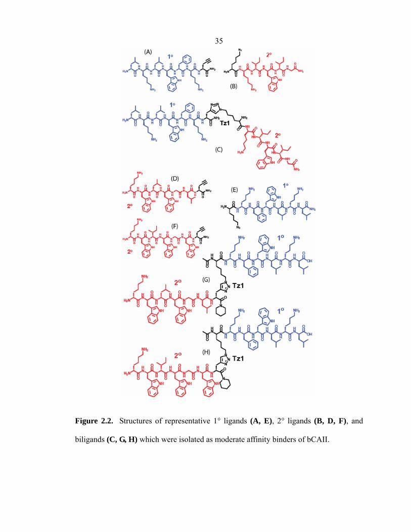
lklwfk-(D-Pra) (Figure 2.2A). MALDI-MS of the purified 1° ligand gave peaks
at m/z 928.7 for [M + H]+ and 950.7 for [M + Na]+.
Az4-kiwiG (Figure 2.2B). ESI-MS of the purified 2° ligand gave peaks at m/z
385.2 for [M + 2H]2+ and 769.5 for [M + H]+.
lklwfk-Tz1-kiwiG (Figure 2.2C). MALDI-MS of the purified biligand gave a
peak at m/z 1808.4 for [M + H]+.
35
Figure 2.2. Structures of representative 1° ligands (A, E), 2° ligands (B, D, F), and
biligands (C, G, H) which were isolated as moderate affinity binders of bCAII.
36 Az4-kfwlkl (Figure 2.2E). ESI-MS of the purified 1° ligand gave peaks at m/z
329.9 for [M + 3H]3+, 494.3 for [M + 2H]2+, and 987.6 for [M + H]+.
kwlwGl-(D-Pra) (Figure 2.2D). MALDI-MS of the purified 2° ligand gave
peaks at m/z 897.0 for [M + H]+, 919.0 for [M + Na]+, and 935.0 for [M + K]+.
kwiwGw-(D-Pra) (Figure 2.2F). MALDI-MS of the purified 2° ligand gave
peaks at m/z 970.1 for [M + H]+ and 992.1 for [M + Na]+.
kwlwGl-Tz1-kfwlkl (Figure 2.2G). MALDI-MS of the purified biligand gave a
peak at m/z 1993.6 for [M + H]+.
kwiwGw-Tz1-kfwlkl (Figure 2.2H). MALDI-MS of the purified biligand gave
peaks at m/z 2066.9 for [M + H]+ and 2088.7 for [M + Na]+.
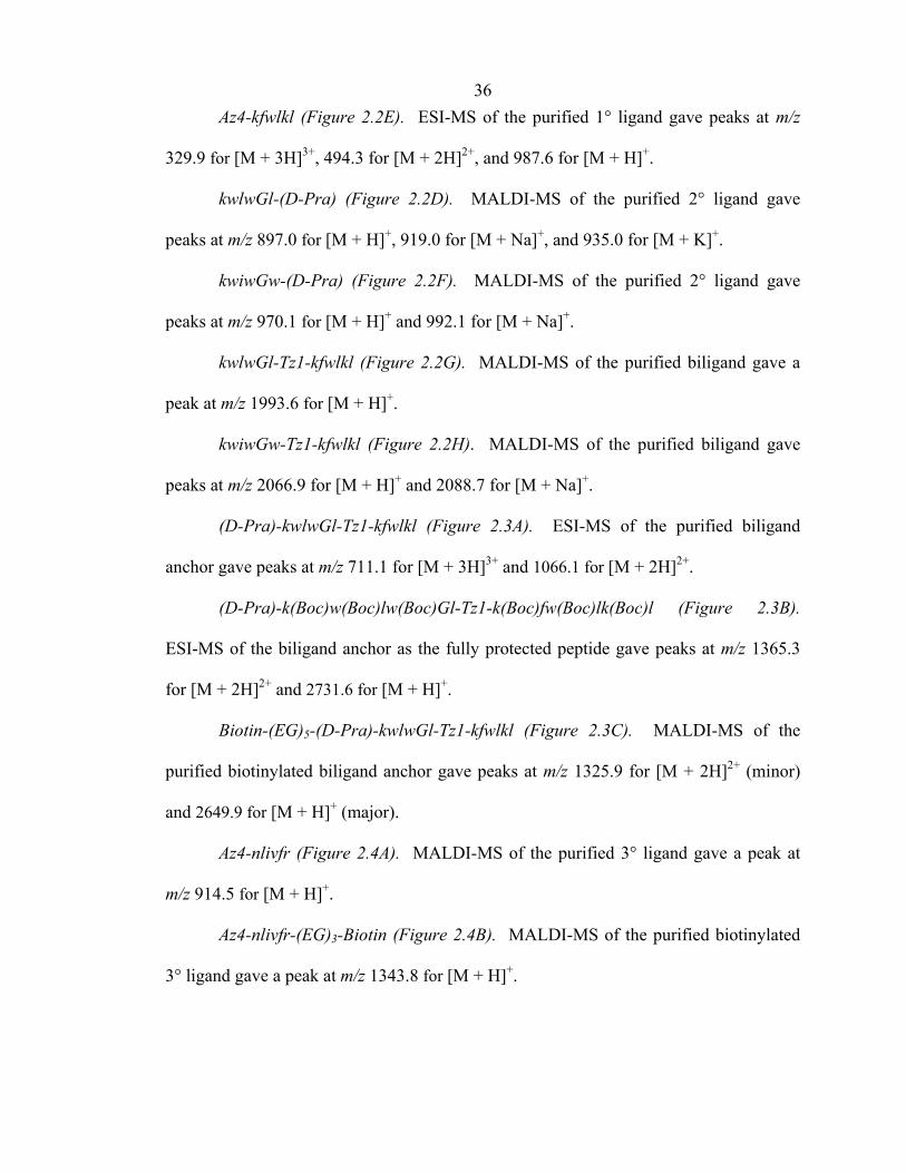
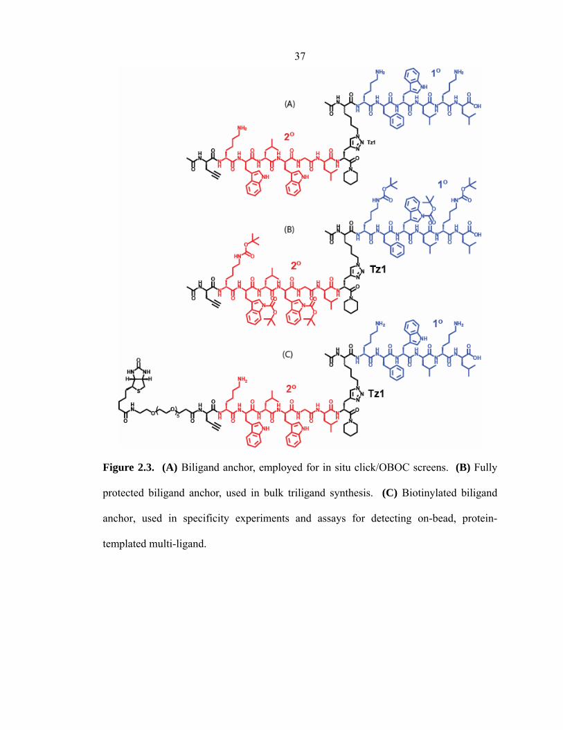
(D-Pra)-kwlwGl-Tz1-kfwlkl (Figure 2.3A). ESI-MS of the purified biligand
anchor gave peaks at m/z 711.1 for [M + 3H]3+ and 1066.1 for [M + 2H]2+.
(D-Pra)-k(Boc)w(Boc)lw(Boc)Gl-Tz1-k(Boc)fw(Boc)lk(Boc)l (Figure 2.3B).
ESI-MS of the biligand anchor as the fully protected peptide gave peaks at m/z 1365.3
for [M + 2H]2+ and 2731.6 for [M + H]+.
Biotin-(EG)5-(D-Pra)-kwlwGl-Tz1-kfwlkl (Figure 2.3C). MALDI-MS of the
purified biotinylated biligand anchor gave peaks at m/z 1325.9 for [M + 2H]2+ (minor)
and 2649.9 for [M + H]+ (major).
Az4-nlivfr (Figure 2.4A). MALDI-MS of the purified 3° ligand gave a peak at
m/z 914.5 for [M + H]+.
Az4-nlivfr-(EG)3-Biotin (Figure 2.4B). MALDI-MS of the purified biotinylated
3° ligand gave a peak at m/z 1343.8 for [M + H]+.
37
Figure 2.3. (A) Biligand anchor, employed for in situ click/OBOC screens. (B) Fully
protected biligand anchor, used in bulk triligand synthesis. (C) Biotinylated biligand
anchor, used in specificity experiments and assays for detecting on-bead, protein-
templated multi-ligand.

Figure 2.4. (A, B) Tertiary (3°) ligands. (C, D) Triligand capture agent, where the 1° ligand is colored in blue, the 2° ligand in red,
and the 3° ligand in light green. The connections between the ligands are formed by 1,2,3-triazoles (Tz1 and Tz2).
38

39 rfviln-Tz2-kwlwGl-Tz1-kfwlkl (Figure 2.4C). MALDI-MS of the purified
triligand gave peaks at m/z 1522.9 for [M + 2H]2+ (minor) and 3045.7 for [M + H]+
(major).
rfviln-Tz2-kwlwGl-Tz1-kfwlkl-(EG)3-Biotin (Figure 2.4D). MALDI-MS of the
purified biotinylated triligand gave peaks at m/z 1737.5 for [M + 2H]2+ (minor) and
3472.0 for [M + H]+ (major).
2.2.9 On-Bead Biligand and Triligand Synthesis
For preparing Libraries D, E, and G (Table 2.1), as well as for bulk synthesis of
biligand and triligand candidates, the Cu(I)-catalyzed azide-alkyne cycloaddition
(CuAAC)43–45 was carried out on bead, with 4 general steps: (1) anchor ligand synthesis,
(2) acetylation, (3) click reaction, and (4) addition of 2° ligand sequence. Scheme 2.4
illustrates the acetylation and click reactions for a 6-mer peptide (Z = any amino acid).
The fully protected TentaGel S-NH2 bead-bound anchor ligand (0.420 g, 0.13 mmol)
was capped by a solution of acetic anhydride (1 mmol) in 2,6-lutidine and DMF.46 The
acetylated peptide was reacted with Fmoc-D-Pra-OH (0.218 g,
0.65 mmol) in the presence of CuI (0.124 g, 0.65 mmol), L-ascorbic acid (0.114 g,
0.65 mmol), and DMF/piperidine (8/2) at 25 °C for 6 h.47 The resin was washed with
5 × 5 mL Et2NCSSNa•3H2O (sodium diethyldithiocarbamate trihydrate, 1% w/v),
containing 1% DIEA (v/v) in DMF to remove the coordinated copper from click
reaction.48
The biligand anchor (D-Pra)-kwlwGl-Tz1-kfwlkl was synthesized on 2-
chlorotrityl chloride (1.6 mmol/g) resin (Anaspec, San Jose, CA) using Scheme 2.4. The
biligand anchor was released either as the fully deprotected peptide by cleavage with

40
Scheme 2.4. Acetylation and click reactions for a 6-mer peptide (Z = any amino acid)
by solid-phase synthesis. Peptide synthesis may continue via the Fmoc-protected
primary amine of Zi to generate a linear multi-ligand capture agent.
41 95:5 TFA:water (+ 2 mol equiv TES per side chain protecting group), or as the fully
protected peptide by cleavage with 99:1 DCM:TFA.49 To facilitate the on-bead click
reaction, it is noted that the 1° ligand was synthesized here as Az4-kfwlkl (displaying N-
terminal Azn modification), and to this sequence was coupled D-Pra and the 2° ligand to
produce the linear biligand.
Triligands were synthesized by click reaction between the fully protected
biligand anchor (D-Pra)-kwlwGl-Tz1-kfwlkl (0.274 g, 0.1 mmol, >95% HPLC) and
bead-bound 3° ligand Az4-nlivfr (0.1 g, 0.03 mmol) using CuI (0.021 g, 0.1 mmol) and
L-ascorbic acid (0.020 g, 0.1 mmol) in DMF/piperidine (8/2).
2.3 RESULTS AND DISCUSSION
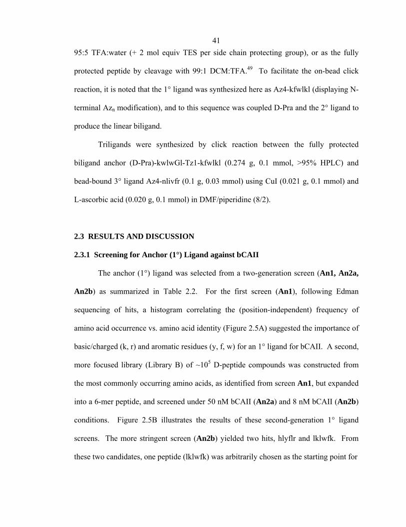
2.3.1 Screening for Anchor (1°) Ligand against bCAII
The anchor (1°) ligand was selected from a two-generation screen (An1, An2a,
An2b) as summarized in Table 2.2. For the first screen (An1), following Edman
sequencing of hits, a histogram correlating the (position-independent) frequency of
amino acid occurrence vs. amino acid identity (Figure 2.5A) suggested the importance of
basic/charged (k, r) and aromatic residues (y, f, w) for an 1° ligand for bCAII. A second,
more focused library (Library B) of ~105 D-peptide compounds was constructed from
the most commonly occurring amino acids, as identified from screen An1, but expanded
into a 6-mer peptide, and screened under 50 nM bCAII (An2a) and 8 nM bCAII (An2b)
conditions. Figure 2.5B illustrates the results of these second-generation 1° ligand
screens. The more stringent screen (An2b) yielded two hits, hlyflr and lklwfk. From
these two candidates, one peptide (lklwfk) was arbitrarily chosen as the starting point for
42
R K W Y F H L S G T M I V A D P N E Q0
20
40
60
80
100
D-amino acid
~500 (0.01%-0.02%)100 nM# Hits, Library AbCAII-Alexa647
~500 (0.01%-0.02%)100 nM# Hits, Library AbCAII-Alexa647
Freq
uenc
y (re
gard
less
of p
ositi
on)
2 (0.005%) → lklwfk, hlyflr8 nM40 (0.09%)50 nM
# Hits, Library BbCAII-Alexa647
2 (0.005%) → lklwfk, hlyflr8 nM40 (0.09%)50 nM
# Hits, Library BbCAII-Alexa647
Figure 2.5. Results of selecting a primary or anchor ligand of bCAII. (A) Diagram
plotting frequency vs. D-amino acid for 51 hit sequences isolated from screening Library
A (first-generation anchor ligand screen). (B) Hit rates for Library A and B (second-
generation anchor ligand) screens, leading to the selection of two anchor ligands (lklwfk
and hlyflr).
(A)
(B)
43 a 1° ligand for use in multi-ligand screens. A complete list of 1° ligand hit sequences
from OBOC selections can be found in Appendix B.
The peptide lklwfk was then functionalized with either an azide (-N3) or
acetylene (-C≡C-H) terminus, fluoresceinated, and produced in bulk quantities for
affinity measurements by fluorescence polarization. Chapter 3 will describe that one
such 1° ligand lklwfk-(D-Pra) displays an equilibrium dissociation constant of KD ≈
500 µM for its interaction with bCAII. This value is an estimate, since weak affinities
are hard to quantify. Surface plasmon resonance (SPR) was also employed to measure
the affinity of bCAII for Az4-kfwlkl and lklwfk-(D-Pra) as 1° ligands, and a similarly
low affinity was recorded (at least >10 µM, see Chapter 3).
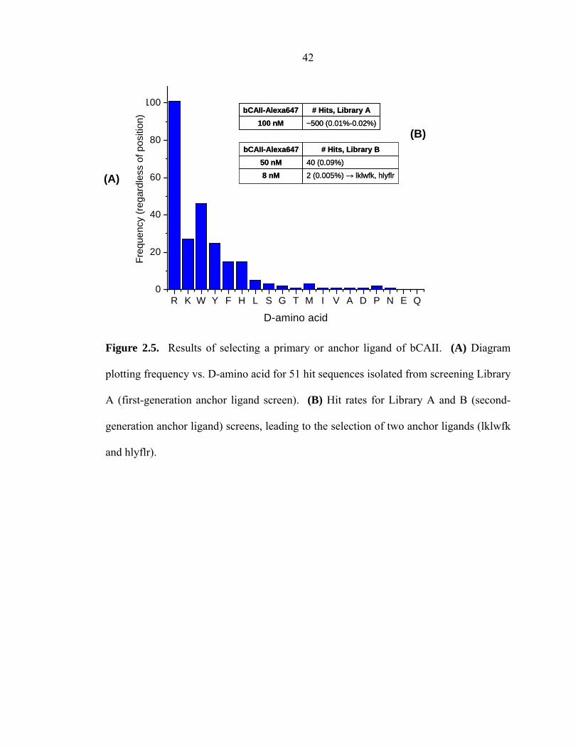
2.3.2 Identification of Secondary (2°) Ligands: Biligand Screens
A biligand is constructed of a 2° ligand that is covalently attached, via a 1,2,3-
triazole linkage, to the 1° ligand. As illustrated by Figure 2.6, secondary (2°) ligands
were identified by two complementary approaches: (1) in situ click/OBOC biligand
screens; (2) on-bead biligand screens. In the first approach (Figure 2.6A), the protein
acts as a catalyst for the in situ click assembly of the biligand on bead. During this
screen, the 1° ligand and protein coexist in solution, while the cognate library of 2°
ligands is on bead. In the second approach (Figure 2.6B), the 1° ligand is covalently
coupled to the on-bead library of 2° ligands via the copper(I)-catalyzed azide-alkyne
cycloaddition (CuAAC). Such a library of pre-assembled biligands is screened against
the protein target to discover 2° ligand candidates. The protein target is not a catalyst in
this approach; this screen was used as a validation tool for comparison against the in situ
click/OBOC screens.
44
Figure 2.6. A schematic illustrating the two types of biligand screen. (A) In situ screen
for a secondary (2°) ligand. (B) The on-bead screen for a secondary (2°) ligand was
utilized as confirmation that the in situ screen was performing its designed function.

45 In situ click/OBOC biligand screen. Based on the protein-catalyzed in situ
click reactions reported by the Sharpless group17–21 only those 2° ligands that bind with
bCAII and are in close proximity with the 1° ligand, and are in the correct orientation,
will react to form the 1,2,3-triazole product. Figure 2.7A illustrates the result of the
first-generation in situ biligand screen Bi1 against bCAII, which utilized 100 µM lklwfk-
(D-Pra) as the 1° ligand and a comprehensive azide-modified Library C. From
histogram and raw analysis of hits, a 2° ligand Az4-kiwiG emerged as the best
candidate, since its inherent motif was repeated several times. Figure 2.7B shows an
abbreviated list of the hit sequences isolated from screening Library C against 50 nM
bCAII-Alexa647 (Bi1). A complete list of biligand hit sequences from the in situ
click/OBOC screens can be found in Appendix B.
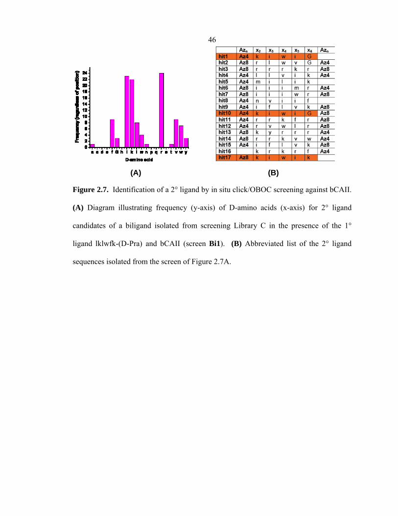
The very high sequence homology observed here was not witnessed for the 1°
ligand screens, but is characteristic of all of the in situ biligand and triligand (see Section
2.3.3) screens discussed in this thesis. Note also that all of the peptides in Figure 2.7B
contain at least one azide group, although, statistically, over one-third of the OBOC
library does not contain azide groups at positions 1 or 7. The high sequence homology,
coupled with the persistence of azide groups in the selected 2° ligands, provides strong
circumstantial evidence that the in situ click/OBOC screen worked to produce a biligand.
On-bead biligand screen. On-bead biligand screens (Bi2a and Bi2b) were
carried out utilizing a focused CuAAC biligand library (Library D) that was prepared
based on the sequencing results from screen Bi1. All 2° ligand sequences obtained by
screens Bi2a and Bi2b (Table 2.2) also display striking sequence homology. Several
sequences were repeated more than once, including kwlwGl and kwiwGw. A residue-
46
(A) (B)
Figure 2.7. Identification of a 2° ligand by in situ click/OBOC screening against bCAII.
(A) Diagram illustrating frequency (y-axis) of D-amino acids (x-axis) for 2° ligand
candidates of a biligand isolated from screening Library C in the presence of the 1°
ligand lklwfk-(D-Pra) and bCAII (screen Bi1). (B) Abbreviated list of the 2° ligand
sequences isolated from the screen of Figure 2.7A.
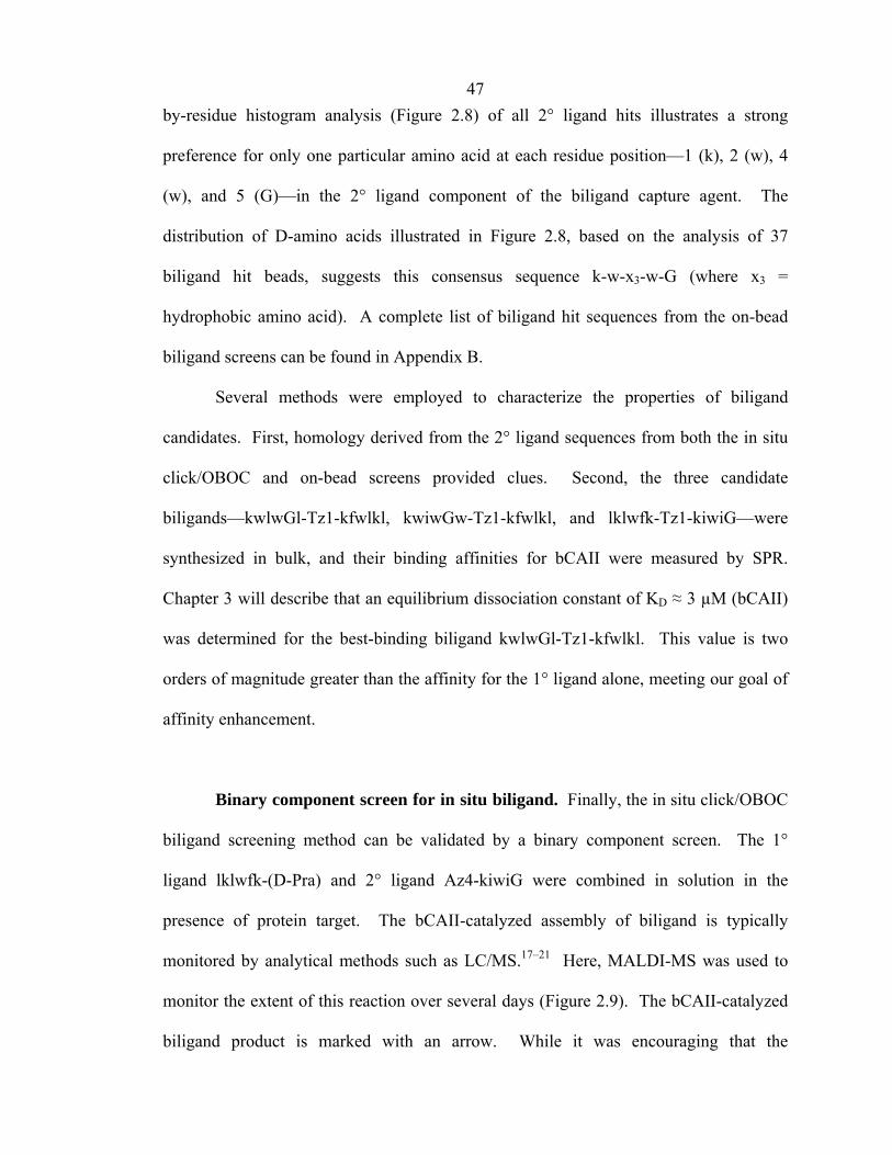
47 by-residue histogram analysis (Figure 2.8) of all 2° ligand hits illustrates a strong
preference for only one particular amino acid at each residue position—1 (k), 2 (w), 4
(w), and 5 (G)—in the 2° ligand component of the biligand capture agent. The
distribution of D-amino acids illustrated in Figure 2.8, based on the analysis of 37
biligand hit beads, suggests this consensus sequence k-w-x3-w-G (where x3 =
hydrophobic amino acid). A complete list of biligand hit sequences from the on-bead
biligand screens can be found in Appendix B.
Several methods were employed to characterize the properties of biligand
candidates. First, homology derived from the 2° ligand sequences from both the in situ
click/OBOC and on-bead screens provided clues. Second, the three candidate
biligands—kwlwGl-Tz1-kfwlkl, kwiwGw-Tz1-kfwlkl, and lklwfk-Tz1-kiwiG—were
synthesized in bulk, and their binding affinities for bCAII were measured by SPR.
Chapter 3 will describe that an equilibrium dissociation constant of KD ≈ 3 µM (bCAII)
was determined for the best-binding biligand kwlwGl-Tz1-kfwlkl. This value is two
orders of magnitude greater than the affinity for the 1° ligand alone, meeting our goal of
affinity enhancement.
Binary component screen for in situ biligand. Finally, the in situ click/OBOC
biligand screening method can be validated by a binary component screen. The 1°
ligand lklwfk-(D-Pra) and 2° ligand Az4-kiwiG were combined in solution in the
presence of protein target. The bCAII-catalyzed assembly of biligand is typically
monitored by analytical methods such as LC/MS.17–21 Here, MALDI-MS was used to
monitor the extent of this reaction over several days (Figure 2.9). The bCAII-catalyzed
biligand product is marked with an arrow. While it was encouraging that the
48
Figure 2.8. Distribution of D-amino acids found in positions 1 to 6 based on the
analysis of 37 biligand hit beads from screens Bi2a and Bi2b.

49
Figure 2.9. Binary component in situ click chemistry screen of 1° ligand lklwfk-(D-Pra)
and 2° ligand Az4-kiwiG, illustrating bCAII-catalyzed formation of biligand (marked by
arrow). (A) Bovine carbonic anhydrase II (bCAII). (B) Bovine serum albumin (BSA)
control. (C) Buffer-only (no protein) control.
(B)
(A)
(C)
50 background reactions (BSA, no protein) were less, the MALDI-MS result did not
provide quantitative measurement of the signal-to-noise ratio and overall yield for the
bCAII-catalyzed reaction. Methods to quantitatively assess these were developed at the
triligand level and are discussed in detail in Chapter 4.
2.3.3 Identification of Tertiary (3°) Ligands: Triligand Screens
Once a biligand is identified, that biligand can serve as the new anchor ligand, as
illustrated in Figure 2.1, and the same OBOC library may be employed to identify a
triligand. This process may be repeated with the same OBOC library until a multi-ligand
with the desired affinity and specificity is reached. With the biligand (D-Pra)-kwlwGl-
Tz1-kfwlkl serving as the anchor ligand, the Figure 2.1 in situ click/OBOC screen was
repeated with Library C (Table 2.1) to identify a triligand rfviln-Tz2-kwlwGl-Tz1-
kfwlkl (Figure 2.4C). It is crucial to note that the comprehensive Library C was applied
again here, demonstrating the versatility of this type of general library.
For the case of the triligand screens, a histogram charting the position-dependent
frequency of amino acids observed in the hit beads was generated. The consensus
tertiary (3°) ligand was Az4-nlivfr (Figure 2.4A). Figure 2.10 shows position-dependent
histograms for the first-generation in situ click/OBOC screens, for peptides (a) with and
(c) without an azide-containing amino acid, to generate a triligand. For the in situ screen
(Tri1, Figure 2.10A), one-third of the beads had no azide at the x1 or x7 positions, but
interestingly, all hit beads contained an azide. On the other hand, the first- and second-
generation on-bead CuAAC library screens (Tri2 and Tri4, Figure 2.10B), where the 3°
ligand variable region was coupled via CuAAC (Tz2) to the biligand, yielded
independent validation of the in situ result. The final, consensus triligand sequence is

51
(C) No azide screen (no consensus)
(B) On-bead screen
(A) In situ click/OBOC screen
Figure 2.10. Method to validate protein-templated formation of a multi-ligand capture
agent. Position-dependent histograms are illustrated for the first-generation in situ
click/OBOC screens, for tertiary ligands (A) with and (C) without an azide-containing
amino acid, to generate a triligand. First- and second-generation on-bead CuAAC
library screens (B) independently confirmed the in situ result. The final consensus
triligand sequence is indicated in red. Sample size: in situ = 25 hits; in situ no azide = 24
hits; CuAAC library = 21 hits.
a CuAAC conditions: Fully protected (D-Pra)-kwlwGl-Tz1-kfwlkl (0.274 g, 0.1 mmol,
>98% HPLC), 0.03 mmol Library C, CuI (0.021 g, 0.1 mmol), and L-ascorbic acid
(0.020 g, 0.1 mmol) were stirred in DMF/piperidine (8/2) overnight at 25 °C.
52 indicated by red font. Both this on-bead triligand screen, and the in situ click/OBOC
screen, yielded the same consensus sequence and confirmed the equivalence of the two
types of screens.
In the absence of azide (Figure 2.10C), the in situ triligand screens yielded
completely different, and much less homologous, hit sequences. This phenomenon
resulted from the prevention of triligand capture agent formation by click chemistry
(control screen TriX). This screen illustrates the importance of the azide and acetylene
functional groups, and their specific interaction on the surface of the target to produce a
multi-ligand capture agent.
The consensus 3° ligand obtained by second-generation in situ screen Tri3
resembles almost exactly the 3° ligand isolated by the first-generation screen (Tri1).
Such sequence homology is unique to the in situ screens, which display target-guided
selection. A complete list of triligand hit sequences from the in situ click/OBOC screens
and on-bead triligand screens can be found in Appendix B.
The interaction between bCAII and triligand rfviln-Tz2-kwlwGl-Tz1-kfwlkl
(Figure 2.4C) was measured by SPR. Chapter 3 will describe that equilibrium
dissociation constants of KD ≈ 45 nM (hCAII) and KD ≈ 64 nM (bCAII) were determined,
and represent a fifty-fold affinity enhancement from the protein/biligand interaction.
2.4 CONCLUSIONS
It was our goal to develop a high-affinity protein capture agent with high affinity
and specificity through the iterative conjugation of modest affinity peptides using in situ
click chemistry. An affinity enhancement due to in situ click conjugation was apparent
at each screening level. Even for a weakly binding anchor ligand (KD ≈ 500 µM), the
53 hits from biligand screens displayed high sequence homologies and affinities (KD ≈ 3 to
10 µM). Both types of biligand screens, in situ and on-bead, demonstrated this effect,
suggesting that although the mechanism of the selection is different, the hits identified
are essentially equivalent.
At the triligand level, a similar concept was explored. When the peptide ligand
became approximately larger than a 15-mer, the OBOC library size was practically
limited to <5 million sequences, and the in situ screen (Tri1) became the only way to
sample increasing diversity and length. Based on analysis of sequence homology, we
discovered that the final triligand capture agent reflected in situ assembly, as the on-bead
CuAAC triligand library (Table 2.2, Library E) was not comprehensive.
The final triligand capture agent (Figure 2.4C) was demonstrated to bind to
bCAII and hCAII with affinities of KD ≈ 64 nM and KD ≈ 45 nM, respectively, and in
Chapter 3, we will provide evidence that it is a specific binder for the enzyme.
2.5 ACKNOWLEDGEMENTS
This work was completed in collaboration with Rosemary D. Rohde, Steven W.
Millward, Arundhati Nag, Wook-Seok Yeo, Jason E. Hein, Suresh M. Pitram, Abdul
Ahad Tariq, Vanessa M. Burns, Russell J. Krom, Valery V. Fokin, and K. Barry
Sharpless.

54 2.6 REFERENCES
1. Borrebaeck, C. A. K. Immunol. Today 2000, 21, 379–382.
2. Kodadek, T.; Reddy, M. M.; Olivos, H. J.; Bachhawat-Sikder, K.; Alluri, P. G.
Acc. Chem. Res. 2004, 37, 711–718.
3. Blow, N. Nature 2007, 447, 741–744.
4. (a) Fan, R.; Vermesh, O.; Srivastava, A.; Yen, B. K. H.; Qin, L.; Ahmad, H.;
Kwong, G. A.; Liu, C.-C.; Gould, J.; Hood, L.; Heath, J. R. Nat. Biotechnol.
2008, 26, 1373–1378. (b) Hood, L.; Heath, J. R.; Phelps, M. E.; Lin, B. Science
2004, 306, 640–643. (c) Phelan, M. L.; Nock, S. Proteomics 2003, 3, 2123–2134.
5. Smith, G. P.; Petrenko, V. A. Chem. Rev. 1997, 97, 391–410.
6. Cox, J. C.; Hayhurst, A.; Hesselberth, J.; Bayer, T. S.; Georgiou, G.; Ellington, A.
D. Nucleic Acids Res. 2002, 30, e108–e108.
7. McCauley, T. G.; Hamaguchi, N.; Stanton, M. Anal. Biochem. 2003, 319, 244–
250.
8. Lee, J. F.; Hesselberth, J. R.; Meyers, L. A.; Ellington, A. D. Nucleic Acids Res.
2004, 32, D95–D100.
9. Gold, L.; Polisky, B.; Uhlenbeck, O.; Yarus, M. Annu. Rev. Biochem. 1995, 64,
763–797.
10. Burmeister, P. E.; Lewis, S. D.; Silva, R. F.; Preiss, J. R.; Horwitz, L. R.;
Pendergrast, P. S.; McCauley, T. G.; Kurz, J. C.; Epstein, D. M.; Wilson, C.;
Keefe, A. D. Chem. Biol. 2005, 12, 25–33.
11. Proske, D.; Blank, M.; Buhmann, R.; Resch, A. Appl. Microbiol. Biotechnol.
2005, 69, 367–374.
12. Lam, K. S.; Lebl, M.; Krchňák, V. Chem. Rev. 1997, 97, 411–448.
55 13. Furka, A.; Sebestyén, F.; Asgedom, M.; Dibó, G. Int. J. Pept. Protein Res. 1991,
37, 487–493.
14. Geysen, H. M.; Mason, T. J. Bioorg. Med. Chem. Lett. 1993, 3, 397–404.
15. Alluri, P. G.; Reddy, M. M.; Bachhawat-Sikder, K.; Olivos, H. J.; Kodadek, T. J.
Am. Chem. Soc. 2003, 125, 13995–14004.
16. Reddy, M. M.; Bachhawat-Sikder, K.; Kodadek, T. Chem. Biol. 2004, 11, 1127–
1137.
17. Lewis, W. G.; Green, L. G.; Grynszpan, F.; Radić, Z.; Carlier, P. R.; Taylor, P.;
Finn, M. G.; Sharpless, K. B. Angew. Chem. Int. Ed. 2002, 41, 1053–1057.
18. Manetsch, R.; Krasiński, A.; Radić, Z.; Raushel, J.; Taylor, P.; Sharpless, K. B.;
Kolb, H. C. J. Am. Chem. Soc. 2004, 126, 12809–12818.
19. Bourne, Y.; Kolb, H. C.; Radić, Z.; Sharpless, K. B.; Taylor, P.; Marchot, P.
Proc. Natl. Acad. Sci. USA 2004, 101, 1449–1454.
20. Mocharla, V. P.; Colasson, B.; Lee, L. V.; Röper, S.; Sharpless, K. B.; Wong, C.-
H.; Kolb, H. C. Angew. Chem. Int. Ed. 2005, 44, 116–120.
21. Whiting, M.; Muldoon, J.; Lin, Y.-C.; Silverman, S. M.; Lindstrom, W.; Olson,
A. J.; Kolb, H. C.; Finn, M. G.; Sharpless, K. B.; Elder, J. H.; Fokin, V. V.
Angew. Chem. Int. Ed. 2006, 45, 1435–1439.
22. Kehoe, J. W.; Maly, D. J.; Verdugo, D. E.; Armstrong, J. I.; Cook, B. N.; Ouyang,
Y.-B.; Moore, K. L.; Ellman, J. A.; Bertozzi, C. R. Bioorg. Med. Chem. Lett.
2002, 12, 329–332.
23. Ramstrom, O.; Lehn, J. M. Nature Rev. Drug Discov. 2002, 1, 26–36.
24. Poulin-Kerstien, A. T.; Dervan, P. B. J. Am. Chem. Soc. 2003, 125, 15811–15821.

56 25. Jain, A.; Huang, S. G.; Whitesides, G. M. J. Am. Chem. Soc. 1994, 116, 5057–
5062.
26. Mammen, M.; Choi, S. K.; Whitesides, G. M. Angew. Chem. Int. Ed. 1998, 37,
2755–2794.
27. Kodadek, T.; Reddy, M. M.; Olivos, H. J.; Bachhawat-Sikder, K.; Alluri, P. G.
Acc. Chem. Res. 2004, 37, 711–718.
28. Parkkila, S.; Rajaniemi, H.; Parkkila, A.-K.; Kivelä, J.; Waheed, A.; Pastoreková,
S.; Pastorek, J.; Sly, W. S. Proc. Natl. Acad. Sci. USA 2000, 97, 2220–2224.
29. Yoshiura, K.; Nakaoka, T.; Nishishita, T.; Sato, K.; Yamamoto, A.; Shimada, S.;
Saida, T.; Kawakami, Y.; Takahashi, T. A.; Fukuda, H.; Imajoh-Ohmi, S.;
Oyaizu, N.; Yamashita, N. Clin. Cancer Res. 2005, 11, 8201–8207.
30. Haapasalo, J.; Nordfors, K.; Järvelä, S.; Bragge, H.; Rantala, I.; Parkkila, A.-K.;
Haapasalo, S. Neuro-Oncol. 2007, 9, 308–313.
31. Krishnamurthy, V. M.; Kaufman, G. K.; Urbach, A. R.; Gitlin, I.; Gudiksen, K.
L.; Weibel, D. B.; Whitesides, G. M. Chem. Rev. 2008, 108, 946–1051.
32. Jude, K. M.; Banerjee, A. L.; Haldar, M. K.; Manokaran, S.; Roy, B.; Mallik, S.;
Srivastava, D. K. Christianson, D. W. J. Am. Chem. Soc. 2006, 128, 3011–3018.
33. Melkko, S.; Scheuermann, J.; Dumelin, C. E.; Neri, D. Nat. Biotechnol. 2004, 22,
568–574.
34. Wilkinson, B. L.; Bornaghi, L. F.; Houston, T. A.; Innocenti, A.; Supuran, C. T.;
Poulsen, S.-A. J. Med. Chem. 2006, 49, 6539–6548.
35. Chenault, H. K.; Dahmer, J.; Whitesides, G. M. J. Am. Chem. Soc. 1989, 111,
6354–6364.
57 36. van Hest, J. C. M.; Kiick, K. L.; Tirrell, D. A. J. Am. Chem. Soc. 2000, 122,
1282–1288.
37. Lee, H.-S.; Park, J.-S.; Kim, B. M.; Gellman, S. H. J. Org. Chem. 2003, 68,
1575–1578.
38. (a) I. Coin, M. Beyermann, M. Bienert, Nat. Protocols 2007, 2, 3247–3256.
(b) Fields, G. B.; Noble, R. L. Int. J. Pept. Protein Res. 1990, 35, 161–214.
39. Carpino, L. A.; El-Faham, A.; Minor, C. A.; Albericio, F. J. Chem. Soc., Chem.
Commun. 1994, 201–203.
40. Dixon, S. M.; Li, P.; Liu, R.; Wolosker, H.; Lam, K. S.; Kurth, M. J.; Toney, M.
D. J. Med. Chem. 2006, 49, 2388–2397.
41. (a) Edman, P. Acta Chem. Scand. 1950, 4, 283–293. (b) Laursen, R. A. Eur. J.
Biochem. 1971, 20, 89–102. (c) Niall, H. D. Methods Enzymol. 1973, 27, 942–
1010.
42. Lee, S. S.; Lim, J.; Cha, J.; Yeo, S. Y.; Tan, S.; Agnew, H. D.; Heath, J. R. Anal.
Chem. 2010, 82, 672–679.
43. Rostovtsev, V. V.; Green, L. G.; Fokin, V. V.; Sharpless, K. B. Angew. Chem. Int.
Ed. 2002, 41, 2596–2599.
44. Tornøe, C. W.; Christensen, C.; Meldal, M. J. Org. Chem. 2002, 67, 3057–3064.
45. Tornøe, C. W.; Meldal, M. “Peptidotriazoles: Copper(I)-catalyzed 1,3-dipolar
cycloadditions on solid-phase” in Peptides: The Wave of the Future (Lebl, M.;
Houghten, R. A., editors), Kluwer, Dordrecht, 2001, p. 263.
46. Atherton, E.; Sheppard, R. C. in Solid Phase Peptide Synthesis—A Practical
Approach, Oxford University Press, USA, 1989, p. 136.
47. Zhang, Z.; Fan, E. Tetrahedron Lett. 2006, 47, 665–669.
58 48. Weterings, J. J.; Khan, S.; van der Heden, G. J.; Drijfhout, J. W.; Melief, C. J. M.;
Overkleeft, H. S.; van der Burg, O. H.; Ossendorp, F.; van der Marel, G. A.;
Filippov, D. V. Bioorg. Med. Chem. Lett. 2006, 16, 3258–3261.
49. García-Martín, F.; Bayó-Puxan, N.; Cruz, L. J.; Bohling, J. C.; Albericio, F.
QSAR Comb. Sci. 2007, 26, 1027–1035.