selection of an aptamer against surface exposed targets...
TRANSCRIPT

SELECTION OF AN APTAMER AGAINST SURFACE
EXPOSED TARGETS ON YERSINIA PESTIS
By
PRAVINA P FERNANDEZ
Bachelor of Veterinary Medicine and Animal Husbandry
Bombay Veterinary College
Mumbai, India
2002
Submitted to the Faculty of theGraduate College of the
Oklahoma State Universityin partial fulfillment ofthe requirements for
the Degree ofMASTER OF SCIENCE
May, 2006

ii
SELECTION OF AN APTAMER AGAINST SURFACE
EXPOSED TARGETS ON YERSINIA PESTIS
Thesis Approved:
Dr. Kenneth D. Clinkenbeard
Thesis Adviser
Dr. Rebecca Morton
Dr. Andrew Mort
Dr. Jean Clarke
Dr. A. Gordon Emslie
Dean of the Graduate College

iii
ACKNOWLEDGEMENTS
I wish to express my sincere thanks to my thesis advisor Dr Kenneth D.
Clinkenbeard giving me an opportunity to work on this project. I value his wisdom,
understanding and guidance. I also extend my appreciation to members of my committee
Dr Rebecca Morton, Dr Andrew Mort and Dr Jean Clarke, who have been a source of
immense help and encouragement to me.
I also wish to thank my colleagues Dr Tim Snider, Dr Jeff Blair and Ms Pat
Clinkenbeard for their support and help. I would like to specially thank Dr Charlotte
Ownby for her unwavering support with regards to my graduate studies.
I would also give a special thanks to my family in India, who have been giving me their
constant encouragement and love even at a distance of more than thousands of miles and
to Mr Juan Carlos Marin my best friend who has always been there whenever I have
needed him.
Finally, I would like to thank the Center of Veterinary Health Services and
Department of Pathobiology for their support during my years of study.

iv
TABLE OF CONTENTS
Chapter Page
I. INTRODUCTION ......................................................................................................1
II. REVIEW OF LITERATURE
History of PlaguePlague Pandemics ............................................................................................2Anthropological Studies on PastPlague Epidemics.............................................................................................3
Bacteriologic and Biochemical properties of Y. pestis ............................................4Genetic Analysis of Y. pestis ...................................................................................6Virulence Factors of Y. pestis
Fraction 1 (F1) antigen.....................................................................................9Murine Toxin .................................................................................................13Lipopolysaccharide (LPS) of Y. pestis ..........................................................15pH6 antigen....................................................................................................17Pesticin ..........................................................................................................18Plasminogen activator ...................................................................................20V-antigen........................................................................................................21Type III secretory system and Y. pestis .........................................................22Iron transport system and regulation in Y. pestis ...........................................26
Pathogenesis of Yersinia pestisFlea vectors and their role in transmission of plague ............................................28Geographic distribution and epidemiology............................................................29Disease Pathogenesis .............................................................................................30Host susceptibility..................................................................................................32Antibiotic treatment against plague ......................................................................32Detection of Y. pestis .............................................................................................34
Aptamer selectionIntroduction to Aptamer selection .........................................................................36Aptamers to specific targets
Aptamer to Protein Kinase C ........................................................................37Thrombin aptamer..........................................................................................37Aptamer binding to Cyanocobalamin ............................................................39Aptamers to Adenosine..................................................................................40Aptamers to Arginine and Citrulline .............................................................41

v
Aptamers to Flavin and Nicotinamide ...........................................................41Aptemer selection using L-selectin as a target ..............................................42Aptamers to Aminoglycoside antibiotics ......................................................42Aptamers to Theophylline and Caffeine ........................................................43DNA aptamers to Cellobiose .........................................................................43Aptamer selection using Biotin as a target ....................................................44
Application of Aptamers .....................................................................................44References............................................................................................................47
III. SELECTION OF AN APTAMER AGAINST SURFACE EXPOSED TARGETSON Y. PESTIS
Abstract .................................................................................................................74Introduction...........................................................................................................75Materials and Methods .........................................................................................79Results and Discussion ........................................................................................85Summary and Conclusions ...................................................................................93References ............................................................................................................98
vi
LIST OF TABLES
Chapter I
Table Page
1. Outer membrane structure of Y. pestis 103
2. SELEX rounds carried out by incubating F1 antigen and 10478 bp oligonucleotide pool having sequenceCATGTACTGTACCCTCGCACTGTG- N30-CTTGACTTCGCTGGACTCACTACG in a 1:10ratio in HMKN binding buffer at RT. Separation ofbound and unbound oligonucleotides was carried out byelectrodialysis.

vii
LIST OF FIGURES
Chapter III
Figure Page
1. 5, 10, 15 and 20 μl of rough LOS fractions on a 15% SDS-PAGE 105gel, stained with silver stain
2. Size exclusion chromatography reveals that fractions of CPS 106are eluted 25-30 minutes after injection of sample into thecolumn
3. Purified CPS samples subjected to MS indicating combination 107of residues of N-acetylglucosamine, N-acetylgalactosamine,Heptose, Hexose, KDO in m/z values comparable to resultsobtained by Knirel et al. 2005
4. 1H NMR analysis of CPS of Y. pestis LOS 108
5. Fractions of multimeric crude F1 antigen purified by HPLC 109obtained in void volume (courtesy R. Kovi)
6a. 15% SDS-PAGE gel of F1 antigen of Y. pestis, Mr 15.5 kDa 110b. Western Blot of 15.5 kDa F1 antigen of Y. pestis (courtesy R. Kovi)
7. PMF of F1 antigen reveals greater than 95% homology to 111known peptides in the database, indicating with high confidencethat the target is purified F1 antigen (courtesy R. Kovi)
8. Schematic depiction of electrodialysis technique with F1 antigen 112and bound and unbound oligonucleotides
9. 1 μl volumes of samples within dialysis membranes, dialysis tubes, 113upper and lower chambers and membrane caps were collectedat 0, 30, 60, 90 and 120 minute intervals and measured on ascintillation counter
10. TCA precipitation of F1 antigen within the dialysis membrane 114after electrodialysis

viii
11. Sequences of Round 4 clones isolated after cloning using pCR® 115TOPO II vector and chemically competent E. coli cells
12a.Dendrogram indicating homology of Round 4 sequences 116b.Sequence alignment by Pretty software indicating consensus
regions.
13. SDS-PAGE of Rounds 1-4 of SELEX after silver staining. 117
14. PCR product of Round 3 SELEX that was amplified after 118incubating with streptavidin coated Dynal® beads on comparedto ten fold dilutions of streptavidin coated Dynal® beads.
15. Analysis of Round 4 sequences by multiple sequence alignment 119dendrogram showing a reduction in pool complexity.
16. Round 8 sequences showing homologous sequences indicative 120of potential aptamer sequences.
17a.Autoradiograph obtained 24 hours after incubating F1 antigen 121immobilized on nitrocellulose membrane with radiolabeledRound 8 and Round 0 pool.
b.Quantitation of binding assay

1
CHAPTER I
INTRODUCTION
Yersinia pestis, a gram-negative bacterium, causes an infectious disease called
plague that affects both humans and animals. Plague is transmitted mainly between
rodents and fleas, with humans unintentionally infected by coming in contact with an
infected rodent or an infected flea. Although, the presence of plague has been
documented since biblical times, the most notable event providing the evidence of plague
was during the late 17th century when millions of people died due to Plague, also called
Black Death. In the US, the last reported epidemic occurred in 1924-25 in Los Angeles,
and recent cases have been reported in endemic areas of Colorado, Arizona, California
and New Mexico. The possible use of Y. pestis to intentionally cause plague through
terrorist attacks, has recently prompted the Center for Disease Control to categorize it as
a Class ‘A’ agent.
While most research on Y. pestis focuses on understanding its role in disease
pathogenesis and vaccine development, the area of plague diagnostics is still in its
infancy with the current diagnoses based on symptomatic observations, bacterial cultures,
PCR analysis and/or immunoassays. Drawbacks of these methods include slow detection,
expensive methods and lack of sensitivity and specificity.
The goal of this study was to utilize aptamer selection to determine the
presence of LOS and F1 antigen on the surface of Y. pestis and thus provide a novel
diagnostic tool for the enhancement of plague diagnosis.

2
CHAPTER II
REVIEW OF LITERATURE
History of plague
Plague Pandemics
The first recorded outbreak of an epidemic consistent with plague was
observed in 430 B.C. claiming an estimated 300 lives. This epidemic was also believed to
claim the life of the great statesman Pericles and contributed to the fall and decline of the
Grecian civilization (Drancourt et al. 2002). The first plague pandemic, called the
Justinian Plague (A.D. 541-544), originated in Ethiopia and spread to the Middle East
and the Mediterranean basin and also affected parts of Mediterranean Europe (Drancourt
et al. 2002; Perry et al. 1997).
An outbreak of plague originated in A.D. 1340 from an infection in marmots
in the steppes of Central Asia that spread to Europe and resulted in the second plague
pandemic that was later called Black Death and had an estimated 30-40% mortality rate
in the European population (Drancourt et al. 2002; Perry et al. 1997). In England, during
the second pandemic, in England, plague epidemics were noted in 2-5 year cycles that
resulted in a recurrent reduction of population continuing into the late 17th century (Perry
et al. 1997). The second plague pandemic, according to McGovern et al. 1997, started to
decline by the early 18th century for three reasons: the failure of the flea vector
Xenopsylla cheopis (Hinnebusch et al. 1993) to survive in a cooler European climate, the
3
emergence of a less virulent strain of Y. pestis resulting in a natural immunity in rodents
and humans, and iron deficiency of some Europeans that interferred with the iron uptake
mechanism of Y. pestis (Buetler et al. 2001).
The third (and current) plague pandemic started in the Yunnan province of
China in 1855 and spread along the coast of southern China into Hong Kong, India,
Africa, Europe, Hawaii and North and South America (Perry et al. 1997). However, while
still an ongoing problem, the mortality rate and dissemination is much lower than the
previous pandemics because of effective public health measures and use of antibiotics
after 1950 (Feldmann et al. 2002; Echenberg M. 2002).
Anthropological Studies on Past Plague Epidemics
The use of molecular biology applications makes it possible to detect microbial
genomic fragments in ancient human remains and aids in making a fair and accurate
diagnosis of ancient diseases (Drancourt et al. 2002).
In 1998, Drancourt et al. detected the presence of Y. pestis in 400-year-old
human skeletal remains from an excavation site in France. DNA from the dental pulp was
extracted and amplified by polymerase chain reaction (PCR) using primers specific for Y.
pestis rpoB gene encoding for the β-subunit of RNA polymerase, and virulence specific
pla gene encoding for plasminogen activator (Drancourt et al. 1998). In another study,
DNA was extracted from dentine of medieval plague victims at a burial site in France.
PCR amplification carried out with two primers specific for the rpoB and pla gene
respectively, and one nonspecific primer for part of the 16s rRNA gene, was
unsuccessful. This led the authors to hypothesize that Y. pestis was not the cause of Black
Death (Gilbert et al. 2004). The DNA extracted by Pusch et al. 2004 from a 17th century

4
skeletal remain in Germany was analyzed by PCR using primers specific to the caf1 gene
which encodes for the F1 antigen and Y. pestis specific amplicons were detected
(Chanteau et al. 2000). Dental DNA isolated from two human skeletal remains from the
6th century in Upper Bavaria were analyzed to detect Y. pestis specific pla fragment
showed a positive result, thus confirming evidence of plague afflicting areas of southern
Germany during the first pandemic (Wiechmann et al. 2005).
Further studies with remains of rodents and ectoparasites might allow better
understanding of the role of these agents in past plague epidemics (Drancourt et al. 2002).
Bacteriologic and Biochemical Properties of Y. pestis
The genus Yersinia belongs to the family Enterobacteriaceae and has 11
species, of which 3 are animal pathogens: Y. pestis, Y. pseudotuberculosis and Y.
enterocolitica (Sulakvelidze et al. 2000). Yersinia enterocolitica is frequently isolated
from soil, water, animals and a variety of foods and causes a range of intestinal and
extraintestinal infections from mild gastroenteritis to mesenteric lymphadenitis and
septicemia (Sulakvelidze et al. 2000). Of the 18 serogroups in Y. enterocolitica, those
that predominate in human illness are O:3, O:8, O:9, O:5, 27 (Weagant et al. 1998;
Aleksic et al. 1984).
Yersinia pseudotuberculosis strains isolated from humans and sylvatic animals
(Tsubokura et al. 1988) have almost identical biochemical properties with the exception
of differences in fermentation of various sugars including mellobiose, raffinose and
salicin (Weagant et al. 1998). Yersinia pseudotuberculosis infections result in
gastroenteritis and a limiting mesenteric lymphadenitis. Most common symptoms include
abdominal pain and fever with absence of diarrhea (Nuorti et al. 2004).

5
Yersinia pestis is a gram-negative, non-sporeforming, non-motile,
coccobacillus that exhibits bipolar staining with common hematologic stains (Perry et al.
1997). The organism can grow from 4 - 40ºC with an optimum growth temperature of 28-
30ºC and optimum growth pH of 7.2 to 7.6. However, it can survive extremes of pH from
5.0-9.6 (Perry et al. 1997). It has a cell wall typical of enteric bacteria, and its
lipopolysaccharide is characterized as rough, with the absence of O-antigen (Vinogradov
et al. 2002). It was determined by Brubaker et al. 1991 that Y. pestis is an obligate
parasite with a nutritional requirement for L-isoleucine, L-valine, L-methionine, L-
phenylalanine and glycine at all temperatures, and biotin, pantothenate, glutamic acid and
thiamine at 37ºC.
Achtman et al. 1999 established that Y. pestis evolved from Y.
pseudotuberculosis about 1500-20,000 years ago, and subsequent microevolution
resulted in the generation of three biotypes of Y. pestis differentiated by ability to convert
nitrate to nitrite and ferment glycerol. Biotype Antiqua is positive for both characteristics,
biotype Orientalis does not ferment glycerol due to a 93 base pair (bp) deletion in the
glpD gene encoding glycerol-3-phosphate dehydrogenase (Motin et al. 2002) and biotype
Mediaevalis does not convert nitrate to nitrite (Perry et al. 1997). Analysis by rRNA
ribotyping indicates that a link exists between the ribotypes and the biovars of Y. pestis
(Guiyoule et al. 1994) with ribotypes F and G found in biovars Antiqua and Orientalis
and ribotype O common to biovars Mediavalis and Antiqua.
The first pandemic was thought to be caused by Antiqua ribotype O, whereas a
mutant of this strain, Mediavalis ribotype O, caused the second pandemic, and Orientalis
ribotype B initiated the third pandemic (Guyiole et al. 1994; Perry et al 1997). Based on

6
the mutation profile of the three isolated biovars of Y. pestis, it was observed that biovars
Mediavalis and Orientalis arose from biovar Antiqua (Tong et al. 2005). Zhou et al. 2004
proposed a new biovar called Microtus that is able to ferment glycerol positive and is
arabinose and nitrate negative.
Genetic analysis of Y. pestis
In 2001, the genome of Y. pestis biovar Orientalis strain CO92 was sequenced
(Parkhill et al. 2001). The isolate was from a fatal case of human pneumonic plague in
Colorado that occurred as a result of exposure to an infected cat (Doll et al. 1994).
Yersinia pestis CO92 possesses a 4.65 Mb chromosome that is almost identical in size to
the 4.60 Mb chromosome of the Y. pestis KIM10+ strain biovar Mediavalis sequenced in
2002. Both of these strains possess three plasmids, pMT1 or the pFra that are
approximately 96.2 Kb and 100.9 Kb respectively, the 70.3 Kb pYV or the pCD1, and the
9.6 Kb pPST or the pPCP1 respectively (Parkhill et al. 2001; Deng et al. 2002; Wren et
al. 2003). The 100 Kb pFra plasmid and the 9.6 Kb pPST plasmid are unique to Y. pestis
(Brubaker et al. 1991; Sodeinde et al. 1992).
The Y. pestis KIM10+ strain shares a 95% sequence homology with the CO92
strain, but it is smaller in size by approximately 50 Kb with fewer insertion sequence (IS)
elements and an absence of an integrated prophage that is believed to play a role in phage
replication (Parkhill et al. 2001; Deng et al. 2002). More than 90% of the Y. pestis
KIM10+ open reading frames (ORF) match the ORFs of Y. pestis CO92 while the rest of
the un-matched ORFs generally encode for hypothetical proteins (Deng et al. 2002).
Another difference between the CO92 and the KIM10+ strain of Y. pestis is the additional
rRNA operon carried on the KIM10+ strain (Deng et al. 2002). An interesting feature of

7
the CO92 strain is the difference in the guanine-cytosine (GC) bias, with an increase in
the guanidine bases on the leading strand of the replication fork (Parkhill et al. 2001;
Lobry et al. 1996). It is considered that this bias could either be the result of acquiring
DNA through prophages or by inversions or translocations of blocks of DNA (Parkhill et
al. 2001). PCR analysis confirmed the presence of the translocation as well as the
direction of the orientation of the DNA blocks, with a high proportion of inverse
orientations that could have occurred during the evolution of Y. pestis (Parkhill et al.
2001). In 2000, Eisen et al. noted certain conserved genes or proteins that were present
equidistant from the origin of replication of closely related species and identified an X-
shaped structure called X-alignment in a scatter-plot of conserved genes. These were
thought to be due to large chromosomal inversions around the origin of replication.
Comparing the genome of Y. pestis with E. coli K-12, Deng et al. 2002 noted that a
distance of approximately 400 Kb from the origin of replication appears conserved in
both species, with a few inversions between the two replichores. This similarity on the
backbone genome of the two species indicates that they might have been separated from a
common ancestor about 500 million years before.
Pathogenicity islands are 5-150 Kb regions that lie scattered within the
backbone sequence of a genome, but unlike the conserved regions of the backbone that
have ‘house-keeping’ activities, these islands show atypical GC content acquired through
lateral transfer and commonly encode for virulence genes (Hacker et al. 1997). A 102 Kb
pgm locus within Y. pestis was identified by Buchrieser et al. 1998 as a pathogenicity
island that is involved in uptake of iron and that shares homology to the high
pathogenicity island (HPI) of Y. enterocolitica. In some cases, a number of genes within

8
these islands were believed to have been acquired from insect pathogens through
horizontal transfer (Parkhill et al. 2001). For example, homologous genes of the
insecticidal toxin complexes (tcs) from Photorhabdus luminescens that destroy the mid-
gut epithelium were identified in Y. pestis (Waterfield et al. 2001; Parkhill et al. 2001). Y.
pseudotuberculosis also possesses many of the genes similar to other insect pathogens
and this soil and water dwelling organism might have obtained the genes prior to the
divergence of Y. pestis (Wren et al. 2003; Hinnebusch et al. 2004).
In addition to the two known systems, the psa gene for the pH6 antigen and the
caf gene for the F1 antigen, 8 additional systems were identified in Y. pestis that may be
involved in formation of fimbriae and adhesins. Five of these systems are bordered by
transposases and integrases that indicate a horizontal acquisition (Parkhill et al. 2001).
Escherichia coli and Salmonella typhimurium also show similar highly redundant genes
containing stop codons and frameshift mutations that are related to fimbrial production
(Townsend et al. 2001).
Yersinia pestis CO92 genome contains a total of 149 pseudogenes, of which 51
are caused by disruption of the genome by IS elements. IS elements form about 3.7% of
the entire genome and are present in much higher number in Y. pestis than other bacterial
species (Parkhill et al. 2001). In 2004, Song et al. isolated a human avirulent strain of Y.
pestis 91001 that possessed large chromosomal deletions and pseudogenes, believed to be
the cause of avirulence, and an additional 21 Kb plasmid pCRY that contained a cryptic
Type IV secretory system. Based on the analysis of the distribution of pseudogenes in Y.
pestis within 11 natural foci of plague in China, it was noted that within the same focus
group, Y. pestis isolates showed equal mutational profiles that allowed it to adapt to a

9
certain lifestyle and thus shape its genome accordingly (Tong et al. 2005; Zhou et al.
2004). Within the pCD1 plasmid that mostly encodes for the Type III Secretory System
(TTSS) and the Low Calcium Response (LCR) genes, IS elements were observed that
might indicate it acquired these genes from Y. pseudotuberculosis and Y. enterocolitica
(Perry et al. 1998). Pseudogenes to the invasin YadA and lipoprotein YlpA were also
observed within the pCD1 plasmid (Hu et al. 1998). About 26% of pseudogenes in Y.
pestis were related to production of surface associated proteins, with five of those present
in the LPS genes (Skurnik et al. 2000; Prior et al. 2001). The pMT plasmid integrates into
the chromosome at multiple sites with a high frequency, indicative of homology between
the IS100 sequences (Du et al. 1995).
Sequencing the genome of Y. pestis has revealed the emergence of a pathogenic
species that has changed its genetic makeup by selectively expanding its genome through
horizontal transfer of its plasmids and chromosomal genes followed by stages of genome
reduction, enabling it to survive in a specialized niche in a flea vector and mammalian
hosts with a resultant highly virulent system that is in contrast to its ‘non-fatal’ ancestral
species (Parkhill et al. 2001).
Virulence factors of Y. pestis
Fraction 1 (F1) antigen
The 100 Kb plasmid pFra encodes for the capsular F1 antigen and the Yersinia
murine toxin (YMT) (Du et al. 2002). The pFra varies in size from a smaller 60 Kb to a
larger 200 Kb plasmid. Based on studies done on plasmid content in various Y. pestis
strains around the USSR and several countries in Asia, Africa and South America, some

10
‘cryptic’ plasmids were found in addition to the three usual plasmids (Filippov et al.
1990; Anisimov et al. 2004).
The pMT1 of Y. pestis strain CO92 is observed to be smaller in size as
compared to the pFra of Y. pestis strain KIM10+ due to the loss of certain IS100
elements; however both plasmids show a greater than 50% homology to the pHCM2
plasmid of S. enterica serovar Typhi (Prentice et al. 2001). Within the plasmid pFra, there
are 115 putative regions that could code for protein, of which only 7% show a significant
(at least 25% amino acids) homology to hypothetical proteins (Lindler et al. 1998). By
attempting to cure the pFra plasmid within the Y. pestis M231 strain, Protsenko et al.
1991 noted that although the plasmid was lost, it still retained the ability to produce small
amounts of F1 antigen phenotypically, indicating that the plasmid was integrated into the
chromosome.
Galyov et al. 1990 cloned and sequenced the Y. pestis caf1 gene noting that it
had a putative ribosome binding site 5 bp upstream from the ATG codon and the
presence of a signal sequence that resembles a prokaryotic consensus signal sequence.
Along with the signal sequence, the caf1 gene encoded for a 170 amino acid F1 antigen
with a molecular weight of 17.6 kDa (Von Heijne et al. 1986). Cleavage of the signal
sequence resulted in a secreted F1 antigen of 15.5 kDa that is characterized by the
presence of more than 50% β-sheet with the C-terminal region of the protein being highly
immunogenic (Galyov et al. 1990). By analyzing the protein mass of F1 antigen by
MALDI-TOF, Tito et al. 2001 also observed higher molecular masses of about 114 and
226 kDa that might correlate to 7-mer and 14-mer sub units of an F1 antigen forming a
helical structure with each turn containing 7 sub-units. Scanning electron microscopy

11
established the presence of the capsular F1 antigen on the surface of Y. pestis expressed
at 37ºC, with large amounts of soluble F1 antigen also released from the bacterial surface
into the surrounding medium (Chen et al. 1977).
Three gene products required for the formation of the F1 antigen include a
transcription unit called caf1R on the caf operon that is 1.6 Kb upstream from the caf1M
gene (Karlyshev et al. 1992). The caf1R encodes for a 37 kDa protein (Wu et al. 1991)
and it stimulates the transcription of caf1M (Karlyshev et al. 1992). By sequencing the
genome of the pMT1 plasmid, Lindler et al. 1998 noted a 40 bp region in the caf1R
operon that showed a high homology to the afrR locus in E. coli, coding for the AfrR
protein that belongs to the AraC family that regulates transcription. The caf1M is an ORF
that encodes for a protein, 258 amino acid residues long with a molecular weight of about
287 kDa and shares homology with the PapD protein of uropathogenic E. coli that
functions as a periplasmic transport protein (Lindberg et al. 1989; Galyov et al. 1991).
The caf1A gene is required for the assembly of capsule (Karlyshev et al. 1992).
Molecular modeling of the F1 antigen revealed that it shares a high homology
to the interleukin-1 (IL-1) family of cytokines (Zav’yalov et al. 1995). However,
Krakaeur et al. 1998 noted that the F1 antigen neither elicited any IL-1 activity nor
stimulated human peripheral blood mononuclear cells (PBMC) to produce other anti
inflammatory cytokines like IL-4 and IL-10. Sodhi et al. 2004 revealed that recombinant
F1 antigen (rF1) protein of Y. pestis induced activation of macrophages in vitro as is
evident by up regulation of TLR5, increased production of nitrous oxide (NO), TNF-α,
IL-1, IFN-γand IFN-γdependent chemokines.

12
Mitogen activated protein kinases (MAPK) are responsible for signal
transduction events controlling cell growth, death and differentiation (Robinson et al.
1997). The JNK or stress activated protein kinase; a sub-group of the MAPK is activated
by various stimuli including T-cell co-stimulation, stress and cytokines. Sharma et al.
2005 have established that in the presence of rF1, the JNK subgroup of MAPK, is
activated by phosphorylation. In the presence of JNK inhibitor SP600125 as well as other
pharmacological inhibitors such as wortmannin, genistein and H7, rF1-induced activation
of murine macrophages was decreased in a dose dependant manner indicating that the
kinase cascade is required for macrophage activation (Sharma et al. 2005). F1 antigen
was observed by Du et al. 2002 to block the uptake of Y. pestis by phagocytosis, by
acting together with the Type III secretory system to inhibit phagocytic adhesion receptor
interactions. However, some virulent strains of Y. pestis have been isolated such as the
Java 9 from Indonesia, that lack caf gene encoding for F1 antigen or the pFra plasmid,
thus indicating F1 antigen might not have a major role in virulence (Welkos et al. 1995).
The role of F1 antigen as an immunogen has resulted in using this protein in the
production of vaccines (Simpson et al. 1990). Sharma et al. 2005 noted that while Y.
pestis has an overall immunosuppressive effect, the F1 antigen induces the expression of
IgG1. Of all Yersinia antigens, mice showed highest immune response titres ranging from
1:2000 to 1:250,000 to F1 antigen after an aerosol challenge of Y. pestis CO92 and post-
challenge treatment with ofloxacin and streptomycin (Benner et al. 1999). IgG1 subclass
of immunoglobulin was predominantly noticed in mice injected intramuscularly with a
F1 antigen/V-antigen fusion protein that indicates a Th2 immune response stimulated by
the release of IL-4 (Williamson et al.1999; Snapper et al. 1988).

13
F1 antigen is an important antigenic determinant in Y. pestis commonly used as
a target in diagnostic tests to detect plague because of its stable nature, high
concentrations and specificity to Y. pestis (MacIntyre et al. 2004). The passive
hemagglutination test to detect anti-F1 antibody is used most commonly around the world
as a primary diagnostic test (MacIntyre et al. 2004). An inexpensive and sensitive fibre
optic biosensor was developed by Anderson et al. 1996 for fluoroimmunoassays that
could accurately detect up to 100 ng/ml of F1 antigen. Chanteau et al. 2003 developed a
rapid diagnostic dipstick test using F1 antigen specific antibodies could detect up to 500
picograms/mL of F1 antigen and showed high specificity in people suspected with plague
in Madagascar.
Murine toxin
In 1966, Kadis et al. observed that the effect of Yersinia murine toxin (YMT)
on mammalian cells was mainly on the mitochondria in which it inhibited mitochondrial
respiration in the electron transfer chain at the NADH2-coenzyme Q reductase complex.
In order to distinguish YMT from total proteins in Y. pestis, Montie et al. 1964 noted that
metabolic inhibitors like tryptophan analogs were repressed by the production of the
YMT in Y. pestis cells. High concentrations of heavy metals like silver or mercury ions
interact with sulfhydryl groups and tryptophan residues on YMT resulting in its
detoxification (Montie et al. 1973). Brown et al. 1977 was critical of this conclusion
noting that the toxin concentration of YMT in the mitochondrial respiration experiments
was 1000 times above normal mean lethal dose (LD50) and that the concentration of the
toxin frequently exceeded the mitochondrial protein used in the study. In contrast, Brown

14
et al. 1977 observed that the YMT interferred with the β-adrenergic receptor agonists of
catecholamines.
YMT from Y. pestis strain M23NP was isolated and purified by Seguin et al.
1987 with high pressure liquid chromatography (HPLC) and they observed that two
components, YMT A and B had molecular weights of 240 kDa and 120 kDa respectively,
and both had an iso-electric point of 3.8. YMT is highly toxic in mice and rats, but non
toxic in all other mammals (Brubaker et al. 1991). However, an in-frame deletion of ymt
in Y. pestis KIM did not lower mouse virulence markedly (Hinnebusch et al. 2000).
YMT is expressed at 26ºC in the flea vector and it was observed that in the
absence of an active YMT, the ability of Y. pestis to block flea proventriculus was
reduced (Hinnebusch et al. 2002). The ymt gene that encodes for the YMT is believed to
have been acquired through several unsuccessful transposition attempts through
horizontal transfer. Lindler et al. observed in 1998 that this might be due to the presence
of incomplete transposons and ORFs flanking the ymt gene, remnants of genes that
shared homology to S. typhimurium, Shigella sonnei and Pseudomonas syringae ORFs
and homology of DNA sequences to other plasmids.
YMT belongs to the Phospholipase D (PLD) super family (Waite et al. 1999)
and possesses two conserved motifs of histidine and lysine (HXKX4DX6G (G/S) (Rudolph
et al. 1999). The catalysis of YMT results in the synthesis and breakdown of an
intermediate phosphoenzyme molecule (Rudolph et al. 1999). The PLD identified in
YMT is similar to the human PLD1 and its functions include hydrolysis of
glycerophosphatides and ‘transphosphatidylation’ reaction wherein there is a transfer of
the phosphatidyl group from phosphatidylcholine to various alcohols, ethanolamine and

15
serine (Rudolph et al. 1999; Yang et al. 1967). PLD is observed to help Y. pestis survive
in the flea gut by protecting it from cytotoxic digestion and spheroplast formation by the
blood plasma (Hinnebusch et al. 2002).
The evolution of Y. pestis from Y. pseudotuberculosis denotes a change from an
enteric pathogen to one that requires an arthropod vector for transmission, and
Hinnebusch et al. 2002 considers that the acquisition of the ymt gene might be involved
in this process.
Lipopolysaccharide (LPS) of Y. pestis
LPS is an integral part of the outer membrane of gram-negative bacteria and is
composed of Lipid A that is linked to the core polysaccharide and the O-antigen (Prior et
al. 2001). LPS from Y. pestis contains the Lipid A bound to the core polysaccharide that
is made up of 3-deoxy-D-mannoctulosonic acid (KDO) and sugars like heptose and N-
acetylglucosamine, but lacks the repeating sugar subunits of the O-antigen, resulting in a
rough phenotype (Prior et al. 2001; Chart et al. 1995; Vinogradov et al. 2002). The
absence of a functional O-antigen in Y. pestis LPS is due to the presence of multiple
mutations within the group of the ddhB, gmd, fcl and ushA genes (Skurnik et al. 2000).
This inactive gene cluster in Y. pestis, lies between the hemH and the gsk genes, similar
in location to the other Yersinia species (Prior et al. 2001), with the most homology to the
Y. pseudotuberculosis serotype O:1b (Skurnik et al. 2000). It was observed by Kukkonen
et al. 2004 that the absence of O-antigen in Y. pestis enabled a plasminogen-mediated
proteolysis, adhesion and invasion and also protected it from recognition by complement
and subsequent killing in mammalian serum (Brubaker et al. 2005).The genes that encode

16
for the Lipid A are scattered around the Y. pestis genome while the genes that encode for
the O-antigen are located in clusters within the genome (Skurnik et al. 2004).
LPS isolated from Y. pestis EV76 was grown at 26ºC, isolated by the hot
phenol water method (Westphal et al. 1962) and analyzed by Darveau et al. 1983. It
appeared reddish brown when visualized by the silver staining procedure by Tsai and
Frasch (Tsai et al. 1982) and appeared grey when isolated from bacteria grown at 37ºC.
Knirel et al. 2005 and Prior et al. 2001 noted that LPS isolated from some Y. pestis
strains grown at 37ºC migrated faster than at 25ºC on a SDS-PAGE gel that may indicate
smaller LPS molecules at 37ºC.
Butler et al. 1977 observed LPS from Y. pestis to be mitogenic for spleen cells,
with a lowered mitogenicity in the presence of polymyxin B. This was thought to be due
to either the attraction of the cationic polymixin molecules to the negatively charged
phosphate residues in LPS, or due to inhibition by lymphocytes to polymixin B (Butler et
al. 1977). LPS of Y. pestis and Y. pseudotuberculosis seemed more resistant to bacterial
cationic peptides as compared to Y. enterocolitica LPS (Bengoechea et al. 1998). LPS
from Y. pestis stimulates TNF-αand IL-6 to a lower level than E. coli LPS, thus avoiding
the cellular host defense mechanisms (Prior et al. 2001).
Lipid A is a polar molecule consisting of a β-(1,6) linked D-glucosamine
disaccharide with about 4-6 saturated fatty acid molecules and two phosphate groups
(Skurnik et al. 2004). The hydroxyl groups of the disaccharide are acylated by
dodecanoic, hexadecenoic and 3-hydroxytetradecanoic acids (Dalla et al. 1985). Lipid A
of Y. pseudotuberculosis and Y. pestis share a uniform fatty acid composition, with a
predominance of 3-hydroxytetradecanoic acid and a low level of dodecanoic acid (Frolov
17
et al. 1989). A change in the fatty acid composition of Lipid A from 26ºC to 37ºC was
observed, with tri-, tetra-, penta-, and hexa-acyl lipid A at 26ºC and tri- and tetra-acyl
lipid A at 37ºC (Kawahara et al. 2002). It was speculated by Rebeil et al. 2004 that the
increased acylation at 26ºC would enable Y. pestis to survive in the flea vector and also in
the external environment. Knirel et al. 2005 noted that LPS isolated from Y. pestis
strains in Eurasia and Madagascar have shown intraspecies variations in oligosaccharide
components and acylation of Lipid A at 25ºC and 37ºC. At 6ºC, Knirel et al. 2005,
isolated 2 forms of LPS from Y. pestis KIM218 strain, LPS 6A and LPS 6B. Lipid A
from LPS 6A had 4-6 acyl groups that had 4-amino-4-deoxyarabinose (Ara4N) instead of
the phosphate groups on its terminal position with the core polysaccharide possessing a
galactose molecule and D-glycero-D-talo-octulosonic acid (Ko). LPS 6B lacked Ara4N
and possessed different tetra acylated fatty acids with a lower percent of N-
acetylglucosamine and both Ko and KDO phosophorylated with phosphoethanolamine
(Knirel et al. 2005).
Additional research needs to be carried out to understand the relationship
between LPS genetics and structure as well as differences in biosynthesis of LPS within
strains of Y. pestis as well as LPS of other pathogenic Yersinia (Skurnik et al. 2004).
pH6 antigen
In 1961, Ben-Efraim et al. isolated a pH6 antigen from Y. pestis that was
synthesized at temperatures and pH similar to the mammalian body and had molecular
masses ranging from 15 kDa to 75 kDa at 37ºC with aggregates formed by the 15 kDa
sub unit (Lindler et al. 1990). The pH6 antigen is expressed on the surface of Y. pestis

18
when the bacteria are grown between pH 5.0 and 6.7 and between 35ºC and 41°C (Perry
et al. 1997). The psaA gene is a structural gene that encodes for the psaA protein of the
pH6 antigen and electron microscopy analysis showed the presence of flexible ‘fibrillar’
organelles made up of individual linear strands, several bundles of strands or thin
aggregates of pH6 antigen (Lindler et al. 1993). The psaE gene is located upstream of the
structural psaA gene and is responsible for maximal production of the pH6 antigen with a
homology of about 44% to MyfA, a major sub unit for the fibrillar structure of Y.
enterocolitica (Lindler et al. 1990; Iriatre et al. 1994). Mice were injected with wild type
and mutant Y. pestis by Lindler et al. 1990 by a retro bulbar injection and he noted that
the mutant strain of Y. pestis lacking the psaE gene required a 200 fold greater LD50 as
compared to the wild type strain.The pH6 antigen has also been reported to bind to
several human IgG subclasses by acting as a bacterial Fc receptor in order to prevent
detection by other specific immune complexes (Zav’yalov et al. 1996). Though not
required for adherence to cells, the main role of the pH6 antigen appears to be
antiphagocytic (Huang et al. 2004). Its expression is induced in the presence of and
within human macrophages (Lindler et al. 1993; Huang et al. 2004). Makoveichuk et al.
2003 determined that the pH6 antigen interacted with lipoproteins like apoB within blood
plasma and also within macrophages and speculated that expression of pH6 antigen could
prevent interaction of bacteria with the host cells.
Pesticin
Bacteriocins are toxins released by bacteria that kill closely related species. For
example colicins produced by E. coli may kill only E. coli and other members of the

19
Enterobacteriaceae family (Braun et al. 1994). Most colicins act by forming pores within
the cytoplasmic membrane by ultimately disrupting the transmembrane potential within
the cell (Braun et al. 1994; Pilsl et al. 1996). However, a few bacteriocins such as colicin
M and pesticin do not form pores in the cell membrane. Colicin M acts by inhibiting the
activity of the lipid carriers to take up additional peptidoglycan precursors and thus
interferes with peptidoglycan synthesis (Harkness et al. 1989) whereas pesticin, a toxin
produced by pPCP1 plasmid of Y. pestis has N-acetylglucosaminidase activity and acts
by cleaving the β-(1,4) linkage between N-acetyl glucosamine and N-acetyl muramic
acid of the peptidoglycan layer (Ferber et al. 1979; Sodiende et al. 1988). Dinitrophenol
inhibits the interaction of colicins with cell membranes by interfering with oxidative
phosphorylation; however pesticin was not affected by the action of dinitrophenol (Elgat
et al. 1969). Pesticin, like colicin M, can also convert sensitive bacterial cells into an
osmotically stable spheroplast (Hall et al. 1978).Within the pPCP1 plasmid, the pst gene
was found to encode pesticin with a molecular weight of 40 kDa, while pim encoded the
Pesticin Immunity Determinant (PIM) with molecular wt of 16 kDa (Pilsl et al.1996).
According to Sodeinde et al. 1988, all bacteriocins and bacteriocin related immunity
genes are expressed on plasmids as a protective mechanism to ensure a selective
advantage over non-bacteriocin carrying seggregants, analogous to using antibiotics to
maintain selective pressure within bacterial cultures. Fetherston et al. 1995 sequenced the
pesticin receptor gene (psn) in Y. pestis and found it to be identical to the FyuA receptor
in Y. enterocolitica. The pesticin receptor increases the sensitivity of Y. pestis to pesticin
and the siderophore Yersiniabactin (Ybt), thus enabling pesticin to gain access into the
20
bacterial cell and hydrolyze murein (Pilsl et al. 1996; Fetherston et al. 1995; Rakin et al.
1994).
Plasminogen activator
The plasminogen activator is a unique surface molecule that provides Y. pestis
with a combination of proteolytic, adhesive and invasive function (Lahteenmaki et al.
2003). The pla gene encodes for two proteins, α-Pla and β-Pla, from a precursor pre-Pla,
and it possesses 69% and 59% homologies to the gene E of S. typhimurium and ompT of
E. coli respectively, whereas the protein homology based on the amino acid alignment
was 71% for Pla and Protein E and 47.5% between Pla and OmpT (Sodiende et al. 1989).
However, both Protein E and OmpT do not show any plasminogen or coagulase activity
(Sodeinde et al. 1989).
Plasmin is a serine protease that functions by cleaving fibrin clots and
noncollagenous proteins in the extracellular matrix and basement membrane, and also by
activating pro-collagenases, resulting in the dissemination of Y. pestis into the internal
tissues (Mignatti et al. 1993; Lahteenmaki et al. 1998). Theα-2 anti plasmin inactivates
plasmin; however, in the presence of the plasminogen activator, plasmin is activated
resulting in a brief proteolytic activity that is contained within a small area by adhesion
of Y. pestis to laminin and heparan sulfate in the basement membrane (Lahteenmaki et al.
1998). Phage display studies carried out by Benedeck et al. 2005 to characterize laminin-
binding motifs on Pla showed that amino acid sequence WSLLTPA on Pla interacted
with laminin more significantly at 37°C than at 26°C.

21
The plasminogen activator decreased the LD50 of bacteria for mice via a
subcutaneous route by a thousand fold indicating that the plasminogen activator is
required for the dissemination of bacteria from the peripheral sites of infection (Welkos
et al. 1997). It was observed by Sodiende et al. 1992 that the clot-forming ability of Y.
pestis pla gene product was due to a pro-coagulant, fibrinogen-proteolysis mechanism
with the absence of crosslinks normally seen in a fibrin clot (Hinnebusch et al. 1998).
V-antigen
When grown at 37ºC under aerated conditions Y. pestis produces the V-antigen
(vwa+), with a molecular weight of 38.5 kDa (Straley et al. 1981). A temperature shift
from 26ºC to 37ºC in the presence of low calcium results in a shut down of synthesis of
rRNA and tRNA, but a constant synthesis of mRNA, release of V-antigen and other
physiological alterations in the bacteria that will ultimately result in apoptosis
(Charnetzky et al. 1982). At 37ºC in the presence of exogenous ATP and 2.5 mM Mg+2,
Y. pestis (vwa+) grown on a synthetic calciumdeficient media released significant
amounts of V-antigen (Zahorchak et al. 1982).
It was determined by Portnoy et al. 1983 that the 70 Kb pVY019/pCD1 plasmid
encodes for the V-antigen, and its production is regulated by the Lcr phenotype (Straley
et al. 1986). It was shown by Yother et al. 1986 that the pCD1 plasmid contained operons
trtA and trtB that are transcriptionally activated at 37ºC, trtB being responsible for the
synthesis of the V-antigen. The lcrF operon lies adjacent to the trtB and acts as a positive
regulator, as any mutation within the lcrF results in a decreased activity for trtA and trtB
(Yother et al. 1986). Mutational analysis of the trtB operon showed it to be influenced
mainly by lcrF and hence, for easy understanding, trtB was renamed lcrF (Yother et al.

22
1986). The lcrGVH operon encodes for the V-antigen and is also transcriptionally
regulated at 37ºC (Price et al. 1989). The gene lcrV that is a part of the lcrGVH operon
was cloned into E. coli and a purified recombinant V-antigen was isolated that is able to
stimulate serum antibody and T-cell responses similar to native V-antigen (Leary et al.
1995).
In 1994, Motin et al. created a fusion peptide (PAV) with Staphylococcal P-
antigen and V-antigen of Yersinia and upon inoculating into mice suppression of TNF-α
and IFN-γ was noted. However, this suppression was not limited only to Yersinia, and
hence it could serve as a generalized anti inflammatory agent (Nakajima et al. 1995). A
mutant rLcrV lacking 30 amino acids in position 271-300 was observed to lack the ability
to release IL-10 and inhibit cytokines, compared to the wild type (Overheim et al. 2005).
It is thought that LcrV might be important for targeting Yersinia outer proteins
(Yops) into eukaryotic cells, by forming a complex with LcrG that acts as a negative
regulator for Yops secretion in the absence of LcrV (Nilles et al. 1998; Matson et al.
2001). It was hypothetized by Fields et al. 1999 that the V-antigen will associate with the
Ysc proteins involved in Yops secretion at the surface of Y. pestis, acting as a link
between the eukaryotic cell and the bacterium.
Type III Secretory system and Y. pestis
The 70 Kb pCD1 virulence plasmid is present in all three pathogenic Yersiniae
and encodes for the TTSS and the Low Calcium Virulence response (LCVR) (Portnoy et
al. 1984; Geimski et al. 1980; Hu et al. 1998). The pCD1 of Y. pestis and Y.
pseudotuberculosis have a GC content of 46-47% as compared to the pCD1 of Y.
enterocolitica that has a GC content of 47-48.5% (Brubaker et al. 2005). The TTSS of

23
pCD1 of the three pathogenic Yersiniae secrete a varied assembly of proteins called Yops
along with Ysc proteins that are needed for introducing them into eukaryotic cells
(Cornelis et al. 2002). Initial studies on Yops in Y. pseudotuberculosis showed that they
were expressed at 37ºC but were thought to be degraded rather than inserted into the
bacterial outer membrane (Bolin et al. 1988). In 1990, Michiels et al. established that
Yops were not secreted proteins, but bound to the outer membrane with some Yops being
weakly soluble. Most of the yop genes, YopB, YopD, YopE, YopH, YopM, and LcrV
appear to be almost identical in Y. pestis, Y. pseudotuberculosis and Y. enterocolitica
(Cornelis et al. 1998). However, Neyt et al. 1997 observed in certain low virulence strains
of Y. enterocolitica, a transposon Tn2501 encoding for arsenic resistance inserted into the
pCD1 plasmid and this was thought to be the only important difference of the pCD1
plasmid among the three pathogenic Yersiniae (Cornelis et al. 1998).
Yops can be further divided into two distinct groups of proteins with a few
Yops forming effectors that are delivered to eukaryotic cells by extracellular Yersinia
adhering to the cell surface, whereas other Yops (translocator Yops) form a delivery
apparatus (Cornelis et al. 1998). The secretion of Yops takes place by the mechanism
called TTSS (Michiels et al. 1991; Cornelis et al. 1998) that has also been developed in
other gram-negative bacteria as a multi-protein highly refined system that can effectively
disable mammalian cells (Cornelis et al. 2000). In Y. enterocolitica, VirA and VirC are
involved in Yops transport across the bacterial membrane, whereas VirB has a regulatory
function (Michiels et al.1991). VirF, a transcriptional regulator for the entire yop virulon,
is sensitive to temperature changes of Yersiniae, but is not affected by calcium responses
24
(Cornelis et al. 1989) and, it encodes for a 30 kDa protein with its carboxy terminal
resembling transcriptional regulators of the Arabinose operon (Wallace et al. 1980).
Yersinia pseudotuberculosis produces proteins YopH and YopE that are able to
inhibit phagocytosis (Rosqvist et al. 1988). When Bliska et al. 1991, introduced YopH
expressed by the yop2b gene into eukaryotic cells, it was observed that the cells showed
multiple dephosphorylation events that interfered with regulatory and signal transduction
pathways of eukaryotic cells. Using catalytically inactive protein tyrosine phosphatases
(PTPases) probes, Black et al. 1997 identified focal adhesion kinase p130 (Cas) on
human epithelial cells as the main target of YopH. YopE is a GTPase activating protein
that belongs to the Rho family of GTPases and mediates a cytotoxic response on a
confluent layer of HeLa cells and on mouse macrophages (Rosqvist et al. 1990). This
family of GTPases hydrolyzes ATP resulting in the switching-off of the actin
polymerization (Viboud et al. 2001) that takes place when YopE is translocated by YopD
into the cytoplasm of the target cell (Rosqvist et al. 1994; Cornelis et al. 1998). YopB
forms a pore in the target cell membrane of effector proteins and enables translocation of
effector Yops (Hakansson et al. 1996) in eukaryotic cells. The lcrGVH operon ends with
the yopD gene and the entire operon encodes for lcrGVH-yopBD gene (Francis et al.
1998). This is based on the fact that the rho-independent transcription terminator was
found 13 bp downstream of the yopD gene with no additional ORFs present. YopD
functions as a negative regulator of Yop production and is also translocated into the
cytosol of eukaryotic HeLa cells (Francis et al. 1998). The YopB protein has 2
hydrophobic regions in the central portion, whereas YopD has just one transmembrane
region indicating that YopsB and D are present within the outer membrane of pathogenic

25
Yersiniae (Bliska et al. 1991; Hakansson et al. 1993). Via confocal microscopy, Black et
al. 1997 also identified integrin receptors that serve as a transmembrane bridge on the
cells with invasins on Y. pestis and Y. pseudotuberculosis (Persson et al. 1997). YadA is
an adhesin that is encoded in Y. pseudotuberculosis and Y. enterocolitica, but in Y. pestis,
the yadA gene has a frameshift mutation, that inactivates the protein production (El Tahir
et al. 2001). YopP in Y. enterocolitica induces apoptosis in mouse monocyte-macrophage
cell line J774A by causing membrane blebbing and nuclear and cytoplasmic shrinkage
(Mills et al. 1997). Syc proteins were isolated by Wattiau et al. 1994 and they act as
chaperones to YopE, YopH, YopD and YopB. SycH and SycD possess a conserved
leucine rich motif in the C-terminal region that might be associated with protein binding
(Wattiau et al. 1994). YscB functions as a chaperone to YopN in Y. pestis by presenting it
in a specific conformation that would enable it to block Yop secretion in the presence of
calcium (Jackson et al. 1998). YopJ in Y. pestis shows a 94% homology to a similar
protein in Y. enterocolitica, however, the chaperone to YopJ is encoded by an ORF
upstream of the YopJ encoding operon and shares the homology to an operon located
upstream of the operon encoding the Avr protein in P. syringae (Alfano et al. 1997). Yop
J acts by blocking the release of TNF-αin the macrophage and IL-8 in the epithelial and
endothelial cells and NF-κB, that is a transcription factor activated at the beginning of
inflammation thus inhibiting immune response (Cornelis et al. 1998; Schesser et al.
1998).
Six effector proteins, YopE, YopH, YpkA/YopO, YopM, YopP/YopJ, and
YopT, are known to be translocated across the eukaryotic membrane by a directional
process (Cornelis et al. 1998). Ysc injectisome is a peptidoglycan spanning protein pump

26
along with a stiff needle like structure protruding out of the bacterium that serves as a
hollow conduit allowing the effector proteins to translocate across the two membranes
and peptidoglycan layer in one step (Cornelis et al. 2002). The essential part of the pump
is an ATPase that resembles αand βsub units of the F1F0 proton translocase (Cornelis et
al. 2002). The YscC at the distal portion of the injectisome is a monomer that belongs to
the family of secretins (Cornelis et al. 2002) and the YscF with a molecular weight of 6
kDa, polymerizes to form the terminal portion of the needle (Hoiczyk et al. 2001). Based
on the fact that YscF is a surface protein in Y. pestis, Matson et al. 2005 reported that
mice inoculated with YscF intra venously showed a high serum titer after being exposed
to wild type Y. pestis. The level of protection offered by YscF was believed to be
comparable to F1 antigen and V-antigen. YscP acts as a ‘molecular ruler’ controlling the
length of the needle that assists in translocation and in Y. pestis it is about 41±8nm
(Journet et al. 2003).
Iron transport system and regulation in Y. pestis
The Ferric uptake repressor (Fur) protein is a negative transcription regulator
that binds to DNA when complexed with ferrous iron or any other divalent metal ion
(Neilands et al. 1990). Fur-regulated genes possess operator regions or iron-binding
boxes called fur boxes that are 21 bp regions that bind to the Fur ferrous complex.
However, in the presence of cytoplasmic iron, the repression is relieved, resulting in the
formation of apo-Fur (Neilands et al. 1994). The sequence of the fur regulatory system in
Y. pestis shares a significant homology to the 1.9 Kb E. coli fur regulatory system (Staggs
et al. 1991; Schaffer et al. 1985).

27
Pathogenic Yersiniae possess high molecular weight iron repressible proteins
(Irp) on the outer membrane that are induced under low iron conditions at 37ºC in Y.
pestis (Perry el al. 1979). Pigmentation+ isolates are known to possess a siderophore
independent mechanism for iron acquisition enabling them to grow in an iron deficient
media (Sikkema et al. 1989). These pigmentation+ colonies develop the pigmentation
phenotype that are brown in color and grow on hemin, whereas pigmentation- colonies
are avirulent, unable to absorb pigments and appear white on hemin containing media
(Jackson et al. 1959). Pigmentation+ strains also possess proteins IrpA-IrpE and a
pigmentation specific Peptide F (Sikkema et al 1989). IrpA is expressed by both
pigmentation and non pigmentation phenotypes, whereas IrpB-IrpE is expressed by only
pigmentation+ cells (Sikkema et al. 1989). It was observed that in the presence of iron
chelators, the pigmentation- phenotype from Y. pestis cannot take up additional hemin at
37ºC once the iron stores within it are utilized (Brubaker et al. 1991). Pigmentation-
mutants often arise via spontaneous deletion of the 102 Kb pigmentation(pgm) locus, that
is known to encode for a 7 Kb hemin storage (hms) locus required for the Hms+
phenotype and the yersiniabactin (Ybt) iron transport system (Perry et al. 1997).
The Ybt transport system is a siderophore-dependant system in pathogenic
Yersiniae (Wake et al. 1975; Bearden et al. 1998). A 22 Kb hms locus has been identified
by Bearden et al. 1997 that encodes for the Ybt in Y. pestis. A pigmentation-independent
iron transport system, yfe, was located on the Y. pestis chromosome that required an
ATP-binding cassette and a ybt, yfe double mutant showed a complete loss of virulence in
mice after intra-venous inoculations, whereas only the the yfe mutant showed a 100-fold
loss in virulence via the sub-cutaneous route (Bearden et al. 1997).

28
Pathogenesis of Yersinia pestis
Flea vectors and their role in transmission of plague
Yersinia pestis is cycled from an infected to an uninfected host through the flea
vector X. cheopis (Hinnebusch et al. 1993) and is termed a dangerous pathogen because
of its ability to cause an acute and fatal infection in its mammalian host including
humans.
Within a flea, in normal circumstances, the proventriculus that lies anterior to
the mid gut is closed, but opens up like a valve during feeding, letting the blood meal into
the mid gut, where digestion takes place by liquefaction and hemolysis of the blood meal,
followed by defecation (Hinnebusch et al. 2005). After ingesting blood from an infected
host, Y. pestis accumulates and multiplies in the proventriculus of the flea and partially
‘blocks’ the proventriculus that causes the flea to regurgitate the infected blood meal into
an uninfected host (Hinnebusch et al. 1996; Hinnebusch et al. 2005).
In 1998, Hinnebusch et al. observed that fleas had a shorter lifespan and a
decreased tendency to block the proventriculus when maintained at temperatures greater
than 30ºC. It was also observed that only pigmentation+ bacteria with an intact hms locus
were capable of causing blockage in fleas by promoting hydrophobicity in the flea mid-
gut by the aggregation of blood cell debris and bacterial mass (Hinnebusch et al. 1996;
Titball et al. 2003). During the first week of infection in a flea with pigmentation+ and
pigmentation- mutants, the mid gut appears dense and agglutinated with reddish brown
erythrocytic remnants and bacterial aggregates forming a biofilm like matrix (Jarrett et al.
2004). The chromosomal hmsT and hmsP genes are responsible for biofilm production.
HmsT belongs to the family that synthesizes cyclic-di-GMP that acts as an effector of

29
extra-cellular polysaccharides while HmsP possesses phosphodiesterase activity that
hydrolyzes cyclic-di-GMP (Kirillina et al. 2004; Hinnebusch et al. 2005). It is believed
that by regulating cyclic-di-GMP production, the hms locus will control biofilm
formation in Y. pestis. After the first week, the pigmentation+ mutant spreads to the
proventriculus whereas the pigmentation- mutant remains within the mid-gut and is
eventually flushed out of the flea (Hinnebusch et al.1996).
A quantitative PCR method was described by Hinnebusch et al. 1998 that
estimated approximately 105 Y. pestis organisms per flea would ensure blockage in a lab
colony of X. cheopis with an average time of 21 days for the development of infection in
fleas after ingestion of an infected blood meal. This assay was considered to be a better
indicator of plague detection as compared to previously used mouse inoculation assays
(Engelthaler et al.1999).
YMT was observed by Hinnesbusch et al. 2002 to be required for survival of Y.
pestis in flea mid gut, by protecting it from spheroplast formation and bacterial lysis. By
analyzing Y. pestis isolates in certain Brazilian strains displaying atypical plasmid content
in the flea vector, de Almeida et al. 2003 also observed that certain strains carrying
plasmids pFra and pYV were essential for transmission in rodent. Further research to
characterize the biochemical role of the PLD and the HMS proteins within the flea vector
as well as molecular and genetic changes of Y. pestis encountered within the flea need to
be carried out (Hinnebusch et al. 2005).
Geographic distribution and epidemiology
Current enzootic plague foci in North America include Southwestern and
Pacific coastal regions of the U.S. with other stable enzootic foci also seen in Africa,

30
Asia, and South America but not in Western Europe (Perry et al. 1997). In Hawaii and
Australia, epizootics of plague died out soon after a few human epidemics as it was not
able to establish an infection in an enzootic host (Perry et al. 1997).
The epidemiology of plague cases during 1980 was observed by Butler et al.
1989, and they noted that the cases seen in Tanzania and Madagascar appear to contain
the most active plague foci (Chanteau et al. 2000). In Madagascar, rats are the main
epizootic hosts responsible; the universal flea X. cheopis is the indoor vector of plague in
urban, as well as in rural zones, whereas the endemic flea Synopsyllus fonquerniei is the
outdoor and rural vector in Madagascar (Chanteau et al. 2000). The first case of plague
reported in the U.S. was in wild rodents in the Berkerly Hills of California in 1908 (Cully
et al. 2000).
Disease pathogenesis
In terms of disease pathogenesis, the major difference between Y. pestis and Y.
pseudotuberculosis is the ability of Y. pestis to spread by inoculation of a flea from the
site of injection (Welkos et al. 1995) and cause virulence in mammalian hosts. Mice
exposed to Y. pestis strain CO92 by sub cutaneous inoculation and aerosol challenge
showed a LD50 of 1.9 and 2x10⁴ colony forming units respectively (Welkos et al. 1995).
After inoculation of a vulnerable host by an infected flea vector (Hinnebusch et
al. 1997), the bacteria produce an acute lymphadenitis characterized by an inflammation
of the lymph node termed as ‘bubos’ packed with bacteria in the extra-cellular portions of
the lymph node resulting in peripheral inflammation, hemorrhage and necrosis
(Hinnebusch et al. 1997). Inguinal and femoral lymph nodes are most commonly affected
resulting in buboes (Butler et al. 1989). White rats and guinea pigs develop a papule at

31
the site of inoculation, followed by an enlargement of the regional lymph node as an
indicator to disease by bubonic plague, in comparison to mice that do not show any bubo
formation (Sebbane et al. 2005). When rats were inoculated with approximately 500 Y.
pestis bacteria intra-dermally, Sebbane et al. 2005 noted that the left inguinal lymph node
appeared necrotic and enlarged with an edematous hemorrhagic capsule that resembled
an infected human lymph node. They also observed the lymph node that was proximal to
the site of inoculation was infected initially, followed by blood, spleen and other tissues.
If the bubonic form of plague at this stage is unchecked, it can develop into a septicemic
form that is then spread into the liver, lungs and spleen, where they start multiplying and
accumulate with about 106 cells per gram of tissue (Brubaker et al. 1991).
Pneumonic plague is characterized by cough, chest pain and bloody sputum
with a short incubation period of 1 to 3 days and a high morbidity and high fatality rate of
100% (Perry et al. 1997; Ratsitorahina et al. 2000). It was reported by Gani et al. 2004
that humans are potentially at a high risk of contracting pneumonic plague in non-
endemic regions by aerosolization of the bacteria or by importing the organism via
people traveling from endemic areas or through infected flea vectors. Though the
estimated transmission rate of a secondary case of pneumonic plague is about 1.3 cases
per case of primary bubonic plague, Gani et al. 2004 observed that with attentive public
health measures and antimicrobial therapy, the risk of transmitting this disease is greatly
reduced.
Host Susceptibility
Over 200 mammals in 73 genera have been reported with plague and rodents
such as mice, rats, gerbils, ground squirrels, marmots and prairie dogs are characterized

32
as important enzootic hosts (Perry et al. 1997). Guinea pigs with infected flea bites will
develop a red papule around the site of inoculation that develops into a fatal septicemia in
2-3 weeks (Perry et al.1997). Ground squirrels carrying O. montanus are frequently
associated with human plague (Perry et al. 1997). Cats are most susceptible to plague and
when Gasper et al. 1993 fed 16 healthy cats with an infected mouse that had died of
plague he observed that 38% developed the illness with swelling of lymph nodes similar
to humans. Carnivores such as domestic dogs, domestic ferrets, Siberian polecats, black
bears, badgers, coyotes, raccoons and skunks are highly resistant to plague (Perry et
al.1997).
Antibiotic treatment against plague
In an event of an outbreak of Y. pestis, historically tetracycline, streptomycin
and chloramphenicol have been established as antibiotics of choice (Perry et al. 1997).
However, Smith et al. 1995 tested the sensitivity of 78 Y. pestis strains to 14
antimicrobial agents and determined that most of the strains were susceptible to
ceftriaxone and ciprofloxacin, while being more resistant to the three traditional drugs of
tetracycline, streptomycin and chloramphenicol. Frean et al. 1996 then tested additional
antimicrobial drugs in Y. pestis strains isolated from 1982-1991 from humans infected
with plague in parts of South Africa. He noted that injectable drugs like cefotaxime and
oral drugs like levofloxacin and ofloxacin could inhibit Y. pestis with their minimum
inhibitory concentration less than 0.03-0.06 µg/ml. Byrne et al. 1998 created a
streptomycin model and noted that mice treated initially with streptomycin for 2-2.5 days
after infection and re-dosed every six hours for 5 days, survived the infection. However,
mice treated with streptomycin and re-dosed for just 3 days did not survive, possibly due

33
to the establishment of pneumonic plague. He also observed that mice treated with
antibiotics like streptomycin, ciprofloxacin, ofloxacin and ceftriaxone during early stages
of infection showed 100% survival rate. In contrast, mice treated with the same
antibiotics only during the later stages of infection had a 50-60% survival rate.
Emergence of drug resistant strains interferes with antibiotic treatment and this
has been reported in Madasgascar, where a Y. pestis strain 17/95 resistant to ampicillin,
chloramphenicol, kanamycin, streptomycin, spectinomycin, sulfonamides, tetracycline
and minocycline was detected (Galimand et al. 1997). The resistant gene was carried on a
150 kb plasmid pIPI202, and this plasmid could be transferred from Y. pestis 17/95 to E.
coli K802N at frequencies of 1x10⁻², and re-transferred back to Y. pestis with frequency
of 1x10⁻⁴ and E. coli with a frequency of 5.7x10⁻⁵. In 2001, Guiyole et al. reported the
emergence of another Y. pestis strain 16/95 from a case of bubonic plague in Madagascar
that carried a streptomycin resistance gene in a self-transferable plasmid, 40 kb pIP1023.
They also observed that, even though both the plasmids carrying resistance genes in Y.
pestis were found in the same geographical area but different districts, they were not
related in terms of ribotypes (Guiyole et al. 1997), size of plasmid and types of
streptomycin resistance. Hinnebusch et al. 2002 noted that the flea mid gut environment
seemed an ideal place for Y. pestis to acquire resistance genes and speculated that the
resistant genes observed in the cases reported in Madagascar could have been transmitted
within the flea. He also experimentally observed that in 3 days, the resistant plasmid
could be transferred from E. coli to Y. pestis within the flea mid gut with a frequency of
10-3 and that it took only 4 weeks for more than 90% of Y. pestis to acquire the antibiotic
resistant plasmid.

34
Detection of Y. pestis
Detection methods for Y. pestis include isolation and identification by growth
on selective media and biochemical assays (Perry et al. 1997), PCR analysis (Norkina et
al. 1994, Trebesius et al. 1997, Williams et al. 1984) and antigen detection using
Immunogold dipstick method (Chanteau et al. 2000). Laboratory tests carried out to
determine the presence of Y. pestis are very specific but lack sensitivity especially in
endemic areas because of contamination of samples while being transported to the
laboratory, antibiotic treatment of patients prior to analysis giving a false negative result,
and loss of F1 antigen in the surrounding agar medium (Chanteau et al. 2003).
Anderson et al. 1998 developed a fiber optic biosensor as a sandwich
fluoroimmunoassay. In this case, a fluorescently-labeled rabbit anti-plague IgG was
bound to a probe with silane and a hetero-bi-functional cross-linker, N-succinimidyl- 4-
maleimidobutyrate, and then incubated with F1 antigen for an hour. The decrease in
signal meant that F1 antigen is covalently bound to the antibodies on comparing to a
standard. The biosensor could effectively differentiate between positive and negative
samples.
To determine the presence of F1 positive Y. pestis, Albizo et al. 1968
demonstrated a technique of adding plague antiserum to blood agar and noted precipitin
around the F1 positive colonies. Soergel et al. 1982 developed a radio-immunoassay and
could detect less than 2 ng of F1 antigen in Y.pestis in clinical samples using antibody-
coated beads and radiolabeled immunoglobulin. Neubauer et al. 2000 developed PCR
assays to correctly identifty Y. pestis specific genes of 16s rRNA, V antigen, F1 antigen
and plasminogen activator to serve as a rapid diagnostic agent by combining all the PCR

35
cycling parameters into one sample. However, Rahalison et al. 2000 noted that when
comparing PCR amplification of caf1 gene with F1 antigen ELISA assay and growth on
culture media of samples that had been isolated from human cases of plague in
Madagascar, the test was highly specific (about 96%) but not very sensitive.
In order to detect F1 antigen of Y. pestis from urine, bubo aspirate and serum of
suspected plague patients, Chanteau et al. 2000 developed an immunogold dipstick
method using antibodies specific to F1 antigen. On evaluation of this method, it was
found that it had a higher specificity to Y. pestis in bubo aspirates than in serum and
urine. Chanteau et al. 2003 developed a Rapid Diagnostic Test (RDT) that consisted of a
F1 antigen dipstick with two antibodies, an IgG1 and a κ chain isotype that are specific to
F1 antigen and interact with two different epitopes on F1 antigen. These two antibodies
are conjugated to gold particles and Mab G6-18 that is immobilized on a nitrocellulose
membrane. In the presence of up to 0.5 ng/mL F1 antigen, it was observed that two pink
bands are visible; the upper band indicating the presence of the control, while the lower
band detects the presence of F1 antigen. The RDT showed 100% sensitivity on freshly
isolated strains of Y. pestis, with 100% specificity. A specificity of 100% was observed
on comparing Y. pestis containing samples to other Yersinia species as well as other
Enterobacteriae. However some of the drawbacks included the kit being sensitive to
areas of high humidity, difficulty in determining a weakly positive band and
misinterpreting of the two bands.
Due to the lack of a standardized ELISA kit to determine F1 antigen, in 2004,
Steptloesser et al. evaluated a capture kit similar to an ELISA test to quantify F1 antigen
from clinical samples. This kit was able to detect upto 4 ng/mL of F1 antigen with 90%

36
sensitivity from sera and bubo aspirates rapidly, within 2 hours. However to diagnose
pneumonic plague and cases with no bubo formation, first bacterial isolation and
identification would need to be carried out, and in the case of F1 negative strains, PCR
testing would be necessary.
Aptamer selection
Introduction to Aptamer selection
A method for rapidly selecting ideal binding sequences from a pool of random
sequences of oligonucleotides was developed by Tuerk et al. 1990 and termed Systematic
Evolution of Ligands by Exponential Enrichment (SELEX). Gp43/T4 DNA polymerase
was used as a target and after four rounds the pool complexity, was reduced to two
sequences of which one was identical to the operator and the other showed four
differences in the randomized domain (Tuerk et al. 1990; Gold et al. 1995). This process
differs from the in vitro evolution by Kramer et al. 1974 in that, one does not only
differentiate or develop mutant species, but also during SELEX any molecule that binds
to a target is acquired and amplified (Tuerk et al. 1990). Schneider et al. 1992 tested the
accuracy of SELEX in predicting a high affinity binding site using the R17 coat protein
of bacteriophage as a target as its interaction with RNA had already been characterized.
Using mathematical analysis and computer simulation, Irvine et al. 1991 predicted that
some variations of RNA, DNA and peptide molecules had the greatest impact and
developed strategies and guidelines for enhanced effectiveness. In order to select for high
affinity binders, it was suggested by Irvine et al. 1991 that target concentration should be
higher in initial rounds and should decrease in the later rounds in order to improve the
stringency of the selection. A rule of thumb that is followed for predetermining the

37
number of rounds is to consider ten fold enrichment for each round of SELEX, thus
ensuring capture of rare high affinity sequences (Gold et al. 1995). Binding affinity and
specificity for RNA and DNA aptamers show no difference. However, when ‘winning’
RNA and DNA oligonucleotides with the same sequence are tested, they do not bind well
to the target (Gold et al. 1995). A key step in SELEX is ‘partitioning’ of oligonucleotides
with affinity for target from those without significant affinity (Fitzwater et al. 1996). In a
normal solution, aptamers are unstructured however; upon associating with their ligands
they fold into molecular architectures wherein the ligand becomes an inherent part of the
nucleic acid structure (Hermann et al. 2000).
Aptamers to specific targets
Aptamers to protein kinase C
According to Nishizuka et al. 1992, hydrolysis of inositol phospholipids by
phospholipase C results in an improved and lengthened period of inactivation of protein
kinase C (PKC) ultimately resulting in cell differentiation and activation. Conrad et al.
1994 isolated RNA aptamers PA6 (generalist) and PA10 (specific) that could select and
bind to protein kinase C and thus inhibit and disrupt intracellular protein-protein
interactions that determine signal transduction pathways. The aptamers isolated were
used to detect the isozyme of PKC in rat brain extract; PA6 could detect both isoforms of
PKC, while PA10 could detect only theβII isozyme (Conrad et al. 1996).
Thrombin aptamer
Bock et al. 1992 isolated DNA aptamers to thrombin with binding affinities of
25-200 nM. The tertiary structure of the aptamer is sequence specific and
thermodynamically and kinetically stable indicating that the physical characteristics of

38
the aptamer are conserved on binding to thrombin (Wang et al. 1993). 1 and 2-D NMR
spectra showed potassium ion responsible for the stability of the G-quartets in the
aptamer (Macaya et al. 1993). By observing the Nuclear Overhauser Effect Spectroscopy,
it was noted that the TT loops link the G-quartets on one side, thus spanning the narrow
groove, while a TGT loop will cover the wide groove at the other end (Macaya et al.
1993).
Latham et al. 1994 used a modified 5-(1-pentynyl)-2'-deoxyuridine instead of
thymidine and created a synthetic pool that could bind to thrombin with a Kd of 400 nM.
The site of interaction of DNA aptamers is the exosite of the thrombin, an anion rich
region with Lys 21 and Lys 65 present within the binding site and a wide cluster of
hydrophobic residues (Paborsky et al. 1993). Defibrotide is a single strand
polydeoxyribonucleotide molecule that prevents veno-occlusive diseases due to
complications associated with high doses of chemotherapy and stem cell transplantation
(Linker et al. 2002). The DNA aptamers for thrombin were compared in their binding
efficiency to a defibrotide derived aptamer and it was shown that even though twice the
concentration of defibrotide-derived aptamer was required for inhibiting platelet
aggregation, it was 10 times less potent. This indicates that the aptamers selected for
thrombin can be used as a tool in vivo to control thrombin-mediated interactions (Bracht
et al. 1994). Li et al. 1994 tested the efficacy of the thrombin aptamer in inhibiting clot
bound thrombin and platelet aggregation in an ex vivo whole artery angioplasty model by
comparing it to a synthetic thrombin inhibitor PPACK and heparin. It was shown that
thrombin aptamer significantly reduced the accumulation of fibrinopeptide (FPA) as
39
compared to heparin and PPACK and also possessed the ability to prevent platelet
aggregation in a high shear rate of 64%.
Potyrailo et al. 1998 built the first aptamer-based sensor by attaching the
fluorescently-labeled thrombin aptamer to a glass slide by a linker and flowing thrombin
on a slide. Binding was detected by evanescent wave induced fluorescence anisotropy
with a Kd of 47 nM. Bovine thrombin shares 85% homology to human thrombin as seen
with a monoclonal antibody generated against bovine thrombin that also bound to human
thrombin (Moriyama et al. 2001). Liu et al. 2003 isolated two RNA aptamers against
bovine thrombin that did not interact with human thrombin, with a Kd of 165 nM and 240
nM respectively. These aptamers could inhibit bovine thrombin that catalyzed bovine
fibrin clot formation in vitro specifically and their secondary structures were
characterized by the formation of a typical stem loop with distinctive ‘head loops’ in the
randomized region.
Aptamer binding to Cyanocobalamin
Lorsch et al. 1994 isolated an RNA aptamer specific to cyanocobalamin,
wherein binding depended on only high concentration (1M) of Lithium Chloride (LiCl).
LiCl was chosen as a buffer, because it is highly cationic and is thought to neutralize the
anionic phosphates on the RNA aptamers at higher concentrations. Substitution of LiCl
with other salts like NaCl and KCl did not enhance binding. Analysis of the crystal
structure of a vitamin B12-RNA aptamer complex reveals that the aptamer creates a large
surface binding site that rests with one face of its ligand and thus closely resembling
typical protein-ligand complexes. Hydrophobic packing, direct RNA-ligand hydrogen
bonding and electrostatic interactions help in binding. Also, bases and riboses in the

40
vitamin B12 aptamer assemble to create a hydrophobic surface with many bumps and
pockets that complement the ligand (Sussman et al. 2000).
Aptamers to Adenosine
Adenosine triphosphate (ATP) is an important substrate in many reactions and
adenosine is a component of many biological cofactors (Huizenga et al. 1995). Sassanfar
et al. 1993 isolated RNA aptamers to ATP that showed Kd of range from 0.7-8 µM
depending on salt concentrations. DNA aptamers to ATP were isolated by Huizenga et al.
1995 wherein differences observed were, the RNA version of the DNA aptamer and vice
versa did not bind to ATP (Deickmann et al. 1997). RNA aptamer to ATP formed a 11
nucleotide (nt) loop at the ATP binding site, while in the DNA aptamer, the binding site
was in a cleft between two stems on top of a G-quartet (Huizenga et al. 1995).
Deickmann et al. 1996 observed by 3-D Nuclear Magnetic Resonance (NMR)
the conserved structure of the RNA aptamer to adenosine monophosphate (AMP). The
RNA aptamer with its conserved loops and a bulged G formed a novel structure around
the AMP; the backbone of the loop nucleotides resembling the Greek letter zeta (ζ). The
class II cAMP aptamer was able to discriminate between cAMP and its analogs 5'- and 3'-
AMP, that differ only by the hydrolyzed cyclic phosphodiester moiety. Additionally,
fragments of cAMP such as the nucleoside moiety (adenosine) and the base moiety
(adenine) are also recognized by the aptamer with nearly-equal affinity (Koizumi et al.
2000).
Aptamers to Arginine and Citrulline
L-citrulline and L-arginine differ in two ways. The urea group in L-citrulline
and the guanidine group in L-arginine are neutral. The C=O bond in L-citrulline is a

41
hydrogen bond acceptor, while the C=N-H group in L-arginine is a hydrogen bond donor
(Famulok et al. 1994). In 1994, Famulok et al. isolated a binding motif for L-citrulline
and then changed this motif for recognition of L-arginine. Using glycine derivatized
epoxy agarose as their matrix, wherein the amino acid has the potential to remain in its
zwitterion state; they separated bound and unbound RNA molecules by affinity
chromatography for seven rounds. The DNA of one of the citrulline binding sequences
Cit-16 was mutagenized by 30% and used to select for L-arginine, obtaining an aptamer
with a Kd of 56 µM (Famulok et al. 1994).
Burgstaller et al. 1995 carried out structural analysis of the aptamers to L-
arginine and L-citrulline and observed that G29-U13 in L-citrulline and G30-C13 at L-
arginine are protected in the presence of ligands and any alterations at these positions
result in interference with binding to target. In 1996 Geiger et al. went a step further and
isolated RNA aptamers that differentiated between enantiomers D and L-arginine by a Kd
of 12,000.
Aptamers to Flavin and Nicotinamide
Lauhon et al. 1995 selected RNA aptamers that bind to flavin and nicotinamide
cofactors that have RNA components in their structure. The aptamers however could not
differentiate between the oxidized and reduced form of riboflavin and required the
presence of a monovalent cation like K+. Nicotinamide possesses a proper glycosidic
linkage and hence it resembles ribonucleosides. The aptamer clones could also
discriminate NAD from NADH by one order of magnitude (Lauhon et al. 1995).

42
Aptamer selection using L-selectin as a target
Selectins are a family of three proteins, L-, E- and P- selectin that mediate
adhesive interactions between leukocytes and endothelium and between leukocytes and
platelets. Each selectin molecule has a carbohydrate recognition domain at the amino
terminal, an epidermal growth factor-like motif, short consensus repeats similar to
complementary regulatory proteins, a transmembrane domain and a short cytoplasmic tail
(Rosen et al. 1994).
Hicke et al. 1996 first claimed to have demonstrated in vivo use of an aptamer
directed against a cell surface receptor by isolating DNA aptamers with high specificity
to L-selectin with nanomolar affinity. This binding was dependant on the presence of
divalent cation such as calcium. Ringquist et al. 1998 characterized the univalent and
bivalent L-selectin aptamer binding to human leukocytes and observed that bivalent
(produced by the dimerization of the univalent aptamers) showed an improved affinity
and a slower rate of dissociation. Flow cytometry was used in this study to detect the
aptamer ligand binding. Romig et al. 1999 using affinity chromatography selected for
DNA aptamers for a human -Ig fusion protein target.
Aptamers to aminoglycoside antibiotics
A 109 bp RNA aptamer (J6) to tobramycin aminoglycoside antibiotic was
selected by Wang et al. 1996. Binding was determined by constructs labeled with
fluorescein and rhodamine. NMR analysis of the tobramycin-aptamer interaction showed
that tobramycin binds to the major groove of the RNA aptamer at its stem loop junction
(Jiang et al. 1997). Hamasaki et al. 1998 simplified the J6 aptamer with a truncation by
19 nt from the 5’ end to a 39 nt called J6f1, that had terminal GC base pairing. J6f1 could

43
bind to tobramycin with a Kd of 5.1 nM. However, by surface plasmon resonance it was
determined that binding of RNA aptamers to tobramycin showed lack of selectivity
leading to the conclusion that a new approach would have to be selected to design RNA
targeting drugs (Verhelst et al. 2004).
RNA aptamers that could bind to neomycin and kanamycin B aminoglycoside
with a nanomolar affinity were also detected (Jiang et al. 1999, Kwon et al. 2001).
Aptamers to theophylline and caffeine
Jenison et al. 1994 selected 38 nt RNA aptamers that could differentiate
between theophylline and caffeine by presence of one methyl group with the RNA
aptamer binding affinity to theophylline 10,000 times greater than caffeine. The RNA
aptamer forms a binding pocket that locks the theophylline through hydrogen bonds and
stacking interactions, in addition to 1-2-3 stacking motifs and a ‘base zipper’
(Zimmermann et al. 1997), accounting for biased recognition by the RNA aptamer
(Hermann et al. 2000).
DNA aptamers to cellobiose
Yang et al. 1998 selected DNA aptamers that bind to cellobiose a disaccharide,
that is made up of a simple, uniform repeat of up to 250 β(1,4)- glucose linkages. The
dissociation constant was 6x10 7 M-1. This low Kd value is consistent with other protein
binding affinities of carbohydrates wherein the association constants for lectin-
carbohydrate interactions vary from 103 to 105 M-1 and association constants of
anticarbohydrate antibodies are reported to be 102 to 105 M-1 (Yang et al. 1998, Goldstein
et al. 1986, Pavliak et al. 1993). The low affinities of aptamers associated with
carbohydrate molecules is believed to be due to inability of the sugar molecules to form

44
charged interactions with potential aptamer species and the absence of planar ring
structures that would favor binding to the nucleic acids (Yang et al. 1998).
Aptamer selection using biotin as a target
Biotin lacks the planar aromatic rings and charged functional groups that are
common to most targets binding to aptamers. Additionally it possesses two aliphatic head
groups that are present at right angles, making it difficult for biotin to insert itself
between the bases of the nucleotides (Wilson et al. 1998). As this unusual structure does
not present a problem for a streptavidin-biotin binding, Wilson et al. 1998, created a
model of the way aptamer bound to biotin and compared it to the biotin streptavidin
binding. A pseudoknot was the most likely structure present to bind to biotin, wherein a
single base substitution within the pseudoknot would inhibit binding, while the
restoration of the single base helped in reconstituting binding. The dissociation constant
observed was lower than the streptavidin biotin binding of 10-15 M with a half-life of 120
days (Green et al. 1975). Structural analysis of the aptamer-biotin interaction revealed
that at the proximal portion of the fatty acid group of biotin, the aptamer binds less tightly
than to its head group (Nix et al. 2000).
Applications of aptamers
A two site binding assay is a common diagnostic format that involves
sandwiching the analyte between two ligands, wherein one is used as a capture and the
other as a probe (Jayasena et al. 1999). An RNA aptamer to vesicular endothelial growth
factor (VEGF) was selected in this manner. This aptamer was labeled with fluorescein at
its 5’ end and bound to VEGF in a diagnostic format with antifluorescein Fab fragment
coupled with alkaline phosphatase as a capture probe (Drolet et al. 1996). Lin et al. 1995

45
isolated a DNA aptamer to human neutrophil elastase (HNE) that was then labeled with
fluorescein molecules and the interaction of the DNA ligand with HNE was detected by
flow cytometry (Davis et al. 1996). When aptamers were linked to fluorophores, such as
fluorescein and phycoerythrin, they were able to maintain their binding properties and
could thus be used in diagnostic applications (Jayasena et al. 1999).
There is a need for rapid detection and analysis of analytes in an emergency
medical situation or on a battle field (Jayasena et al. 1999). Biosensors must fulfill this
need by meeting the following criteria (a) the ability to initiate a binding event without
adding additional reagents; (b) the ability to detect and quantify the target within the
preferred concentration range and the time period; and (c) the ability produce the same
result repeatedly (Jayasena et al. 1999). Potyrailo et al. 1998 isolated a DNA aptamer
specific for human thrombin that was used to detect binding of the target protein by
evanescent wave-induced fluorescence anisotropy. Here, the 5' end of the aptamer was
labeled with fluorescein and its 3' end was modified with an alkyl amine attached to a
glass surface. The advantage of this sensor is that it does not require its target to be
labeled and therefore, can be extended to in vivo measurements (Jayasena et al. 1999).
Capillary electrophoresis is used as a separation technique in aptamer
selection using the conventional capillary format. Highly efficient and reproducible
separations take place in an environment whereby molecular interactions may selectively
bind to the aptamer without being inhibited by adverse buffer conditions (Heegaard et al.
2003). German et al. 1998 first used capillary electrophoresis as a method to isolate DNA
aptamers to IgE. Wiegand et al. 1996 fluorescently labeled the DNA probe and utilized it
in an affinity probe capillary electrophoresis format, wherein the free and the complexed

46
aptamers were detected by well separated peaks (Jayasena et al. 1999). Mendonsa et al.
2004 isolated DNA aptamers to IgE with a dissociation constant of 29 nM after four
rounds of selection using capillary electrophoresis as a separation method.

47
REFERENCES
1. Achtman M, Zurth K, et al. (1999). "Yersinia pestis, the cause of plague, is
a recently emerged clone of Yersinia pseudotuberculosis." Proc Natl Acad Sci
USA 96 (24): 14043-8.
2. Aleksic S et al. (1984) Proposed Revision of the Wauters et al. "Antigenic
Scheme for Serotyping of Yersinia enterocolitica." Journal of Clinical
Microbiology July, 99-102.
3. Anderson G, Breslin KA et al. (1996). “Assay development for a portable
biosensor filter”. ASAIO J. 42 (6): 942-46.
4. Anderson GP, King KD et al. (1998). Quantifying serum antiplague
antibody with a fiber-optic biosensor.” Clin Diagn Lab Immunol. 5 (5): 609-12.
5. Anisimov AP, Lindler LE et al. (2004). “Intraspecific diversity of Yersinia
pestis.” Clinical Microbiology Reviews 17(2):434-64.
6. Bearden SW, Fetherston JD et al. (1997) "Genetic organization of the
yersiniabactin biosynthetic region and construction of avirulent mutants in
Yersinia pestis" Infect. Immun. 65 (5): 1659-68.
7. Bearden SW, Staggs TM et al. (1998) "An ABC transporter system of
Yersinia pestis allows utilization of chelated iron by Escherichia coli SAB11." J
Bacteriol. 180 (5):1135-47.

48
8. Ben-Efraim S, Aronson M et al. (1961). "New antigenic component of
Pasteurella pestis formed under specific conditions of pH and temperature." J.
Bacteriol. 81:704-14.
9. Bengoechea JA, Brandenburg K et al. (1998). “Yersinia
pseudotuberculosis and Yersinia pestis show increased outer membrane
permeability to hydrophobic agents which correlates with lipo-polysaccharide
acyl chain fluidity.” Microbiology.144 (Pt6): 1517-26.
10. Benner GE, Andrews GP et al. (1999). “Immune response to Yersinia outer
proteins and other Yersinia pestis antigens after experimental plague infection in
mice.” Infect Immun. 67 (4): 1922-28.
11. Black DS, Bliska JB et al. (1997). "Identification of p130Cas as a substrate
of Yersinia YopH (Yop51), a bacterial protein tyrosine phosphatase that
translocates into mammalian cells and targets focal adhesions." EMBO J. 16:
2730-44.
12. Bliska JB, Guan KL et al. (1991). "Tyrosine phosphate hydrolysis of host
proteins by an essential Yersinia virulence determinant." Proc Natl Acad Sci USA
88(4): 1187-91.
13. Bock LC, Griffin LC et al. (1992) "Selection of single-stranded DNA
molecules that bind and inhibit human thrombin.” Nature. 355 (6360): 564-6.
14. Bolin I, Forsberg A et al. (1988). "Identification and mapping of the
temperature-inducible, plasmid-encoded proteins of Yersinia spp." Infect Immun
56(2): 343-8.

49
15. Bracht F, Schror K et al. (1994). "Isolation and identification of aptamers
from defibrotide that act as thrombin antagonists in vitro." Biochem Biophys Res
Commun 200(2): 933-7.
16. Braun V, Pilsl H et al. (1994). "Colicins: structures, modes of action,
transfer through membranes, and evolution." Arch Microbiol 161(3): 199-206.
17. Brown SD, Montie TC et al. (1977) "Beta-adrenergic blocking activity of
Yersinia pestis murine toxin." Infect Immun. 18 (1): 85-93.
18. Brubaker RR. (1991). "Factors promoting acute and chronic diseases by
Yersinae." Clin Microbiol Reviews 4 (3):309-324.
19. Brubaker RR. (1991). “The V antigen of Yersiniae: an overview.” Contrib
Microbiol Immunol.12:127-133.
20. Brubaker RR. (2005). “Influence of Na(+), dicarboxylic amino acids, and
pH in modulating the low-calcium response of Yersinia pestis.” Infect Immun. 73
(8): 4743- 52.
21. Buchrieser C, Prentis M et al (1998) “The 102 kilo-base unstable region of
Yersinia pestis comprises of a high pathogenicity island linked to a pigmentation
segment that undergoes internal rearrangement”. J. Bacteriol. 180: 2321-29.
22. Buetler E, Felitti V et al. (2001). "Genetics of Iron storage and
Hemochromatosis." Drug Metabolism and Disposition 29 (4): 495-99.
23. Burgstaller P, Kochoyan M et al. (1995). "Structural probing and damage
selection of citrulline- and arginine-specific RNA aptamers identify base positions
required for binding." Nucleic Acids Res 23 (23): 4769-76.

50
24. Butler T, Moller G et al. (1977). "Mitogenic response of mouse spleen
cells and gelation of limulus lysate by lipopolysaccharide of Yersinia pestis and
evidence for neutralization of lipopolysaccharide by polymyxin B." Infect
Immun. 18 (2):400-4.
25. Butler T. (1989) “The black death past and present. 1. Plague in the
1980s”. Trans R Soc Trop Med Hyg. 83 (4): 458-60.
26. Chanteau S, Rahalison L et al. (2000) "Early diagnosis of bubonic plague
using F1 antigen capture ELISA assay and rapid immunogold dipstick." Int J Med
Microbiol 290 (3):279-83.
27. Chanteau S, Ratsitorahina M et al. (2000) “Current epidemiology of human
plague in Madagascar”. Microbes Infect. 2 (1):25-31.
28. Chanteau S, Rahalison L et al. (2003) “Development and testing of a rapid
diagnostic test for bubonic and pneumonic plague”. Lancet 361 (9353); 211-16.
29. Charnetzky W, Brubaker R et al. (1982) "RNA synthesis in Yersinia pestis
during growth restriction in Calcium deficient medium." J. Bacteriol 149 (3):
1089-95.
30. Chart H, Cheasty T et al (1995). "Differentiation of Yersinia pestis and Y.
pseudotuberculosis by SDS-PAGE analysis of lipopolysaccharide." Letters Appl
Microb 20: 369–70.
31. Chen TH, Elberg SS (1977). “Scanning electron microscope study of
virulent Yersinia pestis and Yersinia pseudotuberculosis type 1”. Infect Immun 15
(3): 972-77.

51
32. Conrad R, Keranen LM et al. (1994). “Isozyme-specific inhibition of
protein kinase C by RNA aptamers.” J. Biol Chem 269 (51): 32051- 4.
33. Conrad R, Ellington A et al. (1996). "Detecting Immobilized Protein
Kinase C Isozymes with RNA Aptamers." Analytical Biochem 242 (2): 261-65.
34. Cornelis G, Sluiters C et al. (1989) "Homology between virF, the
transcriptional activator of the Yersinia virulence regulon, and AraC, the
Escherichia coli arabinose operon regulator." J Bacteriol. 171(1): 254-62.
35. Cornelis G, Boland A et al (1998). "The Virulence Plasmid of Yersinia, an
Antihost Genome." Microbiology and Molecular Biology Reviews 62 (4): 1315-
52.
36. Cornelis GR, Van Gejsegem F (2000). “Assembly and functions of Type
III secretory systems.”Annu Rev. Microbiol. 54: 735-74.
37. Cornelis G. (2002). "Yersinia Type III secretion: send in the effectors." J.
Cell. Biol. 158 (3): 401-8.
38. Cully JF, Jr.Carter. (2000) “New records of sylvatic plague in Kansas” J
Wildl Dis 36(2): 389-92.
39. Dalla VN, Minka S et al. (1985) "Lipopolysaccharides from Yersinia
pestis. Studies on lipid A of lipopolysaccharides I and II." Eur J Biochem 151
(2):399-404.
40. Darveau RP, Charnetzky WT et al. (1983) "Effects of growth temperature,
47-megadalton plasmid, and calcium deficiency on the outer membrane protein
porin and lipopolysaccharide composition of Yersinia pestis EV76." Infect Immun
42(3):1092-101.

52
41. Davis KA, Abrams B et al. (1996) “Use of a high affinity DNA ligand in
flow cytometry”. Nucleic Acids Res 24:702-706.
42. de Almeida AM, Alves LC et al. (2003) "Transmission of Yersinia pestis
cultures with different plasmid content from Xenopsylla cheopis to Calomys
callosus." Parasitol Res. 89(3):159-62
43. Deng W, Burland V et al (2002). "Genome sequence of Yersinia pestis
KIM." J Bacteriol. 184 (16):4601-11.
44. Deickmann T, Suzuki E et al. (1996) "Solution structure of an ATP-
binding RNA aptamer reveals a novel fold." RNA 2 (7): 628-40.
45. Doll JM, Zeitz PS et al. (1994) "Cat-transmitted fatal pneumonic plague in
a person who traveled from Colorado to Arizona." Am J Trop Med Hyg. 51 (1):
109-14.
46. Drancourt M, Aboudharam G et al. (1998) "Detection of 400-year-old
Yersinia pestis DNA in human dental pulp: an approach to the diagnosis of
ancient septicemia." Proc Natl Acad Sci U S A 95 (21):12637-40.
47. Drancourt M, Raoult D et al. (2002) "Molecular insights into the history of
plague." Microbes Infect 4 (1):105-9.
48. Drolet DW, Moon-McDermott L et al. (1996) “enzyme-linked
oligonucleotides assay”. Nat Biotechnol 14:1021- 25.
49. Du Y, Rosqvist R et al. (2002) "Role of fraction 1 antigen of Yersinia
pestis in inhibition of phagocytosis." Infect Immun. 70(3):1453-60.
50. Echenberg M. (2002) “Pestis Redux: The initial years of the third Bubonic
Plague pandemic, 1894-1901.” J. World History 13 (2): 429-49.

53
51. Elgat M, Ben-Gurion R. (1969) "Mode of action of Pesticins." Journal of
Bacteriology 98 (2): 359-67.
52. El Tahir Y, Skurnik M et al. (2001) "YadA, the multifaceted Yersinia
adhesin." Int J Med Microbiol. 291(3):209-18.
53. Engelthaler DM, Gage KL et al. (1999). "PCR detection of Yersinia pestis
in fleas: comparison with mouse inoculation." J Clin Microbiol. 37 (6):1980-4.
54. Famulok M. (1994) "Molecular recognition of amino acids by RNA-
aptamers: An L-Citrulline binding RNA motif and its evolution into L-arginine
binder." J.Am.Chem.Soc. 116: 1698-1706.
55. Feldmann H, Czub M et al. (2002). “Emerging and re-emerging infectious
diseases”. Med Microbiol Immunol (Berl). 191(2):63-74.
56. Ferber DM, Brubaker RR et al. (1979). "Mode of action of pesticin: N-
acetylglucosaminidase activity." J Bacteriol 139(2): 495-501.
57. Fetherston JD, Lillard Jr. JW et al. (1995). "Analysis of the pesticin
receptor from Yersinia pestis: role in iron-deficient growth and possible regulation
by its siderophore." J Bacteriol 177(7): 1824-33.
58. Fields KA, Nilles ML et al. (1999). "Virulence role of V antigen of
Yersinia pestis at the bacterial surface." Infect Immun. 67 (10):5395-408.
59. Filippov AA, Solodnovnikov et al. (1990). “Plasmid content of Yersinia
pestis of different origin.” FEMS Microbiol Letters. 55 (1-2): 45-8.
60. Francis MS, Wolf-Watz H et al. (1998). "YopD of Yersinia
pseudotuberculosis is translocated into the cytosol of HeLa epithelial cells:
54
evidence of a structural domain necessary for translocation." Mol Microbiol. 29
(3): 799-813.
61. Frolov AF, Ruban NM et al. (1989) "Fatty acid composition of
lipopolysaccharides of the strains of different species of Yersinia." J Hyg
Epidemiol Microbiol Immunol. 33 (1):55-61.
62. Galyov EE, Smirnov O et al. (1990). "Nucleotide sequence of the Yersinia
pestis gene encoding F1 antigen and the primary structure of the protein. Putative
T and B cell epitopes." FEBS Lett. 277(1-2):230-2.
63. Galyov EE, Karlyshev AV et al. (1991). "Expression of the envelope
antigen F1 of Yersinia pestis is mediated by the product of caf1M gene having
homology with the chaperone protein PapD of Escherichia coli." FEBS Lett. 286:
79-82.
64. Gasper PW, Barnes AM et al. (1993). "Plague (Yersinia pestis) in cats:
description of experimentally induced disease." J Med Entomol. 30 (1): 20-6.
65. Geiger A, Burgstaller P et al. (1996). "RNA aptamers that bind L-arginine
with sub-micromolar dissociation constants and high enantioselectivity." Nucleic
Acids Res 24 (6): 1029-36.
66. German I, Buchanan DD et al. (1998). "Aptamers as ligands in affinity
probe capillary electrophoresis." Anal Chem 70 (21): 4540-5.
67. Gilbert MT, Cuccui J et al. (2004). "Absence of Yersinia pestis specific
DNA in human teeth from five European excavations of putative plague victims."
Microbiology 150: 341-54.

55
68. Goldstein IJ, Poretz RD et al. (1986). “The Lectins: Properties, Function,
and Applications in Biology and Medicine” eds Liener IE, Sharon N. & Goldstein
IJ. (Academic, San Diego), pp. 33-247.
69. Green NM. (1975) "Avidin." Advan. Protein Chem. 29 pp. 85–133.
70. Guiyoule A, Grimont F et al. (1994). "Plague pandemics investigated by
ribotyping of Yersinia pestis strains." J Clin Microbiol 32(3): 634-41.
71. Hacker J, Blum-Oehler G et al. (1997). “Pathogenicity islands of virulent
bacteria: structure, function and impact on microbial evolution”. Molecular
Microbiology, 23:1089- 97.
72. Hakansson S, Bergman T et al. (1993). "YopB and YopD constitute a
novel class of Yersinia Yop proteins." Infect Immun. 61 (1): 71-80.
73. Hakansson S, Schesser K et al. (1996). "The YopB protein of Yersinia
pseudotuberculosis is essential for the translocation of Yop effector proteins
across the target cell plasma membrane and displays a contact-dependent
membrane disrupting activity." EMBO J. 15 (21): 5812-23.
74. Hall PJ, Brubaker RR (1978). "Pesticin-dependent generation of
somotically stable spheroplast-like structures." J Bacteriol 136(2): 786-9.
75. Hamasaki K, Killian J et al. (1998). "Minimal RNA constructs that
specifically bind aminoglycoside antibiotics with high affinities." Biochemistry.
37 (2):656-63.
76. Harkness RE and Braun V (1989). "Inhibition of lipopolysaccharide O-
antigen synthesis by colicin M." J Biol Chem 264(25): 14716-22.

56
77. Heegaard NH. (2003) "Applications of affinity interactions in capillary
electrophoresis." Electrophoresis. 24(22-23):3879-91.
78. Hermann T, Patel DJ et al. (2000). "Adaptive recognition by nucleic acid
aptamers." Science. 287 (5454): 820-5.
79. Hinnebusch J, Schwann T et al. (1993). "New method for plague
surveillance using polymerase chain reaction to detect Yersinia pestis in fleas." J
Clin Microbiol 31 (6) 1511-14.
80. Hinnebusch BJ, Perry RD et al. (1996). "Role of the Yersinia pestis hemin
storage (hms) locus in the transmission of plague by fleas." Science 273
(5273):367- 70.
81. Hinnebusch BJ. (1997). "Bubonic plague: a molecular genetic case history
of the emergence of an infectious disease." J Mol Med. 75 (9): 645- 52.
82. Hinnebusch BJ, Gage KL et al. (1998). "Estimation of vector infectivity
rates for plague by means of a standard curve based competitive polymerase chain
reaction method to quantify Yersinia pestis in fleas." Am J.Trop. Med. Hyg; 58
(5) 562-569.
83. Hinnebusch BJ, Fischer ER et al. (1998). Evaluation of the role of the
Yersinia pestis plasminogen activator and other plasmid-encoded factors in
temperature-dependent blockage of the flea. J Infect Dis.178 (5): 1406-15.
84. Hinnebusch J, Cherepanov P et al. (2000). "Murine toxin of Yersinia pestis
shows phospholipase D activity but is not required for virulence in mice". Int J
Med Microbiol. 290 (4-5): 483-7.
57
85. Hinnebusch BJ, Rudolph AE et al. (2002). "Role of Yersinia murine toxin
in survival of Yersinia pestis in the midgut of the flea vector." Science. 296
(5568): 733- 5.
86. Hinnebusch BJ (2005). “The evolution of flea borne transmission model in
Yersinia pestis”. Curr Issues Mol Biol. 7 (2): 197-212.
87. Hu P, Elliott J et al. (1998). "Structural organization of virulence-
associated plasmids of Yersinia pestis. J Bacteriol. 180 (19): 5192- 202.
88. Huang XZ, Lindler LE et al. (2004) "The pH6 antigen is an antiphagocytic
factor produced by Yersinia pestis independent of Yersinia outer proteins and
capsule antigen." Infect Immun. 272 (12): 7212- 9.
89. Huizenga DE, Szostak JW et al. (1995). "A DNA aptamer that binds
adenosine and ATP." Biochemistry. 34 (2): 656- 65.
90. Jackson S, Burrows Y. (1956). "The pigmentation of Pasteurella pestis on
a defined medium containing haemin." Br. J. Exp. Pathol. 37:570-576.
91. Jackson MW, Day JB et al. (1998). "YscB of Yersinia pestis functions as a
specific chaperone for YopN." Journal of Bacteriology. 180 (18): 4912-21.
92. Jarrett CO, Deak E et al. (2004). "Transmission of Yersinia pestis from an
infectious biofilm in the flea vector." J Infect Dis. 190 (4): 783- 92.
93. Jayasena SD. (1999). “Aptamers: an emerging class of molecules that rival
antibodies in diagnostics”. Clin Chem. 45 (9): 1628- 50.
94. Jenison R, Gill S et al (1994). "High Resolution Molecular Discrimination
by RNA". Science. 263(5152): 1425- 29.

58
95. Jiang L, Suri AK et al. (1997). "Saccharide-RNA recognition in an
aminoglycoside antibiotic-RNA aptamer complex." Chem Biol. 4 (1): 35-50.
96. Jiang L, Majumdar A et al. (1999). "Saccharide-RNA recognition in a
complex formed between neomycin B and an RNA aptamer." Structure Fold Des.
7 (7): 817- 27.
97. Kadis S, Ajl SJ. (1966). "Action of the murine toxin of Pasteurella pestis
on enzyme complexes of the electron transport system." J Biol Chem. 241 (7):
1556-61.
98. Karlyshev A, Galyov E et al. (1992). "Caf1 R gene and its role in
regulation of capsule formation." FEBS 305 (1): 37- 40.
99. Karlyshev AV, Galyov EE et al. (1992). "A new gene of the ƒ1 operon of
Y. pestis involved in the capsule biogenesis." FEBS Letters 297 (1-2): 77- 80.
100. Kawahara K, Tsukano H et al. (2002). "Modification of the structure and
activity of lipid A in Yersinia pestis lipopolysaccharide by growth temperature."
Infect Immun. 70 (8): 4092- 8.
101. Knirel YA, Lindner B et al. (2005). “Cold temperature- induced
modifications to the composition and structure of the lipopolysaccharide of
Yersinia pestis.” Carbohydr Res. 340 (9): 1625-30.
102. Knirel YA, Lindner B et al. (2005). “Temperature- dependant variations
and intra species diversity to the structure of the lipopolysaccharide of Y. pestis.”
Biochemistry. 44 (5): 1731- 43.
103. Krakaeur T, Heath D. (1998). "Lack of IL-1 receptor antagonistic activity
of the capsular F1 antigen of Yersinia pestis." Immunol Lett. 60 (2-3):137- 42.

59
104. Kramer FR, Mills DR. (1974). “Evolution in vitro: sequence and
phenotype of a mutant RNA resistant to ethidium bromide.” J. Mol. Bio. 89: 719-
36.
105. Kukkonen M, Suomalainen M et al. (2004). "Lack of O-antigen is
essential for plasminogen activation by Yersinia pestis and Salmonella enterica."
Mol Microbiol. 51 (1): 215- 25.
106. Kwon M, Chun SM et al (2001) "In vitro selection of RNA against
kanamycin B." Mol Cells. 11 (3): 303- 11.
107. Lahteenmaki K, Virkola R et al. (1998). “Expression of plasminogen
activator pla of Yersinia pestis enhances bacterial attachment to the mammalian
extracellular matrix”. Infect Immun. 66 (12): 5755- 62.
108. Lahteenmaki K, Kukkonen M et al. (2001). “The Pla surface
protease/adhesin of Yersinia pestis mediates bacterial invasion into human
endothelial cells”. FEBS Lett. 504 (1-2): 69- 72.
109. Lahteenmaki K, Kukkonen M et al. (2003). “Yersinia pestis Plahas
multiple-virulence associated factors.” Adv Exp Med Biol. 529:141- 5.
110. Lauhon CT, Szostak JW. (1995). "RNA aptamers that bind flavin and
nicotinamide redox cofactors." J Am Chem Soc 117 (4): 1246- 57.
111. Leary SE, Williamson ED et al. (1995). "Active immunization with
recombinant V antigen from Yersinia pestis protects mice against plague." Infect
Immun. 63 (8): 2854- 8.

60
112. Li WX, Kaplan AV et al. (1994). "A novel nucleotide-based thrombin
inhibitor inhibits clot-bound thrombin and reduces arterial platelet thrombus
formation." Blood 83 (3): 677- 82.
113. Lin Y, Padmapriya A et al. (1995). “Peptide conjugation to an in vitro-
selected DNA ligand improves enzyme inhibition”. Proc Natl Acad Sci USA 92:
11044-11048.
114. Lindberg F, Tennent JM et al. (1989). "PapD, a periplasmic transport
protein in P-pilus biogenesis." J Bacteriol. 171 (11): 6052- 8.
115. Lindler LE, Klempner MS et al. (1990) "Yersinia pestis pH6 antigen:
genetic, biochemical, and virulence characterization of a protein involved in the
pathogenesis of bubonic plague." Infect Immun. 58 (8): 2569- 77.
116. Lindler LE, Tall BD. (1993) "Yersinia pestis pH6 antigen forms fimbriae
and is induced by intracellular association with macrophages." Mol Microbiol. 8
(2): 311- 24.
117. Lindler LE, Plano GV et al. (1998). “Complete DNA sequence and
detailed analysis of the Yersinia pestis KIM5 plasmid encoding murine toxin and
capsular antigen”. Infect Immun. 66 (12):5731- 42.
118. Linker C. (2002), "Defibrotide as treatment to hepatic veno-occlusive
disease." Blood. 100 (13): 4255.
119. Liu X, Zhang D et al. (2003). "RNA aptamers specific for bovine
thrombin." J Mol Recognit. 16 (1): 23- 7.
120. Lorsch JR, Szostak JW (1994). "In vitro selection of RNA aptamers
specific for cyanocobalamin." Biochemistry 33 (4): 973- 82.

61
121. Macaya RF, Schultze P et al. (1993). "Thrombin-binding DNA aptamer
forms a unimolecular quadruplex structure in solution." Proc Natl Acad Sci USA.
90 (8): 3745- 9.
122. Matson JS, Niles ML et al. (2001). “LcrG- LcrV is required for the control
of Yops secretion in Yersinia pestis.” J. Bacteriol. 183 (17): 5082- 91.
123. Matson JS, Durick KA et al. (2005). “Immunization of mice with YscF
provides protection from Yersinia pestis infections.” BMC Microbiol. 24 (5):38.
124. McGovern T, Friedlander A. (1997) "Plague, in Textbook of Military
Medicine, Medical Aspects of Chemical and Biological Warfare." Office of the
Surgeon General. 23: 479- 502.
125. MacIntyre S, Knight SD et al. (2004). ”Structure Assembly and
Applications of the Polymeric F1 Antigen of Yersinia pestis.” In Yersinia:
Molecular and Cellular Biology. E Carniel & J Hinnebusch, eds., Chapter 18, pp
363-407, Horizon Bioscience, UK.
126. Makoveichuk E, Cherepanov P et al. (2003). “pH6 antigen of Yersinia
pestis interacts with plasma lipoproteins and cell membranes.” J Lipid Res. 44 (2):
320-30.
127. Mendonsa SD, Bowser MT. (2004) “In vitro selection of high-affinity
DNA ligands for human IgE using capillary electrophoresis”. Anal Chem. 76
(18): 5387- 92.
128. Michiels T, Wattiau P et al. (1990). "Secretion of Yop proteins by
Yersiniae." Infect Immun. 58 (9): 2840- 9.
62
129. Michiels T, Vanooteghem JC et al. (1991). "Analysis of virC, an operon
involved in the secretion of Yop proteins by Yersinia enterocolitica." J. Bacteriol.
173: 4994- 5009.
130. Mills S D, Boland A et al. (1997). "Yersinia enterocolitica induces
apoptosis in macrophages by a process requiring functional type III secretion and
translocation mechanisms and involving YopP, presumably acting as an effector
protein." Proc. Natl. Acad. Sci. USA 94:12638- 643.
131. Mignatti P, Rifkin DB (1993) “Biology and biochemistry of proteinases in
tumor invasion”. Physiol. Rev. 73:161-195.
132. Montie TC, Montie DB. (1973). "Selective detoxification of murine toxin
from Yersinia pestis. Reaction of heavy metals with essential sulfhydryl and
tryptophan residues." Biochemistry.12 (24): 4958- 65.
133. Montie TC, Ajl SJ. (1964). "Selective Inhibition by Tryptophan
Analogues of Murine Toxin Synthesis in Pasteurella Pestis." J Bacteriol 88:
1467- 75.
134. Moriyama T, Nakamura M et al. (2001). “A monoclonal antibody against
bovine thrombin reacting to the C-terminal side of thrombin." Hybrid
Hybridomics. 20 (5-6): 397- 403.
135. Motin L, Nakajima R et al. (1994). "Passive immunity to yersiniae
mediated by anti recombinant V-antigen and protein A-V fusion peptide." Infect
and Immunity 62: 4192- 201.
136. Motin V, Georgescu A et al. (2002). “Genetic variability of Yersinia pestis
isolates as predicted by PCR –based IS-100 genotyping and analysis of structural

63
genes encoding for glycerol-3-phosphate dehydrogenase (glpD)”. J. Bacteriol,
184: 1019- 27.
137. Nakajima R, Motin VL et al. (1995). "Suppression of cytokines in mice by
protein A-V antigen fusion peptide and restoration of synthesis by active
immunization." Infect Immun. 63 (8): 3021- 9.
138. Neilands JB (1990). "Molecular aspects of regulation of high affinity iron
absorption in microorganisms." Adv Inorg Biochem 8: 63-90.
139. Neilands JB (1994). “Identification and isolation of mutants defective in
iron acquisition”. Methods Enzymol 253: 352-6.
140. Neyt C, Iriarte M et al. (1997). “Virulence and Arsenic resistance in
Yersinia’’. J. Bacteriol. 179 (3):612-9.
141. Nilles ML, Fields KA et al. (1998). "The V antigen of Yersinia pestis
regulates Yop vectorial targeting as well as Yop secretion through effects on
YopB and LcrG." J Bacteriol.180 (13): 3410- 20.
142. Nishizuka Y. (1992). "Intracellular signaling by hydrolysis of
phospholipids and activation of protein kinase C." Science. 258 (5082): 607- 14.
143. Nix J, Sussman D et al. (2000). "The 1.3 A° crystal structure of a biotin-
binding pseudoknot and the basis for RNA molecular recognition." Journal of
Molecular Biology 296 (5):1235-1244.
144. Nuorti JP, Niskanen T et al. (2004). “A widespread outbreak of Y.
pseudotuberculosis O:3 infection from Iceberg lettuce.” J. Infectious Dis. 189:
766-74.

64
145. Paborsky L, McCurdy S et al (1993). "The single stranded DNA Aptamer-
binding Site of human Thrombin." J. Biol. Chem. 268 (28): 20808-811.
146. Parkhill J, Wren BW et al. (2001). "Genome sequence of Yersinia pestis,
the causative agent of plague." Nature 413: 523- 27.
147. Pavliak V, Nashed EM et al. (1993). “Binding of O-antigen of Shigella
dysenteriae type 1 and 26 related to synthetic fragments to a monoclonal IgM
antibody”. J. Biol. Chem. 268 (34): 25797- 802.
148. Persson C, Carballeira N et al. (1997). “The PTPase YopH inhibits uptake
of Yersinia, tyrosine phosphorylation of p130Cas and FAK, and the associated
accumulation of these proteins in peripheral focal adhesions." Embo J, 16 (9)
2307- 18.
149. Perry RD, Brubaker RR (1979). "Accumulation of iron by Yersiniae." J
Bacteriol 137(3): 1290-8.
150. Perry RD, Fetherston JD. (1997). "Yersinia pestis--etiologic agent of
plague." Clin Microbiol Rev 10 (1):35-66.
151. Perry RD, Straley SC et al. (1998). "DNA sequencing and analysis of the
low-Ca2+ response plasmid pCD1 of Yersinia pestis KIM5." Infect. Immun.
66:4611-23.
152. Pilsl H, Killmann H et al. (1996). "Periplasmic location of the pesticin
immunity protein suggests inactivation of pesticin in the periplasm." J Bacteriol
178(8): 2431- 5.

65
153. Portnoy DA, Blank HF et al. (1983). "Genetic analysis of essential
plasmid determinants of pathogenicity in Yersinia pestis." J Infect Dis. 148 (2):
297- 304.
154. Portnoy DA, Wolf-Watz H et al. (1984). "Characterization of common
virulence plasmids in Yersinia species and their role in the expression of outer
membrane proteins." Infect Immun 43 (1): 108-14.
155. Potyrailo RA, Conrad RC et al. (1998). "Adapting selected nucleic acid
ligands (aptamers) to biosensors." Anal Chem 70 (16): 3419-25.
156. Prentice JB, James KD et al. (2001). “Yersinia pestis pFra shows biovar
specific differences and recent common ancestry with Salmonella enterica
serovar Typhi plasmid.” J Bacteriol. 183 (8): 2586- 94.
157. Price SB, Leung KY et al. (1989). "Molecular analysis of lcrGVH, the V
antigen operon of Yersinia pestis." J Bacteriol. 1989 171 (10): 5646- 53.
158. Prior JL, Parkhill J et al. (2001). "The failure of different strains of
Yersinia pestis to produce lipopolysaccharide O-antigen under different growth
conditions is due to mutations in the O-antigen gene cluster." FEMS Microbiol
Lett. 197 (2): 229- 33.
159. Protsenko OA, Filippov AA et al. (1991). “Integration of the plasmid
encoding the synthesis of capsular antigen and murine toxin into Yersinia pestis
chromosome.” Microb Pathog. 11 (2): 123- 8.
160. Pusch CM, Rahalison L et al. (2004). "Yersinia F1 antigen and the cause
of Black Death." Lancet Infect Dis. 4 (8): 484- 5.
66
161. Rakin A, Saken E et al. (1994). “The pesticin receptor of Yersinia
enterocolitica: a novel virulence factor with dual function." Mol Microbiol. 13
(2): 253- 63.
162. Rebeil R, Ernst RK et al. (2004). “Variation in Lipid A structure in
pathogenic Yersiniae”. Mol Microbiol. 52 (5): 1363- 73.
163. Ringquist S, Parma D. (1998). "Anti L-Selectin Oligonucleotide Ligands
Recognize CD62L- Positive Leukocytes: Binding affinity of Univalent and
Bivalent Aptamers." Cytometry 33: 394- 405.
164. Romig TS, Bell C et al. (1999). "Aptamer affinity chromatography:
combinatorial chemistry applied to protein purification." J Chromatogr B Biomed
Sci Appl. 731 (2): 275- 84.
165. Rosen S, Bertozzi C. (1994). "The selectins and their ligands. Current
Opinion in Cell Biology, 6: 663- 673.
166. Rosqvist R, Bolin I et al. (1988). "Inhibition of phagocytosis in Yersinia
pseudotuberculosis: a virulence plasmid-encoded ability involving the Yop2b
protein." Infect Immun 56 (8): 2139- 43.
167. Rosqvist R, Forsberg A et al. (1990). "The cytotoxic protein YopE of
Yersinia obstructs the primary host defence." Mol Microbiol 4 (4): 657- 67.
168. Rosqvist R, Magnusson KE et al. (1994). "Target cell contact triggers
expression and polarized transfer of Yersinia YopE cytotoxin into mammalian
cells." EMBO J. 13:964- 972.
67
169. Rudolph AE, Stuckey JA et al. (1999). "Expression, characterization, and
mutagenesis of the Yersinia pestis murine toxin, a phospholipase D superfamily
member." J Biol Chem. 274 (17): 11824- 31.
170. Sassanfar M, Szostak JW. (1993). "An RNA motif that binds ATP."
Nature. 364 (6437): 550- 3.
171. Schaffer S, Hantke K et al. (1985). "Nucleotide sequence of the iron
regulatory gene fur." Mol Gen Genet. 200 (1): 110-3.
172. Schesser K, Spik A et al. (1998). “The YopJ locus is required for Yersinia
mediated inhibition of NF-κB activation and cytokine expression: YopJ contains a
eukaryotic SH2-like domain that is essential for its repressive activity”. Mol.
Microbiol. 28 (6):1067- 79.
173. Seguin MC, Harrison DN et al. (1987). "A rapid and safe procedure for
preparing the murine toxins of the plague bacillus (Yersinia pestis)." Pharmacol
Ther. 12 (3): 491- 9.
174. Sikkema DJ, Brubaker RR. (1989). "Outer membrane peptides of Yersinia
pestis mediating siderophore-independent assimilation of iron." Biol Met. 2 (3):
174- 84.
175. Simpson WJ, Thomas RE et al. (1990). “Recombinant capsular antigen
(fraction 1) from Yersinia pestis induces a protective antibody response in
BALB/c mice.” Am J Trop Med Hyg . 43 (4): 389- 96.
176. Snapper CM, Finkelman FD et al. (1988). “Differential regulation of IgG1
and IgE synthesis by interleukin 4”. J Exp Med. 167 (1): 183- 96.

68
177. Sodeinde OA, Goguen JD. (1988). "Genetic analysis of the 9.5-kilobase
virulence plasmid of Yersinia pestis." Infect Immun 56 (10): 2743- 8.
178. Sodeinde OA, Goguen JD. (1989). “Nucleotide sequence of the
plasminogen activator gene of Yersinia pestis: relationship to ompT of
Escherichia coli and gene E of Salmonella typhimurium.” Infect Immun. 57 (5):
1517- 23.
179. Sodeinde OA, Subrahmanyam YV et al. (1992). “A surface protease and
the invasive character of plague” Science. 258 (5084): 1004- 7.
180. Sodhi A, Sharma RK et al. (2004) "Recombinant fraction 1 protein of
Yersinia pestis activates murine peritoneal macrophages in vitro." Cell Immunol.
229 (1): 52- 61.
181. Song Y, Tong Z et al. (2004). "Complete genome sequence of Yersinia
pestis strain 91001, an isolate avirulent to humans." DNA Res. 11 (3): 179- 97.
182. Staggs TM, Perry RD (1991). "Identification and cloning of a fur
regulatory gene in Yersinia pestis." J Bacteriol 173 (2): 417- 25.
183. Straley SC, Brubaker RR et al. (1981). "Cytoplasmic and membrane
proteins of Yersiniae cultivated under conditions simulating mammalian
intracellular environment." Proc Natl Acad Sci USA. 78 (2) :1224- 8.
184. Straley SC, Bowmer WS et al. (1986). "Virulence genes regulated at the
transcriptional level by Ca2+ in Yersinia pestis include structural genes for outer
membrane proteins." Infect Immun. 51 (2): 445- 54.

69
185. Sulakvelidze A. (2000). “Yersiniae other than Y. enterocolitica, Y.
pseudotuberculosis, and Y. pestis: the ignored species”. Microbes Infect. 2 (5):
497- 513.
186. Titball RW, Hill J et al. (2003). "Yersinia pestis and plague." Biochem
Soc Trans. 31: 104- 7.
187. Tong Z, Zhou D et al. (2005). “Pseudogene accumulation might promote
the adaptive microevolution of Yersinia pestis.” J Med Microbiol. 54 (Pt3): 259-
268.
188. Townsend S, Kramer N et al. (2001) “Salmonella enterica serovar Typhi
possesses a unique repertoire of fimbrial gene sequences.” Infect Immun. 69:
2894- 01.
189. Tsai CM, Frasch CE. (1982). "A sensitive silver stain for detecting
lipopolysaccharides in polyacrylamide gels." Anal. Biochem. 119:115- 9.
190. Tsubokura M, Otsuki K et al. (1989). “Special features of distribution of
Yersinia pseudotuberculosis in Japan.” J Clin Microbiol. 27 (4): 790- 1.
191. Verhelst SH, Michiels PJ et al. (2004). "Surface plasmon resonance
evaluation of various aminoglycoside-RNA hairpin interactions reveals low
degree of selectivity." Chembiochem. 5 (7): 937- 42.
192. Viboud GI, Bliska JB (2001). “A bacterial Type III secretion inhibits actin
polymerization to prevent pore formation in host cell membranes” The EMBO
Journal. 20 (19): 5373- 82.
70
193. Vinogradov EV, Lindner B et al. (2002). "The core structure of the
lipopolysaccharide from the causative agent of plague, Yersinia pestis."
Carbohydr Res. 337 (9): 775- 7.
194. von Heijne G. (1986). "A new method for predicting signal sequence
cleavage sites." Nucleic Acids Res. 14 (11): 4683- 90.
195. Waite M. (1999). "The PLD superfamily: insights into catalysis."
Biochimica et Biophysica Acta (BBA) - Molecular and Cell Biology of Lipids.
1439 (2): 187- 97.
196. Wake A, Misawa M et al. (1975). "Siderochrome production by Yersinia
pestis and its relation to virulence." Infect Immun 12(5): 1211-3.
197. Wallace RG, Lee N et al. (1980). "The araC gene of Escherichia coli:
transcriptional and translational start-points and complete nucleotide sequence."
Gene 12:179- 90.
198. Wang KY, Krawczyk SH et al. (1993). "The tertiary structure of a DNA
aptamer which binds to and inhibits thrombin determines activity." Biochemistry.
32 (42): 11285- 92.
199. Wang Y, Killian J et al. (1996). “RNA molecules that bind specifically
and stoichiometrically to aminoglycoside antibiotics with high affinities.”
Biochemistry 35(38): 12338- 46.
200. Waterfield N, Bowen D et al. (2001). “The tc genes of Photorhabdus: a
growing family”. Trends in Microbiology. 9: 185- 91.
201. Wattiau P, Bernier B et al. (1994). "Individual chaperones required for
Yop secretion by Yersinia." Proc. Natl. Acad. Sci. USA 91:10493- 497.

71
202. Weagant SD, Feng P et al. (1998). "Yersinia enterocolitica and Yersinia
pseudotuberculosis." Bacteriological Analytical Manual, 8th Edition, Revision A,
1998. Chapter 8.
203. Wiegand TW, Williams PB et al. (1996). “High-affinity oligonucleotide
ligands to human IgE inhibit binding to Fce receptor” J. Immunol. 157: 231- 38.
204. Welkos SL, Friedlander AM et al. (1997). “Studies on the role of
plasminogen activator in systemic infection by virulent Yersinia pestis strain
C092.” Microb Pathog. 23 (4): 211- 23.
205. Welkos SL, Davis KM et al. (1995). “Studies on the contribution of the F1
capsule- associated plasmid pFra to the virulence of Yersinia pestis.” Contrib
Microbiol Immunol. 13: 299- 305.
206. Westphal O, Jann K. (1962). "Bacterial lipopolysaccharides: extraction
with phenol-water and further applications of the procedure." Methods in
carbohydrate chemistry. 5: 83- 91.
207. Wiechmann I, Grupe G. (2005). "Detection of Yersinia pestis DNA in two
early medieval skeletal finds from Aschheim (Upper Bavaria, 6th century A.D.)."
Am J Phys Anthropol 126 (1): 48- 55.
208. Wilson C, Nix J et al. (1998). "Functional requirements for specific ligand
recognition by a biotin-binding RNA pseudoknot." Biochemistry 37(41): 14410-
9.
209. Williamson ED, Vesey PM et al. (1999). “An IgG1 titre to the F1 and V
antigens correlates with protection against plague in the mouse model.” Clin Exp
Immunol. 116 (1): 107- 14.

72
210. Wren et al. (2003). “The Yersiniae- a model genus to study the rapid
evolution of bacteria pathogens.” Nat Rev Microbiol. 1: 55- 64.
211. Wu J, Weiss B. (1991). "Two divergently transcribed genes, soxR and
soxS, control a superoxide response regulon of Escherichia coli." J Bacteriol 173
(9): 2864- 71.
212. Yang Q, Goldstein IJ et al. (1998). "DNA ligands that bind tightly and
selectively to cellobiose." Proc Natl Acad Sci USA. 95 (10): 5462- 7.
213. Yang SF, Freer S et al. (1967). “Transphosphatidylation by Phospholipase
D.” J Biol Chem. 242 (3): 477- 484.
214. Yother J, Chamness TW et al. (1986). "Temperature-controlled plasmid
regulon associated with low calcium response in Yersinia pestis." J Bacteriol. 165
(2): 443- 7.
215. Zahorchak RJ, Brubaker RR. (1982). "Effect of exogenous nucleotides on
Ca2+ dependence and V antigen synthesis in Yersinia pestis." Infect Immun. 38
(3): 953- 9.
216. Zav'yalov V, Denesyuk A et al. (1995). "Molecular modeling of the steric
structure of the envelope F1 antigen of Yersinia pestis." Immunol Lett. 45 (1-2):
19- 22.
217. Zav'yalov VP, Abramov VM et al. (1996). "pH6 antigen (PsaA protein) of
Yersinia pestis, a novel bacterial Fc-receptor." FEMS Immunol. Med. Microbiol.
14: 53- 57.
73
218. Zhou D, Han Y et al. (2004). “DNA micro array analysis of genome
dynamics in Yersinia pestis: insights in bacterial genome microevolution and
genome adaption.” J Bacteriol. 186 (15): 5138- 46.
219. Zimmermann GR, Jenison RD et al. (1997). "Interlocking structural motifs
mediate molecular discrimination by a theophylline-binding RNA." Nat Struct
Biol. 4 (8): 644- 9.

74
CHAPTER III
SELECTION OF AN APTAMER AGAINST SURFACE EXPOSED TARGETS ON
YERSINIA PESTIS
Abstract
Yersinia pestis, the causative agent of plague, is one of the CDC ‘category A’ agents of
priority for biodefense interests. Lack of effective diagnostic tools makes the
development of a rapid, highly sensitive, specific and feasible diagnostic approach for
identification of Y. pestis a high priority. In our study, we have attempted to select
aptamers against surface-exposed F1 antigen and rough Lipo-oligosaccharide (LOS) of Y.
pestis by the process of Systematic Evolution of Ligand by Exponential Enrichment
(SELEX). LOS was isolated by the hot-phenol method, identified by SDS-PAGE,
purified by size exclusion chromatography and characterized by mass spectrometry and
NMR. The Lipid A portion of the LOS was found to be unfavorable for aptamer binding.
The core polysaccharide (CPS) of the LPS separated from the Lipid A by mild acid
hydrolysis was also unstable for aptamer binding due to its high flexibility and inability
to form charged interactions with potential aptamer species. Isolation of F1 antigen was
by salt precipitation, purification by chromatography and characterization by Western
Blot, SDS-PAGE and mass spectrometry. The purified F1 antigen was incubated with an
aptamer pool of 1015 oligonucleotides in a 1:10 mole ratio. Bound oligonucleotide-target
complexes were separated from unbound species by electrodialysis. PCR amplification of

75
the bound pool and subsequent strand separation was carried out for 8 rounds of SELEX,
the recovered pool cloned and sequenced with 39 clones showing a consensus motif –
GTGAG—GTTG--. However, binding assays carried out to determine kinetics of F1
antigen- aptamer interactions were unsuccessful. It is thought that the round 8 pool is not
yet simplified to be highly specific for F1 antigen and hence additional four rounds of
SELEX are being carried out. The aptamer thus selected might be used in in vitro
diagnostic assays to detect F1 antigen of Yersinia pestis.
Introduction
Yersinia pestis, the etiologic agent of plague is a nonspore forming, nonmotile,
gram-negative bacteria that belongs to the class Enterobacteriaceae (Perry et al. 1997). In
human beings, the bacterium manifests itself as an acute and sometimes fatal infection of
which there are three forms (Perry et al. 1997). The bubonic form is transmitted by the
flea vector Xenopsylla cheopis between infected and uninfected hosts and is characterized
by severe lymphadenitis termed bubos mostly on regions of the groin, neck and
underarms. The septicemic form is characterized by fever, malaise, chills, tachycardia
and hypotension. The pneumonic form causes severe respiratory distress with a high rate
of morbidity and mortality (Hinnebusch et al. 1993; Hinnebusch et al. 1997; Hull et al.
1987; Perry et al. 1997). Although rodents are highly susceptible to the bubonic form of
plague (Perry et al. 1997), humans are believed to be more susceptible to an aerosol
infection resulting in pneumonic plague, especially in non-endemic areas (Gani et al.
2004). This fact highlights the potential use of Y. pestis as a bioterrorism agent (Inglesby
et al. 2000).

76
The World Health Organization has predicted that 50 kg of Y. pestis released in
the atmosphere over a city inhabited by 5 million people might cause 150,000 cases of
pneumonic plague and approximately 36,000 deaths (Navas et al. 2002). In such an
environment, Y. pestis would remain viable for about one hour, and attempts to flee the
affected regions by infected individuals would only result in spreading the infection over
a larger area (Riedel et al. 2005).
Diagnostic assays to detect the presence of Y. pestis in clinical samples include
staining, isolation and identification using selective media and biochemical assays (Perry
et al. 1997), enzyme immunoassays (Williams et al. 1986) and PCR analysis (Norkina et
al. 1994; Tsukano et al. 1996). However, most of these assays are time-consuming, lack
sensitivity when tested in endemic areas due to contamination, and after a shortage of
resources to set up enzyme or PCR assays (Chanteau et al. 2003). Recent research in Y.
pestis diagnostics in biological samples include development of a Rapid Diagnostic Test
(RDT) (Chanteau et al. 2003), a partial dipstick method using IgG antibodies specific for
F1 antigen (Thullier et al. 2003) and a capture kit identical to an ELISA assay
(Splettsloesser et al. 2004). Although these tests show reasonable sensitivity and
specificity, problems encountered were inability to detect Y. pestis in cases of pneumonic
plague or cases that lacked bubo aspirates, low range of detection and failure of early
detection of Y. pestis infection in clinical samples (Splettstoesser et al. 2004).
The outer membrane of most rram-negative bacteria contains structures that are
believed to play a role in adherence, invasion and evasion of cell defense systems (Abath
et al. 1992). In Y. pestis, outer membrane structures include the plasmid encoded Yersinia
outer proteins (Yops), plasminogen activator, F1 antigen, V-antigen and the

77
chromosomally encoded LPS (Abath et al. 1991). The properties of these structures along
with their roles in virulence are discussed in Table 1.
In order to select a biological target that will be ideal for aptamer binding, three
major criteria need to be considered. The target should be expressed on the surface of the
microorganism; it should be present in high copy numbers and should be very specific to
the particular organism of interest. We have considered F1 antigen and LPS of Y. pestis
as ideal targets over other surface expressed structures based on the reasons given below:
a) Specificity of target to the organism of interest - LPS in Y. pestis is of the rough
phenotype lacking the repeating sugar unit of the O-antigen and termed henceforth as
lipooligosaccharide (LOS) in our paper. This lack of O-antigen differentiates Y. pestis
LOS from its two closest members in the Yersiniae family, Y. pseudotuberculosis and Y.
enterocolitica (Prior et al. 2001; Skurnik et al. 2003). While the lipid A moiety of all
pathogenic Yersiniae are almost identical to other Enterobactericeae, the core
polysaccharide analysis shows the carbohydrate backbone of Y. pestis and Y.
enterocolitica is somewhat different (Aussel et al. 2000; Bruneteau et al. 2003). F1
antigen is a capsular protein encoded by the caf1 structural gene within caf operon in 100
kDa pFra plasmid of Y. pestis (Perry et al. 1997). The pFra plasmid shows common
ancestry with pHCM2 plasmid of Salmonella enterica serovar Typhi (Prentice et al.
2001). BLAST searches of the four caf genes responsible for F1 antigen production and
expression show homology to several known, unknown and hypothetical proteins in E.
coli and S. typhi (Hu et al. 1998). However, to date, an absolute sequence homology has
not been established, indicating F1 antigen to be a unique protein specific to Y. pestis.

78
b) Surface exposure of target - The Lipid A moiety of LPS of most gram-negative
bacteria is embedded within the outer layer of the outer membrane, while the core
polysaccharide and O-antigen are expressed on the surface (Raetz et al. 2002). The
structural feature holds true for Y. pestis LOS as well, with the exception of absence of O-
antigen. F1 antigen is a capsular protein expressed on the surface of Y. pestis at 37°C (Du
et al. 2001). Scanning electron microscopy revealed that a granular layer of F1 antigen is
not only expressed on the surface of Y. pestis but also in the supernatant, when grown in
broth (Chen et al. 1977).
c) High copy number of target for aptamer binding - In order to recognize the target
efficiently it is important to maintain a proper ratio of aptamer:target. There is an average
of 106 molecules of LPS on the surface of a single gram-negative bacterium including Y.
pestis (Raetz et al. 2002). F1 antigen exists as aggregates of about 300 kDa on the Y.
pestis cell surface in normal physiological solution and as a monomer of about 15.5 kDa
in the presence of SDS (Galyov et al. 1990; Tito et al. 2001).
In our study, we have attempted to select an aptamer against LOS and F1 antigen
on Y. pestis using electrodialysis as a novel partitioning technique. The aptamer thus
selected can be used in future in vitro diagnostic assays.

79
Materials and Methods
Extraction and Characterization of LOS of Y. pestis for aptamer selection
Hot phenol water method to extract LOS of Y. pestis - A purified target to be
presented to the aptamer pool is an important consideration when carrying out SELEX.
Hence, in order to isolate crude LOS from the outer membrane of Y. pestis, a modified
hot phenol water method (Galanos et al. 1969) was carried out. Stock solution of Y. pestis
strain KIM D27 biovar Mediaevalis, lacking the 70 kb pVY plasmid, was grown on blood
agar medium by overnight incubation at 27°C. A single colony was selected and grown in
brain heart infusion (BHI) broth at 27°C for 18 hours to an OD600 of 1.2-1.3. Yersinia
pestis cells were pelleted by centrifugation at 9500 x g at room temperature (RT), and the
pellet washed thrice in distilled, deionized (dd) water. Washed cells were then
resuspended in dd water to obtain a thick slurry and sonicated with a Branson sonifier
450 in 30 second pulses for a total of 1.5 minutes, three times to rupture cell particles.
Lysozyme from chicken egg white (Sigma, L-6876) in concentrations of 0.2% (w/v) was
added to the mixture, allowed to incubate overnight at RT and resonicated as above.
Micrococcal nuclease (Sigma, N-3755) that hydrolyzes 5’-phosphodiester bonds of DNA
molecules in 40 U amounts and 0.1% (w/v) RNAase A (Sigma, R-6513) were then
added, and the mix was allowed to incubate at 4°C for 48 hours. Equal volumes of hot
molten phenol at 65°C was added to the bacterial slurry, incubated for 30 minutes and
allowed to cool on ice to separate the aqueous and the phenol phase. This process was
repeated twice to obtain the aqueous phase. The pooled aqueous phase was then dialyzed
extensively against water for 4-5 days using a 25 kDa dialysis membrane (SpectraPor®).
80
The dialyzed sample was ultracentrifuged using a 55.1 Ti rotor at 100,000 x g for 2 hours
at 4°C and the pellet resuspended in minimal amounts of dd water.
Quantitation of crude LOS of Y. pestis by Purpald assay - Equal volumes of 3-Deoxy-
D-manno-oct-2-ulosonic acid (KDO) standards of 0.05, 0.1, 0.2, 0.3, 0.4, 0.5 mM and
sodium periodate (Sigma-Aldrich, S-1878) in concentrations of 32 mM and 64 mM were
added to a 96-well flat-bottomed plate and incubated at RT for 25 minutes. This was
followed by adding 136 mM Purpald reagent (Sigma-Aldrich, 162892) in 2N sodium
hydroxide, incubating for 25 minutes and measuring the OD550.
Mild Acid Hydrolysis to separate Core Polysaccharide (CPS) from Lipid A of Y.
pestis LOS - Glacial acetic acid (GAA) was added to the crude LOS solution to final
concentration of 1.5%. The mixture was boiled for 2 hours, cooled and pelleted at 16,500
x g for 15 minutes. The supernatant with the CPS was collected for further use while the
pellet containing the Lipid A was discarded.
Size Exclusion Chromatography to purify crude CPS of Y. pestis - Size exclusion
chromatography was carried out after equilibration with degassed 50 mM ammonium
acetate on a HW50 column packed with Toyopearl matrix of size 50 µm at a flow rate of
1 mL/min. Fractions were collected, pooled and concentrated on a vacuum concentrator
(Eppendorf).
Characterization of CPS of Y. pestis LOS by Mass Spectrometry (MS) and Nuclear
Magnetic Resonance (NMR) analysis - CPS of Y. pestis was further characterized by
subjecting it to MS (Voyager) and 1 H NMR analysis.

81
Isolation of F1 antigen from Y. pestis by modified Miller’s method (Miller et al.
1998) - Two strains of Y. pestis, D27 and KIM6 (lacking pH6 antigen) were used in this
study. Yersinia pestis was grown in BHI broth at 37°C for 2 days to an OD600 of
approximately 1.3 to generate F1 antigen on the surface of Y. pestis. The bacterial pellet
obtained by centrifuging the broth culture at 14,000 x g for 45 minutes at 4°C was
resuspended in phosphate buffer saline (PBS) pH 7.2 and incubated with gentle rolling at
room temperature for 30 minutes. This suspension was then centrifuged at 14,000 x g for
30 minutes and the supernatant was saturated with 50% ammonium sulfate (Sigma-
Aldrich) and allowed to incubate for 1 hour. The saturated suspension was centrifuged at
14,000 x g for 30 minutes at 4°C; the pellet was resuspended in a minimal volume of
PBS and dialyzed against PBS overnight at 4°C. The dialyzed crude extract of F1 antigen
was centrifuged at 27,000 x g for 30 minutes to remove insoluble material and the
supernatant filtered through a 0.2 µm cellulose acetate filter (Nalgene® Labware).
Purification of crude F1 antigen by High Pressure Liquid Chromatography (HPLC)
- Crude F1 antigen was purified by HPLC on a HW55 column using Toyopearl beads at
the rate of 2 mL/min after equilibration in PBS. The fractions obtained were collected,
pooled and desalted.
Determination of concentration of purified F1 antigen of Y. pestis
Purified F1 antigen was quantified by spectrophotometrically measuring its OD260/280
using the ND-1000 Nanodrop® and by bicinchoninic acid (BCA) assay and color change
measured at 562 nm.

82
Characterization of F1 antigen by SDS-PAGE and Western Blot - The molecular
weight of purified F1 antigen was determined by electrophoretically separating it on a
15% SDS-PAGE gel and staining with silver stain. A western blot analysis was carried
out by using mouse specific IgG1 anti-Y. pestis capsular F1 antigen (QED Bioscience
Inc.) and detected by antimouse whole cell IgG conjugated to alkaline phosphatase using
BCIP/NBT for colorimetric analysis on a semi- dry transfer apparatus.
Confirmation of F1 antigen as a target by Peptide Mass Fingerprinting (PMF) -
PMF was carried out by trypsinizing F1 antigen protein overnight to generate peptides
that were subjected to MALDI-TOF analysis on Voyager and analyzed by MS-Fit and
Mascot programs.
Optimization assays for electrodialysis - Optimization assays were carried out to
determine the effectiveness of electrodialysis. This was done first by end-labeling 33P
ATP to the 78 bp complex oligonucleotide pool. In this process, 10 pMoles of
oligonucleotides were incubated with Optikinase (Perkin-Elmer) and γ33P ATP (Perkin
Elmer) for 10 minutes at 37°C. The enzymatic reaction was stopped by 0.5 M EDTA.
The 5’ end labeled oligonucleotide pool was separated from ATP by using a Sephadex G-
25 column (Amersham). The radiolabeled oligonucleotide pool was placed in a 100 kDa
dialysis bag with 10 mM Tris and 192 mM Glycine at 10 mA for two hours.
Additionally, to determine the effective retention of F1 antigen within dialysis
membrane while F1 antigen in tris - glycine buffer was placed within dialysis membrane
and electrodialyzed at 10mA for 1 hour at RT. The solution within the dialysis membrane
containing the F1 antigen was collected, precipitated by trichloroacetic acid,
electrophoresed on a 12.5% SDS-PAGE gel and silver stained.

83
SELEX methodology -Each round of SELEX was carried out by incubating 1014-1015
molecules of ssDNA;
CATGTACTGTACCCTCGCACTGTG- N30- CTTGACTTCGCTGGACTCACTACG
with forward primer, CATGTACTGTACCCTCGCACTGTG and biotinylated reverse
primer 7B-CGTAGTGAGTCCAGCGAACTGAAG that is a part of the oligonucleotide
pool with F1 antigen in 20 mM Hepes, pH 7.2, 10 mM MgCl2, 50 mM KCl and 100 mM
NaCl (HMKN) in a 10:1 ratio for a period of 2 hours at RT. The DNA bound to F1
antigen was separated from the unbound by electrodialysis and amplified using two-step
Polymerase Chain Reaction (PCR) by initial heating for Immolase (Bioline, USA)
enzyme activation at 95°C for 7 minutes, denaturation at 95°C for 5 seconds, annealing at
67°C for 5 seconds and repeating process 29 times. An aliquot of the PCR product was
separated on a 4.5% agarose gel by electrophoresis to confirm the presence of 78 bp
DNA. The rest of dsDNA was strand separated by incubating dsDNA with strepavidin
coated Dynal® beads (Dynal Biotech) at RT for 30 minutes as per manufacturer’s
instructions. The solution was heated to 95°C for 5 minutes to separate the two DNA
strands and flash cooled with 100% ethanol. Separation of the single stranded
biotinylated product using magnetic bead was carried out, and the product measured
spectrophotometrically using standard of 33 for ssDNA on the Nanodrop
spectrophotometer.
Cloning and sequencing of Round 4 - Round 4 pool from SELEX B (see results) was
PCR amplified with nonbiotinylated reverse primer and cloned by the TOPO-TA cloning
kit (Invitrogen) using the pCRII® TOPO vector with blue-white screening. White
colonies were selected and plasmid purified by Qiagen plasmid Mini kit and sequenced.

84
SELEX C – Rounds 1-8 using F1 antigen of Y. pestis as target with 78 bp
oligonucleotide sequences - Each SELEX round was carried out by ssDNA that is a part
of the aptamer pool with F1 antigen in HMKN buffer in a 10:1 ratio for an hour at RT.
The DNA bound to protein was separated from the unbound by electrodialysis and
amplified by PCR. An aliquot of the PCR product was separated on a 15% native PAGE
gel by electrophoresis, stained with Sybr Gold® and image taken on VersaDoc gel
documentation system (Bio-rad) to confirm the presence of 78 bp DNA. The remaining
dsDNA was strand separated by heating to 95°C for 5 minutes to separate the two DNA
strands and flash cooling with 100% ethanol. The solution was then incubated with ice
cold streptavidin-coated Dynal beads for 5 minutes. The streptavidin-biotinylated
negative sense ssDNA complex was separated from positive sense ssDNA using a
magnetic column and the product measured spectrophotometrically using standard 33 on
the Nanodrop.
Cloning reaction of Round 4 and Round 8 of SELEX D - Round 4 and Round 8 pool
were PCR amplified using nonbiotinylated Primer 2, cloned by the TOPO-TA cloning kit
using the pCRII® TOPO vector and sequenced. Round 8 pool was electrophoresed on a
15% native PAGE gel and gel purified twice to eliminate low molecular weight products
that were inadvertently cloned previously.
Binding assays - To determine the specificity and sensitivity of the interactions, a
binding assay was carried out. The oligonucleotide pools of Round 8 and Round 0 were
radiolabeled with 33P ATP and amplified by PCR using primers of unequal
concentrations and lengths with and electrophoresed on a 20% denaturing urea gel. The
78 bp oligonucleotide band was excised and gel purified by isobutanol extraction and

85
ethanol precipitation. F1 antigen in concentrations of 2.5 µg, 400 ng, 77.5 ng and 40 ng
were immobilized on a nitrocellulose membrane using the Microsample Filtration
Manifold (Schleider and Schuell) for 10 minutes at RT. The membrane was blocked in
PBS blocking buffer (Invitrogen) at RT for one hour, air dried and blotted with
approximately 70,000 counts per minute (cpm) of Round 0 or Round 8 ssDNA for one
hour at RT and washed in PBS twice for 10 minutes duration. An autoradiograph was
taken after 24 hours and Round 8 and Round 0 bound to the membrane immobilized F1
antigen was quantitated by liquid scintillation analysis.
Results and Discussion
Purification and Characterization of LOS of Y. pestis
Crude Y. pestis LOS was isolated by the hot phenol water method and its
molecular weight and purity in terms of lack of protein contamination were determined
by electrophoresing a 12.5% SDS-PAGE gel containing LOS extract (Figure 1). The gel
was stained with silver stain (Owl Separation Systems), and a low molecular weight band
less than 6 kDa was observed at the dye front of the gel. This band is indicative of the
rough phenotype of Y. pestis LOS, with Lipid A and core polysaccharide but absence of
repeating sugar units of the O-antigen (Prior et al. 2001). Coomassie Blue staining
showed no protein contamination within the crude LOS extract (results not shown). In
order to quantify the crude LOS extract, a Purpald assay was carried out. This assay
detects the presence of KDO that connects the core polysaccharide to the Lipid A. The
main principle of the Purpald assay is the oxidation of the unsubstituted vicinal glycol in
KDO and heptose by sodium periodate, releasing formaldehyde that reacts with the
Purpald reagent, giving it a distinctive purple color that can be measured

86
spectrophotometrically (Lee et al. 1999). Based on the Purpald assay, concentrations of
LOS were found to be approximately 6 µg/mL.
The presence of negatively charged phosphate residues on Lipid A as well as its
hydrophobic nature (Schromm et al. 1998; Raetz et al. 2002) made it easier to repel theα-
phosphate residue on the deoxyribosyl phosphate backbone of oligonucleotides. This
resulted in difficulty in selecting an aptamer for LOS. Therefore, one approach was to
separate the CPS from the Lipid A by hydrolyzing the KDO residue connecting them by
mild acid hydrolysis, in order that the CPS could be used as an aptamer selection target.
Crude CPS was purified by size exclusion chromatography (Figure 2) with peaks noted at
25-30 minutes. Further characterization was carried out by MS (Figure 3) and NMR
analysis (Figure 4). Prior et al. 2001 carried out a MS study on LPS of Y. pestis and
noted that the m/z signal was closer to 3000; however, this was in the presence of a Lipid
A and CPS, but not an O-antigen. Kawahara et al. 2002 studied changes in Lipid A
composition of Y. pestis strain Yreka isolated at 27°C and 37°C by MALDI-TOF
analysis. He detected major peaks concurrent with Lipid A at an m/z of 1178, 1404, 1586
and 1823 at 27°C and 1081, 1307 and 1726 at 37°C respectively. Further comparison was
also carried out by observing electron spray ionization (ESI)-MS of Y. pestis CPS by
Vinogradov et al. 2002. In our study, we observed major peaks between m/z of 700-1500
that are indicative of the combinations of CPS sugars lacking the Lipid A and the Ko-
Kdo moiety. Knirel et al. 2005 observed by ESI-MS that the Y. pestis core sugars,
GlcNac1Glc1Gal1Hep3Kdo1 showed a m/z of 1354 while the core lacking GlcNac had a
m/z of 1138.38.

87
Vinogradov et al. 2002 carried out a 1H and 13C NMR analyses on CPS of Y.
pestis and observed that the chemical shift of the Ko residue was identical to the α-Ko of
Burkholderia cepacia in the range of 3.7-4.5 ppm while the rotational nuclear overhauser
effect spectroscopy (ROESY) for Glc-L, D-Hep3 was identical to Klebsiella pneumoniae
with a range of 3.5- 4.0 ppm. 1H NMR analysis was carried out in our study and we
observed two sets of peaks between 1.8-2.3 ppm and 4.3 ppm. As further analyses using
13C NMR was not carried out, we assume these peaks represent chemical shifts from α-
Kdo, Heptose, Hexose and N-acetyl glucosamine sugars.
LOS is not considered a good target for selection
Carbohydrate moieties have been shown to have a low affinity to aptamer
molecules because of lack of charged interactions, planar ring structures (Yang et al.
1998) and constant movements about the β-glycosidic linkages connecting the sugars
residues (Rajyaguru et al. 1997). These constant movements interfere with the induced-fit
binding that is common with aptamer binding (Jayasena et al. 1999). Furthermore, studies
carried out in our laboratory by Snider. et al. 2005 determined that LPS from E. coli
O157 did not interact with an oligonucleotide pool with high specificity and sensitivity.
Aptamer selection using core polysaccharide and O-polysaccharide of Francisella
tularensis subspecies tularensis LPS as a target immobilized on nitrocellulose showed a
high affinity to both, purified core polysaccharide and O-polysaccharide. However, no
binding was observed to whole F. tularensis cells. This might have been due to the
recognition of core polysaccharide by the aptamer Ft1 that is not exposed above the outer
membrane in whole bacterial cells (Thirumalapura et al. 2005, personal communication).
Hence, CPS of Y. pestis LOS was not considered a suitable target for aptamer selection.

88
Selection and Characterization of F1 antigen of Y. pestis
Capsular F1 antigen is expressed on the surface of Y. pestis at 37°C or when the
bacteria come in contact with the eukaryotic host. Crude F1 antigen was obtained by
concentration with ammonium sulfate and further purification carried out by HPLC
(Figure 5). F1 antigen exists as a multimer in physiological solutions and hence is eluted
in the void volume at fractions 30-40 (Miller et al. 1998). Both the BCA assay and the
spectrophotometric analysis estimated the concentration of F1 antigen to be
approximately 40µg/mL. Analysis of F1 antigen by SDS-PAGE and silver staining
(Figure 6a) revealed that on isolating the monomeric 15.5 kDa F1 antigen from Y. pestis
KIM6, a band of 14 kDa was also observed. That band was later determined to be surface
expressed virulence factor, pH6 antigen, with a molecular weight of 15 kDa (Lindler et
al. 1990). A mutant strain of Y. pestis KIM6 lacking the pH6 antigen gene was later used
to isolate F1 antigen and has replaced Y. pestis D27 in this study. Western blot analysis
(Figure 6b) carried out to confirm the presence of F1 antigen showed a specific band at
15.5 kDa, by a colorimetric analysis. The masses of the peptides generated from F1
antigen showed a greater than 95% match to hypothetical peptide masses seen in the
database (Figure 7).
Electrodialysis as a novel partitioning technique to separate aptamers bound to F1
antigen from those unbound
Although partitioning is an important step in SELEX (Fitzwater et al. 1996),
separation strategies for selecting bound oligonucleotides are varied and not 100%
effective. In our study, we have considered a new method of selecting bound
oligonucleotides by a process of electrodialysis (Figure 8). In this system, a complex

89
sample containing oligonucleotides bound to target and unbound oligonucleotides is
placed in a 100 kDa dialysis membrane, within a chamber with an ionic buffer, and an
electric current is passed through it. The force of the electric current will drive the
unbound molecules out of the membrane, while the oligonucleotides bound to large
multimeric F1 antigen are trapped within the dialysis membrane. The bound
oligonucleotides are then recovered and subjected to further analysis. A pre-SELEX
partitioning run was carried out by introducing the radiolabeled ssDNA complex to a
dialysis membrane and electrodialyzing it for an hour. Representative samples (Figure 9)
were collected at regular intervals from within the dialysis membrane, dialysis tube, the
upper and lower chamber and membrane cap separating the two chambers; radioactivity
was measured in counts per minute on the scintillation counter. It was observed that
within 1 hour, almost all the oligonucleotides were driven from within the dialysis
medium into the surrounding buffer. Additionally, we were also able to retain F1 antigen
in the dialysis membrane during electrodialysis (Figure 10). These observations led us to
conclude that electrodialysis is an efficient way of partitioning bound and unbound
oligonucleotides.
Systematic Evolution of Ligand by Exponential Enrichment (SELEX) using purified
F1 antigen as a target
A total of 8 rounds of SELEX (Table 2) were carried out against the F1 antigen
of Y. pestis. After the first four rounds of SELEX called SELEX A, excessive smearing
was noted on the 4.5% agarose gels after electrophoresing PCR product of Rounds 3 and
4 to confirm the presence of the 78 bp oligonucleotide (results not shown). Hence, the
process of selection was repeated for another 4 rounds termed SELEX B to determine the

90
source of the problem. As the problem persisted, it was initially thought that smearing
within agarose gels during electrophoresis could be the result of either excessive
concentration of oligonucleotides, interference of F1 antigen during electrophoresis or
excess polymerase. Ten fold dilutions of PCR product of Round 4 SELEX partially
reduced the smears. F1 antigen was also assumed to be carried over from each round of
SELEX and hence accumulated protein could interfere with electrophoresis. However,
trichloroacetic acid (TCA) precipitation, SDS-PAGE analysis and silver staining did not
show a sufficient concentration of F1 antigen to cause interference during electrophoresis
(results not shown). Clones of SELEX B were sequenced and nineteen sequences (Figure
11) were analyzed manually and by using the Pretty and Pile-up algorithms on SeqWeb
(Figures 12a and 12b). While no homologous motifs were detected within these clones,
reduction in pool complexity was observed by the multiple sequence dendrogram.
However, due to inability to acquire PCR product without intense smearing, the data
collected after repeating four rounds of SELEX could not be validated and hence the
process had to be repeated for the third time.
By running aliquots of PCR product of Round 2, 3 and 4 of SELEX C on a 15%
native PAGE gel, smearing was noted below and in the wells of the gels and through the
lanes, indicating accumulation of DNA possibly due to protein contamination and or
coagulation. In order to further analyze the source of contamination, the PCR products of
all the four rounds of SELEX were run on a 15% SDS-PAGE gel and silver stained
(Figure 13). The resultant product obtained showed a low molecular weight band below
10 kDa that was identical to the molecular weight of streptavidin protein (Figure 14), and
subsequently it was discovered that heating Dynal beads coated with streptavidin

91
probably resulted in a leaching of streptavidin along with ssDNA that was carried over
into the next round of SELEX. Hence the process of strand separation was changed, by
first heating the PCR product containing dsDNA to 95°C and flash cooling it with 100%
ethanol to maintain single stranded products. Streptavidin-coated Dynal beads were then
added to the separated DNA product. Biotinylated negative sense DNA strands
complexed to the streptavidin coated Dynal beads were separated from the non
biotinylated positive sense strands by a magnetic column. Using this altered methodology
of strand separation, SELEX rounds 1-8 called SELEX D were repeated once again.
For Round 1, at a 1:10 ratio of oligonucleotide to target F1 antigen, 7.2 nMoles
of oligonucleotides would require a concentration of 0.72 nMoles of F1 antigen in
SELEX. As the volume required was very large, a ten fold dilution of ssDNA was used in
selection thus requiring a lower mole amount of F1 antigen. This parameter was followed
for subsequent rounds of SELEX as well. Analysis of PCR product on a 15% native
PAGE gel of Round 5 SELEX D indicated an accumulation of oligonucleotides within
the well of the gel. A probable reason might have been excessive amounts of
oligonucleotides, and hence it was decided to repeat SELEX round 3 and gel purify the
band of interest. This was carried out by excising the 78 bp band, eluting it against
minimal amounts of Diethylpyrocarbonate (DEPC) water overnight at RT and ethanol
precipitating the supernatant. The recovered DNA was then measured
spectrophotometrically to determine the A260/280 ratio. Approximately 1.2 µg of dsDNA
was recovered from the gel purified Round 3 SELEX pool that was strand separated and
used in succeeding rounds of SELEX. Due to contamination of the stock primer
solutions, PCR product from the negative controls of Round 7 SELEX D that lack the

92
oligonucleotide product, show a positive 78 bp band. These were assumed to have been
misamplified PCR product or primer-dimers. In addition to Round 3, gel purification of
78 bp oligonucleotide pool was also carried out in Rounds 6, 7 and 8 by using the Qiaex®
II Agarose Gel Extraction method. Cloning and sequencing of rounds 4 and 8 were
carried out and 59 sequences (Figure 15) of Round 4 (repeat) and 20 sequences from
Round 8 were analyzed manually by visual analysis on MS-Word as well as on the Pile
up algorithm from SeqWeb. From the 50 sequences obtained from the Round 8 pool, only
20 were of the 30 bp length that could be analyzed further (data not shown). The length
of the rest of the sequences ranged from 2 bp to 10 bp, indicating there might be a
misstep during PCR amplification, due to secondary structure folding. No primer-dimers
were noted by sequence analysis. Gel purification and subsequent recloning of Round 8
pool revealed that of 63 sequences analyzed from Round 8 pool, 39 sequences showed
homologous sequences (Figure 16).
Binding Assays
Immobilization of F1 antigen within the nitrocellulose membrane did not result
in any binding with Round 8, as determined by autoradiography and quantitative
measurement on a scintillation counter (Figure 17a and b). Since, the maximum amount
of F1 antigen immobilized was 2.5 µg, it might not be in adequate amounts for binding to
occur, and immobilization of aggregated F1 antigen multimers might result in covering
the epitopes that are available for aptamer binding. As sequencing of Round 8 showed the
presence of two motifs, it might indicate that by our process of electrodialysis, we have
not succeeded in isolating a strong ‘binder’ to our target, but have isolated a group of
poor binding species. Additional rounds of selection will have to be carried out to
93
effectively partition out the strong and weak binding aptamer species. Further plans
include characterization of potential aptamer species by sequence analysis and binding
assays. The aptamer will also be tested to determine its specificity for Y. pestis and using
closely related Y. pseudotuberculosis and E.coli.
Summary and Conclusions
Due to its ability to cause fatal primary pneumonic plague infections, Y. pestis
has been considered as a bioterrorism agent by the Center for Disease Control and
Prevention (Rotz et al. 2002). The widespread devastation that is predicted to be caused
by primary pneumonic plague will be further complicated by lack of rapid and suitable
diagnostic tests (Riedel et al. 2005). Aptamer biotechnology is thought to overcome some
of the limitations that are observed when utilizing antibodies as diagnostic agents
(Jayasena et al. 1999).
Aptamer selection is an iterative process of binding, partitioning, amplification
and strand separation called SELEX (Tuerk et al. 1990). This process will result in the
reduction of the complex aptamer pool with approximately 109 or 1013 molecules to a
small family of rare molecules that are highly specific to the target of interest (Jayasena
et al. 1999). Once the pool complexity is sufficiently reduced, assessment of the pool is
carried out by cloning, sequencing, sequence analysis and binding assays.
Factors important to consider when selecting microbial cellular targets for
diagnostic applications are external presentation on surface of organism, high copy
number and uniqueness. In addition it is necessary to have highly purified target
molecules for SELEX. Several targets on the surface of Y. pestis were considered
including translocator Yops, V-antigen, F1 antigen, LOS, plasminogen activator and pH6
94
antigen. LOS and F1 antigen of Y. pestis were initially selected as suitable targets for
aptamer selection because of the above mentioned criteria. The LOS of Y. pestis is
present on the outer membrane, its CPS moiety is unique to Y. pestis, it lacks the O-
antigen that is common to its closest related species in the Yersiniae family, and there are
on an average more than a million molecules of LOS are present per bacterium (Raetz et
al. 2002; Bruneteau et al. 2003; Skurnik et al. 2003). However, based on previous
experience while working on aptamer selection in our laboratory utilizing LPS of E. coli
as a target, it was observed that the presence of negatively charged phosphate groups
attached to the glucosamine dissacharide on the Lipid A moiety and the hydrophobic
characteristics of the acyl side chains may make it unsuitable for aptamer interaction
(Snider T, personal communication).
The core polysaccharide (CPS) of Y. pestis is a heptameric sugar molecule made
up of N-acetylglucosamine, heptose sugars, glucose and 3-deoxy-D-mannoctulosonic
acid (KDO) (Vinogradov et al. 2002). The CPS was considered as a target in aptamer
selection in place of intact LOS because of its hydrophilic nature and presence of several
hydrogen binding regions. The CPS was separated from the Lipid A of LOS by mild acid
hydrolysis and further characterized by MS and 1H NMR analysis. Although CPS has
certain positive attributes such as hydrophilicity, hydroxyl-hydrogen binding sites, upon
further consideration we decided that CPS of Y. pestis was not a favorable target for
aptamer selection as we had initially thought. Our concern was that the rotational
freedom of the glycosidic bonds present between the sugar residues makes it unfavorable
for the induced fit binding of the aptamer to the CPS. Additionally, the hydroxyl groups

95
present in the sugar molecules were thought to be less avid for forming hydrogen bond
interactions with potential aptamer species (Rajyaguru et al. 1997; Yang et al. 1998).
As LOS of Y. pestis was determined unsuitable for aptamer interaction; we
decided to consider F1 antigen of Y. pestis as a target for aptamer selection. F1 antigen is
a multimeric capsular protein expressed on the surface of Y. pestis at 37°C. It is encoded
by the caf genes present on the pFra plasmid unique to Y. pestis (Perry et al. 1997). We
isolated, purified and characterized F1 antigen as a target by ammonium sulfate
precipitation, HPLC and peptide mass fingerprinting respectively.
The first step of SELEX is an affinity binding process that entails incubating the
purified target with the complex aptamer pool under suitable binding conditions. Our
affinity binding process involved incubating target F1 antigen with a 78 bp
oligonucleotide pool made of a 30 bp random sequence flanked by 24 bp of primer
binding sites in a 1: 10 molar ratio in HMKN buffer for 1 hour at room temperature.
Partitioning of bound aptamer species to target from unbound is a very important step in
selection (Fitzwater et al. 1996). In the past, partitioning was carried out by affinity
chromatography, nitrocellulose membrane filtration, centrifugation and capillary
electrophoresis. We separated oligonucleotides bound to F1 antigen from unbound
oligonucleotides by a novel process of electrodialysis. Two-step PCR amplification of the
target bound aptamer pool was carried out with a non-biotinylated forward primer and a
biotinylated reverse primer. The PCR amplified biotinylated dsDNA was strand separated
first by incubating with streptavidin coated Dynal beads. The non-biotinylated positive
sense strand was then separated from the biotinylated negative sense strand by heat
denaturation at 95°C and flash cooling in ice cold ethanol bath. The biotinylated negative

96
sense strand complexed to streptavidin-coated Dynal beads was immobilized on a
magnetic column and archived, while the positive sense strand collected, quantified
spectrophotometrically and used in subsequent rounds of SELEX.
After four rounds of SELEX termed SELEX A, we observed that the 78 bp PCR
amplified bound aptamer pool showed intense smearing when electrophoresed on a 4.5%
agarose gel. The selection process was repeated again for four rounds and termed
SELEX B, with similar results. As results for both SELEX A and B could not be
validated, a third selection process was carried out termed SELEX C. Smearing was
observed after only two rounds of SELEX C. A probable cause was determined as F1
antigen carry over and coagulation or contamination with PCR products such as excess
polymerase enzyme. To confirm our theory, we electrophoresed the PCR products of four
rounds of SELEX C on a 15% SDS-PAGE gel. We observed low molecular weight bands
(<15 kDa) in Rounds 2, 3 and 4 of SELEX. We did not observed these bands in Round 1
PCR product that had not yet undergone strand separation or in our 78 bp oligonucleotide
positive control that lacked the F1 antigen and had not undergone the selection process.
The negative control, that lacked the 78 bp oligonucleotide product did not show these
low molecular weight bands as well. By extensively troubleshooting our methodology,
we found that these low molecular weight bands were similar to a subunit of
commercially available streptavidin. We determined that due to erroneous strand
separation process, wherein incubating PCR product of bound aptamer pool with
streptavidin coated Dynal beads and denaturation by heating resulted in leaching of
streptavidin. This leached streptavidin was carried over in subsequent rounds of SELEX,
possibly resulting in interference of oligonucleotide migration under electrophoresis.
97
The process of strand separation was altered to a more appropriate process of
first denaturing the positive and negative sense strands of the PCR product of bound
aptamer pool. This was then followed by flash cooling in ethanol ice bath and
immobilizing the negative sense strand bound to streptavidin coated Dynal beads on a
magnetic column. Eight rounds of SELEX termed SELEX D were thus successfully
carried out using F1 antigen as a target and electrodialysis as a partitioning method.
However, we did note that in Rounds 7 and 8, we were amplifying low molecular weight
PCR products ~50 bp in addition to our 78 bp oligonucleotide. This was initially thought
to be primer dimers, but on sequence analysis was assumed to be mis-amplified PCR
product. The cause of this product appearing in our PCR reactions in Rounds 7 and 8 is
not exactly clear.
After a certain number of SELEX rounds are carried out to reduce the pool
complexity, an assessment of the reduced pool is carried out by cloning, sequencing,
sequence analysis and binding assays to determine kinetics of interaction. Rounds 4 and 8
from SELEX D were cloned and sequenced. Sequence analysis of Round 4 clones
showed a reduction in pool complexity as observed by multiple sequence alignment
dendrogram. Out of 63 clones of Round 8 that were sequenced, 39 clones showed the
presence of consensus motifs within each sequence. In some sequences, variations of one
or two bp in consensus motifs were also noted.
As we had observed sufficient reduction in Round 8 aptamer pool complexity,
we decided to determine the specificity of Round 8 interaction for F1 antigen by carrying
out a binding assay. Purified F1 antigen in concentrations of up to 2.5 µg were
immobilized on a nitrocellulose membrane, blocked, air dried and incubated with

98
radiolabeled Round 8 and Round 0. An autoradiograph taken 24 hours later did not show
any significant interaction between Round 8 and F1 antigen. Round 8 and Round 0 did
show binding to single stranded DNA binding protein that was used as a positive control.
Non specific binding to the nitrocellulose membrane as well as to bovine serum albumin
(BSA) that was used as a negative control was not observed. In contrast to Round 8
aptamer pool, up to 80 ng of immobilized F1 antigen on nitrocellulose membrane could
be detected after probing with primary anti-F1 antibody. Although consensus motifs exist
between the cloned sequences of Round 8, we concluded that an unsuccessful binding
assay may be due to lack of specificity of Round 8 to F1 antigen. It was determined that
additional rounds of selection would be necessary to ensure a specific aptamer species
that would recognize F1 antigen.
Our future plan includes carrying out 4 additional rounds of SELEX to increase
the specificity of aptamer pool to F1 antigen. This will be followed by assessing Round
12 by cloning, sequence analysis and binding assay. We anticipate that additional rounds
of SELEX will reduce the pool complexity significantly from Round 8 so as to obtain
strong binding species with greater consensus. The aptamer we select will thus be used as
an in vitro probe to detect the presence of F1 antigen of Y. pestis in clinical samples.
References
1. Chanteau S, Rahalison L. “Development and testing of a rapid diagnostic test for
bubonic and pneumonic plague”. 2003, Lancet 361 (9353); 211-16.
2. Fitzwater T, Polisky B. “A SELEX primer.” Methods Enzymol (1996), 267; 175-
301.

99
3. Gani R, Leach S. “Epidemiologic models for modeling pneumonic plague
outbreaks” (2004) Emerging Infectious Diseases 10 (4): 608-614.
4. Hinnebusch J, Schwann T. "New method for plague surveillance using
polymerase chain reaction to detect Yersinia pestis in fleas." (1993) J Clin
Microbiol 31 (6) 1511-14.
5. Hinnebusch BJ. "Bubonic plague: a molecular genetic case history of the
emergence of an infectious disease." J Mol Med. 1997 Sep; 75(9):645-52.
6. Huang XZ, Lindler LE. "The pH6 antigen is an antiphagocytic factor produced by
Yersinia pestis independent of Yersinia outer proteins and capsule antigen." Infect
Immun.(2004); 272 (12): 7212- 9
7. Hull HF, Montes JM. “Septicemic plague in New Mexico.” J Infect Dis. 1987
Jan; 155 (1): 113-8.
8. Inglesby TV, Dennis DT. "Plague as a biological weapon: medical and public
health management. Working Group on Civilian Biodefense. “JAMA. 2000 May
3; 283(17):2281-90.
9. Iriarte M, Vanooteghem JC. “The myf fibrillae of Yersinia enterocolitica”. Mol
Microbiol. 1993 9 (3)507-20.
10. Jayasena SD. “Aptamers: an emerging class of molecules that rival antibodies in
diagnostics”. Clin Chem. 1999 Sep; 45(9):1628-50.
11. Knirel YA, Lindler B. “Temperature- dependant variations and intra-species
diversity of the structure of lipopolysaccharide of Yersinia pestis.” Biochemistry.
2005; 44: 1731- 43.

100
12. Lee CH, Tsai CM. “Quantification of bacterial lipopolysaccharide by the purpald
assay: measuring formaldehyde generated from 2- keto-3 – deoxyoctonate and
heptose at the inner core by periodate oxidation.” Anal Biochem. 1999 Feb; 267
(1): 161-8.
13. Lindler LE, Klempner MS. "Yersinia pestis pH6 antigen: genetic, biochemical,
and virulence characterization of a protein involved in the pathogenesis of
bubonic plague." Infect Immun. 1990; 58 (8): 2569- 77
14. Lindler LE, Tall BD. "Yersinia pestis pH6 antigen forms fimbriae and is induced
by intracellular association with macrophages." Mol Microbiol. 1993. 8 (2): 311-
24.
15. Miller J, Williamson ED. “Macromolecular organization of recombinant Yersinia
pestis F1 antigen and the effect of structure on immunogenicity”. FEMS Immunol
Med Microbiol. 1998; 901:1- 9.
16. Navas E. "Problems associated with potential massive use of antimicrobial
agents as prophylaxis or therapy of a bioterrorist attack." Clin Microbiol Infect.
2002 Aug; 8(8):534-9.
17. Norkina O, Kulichenko A “Development of a diagnostic test for Yersinia pestis
by Polymerase Chain Reaction”. J Applied Bacteriol. 1994 Mar; 76 (3): 240-245.
18. Perry RD, Fetherston JD. "Yersinia pestis--etiologic agent of plague." Clin
Microbiol Rev 1997; 10 (1):35-66.
19. Price SB, Leung KY et al. "Molecular analysis of lcrGVH, the V antigen operon
of Yersinia pestis." J Bacteriol. 1989 171 (10): 5646- 53.
101
20. Price SB, Freeman MD. “Transcriptional analysis of Yersinia pestis pH6 antigen.”
J. Bacteriol 1995; 177 (20) 5997-6000.
21. Prior JL, Parkhill J "The failure of different strains of Yersinia pestis to produce
lipopolysaccharide O-antigen under different growth conditions is due to
mutations in the O-antigen gene cluster." FEMS Microbiol Lett. 2001; 197 (2):
229- 33.
22. Raetz C, Whitfield C “Liposaccharide endotoxins”. Annu. Rev. Biochem. 2002;
71: 635- 700.
23. Rajyaguru JM, Kado M “X ray microscopy and imaging of Escherischia coli,
LPS and DNA.” J Microsc. 1997; 88 (2): 96-105.
24. Riedel S. “Plague: from natural disease to bioterrorism.” Proc (Bayl Univ Med
Cent) 2005; 18 (2); 116-124.
25. Rotz LD, Khan AS. “Public Health Assessment of Potential Biological Terrorism
Agents.” Emerging Infectious Diseases 2002; 8(2): 225- 30.
26. Schromm AB, Brandenburg K “The charge of endotoxin molecules influences
their conformation and IL-6 inducing capacity.” Journal of Immunology. 1998;
161: 5454- 71.
27. Splettstoesser W, Rahalison L “Evaluation of a standardized F1 capsular antigen
ELISA test kit for the rapid diagnosis of plague”. FEMS Immunol Med Microbiol
(2004) 41 (2); 149-155.
28. Thullier P, Guglielmo V. “Serodiagnosis of plague in rats and humans using a
rapid test.” Am J Trop Med Hyg (2003) 69 (4) 450-451.

102
29. Tsukano H, Itoh K “Detection and identification of Yersinia pestis by Polymerase
Chain Reaction (PCR) using Multiplex primers.” Microbiol Immunol 1996; 40
(10) 733-35.
30. Tuerk C, Gold L “Systematic evolution of Ligands by Exponential Enrichment:
RNA ligands to bacteriophage T4 DNA polymerase.” Science (1990); 249 (4968);
505-10.
31. Vinogradov EV, Lindner B et al. "The core structure of the lipopolysaccharide
from the causative agent of plague, Yersinia pestis." Carbohydr Res.(2002); 337
(9): 775- 7
32. Yang Y, Isberg R “Transcriptional regulation of Y. pseudotuberculosis pH6
antigen adhesin by two envelope associated components.” Mol Microbiol. 24(3):
499-510.
33. Yang Q, Goldstein IJ. "DNA ligands that bind tightly and selectively to
cellobiose." Proc Natl Acad Sci USA. (1998); 95 (10): 5462- 7.
34. Zahorchak RJ, Brubaker RR. "Effect of exogenous nucleotides on Ca2+
dependence and V antigen synthesis in Yersinia pestis." Infect Immun. (1988); 38
(3): 953- 9.

103
Table 1: Outer membrane structures of Y. pestis

104
Table 2- SELEX rounds carried out by incubating F1 antigen and 78 bp oligonucleotide
pool having sequence CATGTACTGTACCCTCGCACTGTG- N30-
CTTGACTTCGCTGGACTCACTACG in a 1:10 ratio in HMKN binding buffer at RT.
Separation of bound and unbound oligonucleotides was carried out by electrodialysis.

105
Fig. 1- 5, 10, 15 and 20 µl of rough LOS fractions on a 15% SDS-PAGE gel, stained with
silver stain.

106
Fig. 2- Size exclusion chromatography reveals that fractions of CPS are eluted out 25-30
minutes after injection of sample into the column. The large peak on the right is
indicative of ammonium acetate.
CPS- CorePolysaccharide
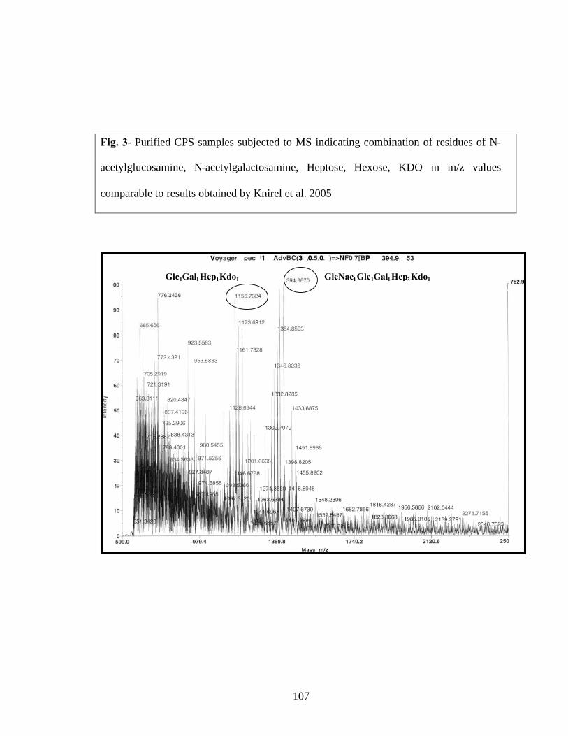
107
Fig. 3- Purified CPS samples subjected to MS indicating combination of residues of N-
acetylglucosamine, N-acetylgalactosamine, Heptose, Hexose, KDO in m/z values
comparable to results obtained by Knirel et al. 2005
GlcNac1Glc1Gal1Hep3Kdo1Glc1Gal1Hep4Kdo1

108
Fig. 4- 1H NMR analysis of CPS of Y. pestis LOS (Referenced to Vinogradov et al. 2002)
Glucose, Heptose

109
Fig. 5- Fractions of multimeric crude F1 antigen purified by HPLC obtained in void
volume (first peak at 25 minutes) (courtesy R. Kovi)

110
Fig. 6a- 15% SDS-PAGE gel of F1 antigen of Y. pestis KIM6, Mr 15.5 kDa
Fig. 6b- Western blot of 15.5 kDa F1 antigen of Y. pestis (courtesy R. Kovi)
15.5 kDa 15.5 kDa

111
Fig. 7- PMF of F1 antigen reveals greater than 95% homology to known peptides in the
database, indicating with high confidence that the target is purified F1 antigen (courtesy
R. Kovi)

112
Fig. 8- Schematic depiction of electrodialysis technique with F1 antigen and bound and
unbound oligonucleotides
Electrodialysis technique
Dialysis membrane with F1-oligo complex
F1 antigen
Oligonucleotide
Unbound oligonucleotides
Lowerchamber
Upperchamber
Membrane cap

113
Fig. 9- 1 µl volumes of samples within the dialysis membrane, dialysis tube, upper and
lower chambers and membrane cap were collected at 0, 30, 60, 90 and 120 minute
intervals and measured on a scintillation counter
Electro-dialysis of 33P-Oligonucleotides from within membrane
1
10
100
1000
10000
Time (m
ins) 0 30 60 90120
Blank
Time (minutes)
CP
M
Within membrane

114
Fig. 10- TCA precipitation of F1 antigen within the dialysis membrane after
electrodialysis and electrophoresing on SDS-PAGE gel
F1 antigen
115
Fig. 11- Sequences of Round 4 clones isolated after cloning using pCR® TOPO II vector
and chemically competent E. coli cells

116
Fig. 12a- Dendrogram indicating homology of Round 4 sequences
Below Fig. 12b- Sequence alignment by Pretty software indicating consensus regions
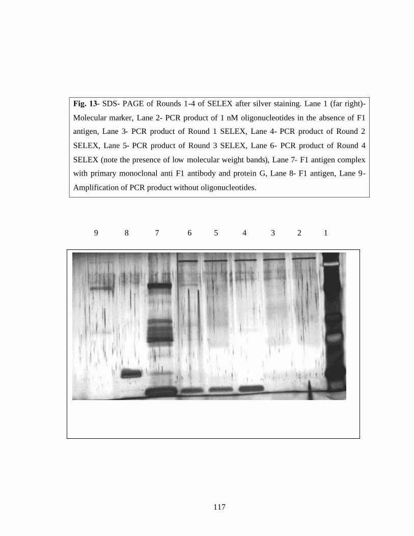
117
Fig. 13- SDS- PAGE of Rounds 1-4 of SELEX after silver staining. Lane 1 (far right)-
Molecular marker, Lane 2- PCR product of 1 nM oligonucleotides in the absence of F1
antigen, Lane 3- PCR product of Round 1 SELEX, Lane 4- PCR product of Round 2
SELEX, Lane 5- PCR product of Round 3 SELEX, Lane 6- PCR product of Round 4
SELEX (note the presence of low molecular weight bands), Lane 7- F1 antigen complex
with primary monoclonal anti F1 antibody and protein G, Lane 8- F1 antigen, Lane 9-
Amplification of PCR product without oligonucleotides.
9 8 7 6 5 4 3 2 1

118
Fig. 14- PCR product of Round 3 SELEX that was amplified after incubating with
streptavidin coated Dynal beads on compared to ten fold dilutions of streptavidin coated
Dynal beads.
Lane 1 (far left)- molecular marker, Lane 2- PCR product of Round 3 SELEX amplified
of streptavidin beads, Lanes 3-5- Ten fold dilutions of PCR product of Round 3, Lane 6-
15.5 kDa product of streptavidin, Lane 7- Streptavidin coated Dynal beads, Lanes 8-10-
Ten fold dilutions of streptavidin coated Dynal beads.
1 2 3 4 5 6 7 8 9 10

119
Fig. 15- Analysis of Round 4 sequences by multiple sequence alignment dendrogram
showing a reduction in pool complexity
120
Fig. 16- Round 8 sequences showing homologous sequences indicative of potential
aptamer sequences.

121
Fig. 17a- Autoradiograph obtained 24 hours after incubating F1 antigen immobilized on
nitrocellulose membrane with radiolabeled Round 8 and Round 0 pool.
Fig. 17b- Quantitation of binding assay
Binding assayof F1 antigen with Round 8 and Round 0
0
500
1000
1500
2000
2500
3000
2.5ug F1 / R8
40 0 ng F1 / R8
80 ng F1/ R8
4n g F1 / R8
BSA/R8
ssDNA BP/ R8
2. 5ug
F1/ R0
40 0 ng F1 / R0
80ng F1/
R0
4 ngF1/ R
0
BSA/ R0
ssDNA BP/ R0
Concentration
cpm CPM
F1 antigen
2 µg 400 ng 80 ng 4 ng
Round 8
Round 0ssDNA
BSA

VITA
Pravina P Fernandez
Candidate for the Degree of
Master of Science
Thesis: SELECTION OF AN APTAMER AGAINST SURFACE EXPOSEDTARGETS ON YERSINIA PESTIS
Major Field: Veterinary Biomedical Sciences
Biographical:
Personal Data: Born in Bombay, India on June 10, 1979, daughter of Peter AFernandez and Leonora P Fernandez.
Education: Graduated as a Bachelor’s in Veterinary Science and AnimalHusbandry at the Bombay Veterinary College, Bombay, India in Sept,2002.
Graduated with a M.S. in Veterinary Biomedical Sciences atOklahoma State University, Stillwater, OK in May, 2006.
Experience: Worked as a part- time veterinarian at the animal clinic run by DrSV Shah, Bombay, India. Volunteer at the Bombay Zoo and Society ofPrevention to Cruelty to Animals (SPCA)
Professional Memberships: ASM (2006).

Name: Pravina P Fernandez Date of Degree: May, 2006
Institution: Oklahoma State University Location: Stillwater, Oklahoma
Title of Study: SELECTION OF AN APTAMER AGAINST SURFACE EXPOSEDTARGETS ON YERSINIA PESTIS
Pages in Study: 121 Candidate for the Degree of Master of Sciences
Major Field: Veterinary Biomedical Sciences
Scope and Method of Study: Yersinia pestis is a CDC ‘category A’ bioterrorism agent.This study attempts to select an aptamer to detect the presence of LOS and F1antigen on Y. pestis. Aptamer selection by Systematic Evolution of Ligand byExponential Enrichment is an iterative process of binding, partitioning,amplification and strand separation.
Findings and Conclusions: The Lipid A as well as the core polysaccharide of LOS wasfound unfavorable for aptamer binding. Purified F1 antigen was considered asuitable target for aptamer selection. Our partitioning method using electrodialysisand eight rounds of selection resulted in thirty nine clones from Round 8 with aconsensus motif –GTGAG—GTTG--. However, binding assays wereunsuccessful due to non-specificity of Round 8 to F1 antigen, and and henceadditional four rounds of SELEX are being carried out. The aptamer thus selectedmight be used in in vitro diagnostic assays to detect F1 antigen of Yersinia pestis.
ADVISER’S APPROVAL: Dr Kenneth D. Clinkenbeard