selective behavioral alterations in the hiv-1 transgenic
TRANSCRIPT
University of South CarolinaScholar Commons
Theses and Dissertations
6-30-2016
Selective Behavioral Alterations In The HIV-1Transgenic Rat: Implications For Diagnosis OfPediatric HIV-1Kristen A. McLaurinUniversity of South Carolina
Follow this and additional works at: https://scholarcommons.sc.edu/etd
Part of the Arts and Humanities Commons, and the Psychology Commons
This Open Access Thesis is brought to you by Scholar Commons. It has been accepted for inclusion in Theses and Dissertations by an authorizedadministrator of Scholar Commons. For more information, please contact [email protected].
Recommended CitationMcLaurin, K. A.(2016). Selective Behavioral Alterations In The HIV-1 Transgenic Rat: Implications For Diagnosis Of Pediatric HIV-1.(Master's thesis). Retrieved from https://scholarcommons.sc.edu/etd/3443

0
SELECTIVE BEHAVIORAL ALTERATIONS IN THE HIV-1 TRANSGENIC RAT: IMPLICATIONS FOR DIAGNOSIS OF PEDIATRIC HIV-1
By:
Kristen A. McLaurin
Bachelor of Arts
Winthrop University, 2014
Submitted in Partial Fulfillment of the Requirements
For the Degree of Master of Arts in
Experimental Psychology
College of Arts and Sciences
University of South Carolina
2016
Accepted by:
Charles F. Mactutus, Director of Thesis
Rosemarie M. Booze, Reader
Steven B. Harrod, Reader
Lacy Ford, Senior Vice Provost and Dean of Graduate Studies

ii
© by Kristen A. McLaurin, 2016 All Rights Reserved.

iii
DEDICATION I dedicate this thesis to my parents, Preston and Kim McLaurin, who constantly
encouraged me to pursue my passion and higher education. Thank you for your listening
ears. Your unconditional love and support. For reminding me to laugh. But mostly, for
instilling in me values of perseverance and strength that have shaped who I am today.
I want to thank the director of my thesis, Charles Mactutus, for his guidance and
encouragement throughout this study. I would also like to thank my thesis committee,
Rosemarie Booze, and Steven Harrod, for their valuable suggestions to improve my
manuscript.
iv
ACKNOWLEDGEMENTS
This work was supported in part by grants from NIH (National Institute on Drug Abuse,
DA013137; National Institute of Child Health and Human Development, HD043680;
National Institute of Mental Health, MH106392) and the interdisciplinary research
training program supported by the University of South Carolina Behavioral-Biomedical
Interface Program.
v
ABSTRACT
Since the advent of combination antiretroviral therapy (cART), pediatric HIV-1
(PHIV) has evolved from a fatal disease to a chronic disease with children perinatally
infected with HIV-1 surviving into adulthood. The HIV-1 transgenic (Tg) rat, which
expresses 7 of the 9 HIV-1 genes constitutively throughout development, was used to
investigate the early development of chronic neurological impairment in PHIV. Male and
female Fischer HIV-1 Tg and F344N control rats, sampled from 35 litters, were
repeatedly assessed during early development using multiple experimental paradigms,
including somatic growth, locomotor activity, cross-modal prepulse inhibition (PPI) and
gap-prepulse inhibition (gap-PPI). A rightward shift towards later eye opening was
observed in HIV-1 Tg animals in comparison to controls. HIV-1 Tg animals exhibited
delays in the development of the cholinergic inhibitory system, assessed using locomotor
activity. Alterations in the development of the interstimulus interval (ISI) function were
observed in HIV-1 Tg rats in comparison to control animals, assessed using PPI.
Presence of the HIV-1 transgene was diagnosed with 91.4% accuracy using multiple
behavioral assessments on PD 20 and 21. Selective early behavioral alterations observed
in the HIV-1 Tg rats provide an opportunity for the development of a clinical diagnostic
screening tool, which may improve the long-term outcome for children perinatally
infected with HIV-1.

vi
TABLE OF CONTENTS
DEDICATION.................................................................................................................. iii ACKNOWLEDGEMENTS.............................................................................................. iv ABSTRACT........................................................................................................................v LIST OF FIGURES .......................................................................................................... vii LIST OF ABBREVIATIONS..........................................................................................viii CHAPTER 1: INTRODUCTION.......................................................................................1 CHAPTER 2: METHODOLOGY ......................................................................................5 CHAPTER 3: RESULTS.................................................................................................. 10 CHAPTER 4: DISCUSSION............................................................................................ 23 REFERENCES ................................................................................................................. 31

vii
LIST OF FIGURES Figure 3.1 Eye Opening .................................................................................................... 15
Figure 3.2 Cumulative Locomotor Activity ..................................................................... 16
Figure 3.3 Total Ambulation in Locomotor Activity ....................................................... 17
Figure 3.4 Visual Prepulse Inhibition ............................................................................... 18
Figure 3.5 Prepulse Inhibition on Postnatal Day 21 ......................................................... 19
Figure 3.6 Auditory Prepulse Inhibition ........................................................................... 20
Figure 3.7 Gap-Prepulse Inhibition .................................................................................. 21
Figure 3.8 Discriminant Function Analysis ...................................................................... 22
Figure 4.1 Hypothetical Serial Neural Circuitry of PPI.................................................... 30

viii
LIST OF ABBREVIATIONS ANOVA .............................................................................................. Analysis of Variance
ASR............................................................................................ Auditory Startle Response
cART..........................................................................Combination Antiretroviral Therapy
DA........................................................................................................................Dopamine
DAT ................................................................................................. Dopamine Transporter
Gap-PPI ........................................................................................ Gap-Prepulse Inhibition
HAART.................................................................... Highly Active Antiretroviral Therapy
HAD ....................................................................................... HIV-1 Associated Dementia
HAND ...........................................................HIV-1 Associated Neurocognitive Disorders
HDS .................................................................................................... HIV Dementia Scale
HIV-1 ............................................................................. Human Immunodeficiency Virus
IACUC ........................................................Institutional Animal Care and Use Committee
IC .............................................................................................................Inferior Colliculus
IHDS .............................................................................. International HIV Dementia Scale
M ................................................................................................................................. Mean
MAO-A............................................................................................ Monoamine Oxidase A
MTCT ................................................................................. Mother-to-Child Transmission
PD .................................................................................................................. Postnatal Day
PET .....................................................................................Positron Emission Tomography
ix
PHE ..............................................................................Progressive HIV-1 Encephalopathy
PHIV ....................................................Pediatric Human Immunodeficiency Virus Type 1
PnC................................................................................. Caudal Pontine Reticular Nucleus
PPI......................................................................................................... Prepulse Inhibition
PPTg........................................................................Pedunculopontine Tegmental Nucleus
pTH .........................................................................Phosphorylated Tyrosine Hydroxylase
SC........................................................................................................... Superior Colliculus
SEM................................................................................................................Standard Error
Tg .........................................................................................................................Transgenic

1
CHAPTER 1
INTRODUCTION
Worldwide, approximately 39 million individuals have died from human
immunodeficiency virus type-1 (HIV-1) and 35 million individuals are living with HIV-
1, including over 3.2 million children (≤15 years of age; CDC, 2013). Despite the
dramatic decrease in mother-to-child transmission (MTCT), the predominant source of
HIV-1 infection in children (Kourtis et al., 2001), 220,000 new cases of pediatric HIV-1
(PHIV) were reported in 2014 (UNAIDS, 2015). Since the advent of combination
antiretroviral therapy (cART), PHIV has evolved from a fatal disease to a chronic disease
with children perinatally infected with HIV-1 surviving into adulthood (Smith & Wilkins,
2015; Crowell et al., 2014). Despite decreased mortality rates, chronic neurological
impairment is still commonly reported in children perinatally infected with PHIV
(Franklin et al., 2005; Paramesparan et al., 2010). Therefore, given the prevalence of
PHIV, understanding the early behavioral alterations may be vital for the development of
a translational screening tool for neurological impairment in HIV-1 seropositive children.
Progressive HIV-1 encephalopathy (PHE), which is often analogous to HIV-1
associated dementia (HAD) in adults, was predominantly observed prior to the advent of
cART, with prevalence rates as high as 50% (Chiriboga et al., 2005; Crowell et al., 2014;
Shanhbhag et al., 2005). Common neurological manifestations of PHE include
microcephaly, resulting from cerebral atrophy, developmental delays, and movement
disorders (Belman et al., 1985; Epstein et al., 1985; Epstein et al., 1986) Furthermore,

2
neuroimaging analyses reveal calcification in the basal ganglia, and focal white matter
lesions in children with PHE (Epstein et al., 1985; Epstein et al., 1986; Kauffman et al.,
1992). Currently, in the post-cART era, the prevalence rate of PHE is between 2-15%
(Chiriboga et al., 2005; Shanbhag et al., 2005), however, chronic neurological
impairment persists.
High rates of chronic neurological impairment, including neurodevelopmental
delays, are still being reported in HIV-1 seropositive children (Franklin et al., 2005;
Paramesparan et al., 2010; review, Van Rie et al., 2007). Neurological assessments,
including the Bayley Scales of Infant Development and Wechsler Intelligence Scale for
Children-Revised, have previously been used to assess the effect of pediatric HIV-1 on
neurodevelopment (Blanchette et al., 2002; Lindsey et al., 2007; Van Rie et al., 2008;
Walker et al., 2013). Despite treatment with highly active antiretroviral therapy
(HAART), HIV-1 infected children exhibit significant delays in cognitive development,
motor skills and language expression in both high- (Lindsey et al., 2007) and low-
resource countries (Van Rie et al., 2008; Walker et al., 2013).
Neurocognitive deficits in HIV-1 seropositive children, including disease
progression, are poorly understood (Crowell et al., 2014), however, there is currently a
wealth of knowledge on HIV-1 associated neurocognitive disorders (HAND) in adults
evidenced in both clinical and preclinical studies (i.e. Heaton et al., 2010; Woods et al.,
2009). Neurocognitive assessments, including the Wisconsin Card Sorting Test and
Stroop Color Word Test, have shown that HIV-1 seropositive individuals display
significant deficits in set shifting (Carter et al., 2003) and response inhibition (Hinkin et
al., 1999; Tozzi et al., 1999). Deficits in complex problem solving and abstraction have
3
been demonstrated using the Wisconsin Card Sorting Test and Tower of London- Drexel
Version neurocognitive assessment (Cattie et al., 2012; Cherner et al., 2004).
Furthermore, HIV-1 seropositive individuals display a greater impulsivity than control
individuals on the Iowa Gambling Test (Hardy et al., 2006; Martin et al., 2004).
The HIV-1 transgenic (Tg) rat, which expresses 7 of the 9 HIV-1 genes, has been
used in preclinical studies to model neurocognitive deficits, including HAND, commonly
observed in HIV-1 seropositive individuals (Moran et al., 2013a; Moran et al., 2013b;
Moran et al., 2014a). Specifically, adult HIV-1 Tg rats exhibit significant deficits in
executive functions, including attention, inhibition, and flexibility in comparison to
controls (Moran et al., 2014a). Furthermore, significant alterations in temporal
processing, a pre-attentive process, have been observed using prepulse inhibition (PPI) of
the auditory startle response (ASR) in the HIV-1 Tg rat (Moran et al., 2013a).
Specifically, we have shown that, on both visual and auditory prepulse trials, HIV-1 Tg
rats exhibit an insensitivity to ISI duration, suggesting a lack of perceptual sharpening
with age (Moran et al., 2013a). Preliminary gap-prepulse inhibition (gap-PPI) data
suggest that HIV-1 Tg rats display alterations in the development of temporal processing,
assessed using startle response and prepulse inhibition.
Due to high rates of chronic neurological deficits in both HIV-1 seropositive
children and adults, there is a critical need for accurate screening tools for the diagnosis
of HAND. Early in the HIV-1 epidemic, two screening tools, the HIV Dementia Scale
(HDS; Power et al., 1995) and the International HDS (IHDS; Sacktor et al., 2005), were
developed to screen for HAD. However, neither the HDS or the IHDS are able to
accurately screen for milder forms of HAND, which are more common in the post-cART

4
era, affecting up to 40%-70% of HIV-1 infected individuals (Heaton et al., 2010; Heaton
et al., 2011; Letendre et al., 2009; McArthur et al., 2010; Sacktor et al., 2005; Zipursky
et al., 2013). Development of a screening tool, specifically for neurological impairment
seen in PHIV has immense clinical significance and may have a significant impact on the
lives of HIV-1 seropositive children and adults (Zipursky et al., 2013).
Thus, the aim of the current study was to establish the early trajectory of
behavioral deficits in the HIV-1 Tg rat. The HIV-1 Tg rat, which express 7 of the 9 HIV-
1 genes constitutively throughout development, provides a useful model for investigating
the development of neurologic impairments in pediatric AIDS (Peng et al., 2010; Royal
et al., 2012; Vigorito et al., 2015). Behavioral assessments, including locomotor activity,
cross-modal PPI, and gap-PPI were conducted prior to weaning from postnatal day (PD)
12 to PD 21. It was hypothesized that HIV-1 Tg rats would exhibit selective, early
alterations in somatic growth, including body weight and eye opening, and behavioral
measures, including locomotor activity, cross-modal PPI, and gap-PPI compared to non-
transgenic F344N controls. Understanding the early trajectory of behavioral deficits in
the HIV-1 Tg rats may not only provide a translational screening tool, but is also vital to
understanding the progression of neurocognitive deficits in children perinatally infected
with HIV-1.
5
CHAPTER 2
METHODOLOGY
2.1 Animals
Behavioral assessments were conducted on Fischer (F344/N; Harlan Laboratories
Inc., Indianapolis, IN) rats (HIV-1 Tg, n=19 litters; control, n=16 litters) during early
development beginning at PD 12. All rats were tested for motor movement, assessed
using locomotor activity (PD 12, 16, 20) and temporal processing deficits, assessed using
cross-modal prepulse inhibition (PPI) of the auditory startle response (PD 14, 17, and 21).
Gap-PPI was conducted on PD 18.
Animals were delivered to the facility between PD 7 and PD 9 over the course of
one year. All animals were housed with their biological dam until PD 21 when animals
were weaned and separated by sex. Subsequently animals were pair- or group-housed
with animals of the same sex throughout experimentation. Rodent food (Pro-Lab Rat,
Mouse, Hamster Chow #3000) and water were provided ad libitum throughout
experimentation.
Animals were maintained according to the National Institute of Health (NIH)
guidelines in AAALAC-accredited facilities. The targeted environmental conditions for
the animal facility were 21°± 2°C, 50% ± 10% relative humidity and have a 12-h
light:12-h dark cycle with lights on at 0700 h (EST). The Institutional Animal Care and
Use Committee (IACUC) of the University of South Carolina approved the project
protocol as consistent with federal assurance (# A3049-01).
6
2.2. Somatic Growth
Body Weight
Body weight was assessed as a measure of somatic growth upon arrival at the
facility between PD 8 and PD 10. Body weight was assessed periodically throughout
development.
Eye Opening
Eye opening was assessed as a developmental milestone on PD 13-19. Eye
opening was assessed separately for the right and left eye. A scale ranging from zero to
two was used with a zero score indicating a closed eye, and a score of one indicating an
open eye.
2.3 Motor Movement
Apparatus
Square (40 x 40 cm) (Hamilton Kinder, San Diego Instruments, San Diego, CA)
activity monitors were used to assess locomotor activity. Clear Plexiglas inserts were
added to convert the chambers into a round (~40 cm diameter) compartment. Free
movement of animals was detected by infrared photocell (32 emitter/detector pairs)
interruptions. Total locomotor activity was measured by assessing the number of
photocell interruptions within a 60-minute period.
Locomotor Activity
Locomotor activity testing occurred on PD 12, 16, and 20. Testing occurred for a
60-minute period between 700 and 1200h (EST) under dim light conditions, in the
absence of direct overhead lighting (<10 lux). All activity monitors were located in an
isolated room.

7
2.4 Temporal Processing
Apparatus
The startle platform (SR-Lab Startle Reflex System, San Diego Instruments, Inc.,
San Diego, CA) was enclosed in a 10 cm-thick double-walled, 81×81×116-cm isolation
cabinet (external dimensions) (Industrial Acoustic Company, INC., Bronx, NY), instead
of the 1.9 cm thick ABS plastic or laminate cabinets offered with this system. Sound
attenuation of 30dB(A) was provided in the isolation chamber relative to the external
environment. An ambient sound level of 22dB (A) was presented in the chamber without
any stimuli presented. The high-frequency loudspeaker of the SR-Lab system (Radio
Shack model#40-1278B), mounted inside the chamber 30 cm above the Plexiglas animal
test cylinder, delivered all auditory stimuli (frequency range of 5k-16k Hz). The animal’s
response to the auditory stimulus produced deflection of the test cylinder, which was
converted into analog signals by a piezoelectric accelerometer integral to the bottom of
the cylinder. The response signals were digitized (12 bit A to D) and saved to a hard disk.
Response sensitivities were calibrated using a SR-LAB Startle Calibration System. Sound
levels were measured and calibrated with a sound level meter (Kjaer Bruel 2203) with the
microphone placed inside the Plexiglas cylinder.
Cross-modal Prepulse Inhibition
Both visual and auditory prepulse stimuli were used to test animals for PPI of the
ASR on PD 14, 17 and 21. PPI was administered using a 30-min test session, beginning
with a 5-min acclimation period in the dark with 70 dB (A) background white noise,
followed by 6 pulse-only ASR trials with a 10s ITI. A total of 72 trials, including an
equal number of visual and auditory prepulse trials, were presented. Trials had an

8
interstimulus interval (ISI) of 0, 30, 50, 100, 200, and 4000 msec and were interdigitated
in an ABBA order of presentation. The 0 and 4000 msec ISI trials were control trials. The
intertrial interval (ITI) was variable from 15 sec to 25 sec. Inside the test cylinder, the
pulse stimulus intensity was 100 dB(A) (20 msec duration). Mean peak ASR amplitude
values were collected for analysis.
Gap-Prepulse Inhibition
Animals were tested for gap-PPI of the ASR on PD 18. Animals were tested for
gap-PPI of the ASR with a preceding gap in background white-noise as a stimulus. A 20-
min test session began with a 5-min acclimation period in the dark with 70 dB(A)
background white noise, followed by six pulse-only ASR trials, used for habituation, with
a 10s intertrial interval (ITI). Thirty-six trials were presented using 6-trial blocks in an
ABBA order of presentation. A 20-msec gap in white noise preceded a startle stimulus
presented at ISIs of 30,50,100, 200 and 4000 msec. Two control trials, the 0 and 4000
msec ISI trials, were included to provide a reference ASR within gap-PPI. The startle
stimulus intensity was 100 dB(A) (20 msec duration) measured inside the test cylinder.
Mean peak ASR amplitude values were collected for analysis. All test sessions were
conducted in the dark.
2.5 Statistical Analysis
Categorical data, including eye opening, an index of somatic growth, was
analyzed using a chi-squared statistical technique. Eye opening data were assessed by
individual pup. An alpha level of p≤0.05 was considered significant.
Analysis of variance (ANOVA) techniques (SPSS Statistics 20, IBM Corp.,
Somers, NY) were used to analyze all continuous data. To account for the nested design

9
within the ANOVA analysis, individual observations were analyzed by using litter means
and standard errors (Denenberg, 1984; Wears, 2002). Potential violations of sphericity of
within-subjects variables (Winer, 1971) were corrected using the Greenhouse-Geisser df
correction factor (Greenhouse & Geisser, 1959). An alpha level of p≤0.05 was considered
significant for all statistical tests.
Locomotor activity data was analyzed using a three-way mixed factor ANOVA.
Cumulative photocell interruptions were used for analysis, with genotype (HIV-1 Tg vs.
control) as the between-subjects factor, and time and age as the within-subjects factor.
Cross-modal PPI data were analyzed using a four-way mixed-factor ANOVA for
both prepulse modalities (auditory, visual). Mean peak ASR amplitude for the 0-4000
msec ISIs were used for analysis, with genotype (HIV-1 Tg vs. control) as the between-
subjects factor, and age, ISI, and trial as the within-subjects factors.
For gap-PPI, a three-way repeated measures ANOVA was performed on mean
peak ASR amplitude for the 0-4000 msec ISIs, with genotype (HIV-1 Tg vs. control) as
the between-subjects factor, and ISI and trial as the within-subjects factors. Gap-PPI
testing began after 11 litters had arrived. The present analysis includes 24 litters (HIV-1
Tg, n=13 litters; control, n=11 litters).
An exploratory discriminant functional analysis was conducted to determine the
diagnostic accuracy of early behavioral alterations and to determine which observed
behavioral assessments were able to correctly identify animals in regard to their genotype
(HIV-1 Tg vs. Control).

10
CHAPTER 3
RESULTS
3.1 HIV-1 Tg animals exhibit selective alterations in somatic growth
Body weight measurements, obtained upon arrival (PD 8 to PD 10), were used to
assess initial somatic growth. Upon arrival, there was not a significant difference in body
weight between control (M=11.9g, SEM=0.7g) and HIV-1 Tg animals (M=12.5g,
SEM=0.6 g).
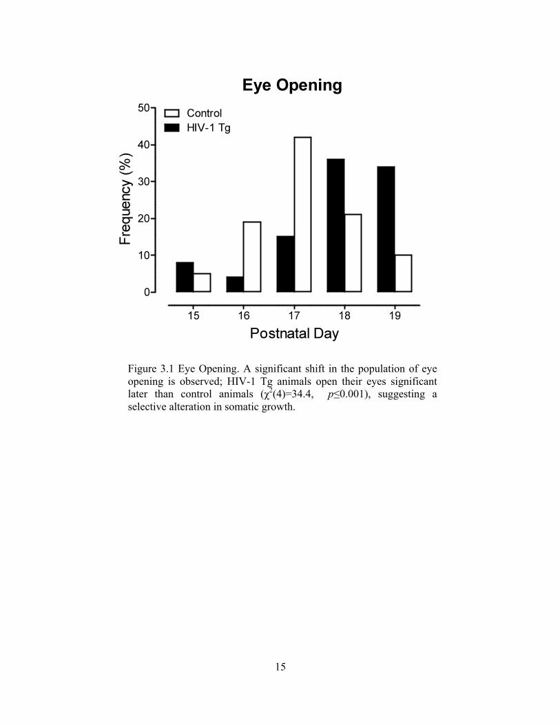
Selective alterations in somatic growth were evident in eye opening assessments,
as illustrated in Figure 1. Eye opening was assessed from PD 13 to PD 19 as a measure of
development. Eye opening started at PD 15 for both HIV-1 Tg and control animals. Eyes
were fully open for all animals on PD 19.
A χ2 revealed a statistically significant difference between eye opening in HIV-1
Tg vs control animals [χ2(4)=34.4, p≤0.001]. A significant shift in the distribution from
earlier eye opening to later eye opening was observed for the HIV-1 Tg rats in
comparison to control animals. Therefore, the HIV-1 Tg rat displays selective alterations
in somatic growth evident in eye opening, but not in body weight.
3.2 HIV-1 Tg animals exhibit altered development of motor movement.
Differential progression of motor movement in the HIV-1 Tg rat, relative to
control animals, was assessed using cumulative frequency for locomotor activity,
illustrated in Figure 2a, 2b, and 2c. The development of motor movement is significantly
11
altered in HIV-1 Tg animals relative to control animals. The overall ANOVA for
locomotor activity revealed a significant age x time x genotype interaction
[F(22,726)=6.3, pGG≤0.004, ηp2=0.16], age x time interaction [F(22,726)]=77.4,
pGG≤0.001, ηp2=0.70], time x genotype interaction [F(11,363)=12.1, pGG≤0.001,
ηp2=0.27], and age x genotype interaction [F(2,66)=6.9, pGG≤0.004, ηp
2=0.17]. Significant
main effects of genotype [F(1,33)=6.1, p≤0.02, ηp2=0.16], age [F(2,66)=103.9,
pGG≤0.001, ηp2=0.76] and time [F(11,363)=607.8, pGG≤0.001, ηp
2=0.95] were also
observed.
Separate analyses at each age were conducted to determine the locus of these
interactions. Analyses revealed a significant time x genotype interaction at PD 20
[F(11,363)=23.1, pGG≤0.001, ηp2=0.41], but not at PD 12 [F(11,363)=1.5, pGG≤0.237] or
PD 16 [F(11,363)=1.6, pGG≤0.217].
Alterations in the development of motor movement, assessed using locomotor
activity, are further evidenced by mean total ambulation, illustrated in Figure 3. A
segmented first-order polynomial was the best fit for the total ambulation in HIV-1 Tg
animals, while a second-order polynomial was the best fit for control animals. Therefore,
both the time x genotype interaction at PD 20, as well as differences in the best fit for
total ambulation, indicates altered development of motor movement in the HIV-1 Tg
animals.
3.3 HIV-1 Tg animals exhibit altered temporal processing development with a visual prepulse.
Altered temporal processing development with a visual prepulse in the HIV-1 Tg
rat, relative to control animals, is illustrated in Figure 4a and 4b. The overall ANOVA on
mean peak ASR amplitude revealed a significant age x ISI x genotype interaction

12
[F(10,330)=5.2, pGG≤0.001, ηp2=0.14], a significant age x ISI interaction
[F(10,330)=19.2, pGG≤0.001, ηp2=0.37] and a significant ISI x genotype interaction
[F(5,165)=12.0, pGG≤0.001, ηp2=0.27]. Significant main effects of genotype
[F(1,33)=19.8, p≤0.001, ηp2=0.38], age [F(2,66)=48.2, pGG≤0.001, ηp
2=0.60], and ISI
[F(5,165)=44.6, pGG≤0.001, ηp2=0.58] were also observed.
Differences in the development of temporal processing were further examined by
separate analysis of each genotype. The overall ANOVA for control animals, illustrated
in Figure 3a, revealed an age x ISI interaction [F(10,140)=4.3, pGG≤0.008, ηp2=0.23].
Main effects of age [F(2,28)=6.6, pGG≤0.009, ηp2=0.32] and ISI were also observed
[F(5,70)=7.6, pGG≤0.001, ηp2=0.35]. In contrast, the overall ANOVA for HIV-1 Tg
animals only revealed a significant main effect of age [F(2,34)=7.0, pGG≤0.004,
ηp2=0.29]. The age x ISI interaction present in the control animals, but not the HIV-1 Tg
animals indicates an altered development of the ISI function.
Separate analyses at each age were also conducted, which revealed a significant
genotype x ISI interaction on PD 17 [F(5,165)=4.6, pGG≤0.003, ηp2=0.12] and on PD 21,
[F(5,165)=10.5, pGG≤0.001, ηp2=0.24], illustrated in Figure 5a, but not on PD 14. The
genotype x ISI interactions, observed on PD 17 and PD 21, provide additional evidence
for the altered development of the ISI function in the HIV-1 Tg group.
3.4 HIV-1 Tg animals exhibit altered temporal processing development with an auditory prepulse.
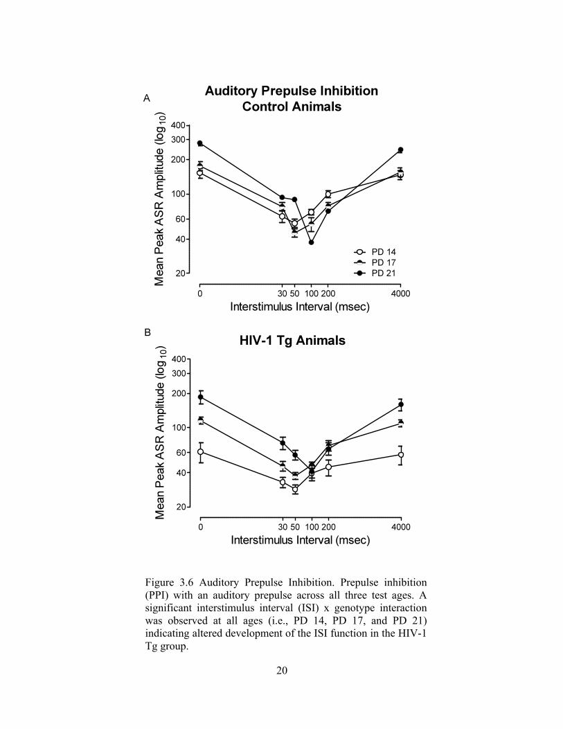
HIV-1 Tg animals exhibit alterations in the development of temporal processing
with an auditory prepulse, as illustrated in Figure 6a and 6b. Both HIV-1 Tg and control
animals exhibited a shift in maximal inhibition (from 30 msec to 50 msec) on postnatal
day 21. However, HIV-1 Tg animals exhibit altered development of the ISI function in
13
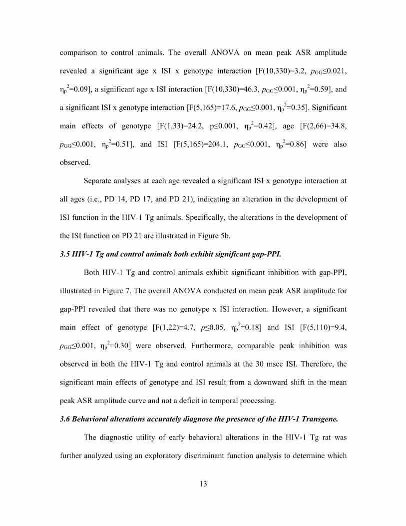
comparison to control animals. The overall ANOVA on mean peak ASR amplitude
revealed a significant age x ISI x genotype interaction [F(10,330)=3.2, pGG≤0.021,
ηp2=0.09], a significant age x ISI interaction [F(10,330)=46.3, pGG≤0.001, ηp
2=0.59], and
a significant ISI x genotype interaction [F(5,165)=17.6, pGG≤0.001, ηp2=0.35]. Significant
main effects of genotype [F(1,33)=24.2, p≤0.001, ηp2=0.42], age [F(2,66)=34.8,
pGG≤0.001, ηp2=0.51], and ISI [F(5,165)=204.1, pGG≤0.001, ηp
2=0.86] were also
observed.
Separate analyses at each age revealed a significant ISI x genotype interaction at
all ages (i.e., PD 14, PD 17, and PD 21), indicating an alteration in the development of
ISI function in the HIV-1 Tg animals. Specifically, the alterations in the development of
the ISI function on PD 21 are illustrated in Figure 5b.
3.5 HIV-1 Tg and control animals both exhibit significant gap-PPI.
Both HIV-1 Tg and control animals exhibit significant inhibition with gap-PPI,
illustrated in Figure 7. The overall ANOVA conducted on mean peak ASR amplitude for
gap-PPI revealed that there was no genotype x ISI interaction. However, a significant
main effect of genotype [F(1,22)=4.7, p≤0.05, ηp2=0.18] and ISI [F(5,110)=9.4,
pGG≤0.001, ηp2=0.30] were observed. Furthermore, comparable peak inhibition was
observed in both the HIV-1 Tg and control animals at the 30 msec ISI. Therefore, the
significant main effects of genotype and ISI result from a downward shift in the mean
peak ASR amplitude curve and not a deficit in temporal processing.
3.6 Behavioral alterations accurately diagnose the presence of the HIV-1 Transgene.
The diagnostic utility of early behavioral alterations in the HIV-1 Tg rat was
further analyzed using an exploratory discriminant function analysis to determine which

14
behavioral assessments and ages were best able to identify group membership.
Assessment of locomotor activity on PD 20 and cross-modal PPI on PD 21 best predicted
group membership, as illustrated in Figure 8. A stepwise discriminant function analysis
selected four variables (Motor Movement (PD 20), Auditory Mean Peak ASR Amplitude
Values at 50 msec (PD 21) and 100 msec (PD 21) and Visual Mean Peak ASR Amplitude
Values at 50 msec (PD 21)) that maximally separated the HIV-1 Tg and control animals
(canonical correlation of 0.831). Animals were classified (jack-knifed) with 91.4%
accuracy (F approximation of Wilks’ λ of 0.309, F (4, 30) =16.8, p≤0.001).
15
Figure 3.1 Eye Opening. A significant shift in the population of eye opening is observed; HIV-1 Tg animals open their eyes significant later than control animals (χ2(4)=34.4, p≤0.001), suggesting a selective alteration in somatic growth.

16
Figure 3.2 Cumulative Locomotor Activity. HIV-1 Tg animals exhibit altered development of motor movement assessed using locomotor activity. Cumulative frequencies of gross motor movement are shown on postnatal day (PD) 12 (a), PD 16 (b), and PD 20 (c). A significant age x time x genotype interaction was observed, indicating that the HIV-1 Tg animals exhibit alterations in motor movement both within a session and throughout development.

17
Figure 3.3 Total Ambulation in Locomotor Activity. HIV-1 Tg animals exhibit significantly altered development of motor movement, assessed using locomotor activity. A significant time x genotype interaction on PD 20 was observed, indicating altered development of motor movement in the HIV-1 Tg animals. Furthermore, a segmented first-order polynomial was the best fit for the total ambulation in HIV-1 Tg animals, while a second-order polynomial was the best fit for control animals.

18
Figure 3.4 Visual Prepulse Inhibition. Prepulse inhibition (PPI) with a visual prepulse across all three test ages. A significant age x interstimulus interval (ISI) interaction was present in control animals (a), but not in HIV-1 Tg animals(b) indicating that HIV-1 Tg animals exhibit altered development of the ISI function with a visual prepulse.

19
Figure 3.5 Prepulse Inhibition on Postnatal Day (PD) 21. A significant genotype x ISI interaction was observed in visual PPI at PD 17 and PD 21 (a). A significant genotype x ISI interaction was observed at all ages (PD 14, PD 17, and PD 21) in auditory PPI (b). Results are indicative of altered development of the ISI function, regardless of modality, in the HIV-1 Tg group.
20
Figure 3.6 Auditory Prepulse Inhibition. Prepulse inhibition (PPI) with an auditory prepulse across all three test ages. A significant interstimulus interval (ISI) x genotype interaction was observed at all ages (i.e., PD 14, PD 17, and PD 21) indicating altered development of the ISI function in the HIV-1 Tg group.

21
Figure 3.7 Gap-prepulse inhibition (gap-PPI). A significant main effect of age and ISI were observed. comparable peak inhibition was observed in both the HIV-1 Tg and control animals at the 30 msec ISI. Therefore, the HIV-1 Tg animals exhibit a downward shift in the mean peak ASR amplitude curve, but not a deficit in temporal processing.

22
Figure 3.8 Discriminant Function Analysis. Animal classification is illustrated as a function of the canonical variable representing the simplest linear function that best separated the HIV-1 Tg and control groups (canonical correlation 0.83) and correctly identified (jackknife classification) group membership with 91.4% accuracy (93.8% of controls, and 89.5% of HIV-1 Tg animals).

23
CHAPTER 4
DISCUSSION
Selective early behavioral alterations in the HIV-1 Tg rat were observed using
multiple experimental paradigms, including somatic growth, locomotor activity, cross-
modal PPI, and gap-PPI. A rightward shift towards later eye opening was observed in the
HIV-1 Tg animals in comparison to controls. HIV-1 Tg animals exhibit maximal
spontaneous activity on PD 20, in comparison to PD 16 for control animals, indicating
delayed maturation of the cholinergic inhibitory system. Alterations in the development
of the ISI function were exhibited by HIV-1 Tg animals with both a visual and auditory
prepulse. Presence of the HIV-1 transgene can be diagnosed with 91.4% accuracy using
multiple behavioral assessments. Selective early behavioral alterations observed in the
HIV-1 Tg rat resemble alterations observed in PHIV, providing the potential for
developing a translational screening tool for the early diagnosis of chronic neurocognitive
impairment observed in children perinatally infected with HIV-1.
4.1 Somatic Growth
Selective alterations in somatic growth are observed in the HIV-1 Tg rat, assessed
using body weight and eye opening. No statistical differences were observed in initial
body weight, assessed from PD 8 to PD 10, results which replicate those previously
reported in male Sprague-Dawley rats stereotaxically injected with Tat and/or gp120,
HIV1 viral proteins, on PD 1 (Fitting et al., 2008) and results previously reported in HIV-
1 infected children (Guillen et al., 2007; Parachure et al., 2015). Previous reports in

24
children with PHIV indicate that early initiation of antiretroviral therapy, and therefore
subsequent virologic control, leads to normal growth (Guillen et al., 2007; Parchure et
al., 2015). The HIV-1 Tg rat, however, did exhibit selective alterations in somatic
growth observed in eye opening measurements. Results of alterations in eye opening
replicate those previously reported in male Sprague-Dawley rats stereotaxically injected
with HIV-1 viral proteins (Moran et al., 2014b). Therefore, the HIV-1 transgene may
alter the development of selective somatic growth measurements.
4.2 Motor Movement
HIV-1 Tg rats exhibit alterations in the development of motor movement,
assessed using locomotor activity. Evidence for alterations in the development of motor
movement was revealed using the cumulative frequency of gross motor movements.
Specifically, significant differences were evident at PD 12 and PD 20, but not at PD 16.
Additional evidence for the alterations in the development of motor movement were
evidenced by total ambulation. A second-order polynomial was best fit for control
animals, with maximal spontaneous activity exhibited on PD 16. In contrast, a
segmented first-order polynomial was the best fit for HIV-1 Tg animals. Therefore, HIV-
1 Tg animals fail to exhibit decreased levels of spontaneous activity on PD 20, suggesting
delayed maturation in the development of the cholinergic inhibitory system.
Alterations in the development of the cholinergic system in the forebrain may
underlie early behavioral alterations in motor movement observed in the HIV-1 Tg rat.
Campbell et al., (1969) studied the effect of amphetamine, an indirect dopamine (DA)
agonist, and scopolamine, a forebrain anticholinergic agent on locomotor activity on PD
10, 15, 20, 25, and 100. Administration of amphetamine increased activity during all

25
testing periods. In contrast, administration of scopolamine increased activity beginning at
PD 20, suggesting that forebrain cholinergic inhibitory areas mature later than hindbrain
structures (Campbell et al., 1969). Furthermore, saline control groups exhibited
maximum activity on PD 15, followed by a dramatic decline, implicating that forebrain
cholinergic inhibitory areas may also modulate activity in a novel environment (i.e.,
locomotor activity; Campbell et al., 1969). In the present study, control animals exhibited
maximum spontaneous activity on PD 16, followed by a dramatic decline. In contrast,
HIV-1 Tg animals exhibited maximum spontaneous activity on PD 20, suggesting
delayed development of the forebrain cholinergic inhibitory areas.
Results of alterations in the development of motor movement in the present study
replicate those previously reported in the HIV-1 Tg rat (Moran et al., 2013b) and
Sprague-Dawley rats stereotaxically injected with the HIV-1 viral proteins (Fitting et al.,
2008). In addition, developmental alterations in the HIV-1 Tg rat replicate those reported
in HIV-1 infected children (Ferguson & Jelsma, 2009; Foster et al., 2006; Whitehead et
al., 2014). Specifically, a longitudinal analysis of HIV-1 infected and sero-reverters in
South Africa reported significant motor deficits in approximately 40% of HIV-1 infected
children (Whitehead et al., 2014).
4.3 Temporal Processing
Altered temporal processing development, assessed with cross-modal PPI, was
observed in HIV-1 Tg animals compared to control animals. In PPI with a visual
prepulse, the ISI functions observed in the HIV-1 Tg and control groups were not
significantly different at PD 14, but subsequently changed in different ways (i.e., PD 17
and PD 21) indicating altered development of the ISI function. In addition, HIV-1 Tg

26
animals exhibit an alteration in the development of the ISI function observed in PPI with
an auditory prepulse. Results in the present study replicate those previously reported in
adult HIV-1 Tg animals (Moran et al., 2013a) and Sprague-Dawley rats stereotaxically
injected with the HIV-1 viral proteins on PD 1 (Fitting et al., 2008; Fitting et al., 2006a;
Fitting et al., 2006b). Both HIV-1 Tg and control animals exhibit significant inhibition in
gap-PPI. Gap-PPI results in the present study are consistent with a preliminary
longitudinal analysis conducted from PD 30 to PD 150.
The brain neural circuitry mediating PPI, illustrated in Figure 1, has been
established using lesioning (i.e., Leitner & Cohen, 1985) and electrical stimulation
studies (i.e., Li et al., 1998; Li & Yeomans, 2000). Specifically, the serial circuit
mediating PPI begins with auditory input relayed to the inferior colliculus (IC). Visual
and tactile input, in contrast, are relayed to the superior colliculus (SC). Sensory input,
regardless of modality, is then relayed from the SC to the pedunculopontine tegmental
nucleus (PPTg), which ultimately triggers a cholinergic projection to the caudal pontine
reticular nucleus (PnC; Fendt et al., 1994; Fendt et al., 2001; Koch et al., 1993; Koch &
Schnitzler, 1997). Activation of the PnC is relayed to motor neurons causing a startle
response.
The role of dopamine in the circuit mediating PPI has been evidenced in previous
behavioral and pharmacological studies (review, Geyer et al., 2001; Moran et al., 2009;
Zhang et al., 2000). Administration of direct (i.e., apomorphine) and indirect dopamine
(DA) agonists (i.e., amphetamine) have been used to manipulate dopamine, subsequently
disrupting PPI (review, Geyer et al., 2001). An insensitivity to the manipulation of ISI,
assessed using cross-modal PPI, has also been observed in rats administered

27
apomorphine; results that are comparable to those observed in the HIV-1 Tg rat (Moran
et al., 2009).
4.5 The Dopamine System and Chronic Neurological Impairment
Clinical and preclinical studies have implicated disruptions in the DA system as
an important factor in chronic HIV-1 associated neurological impairment (review, Fitting
et al., 2015; review, Ferris et al., 2008). Significantly greater brain atrophy in HIV-1
seropositive individuals has been reported in areas rich in dopamine, including the basal
ganglia (Kumar et al., 2009), substantia nigra (Kumar et al., 2011), and caudate nucleus
(Kumar et al., 2009). Furthermore, decreased DA transporter levels in HIV-1 seropositive
individuals have been correlated with decreased performance on neuropsychological tests
(Chang et al., 2008). In vivo brain imaging studies replicate postmortem studies in HIV-
1 seropositive individuals, providing further evidence for a DA system disruption in HIV-
1 seropositive individuals (Purohit et al., 2011). HIV-1 seropositive individuals had
decreased brain volumetrics in the thalamus, hippocampus, and corpus callosum (Ortega
et al., 2013). Positron emission tomography (PET) scans provide evidence of a
progressive striatal dopamine deficit that occurs in as the HIV-1 Tg rat ages (Lee et al.,
2014). Furthermore, in vitro studies have replicated the results presented in clinical and
preclinical studies, which provide further evidence that the DA transporter is being
targeted by Tat and gp120, two HIV-1 proteins (Aksenov et al., 2008; Midde et al.,
2013).
Early behavioral alterations observed in the HIV-1 Tg rat may result from
alterations in the development of the DA system. (review, Fitting et al., 2015). Preclinical
studies in the HIV-1 Tg rat have previously implicated DA system impairments as an

28
underlying factor in chronic neurological impairment (Moran et al., 2012; Moran et al.,
2014b; Webb et al., 2010). Specifically, pharmacological assessments were used to
examine alterations in the midbrain DAergic system in the HIV-1 Tg rat (Moran et al.,
2012; Webb et al., 2010). DAergic system dysfunction, assessed using Western blotting,
was evidenced by alterations in phosphorylated tyrosine hydroxylase (pTH), dopamine
transporter (DAT) mRNA, and/or monoamine oxidase A (MAO-A; Moran et al., 2012;
Webb et al., 2010). Although multiple neural systems, including the dopaminergic and
cholinergic system, may mediate the early behavioral alterations observed in the HIV-1
Tg rat, HIV-1 infection often affects DA system function, resulting in subsequent
cognitive deficits (Di Rocco et al., 2000; review, Fitting et al., 2015; Wang et al., 2004).
4.6 Conclusions
The study of selective early behavioral alterations in the HIV-1 Tg rat is vital to
the development of a diagnostic screening tool for chronic neurologic impairment
observed in children perinatally infected with HIV-1. HIV-1 Tg rats used in the present
study are a healthier derivation of those originally described (Reid et al., 2001),
exhibiting no general wasting or pathological phenotypes. HIV-1 Tg litters used in the
current study displayed no significant health disparities in comparison to F344 controls
(i.e., similar initial body weight, similar litter size). Thus, the HIV-1 Tg rat provides a
vehicle for investigating the development and underlying mechanisms involved in
chronic neurologic impairments observed in PHIV (Vigorito et al., 2015).
Selective early behavioral alterations observed in the HIV-1 Tg rat may provide a
novel screening tool for the diagnosis of neurocognitive deficits in children perinatally
infected with HIV-1. The potential utility of selective early behavioral alterations was

29
assessed using a discriminant function analysis, which correctly identified animals in
regards to their genotype (HIV-1 Tg vs. control) with 91.4% accuracy. The presence of
the HIV-1 transgene was best predicted using four variables at PD 20 and PD 21, brain
development which approximates 2-3 years of age in humans (Review: Semple et al.,
2013). Therefore, selective early behavioral alterations observed in the HIV-1 Tg rats
provide an opportunity for the development of a translational screening tool, which will
allow early cART initiation, improving long-term outcomes for children perinatally
infected with HIV-1 (Edwards et al., 2015; Kitahata et al., 2009).

30
Figure 4.1. Hypothetical Serial Neural Circuitry of PPI. Adapted from Fendt et al.
(2001) and Koch (1999).
Reflex Motor
Neurons

31
REFERENCES Aksenov, M.Y., Aksenova, M.V., Silvers, J.M., Mactutus, C.F., & Booze, R.M. (2008).
Different effects of selective dopamine uptake inhibitors, GBR 12909 and WIN 35428, on HIV-1 Tat toxicity in rat fetal midbrain neurons. Neurotoxicology, 29(6), 971-977.
Belman, A.L., Ultmann, M.H., Horoupian, D., Novick, B., Spiro, A.J., Rubinstein, A.,
Kurtzberg, D., & Cone-Wesson, B. (1985). Neurological complications in infants and children with acquired immune deficiency syndrome. Annals of Neurology, 18(5), 560-566.
Blanchette, N., Smith, M.L., King, S., Fernandes-Penney, A., & Read, S. (2002).
Cognitive development in school-age children with vertically transmitted HIV infection. Developmental Neuropsychology, 21(3), 223-241.
Campbell, B.A., Lytle, L.D., & Fibiger, H.C. (1969). Ontogeny of adrenergic arousal and
cholinergic inhibitory mechanisms in the rat. Science, 166(3905), 635-637.
Carter, S.L., Rourke, S.B., Murji, S., Shore, D. & Rourke, B.P. (2003). Cognitive complaints, depression, medical symptoms, and their association with neuropsychological functioning in HIV infection: a structural equation model analysis. Neuropsychology, 17(3), 410-419.
Cattie, J.E., Doyle, K., Weber, E., Grant, I., Woods, S.P., & the HIV Neurobehavioral Research Program (HNRP) Group. (2012). Planning deficits in HIV-associated neurocognitive disorders: Component processes, cognitive correlates, and implications for everyday functioning. Journal of Clinical and Experimental Neuropsychology, 34(9), 906-918.
Chang, L., Wang, G.J., Volkow, N.D., Ernst, T., Telang, F., Logan, J., & Fowler, J.S. (2008). Decreased brain dopamine transporters are related to cognitive deficits in HIV patients with or without cocaine abuse. NeuroImage, 42(2), 869-878.
Cherner, M., Ellis, R.J., Lazzaretto, D., Young, C., Mindt, M.R., Atkinson, J.H., Grant, I.,
Heaton, R.K., & the HNRC Group. (2004) Effects of HIV-1 infection and aging on neurobehavioral functioning: preliminary findings. AIDS, 18, S27-34.
32
Chiriboga, C.A., Fleishman, S., Champion, S., Gaye-Robinson, L., & Abrams, E.J.
(2005). Incidence and prevalence of HIV encephalopathy in children with HIV infection receiving highly active anti-retroviral therapy (HAART). The Journal of Pediatrics, 146(3), 402-407.
Centers for Disease Control and Prevention (2013). Diagnosis of HIV infection in the
United States and Dependent Areas (25). Retrieved from http://www.cdc.gov/hiv/pdf/g-l/hiv_surveillance_report_vol_25.pdf
Crowell, C.S., Malee, K.M., Yogev, R., & Muller, W.J. (2014). Neurologic disease in
HIV-infected children and the impact of combination antiretroviral therapy. Review in Medical Virology, 24(5), 316-331.
Denenberg, V. H. (1984). Some statistical and experimental considerations in the use of
the analysis-of-variance procedure. American Journal of Physiology-Regulatory, Integrative and Comparative Physiology, 246(4), R403-R408.
Di Rocco, A., Bottiglieri, T., Dorfman, D., Werner, P., Morrison, C. & Simpson, D.
(2000). Decreased homovanilic acid in cerebrospinal fluid correlates with impaired neuropsychologic function in HIV-1-infected patients. Clinical Neuropharmacology, 23(4), 190-194.
Edwards, J.K., Cole, S.R. Westreich, D, Mugavero, M.J., Eron, J.J., Moore, R.D.,
Mathews, W.C., Hunt, P., & Williams, C. (2015). Age at entry into care, timing of antiretroviral therapy initiation, and 10-year mortality among HIV-seropositive adults in the United States. Clinical Infectious Diseases, 61(7), 1189-1195.
Epstein, L.G., Sharer, L.R., Joshi, V.V., Fojas, M.M., Koenigsberger, M.R., & Oleske,
J.M. (1985). Progressive encephalopathy in children with acquired immune deficiency syndrome. Annals of Neurology, 17(5), 488-496.
Epstein, L.G., Sharer, L.R., Oleske, J.M., Connor, E.M., Goudsmit, J., Bagdon, L.,
Robert-Guroff, M., & Koenigsberger, M.R. (1986). Neurologic manifestations of human immunodeficiency virus infection in children. Pediatrics, 78(4), 678-687.
Fendt, M., Koch, M., & Schnitzler, H.U. (1994). Sensorimotor gating deficit after lesions
of the superior colliculus. NeuroReport, 5(14), 1725-1738. Fendt, M., Li, L., & Yeomans, J.S. (2001). Brain stem circuits mediating prepulse
inhibition of the startle reflex. Psychopharmacology, 156 (2-3), 216-224. Ferguson, G. & Jelsma, J. (2009). The prevalence of motor delay among HIV infected
children living in Cape Town, South Africa. International Journal of Rehabilitation, 32(2), 108-114.
33
Ferris, M.J, Mactutus, C.F., & Booze, R.M. (2008). Neurotoxic profiles of HIV,
psychostimulant drugs of abuse, and their concerted effect on the brain: Current status of dopamine system vulnerability in NeuroAIDS. Neuroscience & Biobehavioral Reviews, 32(5), 883-909.
Fitting, S., Booze, R.M., & Mactutus, C.F. (2006a). Neonatal hippocampal Tat injections:
developmental effects on prepulse inhibition (PPI) of the auditory startle response. International Journal of Developmental Neuroscience, 24(4), 275-283.
Fitting, S., Booze, R.M., & Mactutus, C.F. (2006b). Neonatal intrahippocampal
glycoprotein 120 injection: the role of dopaminergic alteration in prepulse inhibition in adult rats. The Journal of Pharmacology and Experimental Therapeutics, 318(3), 1352-1358.
Fitting, S., Booze, R.M., & Mactutus, C.F. (2008). Neonatal intrahippocampal injection
of the HIV-1 proteins gp12 and Tat: Differential effects on behavior and the relationship to stereological hippocampal measures. Brain Research, 1232, 139-154.
Fitting, S., Booze, R.M. & Mactutus, C.F. (2015). HIV-1 proteins, Tat and gp120, target
the developing dopamine system. Current HIV-1 Research, 13(1), 21-42. Foster, C.J., Biggs, R.L., Melvin, D., Walters, S., Tudor-Williams, G., & Lyall, E.G.H.
(2006). Neurodevelopmental outcomes in children with HIV infection under 3 years of age. Developmental Medicine & Child Neurology, 48(8), 677-682.
Franklin, S., Lim, H.J., Rennie, K.M., Eastwood, D., Cuene, B., & Havens, P.L. (2005).
Longitudinal intellectual assessment of children with HIV infection. Journal of Clinical Psychology in Medical Settings, 12(4), 367-376.
Geyer, M.A., Krebs-Thomson, K., Braff, D.L. & Swerdlow, N.R. (2001).
Pharmacological studies of prepulse inhibition models of sensorimotor gating deficits in schizophrenia: a decade in review. Psychopharmacology, 156(2), 117-154.
Greenhouse, S.W. & Geisser, S. (1959). On methods in the analysis of profile data.
Psychometrika, 24(2), 95-112. Guillen, S., Ramos, J.T., Resino, R., Bellon, J.M., & Munoz, M.A. (2007). Impact on
weight and height with the use of HAART in HIV-infected children. The Pediatric Infectious Disease Journal, 26(4), 334-338.

34
Hardy, D.J., Hinkin, C.H., Levine, A.J., Castellon, S.A. & Lam, M.N. (2006). Risky decision making assessed with the gambling task in adults with HIV. Neuropsychology, 20(3), 355-360.
Heaton, R.K., Clifford D.B., Franklin D.R., Woods S.P., Ake C., Vaida F., Ellis, R.J., Letendre, S.L., Marcotte, T.D., Atkinson, J.H., Rivera-Mindt, M., Vigil, O.R., Taylor, M.J., Collier, A.C., Marra, C.M., Gelman, B.B., McArthur, J.C., Morgello, S., Simpson, D.M., McCutchan, J.A., Abramson, I., Gamst, A., Fennema-Notestine, C., Jernigan, T.L., Wong, J., Grant, I., & The CHARTER Group. (2010). HIV-associated neurocognitive disorders persist in the era of potent antiretroviral therapy CHARTER study. Neurology, 75(23), 2087-2096.
Heaton, R.K., Franklin, D.R., Ellis, R.J., McCutchan, J.A., Letendre, S.L., LeBlanc, S.,
Corkran, S.H., Duarte, N.A., Clifford, D.B., Woods, S.P., Collier, A.C., Marra, C.M., Morgello, S., Rivera-Mindt, M., Taylor, M.J., Marcotte, T.D., Atkinson, J.H., Wolfson, T., Gelman, B.B., McArthur, J.C., Simpson, D.M., Abramson, I., Gamst, A., Fennema-Notestine, C., Jernigan, T.L., Wong, J., Grant, I., & The CHARTER and HNRC Groups. (2011). HIV-associated neurocognitive disorders before and during the era of combination antiretroviral therapy: differences in rates, nature, and predictors. Journal of NeuroVirology, 17(1), 3-16.
Hinkin, C.H., Castellon, S.A., Hardy, D.J., Granholm, E. & Siegle, G. (1999). Computerized and traditional Stroop task dysfunction in HIV-1 infection. Neuropsychology, 13(2), 306-316.
Kauffman, W.M., Sivit, C.J., Fitz, C.R., Rakusan, T.A., Herzog, K. & Chandra, R.S. (1992). CT and MR evaluation of intracranial involvement in pediatric HIV infection: A clinical-imaging correlation. American Journal of Neuroradiology, 13(3), 949-957.
Kitahata, M.M. Gange, S.J., Abraham, A.G., Merriman, B., Saag, M.S., Justice, A.C.,
Hogg, R.S., Deeks, S.G., Eron, J.J., Brooks, J.T., Rourke, S.B., Gill, M.J., Bosch, R.J., Martin, J.N., Klein, M.B., Jacobson, L.P., Rodriguez, B., Sterline, T.R., Kirk, G.D., Napravnik, S., Rachlis, A.R., Calzavara, L.M., Horberg, M.A., Silverberg, M.J., Gebo, K.A., Goedert, J.J., Benson, C.A., Collier, A.C., Van Rompaey, S.E., Crane, H.M., McKaig, R.G., Lau, B., Freeman, A.M., Moore, R.D., NA-ACCORD Investigators. (2009). Effect of early versus deferred antiretroviral therapy for HIV on survival. New England Journal of Medicine, 360(18), 1815-1826.
Koch, M., Kungel, M., & Herbert, H. (1993). Cholinergic neurons in the
pedunculopontine tegmental nucleus are involved in the mediation of prepulse inhibition of the acoustic startle response in the rat. Experimental Brain Research, 97(1), 71-82.

35
Koch, M. & Schnitzler, H.U. (1997). The acoustic startle response in rats: Circuits mediating evocation, inhibition and potentiation. Behavioural Brain Research, 89(1-2), 35-49.
Koch, M. (1999). The neurobiology of startle. Progress in Neurobiology, 59(2), 107-128. Kourtis, A.P., Bulterys, M., Nesheim, S.R., & Lee, F.K. (2001). Understanding the
timing of HIV transmission from mother to infant. Journal of the American Medical Association, 285(6), 709-712.
Kumar, A.M., Fernandez, J.B., Singer, E.J, Commins, D., Waldrop-Valverde, D., Ownby,
R.L., & Kumar, M. (2009). Human immunodeficiency virus type 1 in the central nervous system leads to decreased dopamine in different regions of postmortem human brains. Journal of NeuroVirology, 15(3), 257-274.
Kumar, A.M., Ownby, R.L., Waldrop-Valverde, D., Fernandez, B., & Kumar, M. (2011).
Human immunodeficiency virus infection in the CAN and decreased dopamine availability: relationship with neuropsychological performance. Journal of NeuroVirology, 17(1), 26-40.
Lee, D.E., Reid, W.C., Ibrahim, W.G., Peterson, K.L., Lentz, M.R., Maric, D., Choyke,
P.L., Jagoda, E.M. & Hammoud, D.A. (2014). Imaging dopaminergic dysfunction as a surrogate marker of neuropathology in a small-animal model of HIV. Molecular Imaging, 13, 1-10.
Leitner, D.S., & Cohen, M.E. (1985). Role of the inferior colliculus in the inhibition of acoustic startle in the rat. Physiology & Behavior, 34(1), 65-70.
Letendre, S.L., Ellis, R.J., Everall, I., Ances, B.M., Bharti, A., & McCutchan, J.A.
(2009). Neurologic complications of HIV disease and their treatment. Topics in HIV Medicine, 17(2), 46-56.
Li, L., Priebe, P.M., & Yeomans, J.S. (1998). Prepulse inhibition of acoustic or trigeminal startle of rats by unilateral electrical stimulation of the inferior colliculus. Behavioral Neuroscience, 112(5), 1187-1198.
Li, L., & Yeomans, J.S. (2000). Using intracranial electrical stimulation to study the
timing of prepulse inhibition of the startle reflex. Brain Research Protocols, 5(1), 67-74.
Lindsey, J.C., Malee, K.M., Brouwers, P., & Hughes, M.D. (2007). Neurodevelopmental
functioning in HIV-infected infants and young children before and after the introduction of protease inhibitor-based highly active antiretroviral therapy. Pediatrics, 119(3), 681-693.
36
Martin, E.M., Pitrak, D.L., Weddington, W., Rains, N.A., Nunnally, G., Nixon, H., Grbesic, S., Vassileva, J., & Bechara, A. (2004). Cognitive impulsivity and HIV serostatus in substance dependent males. Journal of the International Neuropsychological Society, 10(7), 931-938.
McArthur, J.C., Steiner, J., Sacktor, N. & Nath, A. (2010). Human immunodeficiency virus-associated neurocognitive disorders mind the gap. Annals of Neurology, 67(6), 699-714.
Midde, N.M., Huang, X., Gomez, A.M., Booze, R.M., Zhan, C.G., & Zhu J. (2013). Mutation of tyrosine 470 of human dopamine transporter is critical for HIV-1 Tat-induced inhibition of dopamine transport and transporter conformations transitions. Journal of Neuroimmune Pharmacology, 8(4), 975-987.
Moran, L.M., Mactutus, C.F., & Booze, R.M. (2009). Generality of disruption of prepulse
inhibition by the dopamine agonist apomorphine. http://www.cpdd.vcu.edu/Pages/ Meetings/CPDD09AbstractBook.pdf.
Moran, L.M., Aksenov, M.Y., Booze, R.M., Webb, K.M., & Mactutus, C.F. (2012).
Adolescent HIV-1 transgenic rats: Evidence for dopaminergic alterations in behavior and neurochemistry revealed by methamphetamine challenge. Current HIV Research, 10(5), 415-424.
Moran, L.M., Booze, R.M., & Mactutus, C.F. (2013a). Time and time again: Temporal
processing demands implicate perceptual and gating deficits in the HIV-1 Transgenic rat. Journal of Neuroimmune Pharmacology, 8(4), 988-997.
Moran, L.M., Booze, R.M., Webb, K.M., & Mactutus, C.F. (2013b). Neurobehavioral
alterations in HIV-1 transgenic rats: evidence for dopaminergic dysfunction. Experimental Neurology, 239,139-147.
Moran, L.M., Booze, R.M., & Mactutus, C.F. (2014a). Modeling deficits in attention,
inhibition, and flexibility in HAND. Journal of Neuroimmune Pharmacology, 9(4), 508-521.
Moran, L.M., Fitting, S., Booze, R.M., Webb, K.M., & Mactutus, C.F. (2014b). Neonatal
intrahippocampal HIV-1 protein Tat1-86 injection: Neurobehavioral alterations in the absence of increased inflammatory cytokine activation. International Journal of Developmental Neuroscience, 38, 195-203.
Ortega, M., Heaps, J.M., Heaps, Joska, J., Vaida, F., Seedat, S., Stein, D.J., Paul, R., &
Ances, B.M. (2013). HIV clades B and C are associated with reduced brain volumetrics. Journal of Neurovirology, 19(5), 479-487

37
Parachure, R.S., Kulkarni, V.V., Darak, T.S., Mhaskar, R., Miladinovic, B., & Emmanuel, P.J. (2015). Growth patterns of HIV infected Indian children in response to ART: A clinic based cohort study. Indian Journal of Pediatrics, 82(6), 519-524.
Paramesparan, Y., Garvey, L.J., Ashby, J., Foster, C.J., Fidler, S., & Winston, A. (2010).
High rates of asymptomatic neurocognitive impairment in vertically acquired HIV-1-infected adolescents surviving into adulthood. Journal of Acquired Immune Deficiency Syndromes, 55(1), 134-136.
Peng, J.S., Vigorito, M., Liu, XQ, Zhous, DJ, Wu, XW, & Chang, SL. (2010). The HIV-1
transgenic rat as a model for HIV-1 infected individuals on HAART. Journal of Neuroimmunology, 218(1-2), 94-101.
Power, C., Selnes, O.A., Grim, J.A., & McArthur, J.C. (1995). HIV Dementia Scale: A
rapid screening test. Journal of Acquired Immune Deficiency Syndromes & Human Retrovirology, 8(3), 273-278.
Purohit, V., Rapaka, R., & Shurtleff, D. (2011). Drugs of abuse, dopamine, and HIV-
associated neurocognitive disorders/HIV-associated dementia. Molecular Neurobiology, 44(1), 102-110.
Reid, W., Sadowska, M., Denaro, F., Rao, S., Foulke, J., Hayes, N., Jones, O.,
Doodnauth, D., Davis, H., Sill, A., O’Driscoll, P., Huso, D., Fouts, T., Lewis, G., Hill, M., Kamin-Lewis, R., Wei, C., Ray, P., Gallo, R.C., Reitz, M., & Bryant, J. (2001). An HIV-1 transgenic rat that develops HIV-related pathology and immunologic dysfunction. Proceedings of the National Academy of Sciences of the United States of America. 98(16), 9271-9276.
Royal, W., Zhang, L., Guo, M., Jones, O., Davis, H., & Bryant, JL. (2012). Immune
activation, viral gene product expression and neurotoxicity in the HIV-1 transgenic rat. Journal of Neuroimmunology, 247(1-2), 16-24.
Sacktor, N.C., Wong, M., Nakasujja, N., Skolasky, R.L., Selnes, O.A., Musisi, S.,
Robertson, K., McArthur, J.C., Ronald, A., & Katabira, E. (2005). The International HIV Dementia Scale: a new rapid screening test for HIV dementia. AIDS, 19(13), 1367-1374.
Semple, B.D., Blomgren, K., Gimlin, K., Ferriero, D.M., & Noble-Haeusslein, L.J.
(2013). Brain development in rodents and humans: Identifying benchmarks of maturation and vulnerability to injury across species. Progress in Neurobiology, 106-107, 1-16.
Shanbhag, M.C., Rutstein, R.M., Zaoutis, T., Zhao, H., Chao, D., & Radcliffe, J. (2005).
Neurocognitive functioning in pediatric human immunodeficiency virus infection: Effects of combined therapy. JAMA Pediatrics, 159(7), 651-656.

38
Smith, R & Wilkins, M. (2015). Perinatally acquired HIV infection: long-term
neuropsychological consequences and challenges ahead. Child Neuropsychology, 21(2), 234-268.
Tozzi, V., Balestra, P., Galgani, S., Narciso, P., Ferri, F., Sebastiani, G., D,Amato, C.,
Affricano, C., Pigorini, F., Pau, F.M., De Felici, A., & Benedetto, A. (1999). Positive and sustained effects of highly active antiretroviral therapy on HIV-1-associated neurocognitive impairment. AIDS, 13(14), 1889-1897.
UNAIDS. (2015). Fact sheet 2015. Retrieved from: http://www.unaids.org/en/resources/
campaigns/ HowAIDSchangedeverything/factsheet Van Rie, A., Harrington, P.R., Dow, A., & Robertson, K. (2007). Neurologic and
neurodevelopmental manifestations of pediatric HIV/AIDS: A global perspective. European Journal of Paediatric Neurology, 11(1), 1-9.
Van Rie, A., Mupuala, A., & Dow, A. (2008). Impact of the HIV/AIDS epidemic on the
neurodevelopment of preschool-aged children in Kinshasa, Democratic Republic of the Congo. Pediatrics, 122(1), e123-128.
Vigorito, M., Connaghan, K.P., & Chang, S.L. (2015). The HIV-1 transgenic rat model of
neuroHIV. Brain, Behavior, & Immunity, 48, 336-349. Walker, S.Y., Pierre, R.B., Christie, C.D., & Chang, S.M. (2013). Neurocognitive
function in HIV-positive children in a developing country. International Journal of Infectious Diseases, 17(10), e862-867.
Wang, G.J., Chang, L., Volkow, N.D., Telang, F., Logan, J., Ernst, T., & Fowler, J.S.
(2004). Decreased brain dopaminergic transporters in HIV-associated dementia patients. Brain, 127(11), 2452-2458.
Wears, R.L. (2002). Advanced statistics: Statistical methods for analyzing cluster and
cluster-randomized data. Academic Emergency Medicine, 9(4), 330-341. Webb, K.M., Aksenov, M.Y., Mactutus, C.F., & Booze, R.M. (2010). Evidence for
developmental dopaminergic alterations in the human immunodeficiency virus-1 transgenic rat. Journal of Neurovirology, 16(2), 168-173.
Whitehead, N., Potterton, J., & Coovadia, A. (2014). The neurodevelopment of HIV-
infected infants on HAART compared to HIV-exposed but uninfected infants. AIDS Care: Psychological and Socio-medical Aspects of AIDS/HIV, 26(4), 497-504.
Winer, B.J. (1971). Statistical Principles in Experimental Design (2nd ed.) New York:
McGraw-Hill.

39
Woods, S.P., Moore, D.J., Weber, E., & Grant, I. (2009). Cognitive neuropsychology of
HIV-associated neurocognitive disorders. Neuropsychology Review, 19(2), 152-168.
Zhang, J., Forkstam, C., Engel, J.A. & Svensson, L. (2000). Role of dopamine in prepulse
inhibition of acoustic startle. Psychopharmacology, 149(2), 181-188. Zipursky, A.R., Gogolishvili, D., Rueda, S., Brunetta, J., Carvalhal, A., McCombe, J.A.,
Gill, M.J., Rachlis, A., Rosenes, R., Arbess, G., Marcotte, T., & Rourke, S.B. (2013). Evaluation of brief screening tools for neurocognitive impairment in HIV/AIDS: a systematic review of the literature. AIDS, 27(15), 2385-2401.