selective effects of baclofen on use-dependent modulation of
TRANSCRIPT

Development/Plasticity/Repair
Selective Effects of Baclofen on Use-Dependent Modulationof GABAB Inhibition after Tetraplegia
Melissa D. Barry,1 Karen L. Bunday,1 Robert Chen,2 and Monica A. Perez1
1University of Pittsburgh, Department of Physical Medicine and Rehabilitation, Center for the Neural Basis of Cognition, Systems Neuroscience Institute,Pittsburgh, Pennsylvania 15261, and 2University of Toronto, Department of Medicine, Toronto Western Research Institute 7MC411, Toronto, OntarioCanada
Baclofen is a GABAB receptor agonist commonly used to relief spasticity related to motor disorders. The effects of baclofen on voluntarymotor output are limited and not yet understood. Using noninvasive transcranial magnetic and electrical stimulation techniques, weexamined electrophysiological measures probably involving GABAB (long-interval intracortical inhibition and the cortical silent period)and GABAA (short-interval intracortical inhibition) receptors, which are inhibitory effects mediated by subcortical and cortical mecha-nisms. We demonstrate increased active long-interval intracortical inhibition and prolonged cortical silent period during voluntaryactivity of an intrinsic finger muscle in humans with chronic incomplete cervical spinal cord injury (SCI) compared with age-matchedcontrols, whereas resting long-interval intracortical inhibition was unchanged. However, long-term (�6 years) use of baclofen decreasedactive long-interval intracortical inhibition to similar levels as controls but did not affect the duration of the cortical silent period. Wefound a correlation between signs of spasticity and long-interval intracortical inhibition in patients with SCI. Short-interval intracorticalinhibition was decreased during voluntary contraction compared with rest but there was no effect of SCI or baclofen use. Together, theseresults demonstrate that baclofen selectively maintains use-dependent modulation of largely subcortical but not cortical GABAB neuro-nal pathways after human SCI. Thus, cortical GABAB circuits may be less sensitive to baclofen than spinal GABAB circuits. This maycontribute to the limited effects of baclofen on voluntary motor output in subjects with motor disorders affected by spasticity.
IntroductionBaclofen is a GABAB receptor agonist commonly used to reducethe symptoms of spasticity after spinal cord injury (SCI) andother motor disorders (Aydin et al., 2005; Roy and Edgerton,2012). Its effects on synaptic transmission have been attributed todecreasing neurotransmitter release from primary afferent termi-nals (Curtis et al., 1997) and to increasing the sodium current toa larger extent than reducing the calcium inflow by postsynapticeffects in motoneurons (Li et al., 2004).
Studies in humans with SCI have shown a decrease in cor-tical (Shimizu et al., 2000; Saturno et al., 2008; Roy et al., 2011)and subcortical (Calancie et al., 1993; Faist et al., 1994; Ay-mard et al., 2000) GABAergic inhibition, which may be part ofa compensatory effect to the loss of descending and ascendingmotor and sensory pathways. Although these studies weredone in a resting condition, it is thought that baclofen reducesthe symptoms of spasticity by increasing GABAergic inhibi-
tion (Orsnes et al., 2000; Kumru and Kofler, 2012). Baclofendecreases the synaptic effectiveness of afferent fibers (Jimenezet al., 1991; Quevedo et al., 1992) and has inhibitory or excit-atory effects in motoneurons depending on the dose (Li et al.,2004).
The effects of long-term use of baclofen on transmission inspecific GABAergic neuronal circuits during voluntary activityremain unknown. In uninjured individuals, transmission in cor-tical and subcortical inhibitory pathways mediated by GABAB
receptors during voluntary activity contributes to modulate ex-citability of corticospinal and spinal motoneurons involved in theintended movement (Pierrot-Deseilligny and Burke, 2005; Reiset al., 2008). GABAB receptors are abundantly present in the ce-rebral cortex and dorsal horn of the spinal cord (Price et al., 1984,1987; Misgeld et al., 1995; Yang et al., 2001); therefore, baclofenmay affect GABAB inhibition in both cortex and spinal cord.Some lines of evidence suggest, however, a more selective effect ofbaclofen on subcortical pathways. In animals, administration ofbaclofen affect to a lesser degree synaptic efficacy of descendingmotor pathways compared with sensory afferent fibers (Jimenezet al., 1991; Quevedo et al., 1992). In humans, the effects ofbaclofen on spasticity are stronger after intrathecal comparedwith oral administration (Penn et al., 1989; Azouvi et al., 1996) inpatients with complete and partial SCI (Burke et al., 1971) withlimited effects in voluntary motor function (Burke et al., 1971;Latash et al., 1989; Domingo et al., 2012). Thus, we hypothesizedthat long-term use of baclofen in patients with SCI will maintaintransmission in subcortical but not cortical GABAB neuronal cir-
Received April 10, 2013; revised May 18, 2013; accepted June 8, 2013.Author contributions: M.D.B., K.L.B., R.C., and M.A.P. designed research; M.D.B., K.L.B., R.C., and M.A.P. per-
formed research; M.D.B., K.L.B., R.C., and M.A.P. contributed unpublished reagents/analytic tools; M.D.B., K.L.B.,R.C., and M.A.P. analyzed data; M.D.B., K.L.B., R.C., and M.A.P. wrote the paper.
This work was funded by the National Institute of Neurological Disorders and Stroke, National Institutes of Health(Grant R01-NS-076589-1 to M.A.P.).
The authors declare no competing financial interests.Correspondence should be addressed to Dr Monica A. Perez, Department of Physical Medicine and Rehabilitation,
Center for the Neural Basis of Cognition, University of Pittsburgh, Pittsburgh, PA 15261. E-mail: [email protected]:10.1523/JNEUROSCI.1552-13.2013
Copyright © 2013 the authors 0270-6474/13/3312898-10$15.00/0
12898 • The Journal of Neuroscience, July 31, 2013 • 33(31):12898 –12907

cuits during voluntary activity compared with uninjured con-trols. We also predicted that the effects of baclofen will be specificneuronal pathways mediated by GABAB receptors.
To test our hypothesis, we used transcranial magnetic andelectrical stimulation to examine excitability in cortical and sub-cortical electrophysiological pathways probably mediated byGABAB and GABAA receptors. Baclofen effects on clonus presentduring voluntary activity and muscle spasms were measured. Wedemonstrate that SCI results in increased subcortical and corticalGABAB inhibition during voluntary activity compared with un-injured controls, whereas long-term (�6 years) use of baclofenmaintains use-dependent modulation of subcortical but not cor-tical GABAB neuronal pathways.
Materials and MethodsSubjects. Sixteen patients with cervical SCI and 18 age-matched right-handed controls (SCI: mean age � 46.8 � 12.8 years, 2 female; Table 1;controls: mean age � 39.4 � 15.4yr, 9 female; p � 0.14) participated inthe study. All subjects gave informed consent to experimental proce-dures, which were approved by the local ethics committee at the Univer-sity of Pittsburgh. Patients had a chronic (�1 year), cervical (C4 –C8)injury with remaining sensory innervation of the C6 dermatome for lighttouch and pin-prick tests using the American Spinal Injury Association(ASIA) classification. Fourteen patients had a traumatic injury whereastwo had a degenerative disease (Patients 13 and 16; Table 1). Three pa-tients were classified as ASIA A due to lack of sacral sparing (Marino et al.,2003) but were able to perform voluntary contraction with their indexfinger, and 13 were classified as ASIA C or D. Eight patients took baclofen[SCI (Baclofen)] as part of their daily drug therapy for 6.5 � 5.4 years(Table 1, Baclofen dose) and eight never took baclofen since their diag-nosis [SCI (No-Baclofen)]. All patients were able to exert an isometricmaximal voluntary contraction (MVC) by moving their index finger intoabduction against resistance. Electromyographic (EMG) activity exertedduring MVCs was larger in controls than in patients [controls � 646.4 �33.6 �V, SCI (Baclofen) � 300.5 � 21.9 �V, SCI (No-Baclofen) � 398 �22.7 �V, F � 4.6, p � 0.01]. Thus, testing was completed by matchingvoluntary activity as a percentage of MVC across groups.
EMG recordings. EMG was recorded from the first dorsal interosseousmuscle (FDI) of the right side in controls and from the less affected handin participants with SCI through surface electrodes (Ag–AgCl, 10 mmdiameter) arranged in a monopolar configuration. One electrode wassecured to the skin over the belly of the FDI with a reference electrodepositioned over the proximal interphalangeal joint of the index finger.
The signals were amplified (Neurolog System, NL844, NL820, Digi-timer), filtered (30 –1000 Hz, Neurolog System NL844, NL136, Digi-timer), and sampled at 2 kHz for off-line analysis using Signal 4.09software (CED 1401, Cambridge Electronic Design).
Experimental paradigm. Subjects were seated in chair with the testedarm flexed at the elbow at 90°, forearm pronated, wrist restrained bystraps, and with the index finger resting against a custom lever (Fig.1A). At the start of the experiment participants performed three briefMVCs for 3–5 s into index finger abduction, separated by 30 s. Themaximal forces were used to set targets for subsequent submaximalcontractions. Testing was completed at rest and when individualsperformed 25% of MVC into index finger abduction. During volun-tary contraction, integrated EMG signal (Neurolog System, NL703,Digitimer) was displayed continuously on an oscilloscope and verbalfeedback was provided to the subjects (Fig. 1A, top traces) to assurethat physiological measurements in the FDI were acquired during thesame level of background EMG activity at all times. A familiarizationtrial was completed at the beginning of each experiment to ensure thatsubjects were able to complete the task. A total of 5.0 � 5.2% trials inwhich the mean rectified EMG was �2 SD of the mean resting EMG,measured 200 ms before the stimulus artifact, were excluded fromfurther analysis (Bunday and Perez, 2012).
Transcranial magnetic stimulation. Transcranial magnetic stimuli weredelivered from a Magstim 200 stimulator (Magstim) through a figure-eight coil (loop diameter, 7 cm; type no. 16342) with a monophasiccurrent waveform. Transcranial magnetic stimulation (TMS) was deliv-ered to the optimal scalp position for activation of the left or right FDImuscle. The scalp position for FDI was determined with the coil heldtangential to the scalp and the handle pointing backward and 45° awayfrom the midline. With this coil position the induced current flowed in aposterior-medial direction and probably produced D and early I waveactivation of corticospinal neurons (Sakai et al., 1997). During testing,the TMS coil was held to the head of the subject with a custom coil holderwith the head held with straps against a headrest to restrict movements.TMS measurements included resting motor threshold (RMT) and activemotor threshold (AMT), maximal motor-evoked potential (MEP-max)size, long-interval intracortical inhibition (LICI), cortical silent period(CSP), and short-interval intracortical inhibition (SICI).
MEPs. RMT [controls � 49.3 � 8.0%, SCI (Baclofen) � 68 � 21.8%,SCI (No-Baclofen) � 55.1 � 7.7%, F � 6.6, p � 0.01] was defined as theminimal stimulus intensity required to induce MEPs �50 �V peak-to-peak amplitude in 5 of 10 consecutive trials in the relaxed FDI muscle andAMT [controls � 40.3 � 5.6%, SCI (Baclofen) � 56.5 � 18.0%, SCI(No-Baclofen) � 56.9 � 22.3%, F � 5.1, p � 0.01] was defined as the
Table 1. SCI participant demographics
Pt LevelAge,years Gender Aetiology
Injury,years ASIA
Motorscore(FDI,/5)
Lighttouch (12)
Pin-prick(12)
Baclofendose(mg/d)
Yearstakingbaclofen
Spasmfrequencyscore
SCI (Baclofen) 1 C7 51 M T 11 C 5/5 2 2 60 2 32 C6 30 M T 6 A 5/5 1 1 60 6 43 C7 57 F T 13 D 3/5 2 2 120 13 24 C5/6 39 M T 10 C 1/5 1 1 500* 10 36 C4 59 M T 7 D 3/5 1 2 160 15 47 C7 51 M T 1 D 4/5 2 2 80 2.5 48 C5 38 M T 1 D 4/5 2 1 120 1 2
SCI (No-Baclofen) 9 C7 41 F T 20 A 3/5 1 1 — — 410 C4 58 M T 3 D 5/5 2 2 — — 211 C5 50 M T 1 D 5/5 2 2 — — 112 C5/6 34 M T 4 D 4/5 2 2 — — 213 C6 59 M NT 17 D 5/5 2 2 — — 114 C7 45 M T 12 D 5/5 2 2 — — 315 C4 66 M T 3 D 5/5 1 2 — — 216 C4 64 M NT 6 D 5/5 2 2 — — 2
*Milliquarts via surgically implanted baclofen pump.
M, Male; F, female; T, traumatic; NT, non traumatic; Light touch and pinprick: 1 � impaired, 2 � intact; Spasm frequency score: 0 � no spasms, 1 � one or fewer spasms per day, 2 � between 1 and 5 spasms per day, 3 � 5 to �10 spasmsper day, and 4 � 10 or more spasms per day.
Barry et al. • Baclofen and GABAB Mechanisms After SCI J. Neurosci., July 31, 2013 • 33(31):12898 –12907 • 12899

minimal stimulus intensity able to evoke MEPs�200 �V peak-to-peak amplitude in at least 5of 10 consecutive trials during 25% of MVCswith the FDI muscle (Rothwell et al., 1999).The MEP-max was defined in all participants atrest by increasing stimulus intensities in 5%steps of maximal device output until the MEPamplitude did not show additional increase.The MEP-max was different across groups[controls � 4.2 � 2.5 mV, SCI (Baclofen) �2.1 � 2.5 mV, SCI (No-Baclofen) � 1.8 � 2.1�V, F � 3.8, p � 0.03]. Post hoc testing showedno differences in RMT ( p � 0.2), AMT ( p �0.9), and MEP-max ( p � 0.7) between patientgroups.
LICI. LICI was tested using previously de-scribed methods (Valls-Sole et al., 1992; Was-sermann et al., 1996). A conditioning stimulus(CS) was delivered by TMS at an intensity thatelicited �40% of inhibition at rest in all groups[controls � 59.6 � 11.3% of maximal stimulatoroutput (MSO), n � 18; SCI (Baclofen) �78.9 � 20% of MSO, n � 8; SCI (No-Baclofen) � 72.2 � 10.6% of MSO, n � 8;F � 6.2, p � 0.01]. Post hoc testing showed nodifferences in the CS intensity between patientgroups ( p � 0.7). The test stimulus (TS) waselicited by using TMS and set at an intensity toproduce an MEP of �50% of the MEP-max atrest [controls � 63.3 � 8.4% of MSO, n � 18;SCI (Baclofen) � 83.0 � 17.2% of MSO, n � 8;SCI (No-Baclofen) � 72.5 � 10.4% of MSO,n � 8; F � 8.5, p � 0.001]. Post hoc testingshowed no differences in the TS intensity be-tween patient groups ( p � 0.08). The same CSand TS intensity was used at rest and during25% of MVC. The CS was delivered 100 msbefore the TS (Fig. 1B1). The CS and TS inten-sity was different across groups; therefore, LICIwas also tested in controls by adjusting the in-tensity of the CS and TS to match the intensityused in patients. Because the size of the MEPelicited by the CS and the TS increased whentesting was completed during 25% of MVC, LICI was also tested byadjusting the size of the test MEP and the MEP elicited by the CS in allgroups (condition referred as to 25% of MVCADJ; Table 2). LICI wascalculated by expressing the size of the conditioned MEP as a percentageof the size of the test MEP [(conditioned MEP � 100)/(test MEP)].Twenty test MEPs and 20 conditioned MEPs were measured in eachcondition. Measurements were repeated two to three times on each con-dition and averaged.
LICI was also tested using a CS elicited by TMS (at the same intensitydescribed above) and a TS elicited by transcranial electrical stimulation(TES; Fig. 1B2; Table 2). The TS evoked by a high-voltage electrical current(200 �s duration, Digitimer DS7AH) that passed between 9 mm brass elec-trodes fixed to the scalp with electrode conductive gel. The cathode waslocated at the vertex and the anode 7 cm laterally (Rothwell, 1997). Thestimulation intensity was set to elicit an MEP of 3–5% of the maximal motorresponse (M-max) at rest tested by supramaximal stimulation of the ulnarnerve at the wrist and recorded in the FDI muscle [controls � 248.3 � 158.1mA, n�10; SCI (Baclofen)�306.7�45.1 mA, n�3; SCI (No-Baclofen)�264.5 � 45.1 mA, n � 3]. The latency of MEPs elicited by TMS and TES weredifferent in all groups [controls: TMS� 22.3 � 1.9 ms, TES � 20.2 � 1.8 ms,p � 0.01; SCI (Baclofen): TMS � 26.8 � 1.6 ms, TES � 25.1 � 1.8, p � 0.01;SCI (No-Baclofen): TMS � 27.1 � 2.2 ms, TES � 25.3 � 2.4, p � 0.04]indicating that TES activated corticospinal axons bypassing the motor cor-tex. LICI was calculated using the same formula described above. Ten testMEPs and 10 conditioned MEPs were measured in each condition. Mea-surements were repeated two times at rest and during 25% of MVC.
Table 2. LICI: MEP amplitudes
Control SCI (Baclofen) SCI (No-Baclofen) p values
LICI (CS and TS elicited by TMS)
RestCS (mV) 0.5 � 0.5 0.2 � 0.4 0.3 � 0.3 p � 0.35TS (mV) 1.3 � 1.1 0.9 � 1.8 0.6 � 0.8 p � 0.37
25% of MVCCS (mV) 2.6 � 2.3 0.6 � 0.8 0.8 � 1.1 p � 0.02TS (mV) 4.3 � 2.9 1.5 � 2.3 2.4 � 2.0 p � 0.04
25% of MVCADJ
CS (mV) 1.1 � 0.9 0.7 � 1.2 0.4 � 0.5 p � 0.62TS (mV) 1.2 � 0.9 1.1 � 1.9 0.7 � 0.7 p � 0.21
LICI (CS elicited by TMS and TS elicited by TES)
RestCS (mV) 0.3 � 0.3 0.4 � 0.5 0.3 � 0.4 p � 0.14TS (mV) 0.9 � 0.6 0.9 � 0.9 0.7 � 0.6 p � 0.71
25% of MVCCS (mV) TS (mV) 3.3 � 1.9 1.1 � 0.6 1.4 � 2.4 p � 0.23TS (mV) 5.9 � 2.7 1.1 � 0.8 2.5 � 3.5 p � 0.03
Mean (�SD) size of MEPs elicited by the TS and CS during testing of LICI using TMS and TES stimulation. FDI MEP sizeis reported at rest, 25% of MVC, and 25% of MVCADJ in all groups. p values represent ANOVA tests performed acrossgroups on each condition. Note that size of the MEP elicited by the TS, using TMS, was used was similar at rest and inthe 25% of MVCADJ condition across groups but increased during 25% of MVC. Also, note that that size of the MEPelicited by the TS using TES was increased during 25% of MVC compared to rest.
Figure 1. Experimental setup. A, Raw EMG traces showing (top left traces) with the index finger into abduction by activating theFDI muscle and the visual display presented to all subjects (top right traces) during testing. Subjects were instructed by anoscilloscope to maintain at rest and to perform 25% of MVC with the index finger into abduction. Schematic of the experimentalsetup showing the posture of both hands and TMS coil during testing (illustration). Note that control subjects completed the testwith the right dominant hand and patients with SCI used their less affected hand. B, Raw MEP traces elicited by TMS and TESstimulation recorded from the FDI muscle in a representative subject during all conditions tested. MEPs elicited by the TS (blacktraces) and CS (red traces) are indicated by arrows during testing of LICI using TMS (B1) and TES (B2). Note that during testing ofLICI the CS was given 100 ms before the TS (B1, B2). An example of the CSP (B3) elicited by using TMS during 25% of MVC ispresented. The CSP was measured between the stimulus artifact (left dotted line) and the return of background EMG (right dottedline).
12900 • J. Neurosci., July 31, 2013 • 33(31):12898 –12907 Barry et al. • Baclofen and GABAB Mechanisms After SCI

CSP. We measured the duration of the CSP during 25% of MVC,whereas the size of MEPs elicited by TMS was maintained similar acrossgroups [p � 0.26; controls, n � 18; SCI (Baclofen), n � 8; SCI (No-Baclofen), n � 8]. The duration of the CSP was measured by calculatingthe mean amplitude of the rectified EMG activity �100 ms prior the TMSstimulus artifact and by detecting when the EMG returned to 50% ofprestimulus values from the stimulus artifact for a period of 10 s (Butleret al., 2012; Fig. 1B3). In addition, the CSP was measured using a customscript to determine the mean rectified EMG activity averaged 100 msbefore the artifact and the end of the silent period when the mean recti-fied EMG activity was 2 SD of the baseline. These measurements wereconfirmed by visual inspection. The same result in all groups was foundwhen the data were analyzed with either criterion. Twenty MEPs weretested in each group. The CSP was also measured after MEPs elicited byTES during 25% of MVC [controls, n � 10; SCI (Baclofen), n � 3; SCI(No-Baclofen), n � 3]. Ten MEPs were tested in each group.
SICI. SICI was tested using a previously described method (Kujirai etal., 1993). A CS was delivered by TMS at subthreshold intensity thatelicited �40% of inhibition at rest in all groups [controls � 34.5 � 4.5,n � 10; SCI (Baclofen) � 44.6 � 11.9%, n � 6; SCI (No-Baclofen) �43.2 � 14.0, n � 6; F � 2.5, p � 0.11]. The TS intensity was adjusted toproduce an MEP of �50% of the MEP-max [controls � 67.1 � 7.5, n �10; SCI (Baclofen) � 81.3 � 19.6%, n � 6; SCI (No-Baclofen) � 68.7 �
17.8, n � 6; F � 1.9, p � 0.17]. The same CSand TS intensity was used at rest and during25% of MVC. The CS was delivered 2.5 msbefore the TS. Because the intensity used forthe CS and TS was similar across groups, nocontrol studies were conducted. Because MEPsize increased during voluntary contractionSICI was also tested by adjusting the size of thetest MEP to match the MEP amplitude pro-duced during rest (condition referred to as25% of MVCADJ). SICI was calculated by ex-pressing the size of the conditioned MEP as apercentage of the size of the test MEP [(condi-tioned MEP � 100)/(test MEP)]. Twenty testMEPs and 20 conditioned MEPs were tested ineach condition. Measurements were repeatedtwo to three times on each condition andaveraged.
Clonus EMG analysis. Clonus EMG activityhas been reported during voluntary activity inpatients with SCI (Beres-Jones et al., 2003;Wallace et al., 2012). We detected clonus in theFDI muscle in some of the trials during volun-tary contraction in five of eight patients withSCI not taking baclofen and in one of eightpatients taking baclofen. Using a custom scriptEMG burst duration, duration of EMG periodbetween bursts or interburst (a period of a de-crease or a relative silence following the burstof EMG), mean rectified EMG activity duringthe interburst, and burst frequency were mea-sured in individual trials. The custom scriptrectified and smoothed EMG data in the FDImuscle using a time constant of 8 ms. EMGdata were analyzed for 1.5 s after the CSP inindividual frames. Within this period, the burstonset was defined as the time when the meanrectified EMG reached a value of 2 SD abovethe mean rectified EMG for at least 25 ms. Theburst offset was defined as the time when EMGactivity remained below these values at least 25ms. Markers representing the burst durationwere created within a memory buffer channel.The interburst duration was calculated fromthe end of one burst to the start of the nextconsecutive burst. Mean burst frequency wascalculated by counting the number of burst in
each frame from the beginning of the first burst and the end of the lastburst detects during the 1.5 s period of EMG analysis. Based on previouscriteria (Beres-Jones et al., 2003; Wallace et al., 2012), we defined clonicEMG to be at least two consecutive EMG bursts (duration from 25 to 130ms) with a silent period between them (duration from 25 to 280 ms).Measurements were confirmed by visual inspection.
Data analysis. Normal distribution was tested using the Shapiro–Wi-lk’s test and homogeneity of variances using the Levene median test.Repeated-measures ANOVAs were performed to determine the effect ofgroup [controls, SCI (Baclofen), and SCI (No-Baclofen)] and conditions[rest, 25% of MVC, and 25% of MVCADJ] on LICI, CSP, SICI, and thesize of the MEP elicited by the CS and TS (during LICI measurements),and the size of the MEP during CSP measurements. The same analysiswas completed to compare RMT, AMT, MSO intensities for CS and TS,MEP latencies, MEP-max, mean rectified EMG, and MVCs acrossgroups. Post hoc Holm–Sidak test was used to test for significant compar-isons. Unpaired t tests were used to compare the MEP amplitudes acrossgroups, and paired t tests were used to compare LICI between rest and25% of MVC during the intensity control experiments, and LICI testedby TES. Pearson correlation analysis was used as needed. Significancewas set at p � 0.05. Group data are presented as the means � SD in thetext.
Figure 2. LICI using TMS. A, LICI tested in the resting FDI in a representative control subject (control, top left traces) and in apatient with SCI taking [SCI (Baclofen), top middle traces] and not taking [SCI (No-Baclofen), top right traces] baclofen when theconditioning and test stimulus were given by TMS. The test MEP (black traces) and conditioned MEP (Cond MEP, red traces) areindicated by black arrows. Traces show the average 20 test MEP and 20 Cond. MEP. B, Group data (controls, n � 18, bottom left;SCI Baclofen, n � 8, bottom left; SCI No-Baclofen, n � 8, bottom right). The abscissa shows all conditions tested (rest, black bars;25% of MVC, light gray bars; 25% of MVCADJ, dark gray bars). The ordinate shows the magnitude of the conditioned MEP expressedas a percentage of the test MEP. The horizontal dashed line represents the size of the test MEP. Note that LICI decreased duringindex finger abduction compared with rest in controls and in patients taking baclofen but remains unchanged during voluntarycontraction and rest in patients not taking baclofen. Error bars indicate SEs; *p � 0.05.
Barry et al. • Baclofen and GABAB Mechanisms After SCI J. Neurosci., July 31, 2013 • 33(31):12898 –12907 • 12901

ResultsLICIFigure 2A illustrates raw data from LICImeasured in the FDI muscle in a controlsubject (left) and in a patient with SCI tak-ing (middle) and not taking baclofen(right). Note that LICI decrease duringvoluntary contraction (25% of MVC and25%MVCADJ) compared with rest in thecontrol subject and in the SCI patient tak-ing baclofen but not in the patient thatnever took baclofen.
Repeated-measures ANOVA showed asignificant effect of group (F � 7.29, p �0.01), conditions (F � 28.5, p � 0.001),and in their interaction (F � 6.1, p �0.001) on LICI. Post hoc testing showedthat LICI was decreased during 25% ofMVC compared with rest in controls(rest � 30.6 � 9.2% and 25% of MVC �61.0 � 27.3%, p � 0.001; Fig. 2B, left) andin patients taking baclofen (rest � 27.9 �14.1% and 25% of MVC � 63.3 � 36.4%,p � 0.001; Fig. 2B, middle) but not inthose not taking baclofen (rest � 36.3 �10.3% and 25% of MVC � 29.7 � 18.6%,p � 0.52; Fig. 2B, right). During voluntarycontraction mean background rectifiedEMG activity in the FDI remained similaracross conditions (p � 0.10) and groups(p � 0.32).
Because MEP size increased duringvoluntary contraction in all groups (p �0.001) LICI was also tested by adjustingthe size of the MEPs evoked by the TS dur-ing 25% of MVC to match resting values(25% of MVCADJ). Similar to our previ-ous results, during 25% of MVCADJ LICI was decreased com-pared with rest in controls (p � 0.001) and in patients takingbaclofen (p � 0.001) but not in those patients not taking baclofen(p � 0.42). When the CS and TS intensity were increased incontrols to match the intensity used in patients we found thatLICI was decreased during 25% of MVC (61.7 � 37.9%) com-pared with rest (29.6 � 20.6%, p � 0.03). When LICI was testedby comparing the conditioned MEP during voluntary activityto the test MEP elicited at rest, we found that LICI was de-creased to a similar extent in all groups [controls � 207.1 �102.6; SCI (Baclofen) � 181.1 � 134.3%; SCI (No-Baclofen) � 160.9 � 151.3, F � 0.4, p � 0.6].
Figure 3A illustrates examples of LICI tested using TES for theTS across conditions in representative subjects. Note that LICIwas decreased during 25% of MVC in the control subject and inthe patient taking baclofen but not in the participant not takingbaclofen. Similarly, in all subjects, LICI was decreased during25% of MVC compared with rest in the control group (rest �39.2 � 15.6% and 25% of MVC � 66.3 � 32.1%, p � 0.02) andin patients taking baclofen (rest � 41.2 � 24.4% and 25% ofMVC � 107.8 � 18.1%, p � 0.01). In contrast, LICI remainedthe same in both conditions in patients not taking baclofen(rest � 40.2 � 16.3% and 25% of MVC � 26.5 � 30.8%, p �0.57). Mean background rectified EMG activity in the FDI re-mained similar across groups (p � 0.23). Overall, together these
results together show that the magnitude of LICI was decreasedduring voluntary activity compared with rest in controls and pa-tients taking baclofen but not patients who never took baclofenregardless if LICI was tested using a TS elicited by TMS or TES.
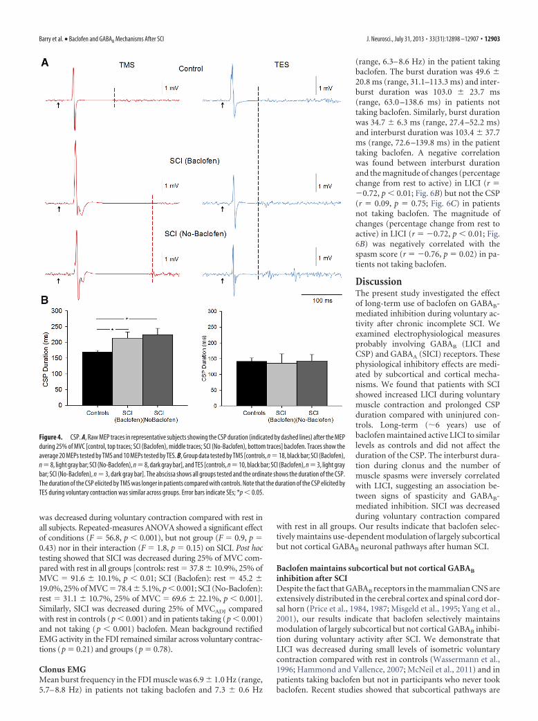
CSPFigure 4 illustrates examples of the CSP elicited by TMS and TESduring 25% of MVC in representative participants. Note that theduration of the CSP was increased in patients compared with acontrol subject when the CSP was elicited by TMS but not by TES.Repeated-measures ANOVA showed a significant effect of group(F � 7.3, p � 0.01) on the duration of the CSP. Post hoc testingshowed that the CSP duration was increased during 25% of MVCin patients taking (213.5 � 50.1 ms, p � 0.01) and not takingbaclofen (224.3 � 55.7 ms, p � 0.01) compared with controls(169.3 � 19.1 ms). No differences were observed between patientgroups (p � 0.3). The duration of the CSP elicited by TES wassimilar across groups [controls � 141.1 � 33.5 ms; SCI(Baclofen) � 135.2 � 51.9 ms; SCI (No-Baclofen) � 142.8 �44.8 ms, p � 0.9]. Overall, these results show that the duration ofthe CSP tested by TMS was longer in patients that controls, re-gardless of their intake of baclofen, but the duration of CSP testedby TES was similar across groups.
SICIFigure 5 illustrates representative examples of SICI measuredin the FDI muscle across conditions tested. Note that SICI
Figure 3. LICI using TES. A, LICI tested in the resting FDI in representative subjects when the conditioning stimulus was given byTMS and test stimulus was given by TES [control, top left traces; SCI (Baclofen), top middle traces; SCI (No-Baclofen), top righttraces]. The test MEP (black traces) and conditioned MEP (red traces) are indicated by black arrows. Traces show the average 10 testMEP and 10 Cond. MEP. B, Group data (controls, n � 10, bottom left; SCI Baclofen, n � 3, bottom left; SCI No-Baclofen, n � 3,bottom right. The abscissa shows all conditions tested (rest, black bars; 25% of MVC, light gray bars). The ordinate shows themagnitude of the conditioned MEP expressed as a percentage of the test MEP. The horizontal dashed line represents the size of thetest MEP. Note that LICI decreased during index finger abduction compared with rest in controls and in patients taking baclofen butremains unchanged in participant’s not taking baclofen. Error bars indicate SEs; *p � 0.05.
12902 • J. Neurosci., July 31, 2013 • 33(31):12898 –12907 Barry et al. • Baclofen and GABAB Mechanisms After SCI
was decreased during voluntary contraction compared with rest inall subjects. Repeated-measures ANOVA showed a significant effectof conditions (F � 56.8, p � 0.001), but not group (F � 0.9, p �0.43) nor in their interaction (F � 1.8, p � 0.15) on SICI. Post hoctesting showed that SICI was decreased during 25% of MVC com-pared with rest in all groups [controls: rest � 37.8 � 10.9%, 25% ofMVC � 91.6 � 10.1%, p � 0.01; SCI (Baclofen): rest � 45.2 �19.0%, 25% of MVC � 78.4 � 5.1%, p � 0.001; SCI (No-Baclofen):rest � 31.1 � 10.7%, 25% of MVC � 69.6 � 22.1%, p � 0.001].Similarly, SICI was decreased during 25% of MVCADJ comparedwith rest in controls (p � 0.001) and in patients taking (p � 0.001)and not taking (p � 0.001) baclofen. Mean background rectifiedEMG activity in the FDI remained similar across voluntary contrac-tions (p � 0.21) and groups (p � 0.78).
Clonus EMGMean burst frequency in the FDI muscle was 6.9 � 1.0 Hz (range,5.7– 8.8 Hz) in patients not taking baclofen and 7.3 � 0.6 Hz
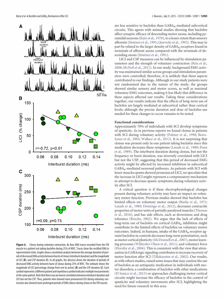
(range, 6.3– 8.6 Hz) in the patient takingbaclofen. The burst duration was 49.6 �20.8 ms (range, 31.1–113.3 ms) and inter-burst duration was 103.0 � 23.7 ms(range, 63.0 –138.6 ms) in patients nottaking baclofen. Similarly, burst durationwas 34.7 � 6.3 ms (range, 27.4 –52.2 ms)and interburst duration was 103.4 � 37.7ms (range, 72.6 –139.8 ms) in the patienttaking baclofen. A negative correlationwas found between interburst durationand the magnitude of changes (percentagechange from rest to active) in LICI (r ��0.72, p � 0.01; Fig. 6B) but not the CSP(r � 0.09, p � 0.75; Fig. 6C) in patientsnot taking baclofen. The magnitude ofchanges (percentage change from rest toactive) in LICI (r � �0.72, p � 0.01; Fig.6B) was negatively correlated with thespasm score (r � �0.76, p � 0.02) in pa-tients not taking baclofen.
DiscussionThe present study investigated the effectof long-term use of baclofen on GABAB-mediated inhibition during voluntary ac-tivity after chronic incomplete SCI. Weexamined electrophysiological measuresprobably involving GABAB (LICI andCSP) and GABAA (SICI) receptors. Thesephysiological inhibitory effects are medi-ated by subcortical and cortical mecha-nisms. We found that patients with SCIshowed increased LICI during voluntarymuscle contraction and prolonged CSPduration compared with uninjured con-trols. Long-term (�6 years) use ofbaclofen maintained active LICI to similarlevels as controls and did not affect theduration of the CSP. The interburst dura-tion during clonus and the number ofmuscle spasms were inversely correlatedwith LICI, suggesting an association be-tween signs of spasticity and GABAB-mediated inhibition. SICI was decreasedduring voluntary contraction compared
with rest in all groups. Our results indicate that baclofen selec-tively maintains use-dependent modulation of largely subcorticalbut not cortical GABAB neuronal pathways after human SCI.
Baclofen maintains subcortical but not cortical GABAB
inhibition after SCIDespite the fact that GABAB receptors in the mammalian CNS areextensively distributed in the cerebral cortex and spinal cord dor-sal horn (Price et al., 1984, 1987; Misgeld et al., 1995; Yang et al.,2001), our results indicate that baclofen selectively maintainsmodulation of largely subcortical but not cortical GABAB inhibi-tion during voluntary activity after SCI. We demonstrate thatLICI was decreased during small levels of isometric voluntarycontraction compared with rest in controls (Wassermann et al.,1996; Hammond and Vallence, 2007; McNeil et al., 2011) and inpatients taking baclofen but not in participants who never tookbaclofen. Recent studies showed that subcortical pathways are
Figure 4. CSP. A, Raw MEP traces in representative subjects showing the CSP duration (indicated by dashed lines) after the MEPduring 25% of MVC [control, top traces; SCI (Baclofen), middle traces; SCI (No-Baclofen), bottom traces] baclofen. Traces show theaverage 20 MEPs tested by TMS and 10 MEPs tested by TES. B, Group data tested by TMS [controls, n �18, black bar; SCI (Baclofen),n�8, light gray bar; SCI (No-Baclofen), n�8, dark gray bar], and TES [controls, n�10, black bar; SCI (Baclofen), n�3, light graybar; SCI (No-Baclofen), n � 3, dark gray bar]. The abscissa shows all groups tested and the ordinate shows the duration of the CSP.The duration of the CSP elicited by TMS was longer in patients compared with controls. Note that the duration of the CSP elicited byTES during voluntary contraction was similar across groups. Error bars indicate SEs; *p � 0.05.
Barry et al. • Baclofen and GABAB Mechanisms After SCI J. Neurosci., July 31, 2013 • 33(31):12898 –12907 • 12903

involved in LICI (McNeil et al., 2009,2011) in addition to cortical mechanisms(Nakamura et al., 1997; Chen et al., 1999;Di Lazzaro et al., 2002). Our data are inagreement because we found that LICIwas modulated to a similar extent whenthe TS was elicited by TMS or TES. Be-cause TES activates axons of pyramidaltract cells in the subcortical white matter(Burke et al., 1993; Di Lazzaro et al., 1998)it is most likely that the changes we ob-served here involved subcortical influ-ences. Pharmacological studies indicate arole of GABAB receptors in mediatingLICI (Werhahn et al., 1999; McDonnell etal., 2006); therefore, it is possible that theprolonged use of baclofen contributed tomodulate LICI in patients taking baclofento similar level as controls. Baclofen canbind to presynaptic GABAB receptorsleading to a decrease in the release ofGABA by negative feedback (Deisz, 1999)altering GABAergic synaptic transmis-sion according to physiological needs(Ohliger-Frerking et al., 2003). Impor-tantly, LICI remained increased duringvoluntary activity in patients who nevertook baclofen. Our results are in line withanimal studies showing that GABAergicinhibitory events occurring at the spinalcord level are increased after SCI (Tilla-karatne et al., 2000; Diaz-Ruiz et al., 2007;Sadlaoud et al., 2010). Moreover, wefound little evidence that motor corticalinhibition could have contributed to theincreased LICI during voluntary contrac-tion in these patients. First, the size of theconditioned MEP tested during LICI dur-ing voluntary activity was larger than theunconditioned MEP elicited at rest in allgroups, suggesting that additional motorcortical elements that can be activated byTMS are facilitated. Second, we found that motor cortical SICI, aprobably GABAA-mediated effect, decreases during voluntary activ-ity in all groups. Although, LICI and SICI inhibitory effects are prob-ably mediated by GABAergic connections involving GABAB andGABAA receptors the involvement of different receptor subtypesdoes not exclude the possibility that a common neuronal populationmediate these inhibitory effects.
Studies in humans have shown that GABAergic inhibition isdecreased after SCI (Calancie et al., 1993; Faist et al., 1994; Ay-mard et al., 2000). At first sight these results might seem in con-tradiction to our findings and raise the question of howGABAergic inhibition is affected after SCI. It is important toconsider that previous studies tested measurements at rest, didnot separate patients according to the use of baclofen, and testedanother measurement of GABAergic inhibition by examiningpresynaptic inhibition of Ia afferents. Animal (Stuart and Red-man, 1992) and human (Orsnes et al., 2000) studies on spasticityhave shown that baclofen has no effect on classical presynapticinhibition. Presynaptic inhibition of Ia afferents, accompanied byprimary afferent depolarization, is caused by axo-axonal GABAAsynapses and activation of GABAA receptors via GABAergic in-
terneurons (Rudomin and Schmidt, 1999). At present, the pre-cise role of GABAB receptors in mediating presynaptic inhibitionis not clear. Thus, changes in GABAergic inhibition after injuryneed to be considered in a task-dependent context with attentionto the type of GABA receptors involved and the medication takeby patients.
We also found that CSP durations were similar in patientsregardless of baclofen use and were longer than controls. The firstpart of the CSP may be mediated by spinal contributions, whereasthe later part results from suppression of neural output by in-terneurons at the cortical level (Fuhr et al., 1991; Chen et al.,1999; Tergau et al., 1999). Our results show that the duration ofthe CSP tested with TES was similar across groups, suggestingthat differences observed between patients and controls (whenthe CSP was tested by TMS) involve cortical mechanisms. Thisagrees with previous results showing that GABAergic inhibitiontested during voluntary activity is increased in patients with SCIcompared with controls (Freund et al., 2011; Bunday and Perez,2012). GABAB receptors play a role in the inhibition tested duringthe CSP (Ziemann et al., 1996; Siebner et al., 1998); thus, ourfindings suggest that GABAB-mediated effects by cortical circuits
Figure 5. SICI. A, SICI recorded from the resting FDI in a representative control subject (top left traces) and in a patient taking(top middle traces) and not taking (top right traces) baclofen. The test MEP (black traces) and conditioned MEP (red traces) areindicated by black arrows. Traces show the average 20 test MEP and 20 Cond. MEP. B, Group data [controls, n � 10, bottom left;SCI Baclofen, n � 6, bottom left; SCI No-Baclofen, n � 6, bottom right]. The abscissa shows all conditions tested (rest, black bars;25% of MVC, light gray bars; 25% of MVCADJ, dark gray bars). The ordinate shows the magnitude of the conditioned MEP expressedas a percentage of the test MEP. The horizontal dashed line represents the size of the test MEP. Note that SICI decreased duringindex finger abduction compared with rest in all groups tested. Error bars indicate SEs; *p � 0.05.
12904 • J. Neurosci., July 31, 2013 • 33(31):12898 –12907 Barry et al. • Baclofen and GABAB Mechanisms After SCI
are less sensitive to baclofen than GABAB-mediated subcorticalcircuits. This agrees with animal studies showing that baclofenaffect synaptic efficacy of descending motor axons, including py-ramidal neurons (Kato et al., 1978), to a lesser extent than sensoryafferents (Jimenez et al., 1991; Quevedo et al., 1992). This may inpart be related to the larger density of GABAB receptors found interminals of afferent axons compared with the terminals of de-scending axons (Jimenez et al., 1991).
LICI and CSP measures can be influenced by stimulation pa-rameters and the strength of voluntary contraction (Reis et al.,2008; McNeil et al., 2011). In our study, background EMG activ-ity was maintained similar across groups and stimulation param-eters were controlled; therefore, it is unlikely that these aspectscontributed to our findings. Although in our study patients werenot randomized due to the nature of the study, the groupsshowed similar sensory and motor scores, as well as maximalvoluntary EMG outcomes, making it less likely that difference inthese aspects affected our results. Taking these considerationstogether, our results indicate that the effects of long-term use ofbaclofen are largely mediated at subcortical rather than corticallevels; although the precise duration and dose of baclofen-useneeded for these changes to occur remains to be tested.
Functional considerationsApproximately 70% of individuals with SCI develop symptomsof spasticity. As in previous reports we found clonus in patientswith SCI during voluntary activity (Palmer et al., 1998; Beres-Jones et al., 2003; Wallace et al., 2012). It is not surprising thatclonus was present only in one patient taking baclofen since thismedication decreases these symptoms (Latash et al., 1989; Pennet al., 1989). The interburst duration during clonus, but not thefrequency or burst duration, was inversely correlated with LICIbut not the CSP, suggesting that this period of decreased EMGactivity might by affected by increased inhibition in subcorticalGABAB-mediated neuronal pathways. As patients with SCI withlesser muscles spams showed pronounced LICI, we speculate thatthe increase in LICI might represent a compensatory mechanismto attempt to decrease spastic symptoms during voluntary activ-ity after SCI.
A critical question is if these electrophysiological changespresent during voluntary activity may have an impact on volun-tary motor function. Previous studies showed that baclofen haslimited effects on voluntary motor output (Burke et al., 1971;Latash et al., 1989; Domingo et al., 2012), decreases contractileproperties of motor units of partially paralyzed muscles (Thomaset al., 2010), and has side effects, such as drowsiness and drugtolerance (Rosche, 2002). We argue that the lack of effects oflong-term use of baclofen on cortical GABAB inhibition mightcontribute to the limited effects of baclofen on voluntary motoroutcomes. Indeed, in humans, intake of the GABAB receptor ag-onist baclofen in controls decreases long-term potentiation, suchas motor cortical plasticity (McDonnell et al., 2007), motor learn-ing processes (Willerslev-Olsen et al., 2011), and voluntary force(Hornby et al., 2004). This is consistent with the view that atten-uation in GABAergic signaling contributes to increase recovery ofmotor function after SCI (Tillakaratne et al., 2002). Our results,as with others studies, raised some issues that may caution the useof baclofen as an antispastic medication in individuals with mo-tor disorders; a combination of baclofen with other medications(D’Amico et al., 2013) or approaches challenging motor corticalcircuits might increase the efficacy of baclofen in the control ofspasticity and voluntary movements after SCI, highlighting theneed for future research in this area.
Figure 6. Clonus during voluntary contraction. A, Raw EMG traces recorded from the FDImuscle in a patient not taking baclofen during 25% of MVC. Traces show the rectified EMG inrepresentative trials. Graphs show a correlation analysis between the average duration of peri-ods of decreased EMG activity between bursts of clonus (interburst duration) and the magnitudeof LICI (B), and CSP duration (C). In all graphs, the abscissa shows the duration of periods ofdecreased EMG activity between burst of clonus during 25% of MVC. The ordinate shows themagnitude of LICI (percentage change from rest to active) (B) and the CSP duration (C). Eachsymbol represents a different patient and repetition a symbol indicates multiple measurementsin the same patient. Note that there was an inverse correlation between interburst duration andLICI but not the CSP. Thus, patients who showed more pronounced LICI during voluntary con-traction also showed more prolonged periods of EMG silence during clonus in the FDI muscle.
Barry et al. • Baclofen and GABAB Mechanisms After SCI J. Neurosci., July 31, 2013 • 33(31):12898 –12907 • 12905

ReferencesAydin G, Tomruk S, Keles I, Demir SO, Orkun S (2005) Transcutaneous
electrical nerve stimulation versus baclofen in spasticity: clinical and elec-trophysiologic comparison. Am J Phys Med Rehabil 84:584 –592.CrossRef Medline
Aymard C, Katz R, Lafitte C, Lo E, Penicaud A, Pradat-Diehl P, Raoul S(2000) Presynaptic inhibition and homosynaptic depression: a compar-ison between lower and upper limbs in normal human subjects and pa-tients with hemiplegia. Brain 123:1688 –1702. CrossRef Medline
Azouvi P, Mane M, Thiebaut JB, Denys P, Remy-Neris O, Bussel B (1996)Intrathecal baclofen administration for control of severe spinal spasticity:functional improvement and long-term follow-up. Arch Phys Med Reha-bil 77:35–39. CrossRef Medline
Beres-Jones JA, Johnson TD, Harkema SJ (2003) Clonus after human spinalcord injury cannot be attributed solely to recurrent muscle-tendonstretch. Exp Brain Res 149:222–236. Medline
Bunday KL, Perez MA (2012) Impaired crossed facilitation of the corti-cospinal pathway after cervical spinal cord injury. J Neurophysiol 107:2901–2911. CrossRef Medline
Burke D, Andrews CJ, Knowles L (1971) The action of a GABA derivative inhuman spasticity. J Neurol Sci 14:199 –208. CrossRef Medline
Burke D, Hicks R, Gandevia SC, Stephen J, Woodforth I, Crawford M (1993)Direct comparison of corticospinal volleys in human subjects to transcra-nial magnetic and electrical stimulation. J Physiol 470:383–393. Medline
Butler JE, Petersen NC, Herbert RD, Gandevia SC, Taylor JL (2012) Originof the low-level EMG during the silent period following transcranial mag-netic stimulation. Clin Neurophysiol 123:1409 –1414. CrossRef Medline
Calancie B, Broton JG, Klose KJ, Traad M, Difini J, Ayyar DR (1993) Evi-dence that alterations in presynaptic inhibition contribute to segmentalhypo- and hyperexcitability after spinal cord injury in man. Electroen-cephalogr Clin Neurophysiol 89:177–186. CrossRef Medline
Chen R, Lozano AM, Ashby P (1999) Mechanism of the silent period fol-lowing transcranial magnetic stimulation: evidence from epidural record-ings. Exp Brain Res 128:539 –542. CrossRef Medline
Curtis DR, Gynther BD, Lacey G, Beattie DT (1997) Baclofen: reduction ofpresynaptic calcium influx in the cat spinal cord in vivo. Exp Brain Res113:520 –533. CrossRef Medline
D’Amico JM, Li Y, Bennett DJ, Gorassini MA (2013) Reduction of spinalsensory transmission by facilitation of 5HT1 receptors in non-injured andspinal cord injured humans. J Neurophysiol 109:1485–1493. CrossRefMedline
Deisz RA (1999) GABA(B) receptor-mediated effects in human and rat neo-cortical neurones in vitro. Neuropharmacology 38:1755–1766. CrossRefMedline
Diaz-Ruiz A, Salgado-Ceballos H, Montes S, Maldonado V, Tristan L,Alcaraz-Zubeldia M, Ríos C (2007) Acute alterations of glutamate, glu-tamine, GABA, and other amino acids after spinal cord contusion in rats.Neurochem Res 32:57– 63. CrossRef Medline
Di Lazzaro V, Oliviero A, Profice P, Saturno E, Pilato F, Insola A, Mazzone P,Tonali P, Rothwell JC (1998) Comparison of descending volleys evokedby transcranial magnetic and electric stimulation in conscious humans.Electroencephalogr Clin Neurophysiol 109:397– 401. CrossRef Medline
Di Lazzaro V, Oliviero A, Mazzone P, Pilato F, Saturno E, Insola A, VisocchiM, Colosimo C, Tonali PA, Rothwell JC (2002) Direct demonstration oflong latency cortico-cortical inhibition in normal subjects and in a patientwith vascular parkinsonism. Clin Neurophysiol 113:1673–1679. CrossRefMedline
Domingo A, Al-Yahya AA, Asiri Y, Eng JJ, Lam T (2012) A systematic reviewof the effects of pharmacological agents on walking function in peoplewith spinal cord injury. J Neurotrauma 29:865– 879. CrossRef Medline
Faist M, Mazevet D, Dietz V, Pierrot-Deseilligny E (1994) A quantitativeassessment of presynaptic inhibition of Ia afferents in spastics: differencesin hemiplegics and paraplegics. Brain 117:1449 –1455. CrossRef Medline
Freund P, Rothwell J, Craggs M, Thompson AJ, Bestmann S (2011) Corti-comotor representation to a human forearm muscle changes followingcervical spinal cord injury. Eur J Neurosci 34:1839 –1846. CrossRefMedline
Fuhr P, Agostino R, Hallett M (1991) Spinal motor neuron excitability dur-ing the silent period after cortical stimulation. Electroencephalogr ClinNeurophysiol 81:257–262. CrossRef Medline
Hammond G, Vallence AM (2007) Modulation of long-interval intracorti-
cal inhibition and the silent period by voluntary contraction. Brain Res1158:63–70. CrossRef Medline
Hornby TG, Heckman CJ, Harvey RL, Rymer WZ (2004) Changes in vol-untary torque and electromyographic activity following oral baclofen.Muscle Nerve 30:784 –795. CrossRef Medline
Jimenez I, Rudomin P, Enriquez M (1991) Differential effects of (-)-baclofen on Ia and descending monosynaptic EPSPs. Exp Brain Res 85:103–113. Medline
Kato M, Waldmann U, Murakami S (1978) Effects of baclofen on spinalneurones of cats. Neuropharmacology 17:827– 833. CrossRef Medline
Kujirai T, Caramia MD, Rothwell JC, Day BL, Thompson PD, Ferbert A,Wroe S, Asselman P, Marsden CD (1993) Corticocortical inhibition inhuman motor cortex. J Physiol 471:501–519. Medline
Kumru H, Kofler M (2012) Effect of spinal cord injury and of intrathecalbaclofen on brainstem reflexes. Clin Neurophysiol 123:45–53. CrossRefMedline
Latash ML, Penn RD, Corcos DM, Gottlieb GL (1989) Short-term effects ofintrathecal baclofen in spasticity. Exp Neurol 103:165–172. CrossRefMedline
Li Y, Li X, Harvey PJ, Bennett DJ (2004) Effects of baclofen on spinal reflexesand persistent inward currents in motoneurons of chronic spinal rats withspasticity. J Neurophysiol 92:2694 –2703. CrossRef Medline
Marino RJ, Barros T, Biering-Sorensen F, Burns SP, Donovan WH, GravesDE, Haak M, Hudson LM, Priebe MM (2003) International standardsfor neurological classification of spinal cord injury. J Spinal Cord Med26:50 –56. Medline
McDonnell MN, Orekhov Y, Ziemann U (2006) The role of GABAB recep-tors in intracortical inhibition in the human motor cortex. Exp Brain Res173:86 –93. CrossRef Medline
McDonnell MN, Orekhov Y, Ziemann U (2007) Suppression of LTP-likeplasticity in human motor cortex by the GABAB receptor agonistbaclofen. Exp Brain Res 180:181–186. CrossRef Medline
McNeil CJ, Martin PG, Gandevia SC, Taylor JL (2009) The response topaired motor cortical stimuli is abolished at a spinal level during humanmuscle fatigue. J Physiol 587:5601–5612. CrossRef Medline
McNeil CJ, Martin PG, Gandevia SC, Taylor JL (2011) Long-interval intra-cortical inhibition in a human hand muscle. Exp Brain Res 209:287–297.CrossRef Medline
Misgeld U, Bijak M, Jarolimek W (1995) A physiological role for GABABreceptors and the effects of baclofen in the mammalian central nervoussystem. Prog Neurobiol 46:423– 462. CrossRef Medline
Nakamura H, Kitagawa H, Kawaguchi Y, Tsuji H (1997) Intracortical facil-itation and inhibition after transcranial magnetic stimulation in con-scious humans. J Physiol 498:817– 823. Medline
Ohliger-Frerking P, Wiebe SP, Staubli U, Frerking M (2003) GABA(B)receptor-mediated presynaptic inhibition has history-dependent effectson synaptic transmission during physiologically relevant spike trains.J Neurosci 23:4809 – 4814. Medline
Orsnes G, Crone C, Krarup C, Petersen N, Nielsen J (2000) The effect ofbaclofen on the transmission in spinal pathways in spastic multiple scle-rosis patients. Clin Neurophysiol 111:1372–1379. CrossRef Medline
Palmer DT, Horn LJ, Harmon RL (1998) Botulinum toxin treatment oflumbrical spasticity: a brief report. Am J Phys Med Rehabil 77:348 –350.CrossRef Medline
Penn RD, Savoy SM, Corcos D, Latash M, Gottlieb G, Parke B, Kroin JS(1989) Intrathecal baclofen for severe spinal spasticity. N Engl J Med320:1517–1521. CrossRef Medline
Pierrot-Deseilligny E, Burke D (2005) Propriospinal relay for descendingmotor commands. In: The circuitry of the human spinal cord, pp 452–510. New York: Cambridge UP.
Price GW, Wilkin GP, Turnbull MJ, Bowery NG (1984) Are baclofen-sensitive GABAB receptors present on primary afferent terminals of thespinal cord? Nature 307:71–74. CrossRef Medline
Price GW, Kelly JS, Bowery NG (1987) The location of GABAB receptorsbinding sites in mammalian spinal cord. Synapse 1:530 –538. CrossRefMedline
Quevedo J, Eguibar JR, Jimenez I, Rudomin P (1992) Differential action of(-)-baclofen on the primary afferent depolarization produced by segmen-tal and descending inputs. Exp Brain Res 91:29 – 45. CrossRef Medline
Reis J, Swayne OB, Vandermeeren Y, Camus M, Dimyan MA, Harris-Love M,Perez MA, Ragert P, Rothwell JC, Cohen LG (2008) Contribution oftranscranial magnetic stimulation to the understanding of cortical mech-
12906 • J. Neurosci., July 31, 2013 • 33(31):12898 –12907 Barry et al. • Baclofen and GABAB Mechanisms After SCI

anisms involved in motor control. J Physiol 586:325–351. CrossRefMedline
Rosche J (2002) Treatment of spasticity. Spinal Cord 40:261–262. CrossRefMedline
Rothwell JC (1997) Techniques and mechanisms of action of transcranialstimulation of the human motor cortex. J Neurosci Methods 74:113–122.CrossRef Medline
Rothwell JC, Hallett M, Berardelli A, Eisen A, Rossini P, Paulus W (1999)Magnetic stimulation: motor evoked potentials. The International Feder-ation of Clinical Neurophysiology. Electroencephalogr Clin Neuro-physiol Suppl 52:97–103. Medline
Roy FD, Zewdie ET, Gorassini MA (2011) Short-interval intracorticalinhibition with incomplete spinal cord injury. Clin Neurophysiol 122:1387–1395. CrossRef Medline
Roy RR, Edgerton VR (2012) Neurobiological perspective of spasticity asoccurs after a spinal cord injury. Exp Neurol 235:116 –1122. CrossRefMedline
Rudomin P, Schmidt RF (1999) Presynaptic inhibition in the vertebrate spi-nal cord revisited. Exp Brain Res 129:1–37. CrossRef Medline
Sadlaoud K, Tazerart S, Brocard C, Jean-Xavier C, Portalier P, Brocard F,Vinay L, Bras H (2010) Differential plasticity of the GABAergic and gly-cinergic synaptic transmission to rat lumbar motoneurons after spinalcord injury. J Neurosci 30:3358 –3369. CrossRef Medline
Sakai K, Ugawa Y, Terao Y, Hanajima R, Furubayashi T, Kanazawa I (1997)Preferential activation of different I waves by transcranial magnetic stim-ulation with a figure-of-eight-shaped coil. Exp Brain Res 113:24 –32.CrossRef Medline
Saturno E, Bonato C, Miniussi C, Lazzaro V, Callea L (2008) Motor cortexchanges in spinal cord injury: a TMS study. Neurol Res 30:1084 –1085.CrossRef Medline
Shimizu T, Hino T, Komori T, Hirai S (2000) Loss of the muscle silentperiod evoked by transcranial magnetic stimulation of the motor cortex inpatients with cervical cord lesions. Neurosci Lett 286:199 –202. CrossRefMedline
Siebner HR, Dressnandt J, Auer C, Conrad B (1998) Continuous intrathecalbaclofen infusions induced a marked increase of the transcranially evokedsilent period in a patient with generalized dystonia. Muscle Nerve 21:1209 –1212. CrossRef Medline
Stuart GJ, Redman SJ (1992) The role of GABAA and GABAB receptors in
presynaptic inhibition of Ia EPSPs in cat spinal motoneurones. J Physiol447:675– 692. Medline
Tergau F, Wanschura V, Canelo M, Wischer S, Wassermann EM, Ziemann U,Paulus W (1999) Complete suppression of voluntary motor drive dur-ing the silent period after transcranial magnetic stimulation. Exp BrainRes 124:447– 454. CrossRef Medline
Thomas CK, Hager-Ross CK, Klein CS (2010) Effects of baclofen on motorunits paralysed by chronic cervical spinal cord injury. Brain 133:117–125.CrossRef Medline
Tillakaratne NJ, Mouria M, Ziv NB, Roy RR, Edgerton VR, Tobin AJ (2000)Increased expression of glutamate decarboxylase (GAD(67)) in felinelumbar spinal cord after complete thoracic spinal cord transection. J Neu-rosci Res 60:219 –230. CrossRef Medline
Tillakaratne NJ, de Leon RD, Hoang TX, Roy RR, Edgerton VR, Tobin AJ(2002) Use-dependent modulation of inhibitory capacity in the felinelumbar spinal cord. J Neurosci 22:3130 –3143. Medline
Valls-Sole J, Pascual-Leone A, Wassermann EM, Hallett M (1992) Humanmotor evoked responses to paired transcranial magnetic stimuli. Electro-encephalogr Clin Neurophysiol 85:355–364. Medline
Wallace DM, Ross BH, Thomas CK (2012) Characteristics of lower extrem-ity clonus after human cervical spinal cord injury. J Neurotrauma 29:915–924. CrossRef Medline
Wassermann EM, Samii A, Mercuri B, Ikoma K, Oddo D, Grill SE, HallettM (1996) Responses to paired transcranial magnetic stimuli in rest-ing, active, and recently activated muscles. Exp Brain Res 109:158 –163. Medline
Werhahn KJ, Kunesch E, Noachtar S, Benecke R, Classen J (1999) Differen-tial effects on motorcortical inhibition induced by blockade of GABAuptake in humans. J Physiol 517:591–597. CrossRef Medline
Willerslev-Olsen M, Lundbye-Jensen J, Petersen TH, Nielsen JB (2011) Theeffect of baclofen and diazepam on motor skill acquisition in healthysubjects. Exp Brain Res 213:465– 474. CrossRef Medline
Yang K, Wang D, Li YQ (2001) Distribution and depression of the GABABreceptor in the spinal dorsal horn of adult rat. Brain Res Bull 55:479 – 485.CrossRef Medline
Ziemann U, Lonnecker S, Steinhoff BJ, Paulus W (1996) Effects of antiepi-leptic drugs on motor cortex excitability in humans: a transcranial mag-netic stimulation study. Ann Neurol 40:367–378. CrossRef Medline
Barry et al. • Baclofen and GABAB Mechanisms After SCI J. Neurosci., July 31, 2013 • 33(31):12898 –12907 • 12907