semaphorin 4d/plexin-b1–mediated r-ras gap activity inhibits … · migration, and tumor...
TRANSCRIPT

TH
EJ
OU
RN
AL
OF
CE
LL
BIO
LO
GY
JCB: ARTICLE
© The Rockefeller University Press $8.00The Journal of Cell Biology, Vol. 173, No. 4, May 22, 2006 601–613http://www.jcb.org/cgi/doi/10.1083/jcb.200508204
JCB 601
IntroductionSemaphorins comprise a large family of secreted and trans-
membrane molecules that play central roles in axon guidance
in the developing nervous system (Kolodkin et al., 1993;
Tamagnone et al., 1999). The function of semaphorins is mediated
by plexins, which are classifi ed into four subfamilies: Plexin-A,
-B, -C, and -D (Tamagnone et al., 1999). Semaphorins were
originally identifi ed as repulsive axonal guidance molecules,
but they have recently been shown to regulate integrin- mediated
cell migration in a variety of cells (Tamagnone and Comoglio,
2004). Sema3A exerts an essential permissive role in the
execution of vasculature remodeling by inhibiting integrin-
mediated adhesion of endothelial cells to the ECM (Serini
et al., 2003). Activation of Plexin-B1 negatively regulates
integrin-based cell adhesion and migration of NIH-3T3 cells
(Barberis et al., 2004). Plexin-C1 inhibits integrin-mediated
adhesion and chemokine-induced migration of dendritic cells
(Walzer et al., 2005). Thus, semaphorin/plexin signaling plays
an important role in the migration of a variety of cells.
However, the molecular mechanisms underlying the inhibition
of integrin-mediated cell migration by semaphorins through
plexins remain unclear.
Rho family small GTPases are signal transduction mole-
cules that remodel the actin cytoskeleton and play fundamental
roles in numerous cellular processes (Negishi and Katoh, 2002).
The small GTPase Rnd1, a constitutively active GTPase (Nobes
et al., 1998), is known to interact directly with the cytoplasmic
domain of Plexin-B1 (Oinuma et al., 2003). We recently re-
vealed that Plexin-B1 functions as an R-Ras GTPase-activating
protein (GAP) and directly and specifi cally down-regulates
R-Ras activity in response to Sema4D, inducing repulsive re-
sponse in hippocampal neurons, and that the expression of
R-Ras GAP activity of Plexin-B1 requires Rnd1 association with
the receptor (Oinuma et al., 2004a). Furthermore, expression of
constitutively active R-Ras prevents growth cone collapse
induced by Sema4D/Plexin-B1 as well as Sema3A/Plexin-A1,
whereas R-Ras siRNA caused a growth cone collapse similar to
those induced by semaphorins (Oinuma et al., 2004a).
Integrins are a family of α/β heterodimeric cell surface
receptors that bind to the ECM, such as collagens and fi bronec-
tins, and play a central part in regulating cell growth, survival,
migration, and tumor metastasis (Hood and Cheresh, 2002).
Activation of integrins is essential for cell adhesion and cell
mig ration, and several studies show that the Ras family of small
Semaphorin 4D/Plexin-B1–mediated R-Ras GAP activity inhibits cell migration by regulating β1 integrin activity
Izumi Oinuma, Hironori Katoh, and Manabu Negishi
Laboratory of Molecular Neurobiology, Graduate School of Biostudies, Kyoto University, Sakyo-ku, Kyoto 606-8502, Japan
Plexins are cell surface receptors for semaphorins and
regulate cell migration in many cell types. We re-
cently reported that the semaphorin 4D (Sema4D)
receptor Plexin-B1 functions as a GTPase-activating pro-
tein (GAP) for R-Ras, a member of Ras family GTPases
implicated in regulation of integrin activity and cell migra-
tion (Oinuma, I., Y. Ishikawa, H. Katoh, and M. Negishi.
2004. Science. 305:862–865). We characterized the role
of R-Ras downstream of Sema4D/Plexin-B1 in cell migra-
tion. Activation of Plexin-B1 by Sema4D suppressed the
ECM-dependent R-Ras activation, R-Ras–mediated phos-
phatydylinositol 3-kinase activation, and β1 integrin acti-
vation through its R-Ras GAP domain, leading to inhibition
of cell migration. In addition, inactivation of R-Ras by
overexpression of the R-Ras–specifi c GAP or knockdown
of R-Ras by RNA interference was suffi cient for suppress-
ing β1 integrin activation and cell migration in response to
the ECM stimulation. Thus, we conclude that R-Ras activity
is critical for ECM-mediated β1 integrin activation and
cell migration and that inactivation of R-Ras by Sema4D/
Plexin-B1–mediated R-Ras GAP activity controls cell mi-
gration by modulating the activity of β1 integrins.
Correspondence to Manabu Negishi: [email protected]
Abbreviations used in this paper: GAP, GTPase-activating protein; HS, horse serum; PE, phycoerythrin; PI3-K, phosphatydylinositol 3-kinase; WT, wild type.
The online version of this article contains supplemental material.

JCB • VOLUME 173 • NUMBER 4 • 2006 602
GTPases regulates integrin activity (Kinbara et al., 2003).
Among the Ras family GTPases, activated R-Ras was shown to
induce integrin activation and increase cell adhesion and matrix
assembly, suggesting that R-Ras plays an important role in the
regulation of integrin activity (Zhang et al., 1996; Sethi et al.,
1999). However, how R-Ras activity is regulated and how
R-Ras activates integrins remain obscure. Signifi cantly, Sema4D
was the fi rst extracellular stimulus shown to infl uence the activ-
ity of R-Ras. These facts collectively prompted us to speculate
that plexins regulate integrin-mediated cell migration by their
R-Ras GAP activity.
In this study, we characterized the role of R-Ras down-
stream of Sema4D/Plexin-B1 in regulation of integrin activa-
tion and cell migration. The activation of R-Ras by ECM is
required for ECM-mediated integrin activation and cell migra-
tion, and Sema4D/Plexin-B1 inhibits integrin activation and
cell migration through R-Ras GAP activity. We also revealed
that down-regulation of phosphatydylinositol 3-kinase (PI3-K)
activity is responsible for Sema4D/Plexin-B1–induced suppres-
sion of β1 integrin activity and cell migration.
ResultsSema4D antagonizes integrin-mediated cell migrationWe examined the effect of Sema4D on integrin-mediated migra-
tion of PC12 cells in a cell migration assay (Fig. 1, A and B).
Transwell chambers were coated on the lower side with varying
concentrations of collagen I. PC12 cells exhibited a collagen
concentration–dependent promotion of cell migration, which
was antagonized by Sema4D. The collagen-dependent PC12
cell migration is mediated by α1 and β1 integrin subunits, as
functional blocking antibodies against α1 (Fig. 1, C and D) and
β1 (Fig. 1, E and F) integrin subunits strongly impaired the
migration. These results indicate that Sema4D antagonizes the
collagen receptor, α1/β1 integrin–dependent PC12 cell migration.
Sema4D through Plexin-B1 inhibits ECM-mediated activation of R-Ras and 𝛃1 integrinsR-Ras is implicated in integrin-mediated cell migration, and
expression of a constitutively active form of R-Ras has been
shown to stimulate cell migration (Keely et al., 1999). We
previously reported that Sema4D stimulation down-regulates
NGF-stimulated R-Ras activity via the R-Ras GAP activity of
Plexin-B1 to induce neurite retraction (Oinuma et al., 2004a).
We next tested whether stimulation of PC12 cells with colla-
gen and Sema4D affects R-Ras activity. PC12 cells were
plated onto collagen-coated dishes and lysed, and the lysates
were incubated with the GST-fused Ras binding domain of
c-Raf-1 (GST-RBD) to pull down activated R-Ras (de Rooij and
Bos, 1997). As shown in Fig. 2 A, cells plated on collagen-
coated dishes showed a collagen concentration–dependent
increase in endogenous R-Ras activity, whereas those kept
in suspension or plated onto the non–integrin-dependent
substrate poly-d-lysine did not. Furthermore, the colla gen-
dependent activation of R-Ras was inhibited by a func-
tional blocking antibody against β1 integrins, P5D2, and was
enhanced by affi nity-related activation of β1 integrins by the
monoclonal antibody 8A2, which mechanically induces a
Figure 1. Sema4D antagonizes integrin- mediated cell migration. (A) PC12 cells were tested in the transwell assay either in the pres-ence or absence of Sema4D. Transwell chambers were coated on the lower side with varying concentrations of collagen I. Migrated cells were visualized by the staining of crystal violet. (C and E) PC12 cells were pretreated with 5 μg/ml of functional block-ing monoclonal antibodies against α1 and β1 integrin subunits (3A3 and P5D2, respec-tively) or control mouse IgG1 and subjected to the transwell assay. Migrated cells were visu-alized by the staining of crystal violet. (B, D, and F) Relative cell migration was determined by the number of the migrated cells normal-ized to the total number of cells. Results are the means ± SEM of three independent experiments. Bars, 1 mm.

MECHANISM OF INTEGRIN INHIBITION BY SEMA4D • OINUMA ET AL. 603
high-affi nity state of β1 integrins. These data suggest that
β1 integrins are required for R-Ras activation upon ECM-
mediated adhesion. Sema4D stimulation strongly in hibited
the collagen-induced activation of R-Ras, and affi nity- related
activation of β1 integrins by the 8A2 antibody attenuated the
inhibitory effect of Sema4D on ECM-mediated R-Ras activation.
R-Ras is known to regulate β1 integrin activation (Zhang
et al., 1996). To examine the effect of Sema4D on β1 integrin
activity, we measured the activity of β1 integrins in cells with
or without Sema4D stimulation by the immunoprecipitation
assay with the monoclonal antibody against active conforma-
tions of β1 integrins, HUTS-4, which detects hybrid domain
swing-out in β1 integrins, a process most commonly associ-
ated with ligand binding (Mould et al., 2003). Sema4D antag-
onized the collagen-dependent activation of β1 integrins (Fig.
2 B). Inhibition of β1 integrin activity by Sema4D was also
observed in the ELISA using the HUTS-4 antibody, which
was performed under a detergent-free condition (Fig. 2 C).
To further ascertain that Sema4D indeed affects the activity of
β1 integrins, we performed fl ow cytometry analysis using the
HUTS-4 antibody. As shown in Fig. 2 D, cells treated with
Sema4D showed a decrease in the level of HUTS-4 binding
(FITC staining). Mn2+ treatment, which induces the activa-
tion of β1 integrins, resulting in the effective interaction with
the ECM ligands and increased HUTS-4 binding (Luque
et al., 1996), completely overcame the Sema4D-induced
decrease in HUTS-4 binding. These results suggest that
decreased HUTS-4 binding induced by Sema4D is due to
affi nity modulation of β1 integrins. FAK is known to be
autophosphorylated at tyrosine upon integrin activation
(Hildebrand et al., 1993), and FAK phosphorylation down-
stream of β1 integrins is the important step for integrin-
mediated cell migration (Parsons et al., 2000; Sieg et al.,
2000). As shown in Fig. 2 E, Sema4D inhibited the collagen-
mediated FAK tyrosine phosphorylation. These results sug-
gest that Sema4D inhibits ECM-mediated activation of R-Ras
and β1 integrins.
We also confi rmed the involvement of the endogenous
Plexin-B1 receptor in Sema4D-dependent inhibition of R-Ras
activity and integrin functions. As shown in Fig. 3, both
Sema4D- dependent inhibition of collagen-mediated activation
of R-Ras and cell migration were blocked by the monoclonal
antibody against Plexin-B1, which recognizes the extracellular
ligand binding region of the receptor. These results suggest that
Sema4D through Plexin-B1 inhibits ECM-mediated activation
of R-Ras, functional activation of β1 integrins, and inhibition of
cell migration.
Inhibition of cell migration by Sema4D is mediated by suppression of 𝛃1 integrin activityWe tested whether Sema4D-mediated inhibition of cell migra-
tion is mediated by suppression of β1 integrin activity. PC12
cells preincubated with 5 μg/ml β1 integrin activating mono-
clonal antibody (8A2) were subjected to the transwell assay. We
tested the migration at relatively low concentrations of collagen
(�3.0 μg/ml) because this antibody inhibits cell migration at
Figure 2. Sema4D inhibits ECM-mediated activation of R-Ras and functional activation of 𝛃1 integrins. (A) PC12 cells were collected and kept in suspension (SUSP) or replated onto poly-D-lysine (PDL)– or collagen (COL)–coated (1 or 10 μg/ml) dishes with or without Sema4D in the plat-ing media. 15 min after plating, the cells were lysed and the lysates were incubated with GST-RBD, and bound R-Ras protein and total lysates were analyzed by immunoblotting. For the indicated samples, cells were treated with 5 μg/ml of monoclonal β1 integrin blocking (P5D2) or activating (8A2) antibody before replating. Relative R-Ras activity was determined by the amount of R-Ras bound to GST-RBD normalized to the amount of R-Ras in cell lysates analyzed by NIH Image software (bottom). (B and E) PC12 cells were seeded onto noncoated or collagen-coated (10 μg/ml) dishes, with or without Sema4D in the plating media. Cells were lysed, and the lysates were immunoprecipitated with an antibody against the active β1 integrins, HUTS-4 (B), or an antibody against FAK (E) to measure the activity of β1 integrins or tyrosine phosphorylated FAK, respectively. (C) The ELISA using HUTS-4 antibody was performed to confi rm the effect of Sema4D on activity of β1 integrins under a detergent-free condition. Results are the means ± SEM of three independent experiments. (D) PC12 cells were treated for 3 h at 37°C with control medium or with medium containing Sema4D or Sema4D plus 1 mM Mn2+. Cells were incubated with HUTS-4 antibody or buffer alone (−1st Ab), followed by labeling with the FITC- conjugated secondary antibody. Fluorescence intensity was determined by fl ow cytometry analysis. Error bars indicate SEM.

JCB • VOLUME 173 • NUMBER 4 • 2006 604
high concentration of the ECM ligands by freezing β1 at a high-
affi nity state (Kuijpers et al., 1993). As shown in Fig. 4, affi nity-
related activation of β1 integrins by 8A2 stimulation overcame
the inhibitory effect of Sema4D on collagen-mediated cell mi-
gration, whereas a control IgG2a antibody did not. These results
suggest that the inhibition of β1 integrin activity is required for
the inhibition of cell migration by Sema4D.
Plexin-B1 inhibits ECM-dependent activation of R-Ras and 𝛃1 integrins through its R-Ras GAP activityWe recently reported that Plexin-B1 encodes R-Ras GAP within
its cytoplasmic tail and that Plexin-B1 associated with the Rho
family GTPase Rnd1 functions as a specifi c GAP toward R-Ras
(Oinuma et al., 2004a). We examined whether Sema4D/Plexin-
B1–Rnd1–mediated R-Ras GAP activity suppresses adhesion-
dependent R-Ras activation. COS-7 cells expressing R-Ras–wild
type (WT) were plated onto fi bronectin-coated dishes or non-
adherent control dishes and lysed, and the lysates were incubated
with GST-RBD to pull down activated R-Ras. The same cell ly-
sates were also used for the immunoprecipitation assay using
HUTS-4 for measurement of the activity of β1 integrins. In COS-7
cells, fi bronectin stimulation activated both R-Ras and β1 integrins
(Fig. 5 A). As shown in Fig. 5 B, expression of Plexin-B1–WT and
Rnd1 inhibited the fi bronectin-mediated R-Ras activation in the
presence of Sema4D. However, this inhibitory effect was not ob-
served in cells expressing Plexin-B1–GGA, a mutant lacking the
ability to associate with Rnd1, or Plexin-B1–RA, a mutant lacking
primary and secondary arginine residues required for the catalytic
activity of GAP. These results suggest that Sema4D/Plexin-B1–
Rnd1–mediated R-Ras GAP activity inhibits adhesion-dependent
R-Ras activation. To examine the effect of Sema4D/Plexin-B1–
Rnd1–mediated R-Ras GAP activity on β1 integrin activity, we
measured the activity of β1 integrins in cells expressing Plexin-B1
and Rnd1 with or without Sema4D stimulation by the immuno-
precipitation assay. As shown in Fig. 5 C, expression of Plexin-B1
Figure 3. Sema4D-dependent inhibition of R-Ras activity and ECM- mediated cell migration involves the endogenous Plexin-B1 receptor. (A) PC12 cells were pretreated with 5 μg/ml of a mouse monoclonal anti-body against the extracellular ligand binding region of Plexin-B1 (PlexB1 Ab) or the control mouse IgG2b (Control Ab). PC12 cells were seeded onto noncoated or collagen-coated (10 μg/ml) dishes with or without Sema4D in the plating media, and relative R-Ras activity was determined as described in the legend to Fig. 2 A. (B) PC12 cells were pretreated with 5 μg/ml of the monoclonal antibody against Plexin-B1 and were subse-quently subjected to the transwell assay in either the presence or absence of Sema4D. Migrated cells were visualized by the staining of crystal violet. (C) Relative cell migration was determined by the number of the migrated cells normalized to the total number of cells. Results are the means ± SEM of three independent experiments. Bar, 1 mm.
Figure 4. Affi nity-related activation of 𝛃1 integrins overcomes the inhibi-tory effect of Sema4D on collagen-mediated PC12 cell migration. (A and B) PC12 cells were pretreated with 5 μg/ml of β1 integrin activating mono-clonal antibody (8A2) or control IgG2a (Control Ab) and were sub-sequently subjected to the transwell assay in either the presence or absence of Sema4D. Migrated cells were visualized by the staining of crystal violet. (B) Relative cell migration was determined by the number of migrated cells normalized to the total number of cells. Results are the means ± SEM of three independent experiments. Bar, 1 mm.

MECHANISM OF INTEGRIN INHIBITION BY SEMA4D • OINUMA ET AL. 605
and Rnd1 strongly inhibited the fi bronectin-mediated β1 integrin
activation in the presence of Sema4D, whereas inhibition of β1 in-
tegrin activation was not observed in cells expressing Plexin-B1–
GGA or Plexin-B1–RA. The same results were also obtained by
the ELISA using the HUTS-4 antibody performed under a deter-
gent-free condition (Fig. 5 D). We also confi rmed the results by
fl ow cytometry analysis. COS-7 cells transiently cotransfected
with GFP-Rnd1 and Plexin-B1 were treated with Sema4D, and
GFP expression and HUTS-4 binding (phycoerythrin [PE] stain-
ing) were simultaneously analyzed by two-color fl ow cytometry.
HUTS-4 binding (PE staining) was analyzed on a gated subset
of cells positive for GFP expression to discriminate β1 integrin
activity of transfected cells from that of untransfected cells.
As shown in Fig. 5 E, a Sema4D-dependent decrease in HUTS-4
binding was observed in Plexin-B1–WT and Rnd1-expressing
cells. However, cells coexpressing Rnd1 with Plexin-B1–RA or
Plexin-B1–GGA, which lacks R-Ras GAP activity, did not show
a Sema4D-dependent reduction in HUTS-4 binding. In addition,
Sema4D/Plexin-B1–Rnd1 also inhibited the fi bronectin-mediated
FAK tyrosine phosphorylation, whereas inhibition of FAK phos-
phorylation was not observed in cells expressing Plexin-B1–GGA
or Plexin-B1–RA (Fig. 5 F). These results suggest that
Sema4D/Plexin-B1–Rnd1–mediated R-Ras GAP activity inhibits
adhesion-dependent activation of R-Ras and thereby inhibits func-
tional activation of β1 integrins.
R-Ras activity is required for the ECM-mediated activation of 𝛃1 integrinsWe next examined whether regulation of R-Ras activity plays
key roles in the ECM-mediated activation of β1 integrins. As
shown in Fig. 6 A, in untransfected cells, activity of β1 integrins
was increased upon adhesion to fi bronectin. This activation was
Figure 5. Plexin-B1–mediated R-Ras GAP activity inhibits ECM-dependent activation of R-Ras and 𝛃1 integrins. (A) COS-7 cells trans-fected with HA-tagged wild-type R-Ras were detached with 1.5 mM EDTA, replated onto the dishes coated with or without 10 μg/ml fi bronectin (FN), and incubated at 37°C for 15 min. (top) The cell lysates were incubated with either the antibody against the active con-formations of β1 integrins HUTS-4 or GST-RBD to see the activity of β1 integrins or R-Ras, respectively. (bottom) Relative activity of R-Ras or β1 integrins was normalized to the amount of R-Ras or β1 integrins in cell lysates analyzed by NIH Image software. (B, C, and F) COS-7 cells transfected with mutants of Myc-tagged Plexin-B1 and HA-tagged Rnd1 were detached with 1.5 mM EDTA. The cells were either kept in suspension (SUSP) or replated onto the dishes coated with 10 μg/ml fi bronectin and incu-bated at 37°C for 15 min in the presence or absence of Sema4D. Relative activity of R-Ras (B) and β1 integrins (C) and tyrosine phosphor-ylation of FAK (F) were measured. (D, top) The ELISA using HUTS-4 antibody was performed to confi rm the effect of Sema4D/Plexin-B1– mediated R-Ras GAP activity on activity of β1 integrins under the detergent-free condition. (bottom) Expression levels of each construct were verifi ed by immunoblot analysis. Results are the means ± SEM of three independent experiments. (E) COS-7 cells transiently co-transfected with GFP-Rnd1 and Plexin-B1 were stimulated for 5 min with Sema4D and ana-lyzed by two-color fl ow cytometry. HUTS-4 binding (PE staining) was analyzed on a gated subset of cells, positive for GFP expression to discriminate β1 integrin activity of transfected cells from that of untransfected cells. Level of HUTS-4 binding in cells expressing GFP-Rnd1 and various Plexin-B1 expression constructs, with or without Sema4D stimulation, was ana-lyzed. Error bars indicate SEM.

JCB • VOLUME 173 • NUMBER 4 • 2006 606
completely blocked by the down-regulation of endogenous
R-Ras activity by the expression of the myristoylated GAP
domain of p98–R-RasGAP (Myr–R-RasGAP), which exhibits
a specifi c GAP activity toward R-Ras (Yamamoto et al., 1995).
R-Ras is implicated in integrin regulation, and the constitutively
active form of R-Ras has been shown to increase the affi nity of
β1 integrins for fi bronectin (Zhang et al., 1996) and to stimulate
cell migration (Keely et al., 1999). Expression of R-Ras–QL
actually induced remarkable activation of β1 integrins, and this
was not further enhanced by fi bronectin. We also tested whether
R-Ras activity affects FAK tyrosine phosphorylation. As shown
in Fig. 6 B, expression of Myr–R-RasGAP completely blocked
the fi bronectin-induced FAK phosphorylation, whereas R-Ras–QL
markedly stimulated FAK phosphorylation independent of
fi bronectin, indicating that endogenous R-Ras activity is also
required for ECM-mediated FAK phosphorylation. We further
confi rmed requirement of R-Ras in the ECM-mediated func-
tional activation of β1 integrins. We reduced expression of
R-Ras in COS-7 cells by R-Ras–specifi c siRNA expression vec-
tor and examined the effect on the activation of β1 integrins and
phosphorylation of FAK. As shown in Fig. 6 (C and D), expres-
sion of R-Ras siRNA effectively reduced endogenous R-Ras
protein, and reduction in R-Ras protein blocked both the fi bro-
nectin-dependent activation of β1 integrins and phosphorylation
of FAK. The ELISA using HUTS-4, under detergent-free condi-
tions, also confi rmed suppression of β1 integrin activation
by inactivation of R-Ras by expression of Myr–R-RasGAP
or knockdown of R-Ras by R-Ras RNA interference (Fig. 6,
E and F). We also confi rmed these results by two-color fl ow
cytometry. COS-7 cells transiently transfected with Myr–
R-RasGAP or an R-Ras siRNA together with GFP were stained
with HUTS-4, and HUTS-4 binding (PE staining) was analyzed
Figure 6. R-Ras activity is required for ECM-mediated activation of 𝛃1 integrins. (A–D) COS-7 cells expressing the indicated ex-pression plasmids were detached with 1.5 mM EDTA in PBS and kept in suspension (SUSP) or replated onto the dishes coated with 20 μg/ml poly-D-lysine (PDL) or 10 μg/ml fi bronectin (FN). After 15 min, activity of β1 integrins (A and C) and tyrosine phosphorylated FAK (B and D) were measured. (E and F, top) The ELISA using HUTS-4 was performed under a detergent-free condition. (bottom) Expression levels of each construct or the levels of endog-enous R-Ras protein in cells transfected with siRNAs were verifi ed by immunoblot analysis. Results are the means ± SEM of three indepen-dent experiments. (G) COS-7 cells transiently transfected with Myr–R-RasGAP or an R-Ras siRNA together with GFP were stained with HUTS-4, and HUTS-4 binding (PE staining) was analyzed on GFP-positive cells.

MECHANISM OF INTEGRIN INHIBITION BY SEMA4D • OINUMA ET AL. 607
on GFP-positive cells. As shown in Fig. 6 G, the level of HUTS-4
binding was reduced in cells expressing Myr–R-RasGAP or
R-Ras siRNA. These results demonstrate that activation of the
endogenous R-Ras protein is essential for the ECM-mediated
functional activation of β1 integrins.
Plexin-B1 inhibits cell migration through R-Ras GAP activityWe next examined the effect of Sema4D/Plexin-B1 signaling on
integrin-mediated cell migration. COS-7 cells expressing a con-
trol GFP alone exhibited a fi bronectin concentration–dependent
promotion of cell migration, and ectopic expression of GFP–
R-Ras–WT enhanced this fi bronectin-dependent cell migration
(Fig. 7, A and B). Coexpression of Plexin-B1–WT and Rnd1
with R-Ras–WT blocked the R-Ras–induced promotion of cell
migration toward fi bronectin, in the presence of Sema4D at the
lower well (Fig. 7 C). On the other hand, expression of Plexin-
B1–RA, a mutant of Plexin-B1 that lacks R-Ras GAP activity,
did not exhibit the Sema4D-dependent inhibition of cell migra-
tion toward fi bronectin (Fig. 7 D). Association of Rnd1 with
Plexin-B1 is essential for the expression of R-Ras GAP activity
of Plexin-B1 (Fig. 5 B), and inhibition of cell migration was not
observed in the cells without Rnd1 or in the cells expressing
Plexin-B1–GGA, a mutant of Plexin-B1 unable to interact with
Rnd1 (Fig. 7 E). The Plexin-B subfamily has been shown to ac-
tivate RhoA via its COOH-terminal PDZ domain binding motif
(Perrot et al., 2002; Swiercz et al., 2002; Oinuma et al., 2003).
However, Plexin-B1–∆C, a mutant of Plexin-B1 that lacks the
PDZ domain binding motif but still has R-Ras GAP activity
(Oinuma et al., 2004a), inhibited fi bronectin-dependent cell
migration in the presence of Sema4D (Fig. 7 E). Cell migration
mediated by constitutively active R-Ras, R-Ras–QL, was
not suppressed by the Sema4D/Plexin-B1–Rnd1 complex (Fig.
7 E). Expression levels of these constructs used in the assay
were similar, as verifi ed by immunoblot analysis (not depicted).
Furthermore, R-Ras activity is essential for ECM-mediated cell
migration, as both inactivation of R-Ras by expression of Myr–
R-RasGAP or knockdown of R-Ras by R-Ras RNA interference
almost completely suppressed the fi bronectin-dependent cell
migration (Fig. 7 F).
We further confi rmed that the R-Ras GAP activity exhib-
ited by endogenous Plexin-B1 is required for Sema4D- mediated
Figure 7. Plexin-B1 inhibits ECM-mediated cell migration through R-Ras GAP activity. (A and B) COS-7 cells expressing GFP or GFP–R-Ras–WT were used in a cell migration assay, using transwell chambers coated on the lower side with varying concentrations of fi bronectin (FN). Migrated cells were visualized by the fl uorescence of GFP (A), and relative cell migration was determined (B). (C) COS-7 cells expressing GFP–R-Ras–WT together with Plexin-B1–WT and Rnd1 were tested in the transwell assay in either the presence or ab-sence of Sema4D. (D) COS-7 cells expressing the listed plasmids were tested in the transwell assay using the chambers coated with vary-ing concentrations of fi bronectin in either the presence (closed symbols) or absence (open symbols) of Sema4D. Migrated cells were visu-alized by the fl uorescence of GFP. (E) Relative cell migration at the point of 20 μg/ml fi bro-nectin with or without Sema4D was deter-mined. Expression levels of the constructs were verifi ed by immunoblot analysis (not depicted). (F) COS-7 cells transfected with the indicated plasmids were tested in a cell migration assay using the transwells coated on the lower sides with varying concentrations of fi bronectin. Relative cell migration was determined by the number of the migrated cells normalized to the total number of the transfected cells. Results are the means ± SEM of three independent experiments. Bars, 1 mm.

JCB • VOLUME 173 • NUMBER 4 • 2006 608
inhibition of ECM-mediated PC12 cell migration. We recently
reported that the cytoplasmic region of Plexin-B1 by nature
takes the intramolecularly tethered form and that disruption of
the interaction between the NH2-terminal region (N-Cyt) and
the COOH-terminal region (C-Cyt) within the cytoplasmic
domain (Fig. 8 A) by Rnd1 binding to N-Cyt is essential for
exhibiting the R-Ras GAP activity. C-Cyt associates with
N-Cyt–GGA, which has no ability to interact with Rnd1, and
Rnd1 cannot disrupt this interaction (Oinuma et al., 2004b). As
shown in Fig. 8 B, overexpression of Plexin-B1–N-Cyt–GGA
could effectively block the Sema4D/Plexin-B1–Rnd1 complex–
mediated R-Ras GAP activity, suggesting that Plexin-B1–
N-Cyt–GGA could be an effective tool to inhibit the R-Ras
GAP activity of Plexin-B1 in a dominant-negative manner.
Overexpression of Plexin-B1–N-Cyt–GGA in PC12 cells
almost completely blocked the Sema4D-mediated inhibition of
ECM-mediated cell migration (Fig. 8, C and D).
We also examined the role of endogenous R-Ras protein
in PC12 cell migration. Transfection of the R-Ras siRNA
effectively reduced the expression of endogenous R-Ras
protein in PC12 cells, whereas the control siRNA did not
work (Fig. S1 A, available at http://www.jcb.org/cgi/content/
full/jcb.200508204/DC1), and expression of R-Ras siRNA
almost completely suppressed the collagen-dependent cell
migration (Fig. S1, B and C), suggesting that R-Ras is a prime
regulator for integrin-mediated cell migration in PC12 cells.
These results demonstrate that activation of endogenous
R-Ras protein is essential for the ECM-mediated cell migra-
tion and that regulation of R-Ras activity through Sema4D/
Plexin-B1– mediated R-Ras GAP activity plays a key role in
ECM-mediated cell migration.
Sema4D/Plexin-B1–Rnd1 inhibits PI3-K activity through its R-Ras GAP activityPI3-K is the predominant effector of R-Ras (Marte et al., 1997;
Suire et al., 2002), and R-Ras–mediated cell migration is sen-
sitive to pharmacological PI3-K inhibitors (Keely et al., 1999;
Rincón-Arano et al., 2003). Expression of R-Ras–QL induces
the ECM-independent functional activation of β1 integrins and
tyrosine phosphorylation of FAK (Fig. 6, A and B) and causes
COS-7 cell migration in the absence of ECM ligands (Fig. 9 A).
The D64A mutation of R-Ras or the pharmacological PI3-K
inhibitor LY294002 abrogated the cell migration induced by
R-Ras–QL (Fig. 9, A and B). R-Ras–QL–64A, the effector loop
mutant of R-Ras, impairs the ability of R-Ras to activate PI3-K
(Oertli et al., 2000), and R-Ras–QL–mediated phosphorylation
of the PI3-K effector Akt (PKB) was abolished by the D64A
mutation (Fig. 9 C). We further examined the involvement of
PI3-K in R-Ras–QL–induced activation of β1 integrins and
subsequent FAK phosphorylation. As shown in Fig. 9 (D and E),
D64A mutation or LY294002 treatment markedly blocked both
R-Ras–QL–induced activation of β1 integrins and phosphory lation
Figure 8. R-Ras GAP activity of endogenous Plexin-B1 is required for Sema4D-mediated inhibition of integrin-mediated PC12 cell migration. (A) Schematic representation of the Plexin-B1–N-Cyt–GGA construct used in the experiment. The Rnd1 binding region and the R-Ras GAP domains (C1 and C2) are indicated. Letters indicate the specifi c amino acid residues within domains (A, Ala; F, Phe; G, Gly; L, Leu; P, Pro; R, Arg; V, Val), and numbers indicate amino acid positions within the sequence. (B) Relative activity of R-Ras was determined as described in the legend to Fig. 5 B. (C) PC12 cells transfected with GFP alone or GFP plus HA–Plexin-B1–N-Cyt–GGA were tested in the transwell assay in either the presence or absence of Sema4D. (D) Relative cell migration was determined by the number of the migrated cells normalized to the total number of cells. Migrated cells were visual-ized by the fl uorescence of GFP. Results are the means ± SEM of three independent experiments. Bar, 1 mm.
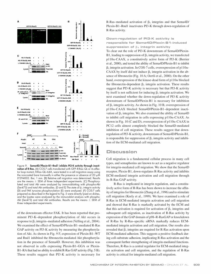
MECHANISM OF INTEGRIN INHIBITION BY SEMA4D • OINUMA ET AL. 609
of the downstream effector FAK. It has been reported that pro-
minent PI3-K–dependent phosphorylation of Akt occurs in
response to β1 integrin–mediated adhesion (Velling et al., 2004).
We examined the effect of Sema4D/Plexin-B1–mediated R-Ras
GAP activity on PI3-K activity by measuring the phosphoryla-
tion of Akt. As shown in Fig. 9 F, expression of Plexin-B1–WT
and Rnd1 inhibited the fi bronectin-mediated Akt phosphoryla-
tion in the presence of Sema4D. However, this inhibition was
not observed in cells expressing Plexin-B1–GGA or Plexin-
B1–RA that had no ability to exhibit R-Ras GAP activity (Fig. 5 B).
These results suggest that PI3-K activity is necessary for
R-Ras–mediated activation of β1 integrins and that Sema4D/
Plexin-B1–Rnd1 inactivates PI3-K through down-regulation of
R-Ras activity.
Down-regulation of PI3-K activity is responsible for Sema4D/Plexin-B1–induced suppression of 𝛃1 integrin activityTo clear out the role of PI3-K downstream of Sema4D/Plexin-
B1, leading to suppression of β1 integrin activity, we transfected
p110α-CAAX, a constitutively active form of PI3-K (Berrier
et al., 2000), and tested the ability of Sema4D/Plexin-B1 to inhibit
β1 integrin activation. In COS-7 cells, overexpression of p110α-
CAAX by itself did not induce β1 integrin activation in the ab-
sence of fi bronectin (Fig. 10 A; Oertli et al., 2000). On the other
hand, overexpression of the kinase-dead form of p110α blocked
the fi bronectin-dependent β1 integrin activation. These results
suggest that PI3-K activity is necessary but that PI3-K activity
by itself is not suffi cient for inducing β1 integrin activation. We
next examined whether the down-regulation of PI3-K activity
downstream of Sema4D/Plexin-B1 is necessary for inhibition
of β1 integrin activity. As shown in Fig. 10 B, overexpression of
p110α-CAAX blocked Sema4D/Plexin-B1–dependent inacti-
vation of β1 integrins. We also examined the ability of Sema4D
to inhibit cell migration in cells expressing p110α-CAAX. As
shown in Fig. 10 (C and D), overexpression of p110α-CAAX in
PC12 cells almost completely blocked the Sema4D-mediated
inhibition of cell migration. These results suggest that down-
regulation of PI3-K activity, downstream of Sema4D/Plexin-B1,
is responsible for suppression of β1 integrin activity and inhibi-
tion of the ECM-mediated cell migration.
DiscussionCell migration is a fundamental cellular process in many cell
types, and semaphorins are known to act as a negative regulator
for integrin-mediated cell migration. We show that the Sema4D
receptor, Plexin-B1, down-regulates R-Ras activity and inhibits
ECM-mediated integrin activation and cell migration through
its R-Ras GAP activity.
R-Ras is implicated in integrin regulation, and a constitu-
tively active form of R-Ras has been shown to increase the affi n-
ity of integrins for fi bronectin (Zhang et al., 1996) and to stimulate
cell migration (Keely et al., 1999). We have examined a role of
R-Ras in ECM-mediated integrin activation and cell migration
and showed that R-Ras is markedly activated by the ECM and
that this activation is required for activation of β1 integrins and
subsequent cell migration, as inactivation of R-Ras activity by
expression of the GAP domain of p98–R-RasGAP or knockdown
of R-Ras by R-Ras–specifi c siRNA markedly reduces ECM-
mediated integrin activation and cell migration. Our results also
revealed that β1 integrins are required for R-Ras activation upon
ECM-mediated adhesion. This suggests a positive feedback dur-
ing cell-substrate adhesion, implicating R-Ras activation and the
consequent further strengthening of integrin-mediated functions.
Therefore, R-Ras is a central regulator for ECM-mediated integ-
rin activation and cell migration, and the regulation of R-Ras
activity is critical for integrin-mediated cell migration.
Figure 9. Sema4D/Plexin-B1–Rnd1 inhibits PI3-K activity through inacti-vation of R-Ras. (A) COS-7 cells transfected with GFP–R-Ras–QL or its effec-tor loop mutant, R-Ras–QL–64A, were tested in a cell migration assay using the noncoated bare transwells in either the presence or absence of 20 μM LY294002. Bar, 1 mm. (B) Relative cell migration was determined. Results are the means ± SEM of three independent experiments. (C) Phosphory-lated and total Akt were analyzed by immunoblotting with phospho-Akt (Ser473) and total Akt antibodies. (D and E) The state of β1 integrin activity (D) and FAK tyrosine phosphorylation (E) were analyzed. (F) COS-7 cells prepared as described in the legend to Fig. 5 were directly lysed on dishes, and the lysates were analyzed by the immunoblot analysis with phospho-Akt (Ser473) and total Akt antibodies. Results are the means ± SEM of three independent experiments.

JCB • VOLUME 173 • NUMBER 4 • 2006 610
Semaphorins are implicated in migration of a variety of
cells. Stimulation of Plexin-B1 by Sema4D is reported to ham-
per integrin-based adhesion and cell migration in NIH-3T3
cells (Barberis et al., 2004). We have reported that Plexin-B1
encodes an R-Ras GAP in the cytoplasmic tail and that stimu-
lation of the Plexin-B1–Rnd1 complex by Sema4D induces the
R-Ras GAP activity and resultant repulsive response of neuro-
nal growth cone (Oinuma et al., 2004a). We demonstrate here
that Plexin-B1/Rnd1–mediated R-Ras GAP activity is also
involved in Sema4D-induced inhibition of integrin activation
and cell migration. Furthermore, the COOH-terminal PDZ
domain binding motif of Plexin-B1 is dispensable for suppres-
sion of integrin activity and cell migration by Sema4D. In ad-
dition to Sema4D, class 3 semaphorins have been shown to
control adhesion and migration of endothelial cells by inhibit-
ing integrin function (Serini et al., 2003), and Sema3A signal-
ing–defi cient mice have shown defective migration of neural
crest cells (Kawasaki et al., 2002). Furthermore, Plexin-C1,
a receptor of semaphorin A39R, was recently reported to inhibit
integrin-mediated adhesion and chemokine-induced migration
(Walzer et al., 2005). The R-Ras GAP–homologous domains
are well conserved among plexin families, including Plexin-A
and -C1. In addition, we recently reported that the down-
regulation of R-Ras activity is also required for the Sema3A/
Plexin-A–induced repulsive response in hippocampal neurons
(Oinuma et al., 2004a). We speculate that the direct regulation
of R-Ras activity by plexins is likely to be a mutual signaling
pathway among plexin families and that this R-Ras GAP activ-
ity of plexin families may be a critical signaling system for
semaphorin-regulated cell migration.
Semaphorins were initially identifi ed as repulsive factors
for axon guidance, and many neurons use members of the inte-
grin family of cell surface receptors for responses to neurite
growth promoting factors, and integrin activation regulates neu-
rite outgrowth (Hynes, 2002). Recently, expression of constitu-
tively active R-Ras was shown to promote integrin-dependent
neurite outgrowth of retinal neurons, suggesting that R-Ras
activity plays an important role in integrin-dependent neurite
outgrowth (Ivins et al., 2000). Therefore, it is proposed that the
down-regulation of R-Ras activity by Plexin-B1 via R-Ras GAP
activity suppresses R-Ras–mediated integrin activation and
thereby induces growth cone collapse and inhibition of neurite
outgrowth. With respect to signaling of other repulsive factors,
the ephrin-B1 receptor EphB2, another family of the repulsive
factor receptor, was also reported to suppress integrin-mediated
functions by inactivating R-Ras (Zou et al., 1999), suggesting
that repulsive guidance cues inhibit integrin-mediated functions
by inactivating R-Ras in general and that R-Ras acts as a com-
mon regulator of integrin activation and cell migration (Serini
and Bussolino, 2004).
We also examined the downstream signaling of Sema4D/
Plexin-B1–mediated R-Ras GAP activity leading to inactivation
of β1 integrins and found that down-regulation of PI3-K activity
is responsible for Sema4D/Plexin-B1–induced suppression of
Figure 10. Down-regulation of PI3-K activity is responsible for Sema4D/Plexin-B1–induced suppression of 𝛃1 integrin activity. (A, left) Lysates of COS-7 cells expressing p110α-CAAX, a constitutively active form of PI3-K, or its kinase-dead (KD) form were immunopre-cipitated with HUTS-4. (right) Relative β1 integrin activity was analyzed. (B) Activity of β1 integ-rins in COS-7 cells expressing the indicated expression plasmids with or without Sema4D stimulation were examined. (C and D) PC12 cells transfected with GFP alone (open symbols) or GFP plus p110α-CAAX (closed symbols) were tested in the transwell assay in either the presence or absence of Sema4D. Results are the means ± SEM of three independent experiments. Bar, 1 mm.
MECHANISM OF INTEGRIN INHIBITION BY SEMA4D • OINUMA ET AL. 611
β1 integrin activity and cell migration. PI3-K activity is known to
be required for R-Ras–mediated enhancement of cell migration
(Keely et al., 1999; Rincón-Arano et al., 2003). PI3-K has emerged
as the predominant effector for R-Ras, and R-Ras is a more potent
activator of PI3-K than other Ras family members (Marte et al.,
1997; Suire et al., 2002). On the other hand, PI3-K activity has
been shown to promote interaction between talin with the β1 inte-
grin cytoplasmic tail, leading to the clustering and activation of in-
tegrins (Calderwood et al., 1999; Martel et al., 2001; Calderwood
et al., 2002). Integrin activation by mechanical stretch is also me-
diated by PI3-K and is followed by an increase in integrin binding
to the extracellular matrix proteins (Katsumi et al., 2005). There-
fore, elevated PI3-K activity by activated R-Ras may trigger a se-
quence of events leading to clustering and activation of integrins,
although overexpression of p110α-CAAX by itself is not suffi -
cient for inducing β1 integrin activation (Fig. 10 A; Oertli et al.,
2000). We used the monoclonal antibody HUTS-4, which detects
hybrid domain swing-out in β1 integrins, a process most com-
monly associated with ligand binding affi nity (Mould et al., 2003),
to measure activity of β1 integrins and revealed that Sema4D/
Plexin-B1–mediated R-Ras GAP activity suppresses affi nity of
β1 integrins through inactivation of PI3-K activity. Consistent with
our results, a previous report demonstrated that an R-Ras– mediated
increase in affi nity of the β1 integrins is dependent on PI3-K activ-
ity by performing the ligand binding assay in mast cells (Kinashi
et al., 2000). On the other hand, Oertli et al. (2000) have shown
that PI3-K activity is not required for R-Ras–mediated integrin
activation in CHO cells by using a ligand-mimetic antibody,
PAC-1. Therefore, we speculate that this discrepancy may be due
to the differences in ways to measure integrin activity or that
R-Ras may regulate integrin activity via both PI3-K–dependent
and –independent pathways, depending on the cell type.
In conclusion, our results demonstrate that R-Ras activity
is required for ECM-mediated integrin activation and cell migra-
tion and that the Sema4D/Plexin-B1–Rnd1 complex regulates
integrin activation and cell migration through the R-Ras GAP
activity. However, a variety of molecules such as ErbB-2 and Met
have been known to be involved in plexin signaling, inducing
diverse physiological functions (Giordano et al., 2002, Swiercz
et al., 2004). It was recently shown that Plexin-B1 enhances
chemotaxis of endothelial cells through the activation of multiple
intracellular tyrosine kinase cascades independent of the R-Ras
GAP activity (Basile et al., 2005). Regulation of R-Ras activity,
tyrosine kinases, and other signaling mechanisms may partici-
pate in diverse actions of plexins. Further work will be required
to delineate the precise mechanism of R-Ras–mediated integrin
activation and its regulation by plexins for cell migration during
physiological and pathological processes, including neural cell
migration, angiogenesis, and tumor metastasis.
Materials and methodsDNA constructs and site-directed mutagenesisPlexin-B1 cDNA was provided by L. Tamagnone (Torino University, Torino, Italy). HA-tagged Rnd1; HA- and GFP-tagged human R-Ras and R-Ras–QL (Q87L); the GST-fused Ras binding domain of c-Raf-1 (amino acids 53–130); the NH2-terminal HA-tagged myristoylated form of R-RasGAP; and Myc-tagged Plexin-B1, Plexin-B1–GGA (L1849G, V1850G, and P1851A), Plexin-B1–RA (R1677A, R1678A, and R1984A), Plexin-B1–∆C
(lacking the last seven COOH-terminal amino acids), and Plexin-B1–N-Cyt–GGA (amino acids 1511–1915) were described previously (Oinuma et al., 2004a,b). The effector loop mutant of R-Ras, R-Ras–DA (D64A), was generated by a PCR-mediated mutagenesis. NH2-terminal FLAG-tagged p110α was a gift from T. Katada (Tokyo University, Tokyo, Japan), and CAAX sequence was fused to the COOH terminus to create a constitutively active form as described previously (Katoh et al., 2002). The specifi c siRNA for R-Ras was designed to target 19 nucleotides at nucleo-tides 359 and 377 (5′-gcaagctcttcactcagat-3′), whereas the control siRNA was designed at nucleotides 426 and 444 (5′-caaggcagatctggagaca-3′), and both were expressed by using a siRNA expression vector (Ambion) as described previously (Oinuma et al., 2004a).
Antibodies and reagentsThe pharmacological PI3-K inhibitor LY294002 was purchased from Calbiochem. A soluble form of Sema4D fused to human IgG1-Fc was a gift from H. Kikutani (Osaka University, Osaka, Japan). We used the following antibodies: mouse monoclonal antibodies against Myc and phosphotyro-sine; a rabbit polyclonal antibody against p125-FAK (Upstate Biotech-nology); mouse monoclonal antibodies against α-tubulin (Sigma-Aldrich), β1 integrins (BD Biosciences), and active β1 integrins, HUTS-4 (Chemicon); a rabbit polyclonal antibody against R-Ras (Santa Cruz Biotechnology, Inc.); a rat monoclonal antibody against HA (Roche); and HRP-conjugated secondary antibodies (DakoCytomation). For functional studies in the transwell assay, we used the following antibodies: the affi nity-related β1 integrin– activating monoclonal antibody 8A2 (IgG2a); the functional blocking monoclonal antibody against the integrin α1 subunit, 3A3 (IgG1; Serotec); the functional blocking monoclonal antibody against the integrin β1 sub-unit, P5D2 (IgG1; Chemicon); and a mouse monoclonal antibody against the extracellular ligand binding region (raised against amino acids 771–1070 of human origin) of Plexin-B1 (IgG2b; Santa Cruz Biotechnology, Inc.). FITC- and PE-conjugated F(ab’)2–specifi c secondary antibodies for fl ow cytometry were purchased from Jackson ImmunoResearch Laboratories. The PhosphoPlus Akt Antibody kit (Cell Signaling) was used for the analysis of the phosphorylation state of Akt.
ImmunoblottingProteins were separated by 12.5% SDS-PAGE and were electrophoreti-cally transferred onto a polyvinylidene difl uoride membrane (Millipore). The membrane was blocked with 3% low-fat milk in TBS and incubated with primary antibodies. The primary antibodies were detected with HRP-conjugated secondary antibodies and a chemiluminescence detection kit (Chemi-Lumi One; Nacalai Tesque). Images were captured using a LAS 1000 analyzer (Fuji) equipped with Image Gauge 4.0 software (Fuji).
Immunofl uorescence microscopyCells on coverslips were fi xed with 4% PFA in PBS for 15 min and washed with PBS fi ve times. Cells were permeabilized with 0.2% Triton X-100 in PBS for 10 min and incubated with 10% FBS in PBS for 30 min to block nonspecifi c antibody binding. Cells were incubated with an anti–R-Ras antibody (1:200 dilution) for 1 h and then incubated with an Alexa Fluor 594–conjugated secondary antibody for 1 h. Cells were washed in PBS for 1 h and mounted in 90% glycerol containing 0.1% p-phenylenedi-amine dihydrochloride in PBS. Images were captured at RT using a micro-scope (Eclipse E800; Nikon) and a 40 × 0.75 objective (Nikon) equipped with a digital camera (DC350F; Leica). The images were arranged and labeled using Photoshop software (Adobe).
Cell culture and transfectionCOS-7 cells were cultured in DME containing 10% FBS, 4 mM glutamine, 100 U/ml penicillin, and 0.2 mg/ml streptomycin under humidifi ed condi-tions in 95% air and 5% CO2 at 37°C. PC12 cells were maintained in RPMI 1640 with 10% horse serum (HS) and 5% FBS. Transient transfections were performed with Lipofectamine 2000 (Invitrogen) according to the manufacturer’s instructions. A soluble form of Sema4D was expressed as a fusion protein with the Fc fragment of human IgG1. Stimulation with Sema4D was performed by incubation of the cells with Sema4D-Fc– containing medium at 37°C.
Cell migration assay104 cells were detached with 1.5 mM EDTA in PBS, washed three times with serum-free medium, resuspended in DME containing 1% BSA, seeded on the upper side of 8-μm pore fi lters of Transwell chambers (Costar), which were coated on the lower side with varying concentrations of either fi bronectin or collagen I (Sigma-Aldrich), and incubated for 7 h. Cells on the upper side of the fi lters were mechanically removed, and cells on the

JCB • VOLUME 173 • NUMBER 4 • 2006 612
lower side were fi xed with 4% PFA. The numbers of migrated cells through the fi lter were counted by the fl uorescence of GFP or the staining with crys-tal violet (A). At the same time, the cells were seeded onto 24-well plastic culture plates to count the total number of transfected cells (B). Relative cell migration was then determined by the number of migrated cells normal-ized to the total number of transfected cells (A/B). Unless described, the value from the GFP-transfected cells in the absence of coating was defi ned as 1. For functional studies using activating or inhibitory monoclonal anti-bodies, cells were pretreated with 5 μg/ml of antibodies or corresponding negative IgG controls for 5 min before seeding onto the transwells. Images were captured at RT in PBS using a microscope (Eclipse TE300-FN; Nikon) and a Plan Fluor 10 × 0.30 objective (Nikon) equipped with digital cam-era (DS-L1 and DS-5M; Nikon). The images were arranged and labeled using Photoshop 7.0 software.
Measurement of the activity of 𝛃1 integrins by immunoprecipitationMeasurement of β1 integrin activity by immunoprecipitation was performed as described previously (Serini et al., 2003). 3 × 10⁶ COS-7 cells were maintained in DME containing 1% FBS after transfection. 16 h after trans-fection, cells were detached with 1.5 mM EDTA in PBS, washed three times with serum-free medium, and resuspended in 10 ml of 1% BSA in DME with or without Sema4D-Fc. The cell suspension was plated onto 10-cm plates coated with or without 10 μg/ml fi bronectin and incubated at 37°C for 15 min. The cells were lysed directly on dishes with ice-cold cell lysis buffer (50 mM Tris-HCl, pH 7.5, 150 mM NaCl, 1% Triton X-100, 10% glycerol, 1 mM sodium vanadate, 25 mM NaF, 10 μg/ml pepstatin, 1 mM PMSF, 10 μg/ml aprotinin, and 10 μg/ml leupeptin) containing 5 μg/ml HUTS-4, immunoprecipitated for 2 h, and subsequently incubated with pro-tein G–Sepharose beads (GE Healthcare) for 1 h at 4°C. After the beads were washed twice with the ice-cold cell lysis buffer, the bound proteins were eluted in Laemmli sample buffer and analyzed by SDS-PAGE and immunoblotting with the monoclonal antibody against β1 integrins. To measure the activity of β1 integrins in PC12 cells, 106 cells were maintained in RPMI 1640 containing 1% HS for 12 h, detached with 1.5 mM EDTA in PBS, washed three times with serum-free medium, and resuspended in 10 ml of 1% BSA in RPMI 1640 with or without Sema4D-Fc. The cell suspen-sion was plated onto 10-cm plates coated with or without 10 μg/ml colla-gen I and incubated at 37°C for 3 h. The cells were lysed directly on dishes with ice-cold cell lysis buffer.
Measurement of the activity of 𝛃1 integrins by ELISAsMeasurement of the activity of β1 integrins by ELISAs under detergent-free condition was performed as described previously (Shih et al., 1999). 105 cells transfected in 24-well plastic culture plates were detached with 1.5 mM EDTA in PBS, washed three times with serum-free medium, and resus-pended in 1 ml DME containing 1% BSA, with or without Sema4D-Fc. One tenth of the resuspended cells (100 μl) were seeded onto the 96-well assay plates, which were coated with 10 μg/ml of either fi bronectin or collagen I. Cell adhesion was allowed for 15 min at 37°C. Then, the cells were deli-cately washed once with PBS and the adherent cells were fi xed with 4% PFA. After the fi xative, the cells were thoroughly rinsed with PBS containing 0.1% BSA. To avoid nonspecifi c binding, the cells were incubated with PBS containing 5% BSA for 3 h at RT. Cells were then incubated overnight at 4°C with 2 μg/ml HUTS-4. After the incubation with primary antibody, the wells were rinsed and blocked with PBS containing 5% BSA for 3 h at RT before they were exposed to an HRP- conjugated secondary antibody. After the incubation, cells were rinsed again with PBS followed by distilled H2O. The peroxidase color reaction was developed in the dark using O-phenylenediamine according to the manufacturer’s instructions (ELISA OPD kit; Nacalai Tesque), and the plate was read on a kinetic microtiter plate reader (GENios; Tecan) using the XFluor4 program (Tecan). The anti-body concentration and incubation times were optimized to ensure testing in the linear range. Expression levels of the constructs used in the assay were also verifi ed by immunoblot analysis.
Flow cytometry analysisAnalysis of cell surface expression of active β1 integrins by fl ow cyto-metry was performed as described previously (Wang et al., 2002). 106 PC12 cells were seeded onto 6-cm noncoated plates in RPMI 1640 containing 10% HS and 5% FBS. 18 h after seeding, cells were treated with medium containing Sema4D-Fc or Sema4D-Fc plus 1 mM Mn2+ for 3 h at 37°C. Cells were washed once with PBS and resuspended in blocking solution containing 5% dissociation buffer (Invitrogen) and 2% sheep serum in PBS. Cells were then incubated with 2.5 μg HUTS-4 or buffer alone for 1 h at 4°C, washed with the blocking solution, and labeled with FITC-conjugated secondary antibody for 30 min at 4°C. Cells were
then washed and analyzed with an EPICS ELITE fl ow cytometer using the EXPO32 analysis program (Beckman Coulter). For the analysis of active β1 integrins in transiently transfected COS-7 cells, 106 cells were trans-fected with a GFP expression vector together with various other expres-sion vectors. Cells were kept for 18 h in DME containing 10% FBS after transfection, stimulated for 5 min at 37°C with or without Sema4D-Fc, and were collected and incubated with HUTS-4 antibody or buffer alone as described previously in this section. Cells were labeled with a PE- conjugated secondary antibody, and expression of GFP and activity of β1 integrins (PE staining) were simultaneously analyzed by two-color fl ow cytometry. Analysis of the intensity of PE staining in a GFP-positive popu-lation was performed as described previously (Ohgushi et al., 2005). Approximately 10,000 cells were analyzed in each experiment, and the results shown are representative of two independent experiments.
Measurement of R-Ras activityMeasurement of R-Ras activity in cells was performed as described pre-viously (Oinuma et al., 2004a). 7 × 10⁵ COS-7 cells were maintained in DME containing 1% FBS after transfection. The cell suspension was pre-pared as described (see Measurement of the activity of β1 integrins by immunoprecipitation) and plated onto plastic dishes coated with or with-out 10 μg/ml fi bronectin and incubated at 37°C for 15 min. The cells were lysed directly on dishes with ice-cold cell lysis buffer (25 mM Hepes-NaOH, pH 7.5, 150 mM NaCl, 1% NP-40, 0.25% sodium deoxycho-late, 0.1% SDS, 10% glycerol, 10 mM MgCl2, 1 mM EDTA, 1 mM DTT, 10 μg/ml aprotinin, and 10 μg/ml leupeptin) containing 75 μg of GST-fused Ras binding domain of c-Raf-1 (GST-RBD). To examine the effect of collagen I and Sema4D stimulation on R-Ras activity in PC12 cells, 106 cells were maintained in RPMI 1640 containing 1% HS for 12 h, detached with 1.5 mM EDTA in PBS, washed three times with serum-free medium, and resuspended in 10 ml of 1% BSA in RPMI 1640 with or without Sema4D-Fc. For samples indicated, cells were treated with 5 μg/ml of monoclonal β1 integrin blocking (P5D2) or activating (8A2) antibody before replating. Cells were either kept in suspension or plated onto 6-cm plates coated with (1 or 10 μg/ml) or without collagen I and incubated at 37°C for 15 min. The cells were lysed directly on dishes with ice-cold cell lysis buffer, and the lysates were used in a pull-down assay using GST-RBD.
Detection of FAK tyrosine phosphorylationDetection of tyrosine phosphorylation of FAK was performed as described elsewhere (Sieg et al., 2000). The cells were lysed directly on dishes with ice-cold cell lysis buffer (50 mM Tris-HCl, pH 7.5, 150 mM NaCl, 1% Triton X-100, 0.25% sodium deoxycholate, 10% glycerol, 1 mM sodium vanadate, 25 mM NaF, 10 μg/ml pepstatin, 1 mM PMSF, 10 μg/ml apro-tinin, and 10 μg/ml leupeptin) containing 4 μg/ml of the polyclonal anti-body against FAK, immunoprecipitated for 2 h, and subsequently incubated with protein A–Sepharose beads (GE Healthcare) for 1 h at 4°C.
Detection of Akt serine phosphorylationCOS-7 cells were maintained in DME with 0.5% FBS after transfection for 36 h. We added 20 μM LY294002 directly to the culture medium after transfection and changed it at every 12 h to reduce the basal levels of PI3-K activity. Cells were directly lysed on dishes with 1× Laemmli sample buffer and analyzed by SDS-PAGE and immunoblotting.
Online supplemental materialFig. S1 shows reduction in endogenous R-Ras protein by RNA interference in PC12 cells and requirement of endogenous R-Ras protein in collagen-mediated PC12 cell migration. Online supplemental material is available at http://www.jcb.org/cgi/content/full/jcb.200508204/DC1.
We thank L. Tamagnone for Plexin-B1 cDNA, H. Kikutani for the soluble forms of Sema4D expression plasmids, and T. Katada for wild-type and kinase-dead forms of p110α expression plasmids. We also thank M. Ohgushi and K. Sakamaki (Laboratory of Molecular and Cellular Biology, Graduate School of Biostudies, Kyoto University) for experimental help with fl ow cytometry analysis.
This work was in part supported by Grants-in-aid for Scientifi c Research from the Ministry of Education, Science, Sports, and Culture of Japan (17079003, 16390021, 18060018, 18022018, and 18013028).
The authors declare that there are no confl icts of interest regarding this article.
Submitted: 31 August 2005Accepted: 18 April 2006

MECHANISM OF INTEGRIN INHIBITION BY SEMA4D • OINUMA ET AL. 613
ReferencesBarberis, D., S. Artigiani, A. Casazza, S. Corso, C. Giordano, C.A. Love, E.Y. Jones,
P.M. Comoglio, and L. Tamagnone. 2004. Plexin signaling hampers integrin-based adhesion, leading to Rho-kinase independent cell rounding, and inhibiting lamellipodia extention and cell motility. FASEB J. 18:592–594.
Basile, J.R., T. Afkhami, and J.S. Gutkind. 2005. Semaphorin 4D/Plexin-B1 induces endothelial cell migration through the activation of PYK2, Src, and the phos-phatidylinositol 3-kinase-Akt pathway. Mol. Cell. Biol. 25:6889–6898.
Berrier, A.L., A.M. Mastrangelo, J. Downward, M. Ginsberg, and S.E. LaFlamme. 2000. Activated R-Ras, Rac1, PI 3-kinase and PKCε can each restore cell spreading inhibited by isolated integrin β1 cytoplasmic domains. J. Cell Biol. 151:1549–1560.
Calderwood, D.A., R. Zent, R. Grant, D.J. Rees, R.O. Hynes, and M.H. Ginsberg. 1999. The talin head domain binds to integrin β subunit cytoplasmic tails and regulates integrin activation. J. Biol. Chem. 274:28071–28074.
Calderwood, D.A., B. Yan, J.M. de Pereda, B.G. Alvarez, Y. Fujioka, R.C. Liddington, and M.H. Ginsberg. 2002. The phosphotyrosine binding-like domain of talin activates integrins. J. Biol. Chem. 277:21749–21758.
de Rooij, J., and J.L. Bos. 1997. Minimal Ras-binding domain of Raf-1 can be used as an activation-specifi c probe for Ras. Oncogene. 14:623–625.
Giordano, S., S. Corso, P. Conrotto, S. Artigiani, G. Gilestro, D. Barberis, L. Tamagnone, and P.M. Comoglio. 2002. The semaphorin 4D receptor con-trols invasive growth by coupling with Met. Nat. Cell Biol. 4:720–724.
Hildebrand, J.D., M.D. Schaller, and J.T. Parsons. 1993. Identifi cation of se-quences required for the effi cient localization of the focal adhesion kinase, pp125FAK, to cellular focal adhesions. J. Cell Biol. 123:993–1005.
Hood, J.D., and D.A. Cheresh. 2002. Role of integrins in cell invasion and migration. Nat. Rev. Cancer. 2:91–100.
Hynes, R.O. 2002. Integrins: bidirectional, allosteric signaling machines. Cell. 110:673–687.
Ivins, J.K., P.D. Yurchenco, and A.D. Lander. 2000. Regulation of neurite out-growth by integrin activation. J. Neurosci. 20:6551–6560.
Katoh, H., A. Harada, K. Mori, and M. Negishi. 2002. Socius is a novel Rnd GTPase-interacting protein involved in disassembly of actin stress fi bers. Mol. Cell. Biol. 22:2952–2964.
Katsumi, A., T. Naoe, T. Matsushita, K. Kaibuchi, and M.A. Schwartz. 2005. Integrin activation and matrix binding mediate cellular responses to mechanical stretch. J. Biol. Chem. 280:16546–16549.
Kawasaki, T., Y. Bekku, F. Suto, T. Kitsukawa, M. Taniguchi, I. Nagatsu, T. Nagatsu, K. Itoh, T. Yagi, and H. Fujisawa. 2002. Requirement of neuropilin 1-mediated Sema3A signals in patterning of the sympathetic nervous system. Development. 129:671–680.
Keely, P.J., E.V. Rusyn, A.D. Cox, and L.V. Parise. 1999. R-Ras signals through specifi c integrin α cytoplasmic domains to promote migration and inva-sion of breast epithelial cells. J. Cell Biol. 145:1077–1088.
Kinashi, T., K. Katagiri, S. Watanabe, B. Vanhaesebroeck, J. Downward, and K. Takatsu. 2000. Distinct mechanisms of α5β1 integrin activation by Ha-Ras and R-Ras. J. Biol. Chem. 275:22590–22596.
Kinbara, K., L.E. Goldfi nger, M. Hansen, F.L. Chou, and M.H. Ginsberg. 2003. Ras GTPases: integrins’ friends or foes? Nat. Rev. Mol. Cell Biol. 4:767–776.
Kolodkin, A.L., D.J. Matthes, and C.S. Goodman. 1993. The semaphorin genes encode a family of transmembrane and secreted growth cone guidance molecules. Cell. 75:1389–1399.
Kuijpers, T.W., E.P. Mul, M. Blom, N.L. Kovach, F.C. Gaeta, V. Tollefson, M.J. Elices, and J.M. Harlan. 1993. Freezing adhesion molecules in a state of high-avidity binding blocks eosinophil migration. J. Exp. Med. 178:279–284.
Luque, A., M. Gómez, W. Puzon, Y. Takada, F. Sánchez-Madrid, and C. Cabanas. 1996. Activated conformations of very late activation integrins detected by a group of antibodies (HUTS) specifi c for a novel regulatory region (355-425) of the common β1 chain. J. Biol. Chem. 271:11067–11075.
Marte, B.M., P. Rodriguez-Viciana, S. Wennstrom, P.H. Warne, and J. Downward. 1997. R-Ras can activate the phosphoinositide 3-kinase but not the MAP kinase arm of the Ras effector pathways. Curr. Biol. 7:63–70.
Martel, V., C. Racaud-Sultan, S. Dupe, C. Marie, F. Paulhe, A. Galmiche, M.R. Block, and C. Albiges-Rizo. 2001. Conformation, localization, and in-tegrin binding of talin depend on its interaction with phosphoinositides. J. Biol. Chem. 276:21217–21227.
Mould, A.P., S.J. Barton, J.A. Askari, P.A. McEwan, P.A. Buckley, S.E. Craig, and M.J. Humphries. 2003. Conformational changes in the integrin βA domain provide a mechanism for signal transduction via hybrid domain movement. J. Biol. Chem. 278:17028–17035.
Negishi, M., and H. Katoh. 2002. Rho family GTPases as key regulators for neuronal network formation. J. Biochem. (Tokyo). 132:157–166.
Nobes, C.D., I. Lauritzen, M.G. Mattei, S. Paris, A. Hall, and P. Chardin. 1998. A new member of the Rho family, Rnd1, promotes disassembly
of actin fi lament structures and loss of cell adhesion. J. Cell Biol. 141:187–197.
Oertli, B., J. Han, B.M. Marte, T. Sethi, J. Downward, M. Ginsberg, and P.E. Hughes. 2000. The effector loop and prenylation site of R-Ras are in-volved in the regulation of integrin function. Oncogene. 19:4961–4969.
Ohgushi, M., S. Kuroki, H. Fukamachi, L.A. O’Reilly, K. Kuida, A. Strasser, and S. Yonehara. 2005. Transforming growth factor β-dependent sequential activation of Smad, Bim, and Caspase-9 mediates physiological apoptosis in gastric epithelial cells. Mol. Cell. Biol. 25:10017–10028.
Oinuma, I., H. Katoh, A. Harada, and M. Negishi. 2003. Direct interaction of Rnd1 with Plexin-B1 regulates PDZ-RhoGEF-mediated Rho activation by Plexin-B1 and induces cell contraction in COS-7 cells. J. Biol. Chem. 278:25671–25677.
Oinuma, I., Y. Ishikawa, H. Katoh, and M. Negishi. 2004a. The semaphorin 4D receptor Plexin-B1 is a GTPase activating protein for R-Ras. Science. 305:862–865.
Oinuma, I., H. Katoh, and M. Negishi. 2004b. Molecular dissection of the sema-phorin 4D receptor Plexin-B1-stimulated R-Ras GTPase-activating pro-tein activity and neurite remodeling in hippocampal neurons. J. Neurosci. 24:11473–11480.
Parsons, J.T., K.H. Martin, J.K. Slack, J.M. Taylor, and S.A. Weed. 2000. Focal adhesion kinase: a regulator of focal adhesion dynamics and cell movement. Oncogene. 19:5606–5613.
Perrot, V., J. Vazquez-Prado, and J.S. Gutkind. 2002. Plexin B regulates Rho through the guanine nucleotide exchange factors leukemia-associated Rho GEF (LARG) and PDZ-RhoGEF. J. Biol. Chem. 277:43115–43120.
Rincón-Arano, H., R. Rosales, N. Mora, A. Rodriguez-Castañeda, and C. Rosales. 2003. R-Ras promotes tumor growth of cervical epithelial cells. Cancer. 97:575–585.
Serini, G., and F. Bussolino. 2004. Common cues in vascular and axon guidance. Physiology (Bethesda). 19:348–354.
Serini, G., D. Valdembri, S. Zanivan, G. Morterra, C. Burkhardt, F. Caccavari, L. Zammataro, L. Primo, L. Tamagnone, M. Logan, et al. 2003. Class 3semaphorins control vascular morphogenesis by inhibiting integrin function. Nature. 424:391–397.
Sethi, T., M.H. Ginsberg, J. Downward, and P.E. Hughes. 1999. The small GTP-bind-ing protein R-Ras can infl uence integrin activation by antagonizing a Ras/Raf-initiated integrin suppression pathway. Mol. Biol. Cell. 10:1799–1809.
Shih, P.T., M.J. Elices, Z.T. Fang, T.P. Ugarova, D. Strahl, M.C. Territo, J.S. Frank, L. Kovach, C. Cabanas, J.A. Berliner, and D.K. Vora. 1999. Minimally mod-ifi ed low-density lipoprotein induces monocyte adhesion to endothelial con-necting segment-1 by activating β1 integrin. J. Clin. Invest. 103:613–625.
Sieg, D.J., C.R. Hauck, D. Ilic, C.K. Klingbeil, E. Schaefer, C.H. Damsky, and D.D. Schlaepfer. 2000. FAK integrates growth-factor and integrin signals to promote cell migration. Nat. Cell Biol. 2:249–256.
Suire, S., P. Hawkins, and L. Stephens. 2002. Activation of phosphoinositide 3-kinase gamma by Ras. Curr. Biol. 12:1068–1075.
Swiercz, J.M., R. Kuner, J. Behrens, and S. Offermanns. 2002. Plexin-B1 di-rectly interacts with PDZ-RhoGEF/LARG to regulate RhoA and growth cone morphology. Neuron. 35:51–63.
Swiercz, J.M., R. Kuner, and S. Offermanns. 2004. Plexin-B1/RhoGEF- mediated RhoA activation involves the receptor tyrosine kinase ErbB-2. J. Cell Biol.165:869–880.
Tamagnone, L., and P.M. Comoglio. 2004. To move or not to move? Semaphorin signalling in cell migration. EMBO Rep. 5:356–361.
Tamagnone, L., S. Artigiani, H. Chen, Z. He, G.I. Ming, H. Song, A. Chedotal, M.L. Winberg, C.S. Goodman, M. Poo, et al. 1999. Plexins are large fam-ily of receptors for transmembrane, secreted, and GPI-anchored sema-phorins in vertebrates. Cell. 99:71–80.
Velling, T., S. Nilson, A. Stefansson, and S. Johansson. 2004. β1-integrins induce phosphorylation of Akt on serine 473 independently of focal adhesion kinase and Src family kinases. EMBO Rep. 5:901–905.
Walzer, T., L. Galibert, M.R. Comeau, and T. De Smedt. 2005. Plexin C1 engage-ment on mouse dendritic cells by viral semaphorin A39R induces actin cytoskeleton rearrangement and inhibits integrin-mediated adhesion and chemokine-induced migration. J. Immunol. 174:51–59.
Wang, W.J., J.C. Kuo, C.C. Yao, and R.H. Chen. 2002. DAP-kinase induces apoptosis by suppressing integrin activity and disrupting matrix survival signals. J. Cell Biol. 159:169–179.
Yamamoto, T., T. Matsui, M. Nakafuku, A. Iwamatsu, and K. Kaibuchi. 1995. A novel GTPase-activating protein for R-Ras. J. Biol. Chem. 270:30557–30561.
Zhang, Z., K. Vuori, H. Wang, J.C. Reed, and E. Ruoslahti. 1996. Integrin activa-tion by R-Ras. Cell. 85:61–69.
Zou, J.X., B. Wang, M.S. Kalo, A.H. Zisch, E.B. Pasquale, and E. Ruoslahti. 1999. An Eph receptor regulates integrin activity through R-Ras. Proc. Natl. Acad. Sci. USA. 96:13813–13818.