cholelithiasisksumsc.com/download_center/3rd/females/1st semester...dr. abdulsalam alsharabi harvest...
TRANSCRIPT
Cholelithiasis
Dr. Abdulsalam Alsharabi
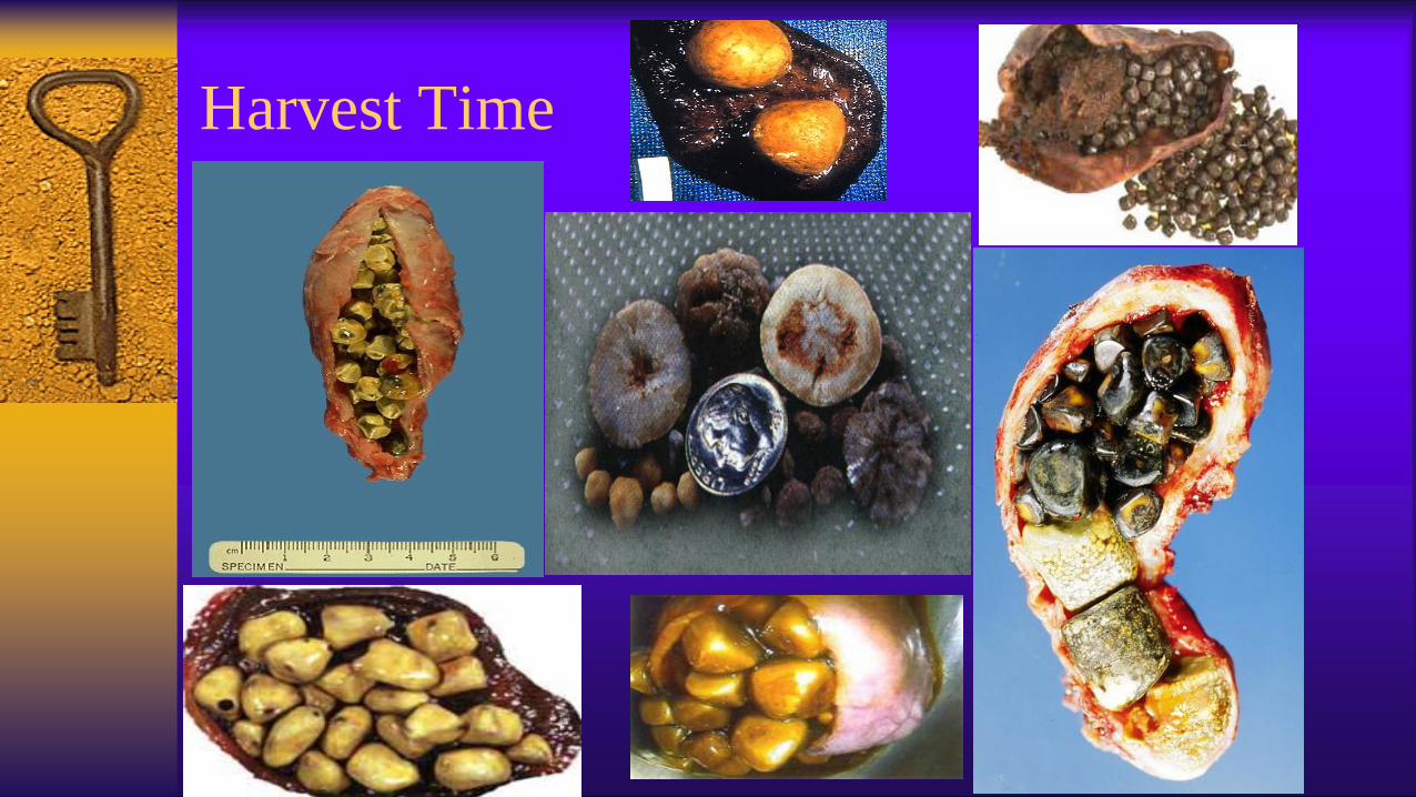
Harvest Time
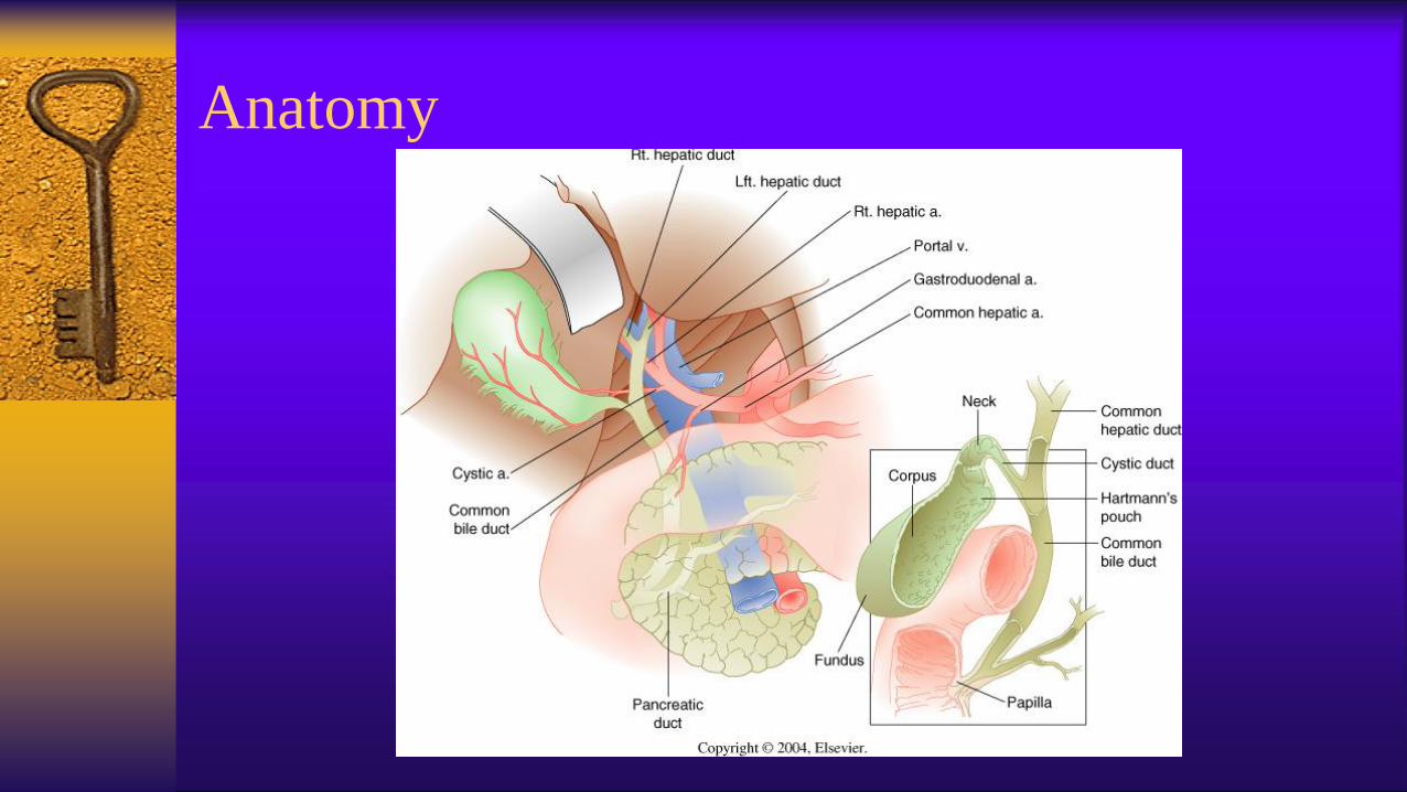
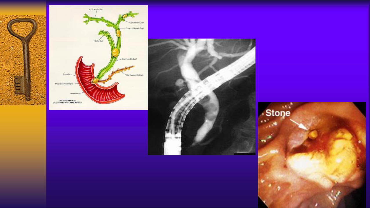
Anatomy

Variations in Bile Ducts

Gallstone Pathogenesis


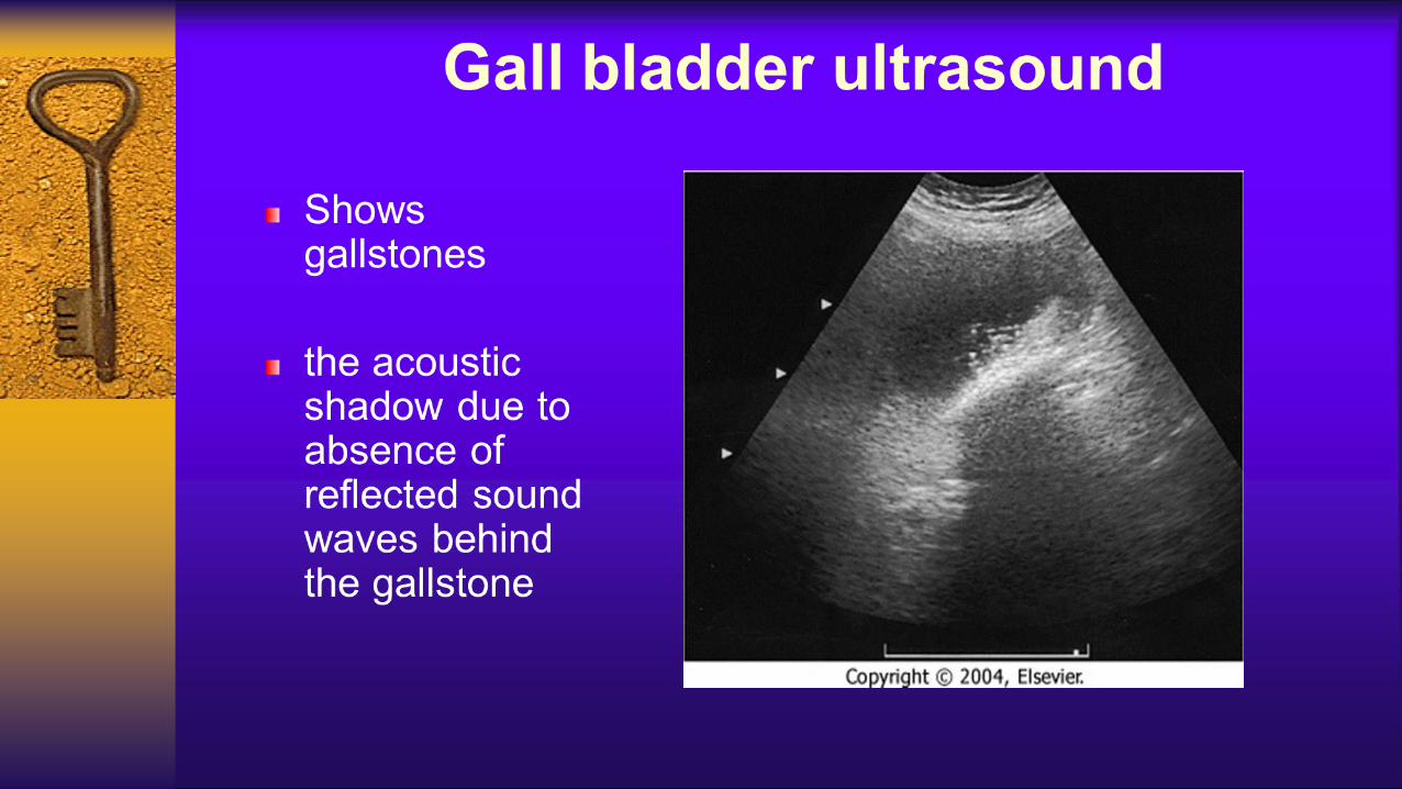

Ultrasound

Ultrasound


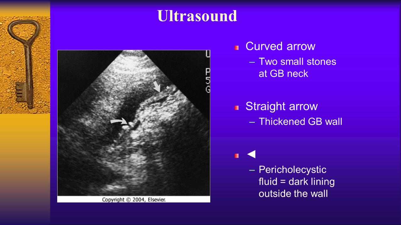
Ultrasound is the first choice for imaging- Distended gallbladder
- Increased wall thickness (> 4 mm)
- Pericholecystic fluid
- Positive sonographic Murphy’s sign (very specific)

Ultrasound


Complications of acute cholecystitis


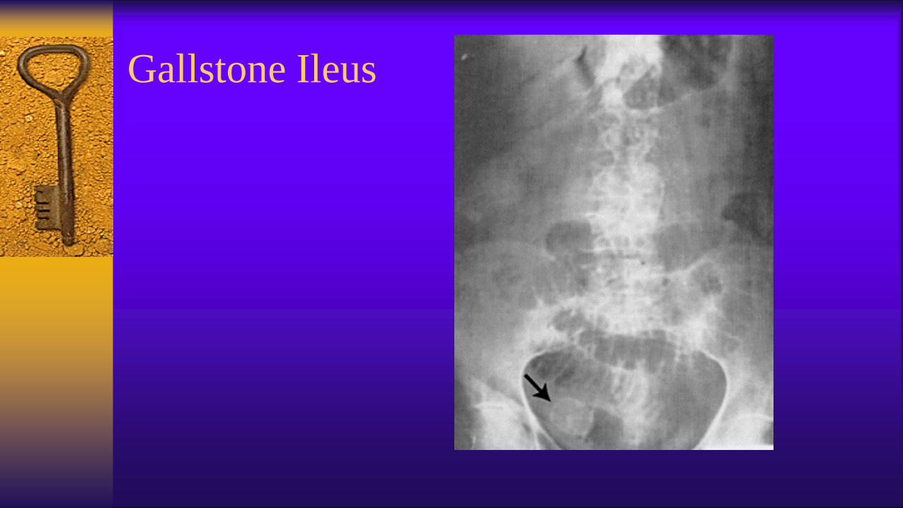
Gallstone Ileus

Acalculous cholecystitis
- A form of acute cholecystitis
- GB inflammation due to biliary stasis(5% of
time) and not stones(95%).
- Often seen in critically ill patients


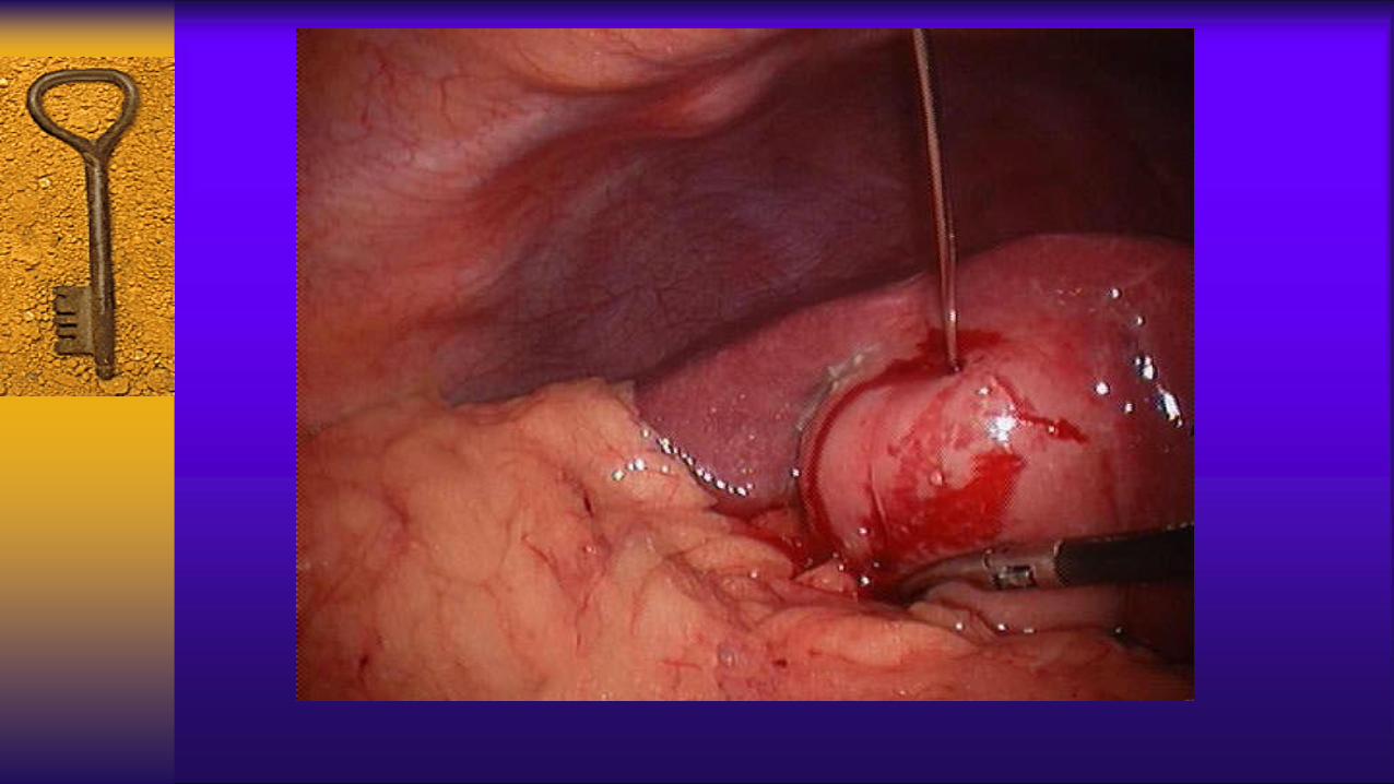
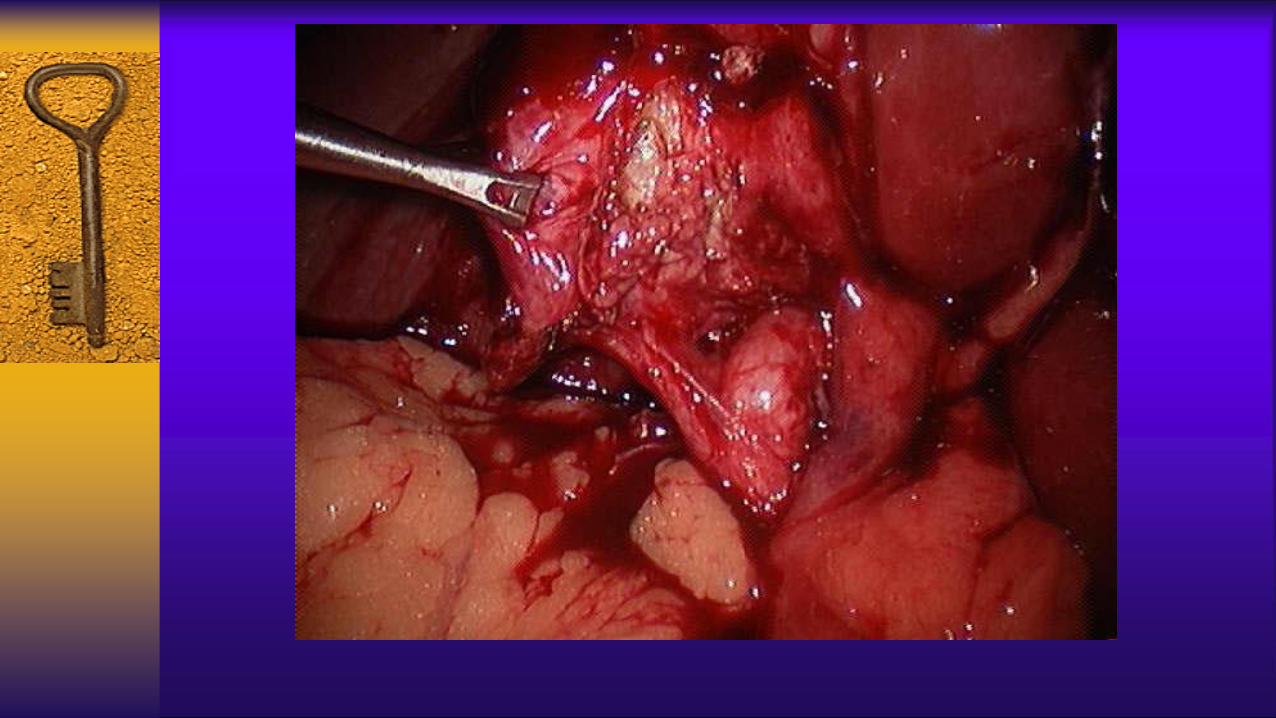
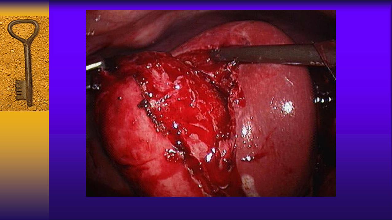
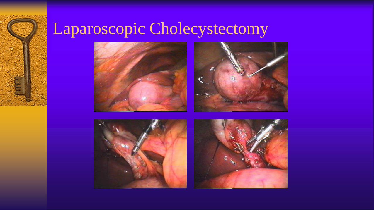
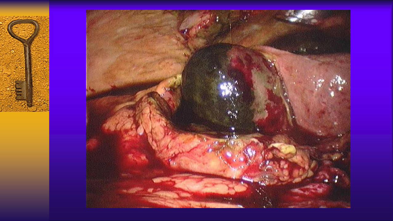
Laparoscopic Cholecystectomy

Laparoscopic Cholecystectomy


Pathogenesis:• Stone obstructing CBD (bear in mind there are other causes for obstructive jaundice) –
danger is progression to ascending cholangitis.
USS• Will confirm gallstones in the gallbladder
• CBD dilatation i.e. >8mm (not always!)
• May visualise stone in CBD (most often does not)
MRCP• In cases where suspect stone in CBD but USS indeterminate
• E.g.1 obstructive LFTs but USS shows no biliary dilatation and no stone in CBD
• E.g. 2 normal LFTS but USS shows biliary dilatation
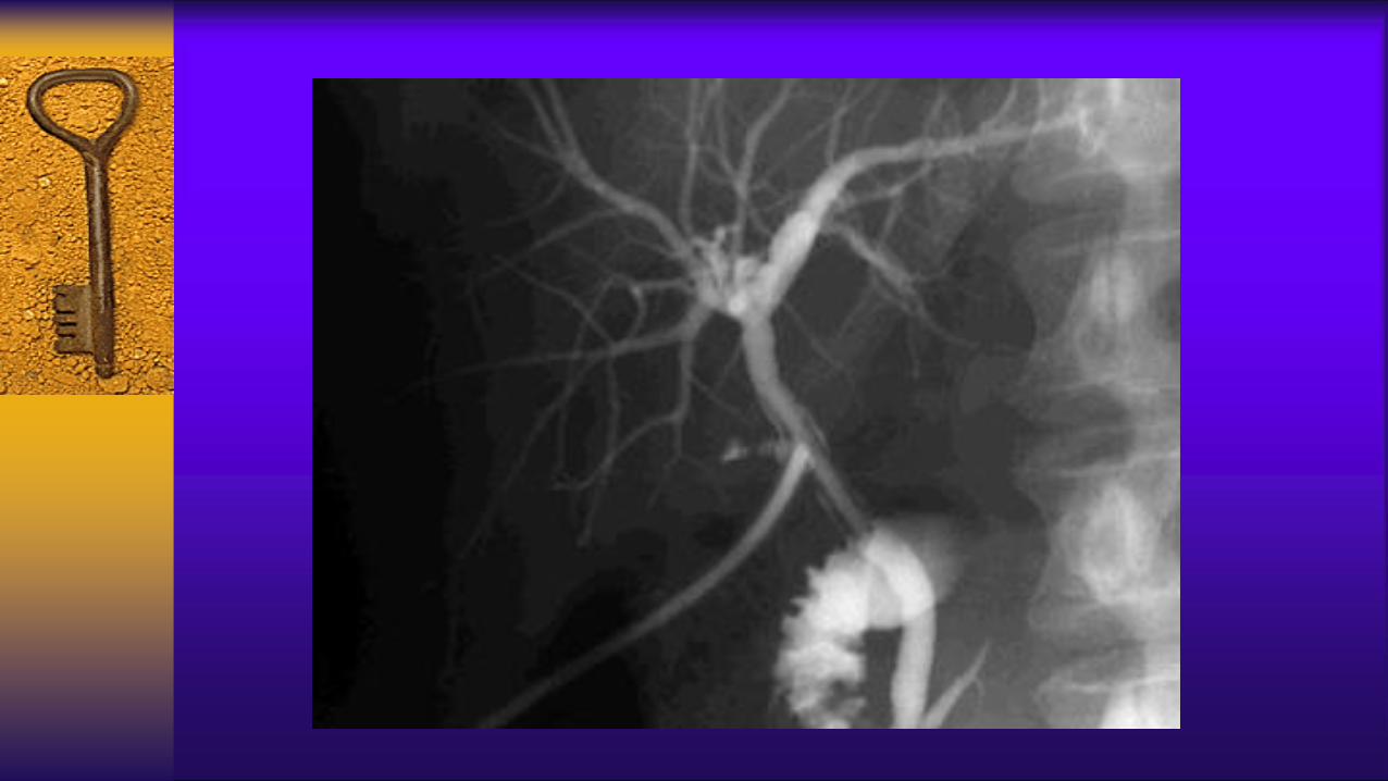
ERCP• If confirmed stone in CBD on USS or MRCP proceed to ERCP which will confirm this
(diagnostic) and allow extraction of stones and sphincterotomy (therepeutic)
Treatment• Must unobstruct biliary tree with ERCP to prevent progression to ascending cholangitis
• Whilst awaiting ERCP monitor for signs of sepsis suggestive of cholangitis
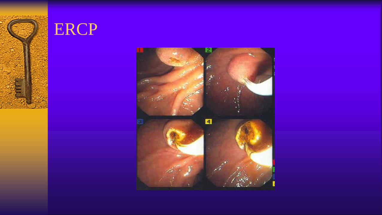
ERCP


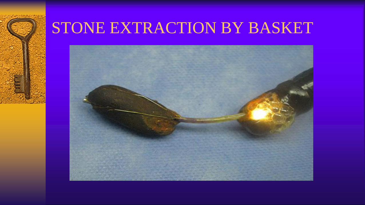
STONE EXTRACTION BY BASKET

STONE EXTRACTION BY BALLON