seminario biomol acabado
TRANSCRIPT

ANDREA CAROLINA VARGAS OLMOS CARMEN SOFIA TORRES DAZA
MEDICAL STUDENTS

INTRODUCTION
The mutant P53 gene and mdm2 gene are found in the majority of human tumor tissues.In this study were compared the mRNA of p53, mdm2 inpatients with normal oral mucosa, simple oral leukoplakia,no-simple oral leukoplakia and leukoplakia cancer, toexplore their role in the carcinogenesis of oral leukoplakia.

CANCER
Cancer is a disease caused by a group of cell that proliferating uncontrollably and autonomously, invading other tissues locally and remotely.

LEUCOPLAKIA
They are patches on the tongue, in the mouth or on the inside of the cheek that occur in response to prolonged irritation.
Occurs in the epithelial surface.
is an injury related to the use of cigarettes, alcohol or HIV.

p53• The p53 gene, also called the "guardian of the genome",
encodes a nuclear transcription factor(The transcription factor p53)
• p53 is crucial in multicellular organisms, where it regulates the cell cycle and, this functions as a tumor suppressor that is involved in preventing cancer

The transcription factor p53 has several important functions:
•It can activate DNA repair proteins.
•It can initiate apoptosis (the programmed cell death).
•Cell cycle arrest in the G1/S o G2/M phase.
activation signal is damaged DNA

mdm2• Mdm2 protein is an important negative regulator of the p53
tumor suppressor.• can activate p53 degradation by the ubiquitination system.• the ubiquitin, works as a signal for protein destruction.

P53, mdm2 and cáncer• In the normal cells:
• In the DNA damage:
p53 is low, because it is associated to the mdm2 protein, which induces its ubiquitination
and destruction(by the proteosome)
to prevent the proliferation of cells with damaged DNA
CANCER


• Mutations in p53, can generate cancer(like leukoplakia cancer)
• about 50% of all human tumors contain mutations in p53.

OBJECTIVE
To study the relationship of the expressions of p53 and mdm2 in leukoplakia cancer.

MATERIALES Y METODOS
DATOS CLINICOS
110 Pacientes con leucoplasia
69 hombres 41 mujeres
edad 23-83 años, con un curso de la enfermedad de
2 meses a 10 años

REACTIVOS
•Trizol•Isotiocianato de fluoresceína•medio de separación de linfocitos•El anticuerpo monoclonal de ratón anti-humano p53•El anticuerpo monoclonal de ratón anti-humano mdm2•Kit de reactivos de RT-PCR•PCR termociclador•Citometría de flujo

MUESTRADos días después de la admisión, se tomaron muestras de sangre venosa del antebrazo en ayunas y se colocaron en tubos heparinizados.
HEPARINA: Anticoagulante.
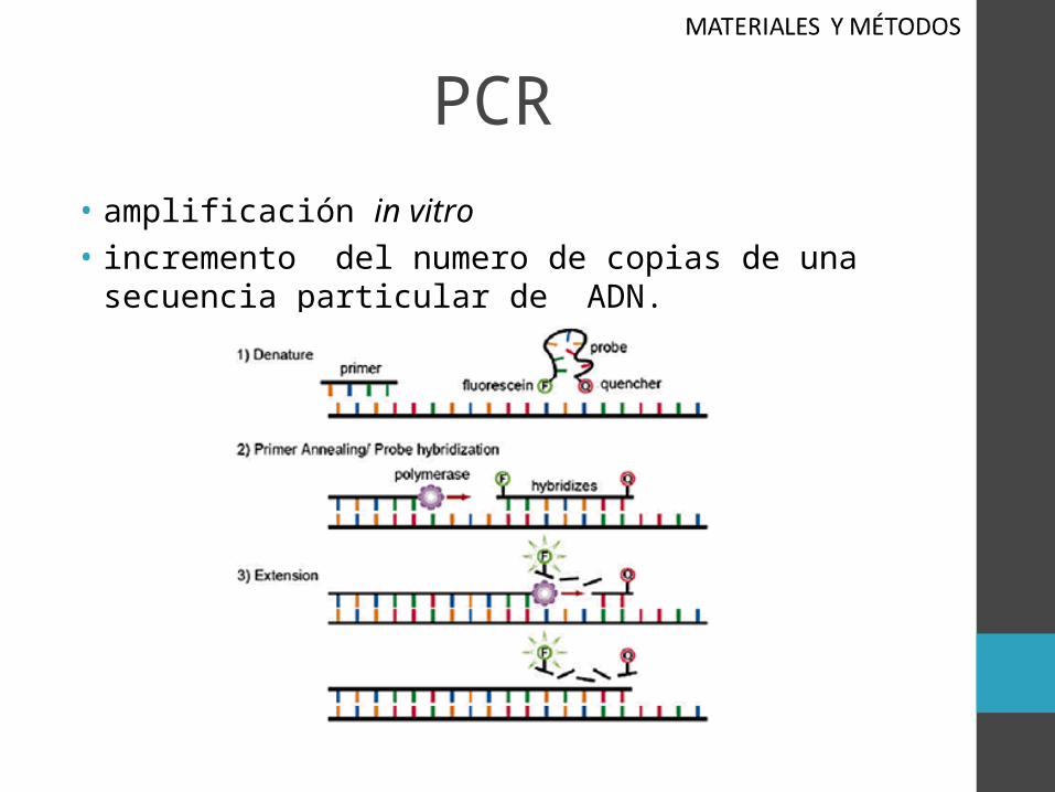
PCR• amplificación in vitro• incremento del numero de copias de una secuencia particular
de ADN.
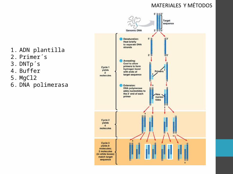
1. ADN plantilla2. Primer´s3. DNTp´s4. Buffer5. MgCl26. DNA polimerasa

APLICACIONES
Clonación de fragmentos de DNA
Secuenciación de ácidos nucleídos
Establecimiento de polimorfismos
Rastreo de mutaciones
Diagnostico de enfermedades genéticas
Detección de células tumorales
DIFICULTADES DIAGNOSTICAS
Falsos positivos por contaminación
costos
La interpretación clínica es difícil
Falta de estandarización
Falta de control de calidad

RT-PCR Reacción en cadena de la polimerasa con transcriptasa
inversa :
generar una gran cantidad de copias
de ADN:"amplificación".
generar una gran cantidad de copias
de ADN:"amplificación".
una hebra de ARN es retrotranscripta
en ADN complementario usando
una enzima: transcriptasa inversa.
una hebra de ARN es retrotranscripta
en ADN complementario usando
una enzima: transcriptasa inversa.

RT-PCR se utiliza para detectar cualitativamente la expresión de genes a través de la creación de ADN complementario

DETECCION CON RT-PCR DE LA EXPRESION DE P53 Y
MDM2ARN fue extraído mediante la
introducción de trizol
Se utilizaron los siguientes primers:
Primer de p53= 409pbPrimer de mdm2= 450pb
Actina-β como referencia= 330pb
“la amplificación por la PCR”

Electroforesis:
• Presencia de banda en: 409pb, 450pb, 330pb
• No presencia de bandas en: 409pb, 450pb,330pb
El contenido relativo de la expresión de ARNm de p53 y mdm2 Se obtuvo gracias a la densidad óptica.
La expresión era “+” para ARNm de p53, mdm2 y actina-β.
La expresión era “+” para ARNm de p53, mdm2 y actina-β.
La expresión era “-” para ARNm p53, mdm2
y actina-β.
La expresión era “-” para ARNm p53, mdm2
y actina-β.

PROPORCION DE P53, MDM2 EN SANGRE PERIFERICA
PBS, linfocitosCentrifugación= 1000rpm,
eliminación de sobrenadante
Dilución 1:100 anticuerpo monoclonal de ratónanti-humano mdm2,
p53(100µl)
Temperatura ambiente durante 30 min.Bañados PBS ( dos veces)Descartar
sobrenadante

37°C por 30min, descartar
sobrenadante, bañado en PBC
+1ml de PBS. paso por la malla filtradora (400
veces)

• “la proporción del p53 y mdm2 fue detectado por citometría de flujo”

RESULTADOS
- P53:

RESULTADOS
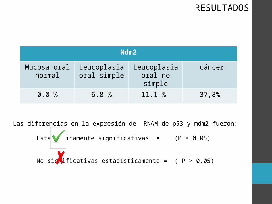
Mdm2
Mucosa oral normal
Leucoplasia oral simple
Leucoplasia oral no simple
cáncer
0,0 % 6,8 % 11.1 % 37,8%
RESULTADOS
Las diferencias en la expresión de RNAM de p53 y mdm2 fueron:
Estadísticamente significativas = (P < 0.05)
No significativas estadísticamente = ( P > 0.05)

RESULTADOS
Figura 1. Resultado de RT- PCR.•P53: 409 pb•Mdm2: 450 pb•β-actina : 330 pb

PROPORCIÓN DE P53 Y MDM2 EN SANGRE PERIFERICA EN CADA GRUPO FUERON DETECTADAS CON CITOMETRIA DE FLUJO
Mucosa oral normal
Leucoplasia oral simple
Leucoplasia oral no simple
Cáncer
P 53 ( 0.3 ± 0.1) % ( 1.6 ± 0.9) % ( 1.9 ± 1.1)% ( 3.4 ± 1.8 ) %
Mdm2 ( 0.1 ± 0.1) % ( 0.8 ± 0.6) % ( 1.2 ± 0.8)% ( 1.2 ± 0.8 ) %
Las frecuencias de p53 y mdm2 fueron:
RESULTADOS

RELACIÓN DE P53, MDM2 Y CARACTERÍSTICAS CLÍNICAS PATOLÓGICAS DE PACIENTES CON CÁNCER DE LEUCOPLASIA
RESULTADOS

DISCUSSIONMAN ¿WHAT DID HE SAY? YES or NO
Kovesi
Consider that the different expression of p53 in oral leukoplakia showed the increased genomic instability is consistent with the oral leukoplakia cancer clinical manifestations.
Liu
confirmed that p53 gene mutation has a correlation with the ocurrence and development of oral precancerous lesions.

MAN WHAT DID HE SAY? YES or NO
Jiang et al, li
Reported mdm2 play an important role in the ocurrence and development of tumors, there was a correlation between them.
wang
Confirmed that p53 , mdm2 protein expression are closely related to the ocurrence and development of oral lichen planus and oral squamous cell carcinoma.
DISCUSSION

CONCLUSION• The p53 tumor suppressor gene is the most important
regulator in DNA damage because this gene generates mechanisms involving growth arrest and apoptosis. Mutation in this gene is associated with the development of malignant disease.
• Nowadays, To study the interaction between p53 and MDM2 has become an important target for cancer therapy and other studies.

• flow cytometry is important in determining the proportion of p53 and mdm2 cells in peripheral blood, this last, can determine the possibility of developing cancer.
• The RT-PCR Is widely used in the diagnosis of genetic diseases and is using in cancer detection to help improve prognosis.

Andrea Vargas

• M.O.N = Mucosa Oral Normal• L.O.S = Leucoplasia Oral Simple• L.O.N.S = Leucoplasia Oral no Simple • C.P.L= Cancer por Leucoplasia Sofía Torres

“All you need is LOVE” GRACIAS