sensitivity and specificity of pulse oximetry and ankle-brachial index...
TRANSCRIPT
Journal of The Association of Physicians of India ■ Vol. 64 ■ August 201638
Sensitivity and Specificity of Pulse Oximetry and Ankle-Brachial Index for Screening Asymptomatic Peripheral Vascular Diseases in Type 2 Diabetes MellitusM Satheesh Kumar1, Ayush Lohiya2, Viviktha Ramesh2, Priyamadhaba Behera2, Sarika Palepu2, SA Rizwan3
AbstractObjectives: To compare pulse oximetry and Ankle-Brachial Index (ABI) with duplex ultrasonography as reference standard to determine the diagnostic accuracy for screening asymptomatic PVD in type 2 diabetes mellitus.
Methods: This cross-sectional study was conducted in 2012 at tertiary hospital in Madurai among diabetic patients attending the medicine outpatient department (OPD). Type 2 Diabetes Mellitus patients, asymptomatic with regards to symptoms and signs of PVD, aged above 40 years were included.
Pulse Oximetry was performed using a pulse oximeter and ABI using sphygmomanometer cuffs and duplex ultrasonography of femoral, popliteal, tibial, posterior tibial and dorsalis pedis arteries. A diagnosis of PVD was based on: monophasic waveforms in any artery by duplex ultrasonography, toe saturation being less than finger saturation by >2% or if foot saturation decreased by >2% in an elevated position and an ABI <0.9.
Results: Among 120 patients included in the study, prevalence of PVD was 22.5% (95% CI: 15.9, 30.8). The PVD group had a higher proportion of elderly, males, current smokers, long-standing diabetics and comorbidities. The sensitivity, specificity, positive predictive value (PPV) and negative predictive value (NPV) of pulse oximetry were 74.1% (95% CI: 55.3, 86.8), 95.7% (89.4, 98.3), 83.3% (64.1, 93.3) and 92.7% (85.7, 96.4) respectively, while those of ABI were 70.3% (51.5, 84.2), 87.1 (78.8, 92.5), 61.3% (43.8, 76.3) and 91.0% (83.3, 95.4) respectively. Parallel testing had net sensitivity increased to 92.3% and net specificity decreased to 83.3%. Performances did not differ across the subgroups.
Conclusion: Pulse oximetry was atleast as good as ABI for the screening for asymptomatic PVD among diabetics.
1Post-Graduate Trainee, Madurai Medical College, Madurai, Tamil Nadu; 2Post-Graduate Trainee, All India Institute of Medical Sciences, New Delhi; 3Assistant Professor, Department of Community Medicine, Velammal Medical College Hospital & Research Institute, Madurai, Tamil NaduReceived: 25.06.2015; Revised: 21.07.2015; Accepted: 23.10.2015
of d iabet i c pat ients g loba l ly , with a prevalence of about 8% among adults next only to China. Currently about 35 million Indians a r e r e p o r t e d t o s u f f e r f r o m diabetes, a significant proportion of whom are either undiagnosed or diagnosed but under-treated l e a d i n g t o p o o r g l y c e m i c control.1 This leads to accelerated development of complicat ions like Peripheral Vascular Disease (PVD). PVD, defined as a clinical disorder in which there is stenosis or occlusion in the arteries of the limbs,2 is a common complication of long standing diabetes mellitus. Individuals with diabetes tend to have a two to four fold increase
O r i g i n a l a r t i c l e
Editorial Viewpoint• Diabetic patients tend to
have a two to four fold increase in the r isk of PVD.
• Pulseoximetryisaneasierway to access PVD.
• T h i s s t u d y s h o w spulse oximetry to be as good as ankle-brachial index for screening of asymptomatic PVD.
Introduction
India, dubbed to be the diabetic capital of the world, has the
second largest absolute number

Journal of The Association of Physicians of India ■ Vol. 64 ■ August 2016 39
in the risk of PVD.3 Prevalence of PVD among overt diabetic patients was estimated to be about 6 to 8% in south Indians.4,5 In diabetes, PVD has certain peculiar features, which make i t more d i f f i cu l t to treat; i t is multi-segmental, bilateral, extensive and involves p r e d o m i n a n t l y d i s t a l ve s s e l s leading to poorer prognosis as compared to non-diabetic PVD. A large proportion of lower extremity amputations among these patients lead to significant disability and economic burden to the individual and the health system in developing countries like India.6 Stenosis in PVD usually develops gradually and is accompanied by formation of extensive collaterals so most of the patients are asymptomatic. Hence, early detection and treatment of PVD in patients with diabetes m e l l i t u s c a n s u r e l y l i m i t i t s debilitating effects.
Various methods are available for diagnosis of PVD. Duplex ultrasonography, an ultrasound-based technique, is a very accurate method for detecting vascular les ions . I t i s the combinat ion of real - t ime B mode scanning
in colour and Doppler spectral analysis, but it is expensive and hence not appropriate for screening purposes. Other imaging modalities like arteriography, Computerized Tomography (CT) angiography and Magnetic Resonance Angiography (MRA) are also accurate though expensive in clinical practice.
Among the methods used for screening, Ankle-Brachial Index (ABI) and pulse oximetry are common. ABI, the ratio of blood pressure (BP) in the legs to that of in the arms was shown to have a sensitivity of over 90% in detecting PVD 7,8 but some s tudies were done in overtly symptomatic cases and therefore liable to spectrum bias, for e .g. , a study done in United States of America (USA) reported that ABI had a sensitivity of only 28.4% among asymptomatic patients.9
The pulse oximeter, which works by combining spectrophotometry and opt ica l p le thysmography has a repor ted sens i t iv i ty o f about 80% and specificity of over 90%.10 It is simple, non-invasive, cheaper, more readily available and can be performed without any special training. However, i t performs poorly in cases of dyshemoglobinemia, pulsating v e i n s a n d p o o r p e r i p h e r a l perfusion.11 Only a few studies have assessed ABI and pulse oximeter as a screening tool for asymptomatic PVD in diabetic patients in India and hence the need for this study.10,12 This study was designed to assess the diagnostic accuracy of pulse oximetry and ABI as a screening tool to detect asymptomatic PVD in patients with type 2 diabetes mellitus against duplex ultrasonography as the reference standard and to compare the performance of these tests across different subgroups.
Material and Methods Patients and setting
This cross-sectional study was conducted between March and
November 2012 in the medicine O P D o f G o v e r n m e n t R a j a j i Hospital, Madurai, a tertiary care hospital in southern India. Diabetic patients who attended the OPD were screened in a consecutive manner for eligibility according to predefined inclusion and exclusion criteria using a screening checklist. (Box 1) All eligible consenting patients were then subjected to all three tests. This process was continued till the requisite sample size was achieved.Sample size
Using the formula,13
n=4x[sensitivity×(1-sensitivity)]
precision^2and assuming a sensi t ivi ty of 90%,7,8 relative precision of 20%, a lpha of 5% and beta of 20%, we calculated that 11 patients of PVD were required. Prevalence of PVD among diabetic patients was considered to be about 10%,5 so the number of diabetic patients needed was 110. Applying a non-response rate of 10%, the final number of diabetic patients required for the study was calculated as 122.Instruments
A handheld pulse oximeter was used to measure SpO2 (peripheral arter ia l oxygen saturat ion) of fingers and toes. Systolic blood pressures of the arms and legs were measured with appropriate size sphygmomanometer cuffs and a handheld 8 MHz Doppler probe. Duplex ultrasonography of lower extremity arteries was performed using a Doppler ultrasonogram machine.Procedures
I n f o r m a t i o n o n p a t i e n t demographics, duration of diabetes and smoking history was collected using a semi-structured, pre-tested questionnaire. A comprehensive physical examination was done a l o n g w i t h m e d i c a l r e c o r d analysis to check for presence of comorbidities. Patients’ medical records were used to conf irm the presence of Coronary Heart Disease (CHD), Cerebrovascular
Box 1: Eligibility criteria
Sr. No.
Inclusion criteria
1. Adults pre-diagnosed type 2 diabetes mellitus, either physician diagnosed or based on blood sugar records as per American Diabetic Association (ADA) criteria,16 irrespective of control of blood sugar, duration of diagnosis, treatment and presence of other complications.
2. Not previously investigated for or diagnosed as PVD and asymptomatic with regard to symptoms of PVD such as pain, swelling, ulcers, previous amputations etc.
Sr. No.
Exclusion criteria
1. Age less than 40 years.2. Patientssufferingfrom
hypercoagulable states, congestive heart failure, valvular heart disease, suspected arteritis and collagen vascular disease.
3. Patients who were unable to lie supine for the period of testing.
4. Extremely sick patients who required intensive care.

Journal of The Association of Physicians of India ■ Vol. 64 ■ August 201640
Disease (CVD) and Hypertension. Af ter th i s in i t ia l assessment , one of the study investigators performed ABI followed by pulse oximetry on the same day. An expert radiographer (not part of the study team) who was blind to the results of index tests, conducted duplex ultrasonography within one-week of the initial assessment. Duplex ultrasonography of lower extremities was done at the level o f f e m o r a l , p o p l i t e a l , t i b i a l , posterior tibial and dorsalis pedis arteries bilaterally. This test has specificity of 97% when compared with arteriography and has been used as the reference standard in many previous studies.9,10 ABI was calculated by measuring systolic BP of the brachial artery at the elbows and that of posterior tibial artery (or dorsalis pedis artery in case posterior tibial had no signal) at the ankles, following the usual precautions prescribed by the seventh report of the Joint National Committee on Prevention, D e t e c t i o n , E v a l u a t i o n , a n d Treatment of High Blood Pressure ( JNC 7) for BP measurement. 14 A hand-held pulse oximeter was used to measure SpO2 to measure the saturation of both index fingers and both big toes. Toe saturation was measured in two positions, one in supine and the other at 12-inch elevation from the horizontal plane. Definitions used for PVD diagnosis
The presence of monophasic waveforms in any one artery by
duplex ultrasonography was taken as confirmatory evidence for PVD. By puls e ox imetry , PVD was considered present if toe saturation was less than finger saturation by >2% or if the foot saturation decreased by >2% in the elevated position.15 ABI values for each leg were calculated by dividing the ankle systolic BP by the elbow pressure and a value of < 0.9 for any leg was considered positive for PVD.16 A patient was considered to be positive for PVD even if any one leg had abnormal results.Research ethics
Ethical clearance was obtained from the Institutional Review Board of the Government Rajaji Hospital, Madurai. Written informed consent was obtained from all participants and confidentiality of information was strictly maintained. Patients who required treatment as per their diagnosis were given appropriate care.Statistical analysis
Characterist ics of the study populat ion were descr ibed in percentages. Diagnostic accuracy of the index tests was calculated in terms of sensitivity, specificity, PPV, and NPV with 95% Confidence Intervals (CI). Each index test was compared separately with the reference standard and a combined measure was also calculated for
a parallel testing scenario. When calculat ing the net sensit ivi ty and net specificity for the parallel testing scenario, positive by any one test was considered positive and negative by both tests was considered negative. The parallel testing formulae17 used were:
Net sensitivity = [sensitivity of test 1 + sensitivity of test 2 – (sensitivity of test 1 × sensitivity of test 2)] and Net specificity = [specificity of test 1 x specificity of test 2] . We also performed subgroup analysis according to a few important covariates. Age, sex , smoking s ta tus , d iabetes duration, cardiovascular disease and hypertension, due to their strong association with PVD were considered as important covariates for subgroup analysis.
Results
A total of 412 diabetic patients were screened and 138 were found eligible for inclusion. Eighteen patients (13%) refused to participate and finally a total of 120 patients (against a calculated sample size of 122) with type 2 diabetes mellitus were included in the study (Figure 1). Main reason for refusal was lack of time to stay for the tests. The prevalence of PVD in the sample was 22.5% (95% CI: 15.9, 30.8). Comparison of baseline parameters showed that the PVD group, as compared to non-PVD patients, had a higher proportion of elderly patients (18.5% vs. 10.8%), males (81.5% vs. 72%), smokers (74.1% vs. 31.2%), long-standing diabetics (81.5% vs. 40.9%) and comorbidities (Table 1).
The sensitivity and specificity of pulse oximetry were 74.1% (95% CI: 55.3, 86.8) and 95.7% (89.4, 98.3) respectively, while those of ABI were 70.3% (51.5, 84.2) and 87.1 (78.8, 92.5) respectively. T h e P P V a n d N P V f o r p u l s e oximetry were 83.3% (64.1, 93.3) and 92.7% (85.7, 96.4) respectively and those for ABI were 61.3% (43.8, 76.3) and 91.0% (83.3, 95.4)
Fig. 1: Flow of participant selection process
Patients screened for eligibility = 412
Patients found eligible = 138
Not eligible = 274
Refusal = 18
Total patients included = 120
Table 1: Distribution of participants according to selected socio-demographic variables and comorbidities
Variable Category Peripheral vascular disease n (%)
Present, n=27
Absent, n=93
Age grp (years)
41-50 7 (25.9) 40 (43.0)51-60 15 (55.6) 43 (46.2)61-70 5 (18.5) 10 (10.8)
Sex Males 22 (81.5) 67 (72.0)Females 5 (18.5) 26 (28.0)
Duration of DM
<10 years 5 (18.5) 55 (59.1)≥10years 22 (81.5) 38 (40.9)
Current smoker 20 (74.1) 29 (31.2)Coronary heart dis. 18 (66.7) 37 (39.8)Cerebrovascular dis. 4 (14.8) 6 (6.5)Hypertension 22 (81.5) 55 (59.1)

Journal of The Association of Physicians of India ■ Vol. 64 ■ August 2016 41
respectively. Therefore, all the validity parameters were higher for pulse oximetry as compared to ABI but the limits of 95% CI were overlapping, indicating no significant statistical difference. A combination of these two tests in parallel testing mode showed an increase in net sensitivity to 92% and a slight decline in net specificity to 83%, as expected (Table 2).
T h e r e wa s n o s t a t i s t i c a l l y
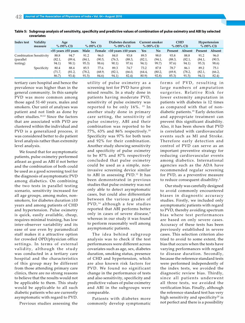
s i g n i f i c a n t d i f f e r e n c e ( i . e . overlapping CIs) in sensitivity, specificity, PPV and NPV between pulse oximetry and ABI across the subgroups of age, sex, diabetes duration, smoking status, presence of CHD and hypertension (Tables 3, 4). In the parallel testing scenario, sensitivity increased for all age groups, male sex, non-smokers, diabetes duration ≥10 years andpatients of CHD and hypertension. The best performance of the parallel testing strategy was seen in the
current non-smokers group with a sensitivity of 98% and specificity of 88% (Table 5).
Discussion
We found that PVD was fairly prevalent among the diabetics included in this s tudy. A few community-based studies have r e p o r t e d a l o we r p r e va l e n c e ranging from about 6% to 8%.4,5 This could be due to the fact that the study sample came from a
Table 2: Sensitivity, specificity and predictive values of pulse oximetry and ABI with duplex ultrasonography as reference standard
Index test PVD (by duplex USG) Sensitivity% (95% CI)
Specificity% (95% CI)
PPV% (95% CI)
NPV% (95% CI)Present nos. Absent nos.
Pulse oximetry 74.1 (55.3, 86.8) 95.7 (89.4, 98.3) 83.3 (64.1, 93.3) 92.7 (85.7, 96.4)Positive 20 4Negative 7 89
ABI 70.3 (51.5, 84.2) 87.1 (78.8, 92.5) 61.3 (43.8, 76.3) 91.0 (83.3, 95.4)Positive 19 12Negative 8 81
Combination (parallel testing)
92.3 (86.4, 96.0) 83.3 (75.7, 88.9) - -Positive 21 13Negative 6 80Total 27 93
Table 3: Subgroup analysis of sensitivity, specificity and predictive values of pulse oximetry by selected covariates
Index test
Validity measure
Age% (95% CI)
Sex% (95% CI)
Diabetes duration% (95% CI)
Current smoker% (95% CI)
CHD% (95% CI)
Hypertension% (95% CI)
<55 years ≥55 years Male Female <10 years ≥10 years Yes No Present Absent Present AbsentPulse oximetry
Sensitivity 66.6 (35.4, 88.7)
80.0 (51.3, 94.6)
72.7 (49.5, 88.3)
80.0 (29.8, 98.9)
60.0 (17.0, 92.7)
77.2 (54.1, 91.3)
70.0 (45.6, 87.1)
85.7 (42.0, 99.2)
77.7 (51.9, 92.6)
66.6 (30.9, 90.0)
72.7 (49.5, 88.3)
80.0 (29.8, 98.9)
Specificity 94.9 (84.9, 98.6)
97.0 (82.9, 99.8)
95.5 (86.6, 98.8)
96.1 (78.4, 99.7)
96.3 (86.3, 99.3)
94.7 (80.9, 99.0)
96.5 (80.3, 99.8)
95.3 (86.0, 98.7)
97.2 (84.1, 99.8)
94.6 (84.1, 98.6)
98.1 (89.0, 99.9)
92.1 (77.5, 97.9)
PPV 72.7 (39.3, 92.6)
92.3 (62.0, 99.5)
84.2 (59.5, 95.8)
80.0 (29.8, 98.9)
60.0 (17.0, 92.7)
89.4 (65.4, 98.1)
93.3 (66.0, 99.6)
66.6 (30.9, 90.9)
93.3 (66.0, 99.6)
66.6 (30.9, 90.0)
94.1 (69.2, 99.6)
57.1 (20.2, 88.1)
NPV 93.3 (82.9, 97.8)
91.6 (76.4, 97.8)
91.4 (81.6, 96.4)
96.1 (78.4, 99.7)
96.3 (86.3, 99.3)
87.8 (72.9, 95.4)
82.3 (64.8, 92.6)
98.3 (90.1, 99.9)
90.0 (75.4, 96.7)
94.6 (84.1, 98.6)
90.0 (78.8, 95.8)
97.2 (83.7, 99.8)
Table 4: Subgroup analysis of sensitivity, specificity and predictive values of ABI by selected covariates
Index test
Validity measure
Age% (95% CI)
Sex% (95% CI)
Diabetes duration% (95% CI)
Current smoker% (95% CI)
CHD% (95% CI)
Hypertension% (95% CI)
<55 years ≥55 years Male Female <10 years ≥10 years Yes No Present Absent Present AbsentABI Sensitivity 66.6
(35.4, 88.7)
73.3 (44.8, 91.0)
68.1(45.1, 85.2)
80.0 (29.8, 98.9)
60.0 (17.0, 92.7)
72.7 (49.5, 88.3)
65.0 (40.9, 83.6)
85.7 (42.0, 99.2)
72.2 (46.4, 89.2)
66.6 (30.9, 90.9)
68.1 (45.1, 85.2)
80.0 (29.8, 98.9)
Specificity 84.7 (72.5, 92.3)
91.1 (75.1, 97.6)
89.5 (79.0, 95.3)
80.7 (60.0, 92.6)
92.7 (81.5, 97.6)
78.9 (62.2, 89.8)
75.8 (56.0, 88.9)
92.1 (81.9, 97.0)
81.0 (64.2, 91.4)
91.0 (79.6, 96.9)
90.9 (79.2, 96.6)
81.5 (65.1, 91.6)
PPV 47.0 (23.8, 71.4)
78.5 (48.8, 94.2)
68.1 (45.1, 85.2)
44.4 (15.3, 77.3)
42.8 (11.8, 79.7)
66.6 (44.6, 83.5)
65.0(40.9, 83.6)
54.5 (24.5, 81.8)
65.0 (40.9, 83.6)
54.5 (24.5, 81.8)
75.0 (50.5, 90.4)
36.3 (12.3 ,68.3)
NPV 92.5 (81.2, 97.6)
88.5 (72.3, 96.2)
89.5 (79.0, 95.3)
95.4 (75.1, 99.7)
96.2 (85.9, 99.3)
83.3 (66.5, 93.0)
75.8 (56.0, 88.9)
98.3 (89.8, 99.9)
85.7 (68.9, 94.6)
94.4 (83.6, 98.5)
87.7 (75.7, 94.5)
96.8 (82.0, 99.8)
Journal of The Association of Physicians of India ■ Vol. 64 ■ August 201642
tertiary care hospital and hence the prevalence was higher than in the general community. In this sample PVD was more common among those aged 51-60 years, males and smokers. Our unit of analyses was patient and not limb as done by other studies.10,12 Since the factors that are associated with PVD are clustered within the individual and PVD is a generalized process, it was considered better to do patient level analysis rather than extremity level analysis.
We found that for asymptomatic patients, pulse oximetry performed atleast as good as ABI if not better and the combination of both could be used as a good screening tool for the diagnosis of asymptomatic PVD among diabetics. On combining the two tests in parallel testing scenario, sensitivity increased for all age groups, among males, non-smokers,fordiabetesduration≥10years and among patients of CHD and hypertension. Pulse oximetry is quick, easily available, cheap, requires minimal training, has low inter-observer variability and its ease of use even by paramedical staff makes it a attractive option for crowded OPD/physician office se t t ings . In terms of external va l i d i t y , a l t h o u g h t h e s t u d y was conducted in a tertiary care hospital and the characteristics of this group may be different from those attending primary care clinics, there are no strong reasons to believe that the results would not be applicable to them. This study would be applicable to all such diabetic patients who are currently asymptomatic with regard to PVD.
Previous studies assessing the
ut i l i ty of pulse oximetry as a screening test for PVD have given mixed results. In a study done in patients having moderate PVD, sensitivity of pulse oximetry was reported to be only 16%. 15 In another study done in primary care sett ing, the sensit ivity of pulse oximetry, ABI and their combination was reported to be 77%, 63% and 86% respectively.10 Specificity was 97% for both tests and 92% for their combination. Another study showing sensitivity and specificity of pulse oximetry to be 87% and 87% respectively concluded that pulse oximetry could be used as a simple, non-invasive screening device similar to ABI in assessing PVD.12 It has been demonstrated in previous studies that pulse oximetry was not only able to detect asymptomatic cases, but could also differentiate between the various grades of PVD, 14 although a few studies reported that ABI performs better only in cases of severe disease,9 whereas in our study it was found to perform reasonably well among asymptomatic patients.
T h e i d e a b e h i n d s u b g r o u p analysis was to check if the test performances were different across covariates such as age, sex, diabetes duration, smoking status, presence of CHD and hypertension, which are also known risk factors for PVD. We found no signif icant change in the performance of tests and also sensitivity, specificity and predictive values of pulse oximetry and ABI in the subgroups were comparable.
Patients with diabetes more commonly develop symptomatic
f o r m s o f P V D , r e s u l t i n g i n large numbers of amputat ion s u r g e r i e s . R e l a t i v e R i s k f o r lower extremity amputation in patients with diabetes is 12 times as compared with that of non-diabetic patients.19 Early detection and appropriate treatment can prevent this significant disability. Also, it has been shown that PVD is correlated with cardiovascular events such as MI and Stroke. Therefore early detect ion and control of PVD can serve as an important preventive strategy for reducing cardiovascular events among diabetics. International agencies such as the ADA have recommended regular screening for PVD, as a preventive measure to reduce consequent disability.2
Our study was carefully designed to avoid commonly encountered b iases in diagnost ic accuracy studies. Firstly, we included only asymptomatic patients with regard to PVD, thereby avoiding spectrum bias where tes t performances are based on only severe cases. Accuracy of these tests has been previously established in severe cases. This selection criterion also tried to avoid to some extent, the bias that occurs when the tests have varying performances with regard to disease duration. Secondly, because the reference standard tests were performed independently of the index tests, we avoided the diagnostic review bias. Thirdly, s i n c e a l l p a t i e n t s u n d e r we n t all three tests, we avoided the verification bias. Finally, although the reference standard test has very high sensitivity and specificity20 is not perfect and there is a possibility
Table 5: Subgroup analysis of sensitivity, specificity and predictive values of combination of pulse oximetry and ABI by selected covariates
Index test Validity measure
Age% (95% CI)
Sex% (95% CI)
Diabetes duration% (95% CI)
Current smoker% (95% CI)
CHD% (95% CI)
Hypertension% (95% CI)
<55 years ≥55 years Male Female <10 years ≥10 years Yes No Present Absent Present AbsentCombination(parallel testing)
Sensitivity 88.8 (82.1, 94.1)
94.7 (89.4, 98.1)
91.2 (84.1, 95.3)
96.0 (90.5, 98.6)
84.0 (76.3, 90.1)
93.8 (88.3, 97.6)
89.5 (82.1, 94.1)
98.0 (94.1, 99.7)
93.8 (88.3, 97.6)
88.8 (82.1, 94.1)
91.2 (84.1, 95.3)
96.0 (90.5, 98.6)
Specificity 80.4 (71.7, 86.7)
88.4 (81.2, 93.4)
85.5 (78.2, 91.5)
77.6 (68.9, 84.6)
89.3 (82.1, 94.1)
74.7 (66.0, 82.4)
73.2 (64.4, 80.9)
87.8 (80.2, 92.8)
78.7 (69.8, 85.3)
86.1 (78.2, 91.5)
89.2 (82.1, 94.1)
75.1 (66.2, 82.4)

Journal of The Association of Physicians of India ■ Vol. 64 ■ August 2016 43
of misclassification bias. However, this misclassification was likely to be non-differential affecting the results only slightly.
There are a few limitations in the study. Firstly, since a single investigator performed both pulse oximetry and ABI, the result of one might have influenced that of the other. Secondly, we could not measure test reliability in terms of inter-observer and intra-observer variability. Thirdly, sample size was probab ly inadequate for subgroup analysis. Finally, we could not study the effect of a few covariates like occupation, control of diabetes, extent of physical activity and lipid profile, which have a bearing on development of PVD. Future research could study the effect of these variables with a larger sample size.Conclusion
In conclusion, it can be stated that pulse oximetry on account of its high sensitivity will serve as a good initial screening test for asymptomatic PVD in patients with type 2 diabetes; better still would be a combination of pulse oximetry and ABI, which has an even higher sensitivity.Research Ethics
Ethical clearance was obtained from the Institutional Review Board of the Government Rajaji Hospital, Madurai. Written informed consent was obtained from all participants and confidentiality of information was strictly maintained. Patients who required treatment as per their diagnosis were given appropriate care.Acknowledgements
We are extremely thankful to all the individuals who had helped and participated in the study. We would also like to thank staff members of Government Rajaji
Hospital, Madurai, for helping in the conduct of the study.
References1. International Diabetes Federation
[Internet]. IDF diabetes atlas. Sixth ed. 2013 [cited 2014 Jan 14]. Available from: www.idf.org/diabetesatlas
2. American Diabetes Association. Peripheral arterial disease in people with diabetes. Diabetes Care 2003; 26:3333–3341.
3. Newman AB, Siscovick DS, Manolio TA, Polak J, Fr ied LP, Borhani NO, et al. Ankle-arm index as a marker of atherosclerosis in the Cardiovascular Health Study. Cardiovascular Heart Study (CHS) Collaborative Research Group. Circulation 1993; 88:837–845.
4. Premalatha G, Shanthirani S, Deepa R, Markovitz J, Mohan V. Prevalence and risk factors of peripheral vascular disease in a selected South Indian population: the Chennai Urban Population Study. Diabetes Care 2000; 23:1295–1300.
5. Pradeepa R, Chella S, Surendar J, Indulekha K, Anjana RM, Mohan V. Prevalence of peripheral vascular disease and its association with carotid intima-media thickness and arterial stiffness in type 2 diabetes: the Chennai Urban Rural Epidemiology Study (CURES 111). Diabetes Vasc Dis Res 2014; 11:190–200.
6. Cavanagh P, Attinger C, Abbas Z, Bal A, Rojas N, Xu Z-R. Cost of treating diabetic foot ulcers in five different countries. Diabetes Metab Res Rev 2012; 28 Suppl 1:107–111.
7. Fowke s F G. The me a su re m e nt of atherosclerotic peripheral arterial disease in epidemiological surveys. Int J Epidemiol 1988; 17:248–254.
8. Yao ST, Hobbs JT, Irvine WT. Ankle systolic pressure measurements in arterial disease affecting the lower extremities. Br J Surg 1969; 56:676–679.
9. Feigelson HS, Criqui MH, Fronek A, Langer RD, Molgaard CA. Screening for peripheral arterial disease: the sensitivity, specificity, and predictive value of noninvasive tests in a defined population. Am J Epidemiol 1994; 140:526–534.
10. Parameswaran GI, Brand K, Dolan J. Pulse oximetry as a potential screening tool for lower extremity arterial disease in asymptomatic patients with diabetes mellitus. Arch Intern Med 2005; 165:442–446.
11. Mardirossian G, Schneider RE. Limitations of pulse oximetry. Anesth Prog 1992; 39:194–196.
12. Kwon J-N, Lee W-B. Utility of digital pulse oximetry in the screening of lower extremity arterial disease. J Korean Surg Soc 2012; 82:94–100.
13. Jones SR, Carley S, Harrison M. An introduction to power and sample size estimation. Emerg Med J 2003; 20:453–458.
14. Chobanian AV, Bakris GL, Black HR, Cushman WC, Green LA, Izzo JL, et al. The Seventh Report of the Joint National Committee on Prevention, Detection, Evaluation, and Treatment of High Blood Pressure: the JNC 7 report. JAMA 2003; 289:2560–2572.
15. Jawahar D, Rachamalla HR, Rafalowski A, Ilkhani R, Bharathan T, Anandarao N. Pulse oximetry in the evaluation of peripheral vascular disease. Angiology 1997; 48:721–724.
16. Writing Committee to Develop Clinical Data Standards for Peripheral Atherosclerotic Vascular Disease, Creager MA, Belkin M, Bluth EI, Casey DE, Chaturvedi S, et al. 2012 ACCF/AHA/ACR/SCAI/SIR/STS/SVM/SVN/SVS key data elements and definitions for peripheral atherosclerotic vascular disease: a report of the American College of Cardiology Foundation/American Heart Association Task Force on Clinical Data Standards ( Writing Committee to Develop Clinical Data Standards for Peripheral Atherosclerotic Vascular Disease). Circulation 2012; 125:395–467.
17. Haynes B, Sackett D, Guyatt G, Tugwell P. Clinical Epidemiology: How to Do Clinical Practice Research. 3rd edition. Philadelphia: LWW; 2005. 480 p.
18. Bossuyt PM, Reitsma JB, Bruns DE, Gatsonis CA, Glasziou PP, Irwig LM, et al. Towards complete and accurate reporting of studies of diagnostic accuracy: the STARD initiative. BMJ 2003; 326:41–44.
19. Centers for Disease Control and Prevention (CDC). Diabetes-related amputations of lower extremities in the Medicare population--Minnesota, 1993-1995. Morb Mortal Wkly Rep 1998; 47:649–652.
20. Collins R, Burch J, Cranny G, Aguiar-Ibáñez R, Craig D, Wright K, et al. Duplex ultrasonography, magnetic resonance angiography, and computed tomography angiography for diagnosis and assessment of symptomatic, lower limb peripheral arterial disease: systematic review. BMJ 2007; 334:1257.